Embed Size (px)
Citation preview
Full Text
ABSTRACT Background: Duodenal mucosal biopsies are routinely taken for diagnosis in children with complaints of the
upper gastrointestinal tract. Surprisingly, little is known about the usefulness of proximal duodenal versus distal duodenal biopsies for routine diagnostic purposes. This study evaluated the comparability of proximal and distal duodenal biopsies with respect to mucosal morphology as well as glycohydrolase expression as an indicator of intestinal epithelial function.
Methods: Specimens obtained in duodenal endoscopic biopsies from 64 children, ranging in age from 3 months to 18 years with normal or affected mucosa, were studied. Biopsies were performed in anatomically defined regions in the bulbus duodeni (the very proximal part of the duodenum) and distally of the papilla of Vater (distal of the pancreatic duct). Biopsy specimens were paraformaldehyde-fixed for histologic examination and immunohistochemical evaluation or were homogenized to isolate RNA. Crypt/villus morphology was assessed as is routinely determined by pathologists. In addition, several aspects of lactase and sucrase-isomaltase expression as paradigms of intestinal brush border enzymes were assessed: localization at the cellular level, semiquantitative immunohistochemistry, and quantitative measurement of the messenger RNA levels of the respective brush border glycohydrolases.
Results: As anticipated, there was a wide interpatient variation in mucosal morphology and expression of
Pediatric Duodenal Biopsies: Mucosal Morphology and Glycohydrolase Expression Do Not Change Along the Duodenum
Keywords: Biopsy, Childhood, Duodenum, Lactase, Morphology, Sucrase-isomaltase
ISSN: 0277-2116 Accession: 00005176-199802000-00013 Author(s):
Van Berrs, Erik H.; Einerhand, Alexandra W. C.; Taminiau, Jan A. J. M.; Heymans, Hugo S. A.; Dekker, Jan; Büller, Hans A.*
Issue: Volume 26(2), February 1998, pp 186-193
Publication Type: [Original Articles]
Publisher: © Lippincott-Raven Publishers
Institution(s):
Laboratory for Pediatric Gastroenterology and Nutrition, Department of Pediatrics, Academic Medical Center, University of Amsterdam, Emma's Childrens Hospital AMC, Amsterdam; and *Sophia Children's Hospital, Rotterdam, The Netherlands Received February 24, 1997; accepted July 3, 1997. Address correspondence and reprint requests to Dr. J. Dekker, Academic Medical Center, Department of Pediatrics, Laboratory for Pediatric Gastroenterology and Nutrition, P.O. Box 22700, 1100 DE Amsterdam, The Netherlands.
pagina 1 van 13Ovid: Pediatric Duodenal Biopsies: Mucosal Morphology and Glycohydrolase Exp...
18-7-2008http://ovidsp.tx.ovid.com/spb/ovidweb.cgi
lactase and sucrase-isomaltase. Nonetheless, the consistent finding was that in each patient, measurements of morphology and lactase and sucrase-isomaltase gene expression were very similar between samples obtained in the proximal and distal biopsies.
Conclusions: Biopsies performed in either location in the duodenum are equally suitable for diagnostic workup of patients suspected of mucosal abnormalities affecting morphology or small intestinal brush border glycohydrolase activities.
The intestinal mucosa is made up of a single-layer epithelium consisting of various specialized cell types and
underlying connective tissue. Many common and widespread diseases affect the morphology and the functions of the intestinal mucosa. Because this mucosa is pivotal in the digestion and absorption of nutrients, gastrointestinal endoscopy is widely valued for its diagnostic merit in gastrointestinal disease.
Diagnosis of upper gastrointestinal disease is generally based on a combination of clinical observations, laboratory tests, and in particular, endoscopic findings. In children, endoscopic examination has become a valuable and often indispensable procedure in the diagnosis of upper gastrointestinal disease (1). Mucosal biopsy specimens taken during endoscopic sessions provide reliable information about the severity of gastrointestinal disease (1,2). Over the years, duodenal biopsies have widely replaced jejunal biopsies in the diagnosis of upper gastrointestinal disease (2-9). The major advantage of performing duodenal biopsies guided by endoscope over performing jejunal biopsies with the Crosby capsule is that the procedure is less distressing to the patients. Jejunal biopsies often require x-ray survey or a capsule mounted on a gastroscope, considerably increasing the risk of tissue damage and the time required to perform the procedure. Moreover, jejunal biopsies are taken without visual contact with the tissue, whereas duodenoscopy enables visual survey of the mucosa before and after the takin of the biopsy specimen.
There is surprisingly little literature describing differences along the cephalocaudal axis of the small intestine as a determining factor in the outcome of the diagnosis obtained by intestinal biopsy. Duodenal biopsies seem to be as suitable for diagnosis as jejunal biopsies (2-8). However, along the duodenum, the effect of the location of the biopsy specimen on the outcome of the diagnosis is uncertain. Especially in children, in whom the intestinal mucosa is particularly important for grwoth and development, the data on this potentially important issue are very scarce.
Here, we describe the morphology of the duodenal mucosa and the expression of the two most important small intestinal brush border glycohydrolases, lactase and sucrase-isomaltase (SI) in pediatric duodenal biopsies. We studied 64 children from various ethnic origins, ranging in age from 3 months to 18 years with normal or affected duodenal mucosae. The object was to compare expression patterns of lactase and SI, along with study of mucosal morphology, in the proximal and distal duodenal mucosae of children, to compare the usefulness of specimens from the two locations for diagnostic workup.
MATERIALS AND METHODS Patients and Ethical Considerations
A prospective study was done on biopsy specimens obtained with endoscopic forceps from the duodenal mucosae of 64 pediatric patients with upper gastrointestinal symptoms. Indications for endoscopic examination included intractable diarrhea, suspected celiac disease, Giardia lamblia orHelicobacter pylori infection, Crohn's disease, chronic abdominal pain, chronic diarrhea, failure to thrive, or esophageal reflux. All patients were analyzed as one group. Each patient underwent gastroduodenal endoscopy and duodenal biopsy for diagnostic purposes. We obtained permission to perform four extra biopsies per patient, the number ethically considered the maximum for this study. Informed consent was gained from the patients and their parents for the performance of the biopsies, and the study was conducted with the permission of the medical ethics committee of our institution.
Tissue Mucosal specimens were obtained in biopsies performed on the proximal and distal duodenum: two in the
pagina 2 van 13Ovid: Pediatric Duodenal Biopsies: Mucosal Morphology and Glycohydrolase Exp...
18-7-2008http://ovidsp.tx.ovid.com/spb/ovidweb.cgi
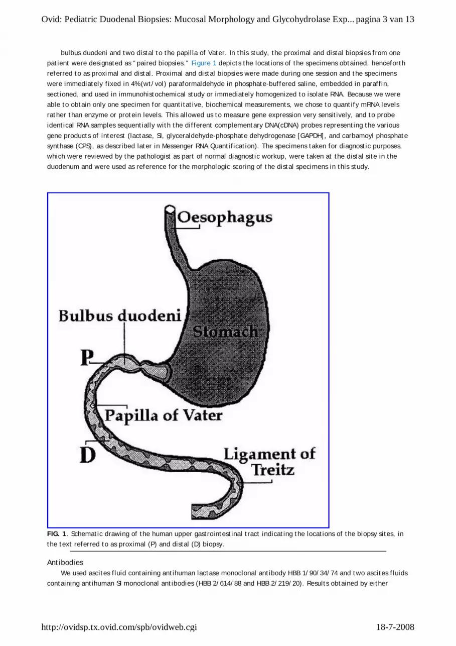
bulbus duodeni and two distal to the papilla of Vater. In this study, the proximal and distal biopsies from one patient were designated as “paired biopsies.” Figure 1 depicts the locations of the specimens obtained, henceforth referred to as proximal and distal. Proximal and distal biopsies were made during one session and the specimens were immediately fixed in 4% (wt/vol) paraformaldehyde in phosphate-buffered saline, embedded in paraffin, sectioned, and used in immunohistochemical study or immediately homogenized to isolate RNA. Because we were able to obtain only one specimen for quantitative, biochemical measurements, we chose to quantify mRNA levels rather than enzyme or protein levels. This allowed us to measure gene expression very sensitively, and to probe identical RNA samples sequentially with the different complementary DNA(cDNA) probes representing the various gene products of interest (lactase, SI, glyceraldehyde-phosphate dehydrogenase [GAPDH], and carbamoyl phosphate synthase (CPS), as described later in Messenger RNA Quantification). The specimens taken for diagnostic purposes, which were reviewed by the pathologist as part of normal diagnostic workup, were taken at the distal site in the duodenum and were used as reference for the morphologic scoring of the distal specimens in this study.
FIG. 1. Schematic drawing of the human upper gastrointestinal tract indicating the locations of the biopsy sites, in the text referred to as proximal (P) and distal (D) biopsy.
Antibodies We used ascites fluid containing antihuman lactase monoclonal antibody HBB 1/90/34/74 and two ascites fluids
containing antihuman SI monoclonal antibodies (HBB 2/614/88 and HBB 2/219/20). Results obtained by either
pagina 3 van 13Ovid: Pediatric Duodenal Biopsies: Mucosal Morphology and Glycohydrolase Exp...
18-7-2008http://ovidsp.tx.ovid.com/spb/ovidweb.cgi
antihuman SI antibody were indistinguishable. These antibodies have been described to recognize the enzyme precursors as well as the mature forms of these brush border enzymes (10). Three independent antihuman lactase monoclonal antibodies prepared from the hybridoma supernatant (mLac1, mLac4, and mLac5) were used in some experiments (11). Furthermore, we used ascites containing antirat lactase monoclonal antibody (12) as a negative control in immunohistochemical studies.
Morphology The morphology of the duodenal mucosa, in particular the crypt/villus ratio, was assessed at least twice by
independent and blinded observers for each specimen obtained, on at least four villi and crypts, using light microscopy at low magnification (×100). We assigned scores as follows: 0, total villus atrophy; 1, moderate villus atrophy (villus/crypt ratio<1); 2, mild villus atrophy; 3, normal morphology, according to established criteria (13).
Immunohistochemical Analysis Seven-micrometer thick sections from formaldehyde-fixed, paraffin-embedded specimens were deparaffinated,
incubated for 30 minutes in 3% H2O2 in phosphate-buffered saline, to inactivate endogenous peroxidase activity and
for 30 minutes in a solution of 10 mM Tris-HCl, 5 mM EDTA, 150 mM NaCl, 0.25% gelatin, and 0.05% Tween-20, to prevent background staining. The sections were incubated overnight with adequate dilutions in phosphate-buffered saline of the primary antibodies, 1.5 hours with rabbit antimouse serum (1:7500, Dako, Glostrup, Denmark), 1.5 hours with goat antirabbit serum (1:1000, Dako) and with rabbit peroxidase-antiperoxidase(1:1000, Dako). All incubations were at room temperature. Antilactase (HBB 1/90/34/74) and anti-SI (and HBB 2/219/88) monoclonal antibodies were diluted 1:1000 from ascites (10). Further steps were as previously described (14). Absence or presence of immunohistochemical staining was determined under light microscope at high magnification (×400). We discriminated four localizations of immunohistochemical staining: 1) intracellular or 2) brush border staining of crypt enterocytes and 3) intracellular or 4) brush border staining of villus enterocytes. Futhermore, we distinguished four semiquantitative classes of staining intensity: 0, absence of staining; 1, very weak, patchy staining; 2, evently distributed, moderate staining; and 3, evenly distributed, dark staining. Semiquantitative measurements of staining intensities were only assessed in specimens that had been immunostained at least in triplicate in independent experiments. All sections were judged at least twice by two independent and blinded observers.
Cloning of a Partial Human CPS Complementary DNA Using the reverse transcription-polymerase chain reaction, technique we have amplified 1394 nt of the 3' end
of the human CPSI gene (15). Polymerase chain reaction primers were as follows: Forward, 3' AATTTGTTGAAGGGGCCC5', Reverse, 5'GGAATTCTGCCCTGTTAAAGTGTCC3'. Reverse transcription-polymerase chain reaction was done in a final volume of 20 µl containing 1 × reverse transcription buffer (Boehringer-Mannheim, Mannheim, Germany) 4 mM of dATP, dCTP, dTTP and dGTP each, 50 nM of each primer, 2 U RNasin (Promega, Madison, WI, U.S.A.), 2 U reverse transcriptase M-MulV (Boehringer-Mannheim), and 1 µg of human jejunal RNA isolated essentially as described by Chirgwin et al. (16) Amplification consisted of 30 consecutive rounds of 1 minute at 94 °C, 1 minute at 55 °C, 2.5 minutes at 72°C, and 7 minutes at 72 °C. The resulting polymerase chain reaction product was digested with ApaI and EcoRI restriction enzymes and ligated into pBluescript KS (Stratagene, La Jolla, CA, U.S.A.). Nucleotide sequencing confirmed the identity of the CPS cDNA fragment.
Messenger RNA Quantification RNA was isolated from individual biopsy specimens using the RNeasy protocol(Qiagen, Chatsworth, CA, U.S.A.).
Messenger RNA quantification was performed on spot blots. In short, between 100 and 500 ng of RNA was spotted onto negatively charged Qiabrane nitrocellulose (Qiagen) using a vacuum-operated BioRad 96-well spotblot system (BioRad, Richmond, CA, U.S.A.). The amount of each mRNA was quantified using specific DNA probes synthesized using random hexamer primers, incorporating [alpha]32P-dATP to at least 1 × 10⁸ cpm/µg (17). Lactase probes were synthesized from the 6.3-kb full-length human lactase cDNA (18), SI probes from 428 bp of a human SI clone (14), probes transcribed from 1394 bp of a CPS cDNA clone synthesized from human jejunal RNA (as described earlier), and GAPDH from 1 kb of human GAPDH cDNA (19). RNA from each specimen was blotted onto Qiabrane filters, dried, and backed at 80 °C for 2 hours. Prehybridization was performed at 65 °C for 1 hour in (7% sodium dodecyl sulfate [SDS], 0.1 mM EDTA, and 0.5 M Na2HPO4 [pH 7.2]). Hybridization was performed for 17 hours at 65 °C.
pagina 4 van 13Ovid: Pediatric Duodenal Biopsies: Mucosal Morphology and Glycohydrolase Exp...
18-7-2008http://ovidsp.tx.ovid.com/spb/ovidweb.cgi

Filters were washed at 65 °C twice for 20 minutes in 6 × SSC and 0.1% SDS, once for 20 minutes in 3 × SSC and 0.1% SDS, and once 20 minutes in 0.1 × SSC and 0.1% SDS. Autoradiographs were prepared using PhosphorImage screens(Molecular Dynamics, Sunnyvale, CA, U.S.A.), that were exposed to hybridized membranes for 48 hours and quantified in a Phosphorlmager using ImageQuant software (Molecular Dynamics). Membranes were stripped of adherent probe by washing at 80 °C in 0.1% SDS for several hours before hybridization with each probe. Stripped blots were checked for absence of radioactivity by exposing PhosphorImager screens for 48 hours. Sequentially, all the blotted RNA samples have been incubated ith probes for SI mRNA, lactase mRNA, CPS mRNA, and GAPDH mRNA. Signals detected for lactase, SI, and CPS were then normalized to the housekeeping gene GAPDH.
RESULTS Patients
The 64 patients that we have studied ranged in age from 3 months to 18 years old (mean, 7 years old, 35 boys). Diagnoses of these patients were obtained at the end of medical workup from their medical records and included esophageal reflux, H. pylori infection of the stomach, celiac disease, Giardia lamblia infestation of the small intestine, intractable diarrhea, and Morbus Crohn and a nonclassified immunodeficiency in one child. The number of biopsies per patient was restricted in this study for ethical reasons: Four duodenal biopsy specimens were obtained during one endoscopic session.
Duodenal Mucosal Morphology and Variation in Lactase and Sucrease-Isomaltase Immunostaining Two strikingly different staining phenotypes of immunodetection for lactase and SI were observed within our
patient population. Figure 2 shows two patients that have similar amounts of immunodetectable lactase or SI in the brush borders of their enterocytes, but additional intracellular localization of lactase and SI was detected in only one patient. These two distinct types of staining were representative for all patients studied. Intracellular staining, if detected, was always observed in specimens from both sites of the duodenum and was always observed for both the glycohydrolases simultaneously. Moreover, intracellular staining was found in all cells expressing lactase and SI on their brush borders, in villus as well as in crypt enterocytes, whereas staining was never detected in the other cell types: goblet, enteroendocrine, or Paneth's cells. Results obtained using two distinct antihuman SI monoclonal antibodies were indistinguishable in all cases. A control experiment with similarly or less-diluted ascites, obtained from a sibling mouse and containing an unrelated monoclonal antibody (12), failed to stain adjacent sections (results not shown), indicating that the intracellular staining indeed represented lactase and SI. Experiments using three independent antihuman lactase monoclonal antibodies, mLac1, mLac4, and mLac5, showed no intracellular staining in any of the specimens, and lactase staining was restricted to the brush border (not shown).
pagina 5 van 13Ovid: Pediatric Duodenal Biopsies: Mucosal Morphology and Glycohydrolase Exp...
18-7-2008http://ovidsp.tx.ovid.com/spb/ovidweb.cgi
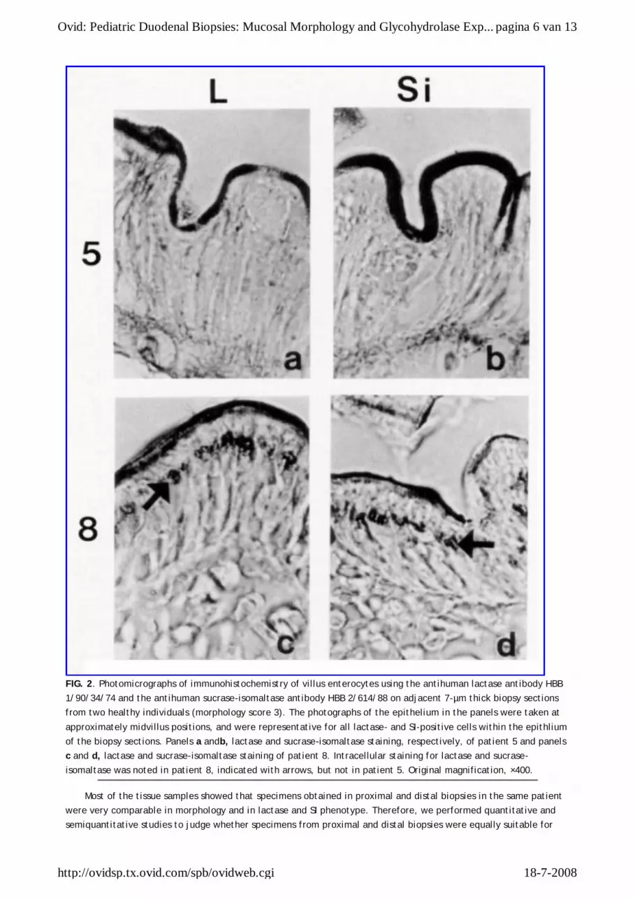
FIG. 2. Photomicrographs of immunohistochemistry of villus enterocytes using the antihuman lactase antibody HBB 1/90/34/74 and the antihuman sucrase-isomaltase antibody HBB 2/614/88 on adjacent 7-µm thick biopsy sections from two healthy individuals (morphology score 3). The photographs of the epithelium in the panels were taken at approximately midvillus positions, and were representative for all lactase- and SI-positive cells within the epithlium of the biopsy sections. Panels a andb, lactase and sucrase-isomaltase staining, respectively, of patient 5 and panels c and d, lactase and sucrase-isomaltase staining of patient 8. Intracellular staining for lactase and sucrase-isomaltase was noted in patient 8, indicated with arrows, but not in patient 5. Original magnification, ×400.
Most of the tissue samples showed that specimens obtained in proximal and distal biopsies in the same patient were very comparable in morphology and in lactase and SI phenotype. Therefore, we performed quantitative and semiquantitative studies to judge whether specimens from proximal and distal biopsies were equally suitable for
pagina 6 van 13Ovid: Pediatric Duodenal Biopsies: Mucosal Morphology and Glycohydrolase Exp...
18-7-2008http://ovidsp.tx.ovid.com/spb/ovidweb.cgi
diagnostic purposes, especially for the assessment of mucosal morphology and the abundance and localization of intestinal glycohydrolases.
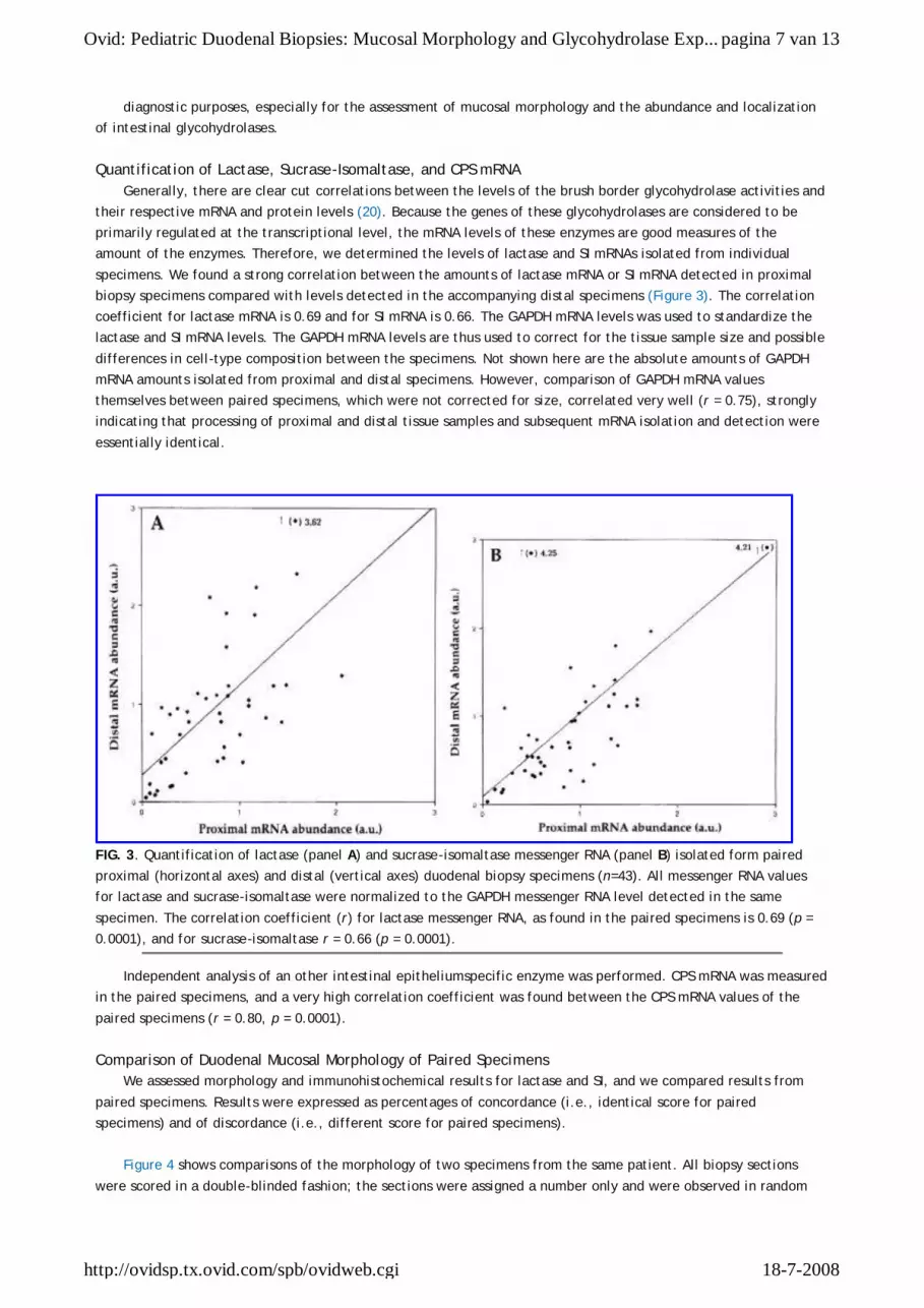
Quantification of Lactase, Sucrase-Isomaltase, and CPS mRNA Generally, there are clear cut correlations between the levels of the brush border glycohydrolase activities and
their respective mRNA and protein levels (20). Because the genes of these glycohydrolases are considered to be primarily regulated at the transcriptional level, the mRNA levels of these enzymes are good measures of the amount of the enzymes. Therefore, we determined the levels of lactase and SI mRNAs isolated from individual specimens. We found a strong correlation between the amounts of lactase mRNA or SI mRNA detected in proximal biopsy specimens compared with levels detected in the accompanying distal specimens (Figure 3). The correlation coefficient for lactase mRNA is 0.69 and for SI mRNA is 0.66. The GAPDH mRNA levels was used to standardize the lactase and SI mRNA levels. The GAPDH mRNA levels are thus used to correct for the tissue sample size and possible differences in cell-type composition between the specimens. Not shown here are the absolute amounts of GAPDH mRNA amounts isolated from proximal and distal specimens. However, comparison of GAPDH mRNA values themselves between paired specimens, which were not corrected for size, correlated very well (r = 0.75), strongly indicating that processing of proximal and distal tissue samples and subsequent mRNA isolation and detection were essentially identical.
FIG. 3. Quantification of lactase (panel A) and sucrase-isomaltase messenger RNA (panel B) isolated form paired proximal (horizontal axes) and distal (vertical axes) duodenal biopsy specimens (n=43). All messenger RNA values for lactase and sucrase-isomaltase were normalized to the GAPDH messenger RNA level detected in the same specimen. The correlation coefficient (r) for lactase messenger RNA, as found in the paired specimens is 0.69 (p = 0.0001), and for sucrase-isomaltase r = 0.66 (p = 0.0001).
Independent analysis of an other intestinal epitheliumspecific enzyme was performed. CPS mRNA was measured in the paired specimens, and a very high correlation coefficient was found between the CPS mRNA values of the paired specimens (r = 0.80, p = 0.0001).
Comparison of Duodenal Mucosal Morphology of Paired Specimens We assessed morphology and immunohistochemical results for lactase and SI, and we compared results from
paired specimens. Results were expressed as percentages of concordance (i.e., identical score for paired specimens) and of discordance (i.e., different score for paired specimens).
Figure 4 shows comparisons of the morphology of two specimens from the same patient. All biopsy sections were scored in a double-blinded fashion; the sections were assigned a number only and were observed in random
pagina 7 van 13Ovid: Pediatric Duodenal Biopsies: Mucosal Morphology and Glycohydrolase Exp...
18-7-2008http://ovidsp.tx.ovid.com/spb/ovidweb.cgi

order. First, we determined the degree of reproducibility for two duplicate distal specimens that were independently processed and their morphology assessed. Morphology scores were concordant in 54 of 64 cases(84%), whereas 6 of the discordant scores differed by only one morphology class and only 3 scores by more than one morphology class (Figure 4A). We then examined the similarity between paired specimens. Proximal versus distal morphology scores were concordant in 53 of 64 cases (83%). In 9 of 64 cases (14%), the morphology differed by one class and in only 2 of 64 cases (3%) by more than one class (Figure 4B). This indicates that the percentage of identical morphology scores between two paired (i.e., proximal and distal) specimens are as high as between duplicate specimens taken at the distal location.
FIG. 4. Comparative morphology of two duodenal biopsies from the same patient. A: Morphology of two closely adjacent, separately processed distal biopsies D1 and D2 (n = 64). Biopsy D1 was obtained for this study, while biopsy D2 was processed and reviewed by an independent pathologist during diagnostic workup. B: Morphology of paired proximal (P) and distal (D) biopsies from the same patient (n = 64).
Comparison of Immunohistochemical Detection of Lactase and Sucrase-Isomaltase Among Paired Specimens
Similarly, we have compared immunohistochemical staining patterns and intesities between the paired specimens. First, we restricted immunohistochemical scoring by distinguishing positive or negative staining (Figure 5), and we then compiled semiquantitative data (Figure 6). Despite our continuous efforts to use reproducible methods of fixation, on several occasions during the 1.5-year period in which specimens were collected, some appeared to immunostain irreproducibly or aberrantly. Therefore, it was occasionally difficult to reach a proper verdict on certain aspects of the expression of lactase and/or SI within some specimens. In such a case, the score of this patient was omitted in the analysis of this particular aspect, explaining why the number of patients per Figure deviates from 64.
pagina 8 van 13Ovid: Pediatric Duodenal Biopsies: Mucosal Morphology and Glycohydrolase Exp...
18-7-2008http://ovidsp.tx.ovid.com/spb/ovidweb.cgi
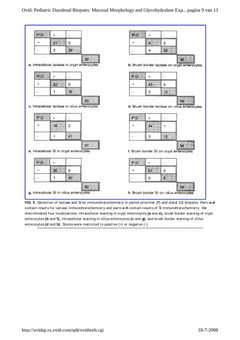
FIG. 5. Detection of lactase and SI by immunohistochemistry in paired proximal (P) and distal (D) biopsies. Parts a-d contain results for lactase immunohistochemistry and parts e-h contain results of SI immunohistochemistry. We discriminated four localizations; intracellular staining in crypt enterocytes (a and e), brush border staining of crypt enterocytes (b and f), intracellular staining in villus enterocytes (c and g), and brush border staining of villus enterocytes (d and h). Scores were restricted to positive (+) or negative (-).
pagina 9 van 13Ovid: Pediatric Duodenal Biopsies: Mucosal Morphology and Glycohydrolase Exp...
18-7-2008http://ovidsp.tx.ovid.com/spb/ovidweb.cgi

FIG. 6. Semiquantitative immunohistochemical comparison of proximal and distal lactase and SI. Comparison between semiquantitative immunohistochemical scores of proximal (P) and distal (D) lactase(a, n = 58) and SI (b, n = 59) expression in villus-associated enterocyte brush border. The four categories (0, 1, 2, and 3) are defined in materials and methods.
Detection of intracellular lactase in crypt enterocytes was carried out on 60 patients in whom there were crypts in both of the paired specimens (Figure 5A). This resulted in 60 of 60 (100%) concordant scores between paired specimens, with 21 pairs positive and 39 pairs negative. In brush bordrs of crypt enterocytes, 56 of 60 (93%) of the pairs were concordant; the majority were nagative (52 of 60; Figure 5B). Detection of intracellular lactase in villus enterocytes appeared concordant in 60 of 61 (98%) of the pairs (Figure 5C), and lactase in brush border of villus enterocytes was concordant in 56 of 58(97%) of the pairs (Figure 5D).
Detection of intracellular SI in crypt enterocytes resulted in 57 of 60(95%) concordant scores between paired specimens, with 41 of 60 pairs negative and 16 of 60 pairs positive (Figure 5E). In brush borders of crypt enterocytes 56 of 60 (93%) of the pairs were concordant; the majority were positive (44 of 60; Figure 5F). Detection of intracellular SI in villus enterocytes appeared concordant in 60 of 61 (98%) of the pairs (Figure 5G) and detection on the brush border of villus enterocytes was concordant in all 59 of 59 (100%) pairs (Figure 5H). The acutal frequencies of the positive staining for lactase or SI varied widely according to cellular location among the patients. However, for each of the locations separately, the paired specimens were at least 93% concordant.
Figure 5 shows that the number of discordant scores for each of the cellular locations is very low. Nevertheless, among these sparse discordant scores, there was a slight bias toward positivity only in the specimen for the detection of lactase and SI (i.e., distal positive, proximal negative). This may reflect the existence of slight quantitative gradients of gene expression for both enzymes increasing toward the distal duodenum in some patients.
As noted earlier (Figure 2), the antilactase monoclonal antibody and the two independent monoclonal antibodies directed against SI appeared to detect high levels of intracellular staining in approximately one third of the patients (Figure 5). Despite the very strong correlation of the intracellular staining of lactase and SI, this staining did not correlate with sex, age, ethnic background, clinical diagnosis, or morphology score (not shown). Moreover, within one specimen, the intracellular staining for lactase or SI was nearly always found in crypt and villus enterocytes, explaining the high similarity of the results in Figure 5A, 5C, 5E, and 5G.
Although brush border expression of lactase is normally detected on villus enterocytes only, 7% (4 of 60) of the patients also have detectable lactase in the brush border of crypts in both specimens (Figure 5B). In contrast, the presence of SI in the brush border of crypt enterocuytes is the prevalent phenotype, in that it occurs in approximately 80% (44 of 60) of the patients (Figure 5F).
pagina 10 van 13Ovid: Pediatric Duodenal Biopsies: Mucosal Morphology and Glycohydrolase E...
18-7-2008http://ovidsp.tx.ovid.com/spb/ovidweb.cgi
Finally, we counted the overall similarities between paired specimens. This analysis was done for 55 patients, in which we were able to determine lactase and SI in all locations according to the scores in Figure 5A through 5H. The comparisons between proximal and distal specimens showed that 43 patients were identical on all eight criteria, 11 were identical on seven criteria, and only 1 patient showed similarity on five criteria.
Semiquantitative evaluation of immunohistochemical staining was performed for staining in the brush border of villus enterocytes. Semiquantitative measurement of lactase was concordant in 47 of 58 (81%) of all studied cases (Figure 6A). There were only 2 of 58 (3%) patients in which we assessed higher amounts of lactase in the proximal specimen than in the distal specimen, whereas 9 of 58 (16%) patients had higher lactase amounts in the distal specimen compared with that in the proximal specimen. Seven out of these 9 differed by one class and only 2 by two classes. Semiquantitative SI measurement was concordant in 52 of 59 (88%) of all cases studied (Figure 6B). There were only 2 of 59 (3%) patients in which the proximal specimen had higher amounts of SI, whereas 5 of 59 (8%) had higher amounts in the distal specimen. Four of these 5 patients differed by one class and 1 patient by two classes. This further shows that at the semiquantitative immunohistochemical level the findings for lactase and SI from either specimen has a high predictive value for the other specimen of the pair.
DISCUSSION In the majority of the patients studied, there appeared to be no major difference between paired specimens in
mucosal morphology, immunohistochemical quantities of lactase and SI, patterns of lactase and SI expression, and levels of lactase, SI, and CPS mRNAs. Because the composition of this prospective study population was determined only by the order in which the patients came to the clinic, this implies that the population studied may be considered representative of children. Therefore, proximal and distal specimens seem to be equally informative about lactase and SI expression as well as mucosal morphology.
In evaluating the usefulness of proximal versus distal duodenal biopsies, it is important to determine whether gradual differences exist along the logitudinal axis of this organ in relation to the criteria that were chosen. A very small number of patients exhibited discordant presence of lactase or SI, with a prevalence for higher expression in the distal specimen. The vast majority of the children in our study showed no gradient along the length of their duodenum. In our predominantly white study population, lactase was present in most children at the mRNA level and was detected by immunohistochemical staining. Importantly, the amounts of lactase detected by either technique were similar along the duodenum as measured in paired specimens, implicating that no proximal-to-distal gradient of lactase expression exists in the duodenum during (at least) the first 18 years of human life. This result was notably different from our recent fidnings in the rat, which showed that lactase expression was restricted after weaning to the central part of the jejunum, generating an expression gradient in the proximal small intestine (21). Similarly, SI was present in most children studied, and expression levels measured at mRNA or SI immunodetection level were not age dependent. In that the levels of enzyme or SI mRNA were very comparable between paired specimens, there is no indication that SI displays a proximal-to-distal expression gradient in the duodenum at any time during postnatal development.
The antihuman lactase monoclonal antibody HBB 1/90/34/74 has been used in previous studies and has been shown to recognize the endoplasmic reticulum-synthesized lactase precursor as well as the mature, brush border enzyme forms of lactase (10,14,18). Both antihuman SI monoclonal antibodies used have been observed to recognize SI precursors and mature, brush border SI (10). Regarding the intracellular staining of lactase and SI, the results indicate that there is genuine detection of the corresponding antigens for several reasons: First, intracellular staining is only detected in enterocytes and is absent from goblet, enteroendocrine, and Paneth's cells. Second, each of the three monoclonal antibodies is directed to a peptide epitope, because they recognize(in addition to the high-mannose and complexly glycosylated forms of the enzymes) the de-N-glycosylated precursor, as shown by immunoprecipitation (10,14). This implicates that these antibodies have the potency to detect antigen at the level of the endoplasmic reticulum and Golgi apparatus and at the brush border membrane. Third, an unrelated ascites, prepared in sibling mice containing monoclonal antibody against rat lactase (12), which does not cross-react with human lactase (Van Beers, 1995, unpublished results), did not show any intracellular staining when used at identical or even at higher concentrations. Thus, we conclude that immunostaining with antilactase and anti-SI antibodies within the enterocytes represents authentic lactase and SI. The cellular location of the staining in the
pagina 11 van 13Ovid: Pediatric Duodenal Biopsies: Mucosal Morphology and Glycohydrolase E...
18-7-2008http://ovidsp.tx.ovid.com/spb/ovidweb.cgi
enterocytes makes it most likely that these lactase and SI molecules are present within the Golgi apparatus.
It still remains to be explained why intracellular staining is present in specimens from some but not other patients, although the antigen under investigation is present in the brush border of the specimens in comparable amounts. The explanation of the origin of the intracellular staining for both glycohydrolases is beyond the scope of this article. In fact, this phenomenon only helped us to discriminate additional aspects of the lactase and SI phenotypes amoung our study population.
Our findings on the similarities between specimens taken in proximal and distal duodenal biopsies were based on results from a large and diverse group of children of various ages. Because there were no inclusion criteria for our prospective study, we believe that the results are representative for children up to 18 years old. Therefore, we conclude that, independent from racial origin, age or disease, specimens taken in the most proximal area of the duodenum, are equally suitable as specimens taken distally of the papilla of Vater in the diagnosis of upper gastrointestinal disease in children.
Acknowledgment: The authors thank all patients and their parents who gave informed consent to participate in this research; Professor F. J. W. ten Kate and the Department of Pathology for observations and review of specimens; Dr. Dallas Swallow for providing the monoclonal antibodies mLac1, mLac4, and mLac5; and Dr. Hans-Peter Hauri for providing monoclonal antibodies HBB 1/90/34/74, HBB 2/219/20, and HBB 2/614/88.
This work was sponsored by Nutricia, Zoetermeer, The Netherlands.
REFERENCES 1. Black DD, Hagglitt RD, Whitington PF. Gastroduodenal endoscopic-histologic correlation in pediatric patients. J Pediatr Gastroenterol Nutr 1988;7:353-8. WUR SFX Bibliographic Links [Context Link]
2. Dandalides SM, Carey WD, Petras R, Achkar E. Endoscopic small bowel mucosal biopsy: A controlled trial evaluating forceps size and biopsy location in the diagnosis of normal and abnormal mucosal architecture.Gastrointest Endosc 1989;35:197-200. WUR SFX Bibliographic Links [Context Link]
3. Newcomer AD, McGill DB. Distribution of disaccharidase activity in the small bowel of normal and lactase-deficient subjects.Gastroenterology 1966;51:481-8. WUR SFX Bibliographic Links [Context Link]
4. Newcomer AD, McGill DB. Disaccharidase activity in the small intestine: Prevalence of lactase deficiency in 100 healthy subjects.Gastroenterology 1967;53:881-9. WUR SFX Bibliographic Links [Context Link]
5. Saverymuttu SH, Sabbat J, Burke M, Maxwell JD. Impact of endoscopic duodenal biopsy on the detection of small intestinal villus atrophy. Postgrad Med J 1991;67:47-9. [Context Link]
6. Oderda G, Forni M, Morra I, Tavassoli K, Pellegrino P, Ansaldi N. Endoscopic and histologic findings in the upper gastrointestinal tract of children with coeliac disease. J Pediatr Gastroenterol Nutr 1993;16:172-7. [Context Link]
7. Smith JA, Mayberry JF, Ansel ID, Long RG. Small bowel biopsy for disaccharidase levels: Evidence that endoscopic forceps biopsy can replace the Crosby capsule. Clin Chim Acta 1989;183:317-22. WUR SFX Bibliographic Links [Context Link]
8. Schmitz-Moorman P, Pittner PM, Reichmann L, Massarat S. Quantitative histological study of duodenitis in biopsies. Pathol Res Pract 1984;178:499-507. WUR SFX Bibliographic Links [Context Link]
9. Forget P, Lombet, J, Grandfils C, Dandrifosse G, Geubelle FJ. Lactase insufficiency revisited. J Pediatr Gastroenterol Nutr 1985;4:868-72. [Context Link]
10. Hauri HP, Sterchi EE, Bienz D, Fransen JAM, Marxer A. Expression and intracellular transport of microvillus
pagina 12 van 13Ovid: Pediatric Duodenal Biopsies: Mucosal Morphology and Glycohydrolase E...
18-7-2008http://ovidsp.tx.ovid.com/spb/ovidweb.cgi
membrane hydrolases in human intestine. J Cell Biol 1985;101:838-51. WUR SFX Bibliographic Links [Context Link]
11. Maiuri L, Raia V, Potter J, et al. Mosaic pattern of lactase expression by villous entercoytes in human adult-type hypolactasia.Gastroenterology 1991;100:359-69. WUR SFX Bibliographic Links [Context Link]
12. Quaroni A, Isselbacher KJ. Study of intestinal cell differentiation with monoclonal antibodies to intestinal cell surface components. Dev Biol 1985;111:267-79. WUR SFX Bibliographic Links [Context Link]
13. Mercer J, Eagles ME, Talbot IC. Brush border enzymes in coeliac disease: Histochemical evaluation. J Clin Pathol 1990;43:307-12. WUR SFX Bibliographic Links [Context Link]
14. Van Beers EH, Al RH, Rings EHHM, Einerhand AWC, Dekker J, Büller HA. Lactase and sucrase-isomaltase gene expression during caco-2 cell differentiation. Biochem J 1995;308:769-75. WUR SFX Full Text Bibliographic Links [Context Link]
15. Haraguchi Y, Uchino T, Takiguchi M, Endo F, Mori M, Matsuda I. Cloning and sequence of a cDNA encoding human carbamoyl phosphate synthetase I: Molecular analysis of hyperammonemia. Gene 1991;107:335-40. WUR SFX Bibliographic Links [Context Link]
16. Chirgwin JM, Przybyla AM, MacDonald RJ, Rutter WJ. Isolation of biologically active ribonucleic acid from sources enriched in ribonuclease. Biochemistry 1979;18:5294-9. WUR SFX Bibliographic Links [Context Link]
17. Sambrook J, Fritsch EF, Maniatis T. Molecular Cloning: A Laboratory Manual. 2nd ed. Cold Spring Harbor, NY: Cold Spring Harbor Laboratory, 1989. [Context Link]
18. Naim HY, Lacey SW, Sambrook JF, Gething MJ. Expression of a full-length cDNA coding for human intestinal lactase-phlorizin hydrolase reveals an uncleaved, enzymatically active, and transport competent protein.J Biol Chem 1991;266:12313-20. WUR SFX Bibliographic Links [Context Link]
19. Tokunaga K, Nakamura Y, Sakata K, Fujimori K, Ohkubo M, Sawada K, Sakiyama S. Enhanced expression of glyceraldehyde3-phosphate dehydrogenase. Cancer Res 1987;47:5616-9. [Context Link]
20. Van Beers EH, Büller HA, Grand RJ, Einerhand AWC, Dekker J. Intestinal brush border glycohydrolases: Structure, function and development. Crit Biochem Mol Biol 1995;30:197-262. [Context Link]
21. Rings EHHM, Krasinski SD, Van Beers EH, et al. Restriction of lactase gene expression along the proximal-to-distal axis of rat small intestine occurs during postnatal development.Gastroenterology 1994;106:1223-32. WUR SFX Bibliographic Links [Context Link]
Key Words: Biopsy; Childhood; Duodenum; Lactase; Morphology; Sucrase-isomaltase
Copyright (c) 2000-2007 Ovid Technologies, Inc.
Version: OvidSP_UI01.01.02, SourceID 35095
pagina 13 van 13Ovid: Pediatric Duodenal Biopsies: Mucosal Morphology and Glycohydrolase E...
18-7-2008http://ovidsp.tx.ovid.com/spb/ovidweb.cgi


