Embed Size (px)
Citation preview

Pascale G. Charest and Michel Bouvier ActivationReceptor Endocytosis and ERK1/2 -Arrestin Recruitment Leading to Efficient
βReceptor Carboxyl Tail Enhances Palmitoylation of the V2 VasopressinMechanisms of Signal Transduction:
doi: 10.1074/jbc.M306589200 originally published online August 4, 20032003, 278:41541-41551.J. Biol. Chem.
10.1074/jbc.M306589200Access the most updated version of this article at doi:
.JBC Affinity SitesFind articles, minireviews, Reflections and Classics on similar topics on the
Alerts:
When a correction for this article is posted•
When this article is cited•
to choose from all of JBC's e-mail alertsClick here
http://www.jbc.org/content/278/42/41541.full.html#ref-list-1
This article cites 55 references, 43 of which can be accessed free at
at University of A
rizona Library on Septem
ber 23, 2014http://w
ww
.jbc.org/D
ownloaded from
at U
niversity of Arizona L
ibrary on September 23, 2014
http://ww
w.jbc.org/
Dow
nloaded from

Palmitoylation of the V2 Vasopressin Receptor Carboxyl TailEnhances �-Arrestin Recruitment Leading to Efficient ReceptorEndocytosis and ERK1/2 Activation*
Received for publication, June 20, 2003, and in revised form, July 21, 2003Published, JBC Papers in Press, August 4, 2003, DOI 10.1074/jbc.M306589200
Pascale G. Charest‡ and Michel Bouvier§
From the Department of Biochemistry and Groupe de Recherche sur le Systeme Nerveux Autonome,Universite de Montreal, Montreal, Quebec H3C 3J7, Canada
A large number of G protein-coupled receptors arepalmitoylated on cysteine residues located in their car-boxyl tail, but the general role of this post-translationalmodification remains poorly understood. Here we showthat preventing palmitoylation of the V2 vasopressinreceptor, by site-directed mutagenesis of cysteines 341and 342, significantly delayed and decreased both ago-nist-promoted receptor endocytosis and mitogen-acti-vated protein kinase activation. Pharmacological block-ade of receptor endocytosis is without effect on thevasopressin-stimulated mitogen-activated protein ki-nase activity, excluding the possibility that the reducedkinase activation mediated by the palmitoylation-lessmutant could result from altered receptor endocytosis.In contrast, two dominant negative mutants of �-arres-tin which inhibit receptor endocytosis also attenuatedvasopressin-stimulated mitogen-activated protein ki-nase activity, suggesting that the scaffolding protein,�-arrestin, represents the common link among receptorpalmitoylation, endocytosis, and kinase activation. Co-immunoprecipitation and bioluminescence resonanceenergy transfer experiments confirmed that inhibitingreceptor palmitoylation considerably reduced the vaso-pressin-stimulated recruitment of �-arrestin to the re-ceptor. Interestingly, the changes in �-arrestin recruit-ment kinetics were similar to those observed forvasopressin-stimulated receptor endocytosis and mito-gen-activated protein kinase activation. Taken togetherthe results indicate that palmitoylation enhances therecruitment of �-arrestin to the activated V2 vasopres-sin receptor thus facilitating processes requiring thescaffolding action of �-arrestin.
Among the molecular mechanisms regulating G protein-cou-pled receptor (GPCR)1 function, post-translational modifica-
tions such as phosphorylation and palmitoylation have beenthe subject of numerous studies. The role of phosphorylation inreceptor desensitization is now well established (1). Upon ac-tivation, GPCRs become phosphorylated by both second mes-senger-activated and GPCR kinases on serine and threonineresidues located in the third intracellular loop and/or carboxyl-terminal tail of the receptors. These phosphorylation eventsprevent further G protein interaction through the involvementof �-arrestin proteins preferentially binding to receptors phos-phorylated by GPCR kinases. In addition to promoting recep-tor/G protein uncoupling, the �-arrestins target such desensi-tized receptors to clathrin-coated pits for endocytosis byfunctioning as adaptor proteins that link the receptors to com-ponents of the endocytic machinery. Furthermore, �-arrestinacts as a scaffolding protein directly linking the receptors to themitogen-activated protein kinases (MAPK) signaling path-ways, a process that has been shown to play an important rolein GPCR-mediated MAPK activation (2).
On the other hand, GPCR palmitoylation has also beenshown to affect receptor function, but its precise role is not welldefined. This post-translational modification, resulting fromthe covalent linkage of palmitic acid through a thioester bond,usually occurs at the level of conserved cysteine residues in thecarboxyl tail of receptors. GPCR palmitoylation has been foundto affect differentially a broad spectrum of biological processes,including G protein coupling efficiency and selectivity, receptorphosphorylation and desensitization, receptor endocytosis, cellsurface expression and trafficking, and receptor down-regula-tion (3). Despite the role that �-arrestin plays in several ofthese processes, the influence of receptor palmitoylation on therecruitment of the scaffolding protein has never beeninvestigated.
For the V2 vasopressin receptor (V2R), palmitoylation hasbeen shown to occur on cysteine residues 341 and 342 in thecarboxyl tail of the receptor (4). In two studies, mutation ofthese palmitoylation sites had no effect on the arginine vaso-pressin (AVP) binding affinity or agonist-stimulated adenylylcyclase activity (4, 5). Similarly, no difference in agonist-pro-moted desensitization was observed between wild-type and thepalmitoylation-less mutant V2R (4). In contrast, Schulein et al.(5) also reported that lack of palmitoylation was associatedwith a reduced rate of agonist-promoted endocytosis. However,this finding was not confirmed by Sadeghi et al. (4), wherewild-type V2R and two palmitoylation-less V2R mutants
* This work was supported in part by grants from the CanadianInstitute for Health Research and the Quebec Heart and Stroke Foun-dation. The costs of publication of this article were defrayed in part bythe payment of page charges. This article must therefore be herebymarked “advertisement” in accordance with 18 U.S.C. Section 1734solely to indicate this fact.
‡ Supported by doctoral fellowships from the Heart and Stroke Foun-dation of Canada and the Fonds de Recherche en Sante du Quebec.
§ Holder of Canada Research Chair in Signal Transduction and Mo-lecular Pharmacology. To whom correspondence should be addressed:Dept. de Biochimie, Universite de Montreal, C. P. 6128 SuccursaleCentre-Ville, Montreal, Quebec H3C 3J7, Canada. E-mail: [email protected].
1 The abbreviations used are: GPCR, G protein-coupled receptor;AVP, arginine vasopressin; �2AR, �2-adrenergic receptor; BRET, biolu-minescence resonance energy transfer; 2-BrP, 2-bromopalmitic acid;BSA, bovine serum albumin; DSP, dithiobis(succinimidyl propionate);
ERK, extracellular signal-regulated kinase; FBS, fetal bovine serum;GFP, green fluorescent protein; HEK293, human embryonic kidney 293cells; MAPK, mitogen-activated protein kinase; PBS, phosphate-buff-ered saline; P-ERK1/2, phosphorylated ERK1/2; V2R, V2 vasopressinreceptor; wt, wild-type.
THE JOURNAL OF BIOLOGICAL CHEMISTRY Vol. 278, No. 42, Issue of October 17, pp. 41541–41551, 2003© 2003 by The American Society for Biochemistry and Molecular Biology, Inc. Printed in U.S.A.
This paper is available on line at http://www.jbc.org 41541
at University of A
rizona Library on Septem
ber 23, 2014http://w
ww
.jbc.org/D
ownloaded from

(C341G/C342G, C341S/C342S) were reported to have identicalAVP-promoted endocytosis.
Although stimulation of adenylyl cyclase through Gs is thebest characterized signaling cascade engaged by the V2R, ex-tracellular signal-regulated kinases 1 and 2 (ERK1/2) have alsobeen shown to be activated upon activation of the V2R by itsnatural ligand, AVP (6). Among diverse mechanisms, �-arres-tin recruitment and receptor endocytosis have often been pro-posed to be implicated in the activation of these MAPKs byGPCR. Despite the growing recognition that activation ofMAPK by GPCR plays important roles in downstream signal-ing events (7) and the putative role of palmitoylation in ago-nist-promoted endocytosis, very little is known of the potentialrole of receptor palmitoylation in MAPK activation.
In the present study, we investigated the role of V2R palmi-toylation on AVP-stimulated MAPK activation, receptor endo-cytosis, and �-arrestin recruitment. We report that the lack ofpalmitoylation at cysteines 341 and 342 significantly decreasesthe rate and extent of �-arrestin recruitment to the V2R, thusleading to slower and reduced receptor endocytosis and MAPKactivation. Our results also indicate that although the scaffold-ing function of �-arrestin plays important roles in both AVP-stimulated endocytosis and MAPK activation, the latter canoccur independently of receptor internalization. We thus sug-gest that V2R palmitoylation serves to induce and/or stabilizea particular receptor conformation, optimizing the interactionof �-arrestin with the receptor and facilitating �-arrestin-de-pendent downstream events such as ERK1/2 activation andendocytosis.
EXPERIMENTAL PROCEDURES
Materials
Dulbecco’s modified Eagle’s medium, fetal bovine serum (FBS), pen-icillin, streptomycin, glutamine, Fungizone, G418, and phosphate-buff-ered saline (PBS) were all from Wisent, Inc. Cell culture plates anddishes were from Corning. Bovine serum albumin (BSA), AVP, 3-isobu-tylmethylxanthine, o-phenylenediamine dihydrochloride tablets (OPDperoxidase substrate), and tunicamycin were from Sigma. The Bio-RadDC Protein Assay kit and the Bradford reagent were from Bio-RadLaboratories. [3H]Adenine, [3H]palmitic acid, 32Pi, and ECL were fromPerkinElmer Life Sciences. Antibodies recognizing ERK1/2 and theirphosphorylated forms (P-ERK1/2), anti-myc mouse monoclonal 9E10,and rabbit polyclonal A14 IgGs were from Santa Cruz BiotechnologyInc. Anti-human (h)V2R AS435 antibody was a generous gift from W.Muller-Esterl (University of Frankfurt Medical School, Frankfurt).
Expression Vectors
wtV2R and C341A/C342A-V2R—Wild-type hV2R was subcloned 3� tothe myc epitope sequence (MEQKLISEEDLNA) into the pBC12BImammalian expression vector as described previously (8). The mycepitope-tagged hV2R (mycV2R) was then subcloned into a pcDNA3vector in which the cytomegalovirus promoter had been replaced by theRous sarcoma virus (RSV) promoter. The V2R mutant lacking thepalmitoylation sites cysteine 341 and cysteine 342 (C341A/C342A-V2R)was constructed by PCR site-directed mutagenesis of the cysteines intoalanines using the wild-type pcDNA3-RSV-mycV2R.
V2R-GFP and C341A/C342A-V2R-GFP—The coding sequence of thegreen fluorescent protein variant GFP10 (9) was inserted in-frame 3� ofthe wild-type V2R coding sequence into pcDNA3.1-V2R so that a14-amino acid linker (GSGTAGPGSPPVAT) separated the carboxyltail of the V2R and the initiator methionine of GFP10. The palmitoy-lation sites cysteine 341 and cysteine 342 were then replaced byalanine residues by PCR site-directed mutagenesis to generate thepcDNA3.1-C341A/C342A-V2R-GFP.
�-Arrestin Constructs—For the �-arrestin-Rluc, the rat �-arrestin2(arrestin3) coding sequence was inserted in-frame 5� of the Renillaluciferase sequence in the pRluc vector (PerkinElmer Life Sciences) sothat a 7-amino acid linker (GAGALAT) separated the carboxyl terminusof �-arrestin and the initiator methionine of Rluc. For the �-arrestin-GFP, the rat �-arrestin2 coding sequence was subcloned in-frame 5� ofthe GFP10 coding sequence within the pcDNA3.1-V2R-GFP (see above)after removing the V2R coding sequence. This led to a construct in
which the carboxyl terminus of the �-arrestin was separated from theinitiator methionine of GFP10 by a 6-amino acid linker (GSGTGS).�-Arrestin (319–418) and �-arrestin V53D (10) were generously pro-vided by J. Benovic (Thomas Jefferson University, Philadelphia).
All constructs were confirmed by sequencing.
Cell Culture and Transfections
Human embryonic kidney 293 cells (HEK293) were cultured in Dul-becco’s modified Eagle’s medium supplemented with 10% FBS, 2 mM
glutamine, 0.1 unit/ml penicillin, 0.1 mg/ml streptomycin, and 0.25�g/ml Fungizone. Stable transfections of myc-tagged wt- or C341A/C342A-V2R were performed using the calcium phosphate precipitationmethod, and neomycin-resistant cells were selected in the presence of450 �g/ml G418. Resistant clones were screened for V2R expression byradioligand binding assay. In cases where receptors and accessoryproteins needed to be overexpressed, transient transfections weremade, and cells were harvested 48 h after transfection. In biolumines-cence resonance energy transfer (BRET) assays, the calcium phosphateprecipitation method (11) was used, whereas FuGENE 6 transfectionreagent (Roche Applied Science) was utilized, according to the manu-facturer’s protocol, in all other cases.
[3H]AVP Radioligand Binding Assay
Radioligand binding assays were carried out in both whole cell andpurified membrane preparations using [3H]AVP (PerkinElmer Life Sci-ences) as radioligand. In the case of membrane binding, cells werewashed twice with cold PBS, lysed in 15 mM Tris-HCl, 2 mM MgCl2, 0.3mM EDTA, pH 7.4, using a Polytron (three times for 5 s). The superna-tant resulting from a 5-min 200 � g centrifugation was recentrifuged at40,000 � g for 20 min. The pelleted membranes were washed once in 50mM Tris-Cl, 5 mM MgCl2, pH 7.4, and a binding assay was carried outin the same buffer. 15 �g of membrane proteins were incubated for 30min at room temperature in the presence of increasing concentrations of[3H]AVP (0.1–40 nM) and 1 mg/ml BSA, in a total volume of 300 �l.Nonspecific binding was determined in the presence of 10 �M cold AVP.For whole cell binding, cells were detached from 100-mm dishes using5 mM EDTA in PBS. Cells (40 �g of cell proteins) were then resuspendedin ice-cold PBS and the radioligand binding initiated by adding asaturating concentration of [3H]AVP (20 nM) in the presence or absence10 �M cold AVP for 2 h at 4 °C. In both cases, the reaction was stoppedby rapid filtration over glass fiber (GF/C) filters (Whatman) and boundradioligand detected by scintillation counting.
[3H]Palmitic Acid Labeling and V2R Purification
Cells were grown in 100-mm dishes, preincubated for 1 h in serum-free medium, and labeled with 0.4 mCi/ml [3H]palmitic acid for 2 h at37 °C. Where indicated, cells were also pretreated for 16 h in thepresence of 100 �M 2-bromopalmitic acid (2-BrP) (Fluka Chemie) or 4 hwith 30 �M tunicamycin at 37 °C. After metabolic labeling, V2Rs werepurified by immunoprecipitation using the AS435 antibody raisedagainst the carboxyl-terminal portion of the human V2R (peptideARG29, ARGRTPPSLGPQDESCTTASSSLAKDTSS). The selectivity ofthe antibody was confirmed by the fact that a band was identified onlyin cells transfected with the V2R and that the myc-tagged V2R wasrecognized by both AS435 and the anti-myc 9E10 antibodies. Labeledcells were washed twice with cold PBS and lysed by sonication (twice for15 s) in 25 mM Tris-HCl, 2 mM EDTA, pH 7.4, in the presence of proteaseinhibitors (10 �g/ml benzamidine, 5 �g/ml soybean trypsin inhibitor, 5�g/ml leupeptin) and 5 mM N-ethylmaleimide at 4 °C. The lysate wascentrifuged for 5 min at 500 � g and then 20 min at 40,000 � g at 4 °C.The recovered membranes were then washed once in the same bufferand solubilized in RIPA buffer (150 mM NaCl, 50 mM Tris-HCl, pH 8.0,5 mM EDTA, 1% Nonidet P-40, 0.5% deoxycholic acid, 0.1% SDS, withprotease inhibitors and N-ethylmaleimide) for 1 h at 4 °C under gentleagitation. The solution was then centrifuged 1 h at 145,000 � g at 4 °Cto get rid of insoluble material, and the supernatant was preincubatedwith a suspension of Staphylococcus aureus (Pansorbin; Calbiochem)for 30 min at 4 °C before incubation with 5 �l of AS435 antibody in thepresence of 1% BSA for 16 h at 4 °C. Antigen-antibody complexes werethen isolated by incubation with 40 �l of Pansorbin for 2 h at 4 °Cfollowed by centrifugation at 4,000 � g for 2 min. Precipitates werewashed five times in RIPA buffer and proteins eluted in 50 �l ofSDS-PAGE loading buffer (125 mM Tris-HCl, pH 6.5, 4% SDS, 1 M urea,5% glycerol, 0.1% bromphenol blue). Proteins were then resolved onSDS-PAGE in nonreducing conditions and transferred to nitrocellulosemembranes. Tritium-labeled proteins were detected using EA-Wax(EABiotech Ltd., Interscience) and exposing the membranes to Kodak
V2R Palmitoylation and �-Arrestin Recruitment41542
at University of A
rizona Library on Septem
ber 23, 2014http://w
ww
.jbc.org/D
ownloaded from

X-Omat film for 1 week. Detection of the receptors by immunoblots wasperformed using the anti-myc monoclonal 9E10 IgG as a primary anti-body followed by an anti-mouse horseradish peroxidase-conjugated IgG(Amersham Biosciences) for chemiluminescence detection.
Intracellular cAMP Accumulation Measurement
Cells were grown in 6-well plates and incubated in the presence of 2�Ci/ml [3H]adenine in complete Dulbecco’s modified Eagle’s medium for16 h. They were then washed twice with PBS containing 1 mM isobu-tylmethylxanthine before being incubated in the presence of increasingconcentration of AVP for 15 min at 37 °C. The reaction was stopped byadding 1 ml of ice-cold 5% trichloroacetic acid and 1 mM unlabeledcAMP to decrease enzymatic degradation of [3H]cAMP. The cells werescraped off the plates and centrifuged at 800 � g for 20 min at 4 °C toclear the lysate. The [3H]cAMP was then separated by sequential chro-matography over Dowex and Alumina columns as described previously(56). The cAMP accumulation was calculated as ([3H]cAMP cpm/([3H]cAMP cpm � [3H]ATP cpm)) � 1,000 and expressed as a percent-age of the maximal AVP-stimulated cAMP production for the wtV2R.
ERK1/2 Phosphorylation and Immunoblots
Cells were grown in 6-well plates and rendered quiescent by serumstarvation for 24 h prior to stimulation with 10% FBS or differentconcentrations of AVP for the indicated time. Cells were then placed onice, washed twice with ice-cold PBS, and solubilized directly in 100 �l ofLaemmli sample buffer containing 50 mM dithiothreitol. The sampleswere sonicated for 10 s, heated for 5 min at 95 °C, and microcentrifuged5 min before fractionation of the proteins on SDS-PAGE. ERK1/2 phos-phorylation was detected by protein immunoblotting using P-ERK1/2-specific antibodies and horseradish peroxidase-conjugated IgG as sec-ondary antibody for chemiluminescence detection. After quantificationof phosphorylation by autoradiography, nitrocellulose membranes werestripped of immunoglobulins and reprobed using anti-ERK1/2. ERK1/2phosphorylation was normalized according to the loading of proteins byexpressing the data as a ratio of P-ERK1/2 over total ERK1/2.
Receptor Internalization Assay
Receptor internalization was measured as described previously (12).Cells were grown in 24-well plates and washed twice with 0.2 M Hepesin Dulbecco’s modified Eagle’s medium prior to incubation with AVP forthe indicated time. Stimulations were stopped on ice, and cells werewashed twice with cold PBS and blocked for 30 min with 1% BSA inPBS before being incubated for 1 h with the anti-myc 9E10 antibody.Cells were then washed three times with 1% BSA in PBS and fixed for15 min at room temperature with 3% paraformaldehyde in PBS. Theywere then washed twice with PBS, reblocked for 15 min, and incubatedwith an anti-mouse horseradish peroxidase antibody. Antibody bindingwas then visualized after three additional washes by adding 0.5 ml ofOPD peroxidase substrate diluted in PBS. Reactions were stopped byadding 0.1 ml of 3 N HCl, and the absorbance of the samples was readat 492 nm in a spectrophotometer.
BRET
The BRET between the V2R-GFP and �-arrestin2-Rluc was meas-ured as described previously (13). Briefly, 48 h post-transfection, cellswere detached and washed twice with PBS at room temperature. Cells(40 �g of proteins) were then distributed in a 96-well microplate (whiteOptiplate from Packard Bioscience) and incubated in the presence orabsence of 1 �M AVP for the indicated time. DeepBlueCTM coelantera-zine (Packard Bioscience) was added at a final concentration of 5 �M,and readings were collected using a modified top count apparatus(BRETCount) that allows the sequential integration of the signals de-tected in the 370–450 and 500–530 nm windows using filters with theappropriate band pass (Chroma). The BRET signal was determined bycalculating the ratio of the light emitted by the receptor-GFP (500–530nm) to the light emitted by the �-arrestin2-Rluc (370–450 nm). Thevalues were corrected by subtracting the background signal detectedwhen the �-arrestin2-Rluc construct was expressed alone. Where indi-cated, cells were pretreated for 16 h in the presence of 100 �M 2-BrP or4 h with 30 �M tunicamycin at 37 °C before being detached.
�-Arrestin2-GFP Coimmunoprecipitation
Coimmunoprecipitation of covalently cross-linked �-arrestin to V2Rswas performed as described previously (14). Cells were transientlytransfected with �-arrestin2-GFP and myc-tagged wt- or C341A/C342A-V2R. 48 h post-transfection, cells were incubated with or with-out 1 �M AVP in PBS for 15 min at 37 °C, and stimulations were
terminated by the addition of the membrane-permeable and reversiblecross-linking agent DSP at a final concentration of 2 mM. Cells werethen incubated for 30 min at room temperature under gentle agitation;washed twice with 50 mM Tris-HCl, pH 7.4 in PBS to neutralize unre-acted DSP; lysed in 0.5 ml of 50 mM Hepes, 50 mM NaCl, 10% (v/v)glycerol, 0.5% (v/v) Nonidet P-40, 2 mM EDTA, 100 �M Na3VO4, 1 mM
phenylmethylsulfonyl fluoride, 10 �g/ml benzamidine, 5 �g/ml soybeantrypsin inhibitor, 5 �g/ml leupeptin, and N-ethylmaleimide; and clari-fied by centrifugation. 25-�l aliquots of whole cell lysate was removedand mixed with an equal volume of 2� reducing loading buffer (withdithiothreitol at a final concentration of 50 mM). To isolate V2R-bound�-arrestin2-GFP, BSA was added to each lysate to a final concentrationof 1%, and immunoprecipitation was performed for 16 h at 4 °C usingthe anti-myc monoclonal 9E10 antibody precoated on protein G-Sepha-rose beads (Amersham Biosciences). Immune complexes were washedthree times with glycerol lysis buffer and eluted in 1� reducing loadingbuffer 15 min at 45 °C. Proteins were resolved on SDS-PAGE andtransferred to nitrocellulose for immunoblotting. Immunoblotting of�-arrestin2-GFP was performed using rabbit polyclonal anti-GFP IgG(Clontech), and immunoblotting of mycV2Rs was performed using rab-bit polyclonal anti-myc A14 IgG. Immune complexes were then visual-ized by chemiluminescence detection using anti-rabbit horseradish per-oxidase-conjugated IgG.
Receptor Phosphorylation Assay
Cells were grown in 100-mm dishes, preincubated for 1 h in 0.2 M
Hepes in phosphate-free medium, and labeled with 0.5 mCi/ml 32Pi for2 h at 37 °C with or without 1 �M AVP for the last 15 min. Labeled cellswere washed twice with cold PBS and incubated in RIPA buffer for 1 hunder gentle agitation. The solution was then centrifuged for 1 h at145,000 � g at 4 °C to get rid of insoluble material, and the V2Rs werepurified by overnight immunoprecipitation at 4 °C using the anti-mycmonoclonal 9E10 antibody precoated on protein G-Sepharose beads.Immune complexes were washed three times with RIPA buffer andeluted in 1� loading buffer 15 min at 45 °C. Proteins were then resolvedon SDS-PAGE and transferred to nitrocellulose membranes where 32P-labeled proteins were detected using the Molecular Imager® FX Phos-phorImager (Bio-Rad). Detection of the receptors by immunoblots wasperformed using rabbit polyclonal anti-myc A14 IgG. Immune com-plexes were then visualized by chemiluminescence detection using anti-rabbit horseradish peroxidase-conjugated IgG.
Protein Determination
Protein concentrations were determined using either the Bio-Rad DC
Protein Assay or the Bradford quantification assay using BSA asstandard.
Data Analysis
Dose-response curves and saturation experiments were analyzed bynonlinear regression using Prism (GraphPad Software), and the EC50
values were derived from the curves. Affinity constant (Kd) and maxi-mal binding (Bmax) values of the radioligand were derived from thecurve fitting. Immunoreactivities were determined by densitometricanalysis of the films using NIH Image software. The extent of receptorphosphorylation was determined by digital analysis of the images gen-erated by the PhosphorImager using the Quantity One software (Bio-Rad). The MAPK activation, internalization, and �-arrestin recruit-ment rates were evaluated by analyzing the linear portion of the curveswith the following rate equation
q�t� � q�t3 �� � q�t � 0�e��R�t (Eq. 1)
where t is the time of incubation (in min), R is the rate, and q representsthe level of MAPK activation, internalized receptors, or �-arrestin re-cruitment. The data in the initial part of the curves were plotted as inEquation 2.
f�t� � ln�q�t�/q�0�� (Eq. 2)
The half-times were estimated as t where q(t) � 50% of the maximaleffect in each data set. Statistical significances of the differences werecarried out using unpaired Student’s t test. p 0.05 was consideredstatistically significant.
RESULTS
Expression and Palmitoylation State of Wild-type and Mu-tant V2Rs—To study the role of palmitoylation of the V2Rs,HEK293 cells were stably transfected with myc-tagged wtV2Rs
V2R Palmitoylation and �-Arrestin Recruitment 41543
at University of A
rizona Library on Septem
ber 23, 2014http://w
ww
.jbc.org/D
ownloaded from
or mutant receptors in which the potential palmitoylation sites(cysteines 341 and 342) were replaced by alanines (C341A/C342A-V2R). Clonal cell lines expressing similar and physio-logical levels (wtV2R, 350 20 fmol/mg; C341A/C342A, 290 20 fmol/mg) of V2Rs as determined by [3H]AVP binding wereselected and used in the various assays. Saturation bindingexperiments performed on isolated membranes of these clonesconfirmed, as reported previously (5), that the replacement ofcysteines 341 and 342 by alanines does not affect the affinity ofthe receptor for AVP (Kd � wtV2R, 0.8 0.1 nM versus C341A/C342A, 0.7 0.1 nM).
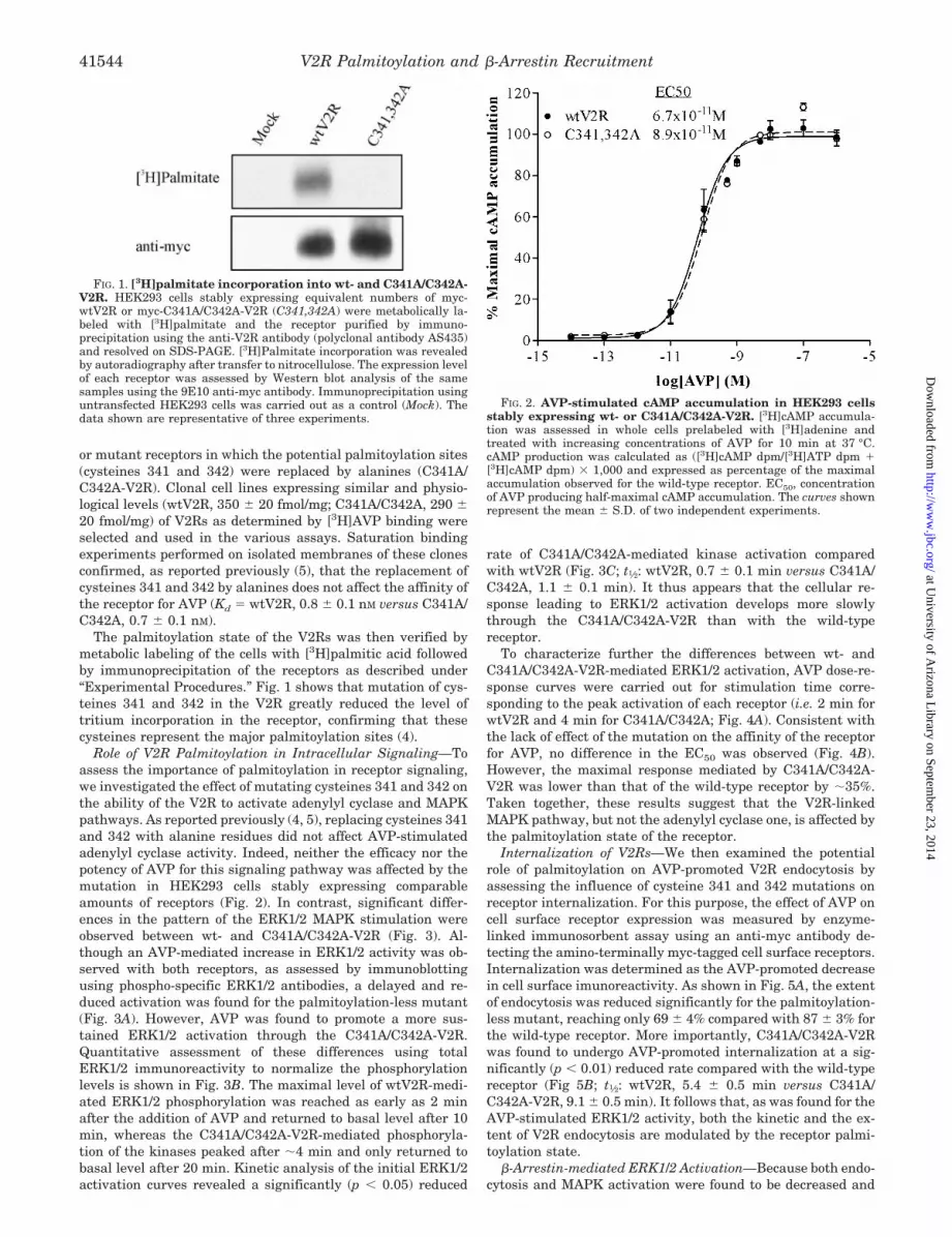
The palmitoylation state of the V2Rs was then verified bymetabolic labeling of the cells with [3H]palmitic acid followedby immunoprecipitation of the receptors as described under“Experimental Procedures.” Fig. 1 shows that mutation of cys-teines 341 and 342 in the V2R greatly reduced the level oftritium incorporation in the receptor, confirming that thesecysteines represent the major palmitoylation sites (4).
Role of V2R Palmitoylation in Intracellular Signaling—Toassess the importance of palmitoylation in receptor signaling,we investigated the effect of mutating cysteines 341 and 342 onthe ability of the V2R to activate adenylyl cyclase and MAPKpathways. As reported previously (4, 5), replacing cysteines 341and 342 with alanine residues did not affect AVP-stimulatedadenylyl cyclase activity. Indeed, neither the efficacy nor thepotency of AVP for this signaling pathway was affected by themutation in HEK293 cells stably expressing comparableamounts of receptors (Fig. 2). In contrast, significant differ-ences in the pattern of the ERK1/2 MAPK stimulation wereobserved between wt- and C341A/C342A-V2R (Fig. 3). Al-though an AVP-mediated increase in ERK1/2 activity was ob-served with both receptors, as assessed by immunoblottingusing phospho-specific ERK1/2 antibodies, a delayed and re-duced activation was found for the palmitoylation-less mutant(Fig. 3A). However, AVP was found to promote a more sus-tained ERK1/2 activation through the C341A/C342A-V2R.Quantitative assessment of these differences using totalERK1/2 immunoreactivity to normalize the phosphorylationlevels is shown in Fig. 3B. The maximal level of wtV2R-medi-ated ERK1/2 phosphorylation was reached as early as 2 minafter the addition of AVP and returned to basal level after 10min, whereas the C341A/C342A-V2R-mediated phosphoryla-tion of the kinases peaked after �4 min and only returned tobasal level after 20 min. Kinetic analysis of the initial ERK1/2activation curves revealed a significantly (p 0.05) reduced
rate of C341A/C342A-mediated kinase activation comparedwith wtV2R (Fig. 3C; t1⁄2: wtV2R, 0.7 0.1 min versus C341A/C342A, 1.1 0.1 min). It thus appears that the cellular re-sponse leading to ERK1/2 activation develops more slowlythrough the C341A/C342A-V2R than with the wild-typereceptor.
To characterize further the differences between wt- andC341A/C342A-V2R-mediated ERK1/2 activation, AVP dose-re-sponse curves were carried out for stimulation time corre-sponding to the peak activation of each receptor (i.e. 2 min forwtV2R and 4 min for C341A/C342A; Fig. 4A). Consistent withthe lack of effect of the mutation on the affinity of the receptorfor AVP, no difference in the EC50 was observed (Fig. 4B).However, the maximal response mediated by C341A/C342A-V2R was lower than that of the wild-type receptor by �35%.Taken together, these results suggest that the V2R-linkedMAPK pathway, but not the adenylyl cyclase one, is affected bythe palmitoylation state of the receptor.
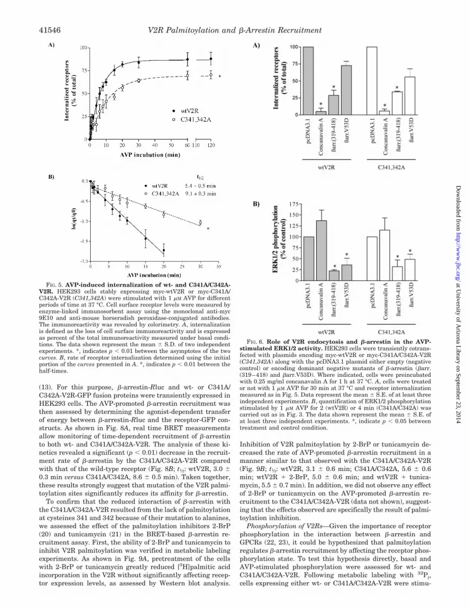
Internalization of V2Rs—We then examined the potentialrole of palmitoylation on AVP-promoted V2R endocytosis byassessing the influence of cysteine 341 and 342 mutations onreceptor internalization. For this purpose, the effect of AVP oncell surface receptor expression was measured by enzyme-linked immunosorbent assay using an anti-myc antibody de-tecting the amino-terminally myc-tagged cell surface receptors.Internalization was determined as the AVP-promoted decreasein cell surface imunoreactivity. As shown in Fig. 5A, the extentof endocytosis was reduced significantly for the palmitoylation-less mutant, reaching only 69 4% compared with 87 3% forthe wild-type receptor. More importantly, C341A/C342A-V2Rwas found to undergo AVP-promoted internalization at a sig-nificantly (p 0.01) reduced rate compared with the wild-typereceptor (Fig 5B; t1⁄2: wtV2R, 5.4 0.5 min versus C341A/C342A-V2R, 9.1 0.5 min). It follows that, as was found for theAVP-stimulated ERK1/2 activity, both the kinetic and the ex-tent of V2R endocytosis are modulated by the receptor palmi-toylation state.
�-Arrestin-mediated ERK1/2 Activation—Because both endo-cytosis and MAPK activation were found to be decreased and
FIG. 1. [3H]palmitate incorporation into wt- and C341A/C342A-V2R. HEK293 cells stably expressing equivalent numbers of myc-wtV2R or myc-C341A/C342A-V2R (C341,342A) were metabolically la-beled with [3H]palmitate and the receptor purified by immuno-precipitation using the anti-V2R antibody (polyclonal antibody AS435)and resolved on SDS-PAGE. [3H]Palmitate incorporation was revealedby autoradiography after transfer to nitrocellulose. The expression levelof each receptor was assessed by Western blot analysis of the samesamples using the 9E10 anti-myc antibody. Immunoprecipitation usinguntransfected HEK293 cells was carried out as a control (Mock). Thedata shown are representative of three experiments.
FIG. 2. AVP-stimulated cAMP accumulation in HEK293 cellsstably expressing wt- or C341A/C342A-V2R. [3H]cAMP accumula-tion was assessed in whole cells prelabeled with [3H]adenine andtreated with increasing concentrations of AVP for 10 min at 37 °C.cAMP production was calculated as ([3H]cAMP dpm/[3H]ATP dpm �[3H]cAMP dpm) � 1,000 and expressed as percentage of the maximalaccumulation observed for the wild-type receptor. EC50, concentrationof AVP producing half-maximal cAMP accumulation. The curves shownrepresent the mean S.D. of two independent experiments.
V2R Palmitoylation and �-Arrestin Recruitment41544
at University of A
rizona Library on Septem
ber 23, 2014http://w
ww
.jbc.org/D
ownloaded from

delayed by mutating cysteines 341 and 342, and given theproposed linked between endocytosis and MAPK activation (1,15), we then investigated the potential implication of V2Rendocytosis in the AVP-stimulated ERK1/2 activation. For this,the effect of a pharmacological blocker (concanavalin A, a lectinthat interferes with endocytosis by binding to the carbohydratemoieties (16)) and of two dominant negative mutants of �-ar-restin with impaired receptor binding capacities (a truncatedform, �-arrestin (319–418) (10), and a point mutant, �-arrestinV53D (10, 17)) were assessed. As shown in Fig. 6A, the con-canavalin A treatment almost completely blocked endocytosisof both wt- and C341A/C342A-V2R, whereas �-arrestin (319–418) and �-arrestin V53D inhibited the internalization of thesereceptors by 70 and 35%, respectively. These results confirmprevious data indicating that V2R undergoes agonist-promotedendocytosis via a �-arrestin-dependent process presumablythrough clathrin-coated vesicles (18, 19). In contrast to itsdramatic effect on receptor endocytosis, concanavalin A waswithout effect on the AVP-stimulated MAPK activation in cellsexpressing either wt- or C341A/C342A-V2R (Fig. 6B), clearlyseparating the two phenomena. However, both �-arrestin(319–418) and �-arrestin V53D significantly inhibited re-
ceptor-mediated ERK1/2 phosphorylation, suggesting an im-portant role for �-arrestin in the AVP-stimulated ERK1/2activation (Fig. 6B).
AVP-induced �-Arrestin Recruitment—The preceding dataindicate that no causal link exists between V2R endocytosisand MAPK activation. However, they clearly place �-arrestinon the path of two processes affected by the palmitoylationstate of the receptor. To test directly whether the decreasedreceptor endocytosis and reduced AVP-stimulated ERK1/2 ac-tivity of C341A/C342A-V2R could result from an altered �-ar-restin interaction, the ability of AVP to promote �-arrestinrecruitment to the wt- and C341A/C342A-V2R was first inves-tigated in coimmunoprecipitation studies using the reversiblemembrane-permeable cross-linker DSP. As shown in Fig. 7A,�-arrestin2-GFP could be coimmunoprecipitated with both wt-and C341A/C342A-myc-tagged V2R. Although a single specific�-arrestin2-GFP band was observed in the whole cell lysates,the coimmunoprecipitated �-arrestin2-GFP appeared as a dou-blet including a less abundant species with a slower mobility.The nature of this �-arrestin species that is revealed in thecoimmunoprecipitation conditions remains to be determined.In any case, AVP induced a significant increase of �-arrestin2-GFP in the myc-tagged receptor immunoprecipitates, reflectingthe agonist-promoted recruitment of �-arrestin to both wt- andC341A/C342A-V2R after agonist exposure. Interestingly, how-ever, less �-arrestin2-GFP was found to be associated withC341A/C342A-V2R (Fig. 7B), suggesting an altered interactionbetween �-arrestin and the V2R palmitoylation-less mutant.
To characterize further �-arrestin interaction with the wt-and C341A/C342A-V2R, we assessed the AVP-promoted �-ar-restin recruitment to the receptors using a BRET-based assay
FIG. 3. Time course of AVP-induced ERK1/2 activity. A,HEK293 cells stably expressing myc-wtV2R or myc-C341A/C342A-V2R(C341,342A) were stimulated with 1 �M AVP or 10% FBS for theindicated times. MAPK activity was then detected using phospho-spe-cific anti-ERK1/2 antibodies (P-ERK1/2). Levels of the MAPK werecontrolled using antibodies directed against the total kinase population(ERK1/2). B, graphic representation of the data expressed as P-ERK1/2over ERK1/2 in percentage of the levels observed with 10% FBS used asthe reference control. The curves shown represent the mean S.E. of atleast four independent experiments. C, rate of ERK1/2 phosphorylationdetermined using the initial portion of the curves presented in B. *,indicates p 0.05 between the half-times.
FIG. 4. Concentration-dependent AVP-induced ERK1/2 activ-ity. A, HEK293 cells stably expressing myc-wtV2R or myc-C341A/C342A-V2R (C341,342A) were stimulated with increasing concentra-tions of AVP for 2 (wtV2R) or 4 min (C341A/C342A) at 37 °C. Referencestimulation was carried out using 10% FBS for 5 min. The MAPKactivity was assessed as in Fig. 3. B, graphic representation of the dataexpressed as P-ERK1/2 over ERK1/2 in percentage of the levels ob-served with 10% FBS used as the reference control. The curves shownrepresent the mean S.E. of four independent experiments. *, indi-cates p 0.01 between the asymptotes of the two curves.
V2R Palmitoylation and �-Arrestin Recruitment 41545
at University of A
rizona Library on Septem
ber 23, 2014http://w
ww
.jbc.org/D
ownloaded from
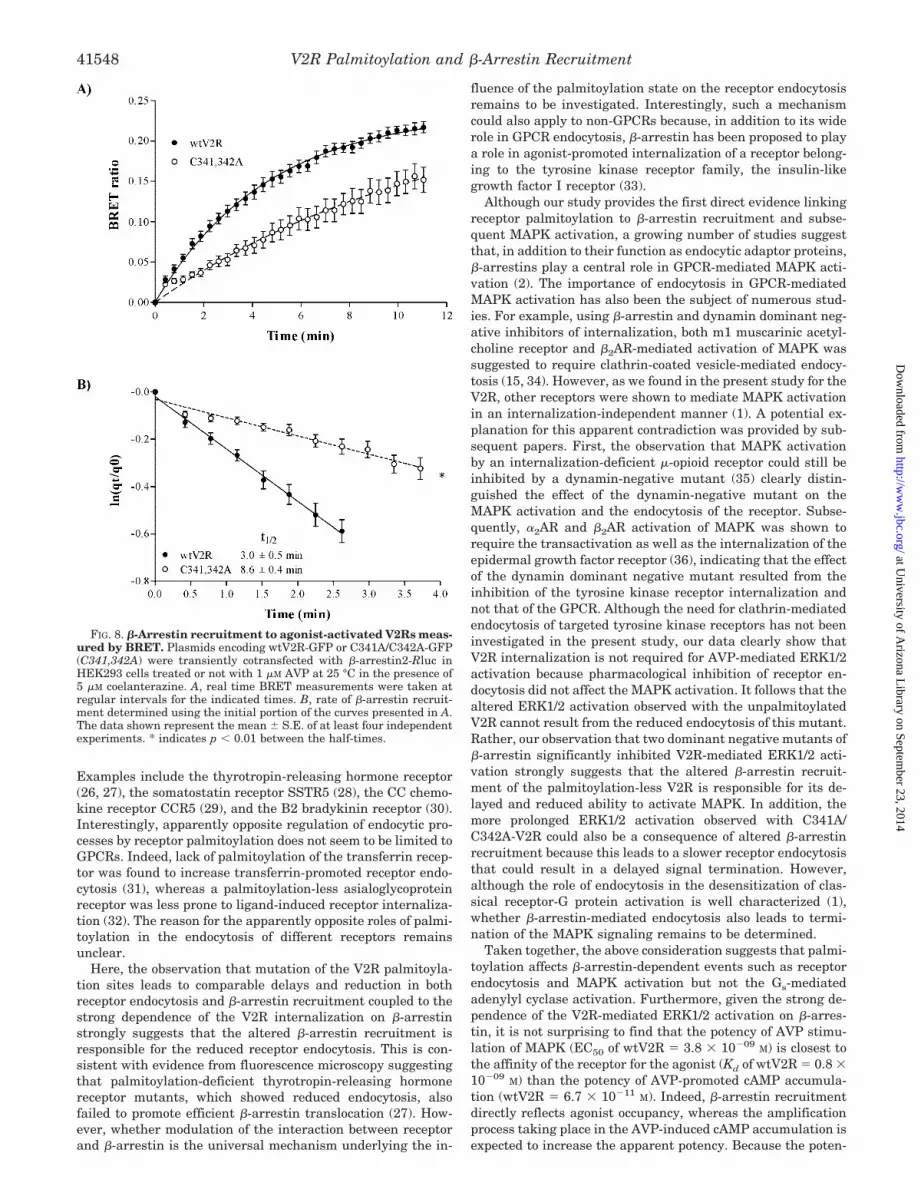
(13). For this purpose, �-arrestin-Rluc and wt- or C341A/C342A-V2R-GFP fusion proteins were transiently expressed inHEK293 cells. The AVP-promoted �-arrestin recruitment wasthen assessed by determining the agonist-dependent transferof energy between �-arrestin-Rluc and the receptor-GFP con-structs. As shown in Fig. 8A, real time BRET measurementsallow monitoring of time-dependent recruitment of �-arrestinto both wt- and C341A/C342A-V2R. The analysis of these ki-netics revealed a significant (p 0.01) decrease in the recruit-ment rate of �-arrestin by the C341A/C342A-V2R comparedwith that of the wild-type receptor (Fig. 8B; t1⁄2: wtV2R, 3.0 0.3 min versus C341A/C342A, 8.6 0.5 min). Taken together,these results strongly suggest that mutation of the V2R palmi-toylation sites significantly reduces its affinity for �-arrestin.
To confirm that the reduced interaction of �-arrestin withthe C341A/C342A-V2R resulted from the lack of palmitoylationat cysteines 341 and 342 because of their mutation to alanines,we assessed the effect of the palmitoylation inhibitors 2-BrP(20) and tunicamycin (21) in the BRET-based �-arrestin re-cruitment assay. First, the ability of 2-BrP and tunicamycin toinhibit V2R palmitoylation was verified in metabolic labelingexperiments. As shown in Fig. 9A, pretreatment of the cellswith 2-BrP or tunicamycin greatly reduced [3H]palmitic acidincorporation in the V2R without significantly affecting recep-tor expression levels, as assessed by Western blot analysis.
Inhibition of V2R palmitoylation by 2-BrP or tunicamycin de-creased the rate of AVP-promoted �-arrestin recruitment in amanner similar to that observed with the C341A/C342A-V2R(Fig. 9B; t1⁄2: wtV2R, 3.1 0.6 min; C341A/C342A, 5.6 0.6min; wtV2R � 2-BrP, 5.0 0.6 min; and wtV2R � tunica-mycin, 5.5 0.7 min). In addition, we did not observe any effectof 2-BrP or tunicamycin on the AVP-promoted �-arrestin re-cruitment to the C341A/C342A-V2R (data not shown), suggest-ing that the effects observed are specifically the result of palmi-toylation inhibition.
Phosphorylation of V2Rs—Given the importance of receptorphosphorylation in the interaction between �-arrestin andGPCRs (22, 23), it could be hypothesized that palmitoylationregulates �-arrestin recruitment by affecting the receptor phos-phorylation state. To test this hypothesis directly, basal andAVP-stimulated phosphorylation were assessed for wt- andC341A/C342A-V2R. Following metabolic labeling with 32Pi,cells expressing either wt- or C341A/C342A-V2R were stimu-
FIG. 5. AVP-induced internalization of wt- and C341A/C342A-V2R. HEK293 cells stably expressing myc-wtV2R or myc-C341A/C342A-V2R (C341,342A) were stimulated with 1 �M AVP for differentperiods of time at 37 °C. Cell surface receptor levels were measured byenzyme-linked immunosorbent assay using the monoclonal anti-myc9E10 and anti-mouse horseradish peroxidase-conjugated antibodies.The immunoreactivity was revealed by colorimetry. A, internalizationis defined as the loss of cell surface immunoreactivity and is expressedas percent of the total immunoreactivity measured under basal condi-tions. The data shown represent the mean S.D. of two independentexperiments. *, indicates p 0.01 between the asymptotes of the twocurves. B, rate of receptor internalization determined using the initialportion of the curves presented in A. *, indicates p 0.01 between thehalf-times.
FIG. 6. Role of V2R endocytosis and �-arrestin in the AVP-stimulated ERK1/2 activity. HEK293 cells were transiently cotrans-fected with plasmids encoding myc-wtV2R or myc-C341A/C342A-V2R(C341,342A) along with the pcDNA3.1 plasmid either empty (negativecontrol) or encoding dominant negative mutants of �-arrestin (�arr.(319–418) and �arr.V53D). Where indicated, cells were preincubatedwith 0.25 mg/ml concanavalin A for 1 h at 37 °C. A, cells were treatedor not with 1 �M AVP for 30 min at 37 °C and receptor internalizationmeasured as in Fig. 5. Data represent the mean S.E. of at least threeindependent experiments. B, quantification of ERK1/2 phosphorylationstimulated by 1 �M AVP for 2 (wtV2R) or 4 min (C341A/C342A) wascarried out as in Fig. 3. The data shown represent the mean S.E. ofat least three independent experiments. *, indicate p 0.05 betweentreatment and control condition.
V2R Palmitoylation and �-Arrestin Recruitment41546
at University of A
rizona Library on Septem
ber 23, 2014http://w
ww
.jbc.org/D
ownloaded from

lated or not with AVP and the receptors purified by immuno-precipitation as described under “Experimental Procedures.”As shown in Fig. 10 and in agreement with the previous reportof Sadeghi et al. (4), comparable basal and AVP-promotedphosphorylation levels were observed for the wild-type andthe palmitoylation-less receptor, excluding a role for phos-phorylation in the reduced �-arrestin recruitment toC341A/C342A-V2R.
DISCUSSION
Taken together, the results of the present study demonstratethat the V2R palmitoylation state regulates distinct receptorfunctions differentially. Although mutation of the palmitoyla-tion sites did not affect the receptor-stimulated adenylyl cy-clase activity, it led to significantly delayed and decreasedagonist-promoted ERK1/2 activation and receptor endocytosis.No causal link between the reduced MAPK activity and recep-
tor endocytosis was found, but both most likely resulted fromthe altered agonist-mediated �-arrestin recruitment observedfor the palmitoylation-less mutant. Indeed, our results showthat lack of palmitoylation of the V2R, either by mutation orpharmacological inhibition, slowed down and decreased itsability to recruit �-arrestin, suggesting a reduced affinity of thereceptor for �-arrestin. Receptor palmitoylation can thereforebe seen as a post-translational modification that favors �-ar-restin-dependent processes.
Modulation of GPCR endocytosis efficiency by palmitoylationhas been reported previously for several receptors. Althoughmutation of palmitoylated cysteines was found to increase theendocytosis of these receptors for the luteinizing hormone/hu-man choriogonadotropin receptor (24) and the V1a vasopressinreceptor (V1aR) (25), in most cases, it was found as in thepresent study to decrease their agonist-promoted endocytosis.
FIG. 7. �-Arrestin coimmunoprecipitation with agonist-activated V2Rs. Plasmids encoding myc-wtV2R or myc-C341A/C342A-V2R(C341,342A) were transiently cotransfected with �-arrestin2-GFP (�arrGFP) in HEK293 cells. Cells were treated or not with 1 �M AVP for 15 minat 37 °C before adding the cross-linking agent DSP. Myc-tagged receptors were then immunoprecipitated using the 9E10 anti-myc antibody andresolved on SDS-PAGE as described under “Experimental Procedures.” A, the amount of myc-V2Rs and the presence of �-arrestin2-GFP in theimmunoprecipitates were assessed by Western blot analysis using the A14 anti-myc and anti-GFP polyclonal antibodies, respectively. Blotting ofthe whole cell lysates was performed to control for the expression levels of myc receptors and �-arrestin2-GFP. B, quantification of coimmuno-precipitated �-arrestin2-GFP expressed as the ratio of anti-GFP over anti-myc immunoreactivities in the immunoprecipitates in percentage of themaximal obtained with the wtV2R. The data shown represent the mean S.E. of three independent experiments.
V2R Palmitoylation and �-Arrestin Recruitment 41547
at University of A
rizona Library on Septem
ber 23, 2014http://w
ww
.jbc.org/D
ownloaded from
Examples include the thyrotropin-releasing hormone receptor(26, 27), the somatostatin receptor SSTR5 (28), the CC chemo-kine receptor CCR5 (29), and the B2 bradykinin receptor (30).Interestingly, apparently opposite regulation of endocytic pro-cesses by receptor palmitoylation does not seem to be limited toGPCRs. Indeed, lack of palmitoylation of the transferrin recep-tor was found to increase transferrin-promoted receptor endo-cytosis (31), whereas a palmitoylation-less asialoglycoproteinreceptor was less prone to ligand-induced receptor internaliza-tion (32). The reason for the apparently opposite roles of palmi-toylation in the endocytosis of different receptors remainsunclear.
Here, the observation that mutation of the V2R palmitoyla-tion sites leads to comparable delays and reduction in bothreceptor endocytosis and �-arrestin recruitment coupled to thestrong dependence of the V2R internalization on �-arrestinstrongly suggests that the altered �-arrestin recruitment isresponsible for the reduced receptor endocytosis. This is con-sistent with evidence from fluorescence microscopy suggestingthat palmitoylation-deficient thyrotropin-releasing hormonereceptor mutants, which showed reduced endocytosis, alsofailed to promote efficient �-arrestin translocation (27). How-ever, whether modulation of the interaction between receptorand �-arrestin is the universal mechanism underlying the in-
fluence of the palmitoylation state on the receptor endocytosisremains to be investigated. Interestingly, such a mechanismcould also apply to non-GPCRs because, in addition to its widerole in GPCR endocytosis, �-arrestin has been proposed to playa role in agonist-promoted internalization of a receptor belong-ing to the tyrosine kinase receptor family, the insulin-likegrowth factor I receptor (33).
Although our study provides the first direct evidence linkingreceptor palmitoylation to �-arrestin recruitment and subse-quent MAPK activation, a growing number of studies suggestthat, in addition to their function as endocytic adaptor proteins,�-arrestins play a central role in GPCR-mediated MAPK acti-vation (2). The importance of endocytosis in GPCR-mediatedMAPK activation has also been the subject of numerous stud-ies. For example, using �-arrestin and dynamin dominant neg-ative inhibitors of internalization, both m1 muscarinic acetyl-choline receptor and �2AR-mediated activation of MAPK wassuggested to require clathrin-coated vesicle-mediated endocy-tosis (15, 34). However, as we found in the present study for theV2R, other receptors were shown to mediate MAPK activationin an internalization-independent manner (1). A potential ex-planation for this apparent contradiction was provided by sub-sequent papers. First, the observation that MAPK activationby an internalization-deficient �-opioid receptor could still beinhibited by a dynamin-negative mutant (35) clearly distin-guished the effect of the dynamin-negative mutant on theMAPK activation and the endocytosis of the receptor. Subse-quently, �2AR and �2AR activation of MAPK was shown torequire the transactivation as well as the internalization of theepidermal growth factor receptor (36), indicating that the effectof the dynamin dominant negative mutant resulted from theinhibition of the tyrosine kinase receptor internalization andnot that of the GPCR. Although the need for clathrin-mediatedendocytosis of targeted tyrosine kinase receptors has not beeninvestigated in the present study, our data clearly show thatV2R internalization is not required for AVP-mediated ERK1/2activation because pharmacological inhibition of receptor en-docytosis did not affect the MAPK activation. It follows that thealtered ERK1/2 activation observed with the unpalmitoylatedV2R cannot result from the reduced endocytosis of this mutant.Rather, our observation that two dominant negative mutants of�-arrestin significantly inhibited V2R-mediated ERK1/2 acti-vation strongly suggests that the altered �-arrestin recruit-ment of the palmitoylation-less V2R is responsible for its de-layed and reduced ability to activate MAPK. In addition, themore prolonged ERK1/2 activation observed with C341A/C342A-V2R could also be a consequence of altered �-arrestinrecruitment because this leads to a slower receptor endocytosisthat could result in a delayed signal termination. However,although the role of endocytosis in the desensitization of clas-sical receptor-G protein activation is well characterized (1),whether �-arrestin-mediated endocytosis also leads to termi-nation of the MAPK signaling remains to be determined.
Taken together, the above consideration suggests that palmi-toylation affects �-arrestin-dependent events such as receptorendocytosis and MAPK activation but not the Gs-mediatedadenylyl cyclase activation. Furthermore, given the strong de-pendence of the V2R-mediated ERK1/2 activation on �-arres-tin, it is not surprising to find that the potency of AVP stimu-lation of MAPK (EC50 of wtV2R � 3.8 � 10�09 M) is closest tothe affinity of the receptor for the agonist (Kd of wtV2R � 0.8 �10�09 M) than the potency of AVP-promoted cAMP accumula-tion (wtV2R � 6.7 � 10�11 M). Indeed, �-arrestin recruitmentdirectly reflects agonist occupancy, whereas the amplificationprocess taking place in the AVP-induced cAMP accumulation isexpected to increase the apparent potency. Because the poten-
FIG. 8. �-Arrestin recruitment to agonist-activated V2Rs meas-ured by BRET. Plasmids encoding wtV2R-GFP or C341A/C342A-GFP(C341,342A) were transiently cotransfected with �-arrestin2-Rluc inHEK293 cells treated or not with 1 �M AVP at 25 °C in the presence of5 �M coelanterazine. A, real time BRET measurements were taken atregular intervals for the indicated times. B, rate of �-arrestin recruit-ment determined using the initial portion of the curves presented in A.The data shown represent the mean S.E. of at least four independentexperiments. * indicates p 0.01 between the half-times.
V2R Palmitoylation and �-Arrestin Recruitment41548
at University of A
rizona Library on Septem
ber 23, 2014http://w
ww
.jbc.org/D
ownloaded from

cies were both determined in whole cell assays and are thusdirectly comparable, the difference then suggests that the AVP-stimulated ERK1/2 activation observed in the present studydoes not depend on cAMP production. This was supportedfurther by the observation that a protein kinase A inhibitor(KT5720) did not affect AVP-promoted ERK1/2 activation.2
Other studies have suggested pathway-specific modulationof receptor functions by palmitoylation. For instance, absenceof palmitoylation in the endothelin A receptor was shown toprevent its coupling to the phospholipase C and ERK1/2 sig-naling pathways without affecting the adenylyl cyclase activa-tion (37, 38). In this case, the authors suggested that palmitoy-lation was required for Gq but not Gs coupling. Similarly,mutation of the three palmitoylated cysteines of CCR5 has2 P. G. Charest and M. Bouvier, unpublished observations.
FIG. 9. Effect of pharmacological inhibition of V2R palmitoylation on �-arrestin recruitment. A, HEK293 cells stably expressingmyc-wtV2R were pretreated or not with 100 �M 2-BrP for 16 h or 30 �M tunicamycin for 4 h at 37 °C followed by metabolic labeling with[3H]palmitate as described under “Experimental Procedures.” Receptors were then purified by immunoprecipitation and resolved on SDS-PAGEbefore being analyzed by autoradiography for [3H]palmitate incorporation or Western blotting to control receptor expression. B, HEK293 cells weretransiently cotransfected with wtV2R-GFP or C341A/C342A-GFP (C341,342A) along with �-arrestin2-Rluc and treated or not with 2-BrP ortunicamycin (Tm) as described above, followed by 1 �M AVP at 25 °C in the presence of 5 �M coelanterazine. Real time BRET measurements weretaken at regular intervals for the indicated times. Inset, rate of �-arrestin recruitment determined using the initial portion of the curves presentedin B. The data shown represent the mean S.E. of three independent experiments. *, indicates p 0.01 compared with wtV2R half-time.
V2R Palmitoylation and �-Arrestin Recruitment 41549
at University of A
rizona Library on Septem
ber 23, 2014http://w
ww
.jbc.org/D
ownloaded from

been shown to affect efficient coupling to only a subset of itssignaling repertoire (39). However, the potential role of �-ar-restin in these pathway-specific effects of palmitoylation wasnot investigated. These observations and the findings reportedherein suggest that different agonist-stimulated processes(such as selective G protein activation, �-arrestin recruitment,and receptor endocytosis) may involve distinct receptor confor-mations and/or domains that can be regulated differentially.Supporting this idea, a modified parathyroid hormone receptoruncoupled from its cognate G protein can still undergo agonist-induced �-arrestin-mediated endocytosis (40). Hence, becausepalmitoylation occurs mostly at the level of the carboxyl-termi-nal tail of GPCR, it will most likely affect processes involvingthis particular domain. Interestingly, it was shown that forseveral receptors, including the V2R, the carboxyl-terminal tailis implicated in the formation of stable receptor-�-arrestininteractions (18, 41).
How receptor palmitoylation could affect the coupling to�-arrestin is unknown. However, palmitoylation has beenshown to play important roles in protein-protein interaction fora number of proteins. For example, palmitoylation of G�s hasbeen shown to favor interactions with G�� (42), whereas palmi-
toylation of G�z was found to decrease the affinity of the Gz-GTPase activating protein for its GTP-bound form (43). Palmi-toylation of Src family tyrosine kinases has also been shown tobe required for their interaction with glycosylphosphatidyli-nositol-anchored proteins (44) as well as with their B cell sub-strate Ig� (45). Finally, palmitoylation of caveolin-1 was foundto be essential for its interaction with c-Src (46) and that oftetraspanin proteins important for their self-association (47).
Binding of �-arrestin to agonist-activated GPCR is thoughtto involve multiple interactions (48). A large region within theamino-terminal half of �-arrestin, termed the activation recog-nition domain, recognizes the activated state of GPCRs. Thisdomain of �-arrestin appears to bind the third intracellularloop of several receptors, including the �2AR, m2 and m3 mus-carinic acetylcholine receptors (49). This is followed by thebinding of a smaller positively charged region in the centralportion of �-arrestin, termed the phosphorylation recognitiondomain, to the receptor carboxyl tail phosphorylated by GPCRkinase (50). Notably, agonist-induced phosphorylated clustersof serine or threonine residues, located downstream of theputative palmitoylation sites of several GPCR including theV2R, were identified as molecular determinants of the stabilityof receptor-�-arrestin complexes (18, 41). In fact, the absence ofsuch phosphorylation clusters within the carboxyl tail of sev-eral receptors, including the �2AR, has been invoked to explainthe rapid dissociation of �-arrestin from these receptors.
Modulation of the phosphorylation state of the receptor bypalmitoylation could thus be proposed as a potential mecha-nism regulating �-arrestin recruitment. Consistent with thishypothesis, GPCR palmitoylation sites are often located prox-imally to receptor phosphorylation sites and have been shownto affect GPCR phosphorylation for a number of receptors. Forinstance, mutation of palmitoylation sites in their carboxyl tailhas been linked to an increased phosphorylation for the �2AR(51), the GluR6 kainate receptor (52), and the A3 adenosinereceptor (53), whereas it led to a decrease phosphorylation forbovine opsin (54), CCR5 (29), and V1aR (25). However, changesin the phosphorylation status of the V2R does not seem toaccount for the altered �-arrestin recruitment of the palmitoy-lation-less mutant because in agreement with the previousfinding of Sadeghi et al. (4), no difference in either basal orAVP-induced phosphorylation levels was observed between wt-and C341A/C342A-V2R. Instead, we propose that the reducedaffinity of �-arrestin for the palmitoylation-less mutant residesin the altered conformation of C341A/C342A-V2R carboxyl tailcompared with the wtV2R.
Our proposition is in part based on the fact that the highaffinity of �-arrestin for the receptor appears to depend on theposition of the phosphorylated cluster of serines within thecarboxyl tail (18). In addition, to show that the V2R clustercould not reintroduce the �-arrestin high affinity when addedat the end of the carboxyl tail of the �2AR, Oakley et al. (41)showed that the position of the clusters within the carboxyl tailof several receptor displaying high affinity for �-arrestin isrelatively well conserved. This conservation is even more re-markable for receptors such as the V2R, the neurotensin-1receptor, and the oxytocin receptor, with carboxyl-terminal tailof similar length and containing putative palmitoylation sites.Because receptor palmitate moieties are inserted in the plasmamembrane where they limit the carboxyl side of the eighth�-helix (55), they also define the distance between the plasmamembrane and downstream residues. Therefore, by controllingthe positioning of the �-arrestin-interacting domains, palmi-toylation could optimize its association with the regulatoryprotein. Lack of palmitoylation could then affect this conforma-tion, leading to decreased affinity of the receptor for �-arrestin
FIG. 10. AVP-induced phosphorylation of wt- and C341A/C342A-V2R. A, HEK293 cells stably expressing myc-wtV2R or myc-C341A/C342A-V2R (C341,342A) were metabolically labeled with 32Piand treated or not with 1 �M AVP for 15 min. The receptors werepurified by immunoprecipitation using the 9E10 anti-myc antibody andresolved on SDS-PAGE. 32Pi incorporation was revealed by autoradiog-raphy using a PhosphorImager after transfer to nitrocellulose. Theexpression level of each receptor was assessed by Western blot analysisof the same membrane using the A14 anti-myc antibody. Immunopre-cipitation using untransfected HEK293 cells was carried out as a con-trol (Mock). B, quantification of receptor phosphorylation is expressedas the ratio of 32P labeling over anti-myc immunoreactivity in percent-age of the maximal obtained with the wtV2R. The data shown representthe mean S.D. of two independent experiments.
V2R Palmitoylation and �-Arrestin Recruitment41550
at University of A
rizona Library on Septem
ber 23, 2014http://w
ww
.jbc.org/D
ownloaded from

and resulting in the slower and reduced recruitment to C341A/C342A-V2R observed in our study.
In conclusion, our study suggests that palmitoylation of V2Rincreases receptor affinity for �-arrestin thus allowing rapidagonist-promoted recruitment of the adaptor protein that leadsto efficient endocytosis and ERK1/2 activation. Given the factthat palmitoylation is dynamically regulated during the recep-tor activation cycle (3), regulated changes in this post-transla-tional modification could modulate receptor endocytosis anddetermine the relative contribution of different signaling path-ways in response to receptor stimulation.
Acknowledgments—We are grateful to Dr. Monique Lagace for thecritical reading of the manuscript and Dr. Werner Muller-Esterl for thegenerous supply of the AS435 anti-V2R antibody.
REFERENCES
1. Ferguson, S. S. (2001) Pharmacol. Rev. 53, 1–242. Luttrell, L. M., and Lefkowitz, R. J. (2002) J. Cell Sci. 115, 455–4653. Qanbar, R., and Bouvier, M. (2003) Pharmacol. Ther. 97, 1–334. Sadeghi, H. M., Innamorati, G., Dagarag, M., and Birnbaumer, M. (1997) Mol.
Pharmacol. 52, 21–295. Schulein, R., Liebenhoff, U., Muller, H., Birnbaumer, M., and Rosenthal, W.
(1996) Biochem. J. 313, 611–6166. Thibonnier, M., Conarty, D. M., Preston, J. A., Wilkins, P. L., Berti-Mattera,
L. N., and Mattera, R. (1998) Adv. Exp. Med. Biol. 449, 251–2767. Marinissen, M. J., and Gutkind, J. S. (2001) Trends Pharmacol. Sci. 22,
368–3768. Morello, J. P., Salahpour, A., Laperriere, A., Bernier, V., Arthus, M. F.,
Lonergan, M., Petaja-Repo, U., Angers, S., Morin, D., Bichet, D. G., andBouvier, M. (2000) J. Clin. Invest. 105, 887–895
9. Mercier, J. F., Salahpour, A., Angers, S., Breit, A., and Bouvier, M. (2002)J. Biol. Chem. 277, 44925–44931
10. Krupnick, J. G., Santini, F., Gagnon, A. W., Keen, J. H., and Benovic, J. L.(1997) J. Biol. Chem. 272, 32507–32512
11. Sambrook, J., Fritsch, E. F., and Maniatis, T. (1989) Molecular Cloning: ALaboratory Manual, 2nd Ed., pp. 16.33–16.36, Cold Spring Harbor Labora-tory Press, Cold Spring Harbor, NY
12. Orsini, M. J., and Benovic, J. L. (1998) J. Biol. Chem. 273, 34616–3462213. Angers, S., Salahpour, A., Joly, E., Hilairet, S., Chelsky, D., Dennis, M., and
Bouvier, M. (2000) Proc. Natl. Acad. Sci. U. S. A. 97, 3684–368914. Tohgo, A., Choy, E. W., Gesty-Palmer, D., Pierce, K. L., Laporte, S., Oakley,
R. H., Caron, M. G., Lefkowitz, R. J., and Luttrell, L. M. (2003) J. Biol.Chem. 278, 6258–6267
15. Daaka, Y., Luttrell, L. M., Ahn, S., Della, R. G., Ferguson, S. S., Caron, M. G.,and Lefkowitz, R. J. (1998) J. Biol. Chem. 273, 685–688
16. Pippig, S., Andexinger, S., and Lohse, M. J. (1995) Mol. Pharmacol. 47,666–676
17. Ferguson, S. S., Downey, W. E., III, Colapietro, A. M., Barak, L. S., Menard, L.,and Caron, M. G. (1996) Science 271, 363–366
18. Oakley, R. H., Laporte, S. A., Holt, J. A., Barak, L. S., and Caron, M. G. (1999)J. Biol. Chem. 274, 32248–32257
19. Bowen-Pidgeon, D., Innamorati, G., Sadeghi, H. M., and Birnbaumer, M.(2001) Mol. Pharmacol. 59, 1395–1401
20. Webb, Y., Hermida-Matsumoto, L., and Resh, M. D. (2000) J. Biol. Chem. 275,261–270
21. Patterson, S. I., and Skene, J. H. P. (1994) J. Cell Biol. 124, 521–53622. Lohse, M. J., Benovic, J. L., Codina, J., Caron, M. G., and Lefkowitz, R. J.
(1990) Science 248, 1547–155023. Lohse, M. J., Andexinger, S., Pitcher, J., Trukawinski, S., Codina, J., Faure,
J. P., Caron, M. G., and Lefkowitz, R. J. (1992) J. Biol. Chem. 267,8558–8564
24. Kawate, N., and Menon, K. M. (1994) J. Biol. Chem. 269, 30651–3065825. Hawtin, S. R., Tobin, A. B., Patel, S., and Wheatley, M. (2001) J. Biol. Chem.
276, 38139–3814626. Nussenzveig, D. R., Heinflink, M., and Gershengorn, M. C. (1993) J. Biol.
Chem. 268, 2389–239227. Groarke, D. A., Drmota, T., Bahia, D. S., Evans, N. A., Wilson, S., and Milligan,
G. (2001) Mol. Pharmacol. 59, 375–38528. Hukovic, N., Panetta, R., Kumar, U., Rocheville, M., and Patel, Y. C. (1998)
J. Biol. Chem. 273, 21416–2142229. Kraft, K., Olbrich, H., Majoul, I., Mack, M., Proudfoot, A., and Oppermann, M.
(2001) J. Biol. Chem. 276, 34408–3441830. Pizard, A., Blaukat, A., Michineau, S., Dikic, I., Muller-Esterl, W., Alhenc-
Gelas, F., and Rajerison, R. M. (2001) Biochemistry 40, 15743–1575131. Alvarez, E., Girones, N., and Davis, R. J. (1990) J. Biol. Chem. 265,
16644–1665532. Yik, J. H., Saxena, A., Weigel, J. A., and Weigel, P. H. (2002) J. Biol. Chem.
277, 40844–4085233. Lin, F. T., Daaka, Y., and Lefkowitz, R. J. (1998) J. Biol. Chem. 273,
31640–3164334. Vogler, O., Nolte, B., Voss, M., Schmidt, M., Jakobs, K. H., and Van Koppen,
C. J. (1999) J. Biol. Chem. 274, 12333–1233835. Whistler, J. L., and Von Zastrow, M. (1999) J. Biol. Chem. 274, 24575–2457836. Pierce, K. L., Maudsley, S., Daaka, Y., Luttrell, L. M., and Lefkowitz, R. J.
(2000) Proc. Natl. Acad. Sci. U. S. A. 97, 1489–149437. Horstmeyer, A., Cramer, H., Sauer, T., Muller-Esterl, W., and Schroeder, C.
(1996) J. Biol. Chem. 271, 20811–2081938. Cramer, H., Schmenger, K., Heinrich, K., Horstmeyer, A., Boning, H., Breit,
A., Piiper, A., Lundstrom, K., Muller-Esterl, W., and Schroeder, C. (2001)Eur. J. Biochem. 268, 5449–5459
39. Blanpain, C., Wittamer, V., Vanderwinden, J. M., Boom, A., Renneboog, B.,Lee, B., Le Poul, E., El Asmar, L., Govaerts, C., Vassart, G., Doms, R. W.,and Parmentier, M. (2001) J. Biol. Chem. 276, 23795–23804
40. Vilardaga, J. P., Frank, M., Krasel, C., Dees, C., Nissenson, R. A., and Lohse,M. J. (2001) J. Biol. Chem. 276, 33435–33443
41. Oakley, R. H., Laporte, S. A., Holt, J. A., Barak, L. S., and Caron, M. G. (2001)J. Biol. Chem. 276, 19452–19460
42. Iiri, T., Backlund, P. S. J., Jones, T. L. Z., Wedegaertner, P. B., and Bourne,H. R. (1996) Proc. Natl. Acad. Sci. U. S. A. 93, 14592–14597
43. Tu, Y., Wang, J., and Ross, E. M. (1997) Science 278, 1132–113544. Shenoy-Scaria, A. M., Gauen, L. K., Kwong, J., Shaw, A. S., and Lublin, D. M.
(1993) Mol. Cell Biol. 13, 6385–639245. Saouaf, S. J., Wolven, A., Resh, M. D., and Bolen, J. B. (1997) Biochem.
Biophys. Res. Commun. 234, 325–32946. Lee, H., Woodman, S. E., Engelman, J. A., Volonte, D., Galbiati, F., Kaufman,
H. L., Lublin, D. M., and Lisanti, M. P. (2001) J. Biol. Chem. 276,35150–35158
47. Yang, X., Claas, C., Kraeft, S. K., Chen, L. B., Wang, Z., Kreidberg, J. A., andHemler, M. E. (2002) Mol. Biol. Cell 13, 767–781
48. Krupnick, J. G., and Benovic, J. L. (1998) Annu. Rev. Pharmacol. Toxicol. 38,289–319
49. Wu, G., Krupnick, J. G., Benovic, J. L., and Lanier, S. M. (1997) J. Biol. Chem.272, 17836–17842
50. Kieselbach, T., Irrgang, K. D., and Ruppel, H. (1994) Eur. J. Biochem. 226,87–97
51. Moffett, S., Mouillac, B., Bonin, H., and Bouvier, M. (1993) EMBO J. 12,349–356
52. Pickering, D. S., Taverna, F. A., Salter, M. W., and Hampson, D. R. (1995)Proc. Natl. Acad. Sci. U. S. A. 92, 12090–12094
53. Palmer, T. M., and Stiles, G. L. (2000) Mol. Pharmacol. 57, 539–54554. Karnik, S. S., Ridge, K. D., Bhattacharya, S., and Khorana, H. G. (1993) Proc.
Natl. Acad. Sci. U. S. A. 90, 40–4455. Palczewski, K., Kumasaka, T., Hori, T., Behnke, C. A., Motoshima, H., Fox,
B. A., Trong, I. L., Teller, D. C., Okada, T., Stenkamp, R. E., Yamamoto, M.,and Miyano, M. (2000) Science 289, 739–745
56. Salomon Y., Londos, C., and Rodbell, M. (1974) Anal. Biochem. 58, 541–548
V2R Palmitoylation and �-Arrestin Recruitment 41551
at University of A
rizona Library on Septem
ber 23, 2014http://w
ww
.jbc.org/D
ownloaded from


