Embed Size (px)
Citation preview

nzbpacbetter edicin m e
Issue 44 May 2012
OXYCODONE | COLORECTAL CANCER | UTI IN CHILDREN | MACROLIDES
www.bpac.org.nz
EDITOR-IN-CHIEFProfessor Murray Tilyard
EDITORRebecca Harris
PROGRAMME DEVELOPMENTGareth Barton, Mark Caswell, Rachael Clarke, Peter Ellison, Julie Knight, Dr Hywel Lloyd, Dr Lik Loh, Dr Sharyn Willis
REPORTS AND ANALYSISTodd Gillies, Tim Powell, Andy Tomlin
DESIGNMichael Crawford
WEBGordon Smith
MANAGEMENT AND ADMINISTRATIONKaye Baldwin, Tony Fraser, Kyla Letman
CLINICAL ADVISORY GROUPProfessor John Campbell, Leanne Hutt, Dr Rosemary Ikram, Dr Cam Kyle, Dr Liza Lack, Dr Chris Leathart, Janet Mackay, Natasha Maraku, Dr Peter Moodie, Barbara Moore, Associate Professor Jim Reid, Associate Professor David Reith, Leanne Te Karu, Professor Murray Tilyard
ACKNOWLEDGEMENTWe would like to acknowledge the following people for their guidance and expertise in developing this edition:
Dr Paul Drury, AucklandKit Hoeben, ChristchurchDr Rosemary Ikram, ChristchurchDr Chris Jackson, DunedinMr Stephen Mark, ChristchurchDr Brandon Orr-Walker, WellingtonDr Geoff Robinson, WellingtonDr Tom Robinson, AucklandDr Howard Wilson, Canterbury
The information in this publication is specifically designed to address conditions and requirements in New Zealand and no other country. BPAC NZ Limited assumes no responsibility for action or inaction by any other party based on the information found in this publication and readers are urged to seek appropriate professional advice before taking any steps in reliance on this information.
Printed in New Zealand on paper sourced from well-managed sustainable forests using mineral oil free, soy-based vegetable inks
SOUTH LINK HEALTH
Issue 44 May 2012
Best Practice Journal (BPJ)ISSN 1177-5645BPJ is published and owned by bpacnz LtdLevel 8, 10 George Street, Dunedin, New Zealand.
Bpacnz Ltd is an independent organisation that promotes health care interventions which meet patients’ needs and are evidence based, cost effective and suitable for the New Zealand context.
We develop and distribute evidence based resources which describe, facilitate and help overcome the barriers to best practice.
Bpacnz Ltd is currently funded through contracts with PHARMAC and DHB Shared Services.
Bpacnz Ltd has five shareholders: Procare Health, South Link Health, General Practice NZ, the University of Otago and Pegasus Health.
CONTACT US: Mail: P.O. Box 6032, Dunedin Email: [email protected] Phone: 03 477 5418 Free-fax: 0800 27 22 69
www.bpac.org.nz

26
8
18
BPJ Issue 44 1
CONTENTS
Issue 44 May 2012
8 Update on oxycodone: what can primary care do about the problem?
The volume of oxycodone prescribed in New Zealand is continuing to rise, despite efforts to encourage clinicians to use this medicine appropriately. Approximately 30% of oxycodone is initiated within general practice. A further 17% of prescriptions are continued by General Practitioners, when initiated outside general practice. Knowledge of a patient’s clinical and medicines history and psychosocial background puts General Practitioners in a strong position to not simply “go with the flow”, but instead re-evaluate the indication for oxycodone.
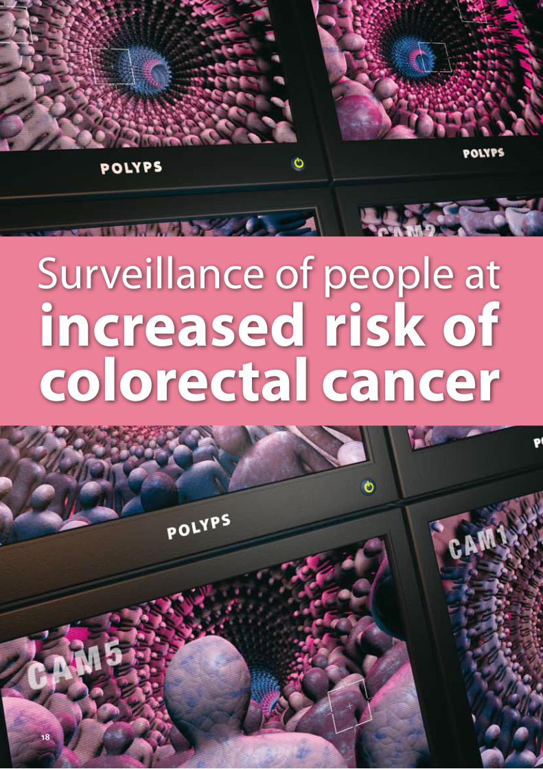
18 Surveillance of people at increased risk of colorectal cancer
The incidence of colorectal cancer in New Zealand is high by international standards. New Zealand females have one of the highest rates in the world (compared to other females). Colorectal cancer occurs less frequently in Māori than in non-Māori, but Māori with colorectal cancer are more likely to die from this disease. Increasing age and a family history are the strongest risk factors for developing colorectal cancer. An assessment of individual risk is required for people at increased risk of colorectal cancer, as well as those with symptoms, and appropriate surveillance and investigation carried out.
26 Managing urinary tract infections in children
Although relatively uncommon in children, urinary tract infection (UTI) should be considered when assessing a young child with fever or any sign of infection without an obvious source. Older children are more likely to be able to describe specific urinary symptoms. Urinalysis (culture and microscopy) is recommended for all children with suspected UTI. Collecting a urine sample can be difficult, however, there are several methods that may be considered. While UTI is usually simple to treat, if a diagnosis is missed or the infection not adequately managed, there is a significant risk of complications.
32
2 BPJ Issue 44
CONTENTS
Issue 44 May 2012
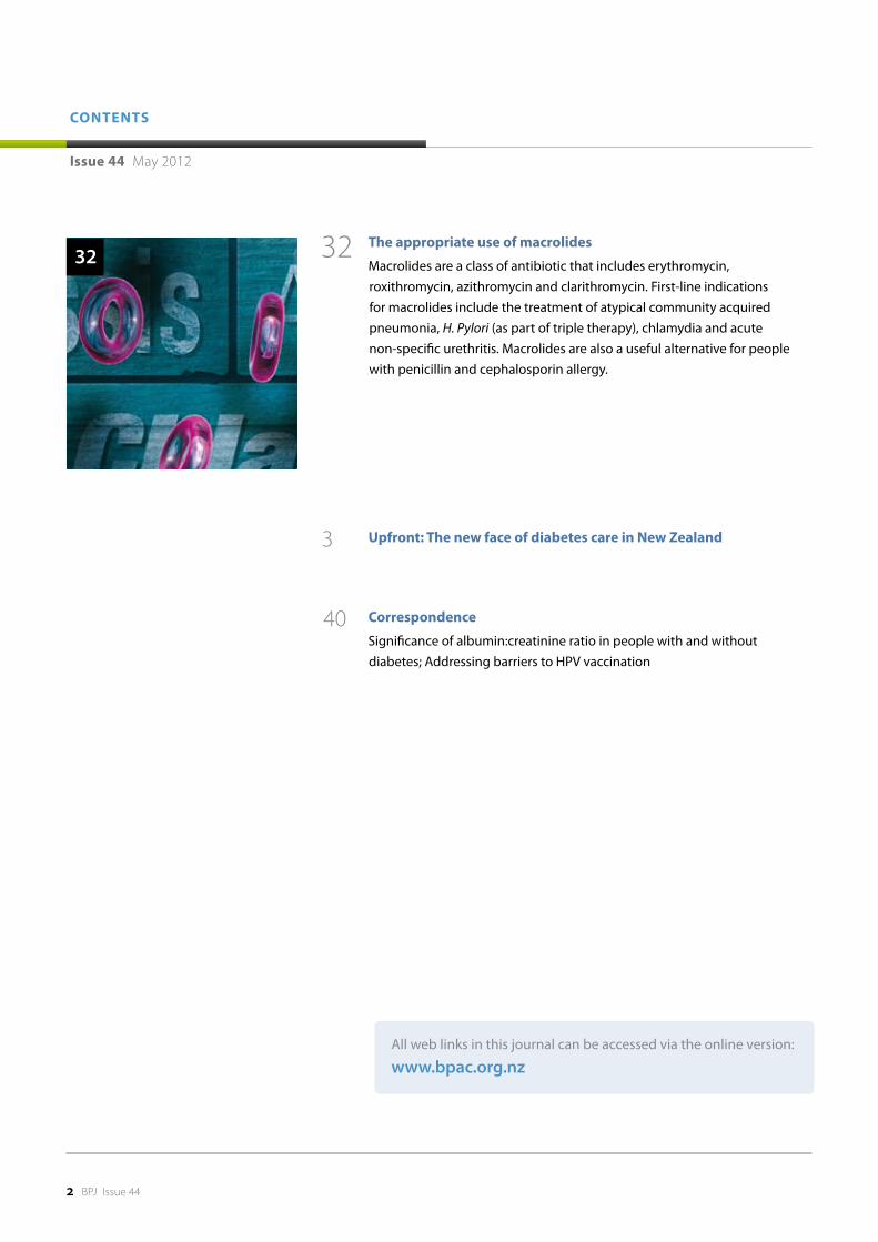
32 The appropriate use of macrolides
Macrolides are a class of antibiotic that includes erythromycin, roxithromycin, azithromycin and clarithromycin. First-line indications for macrolides include the treatment of atypical community acquired pneumonia, H. Pylori (as part of triple therapy), chlamydia and acute non-specific urethritis. Macrolides are also a useful alternative for people with penicillin and cephalosporin allergy.
3 Upfront: The new face of diabetes care in New Zealand
40 Correspondence
Significance of albumin:creatinine ratio in people with and without diabetes; Addressing barriers to HPV vaccination
All web links in this journal can be accessed via the online version:
www.bpac.org.nz
BPJ Issue 44 3
ON 1 JULY, 2012 the “Get Checked” programme, under which diabetes follow-up care in New Zealand is funded, will cease to exist. In its place will be the “Diabetes Care Improvement Package”.
The Get Checked programme, now over a decade old, entitles people with diabetes to a free annual consultation. The decision to stop the programme was partly influenced by a report by Dr Brandon Orr-Walker for the Ministry of Health, which showed that it produced only marginal improvements for people with diabetes in New Zealand, after ten years and a $46 million investment. During the Programme, there has been an absolute reduction in HbA1c levels of 1.4 mmol/mol (from the baseline level of 61 mmol/mol), and only two-thirds of patients are regularly accessing their free check-ups.
An audit undertaken by Waitemata District Health Board found that there was no significant difference in the glucose, lipid and blood pressure levels of those patients enrolled in Get Checked compared to those who were not.
The programme’s replacement, the Diabetes Care Improvement Package is “a primary care based programme, building on core diabetes services that are already being provided, to improve outcomes for people with diabetes”. Essentially, the new programme places the coordination of diabetes care in the hands of District Health Boards (DHBs). Rather than a standard national plan, each DHB will have the opportunity to build their own care model based on the New Zealand Diabetes guidelines and their own unique patient population. The funding for the programme will remain at the same level as for “Get Checked”, but it is hoped that the new models will improve the quality, consistency and direction of care for people with diabetes.
At present there is little information on how the new package will affect patients and healthcare providers, as DHBs are yet to finalise and release their individual plans. With that in mind we invited a group of individuals, with expertise in diabetes and health policy, to discuss what they thought was important in diabetes care, what needed to change and whether DHB-led care plans could work in New Zealand.
The new face of diabetes care in New Zealand
UPFRONT
4 BPJ Issue 44
THE PANEL:
Dr Paul Drury, General Physician and Endocrinologist, Clinical Director, Auckland Diabetes Centre, Medical Director, New Zealand Society for Study of Diabetes. Chair, National Diabetes Services Improvement Group.
Kit Hoeben, Integrated Diabetes Service Manager, Canterbury District Health Board.
Dr Hywel Lloyd, General Practitioner, Chief Medical Officer, BPAC Inc.
Dr Brandon Orr-Walker, Endocrinologist, Clinical Director of Diabetes and Cardiovascular Disease, Ministry of Health.
Dr Tom Robinson, General and Public Health Physician, Waitemata District Health Board.
What the panel said: a summaryThe panel agreed that the replacement of the Get Checked programme with the new diabetes care plan has the potential to improve the health of people with diabetes. However, most expressed concern over the potential for fractured care that came from individualised DHB-led programmes. There was consensus that the “ingredients” for a positive change in diabetes care came down to:
More patient involvement through increased health literacy, health seeking behaviour and self-management of care
A greater role for nurses in coordination and the delivery of resources
Greater use of information technology (IT) in order to streamline care and enhance recall, audit and management procedures, especially in primary care
Involvement of allied care and community care providers, doctors and PHOs in the development phase
Moving towards a “clinical outcome” rather than “output” basis of measuring quality of care
Can DHB-led programmes improve the quality of diabetes care?
One of the most significant changes with the Diabetes Care Improvement Package is the devolvement from Ministry of Health governance to localised DHB-led schemes. This will allow DHBs to provide services tailored to the specific needs of their local population, which are likely to vary considerably across New Zealand.
The panel agreed that a DHB-led programme could improve on government-led schemes, but only if several criteria could be met in development and implementation:
A need for local programmes to be tied to national goals, such as earlier identification of at-risk individuals, and better education services
Adherence to the evidence base, e.g. the 2011 NZGG diabetes guideline
The involvement of PHOs and community-level providers in the development phase
Working from a foundation of national diabetes priorities and goals is crucial and closely tied to the need to base programmes on interventions and management strategies which are supported by evidence of their effectiveness.
“Twenty unconnected plans won’t do this, local programmes could improve care, but they need to be based on the same overall guidance and goals.” – PAUL DRURY
“There is a very strong evidence base about what works in diabetes management in primary care, so there can be a national system which allows modest regional variation.” –
TOM ROBINSON
In terms of the evidence, the Panel agreed that focusing interventions on prevention is key, and will result in long-term savings financially as well as more importantly, reductions in mortality and morbidity. This can come about through earlier identification of people at risk and strenuous application of lifestyle measures before a diagnosis and once the diagnosis of diabetes (or even impaired glucose tolerance) has been made.
“There is growing evidence that lifestyle programmes can drastically reduce the development of diabetes over substantial time frames.” – BRANDON ORR-WALKER
Performance incentives should aim to reduce the key indicators of diabetes health; glucose, blood pressure and lipid levels, rather than just record them. Data should be easy to collect and extract and be made available for analysis and dissemination, to improve and inform health targets.
Community level involvement, i.e. DHBs liaising with care providers on what they require to be able to do their jobs well, is critical to the success of the more localised Diabetes Care Improvement Package.
“I see the Ministry devolving programmes to the DHB level as a good one, so long as the DHBs do the same and
BPJ Issue 44 5
engage with PHOs and enrolled providers to encourage practices to engage in quality improvement. The bottom up approach.” – HYWEL LLOYD
“The ‘individualised’ part, be it at DHB level, PHO level, practice or patient level, needs to acknowledge that in a diverse and vibrant place like New Zealand there may be specific needs, opportunities and challenges that have to be considered beyond providing the core care required by all.” – BRANDON ORR-WALKER
How can the new programme address the disparities in diabetes prevalence?
Māori and Pacific Peoples, people from the Indian sub-continent and people living in lower-decile socioeconomic areas, are disproportionately affected by diabetes and its complications compared to the rest of the population. A PHO Performance Programme Indicator, “Diabetes Follow-Up after Detection”, was implemented during the Get Checked programme to help address this disparity, and will be continuing under the new scheme. The indicator has been successful in increasing the number of “high need” people with diabetes who received an annual review. However, as previously mentioned, it is important that incentives for change focus on improving parameters rather than just recording them. There have been numerous local initiatives within diabetes care that have explicitly targeted high need groups, such as Capital and Coast DHB’s support of the “Pacific Diabetes Fono”, a collaboration that aimed at increasing awareness about diabetes among Pacific people. These initiatives show that focused, community-level schemes can work.
“Great work has shown that these differences can be eliminated, e.g. glycaemic control in Māori in Manaia PHO, so the sector needs to be aspirational, just like has occurred with smoking cessation and immunisation coverage.” –
BRANDON ORR-WALKER
The Panel agreed that districts with the greatest proportion of high need patients would need larger allocations of funding in order to address disparities. Two main themes emerged for how to use this funding to best target high need patient groups:
Increased community and patient engagement, thereby increasing health literacy
Better use of information technology to manage patients
“We need more community buy-in to self-care and we need to raise people’s expectations, though different ethnicities and communities will need different approaches.” – PAUL
DRURY
“[We need] greater use of allied care providers, greater resources in the community and an increase in participation and engagement with focus on self-management.” – KIT
HOEBEN
“Active systematic recall and follow up is one of the few mainstream things that is shown to reduce inequalities.” –
TOM ROBINSON
While the path to eliminating disparities may not be completely clear, DHB-led programmes have the advantage as they allow for more community-level involvement in the planning and implementation stages of programme development. It comes back to the “bottom up approach” and the consensus seems to be that, without engagement from the groups at the greatest risk, with the greatest need, it may be difficult to derive much additional benefit from scrapping Get Checked and starting again.
What are the major factors that contribute to quality diabetes healthcare?The cessation of the Get Checked programme came about in part because it was not delivering clinically significant health benefits to people with diabetes. In 2009 the Office of the Auditor General surveyed General Practitioners on their views and experiences of the Get Checked programme. General Practitioners felt that the programme was not improving diabetes healthcare, because:
The funding did not cover the costs of delivering the checks or completing documentation
They saw the check as an information-collecting exercise
A higher proportion of people failed to attend the pre-arranged appointment than failed to attend for acute complaints (indicating that greater freedom to work opportunistically might be beneficial to healthcare providers)
These lessons need to serve as the basis for the Diabetes Care Improvement Package.
Funding is likely to always be an issue with diabetes care, and the number of people with the disease is growing rapidly. Several members of the Panel felt that a way to maintain quality of care, while operating within funding pressure, was to have patients with diabetes increasingly managed by nurses with specific expertise in diabetes care. Another way to address funding issues is to provide community-level care in a group setting. This needs to focus on giving people with diabetes a greater understanding of their condition, the tools
6 BPJ Issue 44
to change the progression of their condition and a sense of control and achievement when things go well.
“[We’ll see a change in the] amount of care that will be provided by other members of the general practice team, i.e. nursing and pharmacy.” – KIT HOEBEN
“[We need] increased activity from the people providing appropriate advice. This is more about community leadership, and is particularly relevant for high risk ethnicities and circumstances (e.g. where medical care is less available) in the areas of prevention, modification of lifestyle, positive role modelling, and support.” – BRANDON ORR-WALKER
“Patients will be involved to a much greater extent in self-management support. [They need] a greater sense of engagement and participation.” – HYWEL LLOYD
In order to avoid the Diabetes Care Improvement Package becoming an information collecting exercise, the focus needs to change from collecting the information to applying the information.
“This is all about the clinical culture. Entering a patient into a ‘subscription’ to receive something won’t achieve anything on its own. But if that is used to ‘make space’ for the care of diabetes in a proactive way that can catalyse improved care then the result will be a return on investment with better health and less cost.” – BRANDON ORR-WALKER
“Quality is not an end point or a destination but a process of implementing a programme of care that facilitates everyone involved to ask themselves collectively: Are we doing the right things? Are we doing things right? Do we have the capacity to improve?” – HYWEL LLOYD
The Diabetes Care Package needs flexibility in its application, to allow for diabetes detection and follow up to occur at any health encounter. This is particularly important for patients who attend general practice infrequently and who, in the past, have failed to attend scheduled “Get Checked” appointments.
How will care change from a patient perspective?The goal of the Diabetes Care Improvement Package is to improve the quality of care that each person in New Zealand with diabetes receives. Within the constraints of current funding, it is likely that patients will begin to see less of General Practitioners and more of nurses and other healthcare providers. The intensity of care will be based on their disease progression. For example, a patient with diabetic neuropathy on insulin may receive free quarterly consultations with
the practice nurse, whereas a patient without diabetic complications may be seen only annually by their General Practitioner. While this has already been the case in certain PHOs under the Get Checked programme, for many patients this will represent a significant change.
Group education and more community involvement may also be new for some people with diabetes.
“More intense care where it is required. Normal community care where it isn’t.” – TOM ROBINSON
“Those with greatest need will be targeted and receive more frequent support than is delivered currently. There will be a growing interest in group participation programmes where care can be offered to a larger group with less specialised resources.” – HYWEL LLOYD
What are the potential stumbling blocks?
The Panel identified several areas where either more work, a greater commitment from organisational bodies or a different approach to care will be needed.
“The current workload of general practice teams means there isn’t going to be ‘space’ or time to extend their activities unless there is an investment in service redesign, which would likely mean new staff and physical space.” – KIT HOEBEN
As the new programme will retain the same overall level of funding as Get Checked, this is likely to be the major barrier and determinant of the level and type of services that can be offered to patients.
“[There is a current] lack of clinical expertise/time in primary care... and unhelpful funding models; many practices are simply overwhelmed.” – PAUL DRURY
“Long-term condition care still does not receive the resources that it deserves.” – TOM ROBINSON
“Our health funding, and health workforce is unlikely to expand at the same rate [as diabetes is], so to even maintain a [static] level of care we will have to provide care in new ways.” – BRANDON ORR-WALKER
Whatever the stumbling blocks may be, the Diabetes Care Improvement Package offers the opportunity to refocus the way diabetes is managed in New Zealand away from process-based model to a care-based model that is individualised to unique, local patient populations.
BPJ Issue 44 7
Watch this space
We await with interest the look of the new Diabetes Care Improvement Packages as they are rolled out by DHBs. There may be drastic changes that alter the face of diabetes care in New Zealand, or, it may simply be a re-branding of the same old plan. There is a wealth of information and research available, and considerable input has gone into reviewing what worked and did not work under the old scheme. It is hoped that local planners will incorporate some of the ideas outlined here by the Panel, when they implement the Diabetes Care Improvement Package. Finalised DHB annual plans will be published on individual DHB websites in the coming weeks, and should contain programme directions and specific information.
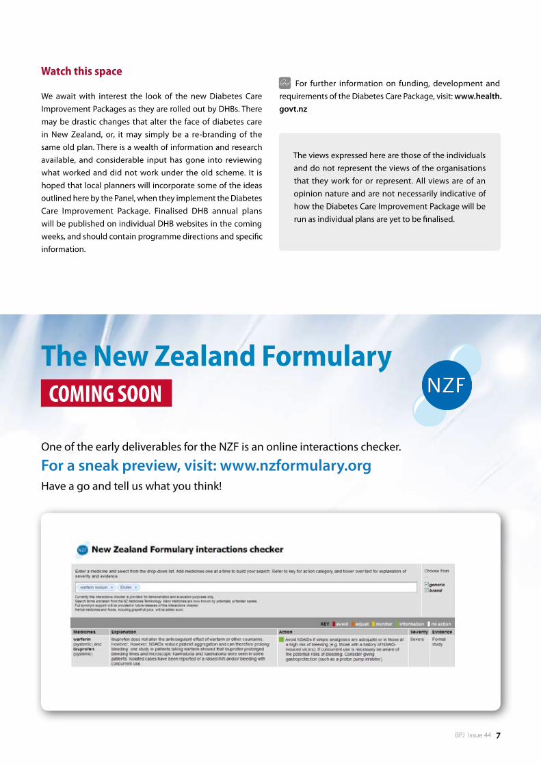
One of the early deliverables for the NZF is an online interactions checker.
For a sneak preview, visit: www.nzformulary.org Have a go and tell us what you think!
For further information on funding, development and requirements of the Diabetes Care Package, visit: www.health.govt.nz
The views expressed here are those of the individuals and do not represent the views of the organisations that they work for or represent. All views are of an opinion nature and are not necessarily indicative of how the Diabetes Care Improvement Package will be run as individual plans are yet to be finalised.
The New Zealand FormularyCOMING SOON

OXYCODONE what can primary care do
about the problem?
Update on
8 BPJ Issue 44
BPJ Issue 44 9
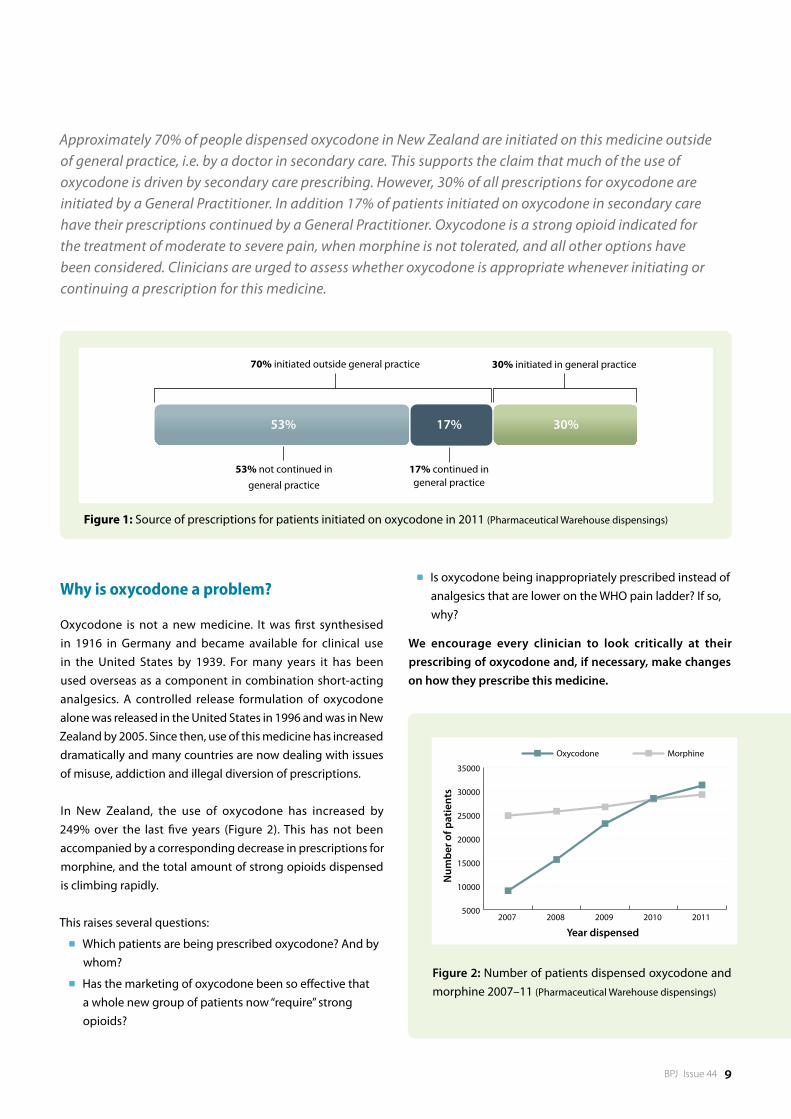
Approximately 70% of people dispensed oxycodone in New Zealand are initiated on this medicine outside of general practice, i.e. by a doctor in secondary care. This supports the claim that much of the use of oxycodone is driven by secondary care prescribing. However, 30% of all prescriptions for oxycodone are initiated by a General Practitioner. In addition 17% of patients initiated on oxycodone in secondary care have their prescriptions continued by a General Practitioner. Oxycodone is a strong opioid indicated for the treatment of moderate to severe pain, when morphine is not tolerated, and all other options have been considered. Clinicians are urged to assess whether oxycodone is appropriate whenever initiating or continuing a prescription for this medicine.
Why is oxycodone a problem?
Oxycodone is not a new medicine. It was first synthesised in 1916 in Germany and became available for clinical use in the United States by 1939. For many years it has been used overseas as a component in combination short-acting analgesics. A controlled release formulation of oxycodone alone was released in the United States in 1996 and was in New Zealand by 2005. Since then, use of this medicine has increased dramatically and many countries are now dealing with issues of misuse, addiction and illegal diversion of prescriptions.
In New Zealand, the use of oxycodone has increased by 249% over the last five years (Figure 2). This has not been accompanied by a corresponding decrease in prescriptions for morphine, and the total amount of strong opioids dispensed is climbing rapidly.
This raises several questions:
Which patients are being prescribed oxycodone? And by whom?
Has the marketing of oxycodone been so effective that a whole new group of patients now “require” strong opioids?
Figure 2: Number of patients dispensed oxycodone and morphine 2007–11 (Pharmaceutical Warehouse dispensings)
Year dispensed
Num
ber o
f pat
ient
s
5000
10000
15000
20000
25000
30000
35000
Oxycodone Morphine
2011 2010 2009 2008 2007
Is oxycodone being inappropriately prescribed instead of analgesics that are lower on the WHO pain ladder? If so, why?
We encourage every clinician to look critically at their prescribing of oxycodone and, if necessary, make changes on how they prescribe this medicine.
53% 17% 30%
30% initiated in general practice
17% continued in general practice
53% not continued in general practice
70% initiated outside general practice
Figure 1: Source of prescriptions for patients initiated on oxycodone in 2011 (Pharmaceutical Warehouse dispensings)
10 BPJ Issue 44
What is the appropriate indication for oxycodone?
There is no dispute that oxycodone is an effective analgesic, however, prescribing figures suggest that it is being chosen as the first-line opioid in many situations when it should not be.
Morphine is the preferred first-line option for the treatment of acute and chronic moderate to severe pain, when a strong opioid is indicated. When compared to morphine, oxycodone:
Has no better analgesic efficacy
Has a similar adverse effect profile
May have more addictive potential1, 2
Is significantly more expensive
Oxycodone should only be prescribed for the treatment of moderate to severe pain in patients who are intolerant to morphine and when a strong opioid is the best option. Although oxycodone has been reported to be potentially safer than morphine in patients with renal impairment, active metabolites can still accumulate.3 Fentanyl or methadone are likely to be safer in patients with renal impairment, who require a strong opioid, because they have no clinically significant active metabolites.3 Discussion with a pain or renal physician is recommended when considering the use of any strong opioid in a patient with severe renal impairment (creatinine clearance < 30 mL/min).
For further information see:
“Fentanyl patches to be available without Special Authority in 2011”, BPJ 33 (Dec, 2010).
“Methadone – safe and effective use for chronic pain”, BPJ 18 (Dec, 2008).
What can General Practitioners do to reduce oxycodone use?
Data from the Pharmaceutical Warehouse show that 30% of prescriptions for oxycodone are initiated within general practice (Figure 1). When considering initiation of oxycodone, always ask yourself if you would use morphine for this patient. If the answer is no then do not prescribe oxycodone. Oxycodone should not be prescribed when a weaker opioid, e.g. codeine, dihydrocodeine or tramadol, would be more appropriate.
Remember that: 5 mg oxycodone is approximately equivalent to 10 mg morphine, 50 – 100 mg tramadol, 100 mg dihydrocodeine or 100 mg codeine.9, 10
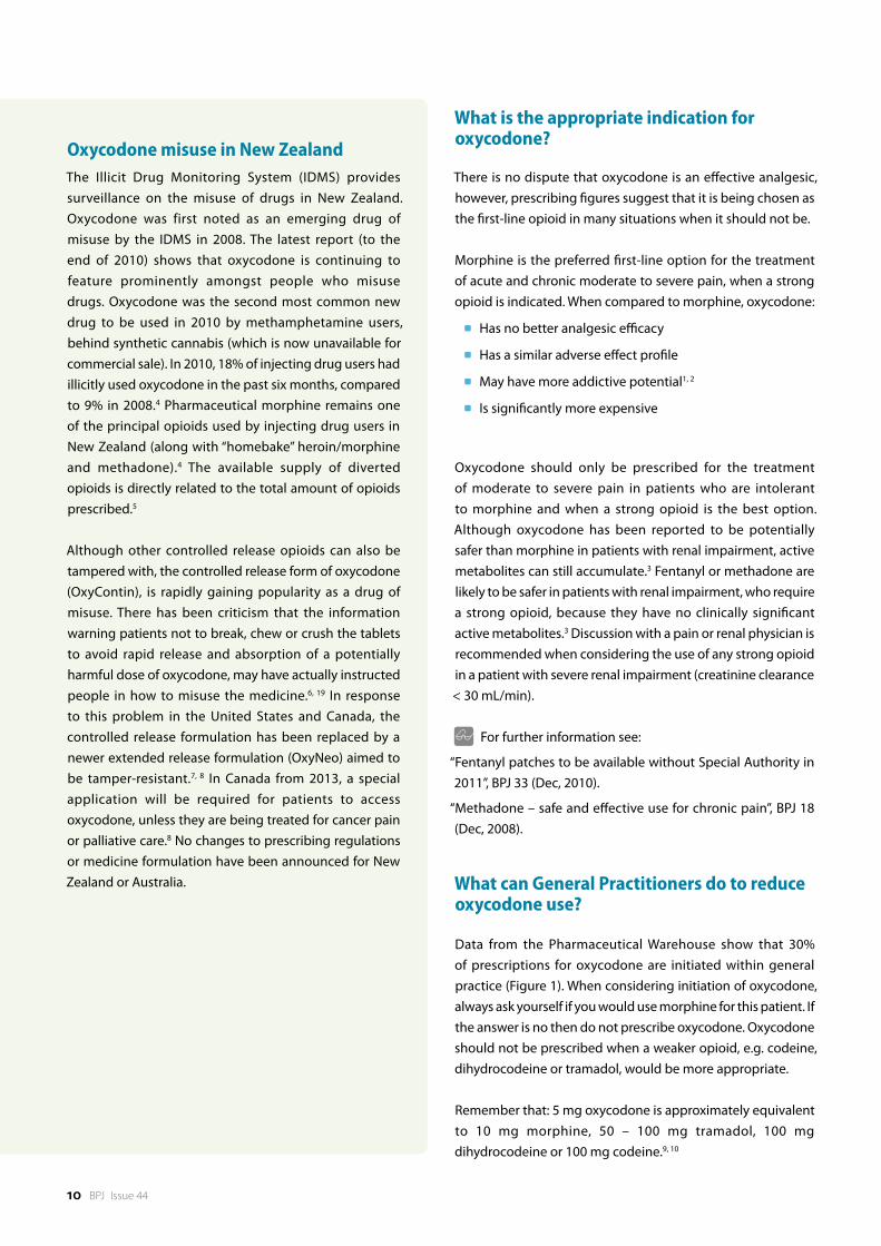
Oxycodone misuse in New ZealandThe Illicit Drug Monitoring System (IDMS) provides surveillance on the misuse of drugs in New Zealand. Oxycodone was first noted as an emerging drug of misuse by the IDMS in 2008. The latest report (to the end of 2010) shows that oxycodone is continuing to feature prominently amongst people who misuse drugs. Oxycodone was the second most common new drug to be used in 2010 by methamphetamine users, behind synthetic cannabis (which is now unavailable for commercial sale). In 2010, 18% of injecting drug users had illicitly used oxycodone in the past six months, compared to 9% in 2008.4 Pharmaceutical morphine remains one of the principal opioids used by injecting drug users in New Zealand (along with “homebake” heroin/morphine and methadone).4 The available supply of diverted opioids is directly related to the total amount of opioids prescribed.5
Although other controlled release opioids can also be tampered with, the controlled release form of oxycodone (OxyContin), is rapidly gaining popularity as a drug of misuse. There has been criticism that the information warning patients not to break, chew or crush the tablets to avoid rapid release and absorption of a potentially harmful dose of oxycodone, may have actually instructed people in how to misuse the medicine.6, 19 In response to this problem in the United States and Canada, the controlled release formulation has been replaced by a newer extended release formulation (OxyNeo) aimed to be tamper-resistant.7, 8 In Canada from 2013, a special application will be required for patients to access oxycodone, unless they are being treated for cancer pain or palliative care.8 No changes to prescribing regulations or medicine formulation have been announced for New Zealand or Australia.
BPJ Issue 44 11
Best Practice Tip: Make it a practice policy, whenever prescribing a strong opioid, to record why the patient has been prescribed this medicine, the usual dose, the expected time frame for treatment, any concerns regarding the patient (such as low mood, poor social support) and specific instructions regarding actions if an increased dose is requested, an early prescription is sought, or if medicines are reported as lost.
Patients on oxycodone initiated in secondary care
Approximately 70% of oxycodone is initiated within secondary care. Prescribing data show that when oxycodone is initiated from outside general practice, 17% of patients have their prescription continued by a General Practitioner (Figure 1).
Knowledge of a patient’s clinical and medicines history and psychosocial background puts General Practitioners in a strong position to not simply “go with the flow”, but instead re-evaluate the indication for oxycodone, even if it has been initiated within secondary care.
Summary: management strategies for patients discharged on oxycodoneWhen a patient is discharged from secondary care on oxycodone, a suggested management strategy is as follows:
When the patient presents for a renewal of a prescription of oxycodone, assess their level of pain and consider whether a strong opioid is still required.
If a strong opioid is no longer required, step down to a weaker opioid or to paracetamol. Depending on the length of time the patient has been on oxycodone, a gradual tapering of the dose may be necessary.
If a strong opioid is still required, consider changing the patient to morphine. Explain to the patient that morphine is equally effective, will not usually result in any other adverse effects and that it is the preferred option when strong opioids are used in general practice. Regularly reassess the patient and step-down treatment as appropriate.
Make sure the patient knows that oxycodone is a strong opioidMany patients are unaware (and shocked to be told) that oxycodone is a strong opioid similar to morphine, but milligram for milligram, twice as potent. Both patients and clinicians have been known to mistakenly associate oxycodone with the weak opioid codeine, rather than with morphine, because of the similarity in the names of the medicines.
Reassess why oxycodone was initially prescribedEstablish the precise clinical problem for which oxycodone was initially prescribed, e.g. post-surgical pain or an acute injury. Does this same problem exist now? Most patients can gradually reduce analgesia in the days to weeks after surgery or acute injury.
What level of pain is the patient experiencing?If there is an ongoing medical condition that requires analgesia, check that the level of pain being experienced warrants the use of a strong opioid.
Consider if oxycodone can be stoppedIf the pain has reduced and oxycodone is no longer required, stop or taper the dose (Page 12). Weaker analgesia, such as codeine and paracetamol, may still be required. Tramadol and dihydrocodeine can also be used as alternatives. Check the patient’s understanding of any analgesic medicines that are used - are they being taken at the right time and in the right dose to gain effective pain relief and to minimise adverse effects?
Consider switching the patient to morphine If a strong opioid analgesic is still indicated, consider switching the patient to morphine. Morphine should be the strong opioid of choice for the majority of patients unless they are allergic to morphine or intolerant to its adverse effects. A dose of 5 mg of controlled release oxycodone is approximately equivalent to 10 mg of long-acting morphine. This conversion rate is, however, only approximate and there is varying guidance on the dose of morphine that should be used when switching.9, 10
If the aim is to eventually discontinue opioids and the degree of pain allows, calculate the equivalent dose of morphine and then start the patient on half of this dose.2 The response of the patient to the change in medicine should be reviewed regularly and the dose adjusted as required to prevent any withdrawal symptoms. The “ABC” of opioid pain medicine use should be remembered:
Anti-emetic prescription if nausea present
Breakthrough dose of morphine may be required
Constipation is likely, prescribe a laxative
12 BPJ Issue 44
If an opioid is continued, establish a pattern of regular reviewEvery patient prescribed a strong opioid analgesic on an ongoing basis requires regular review. The requirement for monthly prescriptions for opioids provides an ideal opportunity to review the need for the medicine, however, in some situations review will need to be more frequent, such as early in the course of treatment. Discuss the dose, the goals of treatment, adverse effects, the time frame for the use of opioid and if appropriate develop a clear plan for stopping the medicine. Check with the patient how they are managing day to day. The Australian and New Zealand College of Anaesthetists recommends a “5A assessment” when prescribing a strong opioid: assess the patient’s analgesia, activity, adverse effects, affect and aberrant drug taking behaviour (see “Detecting aberrant drug-taking behaviour”).11 Referral to a specialist pain clinic may be required if the patient’s pain is unable to be effectively controlled or if there are other concerns with aspects of the “5A” assessment.
How to discontinue oxycodone
Abrupt cessation Patients who have been taking oxycodone at low doses (e.g. 10 – 20 mg daily) for less than one to two weeks can generally stop the medicine without experiencing withdrawal symptoms.16 Gradual tapering of oxycodone to avoid withdrawal symptoms is recommended in most other situations.
Gradual dose reductionPatients who have been taking oxycodone for more than one to two weeks, or at high doses, should have the dose gradually tapered to avoid symptoms of opioid withdrawal.2, 6
How quickly and by how much the oxycodone can be reduced will depend on the current dose, the length of time the medicine has been taken for and individual patient factors, such as anxiety, co-morbidities (e.g. depression or other psychiatric conditions) and the likelihood that the patient is dependent on oxycodone, in which case the dose should be reduced more slowly.2, 6
Advice about tapering of opioids varies widely in the literature, however, in general: 2, 6, 16
Reduce the dose in 20–25% increments or, if required, more slowly by 5–10%
Reductions can be made every two or three days
Detecting aberrant drug taking behaviourBehaviours that may suggest the development of aberrant drug taking behaviour, such as overuse, hoarding, dependence and diversion, include: presenting early for repeats, loss of prescriptions or medicines or requests for an escalation in dose.
Patients with chronic pain who take opioid medicines may over time become tolerant or dependent and require increased doses to enable them to function day to day.6 If the patient reports that their pain is worsening, consider if this would normally be expected with the condition being treated, if a different diagnosis should be considered or whether there is the possibility of misuse.
Addiction to opioids is reported to occur in only a small number of patients with chronic pain. However, many more patients with chronic pain display aberrant drug taking behaviour.12, 13
Personal or family history of alcohol or drug dependence increases the risk of misuse of opioids. The presence of an anxiety disorder or depression further increases this risk.14, 15 However, patients who misuse medicines do not always fit a stereotype and risk factors may not always be apparent. Any person, regardless of gender, age, ethnicity, income, health or employment status can be at risk of aberrant drug taking behaviour. It is therefore recommended that every patient who is prescribed an opioid is assessed for risk factors for aberrant drug taking behaviour, including the possibility of diversion of prescriptions.
BPJ Issue 44 13
Once the patient has been reduced to one-third of the initial dose, the rate of taper should be slowed
Consider holding the dose at the same level if the patient develops withdrawal symptoms, an increase in pain or lowered mood
Most patients can be withdrawn from oxycodone within one month, depending on how high the dose was prior to initiating tapering
Referral to addiction servicesIn some situations it may be more appropriate to refer patients to a community based drug and alcohol programme, to withdraw from oxycodone. Patients who may benefit from referral include those who:17
Are unable to be slowly tapered off oxycodone in general practice due to factors such as a lack of success with tapering, non-compliance with tapering, accessing opioids from other sources
Are misusing oxycodone or other addictive substances (including alcohol)
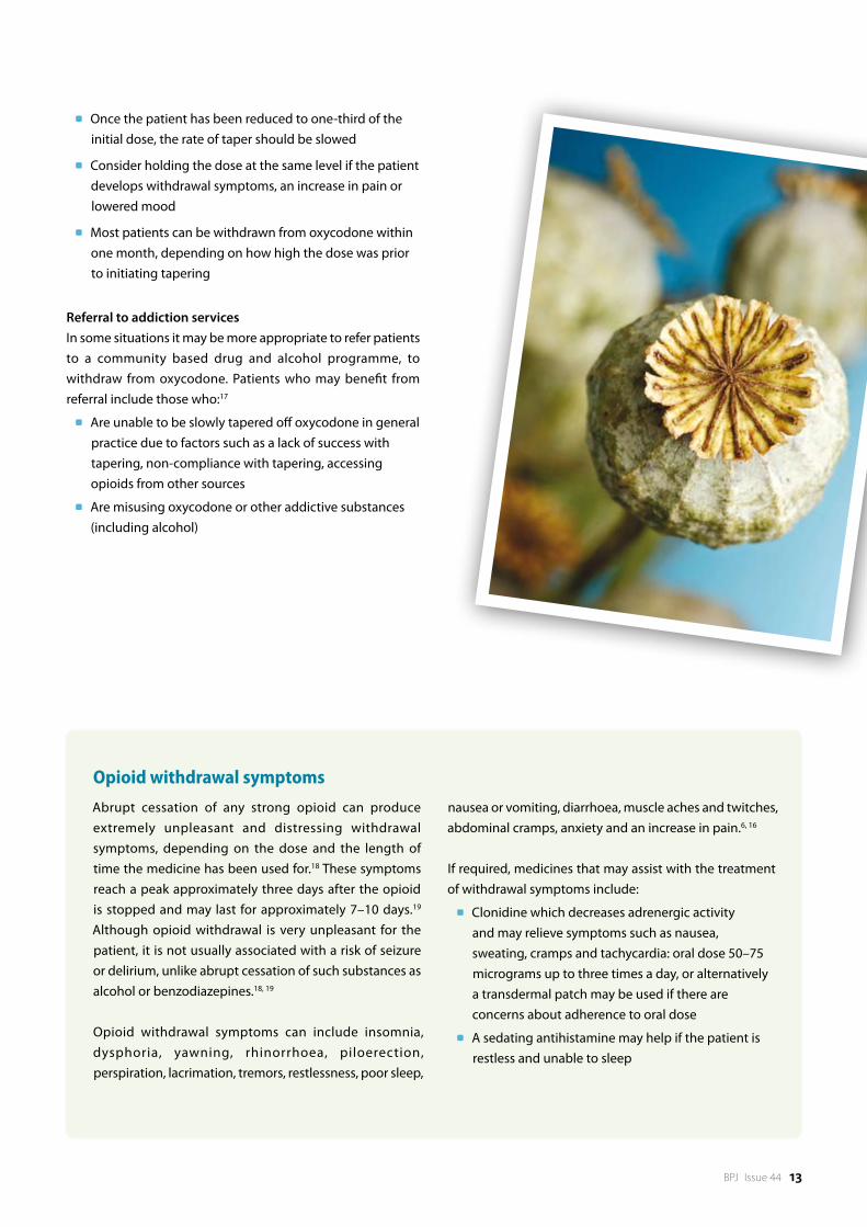
Opioid withdrawal symptoms
Abrupt cessation of any strong opioid can produce extremely unpleasant and distressing withdrawal symptoms, depending on the dose and the length of time the medicine has been used for.18 These symptoms reach a peak approximately three days after the opioid is stopped and may last for approximately 7–10 days.19 Although opioid withdrawal is very unpleasant for the patient, it is not usually associated with a risk of seizure or delirium, unlike abrupt cessation of such substances as alcohol or benzodiazepines.18, 19
Opioid withdrawal symptoms can include insomnia, dysphoria, yawning, rhinorrhoea, piloerection, perspiration, lacrimation, tremors, restlessness, poor sleep,
nausea or vomiting, diarrhoea, muscle aches and twitches, abdominal cramps, anxiety and an increase in pain.6, 16
If required, medicines that may assist with the treatment of withdrawal symptoms include:
Clonidine which decreases adrenergic activity and may relieve symptoms such as nausea, sweating, cramps and tachycardia: oral dose 50–75 micrograms up to three times a day, or alternatively a transdermal patch may be used if there are concerns about adherence to oral dose
A sedating antihistamine may help if the patient is restless and unable to sleep
14 BPJ Issue 44
The use of strong opioids for chronic non-cancer pain is controversial and there is limited quality evidence to support or oppose their use for this type of pain.11, 12 Principles for the use of opioid analgesics in people with chronic non-cancer pain have been developed by the Australian and New Zealand College of Anaesthetists.11 The principles aim to take into account both the widely varying individual response to opioids and the risks for an individual patient. The use of opioids for chronic non-cancer pain should be regarded as an “ongoing individual trial of therapy”.11
Assess all aspects of the pain Consider factors that may influence the nature and intensity of pain and the patient’s reaction to the pain. Ask about the patient’s beliefs about the underlying problem, their mood, their fears and their expectations of pain treatment. Discuss the goals of treatment with the patient – a reduction in pain and an increase in function are realistic and achievable outcomes, while an expectation that the pain will be totally eliminated may be unrealistic.17, 20
Pain can be difficult to assess because it is subjective and is often influenced by factors such as mood, stress and the psychosocial support that the patient has. The most clinically useful pain scales include an assessment of the impact of the pain on daily life. Pain can have a significant effect on daily activities, e.g. altering sleep or appetite. It can induce or exacerbate depression and anxiety, it can influence social interactions, prevent work and impair relationships.
For further information about pain scales, see “Pharmacological management of chronic pain”, BPJ 16 (Sep, 2008).
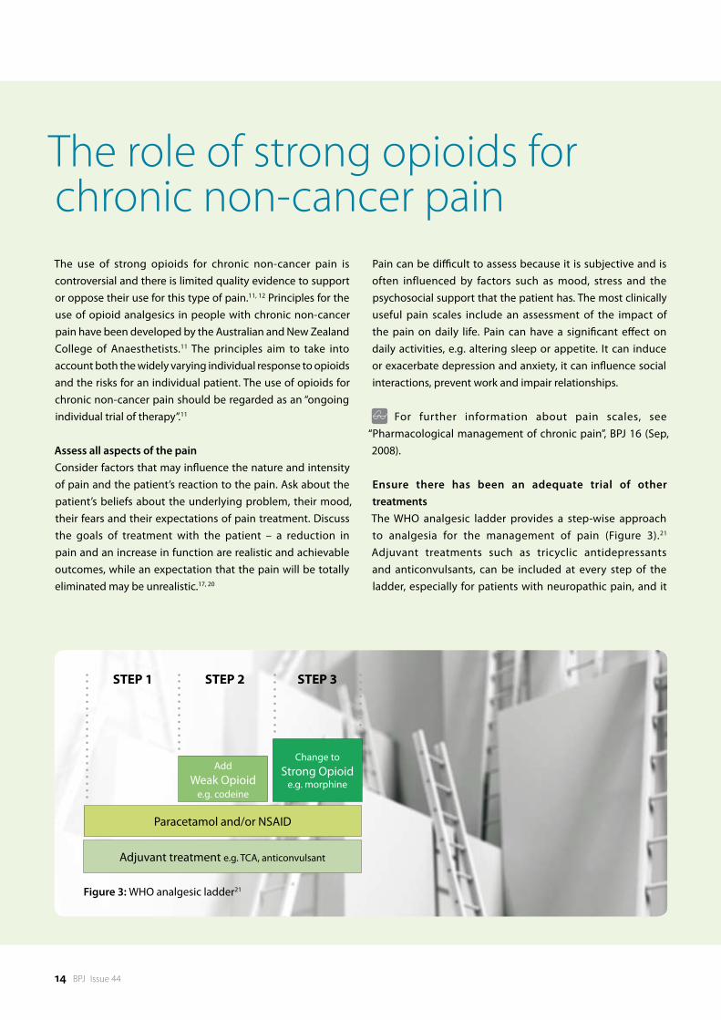
Ensure there has been an adequate trial of other treatments The WHO analgesic ladder provides a step-wise approach to analgesia for the management of pain (Figure 3).21 Adjuvant treatments such as tricyclic antidepressants and anticonvulsants, can be included at every step of the ladder, especially for patients with neuropathic pain, and it
Adjuvant treatment e.g. TCA, anticonvulsant
Paracetamol and/or NSAID
AddWeak Opioid
e.g. codeine
Change toStrong Opioid
e.g. morphine
STEP 1 STEP 2 STEP 3
Figure 3: WHO analgesic ladder21
The role of strong opioids for chronic non-cancer pain
BPJ Issue 44 15
is recommended that they are considered before the use of strong opioids, i.e. Step 3.11 Non-pharmacological treatment of pain is also important. This includes ensuring that the patient understands the underlying problem and the treatment plan, checking on family and social supports, promoting the benefit of healthy lifestyle choices (e.g. exercise, adequate sleep, balanced diet) and the involvement of other health professionals, e.g. physiotherapist, occupational therapist, psychologist, pain clinic specialist.
Consider if a strong opioid is indicated and appropriate for the patientPrior to initiating a strong opioid for chronic pain in particular, consider the following questions:
Have I identified the cause of the pain?
What am I trying to achieve?
Is this what the patient wants?
To what extent are psychosocial factors contributing to the pain level and how can these factors be addressed?
Is there evidence that a particular medicine will help this type of pain?
Are there non-pharmacological alternatives?
Do the potential benefits outweigh the harms of the treatment? Check if the patient has a history of addictive behaviour, alcohol or medicine misuse. If the patient has a current or past history of a psychological problem, a strong opioid may not be appropriate.
Have I provided effective education about the most appropriate way to use analgesics?
Have I considered how long a strong opioid may be required for?
Have I made a plan for follow up?
Reach an agreement with the patient regarding a trial of strong opioid analgesicIf a strong opioid is indicated, ensure the patient has a good understanding of the type of medicine to be used and the goals of treatment, i.e. an increase in function rather than complete resolution of pain. The patient should be made aware of the potential problems with strong opioids, including adverse effects, safety issues and the potential for dependency and misuse. It is also recommended that an agreement is reached so that if the goals are not achieved, adverse effects are intolerable or there are concerns about misuse, the opioid will be discontinued.11, 20 Any agreement should be clearly
documented in the patient notes. This should include guidance about management if the patient requests or presents for an early repeat, if the medicine is reported as lost or there is a request for an increase in dose. When a strong opioid is prescribed, ideally there should be one prescriber and one pharmacy involved.
Start with an appropriate dose and slowly titrate as requiredChoose a low starting dose of a long-acting or extended release preparation of a strong opioid, usually morphine as the first-line choice. Most patients taking opioids will also require a laxative, and possibly an anti-emetic (in the initial stages of treatment), as well as short-acting medicine for breakthrough pain. It is recommended that the dose be slowly titrated over several weeks if required, with a clinical assessment prior to each increase in dose. The Australian and New Zealand College of Anaesthetists recommends a “5A” assessment which includes a review of:11
Analgesia
Activity
Adverse effects
Affect
Aberrant behaviour
A suggested time frame for a trial of a strong opioid is four to six weeks.9 If the treatment has been of no benefit after this time, the dose of the opioid should be tapered and then stopped.
Regularly review the patient Once the patient is established on an effective dose, regularly reassess them using the “5A” assessment. Check that the goals of treatment agreed initially are being achieved and that a strong opioid is still the most appropriate medicine for the patient. If the patient requests an increase in dose consider whether this may reflect:
A change in the underlying condition producing pain
The patient’s current mood, life stressors or other social circumstances
The development of tolerance
Opioid induced hyperalgesia (abnormal sensitivity to pain due to prolonged use of strong opioids)20
Aberrant drug taking behaviour
Learn from the mistakes of others. You can’t live long
enough to make them all yourself.
16 BPJ Issue 44
References1. Zacny JP, Lichtor SA. Within-subject comparison of the
psychopharmacological profiles of oral oxycodone and oral morphine in non-drug-abusing volunteers. Psychopharm 2008;196:105-16.
2. Kahan M, Mailis-Gagnon A, Wilson L, Srivastava A. Canadian guideline for safe and effective use of opioids for chronic noncancer pain. Clinical summary for family physicians. Part 1: general population. Can Fam Physician 2011;57:1257-66.
3. King S, Forbes K, Hanks GW, et al. A systematic review of the use of opioid medication for those with moderate to severe cancer pain and renal impairment: A European Palliative Care Research Collaborative opioid guidelines project. Palliat Med 2011;25(5):525-52.
4. Wilkins C, Sweetsur P, Smart B, Griffiths R. Recent trends in illegal drug use in New Zealand, 2006-2010. Findings from the 2006, 2007, 2008, 2009 and 2010 Illicit Drug Monitoring System (IDMS). Auckland: Social and Health Outcomes Research and Evaluation (SHORE), School of Public Health, Massey University, 2010.
5. Wilkins C, Sweetsur P, Griffiths R. Recent trends in pharmaceutical drug use among frequent injecting drug users, frequent methamphetamine users and frequent ecstasy users in New Zealand, 2006-2009. Drug and Alcohol Review 2011;30:255-63.
6. Manubay JM, Muchow C, Sullivan MA. Prescription drug abuse: epidemiology, regulatory issues, chronic pain management with narcotic analgesics. Prim Care Clin Offic Pract 2011;38:71-90.
7. U.S. Food and Drug Administration (FDA). FDA approves new formulation for OxyContin. FDA news release. 5th April 2010. Available from: www.fda.gov (Accessed May 2012).
8. Ministry of Health and Long-term Care. Change in funding status of oxycodone controlled release tablet (discontinuation of OxyContin and introduction of OxyNEO). Available from: www.health.gov.on.ca/en/public/programs/drugs/ons/oxy_faq.aspx (Accessed May, 2012).
9. Australian Medicines Handbook Adelaide: Australian Medicines Handbook Pty Ltd, 2011.
10. British National Formulary (BNF) 62. London: Pharmaceutical Press, 2011.
11. Faculty of Pain Medicine. Australian and New Zealand College of Anaesthetists. 2010. Principles regarding the use of opioid analgesics in patient with chronic non-cancer pain. Available from: www.fpm.anzca.edu.au (Accessed May, 2012).
12. Manchikanti L, Fellows B, Ailinani H, Pampati V. Therapeutic use, abuse, and nonmedical use of opioids: a ten-year perspective. Pain Physician 2010;13:401-35.
13. Fishbain DA, Cole B, Lewis J, et al. What percentage of chronic non-malignant pain patients exposed to chronic opioid analgesic therapy develop abuse/addiction and/or aberrant drug-related behaviours? A structured evidence-based review. Pain Med 2008;9(4):444-59.
14. Monheit B. Prescription drug misuse. Aust Fam Physician 2010;39(8):540-6.
15. Ling W, Mooney L, Hillhouse M. Prescription opioid abuse, pain and addiction: clinical issues and implications. Drug Alcohol Rev 2011;30:300-5.
16. Gordon D, Dahl J. Opioid withdrawal, #95, 2nd edition. J Pall Med 2011;14(8):965-6.
17. Kahan M, Wilson L, Mailis-Gagnon, Srivastava A. Canadian guideline for safe and effective use of opioids for chronic noncancer pain. Clinical summary for family physicians. Part 2:special populations. Can Fam Physician 2011;57:1269-76.
18. Chou R, Fanciullo GJ, Fine PG et. al. Opioid treatment guidelines. Clinical guidelines for the use of chronic opioid therapy in chronic noncancer pain. J Pain 2009;10(2):113-30.
19. Department of Health and Community Services, Newfoundland and Labrador. Oxycontin Task Force, Final report. June 30, 2004. Available from: www.health.gov.nl.ca/health/publications/oxycontin_final_report.pdf (Accessed May, 2012).
20. British Pain Society. Opioids for persistent pain: good practice. A consensus statement prepared on behalf of the British Pain Society, the Faculty of Pain Medicine of the Royal College of Anaesthetists, the Royal College of General Practitioners and the Faculty of Addictions of the Royal College of Psychiatrists. January 2010. Available from: www.britishpainsociety.org (Accessed May, 2012).
21. WHO analgesic ladder. Available from: www.who.int/cancer/palliative/painladder/en/ (Accessed May, 2012).
ACKNOWLEDGEMENT Thank you to Dr Geoff Robinson, Chief Medical Officer, Addiction Medicine Specialist, Capital & Coast DHB and Dr Howard Wilson, General Practitioner and Pharmacologist, Canterbury, members of the analgesic subcommittee of the Pharmacology and Therapeutics Advisory Committee to PHARMAC for expert guidance in developing this article.

The bpacnz Patient Safety Incident Reporting system is an online resource for people working in community health care to report and review patient safety incidents.
Reports are submitted anonymously, to identify factors which have contributed to patient safety incidents and to share solutions to prevent these incidents from occurring again.
Incidents can be reported and cases reviewed at:
– ELEANOR ROOSEVELT
Learn from the mistakes of others. You can’t live long
enough to make them all yourself. www.bpac.org.nz/safety
18
increased risk of colorectal cancer
Surveillance of people at
BPJ Issue 44 19
Colorectal cancer in New Zealand
Each year approximately 1200 people in New Zealand die of colorectal cancer, a mortality rate similar to breast and prostate cancers combined.1, 2 The incidence of colorectal cancer in New Zealand is high by international standards. In 2008 there were 44.1 cases reported per 100 000 males and 37.5 per 100 000 females. This compares to 36.2 cases per 100 000 males and 23.5 cases per 100 000 females in the United Kingdom.3 Worldwide, colorectal cancer is more common in men than in women. However, the colorectal cancer rates in New Zealand women are higher than for women in any of the other 32 countries within the international cancer screening network.3 Between 2008 and 2010 colorectal cancer was the second most common cancer in New Zealand, behind prostate cancer.4
Colorectal cancer in New Zealand occurs less frequently in Māori compared to non-Māori. From 2008 to 2010 there were on average 39.3 annual registrations of colorectal cancer per 100 000 Māori males and 27.8 per 100 000 Māori females.4 However, once diagnosed, Māori are more likely to die from colorectal cancer than non-Māori. This has been largely attributed to disparities in access to, and quality of, cancer treatment and highlights the need for pro-active follow-up in Māori once a diagnosis of colorectal cancer has been made.5
Surveillance of asymptomatic people at increased risk
Increasing age and a family history of colorectal cancer are the two most significant risk factors for the development of colorectal cancer. A personal history of adenomatous polyps or inflammatory bowel disease also increases risk.
Screening the “average-risk” person based on age
Mortality rates for colorectal cancer increase rapidly from age
In New Zealand, colorectal cancer causes as many deaths each year as breast and prostate cancers combined. In most people, age and family history are the strongest risk factors for developing this cancer. Primary care clinicians need to be able to perform individual risk assessments for people at increased risk of colorectal cancer, and those with symptoms of colorectal cancer, and provide information on the appropriate levels of surveillance and investigation that each person requires.
The pathology of colorectal cancer
Over 95% of cancer in the colon and rectum develops from polyps, which are protrusions in the mucosal surface of the colon, also known as adenomas.6 This process may occur over years to decades.7 Polyps are common and increase in frequency with age. Autopsies show that polyps are present in 30% of people aged over 60 years.8 They are also more common in people with inherited syndromes, who are at increased risk of developing colorectal cancer. The risk of colorectal cancer increases with the size and number of polyps. In a study of 2500 tissue samples, malignant cells were found in 1% of polyps less than 1 cm in diameter, compared to 46% in polyps greater than 2 cm.9
The major types of polyps: Adenomatous polyps account for 60–70% of polyps found in the colon and are the source of the vast majority of adenocarcinomas.10 They can be further classified as tubular (accounting for 70 to 85% of adenomatous polyps), tubulovillous or villous. Villous polyps account for only 5% of adenomatous polyps but are eight to ten times more likely to become malignant than tubular adenomatous polyps.6
Hyperplastic polyps are usually small (less than 0.5 cm) and are frequently found in the rectum and sigmoid portion of the colon. These are usually benign.6
Submucosal polyps have a smooth overlying mucosa. Colour and texture are used to identify these endoscopically. Submucosal polyps are occasionally malignant.
20 BPJ Issue 44
national screening programme for colorectal cancer. However, a four year pilot began in the Waitemata District Health Board region in October 2011, with iFOBT screening offered to all males and females aged 50 to 74 years. People who have a positive iFOBT are referred for a diagnostic colonoscopy. The pilot will determine if the necessary secondary services in New Zealand, e.g. access to colonoscopy, are currently sufficient to support a national screening programme.
Until the results of the pilot study are known, routine FOBT in people aged over 50 years, with no other risk factors for colorectal cancer, is not necessary. However, FOBT may be considered on a case-by-case basis. FOBT is not recommended as a diagnostic test for people with symptoms of bowel cancer, or for surveillance of people with an increased risk or as part of a colorectal cancer follow-up programme.12
FOBT is not recommended for people aged under 50 years as the number of false-positive results is increased in younger people. Age, co-morbidity and life expectancy should also be taken into account when considering FOBT, due to the risk of complications associated with follow-up colonoscopy.
50 years, with 94% of deaths occurring after this age.6 There is evidence that screening asymptomatic people at increased risk of colorectal cancer, based on age, can reduce this mortality rate through early diagnosis.11
Faecal occult blood tests (FOBT) are widely used for the screening of colorectal cancer. FOBT can detect bleeding from colonic lesions, which may suggest the presence of high-risk colorectal adenomas or cancers. A 2007 Cochrane meta-analysis involving 320 000 patients with eight to 18 years follow-up, reported a relative-risk reduction for colorectal cancer of 25% for patients attending at least one round of FOBT screening.11 The mortality reduction equated to 1.25, 5.5 and 17.5 less deaths over ten years per 10 000 people aged 40, 50 and 60 years respectively.11 More recently, immunochemical FOBT (iFOBT) has improved both the sensitivity and specificity of the screening process. This test does not require dietary restrictions.
Colorectal cancer screening programmes or pilots are being run in Australia, the United Kingdom, Korea, Japan, Israel and most countries in the European Union. New Zealand does not have a
Self-testing for bowel cancer
BowelScreen Aotearoa is an organisation founded in 2010 to promote annual self-testing for colorectal cancer. FOBT kits are purchased from pharmacies and then taken home by customers to provide a sample from two different bowel movements. The customer then posts the samples to an Australian based laboratory, including the name of their general practitioner, who is contacted if a test result is positive. Bowel screen Aotearoa advises all people with a positive FOBT to visit their general practitioner.
It is recommended that general practitioners take the following steps if they receive notification that a patient under their care has a positive FOBT result:
1. Contact the patient and arrange a consultation (if
the patient has not already done so)
2. Discuss the patient’s clinical history, risk factors
and symptoms and give healthy lifestyle advice
3. Patients with a personal or family history of
colorectal cancer or other risk factors, e.g. a
personal history of polyps or inflammatory bowel
disease, should be referred for a colonoscopy
4. Symptomatic patients should be referred on the
basis of symptoms and examination findings
rather than the FOBT result alone
5. Asymptomatic patients who are not at increased
risk may still benefit from colonic investigation,
however, local resourcing of colonoscopy
services may influence the pathway of evaluation.
This should be discussed with local DHBs
BPJ Issue 44 21
Optical colonoscopy is the recommended investigation, following referral, for people who have had a positive FOBT result. Optical colonoscopy allows the clinician to visualise the entire colon mucosa, and remove small lesions and perform biopsies as required. It is also recommended for the surveillance of people at increased risk of developing colorectal cancer and as the preferred diagnostic procedure for people with symptoms of bowel cancer (Page 23).12 There is a small risk of bleeding or colorectal perforation associated with colonoscopy, which is dependent on patient age, co-morbidities, performance of polypectomy and clinician proficiency.6
Computed tomography (or virtual) colonoscopy is a useful alternative to optical colonoscopy for the exclusion of malignancy in elderly people, when a less invasive investigation is preferred. It is also useful for people who experience significant pain with optical colonoscopy, e.g. diverticulitis, or present difficulties, e.g. patients taking antithrombotics. If the patient requires a biopsy or polyp removal, then an optical colonoscopy will still need to be performed.
Choosing a healthy diet and making healthy lifestyle choices are proactive steps that all people at risk of developing colorectal cancer can take. Excessive consumption of red and processed meats, high-fat dairy products and highly refined grains, starches and sugars is associated with an increased risk of colon cancer.13 Replacing these foods with protein sources such as poultry and fish, monounsaturated and polyunsaturated fats, e.g. olives, nuts, seeds, avocados, and unrefined grains, legumes and fruits as the primary sources of carbohydrates, is likely to reduce a person’s risk of developing colorectal cancer.13
Maintaining a healthy body weight, regular exercise, abstinence from smoking and drinking less than two standard units of alcohol per day are also healthy lifestyle choices which are likely to reduce a person’s risk of developing colorectal cancer.13
There is currently no evidence to support the routine use of aspirin, vitamin D or calcium for the prevention of colorectal cancer.
The Cancer Society has practical dietary information available for people who want to reduce their cancer risk. Available from: www.cancernz.org.nz/reducing-your-cancer-risk/nutrition-and-physical-activity
Family history of colorectal cancer
Approximately 20% of people with colorectal cancer have two or more first-degree relatives (parents, siblings, children) or second-degree relatives (grandparents, aunts, uncles, nephews and nieces) with colorectal cancer.14 People from these families are said to be at familial risk of colorectal cancer. Colorectal cancer within these families can occur either sporadically or due an inherited syndrome.
Sporadic colorectal cancer in family members influences the risk a person has of developing colorectal cancer:6
One first-degree family member (parents, siblings, children) increases the risk by two to three times
Two first-degree family members increases the risk by three to six times
Two second-degree family members (grandparents, aunts, uncles, nephews, nieces) increases the risk by two times
Inherited colorectal cancers occur via autosomal dominant inheritance and are estimated to account for 5 – 10% of all colorectal cancers.14
Lynch syndrome (hereditary non-polyposis) is the most common hereditary syndrome associated with colorectal cancer. A sample of 500 patients treated consecutively for colorectal cancer found that 3.6% had Lynch syndrome, of which 44% were diagnosed before the age of 50 years.15 Each of these patients had at least three relatives with the syndrome. Females with Lynch syndrome also have an increased risk of developing endometrial cancer.16
Familial adenomatous polyposis (FAP) is caused by a mutation in a tumour suppressor gene and accounts for less than 1% of colorectal cancers. One in 5000 to 7000 people have FAP.17 FAP is characterised by multiple (> 100) adenomatous polyps which develop throughout the colon in the first decade of life.6
Peutz-Jeghers syndrome is characterised by gastrointestinal polyps and dark patches (1 – 5 mm in size) typically around the mouth, eyes, hands, feet and genitals. People with this condition have an increased risk of colorectal and breast cancer. The incidence of this syndrome is estimated to be between one in 50 000 to 200 000 live births.18
Categorising risk for an asymptomatic person (without inflammatory bowel disease) depends on their family history. Table 1 shows the recommended advice for people who know
22 BPJ Issue 44
their family history of colorectal cancer. It should be noted that resourcing constraints may impact on adherence to these guidelines by DHBs around New Zealand.
Adenomatous polyps
People with a previous history of colorectal polyps have an increased risk of developing colorectal cancer and should be offered regular colonoscopy surveillance.
Surveillance frequency is determined by the risk assessment performed at the previous examination. This includes the number and size of any polyps and the histology of any polyps
removed by biopsy. People with a history of adenomatous polyps should be offered colonoscopy at the following intervals:12
Low risk – every five years
Intermediate risk – every three years
High risk – annually
Inflammatory bowel disease
People with inflammatory bowel disease have an increased risk of developing colorectal cancer. Crohn’s disease and ulcerative colitis are the most common forms of inflammatory
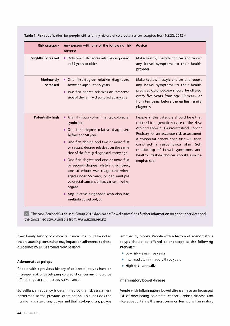
Table 1: Risk stratification for people with a family history of colorectal cancer, adapted from NZGG, 201212
Risk category Any person with one of the following risk factors:
Advice
Slightly increased Only one first-degree relative diagnosed at 55 years or older
Make healthy lifestyle choices and report any bowel symptoms to their health provider
Moderately increased
One first-degree relative diagnosed between age 50 to 55 years
Two first degree relatives on the same side of the family diagnosed at any age
Make healthy lifestyle choices and report any bowel symptoms to their health provider. Colonoscopy should be offered every five years from age 50 years, or from ten years before the earliest family diagnosis
Potentially high A family history of an inherited colorectal syndrome
One first degree relative diagnosed before age 50 years
One first-degree and two or more first or second degree relatives on the same side of the family diagnosed at any age
One first-degree and one or more first or second-degree relative diagnosed, one of whom was diagnosed when aged under 55 years, or had multiple colorectal cancers, or had cancer in other organs
Any relative diagnosed who also had multiple bowel polyps
People in this category should be either referred to a genetic service or the New Zealand Familial Gastrointestinal Cancer Registry for an accurate risk assessment. A colorectal cancer specialist will then construct a surveillance plan. Self monitoring of bowel symptoms and healthy lifestyle choices should also be emphasised
The New Zealand Guidelines Group 2012 document “Bowel cancer” has further information on genetic services and the cancer registry. Available from: www.nzgg.org.nz

BPJ Issue 44 23
bowel disease, with the risk being related to the duration and the anatomical extent of the disease. In a study of over 7500 patients in Sweden with inflammatory bowel disease, followed over a forty year period, 188 patients were diagnosed with colorectal cancer.19 The risk of colorectal cancer begins to increase significantly seven to ten years after the onset of inflammatory bowel disease. The cumulative risk of colorectal cancer is 5 – 10% after 20 years and 20% at 30 years.6
Surveillance colonoscopy should be offered to all people with inflammatory bowel disease beginning eight to ten years following diagnosis.12 Surveillance frequency is determined by the risk assessment based on the extent of the disease using histology and visual inspection at the last colonoscopy:12
Low risk – every five years
Intermediate risk – every three years
High risk – annually
Investigation of people with bowel symptomsIt is common for people to be reluctant to request a consultation with their doctor for abnormal bowel symptoms. Several community-based studies in Australia found that approximately one-third of people with rectal bleeding will wait longer than three months, or never seek medical advice.20 The symptoms of colorectal cancer should be discussed with all patients at increased risk of developing colorectal cancer.
Symptoms of colorectal cancer
Early colorectal cancer is often asymptomatic. Symptomatic presentation may indicate a relatively advanced tumour depending on the location, size and type of cancer. The symptoms of colorectal cancer are often due to the growth of the tumour into the lumen of the gut or adjacent structures. Right-sided lesions are typically larger, while left-sided lesions are more likely to cause partial or full obstruction, resulting in constipation, overflow diarrhoea, narrowed stool, bloating and cramps. Lesions of the lower colon or in the rectum often cause brighter red blood in the stool and occasionally tenesmus (a feeling of constantly needing to pass stools or that the bowel is not completely empty).
Symptoms of colorectal cancer generally include:
Blood mixed with the stool
Change in bowel habit (for at least six weeks)
Abdominal pain or bloating
Weight loss
Physical examination
An abdominal examination, including a rectal examination should be performed on all people with symptoms of colorectal cancer. A rectal examination (proctoscopic and digital) should distinguish rectal masses from haemorrhoids
24 BPJ Issue 44
ACKNOWLEDGEMENT Thank you to Dr Chris Jackson, Consultant Medical Oncologist, Southern DHB, Chair of the South Island Bowel Cancer Working Group and Senior Lecturer, Dunedin School of Medicine, University of Otago for expert guidance in developing this article.
and anal fissures. The presence of blood inside the rectum is suggestive of a diagnosis other than haemorrhoids or anal fissures. In contrast, rectal bleeding with anal symptoms in isolation, i.e. no anorectal mass, no anaemia and no change in bowel habit, has a high likelihood of being due to benign disease.
Diagnostic testing
A full blood count and serum ferritin to investigate iron deficiency anaemia may be useful when a diagnosis is uncertain. This may also assist the triage process if the patient is referred. FOBT and carcinogenic embryonic antigen testing are of little value in a person with symptoms suggestive of colorectal cancer and should not be performed, as a negative result does not exclude colorectal cancer.
Where the decision to refer has been made, examination and investigations should not delay this. Depending on the clinical circumstances, consider ordering a liver function test and a renal function test to assess for liver metastases and assess the patient’s fitness for surgery.
Referral of symptomatic people
Any person with an increased risk of developing colorectal cancer and unexplained gastrointestinal symptoms should be referred to a gastroenterologist. The efficiency of triage is influenced by the level of detail provided by the referring clinician on the extent and duration of any signs or symptoms.
People with the following characteristics require urgent (within two weeks) referral to a gastroenterologist:21
A palpable rectal mass
A right-sided abdominal mass or a left-sided mass once faecal loading has been excluded
Age ≥ 40 years with rectal bleeding and change in bowel habit lasting longer than six weeks
Age ≥ 60 years with rectal bleeding persisting for six weeks or more without a change in bowel habit and without anal symptoms
Age ≥ 60 years with a change in bowel habit persisting for six weeks or more without rectal bleeding
Unexplained iron deficiency anaemia and haemoglobin ≤ 110 g/L (males) or ≤ 100g/L (females)
For further information see: “Guidance on surveillance for people at increased risk of colorectal cancer” available from: www.nzgg.org.nz
References1. Cancer Society of New Zealand. Cancer statistics. Available from: www.
cancernz.org.nz/divisions/auckland/about/cancer-statistics (Accessed May, 2012).
2. Ministry of Health. Cancer: New registrations and deaths 2008. Available from: www.health.govt.nz/publication/cancer-new-registrations-and-deaths-2008 (Accessed May, 2012).
3. National Cancer Institute. International cancer screening network. Available from: http://appliedresearch.cancer.gov/icsn/colorectal/mortality.html (Accessed May, 2012).
4. Ministry of Health. Cancer: Selected Sites 2008, 2009, and 2010. Available from: www.health.govt.nz/publication/cancer-selected-sites-2008-2009-and-2010 (Accessed May, 2012).
BPJ Issue 44 25
A new version of the bestpractice Diabetes Management module is due for release soon. This module will be available as a part of a new Chronic Care Model that allows you to:
Record relevant diabetes review data ■Undertake a cardiovascular risk review ■Assess renal function ■
The module incorporates updated NZGG advice. Data collection and advice is supported by relevant printable resources and website links.
More information: www.bestpractice.net.nz
bestpractice Decision Support is developed by BPAC Inc, which is separate from bpacnz.bpacnz bears no responsibility for bestpractice Decision Support or any use that is made of it.
bestpracticeDECISION SUPPORT FOR HEALTH PROFESSIONALS
DIABETES MODULENEW
5. Hill S, Sarfati D, Blakely T, et al. Survival disparities in Indigenous and non-Indigenous New Zealanders with colon cancer: the role of patient comorbidity, treatment and health service factors. J Epidemiol Community Health 2010;64(2):117–23.
6. Julka M, Cherukuri M, Lameh R. Screening for cancerous and precancerous conditions of the colon. Prim Care 2011;38(3):449–68.
7. Cunningham D, Atkin W, Lenz H, et al. Colorectal cancer. Lancet 2010;375(9719):1030–47.
8. Bond J. Polyp guideline: diagnosis, treatment, and surveillance for patients with colorectal polyps. Practice Parameters Committee of the American College of Gastroenterology. Am J Gastroenterol 2000;95(11):3053–63.
9. Muto T, Bussey H, Morson B. The evolution of cancer of the colon and rectum. Cancer 1975;36(6):2251–70.
10. Heitman S, Ronksley P, Hilsden R, et al. Prevalence of adenomas and colorectal cancer in average risk individuals: a systematic review and meta-analysis. Clin Gastroenterol Hepatol 2009;7(12):1272–8.
11. Hewitson P, Glasziou P, Irwig L, et al. Screening for colorectal cancer using the faecal occult blood test, Hemoccult. Cochrane Database Syst Rev 2007;(1):CD001216.
12. New Zealand Guidelines Group. Guidance on surveillance for people at increased risk of colorectal cancer. Wellington: New Zealand Guidelines Group; 2012.
13. Chan A, Giovannucci E. Primary prevention of colorectal cancer. Gastroenterology 2010;138(6):2029–43.
14. Lynch H, de la Chapelle A. Hereditary colorectal cancer. N Engl J Med 2003;348(10):919–32.
15. Hampel H, Frankel W, Martin E, et al. Feasibility of screening for Lynch syndrome among patients with colorectal cancer. J Clin Oncol 2008;26(35):5783–8.
16. Leenen C, van Lier M, van Doorn H, et al. Prospective evaluation of molecular screening for Lynch syndrome in patients with endometrial cancer ≤70years. Gynecol Oncol 2012;125(2):414-20.
17. Gala M, Chung D. Hereditary colon cancer syndromes. Semin Oncol 2011;38(4):490–9.
18. Giardiello F, Trimbath J. Peutz-Jeghers syndrome and management recommendations. Clin Gastroenterol Hepatol 2006;4(4):408–15.
19. Söderlund S, Brandt L, Lapidus A, et al. Decreasing time-trends of colorectal cancer in a large cohort of patients with inflammatory bowel disease. Gastroenterology 2009;136(5):1561–7.
20. Courtney R, Paul C, Sanson-Fisher R, et al. The current state of medical advice-seeking behaviour for symptoms of colorectal cancer: determinants of failure and delay in medical consultation. Colorectal Dis 2012;14(5):e222-9.
21. New Zealand Guidelines Group. Suspected cancer guideline ebook. Available from: http://ebooks.nzgg.org.nz/suspected_cancer_guideline/ (Accessed May, 2012).

26
urinary tract infections
Managing
in children
BPJ Issue 44 27
Urinary tract infection (UTI) in young children is not always easily recognised as symptoms are usually non-specific. Laboratory urinalysis is recommended for all suspected cases of UTI in children, however, collecting a urine sample can present difficulties. UTI should be considered when investigating a child with fever or any sign of infection without an obvious source. While UTI is usually simple to treat, if a diagnosis is missed or the infection not adequately managed, there is a significant risk of complications.
Urinary tract infection in children aged under 12 years
Urinary tract infection (UTI) affects approximately 8% of females and 2% of males during childhood.1 UTI can occur in either the lower (cystitis) or upper (pyelonephritis) urinary tract. Typical UTI in children aged under 12 years is acute lower UTI, caused by E.coli, which responds promptly to antibiotics.2
Atypical UTI may be due to infection from a bacterium other than E.coli, e.g. Staphylococcus spp., or from an underlying condition, such as a congenital renal tract abnormality. Atypical UTI and recurrent UTI in children is associated with an increased risk of complications, such as septicaemia or renal scarring.
This article will primarily address the management of typical, lower UTIs in children aged three months to 12 years.
Referral to a paediatrician or hospital care is recommended if:1, 3
The child is aged under three months
There is a high risk of severe illness ( see “Identifying the risk of serious illness in children with fever”, BPJ 29 [Apr, 2010])
Acute pyelonephritis (or other atypical UTI) is suspected (fever, loin pain or tenderness, bacteriuria)
A child who has recurrent UTI should be referred to a paediatrician for assessment for an underlying cause. Recurrent UTI is defined as three or more lower UTIs, two or more upper UTIs or one or more upper plus one or more lower UTI during childhood.2
Diagnosing UTI in children
Assess signs and symptoms
Younger children presenting with UTI usually have non-specific symptoms such as fever, lethargy, feeding difficulties or loss of appetite, nausea and vomiting, abdominal pain, waking at night, bed wetting or loss of control during daytime.3 Older children are more likely to be able to describe symptoms specific to the urinary tract such as frequent or painful urination and changes to the colour or smell of urine.3
Risk factors for UTI in children
There are several risk factors that increase the likelihood of a diagnosis of UTI, including:3
History of recurrent fever (undiagnosed origin)
Constipation or dehydration
Congenital abnormality of the renal tract
Previous history of UTI
Family history of renal disease or vesicoureteric reflux (a condition where urine moves from the bladder back up the ureters)
Examination can help to confirm the diagnosis
Findings on examination that may indicate a diagnosis of UTI include:3
Raised temperature
Dehydration
Enlarged or painful bladder upon palpation (child may feel the urge to void)
Abdominal or loin tenderness
28 BPJ Issue 44
Atypical UTI may be suggested by the following signs and symptoms:3
Temperature > 38°C
Poor or minimal urine flow (as reported by parents or child)
Septicaemia (fever, floppy, increased heart/respiratory rate)
Palpable abdominal or bladder mass
If initially treated as typical: failure to respond to treatment within 48 hours (strongly suggests pyelonephritis if fever remains)
Children with signs and symptoms of atypical UTI, and all children aged under three months with suspected UTI, should be referred to hospital.3 Children with recurrent UTI should be referred to a paediatrician for assessment for underlying causes.
Laboratory testing for suspected UTIAll children with suspected UTI should have a urine sample taken for analysis, ideally with microscopy and culture. Urine dipsticks can be used in children aged over three years, to support a diagnosis of UTI and help to indicate empirical treatment, but they have low sensitivity and specificity and do not provide data on the antibiotic sensitivities of the organism.
Collecting a urine sample in children
Collecting a urine sample can be difficult in young children and help from the child’s parent/caregiver is essential.
Clean catch is the first-line method of urine collection in a young child in a community setting, although samples have a contamination rate of approximately 26%.4 The parent or caregiver is given a urine collection container to take home and is instructed to catch a sample of urine in the container when flow begins.
Mid-stream urine may be obtained from older children who can pass urine when asked. The child can collect their own specimen or can be assisted by a parent or caregiver. The initial few drops of urine should be passed into the toilet, and then a sample collected in a labelled collection container.
Urine collection bags are a non-invasive method of urine collection that may be used when other methods of urine sampling are not possible.3, 5 However, this method is associated with a contamination rate of approximately 46%.4 Parents or
caregivers can be instructed in how to collect the sample at home. To apply a urine collection bag, first clean, rinse and dry the infant’s perineum and genital area. The bag should then be placed over the genitals and the adhesive attached to the skin. A nappy can then be applied in the usual way. The bag should be checked frequently and removed immediately after the infant voids. The urine should then be drained from the bag into a urine collection container.
Catheter sample or suprapubic aspiration are associated with less contamination than clean catch or urine collection bags, however, these methods are more invasive and may not be acceptable to some parents. These procedures should only be carried out by General Practitioners experienced in their use.
Catheter sampling is slightly less invasive than a suprapubic aspiration and is therefore preferable, despite a higher contamination rate (12% compared to 1% with suprapubic aspiration).4, 5 The infant should be well hydrated prior to catheterisation. The infant should be placed on their back in the “frog leg” position. Clean the urethral opening. Insert a lubricated foley catheter into the urethra and into the bladder. Urine should flow immediately; discard the first few drops and then capture a sample in a urine collection container.6
Suprapubic aspiration ideally requires use of an ultrasound to confirm that urine is present in the bladder.3 Urine is likely to be obtained in 80–90% of procedures with prior ultrasound, compared to approximately 50% when not used.6 The infant should be placed on their back with legs extended. A collection container should be kept at hand if the infant voids prior to or during the procedure, particularly when first removing the nappy. Apply a topical anaesthetic cream to the lower abdomen. Wipe the skin with alcohol and then insert a 23G needle on the mid-line of the lower abdominal crease.7 Insert perpendicular to the skin and aspirate gently as you advance the needle.7 If unsuccessful, withdraw the needle to just under the skin and then advance again with the needle angled away from the pelvis.7
Parents should be informed that there may be a small amount of blood in the infant’s urine the following day, and asked to return if large amounts of blood are present.
Full guidelines on how to perform a catheter or suprapubic aspiration are available from: www.rch.org.au
Urinalysis
Once a urine sample has been obtained, a urine dipstick can be used in children aged over three years to assess for
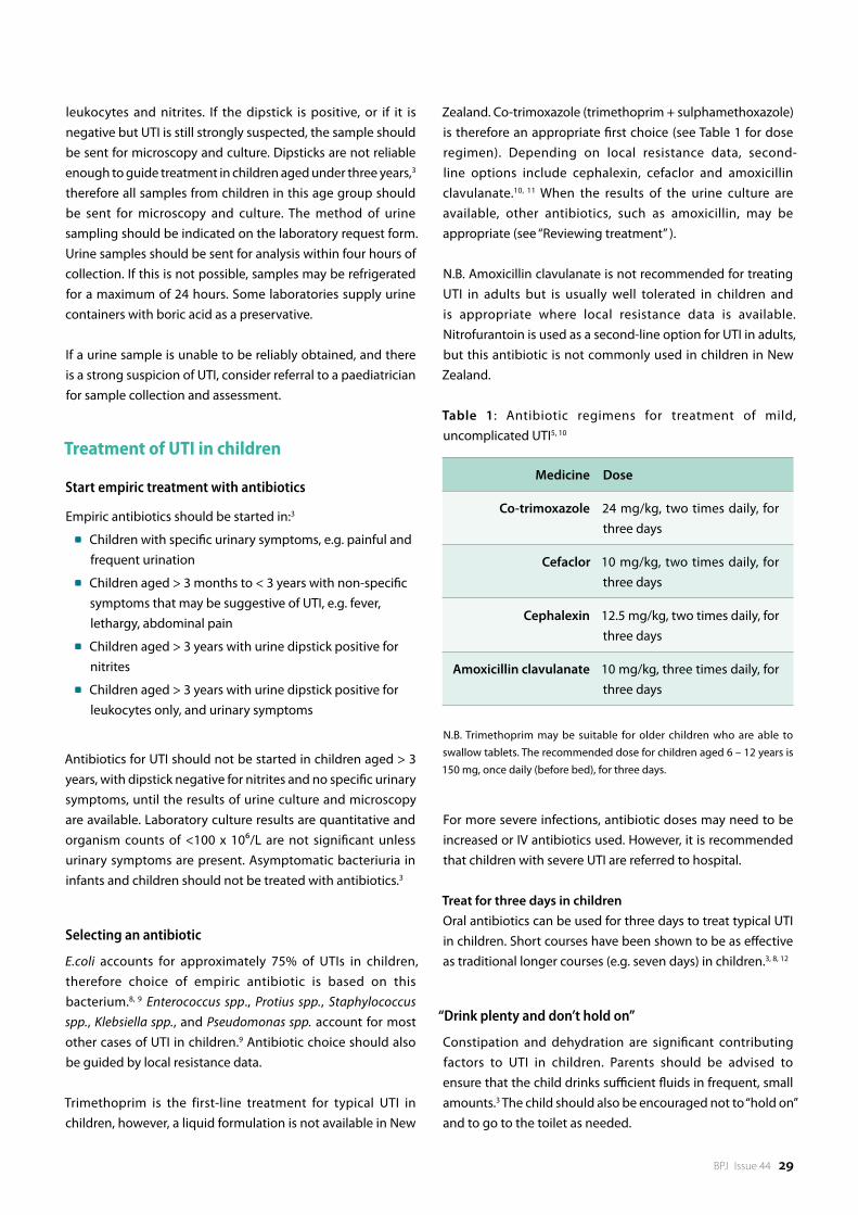
BPJ Issue 44 29
leukocytes and nitrites. If the dipstick is positive, or if it is negative but UTI is still strongly suspected, the sample should be sent for microscopy and culture. Dipsticks are not reliable enough to guide treatment in children aged under three years,3 therefore all samples from children in this age group should be sent for microscopy and culture. The method of urine sampling should be indicated on the laboratory request form. Urine samples should be sent for analysis within four hours of collection. If this is not possible, samples may be refrigerated for a maximum of 24 hours. Some laboratories supply urine containers with boric acid as a preservative.
If a urine sample is unable to be reliably obtained, and there is a strong suspicion of UTI, consider referral to a paediatrician for sample collection and assessment.
Treatment of UTI in children
Start empiric treatment with antibiotics
Empiric antibiotics should be started in:3
Children with specific urinary symptoms, e.g. painful and frequent urination
Children aged > 3 months to < 3 years with non-specific symptoms that may be suggestive of UTI, e.g. fever, lethargy, abdominal pain
Children aged > 3 years with urine dipstick positive for nitrites
Children aged > 3 years with urine dipstick positive for leukocytes only, and urinary symptoms
Antibiotics for UTI should not be started in children aged > 3 years, with dipstick negative for nitrites and no specific urinary symptoms, until the results of urine culture and microscopy are available. Laboratory culture results are quantitative and organism counts of <100 x 10⁶/L are not significant unless urinary symptoms are present. Asymptomatic bacteriuria in infants and children should not be treated with antibiotics.3
Selecting an antibiotic
E.coli accounts for approximately 75% of UTIs in children, therefore choice of empiric antibiotic is based on this bacterium.8, 9 Enterococcus spp., Protius spp., Staphylococcus spp., Klebsiella spp., and Pseudomonas spp. account for most other cases of UTI in children.9 Antibiotic choice should also be guided by local resistance data.
Trimethoprim is the first-line treatment for typical UTI in children, however, a liquid formulation is not available in New
Zealand. Co-trimoxazole (trimethoprim + sulphamethoxazole) is therefore an appropriate first choice (see Table 1 for dose regimen). Depending on local resistance data, second-line options include cephalexin, cefaclor and amoxicillin clavulanate.10, 11 When the results of the urine culture are available, other antibiotics, such as amoxicillin, may be appropriate (see “Reviewing treatment” ).
N.B. Amoxicillin clavulanate is not recommended for treating UTI in adults but is usually well tolerated in children and is appropriate where local resistance data is available. Nitrofurantoin is used as a second-line option for UTI in adults, but this antibiotic is not commonly used in children in New Zealand.
Table 1: Antibiotic regimens for treatment of mild, uncomplicated UTI5, 10
Medicine Dose
Co-trimoxazole 24 mg/kg, two times daily, for three days
Cefaclor 10 mg/kg, two times daily, for three days
Cephalexin 12.5 mg/kg, two times daily, for three days
Amoxicillin clavulanate 10 mg/kg, three times daily, for three days
N.B. Trimethoprim may be suitable for older children who are able to swallow tablets. The recommended dose for children aged 6 – 12 years is 150 mg, once daily (before bed), for three days.
For more severe infections, antibiotic doses may need to be increased or IV antibiotics used. However, it is recommended that children with severe UTI are referred to hospital.
Treat for three days in childrenOral antibiotics can be used for three days to treat typical UTI in children. Short courses have been shown to be as effective as traditional longer courses (e.g. seven days) in children.3, 8, 12
“Drink plenty and don’t hold on”
Constipation and dehydration are significant contributing factors to UTI in children. Parents should be advised to ensure that the child drinks sufficient fluids in frequent, small amounts.3 The child should also be encouraged not to “hold on” and to go to the toilet as needed.
30 BPJ Issue 44
Parents should also be given advice on correct toileting techniques, e.g. always wiping from front to back for girls. The bladder requires regular, complete emptying and this is best taught with a potty or a supported small toilet seat and step so the feet are able to rest on a surface.
Increasing fibre in the diet will help to avoid and alleviate constipation. If constipation persists or is significant, pharmacological management can be considered, e.g. lactulose.
Reviewing treatment once culture results are available
A review of treatment is recommended at 48 hours, when the culture results are available and the child’s response to treatment can be assessed. If the child’s symptoms have not improved, the initial diagnosis and antibiotic choice may need to be reviewed.
When the result of the urinary culture indicate a resistant strain of bacteria, but the child’s condition is improving, the antibiotic course can be continued and a “test of cure” urine culture requested once the course is completed.2 If the child’s
condition is not improving, change the antibiotic and consider discussion with, or referral to, a paediatrician.
Where symptoms have improved and culture indicates an appropriate antibiotic has been given, test for cure is not necessary.
Advise parents that they should bring the child back for reassessment if the child’s condition worsens or if symptoms have not improved after 48 hours of treatment.
Antibiotic prophylaxis to prevent reoccurrenceIn children with typical first-time UTI there is little benefit to prophylactic antibiotic use and it is therefore not recommended.13 Long-term antibiotics may be required in children with underlying renal tract abnormalities or severe recurrent UTIs to prevent reoccurrence.
In children already receiving prophylactic antibiotics, new occurrences of UTI should be treated with a different antibiotic, and not a higher dose of the prophylactic antibiotic.3
Could UTI be a sign of sexual abuse?
UTI is only very rarely a sign of sexual abuse, but if other risk factors are present, it is important to consider this possibility.
Signs and symptoms that can indicate sexual abuse include:
Unusual or excessive genital itching
Bruising, redness, swelling or bleeding in the genital area
Age inappropriate sexual play, knowledge or interest
Fear of certain people or places
If sexual abuse is suspected, refer immediately to a paediatrician and inform Child, Youth and Family.
For further information see: “Detecting child abuse in general practice”, BPJ 38 (Sept, 2011).
BPJ Issue 44 31
References1. Jadresić LP. Diagnosis and management of urinary tract infections
in children. Paediatr Child Health 2010;20(6):274–8.
2. Clinical Knowledge Summaries (CKS). Urinary tract infection - children. CKS; 2009. Available from: www.cks.nhs.uk (Accessed May, 2012).
3. National Institute for Health and Clinical Excellence (NICE). Urinary tract infection in children: Diagnosis, treatment, and long-term management. NICE; 2007. Available from: www.nice.org.uk (Accessed May, 2012).
4. Tosif S, Baker A, Oakley E, et al. Contamination rates of different urine collection methods for the diagnosis of urinary tract infection in young children: An observational cohort study. J Pediatr Child Health; In Corrected Proof: DOI 110.1111/j.1440–1754.2012.
5. Starship Children’s Health Clinical Guidance. Urinary tract infection. Auckland; 2007. Available from: www.adhb.govt.nz/starshipclinicalguidelines/_Documents/Urinary%20Tract%20Infection.pdf (Accessed May, 2012).
6. Dartmouth-Hitchcock Medical Centre. Collection instructions: Urine. Dept. Pathology, Hanover, USA; 2011. Available from: http://labhandbook.hitchcock.org/microUrine.html (Accessed May, 2012).
7. The Royal Children’s Hospital. Suprapubic aspirate guideline. Melbourne, Australia; 2012. Available from: www.rch.org.au/clinicalguide/cpg.cfm?doc_id=5246 (Accessed May, 2012).
8. Larcombe J. Urinary tract infection in children. Am Fam Physician 2010;62(10):1252-4.
9. Sheerin NS. Urinary tract infection. Medicine 2011;39(7):384–9.
10. BMJ Group. British national formulary for children 2011-2012. London: Royal Pharmeceutical Society; 2011.
11. Australian Medicines Handbook Pty Ltd, 2011. Australian medicines handbook. Adelaide: Australian Medicines Handbook Pty Ltd; 2011.
12. Michael M, Hodson E, Craig J, et al. Short versus standard duration oral antibiotic therapy for acute urinary tract infection in children. Cochrane Database Syst Rev 2010;(1):CD003966.
13. Williams G, Craig J. Long-term antibiotics for preventing recurrent urinary tract infection in children. Cochrane Database Syst Rev 2011;(3):CD001534.
14. Shaikh N, Ewing A, Hoberman A. Risk of renal scarring in children with a first urinary tract infection: a systematic review. J Pediatr 2010;126(6):1084–91. ACKNOWLEDGEMENT: Thank you to Associate
Professor David Reith, Paediatrician and Clinical Pharmacologist, Paediatrics and Child Health, Dunedin School of Medicine, University of Otago, Dr Rosemary Ikram, Clinical Microbiologist, Christchurch and Mr Stephen Mark, Consultant Urologist, Paediatric Urology and Clinical Lecturer, University of Otago, Christchurch for expert guidance in developing this article.
Investigating for an underlying cause of UTI after treatment has been initiated
A predisposing abnormality is present in approximately one-third of children presenting with first-time, typical UTIs. Renal ultrasound in a hospital setting is used to identify these abnormalities.2
It is not necessary to refer children aged over six months with a typical, first-time UTI for imaging.3 However, children aged under six months with a typical UTI should be referred approximately six weeks after the infection has cleared. Children aged over six months with more than one confirmed UTI should also be referred for imaging.
Children aged under three months and those with severe infection will have been referred to hospital for treatment of their UTI, and are likely to have undergone renal ultrasound at this time.
Potential complications with UTI in childrenA small number of children who have an acute UTI will develop long-term complications. A meta-analysis assessing the complications of UTIs in children found that approximately 15% had renal scarring post infection.14 Renal scarring can have long-term effects on morbidity and mortality, including increased rates of hypertension and proteinuria, decreased renal function and increased end-stage kidney disease.
The likelihood of complications increases in children with upper UTI, recurrent UTI, vesicoureteral reflux or undiagnosed UTI. Early diagnosis and optimal management greatly reduces the likelihood of long-term complications.

32
The appropriate use of
macrolides

33
What are macrolides and how do they work?
Macrolides are a class of antibiotic that includes erythromycin, roxithromycin, azithromycin and clarithromycin. They are useful in treating respiratory, skin, soft tissue, sexually transmitted, H. pylori and atypical mycobacterial infections. Macrolides share a similar spectrum of antimicrobial activity with benzylpenicillin making them useful alternatives for people with a history of penicillin (and cephalosporin) allergy. Bacteria often display cross-resistance between the macrolides.
Macrolides interfere with bacterial protein synthesis and, depending on concentration and bacterial species, are either bactericidal (kill bacteria), or bacteriostatic (inhibit growth of bacteria). Macrolides also have immunomodulatory and anti-inflammatory effects, which can be beneficial in some situations, e.g. when they are used in the treatment of cystic fibrosis.1
Which infections should macrolides be used for?
Macrolides are effective against gram-posit ive (excluding enterococci) and some gram-negative bacteria. They are also active against Mycoplasma pneumoniae, Treponema pallidum, Bordetella per tussis , Chlamydia trachomatis, Chlamydophila pneumoniae, Legionella spp., Campylobacter spp. and Borrelia spp.1
First-line indications for macrolides for common infections are listed in Table 1. There are numerous infections in which macrolides would be considered for second-line treatment.
Table 1: Common first-line indications for macrolides
Infection First-line treatment Second-line treatment
Pertussis Erythromycin –
Community acquired pneumonia Amoxicillin alone
or
Amoxicillin + erythromycin (for atypical infections)
Erythromycin, roxithromycin, doxycycline or co-trimoxazole
H. pylori Amoxicillin + clarithromycin + omeprazole
Metronidazole + clarithromycin + omeprazole
Chlamydia Azithromycin Doxycycline, amoxicillin, erythromycin
Acute non-specific urethritis Azithromycin Vancomycin (Doxycycline treatment)
For further information see “Antibiotic choices for common infections”, available from: www.bpac.org.nz
34 BPJ Issue 44
First-line indications for macrolides
Pertussis
Erythromycin 10 mg/kg (400 mg for adults), four times daily, for 14 days
Cases of pertussis (whooping cough) persist in New Zealand, despite the vaccine being part of the National Immunisation Schedule. Antibiotics are ineffective at reducing the duration or severity of symptoms if given more than seven days after the infection begins. However, antibiotics are still useful, if started within three to four weeks of infection, to prevent transmission to others. Women diagnosed with pertussis in the third trimester of pregnancy, should be given antibiotic treatment regardless of the time of onset of infection.4
Prophylactic antibiotic treatment should be offered to household contacts of a person with pertussis, if the household includes a child who has not completed a course of pertussis vaccination.4
Erythromycin is considered the medicine of choice for treatment and prophylaxis of pertussis as it is active against the causative organism – Bordetella pertussis. Infants aged under three months treated with erythromycin are at increased risk of developing pyloric stenosis. As the risk associated with pertussis in a young infant is considerably greater, erythromycin is still indicated, but the infant should be monitored for complications for four weeks after completion of treatment.4
Community-acquired pneumonia: atypical infection
Amoxicillin 500 – 1000 mg, three times daily, for seven days + erythromycin 400 mg, four times daily (or 800 mg, twice daily), for seven days
Severe cases of pneumonia require hospitalisation. The first-line treatment choice for pneumonia treated in the community is amoxicillin (to cover Streptococcus pneumoniae). Erythromycin (or roxithromycin) should be added to the treatment regimen when atypical infection is known to be circulating in the community. Erythromycin and roxithromycin provide coverage for Mycoplasma pneumoniae, Legionella spp. and Chlamydophilia pneumoniae.
Resistance of S. pneumoniae to macrolides is a worldwide problem. In 2010, resistance of S. pneumonia (non-invasive disease) to erythromycin in New Zealand was 19%.5
Prescribing erythromycin
Erythromycin is available in New Zealand as erythromycin ethyl succinate (fully subsidised), erythromycin lactobionate (fully subsidised, injection only) and erythromycin stearate (partially subsidised).
The usual oral adult dose of erythromycin is 1 – 2 g daily, in two to four divided doses. The dose may be increased up to 4 g per day according to the severity of the infection. As erythromycin ethyl succinate is now the only fully subsidised oral option, dosing recommendations in this article are altered to take into account the tablet dosages available.
Erythromycin ethyl succinate is available in 400 mg tablets, and two strengths of liquid formulation – 200 mg/5 mL and 400 mg/5 mL. The usual adult dose is 400 mg, four times daily. Alternatively, 800 mg, twice daily, may be a more convenient dose regimen for some patients. In severe infections, the dose may be increased up to a maximum of 4 g per day. Tablets may be taken with or without food.
The usual dose for infants and children is 10 mg/kg, four times daily, although this may be doubled in severe infections. The daily dose may be divided into twice daily or three times daily dosing if desired. Children aged over eight years may be given the usual adult dose.2
The erythromycin doses expressed in this article refer to prescribing of erythromycin ethyl succinate. Therefore, some dosing recommendations may differ slightly from those listed in the bpacnz antibiotic guide.
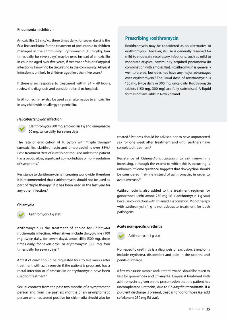
BPJ Issue 44 35
Pneumonia in children
Amoxicillin (25 mg/kg, three times daily, for seven days) is the first-line antibiotic for the treatment of pneumonia in children managed in the community. Erythromycin (10 mg/kg, four times daily, for seven days) may be used instead of amoxicillin in children aged over five years, if treatment fails or if atypical infection is known to be circulating in the community. Atypical infection is unlikely in children aged less than five years.6
If there is no response to treatment within 24 – 48 hours, review the diagnosis and consider referral to hospital.
Erythromycin may also be used as an alternative to amoxicillin in any child with an allergy to penicillin.
Helicobacter pylori infection
Clarithromycin 500 mg, amoxicillin 1 g and omeprazole 20 mg, twice daily, for seven days
The rate of eradication of H. pylori with “triple therapy” (amoxicillin, clarithromycin and omeprazole) is over 85%.7 Post-treatment “test of cure” is not required unless the patient has a peptic ulcer, significant co-morbidities or non-resolution of symptoms.7
Resistance to clarithromycin is increasing worldwide, therefore it is recommended that clarithromycin should not be used as part of “triple therapy” if it has been used in the last year for any other infection.8
Chlamydia
Azithromycin 1 g stat
Azithromycin is the treatment of choice for Chlamydia trachomatis infection. Alternatives include doxycycline (100 mg, twice daily, for seven days), amoxicillin (500 mg, three times daily, for seven days) or erythromycin (800 mg, four times daily, for seven days).9
A “test of cure” should be requested four to five weeks after treatment with azithromycin if the patient is pregnant, has a rectal infection or if amoxicillin or erythromycin have been used for treatment.9
Sexual contacts from the past two months of a symptomatic person and from the past six months of an asymptomatic person who has tested positive for chlamydia should also be
treated.9 Patients should be advised not to have unprotected sex for one week after treatment and until partners have completed treatment.9
Resistance of Chlamydia trachomatis to azithromycin is increasing, although the extent to which this is occurring is unknown.10 Some guidance suggests that doxycycline should be considered first-line instead of azithromycin, in order to avoid overuse.10
Azithromycin is also added to the treatment regimen for gonorrhoea (ceftriaxone 250 mg IM + azithromycin 1 g stat) because co-infection with chlamydia is common. Monotherapy with azithromycin 1 g is not adequate treatment for both pathogens.
Acute non-specific urethritis
Azithromycin 1 g stat
Non-specific urethritis is a diagnosis of exclusion. Symptoms include erythema, discomfort and pain in the urethra and penile discharge.
A first void urine sample and urethral swab* should be taken to test for gonorrhoea and chlamydia. Empirical treatment with azithromycin is given on the presumption that the patient has uncomplicated urethritis, due to Chlamydia trachomatis. If a purulent discharge is present, treat as for gonorrhoea (i.e. add ceftriaxone 250 mg IM stat).
Prescribing roxithromycin
Roxithromycin may be considered as an alternative to erythromycin. However, its use is generally reserved for mild to moderate respiratory infections, such as mild to moderate atypical community acquired pneumonia (in combination with amoxicillin). Roxithromycin is generally well tolerated, but does not have any major advantages over erythromycin.3 The usual dose of roxithromycin is 150 mg, twice daily or 300 mg, once daily. Roxithromycin tablets (150 mg, 300 mg) are fully subsidised. A liquid form is not available in New Zealand.
Endorsement is required when prescribing azithromycin or clarithromycin
36 BPJ Issue 44
Sexual contacts from the past two months should also be treated and tested. This is still necessary if chlamydia and gonorrhoea tests are negative as false negative results are possible and treating the female partner reduces the chance of recurrence in affected males.9
* Check with your local laboratory, a swab may not be necessary
depending on urinanalysis method
Campylobacteriosis
In the majority of cases of campylobacteriosis, antibiotic treatment is not required as diarrhoea will resolve with symptomatic treatment only. Antibiotics have limited effect on the duration and severity of infection, but can remove the infection from the stool and therefore reduce transmission to others.
Treatment with erythromycin 400 mg (children 10 mg/kg), four times daily, for five days, is indicated for people with severe or prolonged infection, in pregnant women nearing term and may be considered for food handlers, childcare workers and people caring for patients who are immuno-compromised.
Second-line indications for macrolidesErythromycin is an alternative antibiotic for people with a history of penicillin allergy in the treatment of otitis media, pharyngitis and boils (when treatment is indicated for these conditions), cellulitis, mastitis and syphilis.
Azithromycin (1 g stat or 500 mg, once daily, for three days) can be used instead of ciprofloxacin as a second-line treatment for severe traveller’s diarrhoea, when antibiotics are required. Azithromycin is recommended for pregnant women (ciprofloxacin is contraindicated) or in areas where there is quinolone resistance, e.g. South East Asia. Azithromycin (10 mg/kg, once daily for three days) is also recommended for young children with traveller’s diarrhoea (ciprofloxacin is not recommended in children), but a liquid formulation is not available in New Zealand. Erythromycin is an alternative. N.B. Azithromycin is not funded for this indication.
Azithromycin 1 g stat can be used instead of doxycycline to treat pelvic inflammatory disease (plus ceftriaxone 250 mg, IM stat and metronidazole 400 mg, twice daily, for two weeks), when chlamydia is present, especially if compliance is likely to be a problem.
Adverse effects of macrolidesThe most common adverse effects associated with macrolides are gastrointestinal, such as abdominal discomfort and cramp, nausea, vomiting and diarrhoea. Symptoms are dose dependent and are more common in children.1 Erythromycin is associated with a higher incidence of gastrointestinal adverse effects than other macrolides, with 5 – 30% of patients reporting symptoms.2 Erythromycin ethyl succinate has a lower incidence of gastrointestinal adverse effects compared to other forms of erythromycin. More frequent daily dosing may alleviate gastrointestinal effects.
Prescriptions for azithromycin must be endorsed to qualify for a full subsidy. An endorsement requires the prescriber to write “certified condition” on the prescription, to indicate that the patient meets the criteria for subsidy. Azithryomycin is fully-subsidised for people with uncomplicated urethritis or cervicitis proven or presumed to be due to chlamydia infection, and their sexual contacts. Azithromycin is also available via Practitioners Supply Order, which must be endorsed.
Clarithromycin is fully subsidised with endorsement for H. pylori eradication. Endorsement occurs automatically
if clarithromycin, amoxicillin (or metronidazole) and a proton pump inhibitor are concurrently prescribed as
“triple therapy”. A maximum of 14 tablets per prescription is allowed. Special Authority criteria also applies for relevant practitioners to prescribe clarithromycin for mycobacterial infections.
See pharmaceutical schedule for full details www.pharmac.govt.nz
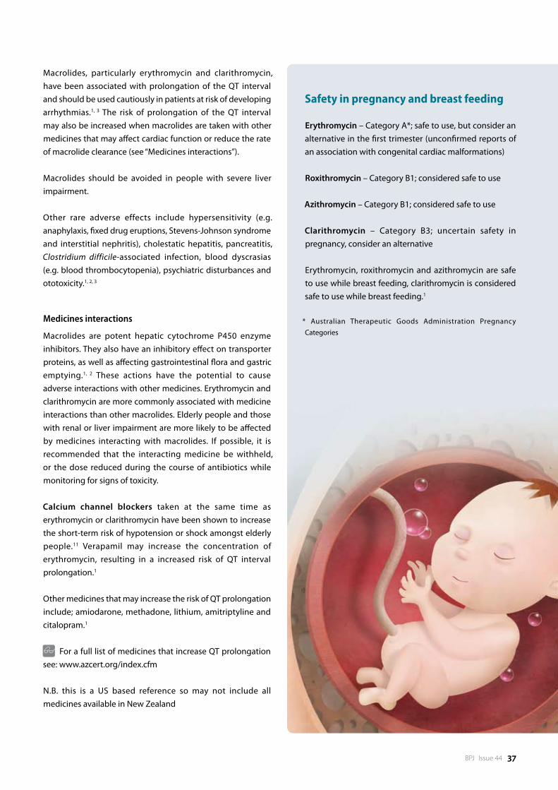
BPJ Issue 44 37
Macrolides, particularly erythromycin and clarithromycin, have been associated with prolongation of the QT interval and should be used cautiously in patients at risk of developing arrhythmias.1, 3 The risk of prolongation of the QT interval may also be increased when macrolides are taken with other medicines that may affect cardiac function or reduce the rate of macrolide clearance (see “Medicines interactions”).
Macrolides should be avoided in people with severe liver impairment.
Other rare adverse effects include hypersensitivity (e.g. anaphylaxis, fixed drug eruptions, Stevens-Johnson syndrome and interstitial nephritis), cholestatic hepatitis, pancreatitis, Clostridium difficile-associated infection, blood dyscrasias (e.g. blood thrombocytopenia), psychiatric disturbances and ototoxicity.1, 2, 3
Medicines interactions
Macrolides are potent hepatic cytochrome P450 enzyme inhibitors. They also have an inhibitory effect on transporter proteins, as well as affecting gastrointestinal flora and gastric emptying.1, 2 These actions have the potential to cause adverse interactions with other medicines. Erythromycin and clarithromycin are more commonly associated with medicine interactions than other macrolides. Elderly people and those with renal or liver impairment are more likely to be affected by medicines interacting with macrolides. If possible, it is recommended that the interacting medicine be withheld, or the dose reduced during the course of antibiotics while monitoring for signs of toxicity.
Calcium channel blockers taken at the same time as erythromycin or clarithromycin have been shown to increase the short-term risk of hypotension or shock amongst elderly people.11 Verapamil may increase the concentration of erythromycin, resulting in a increased risk of QT interval prolongation.1
Other medicines that may increase the risk of QT prolongation include; amiodarone, methadone, lithium, amitriptyline and citalopram.1
For a full list of medicines that increase QT prolongation see: www.azcert.org/index.cfm
N.B. this is a US based reference so may not include all medicines available in New Zealand
Safety in pregnancy and breast feeding
Erythromycin – Category A*; safe to use, but consider an alternative in the first trimester (unconfirmed reports of an association with congenital cardiac malformations)
Roxithromycin – Category B1; considered safe to use
Azithromycin – Category B1; considered safe to use
Clarithromycin – Category B3; uncertain safety in pregnancy, consider an alternative
Erythromycin, roxithromycin and azithromycin are safe to use while breast feeding, clarithromycin is considered safe to use while breast feeding.1
* Australian Therapeutic Goods Administration Pregnancy Categories

38
ACKNOWLEDGEMENT: Thank you to Dr Rosemary Ikram, Clinical Microbiologist, Christchurch for expert guidance in developing this article.
Warfarin and dabigatran may have increased anticoagulant properties when taken with clarithromycin and erythromycin.12 If possible, an alternative antibiotic should be used. Warfarin may need to be temporarily stopped or the dose reduced if there is no alternative. The INR should be monitored if warfarin and macrolides are taken at the same time. Little information is available on interactions with dabigatran, but patients should be monitored for signs of bleeding. This effect may be more pronounced in elderly people, or when renal function is reduced.
Statin metabolism, in particular simvastatin and atorvastatin, may be affected by macrolides inhibiting CYP3A4 enzymes. This can result in an increased risk of statin-induced rhabdomyolysis. Azithromycin interacts less with CYP3A4 enzymes, however, there have also been occasional reports of rhabdomyolysis in patients taking azithromycin.13 Patients can be advised not to take simvastatin or atorvastatin while completing a course of a macrolide antibiotic. Pravastatin is not significantly metabolised by CYP3A4, therefore is less likely to be affected by concurrent macrolide use.
Digoxin is known to interact with clarithromycin, which can lead to digoxin toxicity.14 When these medicines are taken in combination the digoxin dose should be reduced by half and the patient monitored for symptoms of toxicity.15
Other medicines that may have significant interactions with macrolides in elderly people or those with significant co-morbidities include; benzodiazepines, carbamazepine, cimetidine, clozapine, colchicine and theophylline.14
bestpractice Decision Support is developed by BPAC Inc, which is separate from bpacnz.bpacnz bears no responsibility for bestpractice Decision Support or any use that is made of it.
bestpracticeDECISION SUPPORT FOR HEALTH PROFESSIONALS
bestpractice Decision Support facilitates the recording and saving of Well Child checks with the Well Child/Tamariki Ora module. A complete suite of checks is provided, including:
6 week check ■3 month check ■5 month check ■8-10 month check ■12-24 month check ■3 year check ■
Each check provides advice for parents in the form of printable resources.
More information: www.bestpractice.net.nz
WELL CHILD CHECKSTAMARIKI ORA
References:1. Australian medicines handbook. Adelaide: Australian Medicines
Handbook Pty Ltd, 2011.
2. Sweetman S, editor. The complete drug reference. London: Pharmaceutical Press, 2011.
3. Grayson M. Kucers’ the use of antibiotics. 6th ed. Credo; 2010. Available from: www.medicinescomplete.com (Accessed May, 2012).
4. Ministry of Health. Immunisation Handbook 2011. Wellington: Ministry of Health.
5. Environmental Science and Research (ESR). Antimicrobial resistance data from hospital and community laboratories, 2010. Available from: www.surv.esr.cri.nz (Accessed May, 2012).
6. Clinical Knowledge Summaries (CKS). Cough – acute with chest signs in children. Community-acquired pneumonia. 2007. Available from: http://cks.library.nhs.uk (Accessed May, 2012).
7. New Zealand Guidelines Group (NZGG). Management of dyspepsia and heartburn. NZGG, 2004 Available from: www.nzgg.org.nz/search?search=Dyspepsia (Accessed May, 2012).
8. National Institute for Health and Clinical Excellence (NICE). Dyspepsia. NICE, 2004. Available from: www.nice.org.uk (Accessed May, 2012).
9. The New Zealand Sexual Health Society Inc (NZSHS). Best practice guidelines. NZSHS, 2009. Available from: www.nzshs.org.guidelines.html (Accessed May, 2012).
10. Ison CA. Antimicrobial resistance in sexually transmitted infections in the developed world: implications for rational treatment. Curr Opin Infect Dis 2012;25(1):73–8.
11. Wright AJ, Gomes T, Mamdani MM, et al. The risk of hypotension following co-prescription of macrolide antibiotics and calcium-channel blockers. CMAJ 2011;183(3):303–7.
12. British National Formulary (BNF). BNF 62. London: BMJ Publishing Group and Royal Pharmaceutical Society of Great Britain, 2011.
13. Strandell J, Bate A, Hägg S, Edwards IR. Rhabdomyolysis a result of azithromycin and statins: an unrecognized interaction. Br J Clin Pharmacol 2009;68(3):427–34.
14. Westphal JF. Macrolide – induced clinically relevant drug interactions with cytochrome P-450A (CYP) 3A4: an update focused on clarithromycin, azithromycin and dirithromycin. Br J Clin Pharmacol 2000. 50(4):285–95.
15. Lee CYW, Marcotte F, Giraldeau G, et al. Digoxin toxicity precipitated by clarithromycin use: case presentation and concise review of the literature. Can J Cardiol 2011;27(6):870.e15–16.
40 BPJ Issue 44
CORRESPONDENCE
Significance of albumin:creatinine ratio in people with and without diabetes
Dear Editor,I have just read “Testing for CVD, diabetes and renal disease in elderly people” (Best Tests March 2012). I have a question re. renal testing for proteinuria - the article (on page 7) says that in people with diabetes ACR >2.5 mg/mmol is significant BUT for non-diabetics ≥30 mg/mol is significant.
The reasons for the random variation of non-diabetics up to 30 mg WITHOUT significance must also surely apply to diabetics? The corollary is that we ought to seek other reasons for a sudden increase in a diabetic proteinuria, in a patient whose diabetes has not worsened, i.e. all the reasons it might vary in a non-diabetic.
Dr Michael Short General Practitioner, Palmerston North
Thank you for your question. We acknowledge there may have been a lack of clarity within the section to which you refer.
The section states:
In people with diabetes, ACR > 2.5 mg/mmol in males and > 3.5 mg.mmol in females indicates microalbuminuria
In people without diabetes, ACR ≥ 30 mg/mmol indicates clinically significant proteinuria
Proteinuria is a sign of abnormal excretion of protein by the kidney but is a non-specific term including any or all proteins excreted. In contrast, albuminuria specifically refers to an abnormal excretion rate of albumin. Microalbuminuria refers to an abnormally increased excretion rate of albumin in the urine. It is a marker of endothelial dysfunction and increased risk for cardiovascular morbidity and mortality, especially, but not exclusively, in high-risk populations such as people with diabetes and hypertension.
Microalbuminuria is an established risk factor for renal disease progression in type 1 diabetes and its presence is the earliest clinical sign of diabetic nephropathy. In addition, a number of studies suggest that microalbuminuria is an important risk factor for cardiovascular disease and defines a group at high risk for early cardiovascular mortality in both type 2 diabetes and essential hypertension.
Microalbuminuria also signifies abnormal vascular permeability and the presence of atherosclerosis. Among non-diabetic people with essential hypertension, microalbuminuria is associated with higher blood pressures, increased serum total cholesterol and reduced serum high-density lipoprotein cholesterol. Thus, taken together these data support the concept that the presence of microalbuminuria is the kidney's notice to the clinician and patient that there is a problem with the vasculature.
With that said, current thinking and evidence suggests that people with diabetes with a microalbuminuria level indicated by an ACR greater than 2.5 mg/mmol in males and 3.5 mg/mmol in females, have a significantly increased risk, and so individuals with these parameters should be commenced on an ACE inhibitor or an ARB.
In people without diabetes, minor degrees of albuminuria (ACR female 3.5 – 30 mg/mmol or male 2.5 – 30 mg/mmol) are not considered sensitive enough to predict renal disease, particularly given the wide day to day variability of levels and their non-specificity.

BPJ Issue 44 41
CORRESPONDENCE
BPJ Issue 44 41
Addressing barriers to HPV vaccination
Dear Editor,As a female GP I do a lot of gynae and STD work and try to promote HPV vaccination. I've noticed two themes from mums regarding getting their daughters vaccinated - a lot of mum's don't really get it that cervical cancer is a sexually transmitted disease and when I ask them "how much do you trust all the men out there?", their view on vaccination tends to change! I have also had daughters/mums of a strong Christian persuasion saying "I don't need it because I'm saving myself for marriage" - to which I reply by asking if they can absolutely guarantee that their future husband will be a virgin and that they will never divorce or be with anyone else ever. Even the most devout mother can usually see the point!
Dr Phillipa Story,General Practitioner, Hastings
We welcome "best practice tips" on any articles published in Best Practice Journal or Best Tests.
We value your feedback. Write to us at:Correspondence, PO Box 6032, Dunedin
or email: [email protected]

visit us at www.bpac.org.nzCall us on 03 477 5418 Email us at [email protected] Freefax us on 0800 27 22 69