Embed Size (px)
Citation preview

Oxidised fish oil does not influence established markers of oxidativestress in healthy human subjects: a randomised controlled trial
Inger Ottestad1,2, Gjermund Vogt3, Kjetil Retterstøl4, Mari C. Myhrstad1, John-Erik Haugen3,Astrid Nilsson3, Gitte Ravn-Haren5, Berit Nordvi6, Kirsti W. Brønner6, Lene F. Andersen2,Kirsten B. Holven2 and Stine M. Ulven1*1Faculty of Health, Nutrition and Management, Akershus University College, PO Box 423, 2001 Lillestrøm, Norway2Department of Nutrition, Institute for Basic Medical Sciences, University of Oslo, PO Box 1046, Blindern, 0317 Oslo,
Norway3Nofima Mat AS, Osloveien 1, 1430 As, Norway4Lipid Clinic, Medical Department, Rikshospitalet-Oslo University Hospital, PO Box 4950, Nydalen, 0424 Oslo, Norway5Department of Toxicology and Risk Assessment, Technical University of Denmark, National Food Institute,
Mørkhøj Bygade 19, 2860 Søborg, Denmark6TINE SA, Centre for Research and Development, PO Box 7, Kalbakken, N-0902 Oslo, Norway
(Received 3 June 2011 – Revised 7 September 2011 – Accepted 8 September 2011)
Abstract
Intake of fish oil reduces the risk of CHD and CHD deaths. Marine n-3 fatty acids (FA) are susceptible to oxidation, but to our knowledge,
the health effects of intake of oxidised fish oil have not previously been investigated in human subjects. The aim of the present study was
to investigate markers of oxidative stress, lipid peroxidation and inflammation, and the level of plasma n-3 FA after intake of oxidised fish
oil. In a double-blinded randomised controlled study, healthy subjects (aged 18–50 years, n 54) were assigned into one of three groups
receiving capsules containing either 8 g/d of fish oil (1·6 g/d EPA þ DHA; n 17), 8 g/d of oxidised fish oil (1·6 g/d EPA þ DHA; n 18) or
8 g/d of high-oleic sunflower oil (n 19). Fasting blood and morning spot urine samples were collected at weeks 0, 3 and 7. No significant
changes between the different groups were observed with regard to urinary 8-iso-PGF2a; plasma levels of 4-hydroxy-2-hexenal,
4-hydroxy-2-nonenal and a-tocopherol; serum high sensitive C-reactive protein; or activity of antioxidant enzymes in erythrocytes. A sig-
nificant increase in plasma level of EPA þ DHA was observed in both fish oil groups, but no significant difference was observed between
the fish oil groups. No changes in a variety of in vivo markers of oxidative stress, lipid peroxidation or inflammation were observed after
daily intake of oxidised fish oil for 3 or 7 weeks, indicating that intake of oxidised fish oil may not have unfavourable short-term effects in
healthy human subjects.
Key words: n-3: Lipid peroxidation: Oxidative stress: Oxidised oil
Intake of fish and fish oil has been related to a reduced risk of
CHD and CHD deaths(1–3). The recommended daily intake in
primary prevention of CHD is at least two servings of fish per
week, preferably fatty fish(2,3). Such consumption is expected
to provide about 0·5 g of marine n-3 fatty acids (FA) EPA
and DHA per d. n-3 Supplements are recommended for
those who do not include fish in their diet(3), and are used
in clinical practice to prevent CHD and in the treatment of
mild-to-moderate hypertriacylglycerolaemia(2,3).
Long-chain n-3 PUFA are susceptible to oxidation, and lipid
peroxidation leads to the formation of a range of different
oxidation products(4–8). During the initial step of oxidation,
primary oxidation products (hydroperoxides) are formed,
and subsequently more stable secondary oxidation products
are generated, such as aldehydes. The content of total primary
and secondary oxidation products are measured as peroxide
value (PV) and anisidine value (AV), respectively. Maximum
acceptable levels of lipid peroxidation products in refined
marine n-3 oils to be used for dietary supplements are defined
by different monographs such as the European Pharmacopeia,
which recommend that PV and AV should not exceed 10 and
20 mEq/kg, respectively(9). High contents of oxidation products
*Corresponding author: S. M. Ulven, fax þ47 64849001, email [email protected]
Abbreviations: 4-HHE, 4-hydroxy-2-hexenal; 4-HNE, 4-hydoxy-2-nonenal; AV, anisidine value; CAT, catalase; CRP, C-reactive protein; E%, percentage of
energy; FA, fatty acids; FO, 8 g/d of fish oil; GPx, glutathione peroxidase; GR, glutathione reductase; GSH, glutathione; HOSO, 8 g/d of high-oleic
sunflower oil; oxFO, 8 g/d of oxidised fish oil; PV, peroxide value; tGSH, total glutathione.
British Journal of Nutrition, page 1 of 12 doi:10.1017/S0007114511005484q The Authors 2011
British
Journal
ofNutrition

(PV . 10 mEq/kg and/or AV . 20) have been reported in n-3
supplements available for consumers(10–13). Whether the
intake of highly oxidised marine n-3 oils is associated with
unfavourable health effects is unclear.
Oxidative stress is defined as an imbalance between oxidants
and antioxidants, causing oxidative damage(14,15). In healthy
subjects, the endogenous antioxidant defence system, such as
glutathione (GSH), are involved in the detoxification of oxidised
lipids, as are several antioxidant enzymes(16–18). Understanding
of the absorption of dietary lipid oxidation products in human
subjects is limited, and whether the intake of oxidised lipids
can lead to oxidative stress and accumulation of oxidative
damage is uncertain(16,19–22).
No single markers to determine in vivo lipid oxidation exist,
and different methods to assess oxidative stress and lipid per-
oxidation have been suggested. Conjugated dienes and thio-
barbituric acid-reactive substances are common methods to
measure oxidative stress in human subjects, but for several
reasons these methods are considered inappropriate, as
reviewed elsewhere(14,23). At present, 8-iso-PGF2a is suggested
as one of the most reliable markers to measure in vivo
oxidative stress(14,24–26). Elevated levels of 8-iso-PGF2a and
C-reactive protein (CRP) have been observed in a variety of
oxidative stress-related diseases and during inflam-
mation(24–29), but the effects of n-3 FA on these markers
in healthy subjects are inconsistent(30–34). We do not know
of any existing studies investigating whether the intake of
oxidised fish oil may affect the level of 8-iso-PGF2a and CRP.
4-Hydroxy-2-hexenal (4-HHE) and 4-hydoxy-2-nonenal (4-
HNE) are secondary oxidation products derived from n-3
and n-6 FA, respectively(4,6,8,35). These aldehydes have been
suggested as markers of in vivo lipid peroxidation, but
whether these markers are affected by the intake of n-3 FA
or oxidised lipids is not well documented(36–42).
At present, human studies with the aim of investigating
health effects of the intake of oxidised fish oil are lacking,
although it has been suggested that the regular consumption
of oxidised encapsulated n-3 oils may lead to unfavourable
health effects(8,14,43–45). The aim of the present study was to
investigate the effect of intake of oxidised fish oil on a variety
of markers of oxidative stress, lipid peroxidation and inflam-
mation, and the level of plasma n-3 FA in healthy subjects in
a 7-week randomised controlled study.
Subjects and methods
Subjects
Healthy, non-smoking men and women aged 18–50 years,
with a stable body weight over the last 3 months (^5 %),
were recruited among employees and students at Akershus
University College from May to September 2009. Exclusion cri-
teria were chronic illnesses and fasting serum level of total
cholesterol .7·5 mM, TAG .4 mM, glucose .6·0 mM, CRP
.10 mg/l, BMI $30 kg/m2, hypertension ($160/100 mmHg),
pregnancy and lactation. Those with serum levels of thyroxine
stimulating hormone, free T3 and free T4 above or below the
normal reference ranges were also excluded. Thyroxin repla-
cement therapy and contraceptives were accepted, provided
a stable dose was administered during the last 3 months.
Use of lipid-lowering and anti-hypertensive medications was
not permitted. Fe supplementation was accepted among
those with a regular use before inclusion in the study, but
was not to be taken concurrently (at the same meal) with
the test capsules.
The present study was conducted according to the guide-
lines laid down in the Declaration of Helsinki, and all
procedures involving human subjects were approved by the
Regional Committee of Medical Ethics (approval no.
6.2008.2215) and by the Norwegian Social Science Data Ser-
vices (approval no. 21 924). Written informed consent for par-
ticipation was obtained from all subjects. The study was also
registered at www.clinicaltrial.gov (ID no. NCT01034423).
Study design
A 7-week double-blinded, randomised, controlled parallel-
group study was conducted at the Akershus University College
from September to December 2009. A total of eighty-three
subjects were screened for eligibility, sixty-nine subjects
were randomised and fifty-eight subjects received allocated
interventions. Subjects lost during follow-up and the number
of subjects included in the statistical analysis are further
outlined in the flowchart (Fig. 1).
At baseline, subjects were randomly assigned and stratified
by sex into one of three intervention groups receiving sixteen
capsules per d containing 8 g/d of fish oil (FO; n 19; 1·6 g/d
EPA/DHA); 8 g/d of oxidised fish oil (oxFO; n 19; 1·6 g/d
EPA/DHA); or 8 g/d of high-oleic sunflower oil (HOSO; n
20) for 7 weeks. The subjects were instructed to take the cap-
sules with food (minimum two meals), and to store the cap-
sule containers at 48C during the study period. In the 4
weeks leading up to the baseline visit and during the interven-
tion period, the subjects were not allowed to consume fish,
fish products, marine n-3-enriched food or dietary sup-
plements. The subjects received instructions on which food
items to avoid and how to read the food labelling. At each
visit, they were reminded by a clinical nutritionist to avoid
marine n-3 FA products and to keep their weight stable.
Fasting body weight was registered at all visits.
During the first 3 weeks of the intervention period, the sub-
jects conducted a fully controlled isoenergetic diet. All foods
to be consumed were distributed at the Akershus University
College and the food items in the fully controlled diet
period were vegetables (e.g. cucumbers, tomatoes, peppers
and lettuce), fruits (e.g. oranges, bananas and grapes), juices
(apple and orange), low-fat dairy products (milk (1·5 % fat)
and yoghurt (0·1 % fat)), toppings (ham (,4 % fat), cheese
(16 and 27 % fat), strawberry and raspberry jam and eggs),
whole wheat bread (6 % fibre), crackers, chocolate and limited
amounts of tea and coffee. Hot lunches (soup and pasta),
dinner and desserts (creme brulee, chocolate pudding and
mousse) were provided by Fjordland AS (Oslo, Norway).
Dinners were delivered as vacuum-packed ready-made
dishes. To achieve the individual energy level, deliveries
were re-packed and vacuum packed again at Akershus
University College. The diet was planned to provide
I. Ottestad et al.2
British
Journal
ofNutrition

(exclusive of capsules) 24 % of energy (E%) from fat, of which
8 E% from SFA, 5 E% from MUFA and 6 E% from PUFA. The
protein content was 20 E%, and 57 E% was from carbo-
hydrates, including 5 E% from added sugar. The fibre content
was 39 g/d. One alcohol unit was allowed at two occasions
during the period. During the last 4 weeks of the intervention
period, the subjects continued to take the capsules, but
returned to their habitual diet without consuming marine
n-3 FA food items.
Blinding and randomisation
The study was blinded for the subjects and the study investi-
gators by identical appearance of the different capsules and
capsule containers; their contents were only identifiable by
the ID numbers on the containers. Randomisation was per-
formed by LINK Medical Research AS (Oslo, Norway), using
Microsoft Excel and its random generator. The randomisation
code was concealed from the study investigators until the
statistical analyses were completed.
Compliance and side effects
Compliance was assessed by capsule count. The capsules
were dispensed at baseline and after 3 weeks of intervention,
and the subjects were instructed to deliver the unused capsules.
We calculated the number of capsules used during the 7-week
intervention, which was divided by the number of capsules
scheduled for the intervention period(46). The mean daily
capsule count for each subject was expressed as a percentage,
and subjects with compliance (mean daily capsule count)
,70 % were excluded from the study. Average compliance
was 96 (SD 6) % in the FO group (n 17), 100 (SD 3) % in the
oxFO group (n 18) and 97 (SD 6) % in the HOSO group (n 19).
The rate of reported side effects did not differ among the
groups. In all, two subjects, one from each of the different fish
oil groups, reported side effects (belching with fishy taste) and
three subjects in the HOSO group reported side effects (consti-
pation, urgent to stool and burping).
Study products
Refined and deodorised functional food-grade cod liver oil
(Gadidae sp.) TINE EPADHA Oil 1200 was provided from
TINE SA (Oslo, Norway), from which one batch was divided
into two equal parts; one part (FO) was flushed with N2 and
stored at 48C until encapsulation. The other part (oxFO) was
Assessed for eligibility (n 83)
Underwent randomisation (n 69)
Excluded (n 14)(1) Not meeting inclusion criteria (n 8)(2) Declined to participate before randomisation (n 6)
Allocated to intervention HOSO group (n 23)(1) Received allocated intervention (n 20)(2) Did not receive allocated intervention (n 3)
Allocated to intervention FO group (n 24)(1) Received allocated intervention (n 19)(2) Did not receive allocated intervention (n 5)
(1) Baseline (n 18)(2) Analysed visit 3 (n 15)Excluded from analysis due to notcompleting fully controlled diet period (n 3)(3) Analysed visit 4 (n 18)
(1) Baseline (n 17)(2) Analysed visit 3 (n 17) (3) Analysed visit 4 (n 17)
Allocated to intervention oxFO group (n 22)(1) Received allocated intervention (n 19)(2) Did not receive allocated intervention (n 3)
(1) Lost to follow-up (n 0)(2) Discontinued intervention (n 0)(3) Excluded (n 1)Low compliance
(1) Lost to follow-up (n 0)(2) Discontinued intervention (n 1)Hospitalisation, not affected by this study
(1) Lost to follow-up (n 0)(2) Discontinued intervention (n 2)No explanation (n 1), abdominal discomfort(n 1)
En
rollm
ent
Allo
cati
on
Follo
w-u
p
(1) Baseline (n 19)(2) Analysed visit 3 (n 18)Excluded from analysis due to notcompleting fully controlled diet period (n 1)(3) Analysed visit 4 (n 19)
An
alys
is
Fig. 1. Flow chart of the study. Number of subjects included, allocated to intervention, lost during follow-up with explanation for the dropouts, and number included in
the statistical analysis after 3 and 7 weeks of intervention are shown. FO group, fish oil group; oxFO group, oxidised fish oil group; HOSO, high-oleic sunflower oil
group.
Oxidised fish oil and health effects 3
British
Journal
ofNutrition

oxidised by sparkling pure oxygen through the oil for 20 min
twice a day for 21 d, at room temperature, then flushed with
N2 and stored at 48C in dark until encapsulation. The level
of antioxidants (tocopherols and rosemary extracts) was simi-
lar in all three study oils. The level of tocopherols was
measured in the fish oil (before and after oxidation) and in
the refined food-grade HOSO before encapsulation. Toco-
pherols and rosemary extracts were then added to obtain simi-
lar concentration in all three study oils. FO, oxFO and refined
food-grade HOSO (AarhusKarlshamn AB, Malmø, Sweden)
were encapsulated in 500 mg softgel capsules made of
bovine gelatine (Eurocaps Limited, Wales, UK). All capsules
were stored in closed containers at 48C until the start of the
study. Each fish oil capsule contained 45 mg EPA and 56 mg
DHA (FO) and 46 mg EPA and 56 mg DHA (oxFO). The FA
composition was measured in the oils after encapsulation,
which is further outlined in Table 1.
Blood and urine sampling
The day before blood sampling, the subjects were told to
refrain from alcohol consumption and vigorous physical
activity; venous blood samples were drawn after an overnight
fast ($12 h). Serum was obtained from silica gel tubes (Becton
Dickinson Vacutainer Systems, Plymouth, UK) and kept at
room temperature for at least 30 min, until centrifugation
(1500g, 12 min). Plasma was obtained from EDTA tubes
(Becton Dickinson Vacutainer Systems), immediately placed
on ice and centrifuged within 10 min (1500g, 48C, 10 min).
The N2-flushed plasma samples were snap frozen and stored
at 2808C until further analysis. Erythrocytes were isolated
from plasma by centrifugation (1800g, 208C, 20 min), and
either stored at 2808C (for total GSH (tGSH) analysis) or
diluted in water (1:1) and stored at 2808C until analysis
(antioxidant enzyme activities). Morning spot urine samples
were refrigerated (48C) until delivered, and were immediately
aliquoted and stored at 2808C until further analysis.
Routine laboratory analysis
Fasting serum high-sensitive CRP, total cholesterol, LDL-
cholesterol, HDL-cholesterol, TAG, glucose, thyroxine-stimu-
lating hormone, free T3, free T4, alanine aminotransferase,
aspartate aminotransferase, g-glutamyl transferase and alka-
line phosphatase were measured by standard methods at a
routine laboratory (Furst Medical Laboratory, Oslo, Norway).
tGSH in erythrocytes and urinary 8-iso-PGF2a in morning
spot urine samples were determined by Vitas AS (Oslo,
Norway). tGSH was measured using a kit provided by Bio
Rad Laboratories GmbH (Munchen, Germany) validated for
GSH, as described elsewhere(47). Urinary 8-iso-PGF2a was ana-
lysed by liquid chromatography with negative electrospray
ionisation coupled to tandem mass spectrometric detection
(liquid chromatography/MS/MS), according to Bastani
et al.(48). Urinary levels of 8-iso-PGF2a are presented as the
ratio 8-iso-PGF2a:creatinine.
Determination of hydoxy-nonenal and hydroxy-hexenal inplasma
A method based on Luo et al.(49) was further developed
and in-house validated for use on human plasma by Nofima
(As, Norway). 2H-labelled 4-HNE was added to 3–500ml
plasma and the sample was directly derivatised in two steps
to generate the O-pentafluorobenzyl-oxime-trimethylsilyl
derivatives of the syn- and anti-stereoisomers of the respective
4-hydroxy-alkenals before GC–MS analysis – trimethylchloro-
silane. An Agilent 7890A gas chromatograph interfaced
with a 5975C mass-selective detector (Agilent Technologies,
Little Falls, DE, USA) was used. The O-pentafluorobenzyl-
oxime-trimethylsilyl derivatives were injected splitless and
separated on a HP-5MS-fused silica capillary column
(30 m £ 0·25 mm £ 0·25mm) using helium as carrier gas at a
flow rate of 1 ml/min. The oven temperature was programmed
from 508C (1 min) at 108C/min to 2408C (0 min) followed
by 208C/min to 3008C (5 min). Derivatised aldehydes were
Table 1. Characterisation of the encapsulated oil
Fish oil Oxidised fish oil High oleic sunflower oil
Fatty acidSFA (g/100 g) 15·9 15·8 6·6MUFA (g/100 g) 46·5 46·3 75·6PUFA (g/100 g) 28·3 28·4 8·8
n-3 Fatty acidsEPA (20 : 5n-3) (g/100 g) 9·0 9·1 0DHA (22 : 6n-3) (g/100 g) 11·1 11·2 0DPA (22 : 5n-3) (g/100 g) 1·1 1·1 0ALA (18 : 3n-3) (g/100 g) 0·8 0·8 0·3
Oxidation levelPV (mEq/kg) 4 18 4AV 3 9 3Totox 11 45 11
Volatile oxidation products of n-3 fatty acidPentanal (mg/100 g) 6·2 137·8 1·11-Penten-3-ol (mg/100 g) 12·8 132·4 0·1
DPA, docosapentaenoic acid; ALA, a-linolenic acid; PV, peroxide value; AV, anisidine value.
I. Ottestad et al.4
British
Journal
ofNutrition

measured in negative ion chemical ionisation mode. Ion
source temperature was 2308C, with electron ionisation
energy of 100 eV and methane as reagent gas. Stereoisomer
peaks with the highest intensity (anti-) were monitored at
m/z 291 (4-HHE) corresponding to (M 2 C2F4 2 H2O)2, at
m/z 283 (4-HNE) corresponding to (M 2 CF2 2 H2O)2 and
quantification was done by measuring m/z 286 corresponding
to (M 2 CF2 2 H2O)2 of the 2H-labelled 4-HNE internal
standard. CV of measured replicated samples was ,8 %.
Determination of a-tocopherol in plasma
Plasma (500ml) was added to chloroform–methanol (5 ml;
2:1) and a-tocopherol acetate in chloroform–butylated hydro-
xytoluene as internal standard, then mixed (5 min) and
distilled water (1 ml) was added before centrifugation
(1000 rpm, 48C, 10 min). The organic phase was evaporated
under N2 at 338C, re-dissolved in n-heptane–butylated hydro-
xytoluene (500ml) and transferred to HPLC vial. A normal-
phase HPLC method was used as described elsewhere(50).
Plasma concentration of a-tocopherol is normalised to the
a-tocopherol to total lipid concentration (a-tocopherol/total
cholesterol þ TAG), expressed as a-tocopherol:plasma total
lipid ratio. CV were ,5 %.
Erythrocyte antioxidant enzyme activities
Activity of the antioxidant enzymes GSH reductase (GR), GSH
peroxidase (GPx) and catalase (CAT) were spectrophotometri-
cally assayed in erythrocyte lysates on a Cobas Mira S analyser
(Triolab, Brøndby, Denmark) according to Wheeler et al.(51).
All measures of enzyme activities were related to the
amount of Hb in the sample. Hb content was determined
using a commercially available kit (Randox, Ardmore, UK).
Samples from each subject were analysed in the same batch
in random order and a control sample was analysed for
every fifteenth sample. CV were ,7 %.
Fatty acids analysis
Plasma lipids were extracted by use of the Bligh and Dyer
method(52). FA in Bligh and Dyer extract and oils were deriva-
tised and analysed as methyl esters on a GC (HP 6890)
equipped with a BPX-70 column, 60 m £ 0·25 mm inner diam-
eter, 0·25-mm film (SGE Analytical Science Private Limited,
Melbourne, Australia). The temperature program started at
708C for 1 min, increased with 308C/min to 1708C, with
1·58C/min to 2008C and with 38C/min to 2208C, with a final
hold time of 5 min. Peaks were integrated with HP GC Chem-
Station software (rev. B.01.01; Agilent Technologies, Palo Alto,
CA, USA) and identified by use of external standards. CV were
,5 %. The concentration of the individual FA was expressed
in percentage of total FA.
Oxidised lipids in study products and food items
The PV and AV were measured in the oils after encapsulation
using methods according to AOCS Official Method Cd 8-53
and Cd 18-90, respectively. Total antioxidant value (Totox)
was calculated as Totox ¼ 2PV þAV (Table 1). A random
sample of food items considered prone to oxidation was
stored at 2808C until further analysis. Contents of volatile
oxidation products in encapsulated oil and food items were
analysed by dynamic headspace/GC–MS, as described by
Olsen et al.(53), with small modifications in the method. A
measure of 20 g of homogenised food samples or 2 g oil
were heated to 708C and purged with 100 ml/min N2 through
a Drechsel head for 30 min. Volatiles were adsorbed on Tenax
GR (mesh size 60/80; Alltech Associates, Inc., Deerfield, IL,
USA). Trapped compounds were desorbed at 2508C for 5 min
in a Markes Unity/Ultra TD automatic desorber (Markes Inter-
national Limited, Llantisant, UK) and transferred to an Agilent
6890 GC System (Agilent, Palo Alto, CA, USA) with an Agilent
5973 mass-selective detector operated in electron impact
mode at 70 eV. CV were ,10 %. The concentrations of the indi-
vidual volatiles was calculated as mg/g sample based on the
internal standards. The food analysis showed that the food
items used in the fully controlled diet period contributed to no
or only minor levels of volatile oxidation products (data not
shown). The concentrations of volatile n-3-derived oxidation
products in the capsules, pentanal and 1-penten-3-ol, are
shown in Table 1.
Statistics
Sample size was calculated using an expected change in
plasma n-3 FA from baseline to the end of the study of 1·7
(SD 1·2) %. The level of significance was set to 5 % (two-
sided) and the power to 80 %. A total of thirty-nine subjects
were required in the present study, but a high dropout rate
was expected, and it was considered necessary to include a
total of seventy-five subjects (twenty-five per arm). Data are
presented as mean values and standard deviations or as
medians (25th–75th percentiles). Differences between the
randomisation groups were analysed at baseline and after
3 and 7 weeks (baseline-adjusted values) of intervention.
Data were analysed by one-way ANOVA when normally dis-
tributed or by the non-parametric Kruskal–Wallis test when
not normally distributed. Bonferroni post hoc analysis was
performed when the ANOVA analysis was significant. Changes
within groups after 7 weeks of intervention were analysed by
one-sample t test and Wilcoxon signed-rank test when appro-
priate. Correlation analysis was performed using Spearman’s
rank-order correlation. All analyses were performed using
SPSS for Windows (version 18.0; SPSS, Inc., Chicago, IL, USA).
Results
In the present study, fifty-four healthy subjects (thirty-nine
women and fifteen men) participated (Fig. 1). The subjects
were 27 (SD 7) years of age, with a mean BMI within the
normal range (22·6 (SD 2·6) kg/m2). At baseline, no significant
differences in age, BMI, serum level of glucose, lipids (total
cholesterol, LDL-cholesterol, HDL-cholesterol and TAG) or in
serum markers of liver enzyme activity (aspartate aminotrans-
ferase, alanine aminotransferase, g-glutamyl transferase and
Oxidised fish oil and health effects 5
British
Journal
ofNutrition

alkaline phosphatase) between the different randomisation
groups were observed (Table 2). After 7 weeks of interven-
tion, the serum level of lipids, markers of the liver enzyme
activity and BMI were not significantly changed between or
within the different groups (data not shown).
Plasma 4-hydroxy-2-hexenal and 4-hydoxy-2-nonenal
The median plasma 4-HHE and 4-HNE concentrations at base-
line were 3·7 (1·6–5·0) and 3·9 (2·6–5·3) ng/ml, respectively
(n 54), and no significant differences between the different
groups were observed. Changes in plasma 4-HHE and
4-HNE were not significantly different between the randomis-
ation groups after 3 or 7 weeks of intervention. Data after
7 weeks of intervention are presented in Table 3 (data after
3 weeks are not shown). Within the groups, a significant
reduction in 4-HNE was only observed after 7 weeks of inter-
vention in the FO group, when compared to baseline
(P¼0·03).
Urinary 8-iso-PGF2a
The median baseline level of urine 8-iso-PGF2a per mg creati-
nine in morning spot urine samples was 281 (170–370) pg/mg
(n 54). The baseline level of 8-iso-PGF2a per mg creatinine
was not significantly different between the groups, and no
significant change within or between the different groups was
observed during the intervention period. Data after 7 weeks
are shown in Table 3 (data after 3 weeks are not shown).
Antioxidant defence system
Median plasma concentrations of a-tocoferol and tGSH at
baseline were 22·8 (18·9–27·3)mM (n 54) and 1·5 (1·2–
1·7) mM (n 53), respectively. The median baseline levels
of the enzymatic activity of GR, GPx and CAT were
7·8 (7·0–8·8) U/g Hb, 114 (106–124) U/g Hb and 10·1 (8·9–
10·6) U/g Hb (n 53), respectively. For each of the antioxidant
defence markers measured in the present study, we found
no significant differences across the randomisation groups at
baseline or within and between the groups after 3 (data not
shown) or 7 weeks of intervention (Table 3).
Serum high-sensitive C-reactive protein
Median baseline serum level of high sensitive C-reactive pro-
tein was 0·7 (0·3–1·7) mg/l (n 51), and no significant differ-
ence between the groups was observed. No significant
changes within or between the randomisation groups were
observed after 3 (data not shown) or 7 weeks of intervention
(Table 3).
Plasma fatty acids
At baseline, there were no significant differences between the
three different groups in the plasma levels of a-linolenic
acid, EPA, docosapentaenoic acid, DHA or arachidonic acid
(Table 4). Plasma linolenic acid was, however, significantly
higher at baseline in the FO group compared to the oxFO
and HOSO groups (Table 4). After 3 and 7 weeks of interven-
tion, the plasma level of EPA, docosapentaenoic acid and DHA
were significantly increased in both fish oil groups compared
to the HOSO group, but no significant difference in EPA, doc-
osapentaenoic acid and DHA between the FO and oxFO
groups was observed. After 3 weeks of intervention, plasma
level of arachidonic acid was significantly reduced in the FO
group compared to the HOSO group (P¼0·01), and after
7 weeks of intervention, arachidonic acid was significantly
reduced in both fish oil groups compared to the HOSO
group (Table 4). In both fish oil groups, the n-6:n-3 ratio
was significantly reduced from approximately 10:1 to 5:1
after 7 weeks, regardless of the quality of the oil. The ratio
Table 2. Comparison of the randomisation groups at baseline (n 54)*
(Mean values and standard deviations, medians and 25–75th percentiles)
Fish oil Oxidised fish oil High-oleic sunflower oil
Baseline Baseline Baseline
Mean/median SD/25–75 percentile Mean/median SD/25–75 percentile Mean/median SD/25–75 percentile P
Male/female (n) 5/12 5/13 5/14Age (years)† 25 23–32 22 21–28 25 22–31 0·32BMI (kg/m2) 22·1 2·5 22·2 1·7 23·5 3·1 0·20TC (mM) 4·6 0·8 4·7 0·9 4·9 0·8 0·57LDL-C (mM) 2·5 0·8 2·7 0·8 2·7 0·6 0·63HDL-C (mM) 1·5 0·3 1·4 0·4 1·5 0·4 0·88TAG (mM)† 0·8 0·7–0·9 0·9 0·5–1·5 1 0·6–1·2 0·77Glucose (mM) 4·6 0·3 4·8 0·4 4·8 0·5 0·27AST (U/l)† 21 19–23 20 17–22 22 19–23 0·61ALT (U/l)† 19 16–25 16 13–20 18 15–25 0·61G-GT (U/l)† 15 13–25 16 12–20 16 12–20 0·30ALP (U/l)† 64 58–92 53 48–70 60 51–68 0·30
TC, total cholesterol; LDL-C, LDL cholesterol; HDL-C, HDL cholesterol; AST, aspartate aminotransferase; ALT, alanine aminotransferase; G-GT, g-glutamyl transferase; ALP,alkaline phosphatase.
* Differences between the groups were calculated using one-way ANOVA test or the Kruskal–Wallis test. P-values,0·05 were considered significant.† Values are medians and 25–75th percentiles.
I. Ottestad et al.6
British
Journal
ofNutrition
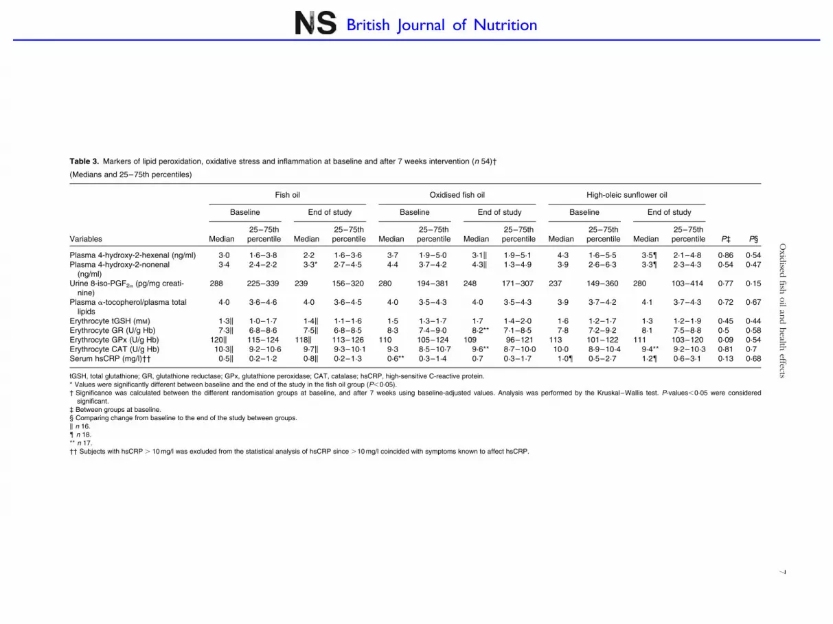
Table 3. Markers of lipid peroxidation, oxidative stress and inflammation at baseline and after 7 weeks intervention (n 54)†
(Medians and 25–75th percentiles)
Fish oil Oxidised fish oil High-oleic sunflower oil
Baseline End of study Baseline End of study Baseline End of study
Variables Median25–75thpercentile Median
25–75thpercentile Median
25–75thpercentile Median
25–75thpercentile Median
25–75thpercentile Median
25–75thpercentile P‡ P§
Plasma 4-hydroxy-2-hexenal (ng/ml) 3·0 1·6–3·8 2·2 1·6–3·6 3·7 1·9–5·0 3·1k 1·9–5·1 4·3 1·6–5·5 3·5{ 2·1–4·8 0·86 0·54Plasma 4-hydroxy-2-nonenal
(ng/ml)3·4 2·4–2·2 3·3* 2·7–4·5 4·4 3·7–4·2 4·3k 1·3–4·9 3·9 2·6–6·3 3·3{ 2·3–4·3 0·54 0·47
Urine 8-iso-PGF2a (pg/mg creati-nine)
288 225–339 239 156–320 280 194–381 248 171–307 237 149–360 280 103–414 0·77 0·15
Plasma a-tocopherol/plasma totallipids
4·0 3·6–4·6 4·0 3·6–4·5 4·0 3·5–4·3 4·0 3·5–4·3 3·9 3·7–4·2 4·1 3·7–4·3 0·72 0·67
Erythrocyte tGSH (mM) 1·3k 1·0–1·7 1·4k 1·1–1·6 1·5 1·3–1·7 1·7 1·4–2·0 1·6 1·2–1·7 1·3 1·2–1·9 0·45 0·44Erythrocyte GR (U/g Hb) 7·3k 6·8–8·6 7·5k 6·8–8·5 8·3 7·4–9·0 8·2** 7·1–8·5 7·8 7·2–9·2 8·1 7·5–8·8 0·5 0·58Erythrocyte GPx (U/g Hb) 120k 115–124 118k 113–126 110 105–124 109 96–121 113 101–122 111 103–120 0·09 0·54Erythrocyte CAT (U/g Hb) 10·3k 9·2–10·6 9·7k 9·3–10·1 9·3 8·5–10·7 9·6** 8·7–10·0 10·0 8·9–10·4 9·4** 9·2–10·3 0·81 0·7Serum hsCRP (mg/l)†† 0·5k 0·2–1·2 0·8k 0·2–1·3 0·6** 0·3–1·4 0·7 0·3–1·7 1·0{ 0·5–2·7 1·2{ 0·6–3·1 0·13 0·68
tGSH, total glutathione; GR, glutathione reductase; GPx, glutathione peroxidase; CAT, catalase; hsCRP, high-sensitive C-reactive protein.* Values were significantly different between baseline and the end of the study in the fish oil group (P,0·05).† Significance was calculated between the different randomisation groups at baseline, and after 7 weeks using baseline-adjusted values. Analysis was performed by the Kruskal–Wallis test. P-values,0·05 were considered
significant.‡ Between groups at baseline.§ Comparing change from baseline to the end of the study between groups.k n 16.{ n 18.** n 17.†† Subjects with hsCRP . 10 mg/l was excluded from the statistical analysis of hsCRP since .10 mg/l coincided with symptoms known to affect hsCRP.
Oxid
ised
fish
oil
and
health
effe
cts7
British Journal of Nutrition
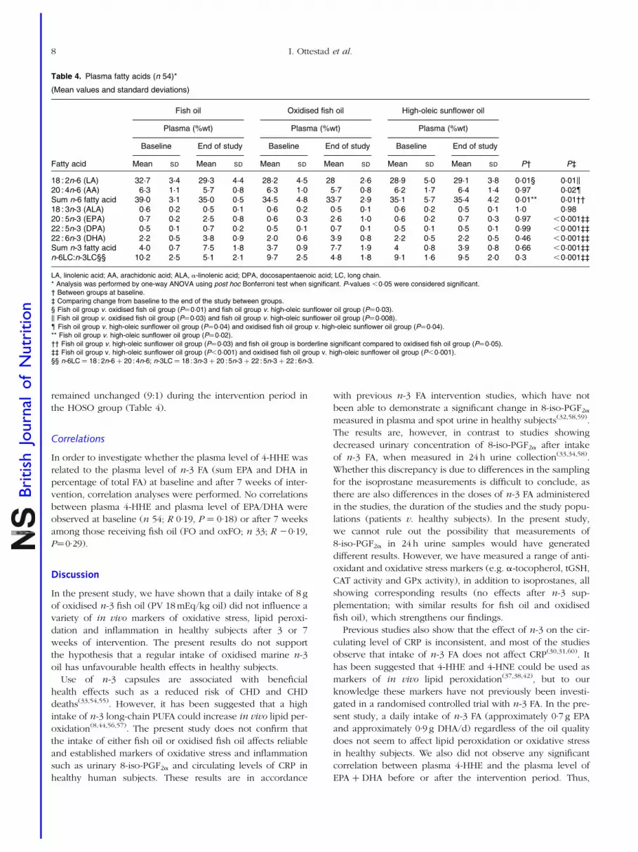
remained unchanged (9:1) during the intervention period in
the HOSO group (Table 4).
Correlations
In order to investigate whether the plasma level of 4-HHE was
related to the plasma level of n-3 FA (sum EPA and DHA in
percentage of total FA) at baseline and after 7 weeks of inter-
vention, correlation analyses were performed. No correlations
between plasma 4-HHE and plasma level of EPA/DHA were
observed at baseline (n 54; R 0·19, P ¼ 0·18) or after 7 weeks
among those receiving fish oil (FO and oxFO; n 33; R 20·19,
P¼0·29).
Discussion
In the present study, we have shown that a daily intake of 8 g
of oxidised n-3 fish oil (PV 18 mEq/kg oil) did not influence a
variety of in vivo markers of oxidative stress, lipid peroxi-
dation and inflammation in healthy subjects after 3 or 7
weeks of intervention. The present results do not support
the hypothesis that a regular intake of oxidised marine n-3
oil has unfavourable health effects in healthy subjects.
Use of n-3 capsules are associated with beneficial
health effects such as a reduced risk of CHD and CHD
deaths(33,54,55). However, it has been suggested that a high
intake of n-3 long-chain PUFA could increase in vivo lipid per-
oxidation(8,44,56,57). The present study does not confirm that
the intake of either fish oil or oxidised fish oil affects reliable
and established markers of oxidative stress and inflammation
such as urinary 8-iso-PGF2a and circulating levels of CRP in
healthy human subjects. These results are in accordance
with previous n-3 FA intervention studies, which have not
been able to demonstrate a significant change in 8-iso-PGF2a
measured in plasma and spot urine in healthy subjects(32,58,59).
The results are, however, in contrast to studies showing
decreased urinary concentration of 8-iso-PGF2a after intake
of n-3 FA, when measured in 24 h urine collection(33,34,58).
Whether this discrepancy is due to differences in the sampling
for the isoprostane measurements is difficult to conclude, as
there are also differences in the doses of n-3 FA administered
in the studies, the duration of the studies and the study popu-
lations (patients v. healthy subjects). In the present study,
we cannot rule out the possibility that measurements of
8-iso-PGF2a in 24 h urine samples would have generated
different results. However, we have measured a range of anti-
oxidant and oxidative stress markers (e.g. a-tocopherol, tGSH,
CAT activity and GPx activity), in addition to isoprostanes, all
showing corresponding results (no effects after n-3 sup-
plementation; with similar results for fish oil and oxidised
fish oil), which strengthens our findings.
Previous studies also show that the effect of n-3 on the cir-
culating level of CRP is inconsistent, and most of the studies
observe that intake of n-3 FA does not affect CRP(30,31,60). It
has been suggested that 4-HHE and 4-HNE could be used as
markers of in vivo lipid peroxidation(37,38,42), but to our
knowledge these markers have not previously been investi-
gated in a randomised controlled trial with n-3 FA. In the pre-
sent study, a daily intake of n-3 FA (approximately 0·7 g EPA
and approximately 0·9 g DHA/d) regardless of the oil quality
does not seem to affect lipid peroxidation or oxidative stress
in healthy subjects. We also did not observe any significant
correlation between plasma 4-HHE and the plasma level of
EPA þ DHA before or after the intervention period. Thus,
Table 4. Plasma fatty acids (n 54)*
(Mean values and standard deviations)
Fish oil Oxidised fish oil High-oleic sunflower oil
Plasma (%wt) Plasma (%wt) Plasma (%wt)
Baseline End of study Baseline End of study Baseline End of study
Fatty acid Mean SD Mean SD Mean SD Mean SD Mean SD Mean SD P† P‡
18 : 2n-6 (LA) 32·7 3·4 29·3 4·4 28·2 4·5 28 2·6 28·9 5·0 29·1 3·8 0·01§ 0·01k20 : 4n-6 (AA) 6·3 1·1 5·7 0·8 6·3 1·0 5·7 0·8 6·2 1·7 6·4 1·4 0·97 0·02{Sum n-6 fatty acid 39·0 3·1 35·0 0·5 34·5 4·8 33·7 2·9 35·1 5·7 35·4 4·2 0·01** 0·01††18 : 3n-3 (ALA) 0·6 0·2 0·5 0·1 0·6 0·2 0·5 0·1 0·6 0·2 0·5 0·1 1·0 0·9820 : 5n-3 (EPA) 0·7 0·2 2·5 0·8 0·6 0·3 2·6 1·0 0·6 0·2 0·7 0·3 0·97 ,0·001‡‡22 : 5n-3 (DPA) 0·5 0·1 0·7 0·2 0·5 0·1 0·7 0·1 0·5 0·1 0·5 0·1 0·99 ,0·001‡‡22 : 6n-3 (DHA) 2·2 0·5 3·8 0·9 2·0 0·6 3·9 0·8 2·2 0·5 2·2 0·5 0·46 ,0·001‡‡Sum n-3 fatty acid 4·0 0·7 7·5 1·8 3·7 0·9 7·7 1·9 4 0·8 3·9 0·8 0·66 ,0·001‡‡n-6LC:n-3LC§§ 10·2 2·5 5·1 2·1 9·7 2·5 4·8 1·8 9·1 1·6 9·5 2·0 0·3 ,0·001‡‡
LA, linolenic acid; AA, arachidonic acid; ALA, a-linolenic acid; DPA, docosapentaenoic acid; LC, long chain.* Analysis was performed by one-way ANOVA using post hoc Bonferroni test when significant. P-values ,0·05 were considered significant.† Between groups at baseline.‡ Comparing change from baseline to the end of the study between groups.§ Fish oil group v. oxidised fish oil group (P¼0·01) and fish oil group v. high-oleic sunflower oil group (P¼0·03).k Fish oil group v. oxidised fish oil group (P¼0·03) and fish oil group v. high-oleic sunflower oil group (P¼0·008).{ Fish oil group v. high-oleic sunflower oil group (P¼0·04) and oxidised fish oil group v. high-oleic sunflower oil group (P¼0·04).** Fish oil group v. high-oleic sunflower oil group (P¼0·02).†† Fish oil group v. high-oleic sunflower oil group (P¼0·03) and fish oil group is borderline significant compared to oxidised fish oil group (P¼0·05).‡‡ Fish oil group v. high-oleic sunflower oil group (P,0·001) and oxidised fish oil group v. high-oleic sunflower oil group (P,0·001).§§ n-6LC ¼ 18 : 2n-6 þ 20 : 4n-6; n-3LC ¼ 18 : 3n-3 þ 20 : 5n-3 þ 22 : 5n-3 þ 22 : 6n-3.
I. Ottestad et al.8
British
Journal
ofNutrition

the present results are in contrast to a previous study in which
the plasma level of 4-HHE was significantly increased after
intake of capsules containing high compared to low doses
of DHA (0·8 and 1·6 g/d v. 0·2 and 0·6 g/d, respectively),
while 4-HNE remained unchanged(41). Whether 4-HHE is a
reliable marker of in vivo lipid peroxidation and whether
the plasma level can be affected by intake of n-3 FA or oxi-
dised lipids is still uncertain and needs to be further
investigated.
GSH is the most important endogenous cellular antioxidant
with the ability to degrade both primary and secondary lipid
oxidation products, and a high capacity of GSH provides opti-
mal activity of GPx and GR(16–18). Plasma level of GSH and the
activity of endogenous antioxidants in erythrocytes have been
used to assess changes in oxidative stress status in dietary
intervention studies and in diseases linked to oxidative
stress(16,47,61,62). Furthermore, cell studies have demonstrated
that GST and GPx are regulators of the homoeostasis of alde-
hydes, such as 4-HNE, and it has been suggested that these
secondary oxidation products could inactivate antioxidant
enzyme activity(63,64). Results from clinical trials with n-3 FA
in patients with oxidative stress-related diseases are, however,
inconclusive. In some studies, antioxidant enzyme activity
is increased, whereas in others it is decreased, and in some
studies the activity remains unchanged after n-3 FA sup-
plementation(65–69). GSH, GPx and CAT are suggested to
play a pivotal role in detoxification of hydroperoxides. We
found no significant changes in plasma level of tGSH and a-
tocoferol or in the enzymatic activity of GR, GPx and CAT.
These findings indicate that the daily intake of FO or oxFO
oil did not affect the in vivo antioxidant defence system. In
the present study, we show that the plasma levels of EPA
and DHA in both fish oil groups were significantly increased;
the increased levels remained stable after 3 and 7 weeks of
intervention. The relative change in plasma EPA and DHA
after daily intake of 1·6 g n-3 FA confirms the findings of
other n-3 supplementation studies(70–72). This clearly shows
that the content of hydroperoxides in fish oil supplements,
even with a PV that exceeds the European Pharmacopeia
for marine n-3 oils, does not apparently influence the
plasma level of n-3 FA. The present understanding of how
dietary oxidised lipids are absorbed in human subjects is
limited(16,17,21,22,73,74). It has been suggested that the gastroin-
testinal tract acts as a barrier against oxidised lipids and that
hydroperoxides from the diet are not transferred into the
circulatory system(64,74,75); we do not know whether the oxi-
dised lipids from the oxidised fish oil were eliminated in the
gastrointestinal tract and prevented from reaching the circula-
tory system. Results from animal and cell studies demonstrate
that hydroperoxides are converted into secondary oxidation
products during the digestion process, and that these com-
ponents are being absorbed, at least partially(75,76). Human
studies have demonstrated that intake of high doses of oxi-
dised vegetable oils (approximately 50–100 g) alters the endo-
thelial function and increases postprandial level of lipid
peroxides in plasma and chylomicrons(77–82). In the present
study, however, we were not able to detect any increase in
secondary oxidation products (4-HHE, 4-HNE or 8-iso-PGF2a)
after 3 or 7 weeks of intervention, nor in chylomicrons after
intake of one dose with 9 g of oxidised fish oil (I Ottestad,
unpublished results). Our findings contrast with previously
published human(45,77–79,81–83) and animal studies(75,84–86)
investigating the effects of oxidised oil. The discrepancy
among the human studies could, at least partially, be
explained by type of oil (vegetable v. fish oil), variation in
the contribution of hydroperoxides and by the study design
(postprandial studies v. intervention study). The relevance of
animal studies in predicting effects in human subjects remains
controversial, as the results from animal studies are not always
reproduced in human subjects. In the present study, we did
not observe any changes in serum TAG. The TAG-lowering
effect from n-3 FA has been shown to be dependent on the
dose and on baseline TAG level(60). We have previously
shown, in line with the present results, that an intake of
0·9 g EPA þ DHA given as fish oil for 7 weeks had no effect
on TAG levels in a healthy non-hyperlipidaemic population
with baseline levels of TAG #1·0 mM(59). The present study
has several strengths such as the study design; using a blinded
randomised controlled study design; and a 3-week fully con-
trolled diet period. Moreover, a high compliance was deter-
mined by capsule count, which was supported by the
significant increase in plasma level of EPA and DHA after
intake of fish oil. Given the few and discrete side effects
reported, the capsules seemed to be well tolerated and the
results in the present study were most probably not affected
by any compliance issue. The fish oils used in the present
study was provided from one single batch, and the use of
highly specific and sensitive methods (GC, GC–MS and
HPLC) to measure several biological markers strengthens our
findings further. Among the limitations of the present study
are the relative short intervention and the fact that the required
sample size was estimated on the expected change in plasma
EPA þ DHA. In addition, whether the composition of the pri-
mary and secondary oxidation products generated during the
oxidation process at our laboratory reflects the content of oxi-
dation products that could be formed in n-3 supplements
available to the customers is uncertain.
To our knowledge, this is the first human study investigating
the health effects of intake of oxidised n-3 oil. The present
study shows that a variety of in vivo markers of oxidative
stress, lipid peroxidation and inflammation are not signifi-
cantly affected in healthy subjects after the intake of 8 g of
highly oxidised fish oil per day for 3 and 7 weeks. Accumu-
lation of lipid peroxidation products has been associated
with the pathogenesis of inflammation and oxidative stress-
related diseases(14,21,22), but in the present study the oxidative
stress status remained unchanged. The relatively short dur-
ation of the present study does not allow us to conclude
that a long-term intake of oxidised n-3 supplements does
not have unfavourable health effects. Whether these results
are applicable for other marine oils remains uncertain and
to what degree the present results are valid in subjects with
elevated levels of inflammation and/or oxidative stress needs
to be further investigated in larger studies before firm
conclusions can be drawn.
Oxidised fish oil and health effects 9
British
Journal
ofNutrition

Acknowledgements
The present study was supported by TINE SA, Centre for
Research and Development (Oslo, Norway), and The
Research Council of Norway (Oslo, Norway). The cod liver
oil used in the present study, TINE EPADHA OIL Oil 1200,
was produced by Martitex AS (Sortland, Norway), and pro-
vided by TINE SA. Maritex AS is a fully owned subsidiary of
TINE SA. The authors thank the research participants for
their dedication to the project. They are also grateful to
Ellen Raael, Daniel Bødtker-Lund, Mona Lundby, Grete Skjeg-
stad, Anne Kristin Sundstøl and Kari Tande-Hansen at Aker-
shus University College and Stine Grimmer, Elin-Merete
Nicolaisen and Frank Lundby at Nofima Mat for valuable
assistance in this project, and Fjordland AS for providing
food products. The study was registered at www.clinicaltrials.-
gov, ID no. NCT01034423. I. O., G. V., A. N., K. R., K. W. B.,
B. N., L. F. A., M. C. M., K. B. H. and S. M. U. designed the
research (project conception, development of overall research
plan and study oversight); I. O., G. V., J.-E. H., A. N., K. R.,
K. B. H., M. C. M., G. R.-H. and S. M. U. conducted research
(hands-on conduct of the experiments and data collection);
G. V., A. N., K. W. B. and B. N. provided essential reagents
or provided essential materials (applies to authors who con-
tributed by providing animals, constructs, databases, etc.,
necessary for the research); I. O., K. B. H. and S. M. U. ana-
lysed data or performed statistical analysis; I. O., G. V., K. R.,
M. C. M., J.-E. H., A. N., G. R.-H., B. N., K. W. B., L. F. A., K.
B. H. and S. M. U. wrote the paper (only authors who made
a major contribution); and I. O., K. B. H. and S. M. U. had pri-
mary responsibility for the final content. K. W. B. is the clinical
nutritionist/project manager and B. N. is the research manager
on ingredients in TINE SA R&D Center (Oslo, Norway). They
have no financial interest to declare. The remaining authors
declare no conflict of interest.
References
1. Skeaff CM & Miller J (2009) Dietary fat and coronary heartdisease: summary of evidence from prospective cohort andrandomised controlled trials. Ann Nutr Metab 55, 173–201.
2. Kris-Etherton PM, Harris WS & Appel LJ (2003) Fish con-sumption, fish oil, omega-3 fatty acids, and cardiovasculardisease. Arterioscler Thromb Vasc Biol 23, 20–30.
3. Lichtenstein AH, Appel LJ, Brands M, et al. (2006) Summaryof American Heart Association Diet and Lifestyle Recommen-dations revision 2006. Arterioscler Thromb Vasc Biol 26,2186–2191.
4. van Kuijk FJ, Holte LL & Dratz EA (1990) 4-Hydroxyhexenal:a lipid peroxidation product derived from oxidized docosa-hexaenoic acid. Biochim Biophys Acta 1043, 116–118.
5. Olsen E, Vogt G, Saarem K, et al. (2005) Autoxidation of codliver oil with tocopherol and ascorbyl palmitate. JAOCS 82,97–103.
6. Pryor WA & Porter NA (1990) Suggested mechanisms for theproduction of 4-hydroxy-2-nonenal from the autoxidation ofpolyunsaturated fatty acids. Free Radic Biol Med 8, 541–543.
7. Yin H, Brooks JD, Gao L, et al. (2007) Identification of novelautoxidation products of the omega-3 fatty acid eicosapen-taenoic acid in vitro and in vivo. J Biol Chem 282,29890–29901.
8. Esterbauer H, Schaur RJ & Zollner H (1991) Chemistry andbiochemistry of 4-hydroxynonenal, malonaldehyde andrelated aldehydes. Free Radic Biol Med 11, 81–128.
9. Council of Europe (2008) European Pharmacopoeia, 6th ed.Supplement 6.3. Strasbourg: Council of Europe.
10. Luley C, Klein B, Hanisch M, et al. (1988) Fatty acid compo-sition and degree of peroxidation in fish oil and cod liver oilpreparations. Arzneimittelforschung 38, 1783–1786.
11. Shukla VK & Perkins EG (1991) The presence of oxidativepolymeric materials in encapsulated fish oils. Lipids 26,23–26.
12. Fierens C & Corthout J (2007) Omega-3 fatty acid prep-arations – a comparative study. J Pharm Belg 62, 115–119.
13. Kolanowski W (2010) Omega-3 LC PUFA contents and oxi-dative stability of encapsulated fish oil dietary supplements.Int J Food Prop 13, 498–511.
14. Halliwell B & Chirico S (1993) Lipid peroxidation: its mech-anism, measurement, and significance. Am J Clin Nutr 57,715S–724S.
15. Blomhoff R (2005) Dietary antioxidants and cardiovasculardisease. Curr Opin Lipidol 16, 47–54.
16. Aw TY (2005) Intestinal glutathione: determinant of mucosalperoxide transport, metabolism, and oxidative susceptibility.Toxicol Appl Pharmacol 204, 320–328.
17. Wingler K, Muller C, Schmehl K, et al. (2000) Gastrointestinalglutathione peroxidase prevents transport of lipid hydroper-oxides in CaCo-2 cells. Gastroenterology 119, 420–430.
18. Dickinson DA & Forman HJ (2002) Cellular glutathione andthiols metabolism. Biochem Pharmacol 64, 1019–1026.
19. Ursini F, Zamburlini A, Cazzolato G, et al. (1998) Postpran-dial plasma lipid hydroperoxides: a possible link betweendiet and atherosclerosis. Free Radic Biol Med 25, 250–252.
20. Staprans I, Pan XM, Rapp JH, et al. (2005) The role of dietaryoxidized cholesterol and oxidized fatty acids in the develop-ment of atherosclerosis. Mol Nutr Food Res 49, 1075–1082.
21. Cohn JS (2002) Oxidized fat in the diet, postprandial lipae-mia and cardiovascular disease. Curr Opin Lipidol 13,19–24.
22. Bowen PE & Borthakur G (2004) Postprandial lipid oxidationand cardiovascular disease risk. Curr Atheroscler Rep 6,477–484.
23. Guichardant M & Lagarde M (2009) Analysis of biomarkersfrom lipid peroxidation: a comparative study. Eur J LipidSci Technol 111, 75–82.
24. Montuschi P, Barnes PJ & Roberts LJ (2004) Isoprostanes:markers and mediators of oxidative stress. FASEB J 18,1791–1800.
25. Minuz P, Fava C & Lechi A (2006) Lipid peroxidation, iso-prostanes and vascular damage. Pharmacol Rep 58, 57–68.
26. Pratico D, Rokach J, Lawson J, et al. (2004) F2-isoprostanesas indices of lipid peroxidation in inflammatory diseases.Chem Phys Lipids 128, 165–171.
27. Pearson TA, Mensah GA, Alexander RW, et al. (2003) Mar-kers of inflammation and cardiovascular disease: applicationto clinical and public health practice: a statement for health-care professionals from the Centers for Disease Control andPrevention and the American Heart Association. Circulation107, 499–511.
28. Roberts LJ & Morrow JD (2000) Measurement of F(2)-iso-prostanes as an index of oxidative stress in vivo. FreeRadic Biol Med 28, 505–513.
29. Basu S (2008) F2-isoprostanes in human health and diseases:from molecular mechanisms to clinical implications. Anti-oxid Redox Signal 10, 1405–1434.
30. Myhrstad MC, Retterstol K, Telle-Hansen VH, et al. (2011)Effect of marine n-3 fatty acids on circulating inflammatory
I. Ottestad et al.10
British
Journal
ofNutrition

markers in healthy subjects and subjects with cardiovascularrisk factors. Inflamm Res 60, 309–319.
31. Basu A, Devaraj S & Jialal I (2006) Dietary factors that pro-mote or retard inflammation. Arterioscler Thromb Vasc Biol26, 995–1001.
32. Higdon JV, Liu J, Du SH, et al. (2000) Supplementation ofpostmenopausal women with fish oil rich in eicosapentae-noic acid and docosahexaenoic acid is not associated withgreater in vivo lipid peroxidation compared with oils richin oleate and linoleate as assessed by plasma malondialde-hyde and F(2)-isoprostanes. Am J Clin Nutr 72, 714–722.
33. Mori TA (2004) Effect of fish and fish oil-derived omega-3fatty acids on lipid oxidation. Redox Rep 9, 193–197.
34. Nalsen C, Vessby B, Berglund L, et al. (2006) Dietary (n-3)fatty acids reduce plasma F2-isoprostanes but not prostaglan-din F2alpha in healthy humans. J Nutr 136, 1222–1228.
35. Long EK & Picklo MJ Sr (2010) trans-4-Hydroxy-2-hexenal, aproduct of n-3 fatty acid peroxidation: make some roomHNE. Free Radic Biol Med 49, 1–8.
36. Draper HH, Csallany AS & Hadley M (2000) Urinary alde-hydes as indicators of lipid peroxidation in vivo. FreeRadic Biol Med 29, 1071–1077.
37. Zarkovic N (2003) 4-Hydroxynonenal as a bioactive markerof pathophysiological processes. Mol Aspects Med 24,281–291.
38. Guichardant M, Bacot S, Moliere P, et al. (2006) Hydroxy-alkenals from the peroxidation of n-3 and n-6 fatty acidsand urinary metabolites. Prostaglandins Leukot Essent FattyAcids 75, 179–182.
39. Kitase A, Hino K, Furutani T, et al. (2005) In situ detection ofoxidized n-3 polyunsaturated fatty acids in chronic hepatitisC: correlation with hepatic steatosis. J Gastroenterol 40,617–624.
40. Yamada S, Funada T, Shibata N, et al. (2004) Protein-bound4-hydroxy-2-hexenal as a marker of oxidized n-3 polyunsa-turated fatty acids. J Lipid Res 45, 626–634.
41. Calzada C, Colas R, Guillot N, et al. (2010) Subgram dailysupplementation with docosahexaenoic acid protects low-density lipoproteins from oxidation in healthy men. Athero-sclerosis 208, 467–472.
42. Spickett CM, Wiswedel I, Siems W, et al. (2010) Advances inmethods for the determination of biologically relevant lipidperoxidation products. Free Radic Res 44, 1172–1202.
43. Turner R, McLean CH & Silvers KM (2006) Are the healthbenefits of fish oils limited by products of oxidation? NutrRes Rev 19, 53–62.
44. Bays HE (2007) Safety considerations with omega-3 fatty acidtherapy. Am J Cardiol 99, 35C–43C.
45. European Food Safety Authority (2010) Scientific opinion onfish oil for human consumption. Food hygene, includingrancidity. EFSA J 8, 1874.
46. Maenpaa H, Heinonen OP & Manninen V (1991) Medicationcompliance and serum lipid changes in the Helsinki HeartStudy. Br J Clin Pharmacol 32, 409–415.
47. Bohn SK, Smeland S, Sakhi AK, et al. (2006) Post-radiotherapyplasma total glutathione is associated to outcome in patientswith head and neck squamous cell carcinoma. Cancer Lett238, 240–247.
48. Bastani NE, Gundersen TE & Blomhoff R (2009) Determi-nation of 8-epi PGF(2alpha) concentrations as a biomarkerof oxidative stress using triple-stage liquid chromatog-raphy/tandem mass spectrometry. Rapid Commun MassSpectrom 23, 2885–2890.
49. Luo XP, Yazdanpanah M, Bhooi N, et al. (1995) Determi-nation of aldehydes and other lipid peroxidation products
in biological samples by gas chromatography–mass spec-
trometry. Anal Biochem 228, 294–298.50. Panfili G, Fratianni A & Irano M (2003) Normal phase high-
performance liquid chromatography method for the determi-
nation of tocopherols and tocotrienols in cereals. J Agric
Food Chem 51, 3940–3944.51. Wheeler CR, Salzman JA, Elsayed NM, et al. (1990) Auto-
mated assays for superoxide dismutase, catalase, glutathione
peroxidase, and glutathione reductase activity. Anal Bio-
chem 184, 193–199.52. Bligh EG & Dyer WJ (1959) A rapid method of total lipid
extraction and purification. Can J Biochem Physiol 37,
911–917.53. Olsen E, Vogt G, Veberg A, et al. (2005) Analysis of early
lipid oxidation in smoked, comminuted pork or poultry sau-
sages with spices. J Agric Food Chem 53, 7448–7457.54. Gruppo Italiano per lo Studio della Sopravvivenza nell’In-
farto miocardico (1999) Dietary supplementation with n-3
polyunsaturated fatty acids and vitamin E after myocardial
infarction: results of the GISSI-Prevenzione trial. Lancet
354, 447–455.55. Yokoyama M, Origasa H, Matsuzaki M, et al. (2007) Effects of
eicosapentaenoic acid on major coronary events in hyperch-
olesterolaemic patients ( JELIS): a randomised open-label,
blinded endpoint analysis. Lancet 369, 1090–1098.56. Meydani M, Natiello F, Goldin B, et al. (1991) Effect of long-
term fish oil supplementation on vitamin E status and lipid
peroxidation in women. J Nutr 121, 484–491.57. Harats D, Dabach Y, Hollander G, et al. (1991) Fish oil inges-
tion in smokers and nonsmokers enhances peroxidation of
plasma lipoproteins. Atherosclerosis 90, 127–139.58. Tholstrup T, Hellgren LI, Petersen M, et al. (2004) A solid
dietary fat containing fish oil redistributes lipoprotein sub-
classes without increasing oxidative stress in men. J Nutr
134, 1051–1057.59. Ulven SM, Kirkhus B, Lamglait A, et al. (2011) Metabolic
effects of krill oil are essentially similar to those of fish oil
but at lower dose of EPA and DHA, in healthy volunteers.
Lipids 46, 37–46.60. Balk EM, Lichtenstein AH, Chung M, et al. (2006) Effects of
omega-3 fatty acids on serum markers of cardiovascular dis-
ease risk: a systematic review. Atherosclerosis 189, 19–30.61. Freese R, Dragsted LO, Loft S, et al. (2008) No effect on oxi-
dative stress biomarkers by modified intakes of polyunsatu-
rated fatty acids or vegetables and fruit. Eur J Clin Nutr 62,
1151–1153.62. Ravn-Haren G, Bugel S, Krath BN, et al. (2008) A short-term
intervention trial with selenate, selenium-enriched yeast and
selenium-enriched milk: effects on oxidative defence regu-
lation. Br J Nutr 99, 883–892.63. Miyamoto Y, Koh YH, Park YS, et al. (2003) Oxidative stress
caused by inactivation of glutathione peroxidase and adap-
tive responses. Biol Chem 384, 567–574.64. Awasthi YC, Yang Y, Tiwari NK, et al. (2004) Regulation
of 4-hydroxynonenal-mediated signaling by glutathione
S-transferases. Free Radic Biol Med 37, 607–619.65. Mabile L, Piolot A, Boulet L, et al. (2001) Moderate intake of
n-3 fatty acids is associated with stable erythrocyte resistance
to oxidative stress in hypertriglyceridemic subjects. Am J Clin
Nutr 74, 449–456.66. Bouzidi N, Mekki K, Boukaddoum A, et al. (2010) Effects of
omega-3 polyunsaturated fatty-acid supplementation on
redox status in chronic renal failure patients with dyslipide-
mia. J Ren Nutr 20, 321–328.
Oxidised fish oil and health effects 11
British
Journal
ofNutrition

67. Barbosa DS, Cecchini R, El Kadri MZ, et al. (2003) Decreasedoxidative stress in patients with ulcerative colitis supplementedwith fish oil omega-3 fatty acids. Nutrition 19, 837–842.
68. Kesavulu MM, Kameswararao B, Apparao C, et al. (2002)Effect of omega-3 fatty acids on lipid peroxidation and anti-oxidant enzyme status in type 2 diabetic patients. DiabetesMetab 28, 20–26.
69. Kesavulu MM, Rao BK, Giri R, et al. (2001) Lipid peroxi-dation and antioxidant enzyme status in type 2 diabeticswith coronary heart disease. Diabetes Res Clin Pract 53,33–39.
70. Cao J, Schwichtenberg KA, Hanson NQ, et al. (2006) Incor-poration and clearance of omega-3 fatty acids in erythrocytemembranes and plasma phospholipids. Clin Chem 52,2265–2272.
71. Flaten H, Hostmark AT, Kierulf P, et al. (1990) Fish-oil con-centrate: effects on variables related to cardiovascular dis-ease. Am J Clin Nutr 52, 300–306.
72. Harris WS & Von SC (2004) The omega-3 index: a new riskfactor for death from coronary heart disease? Prev Med 39,212–220.
73. Wingler K & Brigelius-Flohe R (1999) Gastrointestinal gluta-thione peroxidase. Biofactors 10, 245–249.
74. Chu FF, Esworthy RS & Doroshow JH (2004) Role ofSe-dependent glutathione peroxidases in gastrointestinalinflammation and cancer. Free Radic Biol Med 36,1481–1495.
75. Kanazawa K & Ashida H (1998) Dietary hydroperoxides oflinoleic acid decompose to aldehydes in stomach beforebeing absorbed into the body. Biochim Biophys Acta1393, 349–361.
76. Bull AW & Bronstein JC (1990) Production of unsaturatedcarbonyl compounds during metabolism of hydroperoxyfatty acids by colonic homogenates. Carcinogenesis 11,1699–1704.
77. Naruszewicz M, Wozny E, Mirkiewicz E, et al. (1987) Theeffect of thermally oxidized soya bean oil on metabolism
of chylomicrons. Increased uptake and degradation of oxi-dized chylomicrons in cultured mouse macrophages. Ather-osclerosis 66, 45–53.
78. Staprans I, Rapp JH, Pan XM, et al. (1994) Oxidized lipids inthe diet are a source of oxidized lipid in chylomicrons ofhuman serum. Arterioscler Thromb 14, 1900–1905.
79. Staprans I, Hardman DA, Pan XM, et al. (1999) Effect of oxi-dized lipids in the diet on oxidized lipid levels in postpran-dial serum chylomicrons of diabetic patients. Diabetes Care22, 300–306.
80. Wallace AJ, Sutherland WH, Mann JI, et al. (2001) The effectof meals rich in thermally stressed olive and safflower oils onpostprandial serum paraoxonase activity in patients with dia-betes. Eur J Clin Nutr 55, 951–958.
81. Sutherland WH, de Jong SA, Walker RJ, et al. (2002) Effect ofmeals rich in heated olive and safflower oils on oxidation ofpostprandial serum in healthy men. Atherosclerosis 160,195–203.
82. Sutherland WH, de Jong SA, Hessian PA, et al. (2010) Inges-tion of native and thermally oxidized polyunsaturated fatsacutely increases circulating numbers of endothelial micro-particles. Metabolism 59, 446–453.
83. Wallace JM, McCabe AJ, Robson PJ, et al. (2000) Bioavailabil-ity of n-3 polyunsaturated fatty acids (PUFA) in foodsenriched with microencapsulated fish oil. Ann Nutr Metab44, 157–162.
84. Suomela JP, Ahotupa M & Kallio H (2005) Triacylglyceroloxidation in pig lipoproteins after a diet rich in oxidized sun-flower seed oil. Lipids 40, 437–444.
85. Suomela JP, Ahotupa M, Sjovall O, et al. (2004) Diet and lipo-protein oxidation: analysis of oxidized triacylglycerols in piglipoproteins. Lipids 39, 639–647.
86. Song JH, Fujimoto K & Miyazawa T (2000) Polyunsaturated(n-3) fatty acids susceptible to peroxidation are increasedin plasma and tissue lipids of rats fed docosahexaenoicacid-containing oils. J Nutr 130, 3028–3033.
I. Ottestad et al.12
British
Journal
ofNutrition


