Embed Size (px)
Citation preview

Oxidative Stress Induced Inflammation InitiatesFunctional Decline of Tear ProductionYuichi Uchino1,2, Tetsuya Kawakita1*, Masaki Miyazawa2, Takamasa Ishii2, Hiromi Onouchi2,
Kayo Yasuda2, Yoko Ogawa1, Shigeto Shimmura1, Naoaki Ishii2, Kazuo Tsubota1
1 Department of Ophthalmology, Keio University School of Medicine, Tokyo, Japan, 2 Department of Molecular Life Science, Tokai University School of Medicine,
Kanagawa, Japan
Abstract
Oxidative damage and inflammation are proposed to be involved in an age-related functional decline of exocrine glands.However, the molecular mechanism of how oxidative stress affects the secretory function of exocrine glands is unclear. Wedeveloped a novel mev-1 conditional transgenic mouse model (Tet-mev-1) using a modified tetracycline system (Tet-On/Offsystem). This mouse model demonstrated decreased tear production with morphological changes including leukocyticinfiltration and fibrosis. We found that the mev-1 gene encodes Cyt-1, which is the cytochrome b560 large subunit ofsuccinate-ubiquinone oxidoreductase in complex II of mitochondria (homologous to succinate dehydrogenase C subunit(SDHC) in humans). The mev-1 gene induced excessive oxidative stress associated with ocular surface epithelial damage anda decrease in protein and aqueous secretory function. This new model provides evidence that mitochondrial oxidativedamage in the lacrimal gland induces lacrimal dysfunction resulting in dry eye disease. Tear volume in Tet-mev-1 mice waslower than in wild type mice and histopathological analyses showed the hallmarks of lacrimal gland inflammation byintense mononuclear leukocytic infiltration and fibrosis in the lacrimal gland of Tet-mev-1 mice. These findings stronglysuggest that oxidative stress can be a causative factor for the development of dry eye disease.
Citation: Uchino Y, Kawakita T, Miyazawa M, Ishii T, Onouchi H, et al. (2012) Oxidative Stress Induced Inflammation Initiates Functional Decline of TearProduction. PLoS ONE 7(10): e45805. doi:10.1371/journal.pone.0045805
Editor: Henrik Einwaechter, Klinikum rechts der Isar der TU Munchen, Germany
Received February 16, 2012; Accepted August 24, 2012; Published October 5, 2012
Copyright: � 2012 Uchino et al. This is an open-access article distributed under the terms of the Creative Commons Attribution License, which permitsunrestricted use, distribution, and reproduction in any medium, provided the original author and source are credited.
Funding: This work was supported by Grant-in-Aid for Young Scientists (B) (22791692) from the Ministry of Education, Culture, Sports, Science and Technology ofJapan. The funders had no role in study design, data collection and analysis, decision to publish, or preparation of the manuscript.
Competing Interests: The authors have declared that no competing interests exist.
* E-mail: [email protected]
Introduction
Dry eye disease is a deficiency in tear instability, mainly induced
by low tear production, and a functional decline of the lacrimal
gland induced by age-related chronic inflammation [1–3]. Such
age-related chronic inflammation supported the reported preva-
lence of dry eye disease [4–8]. However, the molecular mechanism
of age-related lacrimal gland inflammation is unclear. The main
cause of chronic inflammation is postulated to involve oxidative
stress, and the main endogenous source of oxidative stress is the
electron transport chain in mitochondria [9]. The mev-1 mutant of
the nematode Caenorhabditis elegans has a genetic dysfunction in
complex II of the mitochondrial electron transport chain [10] and
overproduces a superoxide anion (O22) from the mitochondria
[11]. The lifespan of this mev-1 mutant decreases dramatically as
oxygen concentrations are increased from 1 to 60% [12]. In
addition, mev-1-like dominant negative SdhC (SdhC171E) increases
oxidative stress and reduces the lifespan in Drosophila [13].
To determine whether mouse lacrimal gland functional decline
is related to oxidative-stress-induced inflammation, a mev-1
conditional transgenic mouse (Tet-mev-1) was established with a
modified tetracycline system (Tet-On/Off system) [14], which
equilibrates transgene expression to endogenous levels [15].
Excessive oxidative stress induces mitochondrial respiratory chain
dysfunction and results in excessive apoptosis leading to low birth
weight and growth retardation in Tet-mev-1 mice [14]. Using this
mouse model, we found that the lacrimal gland of Tet-mev-1 mice
produced more O22 and oxidative protein than the lacrimal gland
of wild type mice. This new model provides evidence that
mitochondrial oxidative damage in the lacrimal gland induces
lacrimal dysfunction resulting in dry eye disease.
Methods
Animals and MaterialsC57BL/6L and Tet-mev-1 mice were bred and maintained
under specially pathogen free (SPF) conditions in the Center of
Genetic Engineering for Human Disease (CGHED) (Tokai
University School of Medicine, Kanagawa, Japan). Doxycycline
was administered in a drinking water mix (dose: 2 mg/ml). All
mice used in analyses were 3 month old males.
HistopathologyUnder the operating microscope, the lacrimal gland and
submandibular salivary gland were surgically excised after death.
A portion of each dissected specimen was immediately embedded
in optimal cutting temperature (OCT) compound (Tissue-Tek;
Miles Inc., Elkhart, IN, USA) and snap frozen in pre-cooled
isopentane at 280uC. The remainder of the tissues was analyzed
after being fixed in 4% paraformaldehyde or 10% neutral buffered
formalin and embedded in paraffin wax.
PLOS ONE | www.plosone.org 1 October 2012 | Volume 7 | Issue 10 | e45805

HE staining and Azan staining. Five micrometer-thick
paraffin embedded sections fixed in 4% paraformaldehyde were
cut and stained with HE. Additionally, 5 mm-thick paraffin
embedded sections fixed in 10% neutral buffered formalin
underwent Azan staining to evaluate the severity of fibrosis in
the lacrimal gland.
Immunohistochemical analysis of DNA damage due to
oxidative stress (8-OHdG). The 5 mm-thick paraffin embed-
ded sections fixed in 4% paraformaldehyde were cut and stained
with a mouse anti-8-OHdG monoclonal antibody (Japan Institute
for the Control of Aging [JaICA], Shizuoka, Japan) to analyze
DNA damage due to oxidative stress [16,17]. After removal of
paraffin, the sections were placed in 10 mM citrate buffer solution
and autoclaved at 121uC for 10 min. After blocking with 10%
normal goat serum (Vector Laboratories, Burlingame, CA),
sections were first blocked with Avidin/Biotin blocking reagent
(Vector Labs) and then with a mouse on mouse blocking reagent
(M.O.M.TM). Blocking with the anti-mouse IgG blocking reagent
(Vector Laboratories) was completed overnight at 4uC. Sections
were exposed to diluted mouse anti-8-OHdG monoclonal
antibody (1:10). Antibody binding was detected with a horse
anti-mouse IgG ABC kit (Vector Laboratories) according to the
manufacturer’s protocol. The bound antibodies were visualized by
the addition of diaminobenzidine tetrahydroxychloride.
Analysis of the mononuclear cell fraction using
histochemical staining (CD4, CD8, CD19 and F4/80). Im-
munohistochemical analysis was performed according to a
standard protocol with a panel of mouse monoclonal antibodies
specific for CD4, CD8, CD19 and F4/80, (eBioscience, San Jose,
CA) [18,19]. Briefly, 8 mm-thick frozen sections were air dried,
fixed in acetone for 20 min at room temperature, and rehydrated
in phosphate-buffered saline (PBS). Nonspecific binding was
inhibited by incubating the specimens with 5% goat serum in
PBS for 30 min at room temperature. The sections were incubated
with the optimally diluted primary antibody at room temperature
for 2 h, followed by incubation with a peroxidase-conjugated
rabbit anti-mouse IgG antibody (HistofineH Simple Stain Rat
MAX PO (M)) (Nichirei Biosciences Inc, Tokyo, Japan) for
45 min. The bound antibodies were visualized by the addition of
diaminobenzidine tetrahydroxychloride. All steps were followed by
three washes with PBS. Nuclei were counterstained with
hematoxylin for 1 min [20].
Quantitative real-time RT-PCRRNA extraction. An acid guanidinium-phenol-chloroform
method was used to isolate RNA from tissues and cultured cells.
The following protocol describes isolation of RNA from mouse
lacrimal gland tissue. Immediately after removal from the animal,
the tissue was minced on ice and homogenized (at room
temperature) with 0.85 ml of 4 M guanidinium thiocyanate
(GTC) in a glass-Teflon homogenizer and subsequently trans-
ferred to a 15 ml polypropylene tube with 2 ml of 4 M GTC,
0.15 ml of 10% sarcosyl and 0.72 ml of 2-mercaptoethanol. A total
of 0.3 ml of 2 M sodium acetate, pH 4, 3 ml of phenol (water
saturated), and 0.6 ml of chloroform-isoamyl alcohol mixture (24:l)
were sequentially added to the homogenate, with thorough mixing
by inversion after the addition of each reagent. The final
suspension was shaken vigorously for 10 s and cooled on ice for
15 min. Samples were centrifuged at 7000 rpm for 20 min at 4uC.
After centrifugation, RNA was present in the aqueous phase
whereas DNA and proteins were present in the interphase and
phenol phase. The aqueous phase was transferred to a fresh tube,
mixed with 3 ml of isopropanol, and then placed at 220uC for at
least 2 h to precipitate the RNA. Centrifugation at 7000 rpm for
20 min at 4uC was again performed and the resulting RNA pellet
was washed in 3 ml of 70% ethanol and centrifuged at 7000 rpm
for 20 min at 4uC. After centrifugation, the RNA pellet was air-
dried (1 h) at room temperature. After drying, 88 ml 0.1% diethyl
pyrocarbonate (DEPC) in distilled water was added to the pellet.
The solution was transferred to a 2 ml Eppendorf tube with 2 ml
DNase (20 U), 10 ml DNase buffer and 0.5 ml RNase inhibitor
(Pharmacia) and was heated for 30 min at 37uC. After cooling on
ice, the solution was added to 400 ml of a chloroform-phenol
mixture (1:l) and 300 ml of 0.1% DEPC in distilled water. After
20 min on ice, the solution was centrifuged at 12000 rpm for
20 min at 4uC. The aqueous phase was transferred to a fresh tube
with 35 ml 3 M sodium acetate and 1 ml 100% ethanol. After
mixing, this solution was placed at 220uC for 30 min and
centrifuged at 12000 rpm for 20 min at 4uC. The sediment was
washed with 400 ml 70% ethanol and centrifuged at 12000 rpm
for 5 min at 4uC. The sediment was air-dried for 1 h at room
temperature and 100 ml 0.1% DEPC in distilled water was added.
Complementary DNA (cDNA) preparation and
quantitative real-time RT-PCR. First strand complementary
DNA (cDNA) was synthesized from 4.0 mg of total RNA using
SuperScript III Reverse Transcriptase (Invitrogen) according to
the manufacturer’s protocol. RT-PCR primers and an appropriate
probe were chosen by the Universal Probe Library (UPL) Assay
Design Center web service. Quantitative real-time RT-PCR was
performed with pre-designed primers (Nihon Gene Research
Laboratories, Sendai, Japan) and a TaqManH probe (Applied
Biosystems, Foster City, CA, USA) for the housekeeping gene
GAPDH (NM 008084.2) (forward primer [FP]: AGCTTGTCAT-
CAACGGGAAG, reverse primer [RP]: TTTGATGT-
TAGTGGGGTCTCG) (UPL probe: #9) as an endogenous
control to normalize the expression data for each gene: IL-1b (NM
008361.3) (FP:TGTAATGAAAGACGGCACACC,
RP:TCTTCTTTGGGTATTGCTTGG) (UPL probe #78), tu-
mor necrosis factor (TNF-a) (NM 013693.2)
(FP:TGCCTATGTCTCAGCCTCTTC, RP:GAGGC-
CATTTGGGAACTTCT) (UPL probe #49), IL-6 (NM
031168.1) (FP:GCTACCAAACTGGATATAATCAGGA,RP:C-
CAGGTAGCTATGGTACTCCAGAA) (UPL probe #6), IL-10
(NM 010548.1) (FP:CAGAGCCACATGCTCCTA-
GA,RP:TGTCCAGCTGGTCCTTTGTT) (UPL probe #41)
and interferon-c (IFN-c) (NM 008337.3) (FP:ATCTGGAG-
GAACTGGCAAAA, RP:TTCAAGACTTCAAAGAGTCT-
GAGGTA) (UPL probe #21). Quantitative real-time RT-PCR
was completed using the TaqManH Gene Expression Assay and
the Applied Biosystems 7500 Real-time PCR system (Applied
Biosystems).
Isolation of mitochondriaMitochondria were isolated from mouse lacrimal glands using a
standard procedure involving differential centrifugation [21,22].
After washing with ice-cold PBS, the lacrimal glands were minced
in a volume of isolation buffer (210 mM mannitol, 70 mM
sucrose, 0.1 mM EDTA, and 5 mM Tris-HCl, pH 7.4). The
minced lacrimal glands were homogenized in isolation buffer at
800 rpm with 30 strokes using a Teflon homogenizer. The
homogenate was centrifuged at 2000 rpm for 10 min at 4uC. The
supernatant was transferred to a fresh tube and centrifuged at
14000 rpm for 10 min at 4uC. The mitochondria-containing pellet
was suspended in TE buffer (50 mM Tris-HCl pH 7.4 and
0.1 mM EDTA).
Oxidative Stress Induced Dry Eye Disease
PLOS ONE | www.plosone.org 2 October 2012 | Volume 7 | Issue 10 | e45805

Measurement of activity of complexes I and II of theelectron transport chain
The activity of NADH-coenzyme Q oxidoreductase (complex I)
and succinate-coenzyme Q oxidoreductase (complex II) in
mitochondria was measured as previously described [22,23].
Tissues were homogenized in isolation buffer (10 mM HEPES,
pH 7.4, 0.15 M NaCl). The resulting homogenate was centrifuged
at 2506 g for 10 min to remove debris. The supernatant was
further centrifuged at 310006 g for 20 min. The pellet was
suspended in isolation buffer. Complex I activity was assayed by
measuring NADH-sensitive NADH-cytochrome c reductase activ-
ity at 37uC in 200 ml 0.1 M Tris–SO4 buffer at pH 7.4, containing
0.32 mg cytochrome c and 1 mM sodium cyanate. Complex II
activity was assayed by measuring malonate-sensitive succinate-
cytochrome c reductase activity. The reference cuvette contained
20 ml of 20% sodium malonate solution.
Measurement of O22
Production of O22 was measured using the chemiluminescent
probe 2-methyl-6-p-methoxyphenylethynyl-imidazopyrazinone
(MPEC) (ATTO Co., Tokyo, Japan). MPEC has an advantage
of low background relative to 3, 7-dihydro-2-methyl-6-(4-methox-
yphenol) imidazole [1, 2-a] pyrazin-3-one (MCLA), which is
generally used [15,23–25]. A total of 40 mg of intact mitochondrial
fraction was added to 1 ml assay buffer (50 mM HEPES-NaOH,
pH 7.4 and 2 mM EDTA) containing 0.7 mM of MPEC. The
solutions were placed in a photon counter with an AB-2200 type
Luminescencer-PSN (ATTO Co.) and measured at 37uC. The
rates of O22 production were expressed as counts per second.
Measurement of carbonylated proteinCarbonylated protein as an indicator of oxidized protein was
detected by an enzyme linked immunosorbent assay (ELISA) [25].
Isolated mitochondrial proteins from the lacrimal gland were
treated with 10 mM DNPH. A total of 250 ng of mitochondrial
protein in 50 mM NaHCO3 was coated on an enhanced protein-
binding ELISA plate (Caster) by incubating at 4uC for 8 h.
Nonspecific binding to the plate was minimized by blocking the
wells with 100 ml blocking buffer (3% BSA and 0.1% NaN3 in
PBS) at 37uC for 1 h. After the supernatant was removed, 100 ml
of anti-DNP antibody diluted with buffer G (0.1% BSA, 0.1%
gelatin, 0.1% NaN3 and 1 mM MgCl2 in PBS) was added to each
well and incubated at 37uC for 1 h. After the supernatant was
removed, the plate was washed four times with PBS and 100 ml of
horseradish peroxidase-conjugated secondary antibody diluted
with 0.05% Tween 20 in PBS was added followed by incubation at
37uC for 1 h. The plate was washed four times to remove the
unbound secondary antibody. After 100 ml of ELISA coloring
solution (0.0156 M C6H8O7, 0.1 M Na2HPO4?12H2O, 0.4 mg/
ml o-phenylenediamine dihydrochloride and 0.2 ml/ml 30%
H2O2) was added to each well, the reaction was terminated by
the addition of 100 ml of 1 M H2SO4. The absorbance was
measured using a computer-controlled spectrophotometric plate
reader (Spectra Max 250: Molecular Devices) at a wavelength of
492 nm.
Corneal fluorescein stainingCorneal fluorescein staining was performed as described by
Rashid et al. [26]. Sodium fluorescein (1%) was applied to the
cornea of mice. Three minutes later, eyes were flushed with PBS to
remove excess fluorescein, and corneal staining was evaluated with
a hand slit lamp (Kowa, Tokyo, Japan) using cobalt blue light.
Punctate staining was recorded using a standardized grading
system of 0 to 3 for each of the three areas of the cornea [27–29].
Aqueous tear measurementFor 3 min, tears (0.5 ml) from each mouse were collected in a
microcapillary tube. Tear volume was measured using capillary
length (mm). Tear volume was normalized against the body weight
of each mouse and the experiments were performed three times to
validate the tear measurement.
Results
Histopathology of the lacrimal glands revealed no inflammation
in Tet-mev-1 mice without Dox (Tet-mev-1/Dox(2)) or in wild type
mice (C57BL/6J) with Dox (WT/Dox(+)) or without Dox (WT/
Dox(2)) at 3 months old. Tet-mev-1/Dox(+) mice typically had
multifocal inflammation and fibrosis around acinar cells in the
lacrimal gland (Fig. 1a, b). However, histopathology of the salivary
glands showed no inflammation in all mice (Fig. 1c). Moreover,
although the superoxide anion was overproduced in the whole
body of Tet-mev-1/Dox(+) mice, other main internal organs
examined (i.e., liver, heart, kidney, lung and brain) did not have
an inflammatory response (data not shown). To clarify the
inflammatory status, we investigated the immunostaining by cell
surface antigens (CD4, CD8, CD19, and F4/80). Various
immunocytes such as cytotoxic T cell, helper T cells, activated B
cells, and pan-macrophages had infiltrated the inflammatory focus
(Fig. 1d). This inflammation was not observed in WT/Dox(+)
mice, which suggested that doxycycline administration did not
cause inflammation in the lacrimal gland. In addition, quantitative
real-time RT-PCR analysis of the cytokines in the lacrimal gland
showed an increase in inflammatory cytokines including TNFa,
IL-6 and INFc, which may be related to the inflammatory reaction
in the lacrimal gland of Tet-mev-1/Dox(+) mice. Expression of the
anti-inflammatory cytokine IL-10 was increased. (Fig. 1e, f).
Tet-mev-1 mice contain the mutation site of SDHC V69E, which
is located within the functional ubiquinone (CoQ)-binding region
of complex II [15,30,31]. Tet-mev-1 mice are conditional transgenic
mice and were designed to have decreased affinity of CoQ for
complex II in mitochondria, which would induce electron leakage
and lead to an increase in production of superoxide anion from
complex II in the presence of doxycycline. The activity of
complexes I and II in mitochondria of the lacrimal gland was
compared between WT/Dox(+) and Tet-mev-1/Dox(+) mice. In
the mitochondria of the Tet-mev-1 mouse, only the activity of
complex II was decreased, and, thus, reactive oxygen species
(ROS) was overproduced from complex II with doxycycline.
According to the intended design of the model, complex I activity
of the lacrimal gland was not significantly different between WT/
Dox(+) and Tet-mev-1/Dox(+) mice, and complex II activity in Tet-
mev-1/Dox(+) mice was significantly lower than in WT/Dox(+)
mice (p = 0.008, Fig. 2a). The activity of complex II-induced O22
production in the lacrimal gland significantly increased in Tet-mev-
1/Dox(+) mice compared with that in the other types of mice
(p = 0.014, Fig. 2b). We then measured carbonylated protein as a
marker of oxidized proteins, which accumulate in the mitochon-
drial fractions of wild type mice during aging [25]. Our results
showed that carbonylated protein amounts in the lacrimal gland of
wild type mice were not significantly different between Dox(+) and
Dox(2) mice. Therefore, doxycycline did not affect the quantity of
carbonylated protein. Carbonylated protein content was deter-
mined by ELISA and the ratio of WT/Dox(+) and Tet-mev-1/
Dox(+) was three times higher than the ratio of WT/Dox(2) and
Tet-mev-1/Dox(2) (p,0.01, Figure 2c). The compound 8-OHdG
Oxidative Stress Induced Dry Eye Disease
PLOS ONE | www.plosone.org 3 October 2012 | Volume 7 | Issue 10 | e45805

Oxidative Stress Induced Dry Eye Disease
PLOS ONE | www.plosone.org 4 October 2012 | Volume 7 | Issue 10 | e45805
accumulates with aging [32], and accordingly, 8-OHdG was used
as a marker of oxidative damage in DNA in our study.
Immunohistological labeling intensity for 8-OHdG was higher in
the lacrimal gland of Tet-mev-1/Dox(+) mice compared with that in
the other types of mice (Fig. 2d).
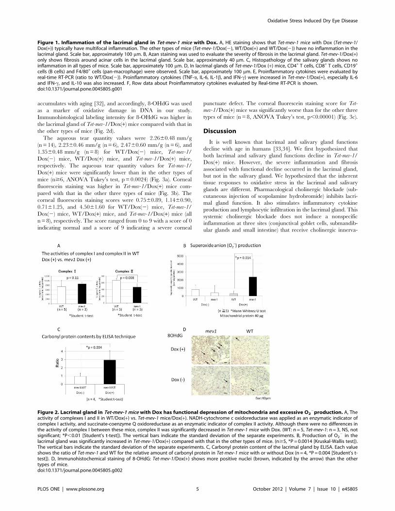
The aqueous tear quantity values were 2.2660.48 mm/g
(n = 14), 2.2360.46 mm/g (n = 6), 2.4760.60 mm/g (n = 6), and
1.3560.48 mm/g (n = 8) for WT/Dox(2) mice, Tet-mev-1/
Dox(2) mice, WT/Dox(+) mice, and Tet-mev-1/Dox(+) mice,
respectively. The aqueous tear quantity values for Tet-mev-1/
Dox(+) mice were significantly lower than in the other types of
mice (n$6, ANOVA Tukey’s test, p = 0.0024) (Fig. 3a). Corneal
fluorescein staining was higher in Tet-mev-1/Dox(+) mice com-
pared with that in the other three types of mice (Fig. 3b). The
corneal fluorescein staining scores were 0.7560.89, 1.1460.90,
0.7161.25, and 4.5061.60 for WT/Dox(2) mice, Tet-mev-1/
Dox(2) mice, WT/Dox(+) mice, and Tet-mev-1/Dox(+) mice (all
n = 8), respectively. The score ranged from 0 to 9 with a score of 0
indicating normal and a score of 9 indicating a severe corneal
punctuate defect. The corneal fluorescein staining score for Tet-
mev-1/Dox(+) mice was significantly worse than for the other three
types of mice (n = 8, ANOVA Tukey’s test, p,0.00001) (Fig. 3c).
Discussion
It is well known that lacrimal and salivary gland functions
decline with age in humans [33,34]. We first hypothesized that
both lacrimal and salivary gland functions decline in Tet-mev-1/
Dox(+) mice. However, the severe inflammation and fibrosis
associated with functional decline occurred in the lacrimal gland,
but not in the salivary gland. We hypothesized that the inherent
tissue responses to oxidative stress in the lacrimal and salivary
glands are different. Pharmacological cholinergic blockade (sub-
cutaneous injection of scopolamine hydrobromide) inhibits lacri-
mal gland function. It also stimulates inflammatory cytokine
production and lymphocytic infiltration in the lacrimal gland. This
systemic cholinergic blockade does not induce a nonspecific
inflammation at three sites (conjunctival goblet cells, submandib-
ular glands and small intestine) that receive cholinergic innerva-
Figure 1. Inflammation of the lacrimal gland in Tet-mev-1 mice with Dox. A, HE staining shows that Tet-mev-1 mice with Dox (Tet-mev-1/Dox(+)) typically have multifocal inflammation. The other types of mice (Tet-mev-1/Dox(2), WT/Dox(+) and WT/Dox(2)) have no inflammation in thelacrimal gland. Scale bar, approximately 100 mm. B, Azan staining was used to evaluate the severity of fibrosis in the lacrimal gland. Tet-mev-1/Dox(+)only shows fibrosis around acinar cells in the lacrimal gland. Scale bar, approximately 40 mm. C, Histopathology of the salivary glands shows noinflammation in all types of mice. Scale bar, approximately 100 mm. D, In lacrimal glands of Tet-mev-1/Dox (+) mice, CD4+ T cells, CD8+ T cells, CD19+
cells (B cells) and F4/80+ cells (pan-macrophage) were observed. Scale bar, approximately 100 mm. E, Proinflammatory cytokines were evaluated byreal-time RT-PCR (ratio to WT/Dox(2)). Proinflammatory cytokines (TNF-a, IL-6, IL-1b, and IFN-c) were increased in Tet-mev-1/Dox(+), especially IL-6and IFN-c, and IL-10 was also increased. F, Row data about Proinflammatory cytokines evaluated by Real-time RT-PCR is shown.doi:10.1371/journal.pone.0045805.g001
Figure 2. Lacrimal gland in Tet-mev-1 mice with Dox has functional depression of mitochondria and excessive O22production. A, The
activity of complexes I and II in WT/Dox(+) vs. Tet-mev-1 mice/Dox(+). NADH-cytochrome c oxidoreductase was applied as an enzymatic indicator ofcomplex I activity, and succinate-coenzyme Q oxidoreductase as an enzymatic indicator of complex II activity. Although there were no differences inthe activity of complex I between these mice, complex II was significantly decreased in Tet-mev-1 mice with Dox. (WT: n = 5, Tet-mev-1: n = 3, NS, notsignificant; *P,0.01 [Student’s t-test]). The vertical bars indicate the standard deviation of the separate experiments. B, Production of O2
2 in thelacrimal gland was significantly increased in Tet-mev-1/Dox(+) compared with that in the other types of mice. (n$5, *P = 0.0014 [Kruskal-Wallis test]).The vertical bars indicate the standard deviation of the separate experiments. C, Carbonyl protein content of the lacrimal gland by ELISA. Each valueshows the ratio of Tet-mev-1 and WT for the relative amount of carbonyl protein in Tet-mev-1 mice with or without Dox (n = 4, *P = 0.004 [Student’s t-test]). D, Immunohistochemical staining of 8-OHdG: Tet-mev-1/Dox(+) shows more positive nuclei (brown, indicated by the arrow) than the othertypes of mice.doi:10.1371/journal.pone.0045805.g002
Oxidative Stress Induced Dry Eye Disease
PLOS ONE | www.plosone.org 5 October 2012 | Volume 7 | Issue 10 | e45805
tions [35]. These results suggest that the lacrimal gland is subject
to inflammation by various stimuli in contrast with the salivary
gland.
Mitochondria generate ATP through aerobic respiration,
whereby glucose, pyruvate, and NADH are oxidized, thus
generating ROS as a byproduct. In normal circumstances, the
deleterious effects caused by the highly reactive nature of ROS are
balanced by the presence of antioxidants. However, high levels of
ROS are observed in chronic human diseases such as neurode-
generation [36], digestive organ inflammation [37], and cancer
[38]. Recent work exploring the mechanisms linking ROS and
inflammation suggest that ROS derived from mitochondria
(mtROS) act as signal transducing molecules to trigger pro-
inflammatory cytokine production [39]. Cells from patients with
TNFR1-associated periodic syndrome (TRAPS) demonstrate that
increased mtROS levels influence the transcription of pro-
inflammatory cytokines such as IL-6 and TNF. TRAPS manifests
as episodes of fever and severe localized inflammation with
mutations in TNFR1. Inhibition of mtROS production inhibited
MAPK activation and production of IL-6 and TNF in cells from
TRAPS patients [40]. The mtROS in Tet-mev-1/Dox(+) mice may
also directly induce increasing production of TNF-a and IL-6 and
continuously induce inflammation in the lacrimal gland.
Protein oxidation is a biomarker of oxidative stress and many
different types of protein oxidative modification can be induced
directly by ROS or indirectly by reactions of secondary by-
products of oxidative stress [41]. Lacrimal gland function has been
reported to decrease gradually with aging, leading to reduced tear
secretion and dry eye disease in the elderly [3,7]. Aging occurs, in
part, as a result of the accumulation of oxidative stress caused by
ROS that are generated continuously during the course of
metabolic processes. Levels of 8-OHdG as a DNA oxidative stress
marker and 4-HNE as a by-product of lipid peroxidation are
higher and tear volume is decreased in middle-aged rats. Caloric
restriction prevents a decline in lacrimal gland function and
morphological changes and might be associated with a reduction
in oxidative stress [42].
We confirmed that 8-OHdG immunohistological labeling
intensity was higher in the lacrimal gland of Tet-mev-1/Dox(+)
mice than in other mice types and the ratio of carbonylated
protein content in mice with Dox was three times the ratio of mice
without Dox. Collectively, mtROS production may damage DNA
and induce the accumulation of carbonylated protein in the
lacrimal gland.
These biochemical and histochemical data suggest that over-
produced superoxide anion from the mitochondria affect directly
and/or indirectly oxidative damage and inflammation in the
lacrimal gland. It is believed that chronic inflammation of the
lacrimal gland is a major contributor to insufficient tear secretion.
Chronic inflammation of the lacrimal gland occurs in several
Figure 3. Tet-mev-1/Dox(+) have dry eye disease. A, Aqueous tear production: Aqueous tear quantity values of Tet-mev-1/Dox(+) weresignificantly lower than those in the other types of mice (n$6, ANOVA Tukey’s test, p = 0.0024). B, Tet-mev-1/Dox(+) mice had more cornealfluorescein staining than in the other mice. C, The corneal fluorescein staining score of Tet-mev-1/Dox(+) was significantly worse than that in the othertypes of mice (all n = 8, ANOVA Tukey’s test, p,0.00001).doi:10.1371/journal.pone.0045805.g003
Oxidative Stress Induced Dry Eye Disease
PLOS ONE | www.plosone.org 6 October 2012 | Volume 7 | Issue 10 | e45805

pathologic conditions such as autoimmune diseases (Sjogren
syndrome, sarcoidosis, and diabetes) or simply as a result of aging
[43]. The relationship between inflammation of the lacrimal gland
and tear secretion deficiency has been described [44,45]. IL-1binduces a severe inflammatory response in the lacrimal gland and
inhibits lacrimal gland secretion and subsequent dry eye disease
[44]. A single injection of interleukin-1 into the lacrimal glands
induces reversible inflammation and leads to destruction of
lacrimal gland acinar epithelial cells, which results in decreased
tear production. However, these inflammatory responses subside
and lacrimal gland secretion and tear production return to normal
levels [45].
For the dry eye model, we first reported the accelerated
oxidation of protein, lipid, and DNA of the ocular surface in the
rat swing model [46,47]. Accumulated oxidative damage caused
the functional decline of the lacrimal gland and dry eye disease in
Tet-mev-1/Dox(+) mice. In the lacrimal gland, age-related chronic
inflammation, and age-related functional alterations including
decreased acetylcholine release and protein secretion, might be
related to dry eye diseases [48,49]. Our study clearly demonstrated
that oxidative stress from mitochondria induced dry eye disease
with morphological changes in the lacrimal gland of mice. In
conclusion, reducing oxidative stress might be one of the possible
treatments for age-related/ROS-induced dry eye disease.
Acknowledgments
We are grateful to Ms. Tamaki Saso for help with immunohistochemical
staining and to Mr. Tadayuki Sato for technical assistance with
quantitative real-time RT-PCR. Presented in part at the Tear Film
and Ocular Surface Society Meeting at Firenze, Italy, in September 2010.
Author Contributions
Conceived and designed the experiments: YU MM TI NI. Performed the
experiments: YU MM TI. Analyzed the data: YU TK SS KT. Contributed
reagents/materials/analysis tools: HO KY YO. Wrote the paper: YU TK.
References
1. Rocha EM, Alves M, Rios JD, Dartt DA (2002) The Aging Lacrimal Gland:
Changes in Structure and Function. Ocul Surf 6:162–174.
2. Obata H, Yamamoto S, Horiuchi H, Machinami R (1995) Histopathologic
study of human lacrimal gland. Statistical analysis with special reference to
aging. Ophthalmology 102: 678–686.
3. Draper CE, Adeghate EA, Singh J, Pallot DJ (1999) Evidence to suggest
morphological and physiological alterations of lacrimal gland acini with ageing.
Exp Eye Res 68: 265–276.
4. Moss SE, Klein R, Klein BE (2000) Prevalence of and risk factors for dry eye
syndrome. Arch Ophthalmol 18: 1264–1268.
5. McCarty CA, Bansal AK, Livingston PM, Stanislavsky YL, Taylor HR (1998)
The epidemiology of dry eye in Melbourne, Australia. Ophthalmology 105:
1114–1119.
6. Lee AJ, Lee J, Saw SM, Gazzard G, Koh D, et al. (2002) Prevalence and risk
factors associated with dry eye symptoms: a population based study in Indonesia.
Br J Ophthalmol 86: 1347–1351.
7. Schaumberg DA, Sullivan DA, Buring JE, Dana MR (2003) Prevalence of dry
eye syndrome among US women. Am J Ophthalmol 136: 318–326.
8. Moss SE, Klein R, Klein BE (2008) Long-term incidence of dry eye in an older
population. Optom Vis Sci 85: 668–674.
9. Turrens JF (1997) Superoxide production by the mitochondrial respiratory
chain. Biosci Rep 17: 3–8.
10. Ishii N, Fujii M, Hartman PS, Tsuda M, Yasuda K, et al. (1998) A mutation in
succinate dehydrogenase cytochrome b causes oxidative stress and ageing in
nematodes. Nature 394: 694–697.
11. Senoo MN, Yasuda K, Tsuda M, Ohkubo T, Yoshimura S, et al. (2001) A defect
in the cytochrome b large subunit in complex II causes both superoxide anion
overproduction and abnormal energy metabolism in Caenorhabditis elegans.
J Biol Chem 276: 41553–41558.
12. Honda S, Ishii N, Suzuki K, Matsuo M (1993) Oxygen-dependent perturbation
of life span and aging rate in the nematode. J Gerontol 48: B57–61.
13. Tsuda M, Sugiura T, Ishii T, Ishii N, Aigaki T (2007) A mev-1-like dominant-
negative SdhC increases oxidative stress and reduces lifespan in Drosophila.
Biochem Biophys Res Commun 363: 342–346.
14. Ishii T, Miyazawa M, Onodera A, Yasuda K, Kawabe N, et al. (2011)
Mitochondrial reactive oxygen species generation by the SDHC V69E mutation
causes low birth weight and neonatal growth retardation. Mitochondrion 11:
155–165.
15. Ishii T, Yasuda K, Akatsuka A, Hino O, Hartman PS, et al. (2005) A mutation
in the SDHC gene of complex II increases oxidative stress, resulting in apoptosis
and tumorigenesis. Cancer Res 65: 203–209.
16. Toyokuni S, Tanaka T, Hattori Y, Nishiyama Y, Yoshida A, et al. (1997)
Quantitative immunohistochemical determination of 8-hydroxy-29-deoxygua-
nosine by a monoclonal antibody N45.1: its application to ferric nitrilotriacetate-
induced renal carcinogenesis model. Lab Invest 76: 365–374.
17. Zhang N, Komine-Kobayashi M, Tanaka R, Liu M, Mizuno Y, et al. (2005)
Edaravone reduces early accumulation of oxidative products and sequential
inflammatory responses after transient focal ischemia in mice brain. Stroke 36:
2220–2225.
18. Anderson G, Gordon K (1996) Tissue processing, microtomy and paraffin
sections. In: Bancroft JD, Stevens A editors. Theory and practice of histological
techniques. 4th ed. New York, Churchill Livingstone. pp. 50–68
19. Bancroft J, Palmer J (1996) Frozen and related sections. In: Bancroft JD, Stevens
A editors. Theory and practice of histological techniques. 4th ed. New York,
Churchill Livingstone. pp. 69–80
20. Ogawa Y, Kuwana M, Yamazaki K, Mashima Y, Yamada M, et al. (2003)
Periductal area as the primary site for T-cell activation in lacrimal gland chronicgraft-versus-host disease. Invest Ophthalmol Vis Sci 44: 1888–1896.
21. Slack EN, Bursell E (1976) The isolation of mitochondria from dipteran flight
muscle. Biochim Biophys Acta 449: 491–499.
22. Trounce IA, Kim YL, Jun AS, Wallace DC (1996) Assessment of mitochondrialoxidative phosphorylation in patient muscle biopsies, lymphoblasts, and
transmitochondrial cell lines. Methods Enzymol 264: 484–509.
23. Yasuda K, Ishii T, Suda H, Akatsuka A, Hartman PS, et al. (2006) Age-relatedchanges of mitochondrial structure and function in Caenorhabditis elegans.
Mech Ageing Dev 127: 763–770.
24. Shimomura O, Wu C, Murai A, Nakamura H (1998) Evaluation of fiveimidazopyrazinone-type chemiluminescent superoxide probes and their appli-
cation to the measurement of superoxide anion generated by Listeriamonocytogenes. Anal Biochem 258: 230–235.
25. Miyazawa M, Ishii T, Yasuda K, Noda S, Onouchi H, et al. (2009) The role of
mitochondrial superoxide anion (O2(-)) on physiological aging in C57BL/6J
mice. J Radiat Res 50: 73–83.
26. Turpie B, Yoshimura T, Gulati A, Rios JD, Dartt DA, et al. (2009) Sjogren’s
syndrome-like ocular surface disease in thrombospondin-1 deficient mice.
Am J Pathol 175: 1136–1147.
27. Shimazaki J, Tsubota K, Kinoshita S, Ohashi Y (2007) Difinition and diagnosisof dry eye 2006. Atarashii Ganka (in Japanese) 24: 181–184.
28. Ibrahim OM, Dogru M, Takano Y, Satake Y, Wakamatsu TH, et al. (2010)
Application of visante optical coherence tomography tear meniscus heightmeasurement in the diagnosis of dry eye disease. Ophthalmology 117: 1923–
1929.
29. Mizuno Y, Yamada M, Miyake Y (2010) Association between clinical diagnostictests and health-related quality of life surveys in patients with dry eye syndrome.
Jpn J Ophthalmol 54: 259–265.
30. Sun F, Huo X, Zhai Y, Wang A, Xu J, et al. (2005) Crystal structure ofmitochondrial respiratory membrane protein complex II. Cell 121: 1043–1057.
31. Yankovskaya V, Horsefield R, Tornroth S, Luna-Chavez C, Miyoshi H, et al.
(2003) Architecture of succinate dehydrogenase and reactive oxygen speciesgeneration. Science 299: 700–704.
32. Wang AL, Lukas TJ, Yuan M, Neufeld AH (2008) Increased mitochondrial
DNA damage and down-regulation of DNA repair enzymes in aged rodentretinal pigment epithelium and choroid. Mol Vis 14: 644–651.
33. Xu KP, Yagi Y, Tsubota K (1996) Decrease in corneal sensitivity and change in
tear function in dry eye. Cornea 15: 235–239.
34. Flink H, Bergdahl M, Tegelberg A, Rosenblad A, Lagerlof F (2008) Prevalenceof hyposalivation in relation to general health, body mass index and remaining
teeth in different age groups of adults. Community Dent Oral Epidemiol 36:
523–531.
35. Pitcher JD 3rd, De Paiva CS, Pelegrino FS, McClellan AJ, Raince JK, et al.(2011) Pharmacological cholinergic blockade stimulates inflammatory cytokine
production and lymphocytic infiltration in the mouse lacrimal gland. InvestOphthalmol Vis Sci 52: 3221–7.
36. Cominelli F (2004) Cytokine-based therapies for Crohn’s disease–new
paradigms. N Engl J Med 351: 2045–8.
37. Drake IM, Mapstone NP, Schorah CJ, White KL, Chalmers DM, et al. (1998)Reactive oxygen species activity and lipid peroxidation in Helicobacter pylori
associated gastritis: relation to gastric mucosal ascorbic acid concentrations andeffect of H pylori eradication. Gut 42: 768–71.
38. Reuter S, Gupta SC, Chaturvedi MM, Aggarwal BB (2010) Oxidative stress,
inflammation and cancer: How are they linked? Free Radic Biol Med 249:
1603–16.
Oxidative Stress Induced Dry Eye Disease
PLOS ONE | www.plosone.org 7 October 2012 | Volume 7 | Issue 10 | e45805

39. Zhou R, Yazdi AS, Menu P, Tschopp J (2011) A role for mitochondria in
NLRP3 inflammasome activation. Nature 469(7329): 221–5.40. Bulua AC, Simon A, Maddipati R, Pelletier M, Park H, et al. (2011)
Mitochondrial reactive oxygen species promote production of proinflammatory
cytoki\nes and are elevated in TNFR1-associated periodic syndrome (TRAPS).J Exp Med 208:519–33.
41. Berlett BS, Stadtman ER (1997) Protein oxidation in aging, disease, andoxidative stress. J Biol Chem 272: 20313–6.
42. Kawashima M, Kawakita T, Okada N, Ogawa Y, Murat D, et al. (2010) Calorie
restriction: A new therapeutic intervention for age-related dry eye disease in rats.Biochem Biophys Res Commun 397: 724–8.
43. Zoukhri D (2006) Effect of inflammation on lacrimal gland function. Exp EyeRes 82: 885–98.
44. Zoukhri D, Macari E, Choi SH, Kublin CL (2006) c-Jun NH2-terminal kinasemediates interleukin-1beta-induced inhibition of lacrimal gland secretion.
J Neurochem 96: 126–35.
45. Zoukhri D, Macari E, Kublin CL (2007) A single injection of interleukin-1
induces reversible aqueous-tear deficiency, lacrimal gland inflammation, and
acinar and ductal cell proliferation. Exp Eye Res 84: 894–904.
46. Nakamura S, Kinoshita S, Yokoi N, Ogawa Y, Shibuya M, et al. (2010)
Lacrimal hypofunction as a new mechanism of dry eye in visual display terminal
users. PLoS ONE 5: e11119
47. Nakamura S, Shibuya M, Nakashima H, Hisamura R, Masuda N, et al. (2007)
Involvement of oxidative stress on corneal epithelial alterations in a blink-
suppressed dry eye. Invest Ophthalmol Vis Sci 48: 1552–1558.
48. Rıos JD, Horikawa Y, Chen LL, Kublin CL, Hodges RR, et al. (2005) Age-
dependent alterations in mouse exorbital lacrimal gland structure, innervation
and secretory response. Exp Eye Res 80: 477–491.
49. Draper CE, Adeghate E, Lawrence PA, Pallot DJ, Garner A, et al. (1998) Age-
related changes in morphology and secretory responses of male rat lacrimal
gland. J Auton Nerv Syst 69: 173–183
Oxidative Stress Induced Dry Eye Disease
PLOS ONE | www.plosone.org 8 October 2012 | Volume 7 | Issue 10 | e45805