Embed Size (px)
Citation preview

Figure 1
“ORTHOTIC MANAGEMENT OF THE LOWER LIMB OF CHILDREN WITH
CEREBRAL PALSY”
Marty Carlson, CPO
Tamarack Habilitation Technologies, Inc.
1670 94th
Lane NE
Blaine, MN 55449-4323
As we all know, the manifestations of cerebral palsy vary widely, and every child
must be closely examined to evaluate his/her particular type and combination of
pathological characteristics. However, a certain pattern of muscle imbalance is extremely
common in children with cerebral palsy. At
least part of this pattern is due to retention
of primitive reflexes. However, some
components of the muscle imbalance pattern
suggest that non-pathological factors,
muscle bulk and mechanical advantage, are
also important in determining which muscle
group dominates.
The pattern of deformity most
commonly occurring in the lower
extremities of children with cerebral palsy is
seen in figure 1 and includes:
1. The subtalar joint is in valgus and
the longitudinal arch is flattened on
weight bearing;
2. The ankle tends to be plantar flexed;
3. The knee is flexed and;
4. The hip is flexed, adducted, and
internally rotated.
If the muscle imbalance persists without
intervention, joint and bone deformities will
develop and progress. We should also remember that orthotic treatment should never be
an isolated intervention technique. It is combined with physical therapy and surgery as
appropriate.

Page 2
Figure 2
Figure 3A Figure 3B Figure 3C
Orthotic treatment has three general goals:
1. Prevent the development and progression of deformities from flexible to structural
(especially during periods of rapid physical growth); Figure 2 is an example of
flexible early deformity in CP
2. Provide support so the child can be more functional;
3. Provide external support following surgery to prevent either over-correction or
recurrence of the deformity during the healing process.
Correction of a firmly established orthopedic deformity in the lower limbs of a child
with cerebral palsy is seldom accomplished orthotically. For that reason it is not listed as
one of our goals.
Every drug which goes on the market must be evaluated with respect to the side
effects it produces. We must understand that orthoses have the potential to produce
undesirable side effects also. Orthoses add weight, they may encumber, they take time
and can even worsen deformities if badly designed/fit. With understanding, we can
design to achieve or extend our goals while we minimize undesirable side effects.
Discussion of the specific joints will start at the child’s foot and work upward to the hip.
The diagram in figure 3A illustrates how, during stance, most of the weight bearing
forces are normally divided between two distinct areas of the

Page 3
Figure 5
Figure 6 Figure 7
Figure 4
foot, the calcaneus and the metatarsal heads.
When the plantar flexor muscles are over-active, the hind foot is pulled upward out of
contact with the floor (figure 3B) and the forefoot must take most of the weight. The
body’s center of gravity is shifted forward to maintain stability and it is a greater
challenge for the child to maintain balance within a smaller base of support. The soft
tissues which maintain the longitudinal arch are stretched. If these conditions persist, the
longitudinal arch will be flattened. With growth and time, the long arch will be reversed,
creating what we call the “rocker bottom” foot deformity seen in figure 3C.
Figure 4 diagrams the deformity in the frontal plane.
The foot inverters are overpowered and the subtalar joint
tends to collapse into valgus. Both the weight bearing force
and the Achilles tendon tension become valgus deforming
forces when the inverters are unable to maintain proper M-L
alignment of the calcaneus under the tibia. Figure 5 is an
example of the consequences for a particular 10 year old.
We know that
physical therapy forces to
stretch the heel cord should
not be applied to the
forefoot alone. That does
stretch the plantar flexors
but also will exaggerate the
collapse of the subtalar joint (see figure 6). A skilled
therapist will grasp the calcaneus to control the subtalar
joint to make sure it doesn’t collapse into valgus as the
ankle is passively dorsiflexed (see figure 7). As
orthotists, we should be aware that these principles
apply in orthotic design
as well. An orthosis
which is intended to
resist plantar flexion
must be designed to
simultaneously control
the subtalar joint and
support the arch. If not,
the orthosis will be
needlessly contributing
to the development of a
foot deformity.
Figure 8 is another example of the classic rigid, post adolescent valgus “rocker
bottom” feet. For many years this patient wore conventional double upright type Ankle-

Page 4
Figure 8
Figure 9
Figure 10
Figure 11 Figure 11
Foot Orthoses attached to a shoe such as shown in
figure 9. A medial “T” strap, is unlikely to
effectively prevent the subtalar joint from
collapsing into valgus.
In North America, almost all
current lower limb orthoses utilize
the polypropylene shell design.
These orthoses must fit the hind foot
and mid foot very supportively. In a
well designed AFO, an instep strap is
installed as necessary to hold the
child’s heel down and back in the orthosis (see figure 10).
It is, unfortunately very common to think that neutral subtalar alignment can be
maintained by supporting the
longitudinal arch of these
developing children. That is wrong.
Arch support relies on ligaments,
most notable the Spring ligament,
(see figure 11) to transmit that
support back to the subtalar joint
[Ref.1]. Over time, with growth and
development these ligaments will be
stretched, and the subtalar joint will
collapse into valgus alignment in
spite of support to the arch area.

Page 5
Figure 14
Figure 12
Figure 13
F2 F1
Figures 12 and 13 show the primary
areas of medial stabilizing pressure, and figures 14 indicate the aggressive sculpting
necessary to adequately grip the calcaneus to support the subtalar joint. This
configuration of support is much more directly applied to the Sustentaculum Talus and
Calcaneous.
There are times when we should be aware of the practical limits of orthotic
treatment. When the heel cord of an ambulatory child is too tight to permit passive
dorsiflexion to neutral with moderate force, even the most skillfully rendered orthotic
treatment will fail in most cases, to achieve/maintain a plantargrade foot. That situation
may call for surgical weakening of the plantar flexor muscles to gain adequate
dorsiflexion range of motion without excessive heel cord tension. Active and passive
examination of the lower limbs of a supine child is not only inadequate, but can actually
be misleading. Observation of muscle activity, coordination, and spasticity during weight
bearing is very important in making the correct decisions about treatment. Also, keep in
mind that the child in clinic is not always typical of the child at home.
If your determination is that there is too much plantar flexion tone or inadequate
ankle range to achieve a plantar grade foot (without unrealistic force or sacrificing sub-
talar neutral alignment), surgery may be advisable. If surgery is not performed, the calf

Page 6
Figure 15
section of the AFO should be brought to the desired A-Palignment by means of a
compensating posterior wedge added to the AFO or shoe. It should also be noted that if
no significant dorsiflexion range is expected during ambulation (because of tone or
contracture) it is probably counter- productive to articulate the AFO. Articulation causes
extra unproductive skin pressure from subtalar support surfaces as the tibia moves into
anterior alignment. Articulation may also reduce Gastrocnemius stretch during
ambulation.
When the ankle has significant, useful dorsiflexion motion, we provide for that
motion in the orthosis with ankle joints as you saw earlier (figure 10). We stop plantar
flexion motion at the neutral (90°) position. It is typical for these children to have less
lower limb muscle tone when crawling and climbing around toys and furniture than when
they are upright and ambulating. Non ambulation hours make up the vast majority of
their days. A well-designed, articulated AFO in these cases allows dorsiflexion ranging
during those hours when tone is reduced.
Moving up to the knee, one of the most common and obvious observations is the
crouched stance. Loss of full knee extension may be primarily due to tight hamstring
muscles or it may have developed secondary to hip flexion contractures. Often, it seems,
both factors contribute. Knee flexion is related to hip flexion because of the muscles they
share and because of their need to compensate for one another in the achievement of
balanced upright stance. Our examination should always determine, if possible, which
postural deviations are structural and which are functional compensations. These
considerations are, of course, of utmost importance whenever surgery is contemplated.
There are some common
misconceptions about why many
children with cerebral palsy stand
and walk with knees flexed. One
theory is that it is an attempt to
lower the center-of-gravity to aid
balance. That theory is hard to
accept; even 50 degrees of knee
flexion will lower the body center-
of-gravity by less than 10%.
Another statement, heard on
occasion, is that the bent-knee
stance results from weak
quadriceps and strengthening
exercises are recommended. This
seems very improbable because a
flexed knee stance, in itself,
requires enhanced knee extensor
strength (see figure 17). Low

Page 7
Figure 17
Figure 18
Figure 17
Figure 16
quadriceps tone/strength is not the problem.
We can identify several biomechanic factors that contribute to flexed knee stance.
Hamstring tightness or spasticity is almost universally observed in these youngsters. The
hamstrings are very strong, so this usually is the primary and major resistance to full knee
extension. The graph in figure 15 qualitatively illustrates the rapid rise in that resistance
as full knee extension is approached.
Straightforward mechanical analysis can relate
knee flexion angle to the flexion moment at the knee
caused by gravitational force on the body (see figure 16).
For example, when 35 kgm., normal youngster, stands
with knees flexed 50 degrees; his quadriceps must
generate a moment of about 3 KgM just to offset the
collapsing effect of gravity
(figure 17). When the
youngster stands in well-aligned
full knee extension, virtually no
effort is needed against gravity.
The graph in figure 18
looks at the combined effects of
gravity and tight hamstrings as
a function of knee extension. The
dashed line is the sum of the two.
The left side of the graph
represents absence of crouch. The
extreme right side of the graph
again represents a deep crouch.
As we follow the dashed line
leftward, we see that, as the
youngster straightens from the
deep crouch, the necessary
quadriceps effort decreases as the
knee centers move closer to the
weight line. However, at some

Page 8
Figure 19
Figure 20 Figure 21
point the hamstrings begin to significantly resist knee extension, and that resistance may
rise very fast as the knees approach full extension. Please note that the dashed line
indicates the youngster will probably choose particular to stand, in this example, with
knees flexed about 40°. Standing with either more or less knee flexion requires even
greater quadriceps effort. Please also note that this analysis is qualitative. Each child
represents unique muscle tone and contracture factors and will find his own particular
stance or posture depending on those factors.
As the months and years progress, the extraordinary tension of the quadriceps will
stretch and elongate the patello-tibial ligament. As the child grows, a patella alta
condition develops. The photograph in figure 19 came out of an old medical chart. It
was taken to document a case of severe foot deformity. However, notice the boy’s knees.
The patellae are riding entirely above the femoral epicondyles. The profile of the femoral
condyles and the inter-condylar notches are clearly seen below the lower border of the
patellae. The patello-tibial ligament has been stretched to perhaps twice the normal
length.
The change in the position of the patella profoundly alters the
mechanics and effectiveness of the quadriceps. It does so in
several compounding ways:
1. As the patellar ligament lengthens, the resting length of
the quadriceps muscle belly is reduced; the muscle is thus
“weakened” similar to what happens in a surgical tendon
lengthening procedure.
2. As the patella moves above the femoral condyles, the
patellar ligament slips into a position in the femoral
intercondylar grove where it has less mechanical
advantage to create an extension moment. Figures 20 and
21 compare the normal with the patella alta configuration.
Page 9
Figure 22
Note that, in the Patella Alta condition, the tendon, lying deep within the
intercondylar groove, passes closer to the center of rotation. Thus the extension
moment produced by any amount of quadriceps pull is reduced. This effect is
greater as full knee extension is approached because the patella is then entirely
above the condyles.
3. Because of the geometry and mechanics of the knee, as the patellar ligament
lengthens, more and more of the quadriceps tension is transferred to the patellar
retinaculum. Retinaculum tension is much less effective in extending the knees
because these structures pass closer to the center of motion as you see in the
diagram to your right. Because of knee kinematics, this transfer of tension from
the patellar ligament to the retinaculum increases as the knee approaches full
extension and there exists the situation diagrammed at the right side of figure 21.
This tension transfer mechanism was pointed out by Dr. Eggers in 1950 [Ref 2.].
Note that both loss of mechanical advantage and tension transfer to the
retinaculum are increasingly occurring as the knee approaches full extension.
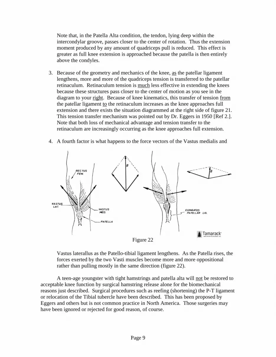
4. A fourth factor is what happens to the force vectors of the Vastus medialis and
Vastus laterallus as the Patello-tibial ligament lengthens. As the Patella rises, the
forces exerted by the two Vasti muscles become more and more oppositional
rather than pulling mostly in the same direction (figure 22).
A teen-age youngster with tight hamstrings and patella alta will not be restored to
acceptable knee function by surgical hamstring release alone for the biomechanical
reasons just described. Surgical procedures such as reefing (shortening) the P-T ligament
or relocation of the Tibial tubercle have been described. This has been proposed by
Eggers and others but is not common practice in North America. Those surgeries may
have been ignored or rejected for good reason, of course.

Page 10
Figure 23B Figure 23A
The graphs in figures 15, 16, 17 and 18 will vary from patient to patient and even
for a given patient, according to level of spasticity and fatigue. This analysis is presented
not as a quantitative tool. It was meant to help us analyze, only qualitatively, the
biomechanics of what we observe.
The use of orthoses which cross the knee joint is advocated in the following
instances only: 1. when the knee is passively fully extendable and supporting the knee
significantly improves the child’s ability to stand and ambulate; 2. to maintain the knee
extension range of motion gained by surgical procedure; and 3. Night positioning to resist
a progressive knee deformity.
We will turn now to orthotic treatment of the hip. In most cases it is impractical
to attempt to control hip flexion or adduction during ambulation. The hardware
necessary to achieve control is prohibitively bulky, heavy, and bothersome for the child
to carry around. However, nighttime positioning orthoses do make sense in many cases
for both marginal ambulators and non-ambulators. The hip and knee deformities we see
developing in children invariably match the child’s sleeping posture. If the sleeping
posture does not initiate the deformities, it seems certain that maintaining those positions
8-10 hours every night will facilitate deformity progression.
It has been common practice in some places to apply abduction pillows or wedges
to resist adductor tightness or following adductor tenotomy and obturator neurectomy.
These abduction orthoses are ineffective. They do not control rotation and they do not
prevent the adduction deformity from recurring on the side where spasticity/tone is most
severe. We cannot expect to control hip position with an orthosis which does not cross
the hip joint. The night time positioning orthosis must cross the hip. Children who begin
to develop hip or hip and knee deformities can more successfully be provided with a
recumbent support orthosis
(RSO) which maintains
them in an extended/neutral
or optimum attainable
position during their
recumbent hours. This
type of orthosis can be
fabricated in many
configurations, depending
on the deformity it is meant
to resist. The design you
see in figure 23B applies
simple biomechanical
principles to resist the
habitual posture shown in
figure 23A. Other designs
may be rendered according
to the need. Recumbent Support Orthoses are relatively inexpensive and can

Page 11
accommodate considerable growth. It is important to make the orthoses as attractive as
possible so that parents, caregivers, and child will accept them. The Recumbent Support
Orthosis may also be used to maintain desired positioning following hip and knee
surgery.
Pommels, straps and bolsters can be used as part of the non-ambulatory patient’s
seating system, but their effect on hip adduction contractures is minimal because they
cannot prevent the pelvis from rotating about the vertical axis allowing the more severe
adduction contracture to create/maintain a “Windswept Hips” deformity.
A final comment on knee and hip deformities in non-ambulatory children should
be made. Some of those who give medical care to these children feel that lower limb
deformities are of no consequence if there is no possibility of ambulation. If that
reasoning prevails, by the time the child is a teenager, the hip and knee deformities which
result from such neglect are likely to be very severe. Efforts by caregivers to adequately
position and provide nursing and toileting care for these people, either seated or
recumbent, are greatly complicated and limited by the deformities. Gradually the
deformities lead to a pattern which allows only a single recumbent posture. The result is
greater care labor costs, and more frequent hospitalization for ulcer care. The long term
cost to society is much greater because of that early neglect.
REFERENCES:
1. Carlson, J.M., and Berglund, G., “An Effective Orthotic Design for Controlling
the Unstable Subtalar Joint”, Orthotics and Prosthetics, Vol. 33, No. 1, pp. 39-49,
March 1979.
2. Eggers, G.W.N., “Surgical Division of the Patellar Retinacula to Improve
Extension of the Knee Joint in Cerebral Spastic Paralysis”, Journal of Bone and
Joint Surgery, Vol. 32-A:80, 1950


