Embed Size (px)
Citation preview

PRECLINICAL STUDY
Oncogene amplification in male breast cancer: analysisby multiplex ligation-dependent probe amplification
Robert Kornegoor • Cathy B. Moelans • Anoek H. J. Verschuur-Maes •
Marieke C. H. Hogenes • Peter C. de Bruin • Joost J. Oudejans •
Luigi Marchionni • Paul J. van Diest
Received: 22 December 2011 / Accepted: 26 March 2012 / Published online: 13 April 2012
� The Author(s) 2012. This article is published with open access at Springerlink.com
Abstract Gene amplification is an important mechanism
for oncogene activation, a crucial step in carcinogenesis.
Compared to female breast cancer, little is known on the
genetic makeup of male breast cancer, because large series are
lacking. Copy number changes of 21 breast cancer related
genes were studied in 110 male breast cancers using multiplex
ligation-dependent probe amplification. A ratio of[1.3 was
regarded indicative for gene copy number gain and a ratio
[2.0 for gene amplification. Data were correlated with
clinicopathological features, prognosis and 17 genes were
compared with a group of female breast cancers. Gene copy
number gain of CCND1, TRAF4, CDC6 and MTDH was seen
in[40 % of the male breast cancer cases, with also frequent
amplification. The number of genes with copy number gain
and several single genes were associated with high grade, but
only CCND1 amplification was an independent predictor of
adverse survival in Cox regression (p = 0.015; hazard ratio
3.0). In unsupervised hierarchical clustering a distinctive
group of male breast cancer with poor prognosis (p = 0.009;
hazard ratio 3.4) was identified, characterized by frequent
CCND1, MTDH, CDC6, ADAM9, TRAF4 and MYC copy
number gain. Compared to female breast cancers, EGFR
(p = 0.005) and CCND1 (p = 0.041) copy number gain was
more often seen in male breast cancer, while copy number
gain of EMSY (p = 0.004) and CPD (p = 0.001) and
amplification in general was less frequent. In conclusion,
several female breast cancer genes also seem to be important
in male breast carcinogenesis. However, there are also clear
differences in copy number changes between male and female
breast cancers, pointing toward differences in carcinogenesis
between male and female breast cancer and emphasizing the
importance of identifying biomarkers and therapeutic agents
based on research in male breast cancer. In addition CCND1
amplification seems to be an independent prognosticator in
male breast cancer.
Keywords Breast cancer � Male � MLPA �Amplification � Copy number � Survival
Introduction
Gene amplification is important in the development and
progression of cancer and could serve as a potential
Electronic supplementary material The online version of thisarticle (doi:10.1007/s10549-012-2051-3) contains supplementarymaterial, which is available to authorized users.
R. Kornegoor � C. B. Moelans � A. H. J. Verschuur-Maes �P. J. van Diest
Department of Pathology, University Medical Center Utrecht,
Heidelberglaan 100, 3584 CX Utrecht, The Netherlands
M. C. H. Hogenes
Laboratory for Pathology East Netherlands, Burgemeester Edo
Bergsmalaan 1, 7512 AD Enschede, The Netherlands
P. C. de Bruin
Department of Pathology, St. Antonius Hospital, Koekoekslaan
1, 3435 CM Nieuwegein, The Netherlands
J. J. Oudejans
Department of Pathology, Diakonessenhuis, Bosboomstraat 1,
3582 KE Utrecht, The Netherlands
L. Marchionni
Johns Hopkins University, 1550 Orleans Street, Baltimore,
MD, USA
P. J. van Diest (&)
Department of Pathology, University Medical Center Utrecht,
PO Box 85500, 3508 GA Utrecht, The Netherlands
e-mail: [email protected]
123
Breast Cancer Res Treat (2012) 135:49–58
DOI 10.1007/s10549-012-2051-3

biomarker for prognosis or as a target for molecular ther-
apy. In female breast cancer, HER2 is the best described
oncogene with frequent amplificaion. HER2 amplification
is correlated with poor survival and good response to tar-
geted therapy [1, 2]. Other genes, like epidermal growth
factor receptor (EGFR), Fibroblast growth factor receptor 1
(FGFR1), topoisomerase IIa (TOP2A) and MYC are also
involved in female breast cancer and have prognostic and
therapeutic implications [3–6].
Compared to female breast cancer, there is yet little
knowledge regarding the genetic makeup of male breast
cancer, because male breast cancer is a rare disease and the
few available studies are based on small single institutional
series [7]. Treatment of male breast cancer has largely been
extrapolated from its female counterpart, while there are
important differences between male and female breast
cancer, with higher ratios of estrogen receptor (ER) and
progesterone receptor (PR) positivity in men [8–10]. Also
the distribution of molecular subtypes by immunohisto-
chemical analysis shows important differences. Luminal
type A and B are by far the most frequently encountered
subtypes and HER2 driven, basal-like and triple-negative
tumors are very rare in men [11, 12]. The few gene
expression studies performed recently in men showed that
there might be important differences in molecular profile
between male and female breast cancer [13–15]. However,
the clinical and prognostic significance of genetic altera-
tions in relevant breast cancer genes still needs to be elu-
cidated in male breast cancer.
Multiplex ligation-dependent probe amplification (MLPA)
analysis is a high throughput genomic technique enabling
relative quantification of copy number or promoter hyper-
methylation in a variety of genes in one reaction, based on the
simultaneous amplification of specifically hybridized probes
on DNA that can be derived from paraffin embedded material
[16, 17]. We previously showed in female breast cancer that
MLPA analysis with a dedicated ‘‘breast cancer kit’’ allows
evaluation of copy numbers in 21 important breast cancer
genes, providing an overview of the most common amplifi-
cations [18]. In the present study, we used MLPA to investi-
gate copy number changes of 21 (female) breast cancer related
genes in a large group of male breast cancer and correlate these
genomic anomalies with clinicopathological features,
patients’ outcome, and with previously obtained MLPA data
from female breast cancers.
Materials and methods
Patients: specimens and clinical information
All consecutive cases of surgical breast specimens of inva-
sive male breast cancer from 1986 to 2010 were collected
from four different pathology labs in The Netherlands (St.
Antonius Hospital Nieuwegein, Diakonessenhuis Utrecht,
University Medical Center Utrecht, Laboratory for Pathol-
ogy East Netherlands) as described in more detail previously
[12]. Hematoxylin and eosin (HE) slides were reviewed by
three experienced observers (PJvD, RK, AM) to confirm the
diagnosis and to type and grade according to current stan-
dards. Pathology reports were used to retrieve information
on age, tumor size, and lymph node status. A total of 110
cases from which the paraffin blocks contained enough
tumor for DNA isolation were included. The age of these
patients ranged from 32 to 89 years (average: 66 years).
Tumor size ranged from 0.8 to 5.5 cm (average: 2.2 cm). In
86 % lymph node status was known and 55 % of these
patients had lymph node metastases. The majority of cases
were diagnosed (according to the WHO) as invasive ductal
carcinoma (90 %). The remaining cases were lobular
(n = 3), mixed type (ductal/lobular) (n = 2), invasive
cribriform (n = 1), papillary (n = 1), mucinous (n = 2),
invasive micropapillary (n = 1) or adenoid cystic carcino-
mas (n = 1). According to the modified Bloom and Rich-
ardson score [19] most tumors were grade 2 (41 %) or grade
3 (36 %). Mitotic activity was assessed as before [20] with a
mean mitotic index of 11 per 2 mm2 (range 0–56). For all
cases hormone receptor and HER2 status were re-assessed as
described previously [12]. Tissue microarray (TMA) slides
were used for immunohistochemical stainings for ER, PR
and chromogenic in situ hybridization (CISH) for HER2
assessment, the latter showing HER2 amplification in only
4/110 cases (4 %). TMA slides were also stained for
E-cadherin. Most tumors were ER positive (102/110, 93 %)
and PR positivity was also common (71/110; 65 %). Only
four cases were E-cadherin negative (three lobular carci-
nomas and one ductal carcinoma).
DNA extraction and MLPA analysis
Representative tumor areas were identified in HE stained
slides and corresponding tumor areas (at least 1 cm2) were
dissected with a scalpel from 8 lm paraffin slides [21].
DNA was extracted by overnight incubation in proteinase
K (10 mg/ml; Roche, Almere, The Netherlands) at 56 �C.
After boiling for 10 min and centrifugation, 5 ll of this
DNA solution was used for MLPA analysis. MLPA was
performed according the manufacturers’ instructions (MRC
Holland, Amsterdam, The Netherlands), using a Veriti�
96-well thermal cycler (Applied Biosystems, Foster City,
CA, USA). The P078-B1 kit (MRC Holland), containing 21
breast cancer related genes (Table 1), was used as before
[18]. All tests were performed in duplicate. Seven negative
references samples (normal breast and blood) were inclu-
ded in each MLPA run. The PCR products were separated
by electrophoresis on an ABI 3730 capillary sequencer
50 Breast Cancer Res Treat (2012) 135:49–58
123

(Applied Biosystems). Mean probe peaks were used for
final gene copy number analysis with Genescan v4.1
(Applied Biosystems) and Coffalyser v9.4 (MRC-Holland)
software. Cut-off values were set as before with 1.3–2.0 for
gene copy number gain, [2.0 for amplification and \0.7
for lost genes. Values between 0.7 and 1.3 were regarded
normal [18, 22].
Control female breast cancers
A group of female breast cancer described previously was
used to study differences in gene copy number change
between male and female breast cancer [18]. This group
consists of 104 cases with a mean age of 58 years (range
30–86 years). Tumor size ranged from 0.2 to 6.5 cm
(average 2.1 cm) and 46 % of the cases had lymph node
metastases. Most cases were diagnosed (according to the
WHO) as invasive ductal carcinoma (78 %) or invasive
lobular carcinoma (11 %). Mean mitotic activity was 21
per 2 mm2 and according to the modified Bloom and
Richardson score most tumors were grade 2 (34 %) or
grade 3 (45 %). ER positivity was common (69 %, 70/101)
and 48 % of the tumors were PR positive (48/101). HER2
amplification defined by immunohistochemistry and CISH
was seen in 19 % of cases (19/102). The same ‘‘breast
cancer kit’’ (P078-A1 kit; MRC Holland) was used, but
because the gene content of the kit had been updated by the
manufacturer in the meanwhile, only 17 genes could be
compared between the groups. In addition, for two genes
(EGFR and HER2) one of the probes was modified and for
five genes one probe was deleted. Some reference probes
were modified as well (Supplementary Table 1).
Statistics
Statistical calculations were performed using SPSS for
Windows v15.0. Correction for multiple comparisons was
applied by resetting the 0.05 threshold according to the
Holm–Bonferroni method. Differences between gene copy
number and clinicopathological characteristics were cal-
culated with ANOVA for continuous variables and with
Pearson Chi-square (or Fisher’s exact test when appropri-
ate) for categorical variables. The following clinicopatho-
logical features were dichotomized: age ([50 years), tumor
size ([2.0 cm), mitotic activity ([8 mitoses/2 mm2), and
histological grade (grade 1/2 vs 3). Correlation between
Table 1 Contents of the ‘‘breast cancer’’ MLPA kit P078-B1 (MRC Holland)
Gene Chrom Gain (%) Amp (%) Loss (%) Function and clinical relevance
ESR1 06q25.1 6 0 3 Transcription factor; under debate [40–42]
EGFR 07p11.2 22 1 0 Signal transduction; poor survival [4]
FGFR1 08p11.23 29 13 0 Signal transduction; poor survival, tamoxifen resistance [5]
ADAM9 08p11.23 39 11 1 Protein metabolism; promotes invasion [38]
IKBKB 08p11.21 32 6 0 Signal transduction [43]
PRDM14 08q13.3 32 9 0 Transcription regulatory protein; chemoresistance [44]
MTDH 08q22.1 49 12 0 Signal transduction; promoting metastases, chemoresistance,
poor survival [36]
MYC 08q24.21 36 10 0 Transcription factor; poor survival [3]
CCND1 11q13.2 46 18 1 Signal transduction; ER positivity, poor survival [35]
EMSY 11q13.5 10 2 3 Transcription regulatory protein; poor survival [45]
CDH1 16q22.1 6 0 9 Cell adhesion [46]
TRAF4 17q11.2 41 4 0 Signal transduction [47]
CPD 17q11.2 9 0 0 Protein metabolism [48]
MED1 17q21.2 23 4 0 Transcriptional coactivator; ER positivity [49]
HER2 17q12 17 4 0 Signal transduction; bad survival; trastuzumab response [2]
CDC6 17q21.2 41 4 0 Signal transduction [50]
TOP2A 17q21.2 26 2 0 Regulation of the topological status of DNA; poor survival,
susceptible for certain chemotherapy [6]
MAPT 17q21.31 16 0 0 Microtubule stabilization; chemoresistance (taxanes) [51]
BIRC5 17q25.3 27 2 0 Signal transduction; predict distant recurrence [52]
CCNE1 19q12 2 0 1 Signal transduction; poor survival [53]
AURKA 20q13.31 10 4 12 Signal transduction [54]
For each gene, chromosome location (Chrom), gene copy number gain (Gain;[1.3), amplification (Amp;[2.0), gene loss (Loss;\0.7), function
and clinical relevance (in female breast cancer) are shown
Breast Cancer Res Treat (2012) 135:49–58 51
123
number of gene amplification and clinicopathological fea-
tures were calculated with Spearman’s rho. Unsupervised
hierarchical clustering using the statistical program R
(www.r-project.org) was performed to identify relevant
clusters and co-amplification. We used the maximum dis-
tance and Ward’s clustering method and calculated the
stability of the clusters with pvclust. Logistic regression
analysis was performed to compare gene amplification in
male and female breast cancer, taking significant differ-
ences in clinicopathological features between the two
groups into account. Information regarding prognosis and
therapy was requested from the Integral Cancer registration
The Netherlands (IKNL). Survival data were available for
101 cases with a mean follow up of 5.7 years. Therefore,
survival analysis was based on 5 years survival rates. For
univariate survival analysis Kaplan–Meier curves were
plotted and analyzed with the log rank test. Multivariate
survival analysis was done with Cox regression including
the variables that were significant in univariate analysis.
Results
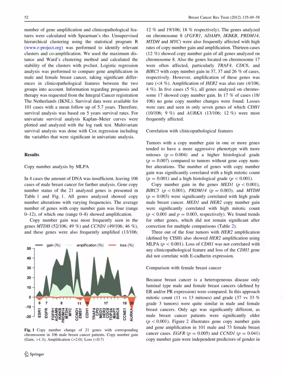
Copy number analysis by MLPA
In 4 cases the amount of DNA was insufficient, leaving 106
cases of male breast cancer for further analysis. Gene copy
number status of the 21 analyzed genes is presented in
Table 1 and Fig. 1. All genes analyzed showed copy
number alterations with varying frequencies. The average
number of genes with copy number gain was four (range
0–12), of which one (range 0–8) showed amplification.
Copy number gain was most frequently seen in the
genes MTDH (52/106; 49 %) and CCND1 (49/106; 46 %),
and these genes were also frequently amplified (13/106;
12 % and 19/106; 18 % respectively). The genes analyzed
on chromosome 8 (FGFR1, ADAM9, IKBKB, PRDM14,
MTDH and MYC) were also frequently affected with high
rates of copy number gain and amplification. Thirteen cases
(12 %) showed copy number gain of all genes analyzed on
chromosome 8. Also the genes located on chromosome 17
were often affected, particularly TRAF4, CDC6, and
BIRC5 with copy number gain in 37, 37 and 26 % of cases,
respectively. However, amplification of these genes was
rare (\4 %). Amplification of HER2 was also rare (4/106;
4 %). In five cases (5 %), all genes analyzed on chromo-
some 17 showed copy number gain. In 17 % of cases (18/
106) no gene copy number changes were found. Losses
were rare and seen in only seven genes of which CDH1
(10/106; 9 %) and AURKA (13/106; 12 %) were most
frequently affected.
Correlation with clinicopathological features
Tumors with a copy number gain in one or more genes
tended to have a more aggressive phenotype with more
mitoses (p = 0.004) and a higher histological grade
(p = 0.007) compared to tumors without gene copy num-
ber alterations. The number of genes with copy number
gain was significantly correlated with a high mitotic count
(p = 0.001) and a high histological grade (p \ 0.001).
Copy number gain in the genes MED1 (p \ 0.001),
BIRC5 (p \ 0.001), PRDM14 (p = 0.003), and MTDH
(p = 0.003) were significantly correlated with high grade
male breast cancer. MED1 and HER2 copy number gain
were significantly correlated with high mitotic count
(p \ 0.001 and p = 0.003, respectively). We found trends
for other genes, which did not remain significant after
correction for multiple comparisons (Table 2).
Three out of the four tumors with HER2 amplification
(defined by CISH) also showed HER2 amplification using
MLPA (p \ 0.001). Loss of CDH1 was not correlated with
any clinicopathological feature and loss of the CDH1 gene
did not correlate with E-cadherin expression.
Comparison with female breast cancer
Because breast cancer is a heterogeneous disease only
luminal type male and female breast cancers (defined by
ER and/or PR expression) were compared. In this approach
mitotic count (11 vs 13 mitoses) and grade (37 vs 33 %
grade 3 tumors) were quite similar in male and female
breast cancers. Only age was significantly different, as
male breast cancer patients were significantly older
(p \ 0.001). Figure 2 illustrates gene copy number gain
and gene amplification in 101 male and 73 female breast
cancer cases. EGFR (p = 0.005) and CCND1 (p = 0.041)
copy number gain were independent predictors of gender in
Fig. 1 Copy number change of 21 genes with corresponding
chromosome in 106 male breast cancer patients. Copy number gain
(Gain, [1.3); Amplification ([2.0); Loss (\0.7)
52 Breast Cancer Res Treat (2012) 135:49–58
123

logistic regression, and these genes were more often gained
in male breast cancer. EMSY (p = 0.004) and CPD
(p = 0.001) copy number gain were also independent
predictors of gender and these genes were more frequently
gained in female breast cancer. Two genes, TRAF4
(p = 0.024) and EMSY (p = 0.041) were more often
amplified in female breast cancer. None of the studied
genes was significantly more frequently amplified in men.
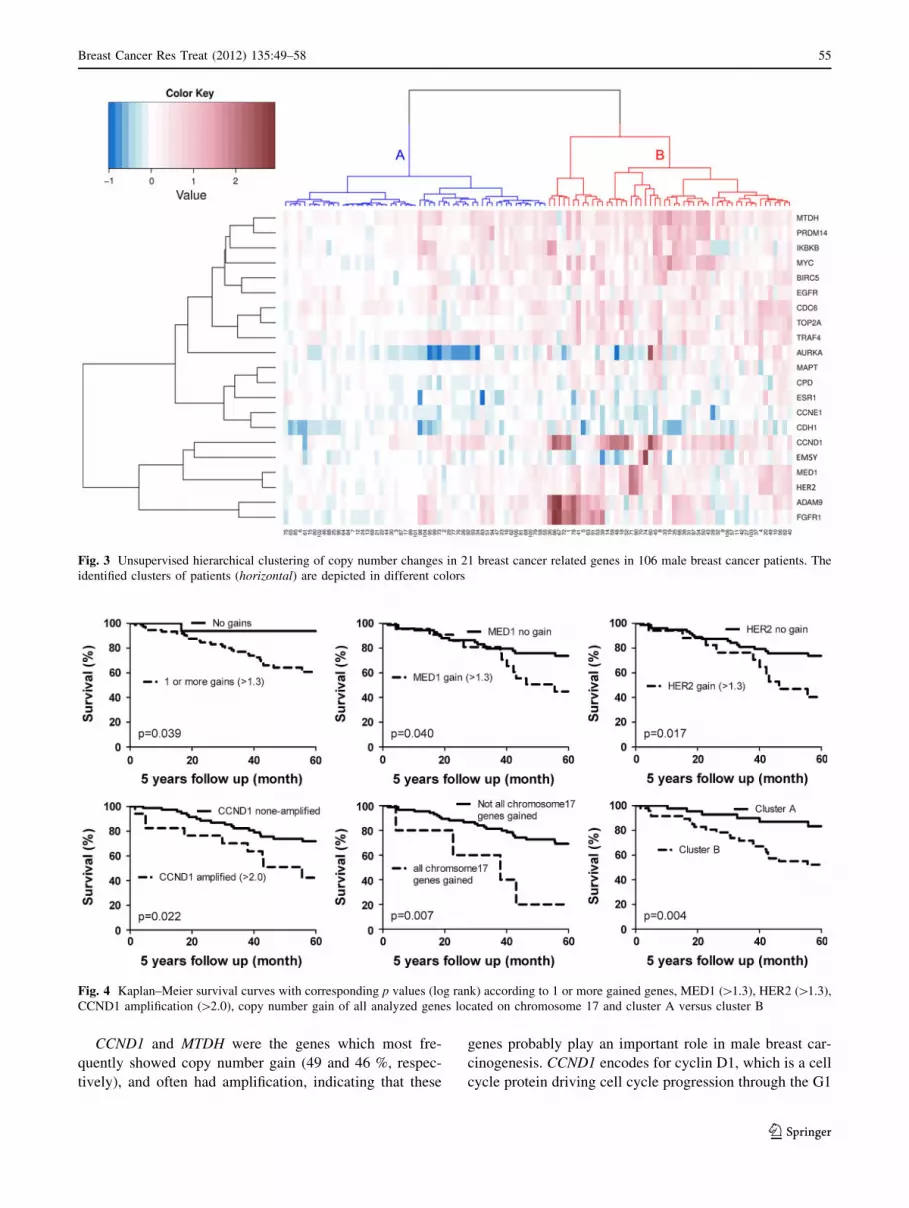
Cluster analysis
Unsupervised hierarchical clustering revealed a separate
gene cluster, consisting of FGFR1, ADAM9, HER2, MED1,
EMSY, and CCND1 (Fig. 3). One small sub-cluster was
formed by FGFR1 and ADAM9 which showed simulta-
neously copy number gain in 29 % of all cases (31/106).
Gains in both genes was correlated with younger age (62 vs
68 years; p = 0.019). No associations with other clinico-
pathological features were found.
Reasoning from the cases, two major clusters were
found (Fig. 3). These clusters were stable according to the
approximately unbiased p values calculated with pvclust
(p \ 0.001). Cluster A consisted of 55 cases and was
characterized by a low rate of gene copy number gain and
gene amplification. Cluster B consisted of 51 cases and
was characterized by CCND1 (73 %), MTDH (69 %),
CDC6 (63 %), ADAM9 (57 %), TRAF4 (57 %) and MYC
(53 %) copy number gain. The male breast cancers in
cluster B showed significantly more mitosis compared to
the tumors in cluster A (8 vs 14 mitosis; p \ 0.001).
Cluster B tumors were also more often grade 3
(p = 0.020) and were larger (2.4 vs 2.0 cm; p = 0.036)
compared to cluster A tumors.
Survival analysis
Grade 3 (p = 0.027), high mitotic count ([8; p = 0.015)
and large tumor size ([2.0 cm; p = 0.036) were correlated
with a decreased 5 years survival. Chemotherapy was
given in 14 % of the cases and 40 % received hormone
therapy. Both treatment regimes did not correlate with
patients’ survival (p = 0.700 and p = 0.140, respectively).
Univariate survival analysis is presented in Fig. 4. Tumors
with one or more gains had a poorer outcome compared
with tumors without gains (p = 0.039). MED1 and HER2
copy number gain also seem to correlate with poor survival
(p = 0.040 and p = 0.017, respectively). In case amplifi-
cation was analyzed the genes CCND1 (p = 0.022) and
EMSY (p = 0.040) were correlated with decreased sur-
vival. However, for EMSY only two cases were amplified.
In case correction for multiple comparisons was performed,
no single prognostic factor remained significant. On the
other hand, tumors with a copy number gain of all genes on
chromosome 17 had a poorer survival (p = 0.007). Cluster
Table 2 Correlation between
gene copy number gain ([1.3)
and clinicopathological features
Tumor size and PR were not
correlated with any of the
studied genes (not shown).
p values were calculated with
Pearson Chi-square or Fisher’s
exact test when appropriate
(number of events \5) for
categorical variables and
ANOVA for continuous
variables. Significant p values
after correction for multiple
comparison (Holm–Bonferroni
method) are depicted in bold.
See Supplementary Table 2 for
full data
LN meta lymph node metastases
Gene Age (mean)
young
Mitoses
high ([8)
Mitoses
(mean) high
Grade
high (3)
LN meta
negative
ER
negative
ESR1
EGFR 0.038
FGFR1 0.019
ADAM9 0.043 0.017 0.004
IKBKB 0.033
PRDM14 0.049 0.003
MTDH 0.019 0.005 0.003
MYC 0.023
CCND1 0.010
EMSY 0.016
CDH1
TRAF4
CPD 0.010
MED1 0.001 <0.001 <0.001
HER2 0.025 0.003 0.014
CDC6 0.027
TOP2A 0.045 0.025 0.013
MAPT
BIRC5 0.018 0.024 <0.001
CCNE1
AURKA
Breast Cancer Res Treat (2012) 135:49–58 53
123

B from unsupervised hierarchical clustering had adverse
patients’ outcome (p = 0.004).
Using a Cox regression analysis, CCND1 amplification
appeared to be the only single gene which was a predictor
of survival aside from grade, mitotic count, and tumor size
(p = 0.015; hazard ratio 3.0). When chemotherapy and
hormone therapy were included in Cox regression, CCND1
was retained as an independent prognosticator. However,
hormone therapy was an independent prognostic factor as
well and was correlated with a favorable prognosis
(p = 0.004; hazard ratio 0.225). Cluster B tumors
(p = 0.009; hazard ratio 3.4) and tumors with copy number
gain of all analyzed genes on chromosome 17 (p = 0.005;
hazard ratio 4.8) were also independent predictors of poor
survival. The multivariate models are supplied in supple-
mentary format (Supplementary Table 3).
Discussion
Gene amplification is an important mechanism of oncogene
activation and is crucial for the development and progres-
sion of cancer. The identification of frequent copy number
change in certain chromosomal regions can lead to iden-
tification of functional important genes in carcinogenesis,
reveal distinctive groups of breast cancer and can be used
as prognostic markers. Knowledge of gene profiling in
male breast cancer is sparse, because male breast cancer is
a rare disease and most studies are based on small single
institutional series. In the present study we used the high
throughput technique MLPA to study gene copy number
alterations of 21 breast cancer related genes in a large
multi-institutional cohort of 106 male breast cancer
patients.
The average amount of genes that showed copy number
gain was four (range 0–12), of which one (range 0–8) was
amplified. 18 cases (17 %) did not show any copy number
change in the studied genes. These 18 cases tended to be
low grade cancers with few mitoses and seem to have
favorable prognosis compared to male breast cancers with
gene copy number gain. The number of genes with copy
number gain was correlated with high grade and a high
mitotic count. This is in line with female breast cancers, as
the genome in high grade female breast cancers is also
more rearranged and these patients have a poor outcome
[23, 24]. Simultaneous copy number gain of all analyzed
genes on chromosome 8, 11, and 17 was seen in 12, 10, and
5 % of the cases, respectively. This points to polysomy or
gain of whole chromosome arms, a finding often seen in
male breast cancer [14]. This is interesting, as polysomy of
e.g., chromosome 17 has been refuted in female breast
cancer [25–30]. In our group of male breast cancer copy
number gain of all genes located on chromosome 17 was an
independent predictor of adverse prognosis.
Using unsupervised hierarchical cluster analysis a small
sub-cluster was formed by FGFR1 and ADAM9. In female
breast cancer, co-amplification of these chromosomal
regions is also a common finding [24, 31]. In addition, two
stable clusters of male breast cancer patients were identi-
fied with additional prognostic value to classical clinico-
pathological prognosticators.
HER2 amplification defined by MLPA in the present
study strongly correlated with HER2 amplification status
defined by CISH on TMA slides. Small differences found
could be due to heterogeneity of tumors which could be
missed or overrepresented in TMA slides. We have also
previously validated MPLA against CISH and FISH [16].
Fig. 2 Comparison of frequency of copy number gain ([1.3, uppergraph) and amplification ([2.0, lower graph) of 17 genes between
luminal type male and female breast cancer. MBC Male breast cancer,
FBC Female breast cancer, Amp amplification. �Genes significantly
more affected in men, *genes significantly more affected in women
54 Breast Cancer Res Treat (2012) 135:49–58
123
CCND1 and MTDH were the genes which most fre-
quently showed copy number gain (49 and 46 %, respec-
tively), and often had amplification, indicating that these
genes probably play an important role in male breast car-
cinogenesis. CCND1 encodes for cyclin D1, which is a cell
cycle protein driving cell cycle progression through the G1
Fig. 3 Unsupervised hierarchical clustering of copy number changes in 21 breast cancer related genes in 106 male breast cancer patients. The
identified clusters of patients (horizontal) are depicted in different colors
Fig. 4 Kaplan–Meier survival curves with corresponding p values (log rank) according to 1 or more gained genes, MED1 ([1.3), HER2 ([1.3),
CCND1 amplification ([2.0), copy number gain of all analyzed genes located on chromosome 17 and cluster A versus cluster B
Breast Cancer Res Treat (2012) 135:49–58 55
123
phase. It also enhances ER-mediated gene transcription and
is especially overexpressed in ER positive female breast
cancer [32]. CCND1 amplification has been linked to ER
positive tumors as well, although some did not find such a
correlation [18, 24, 33]. In the present study, we could not
identify a correlation between CCND1 copy number gain
or amplification and ER status. A clear cut association
between CCND1 amplification and patients’ outcome in
female breast cancer is lacking, but CCND1 amplification
may be associated with a poor prognosis, particularly in ER
positive tumors [23, 24, 33–35]. In the present group of
male breast cancers, tumors with CCND1 copy number
gain tended to have a higher mean mitotic count compared
to tumors without CCND1 amplification, a finding which is
in line with the encoding protein function. More impor-
tantly, amplification of CCND1 was the only single gene
which correlated with poor survival and had additional
prognostic value aside from tumor size, mitotic count, and
histological grade using a Cox regression analysis.
MTDH is involved in several signaling pathways and
amplification of MTDH promotes metastases, enhances
chemo-resistance and is associated with poor outcome in
female breast cancer patients [36]. In line with these
findings in females, we demonstrated that male breast
cancer with MTDH copy number gain showed a more
aggressive phenotype with a high mitotic count and a high
histological grade. However, no correlation with prognosis
and MTDH copy number change was found in our group of
male breast cancer and no correlation with lymph node
metastasis was found either.
The genes located on different amplicons on chromo-
some 8p11 (FGFR1; 29 %, ADAM9; 39 % and IKBKB;
32 %) were also often gained. These genes have been
correlated with ER positive female breast cancers [18].
Since most male breast cancer cases are ER positive (93 %
in the present group), frequent copy number gain of these
genes can be explained by the high ratio of ER positive
tumors [8, 9]. Nevertheless, we could not confirm the
correlation between copy number gain or amplification of
these genes and ER positive tumors in male breast cancer.
However, in view of the low rate of ER negative male
breast cancers in the present study, these results need to be
interpreted with care. It is important to note that FGFR1
amplification enhances tamoxifen resistance, which is
particularly clinically relevant in male breast cancer, as
endocrine therapy is often indicated in these patients [5].
Since FGFR1 copy number gain and amplification seems
to be common in male breast cancer and is suitable for
targeted therapy, this gene could be of further interest in
male breast cancer [37]. ADAM9, which is important in
cell adhesion and tumor cell invasion, has potential in male
breast cancer as well, since this gene is often affected and
could be used for targeted therapy [38, 39].
Among the other genes studied, copy number gain of
MED1, PRDM14, and BIRC5 were associated with a high
grade phenotype, indicating that these genes play a role in
the development or progression of aggressive male breast
cancer. Indeed MED1 copy number gain tends to correlate
with poor survival. We could not confirm the prognostic
relevance of the other genes in male breast cancer patients.
Comparison with 103 female breast cancers revealed
differences in copy number change between male and
female breast cancer in a variety of genes, pointing toward
differences in carcinogenesis. In male breast cancer
CCND1 and EGFR were more often gained than in the
female breast cancer group. In the group of female breast
cancers EMSY and CPD copy number gain were seen more
often than in males. In line with a previous comparative
genomic hybridization study, female breast cancer showed
more frequent amplification in a variety of genes, partic-
ularly in TRAF4 and EMSY [14]. None of the genes studied
were significantly more often amplified in male breast
cancer. Alongside gender specific differences between
male and female breast cancers, differences in genetic
predisposition may also influence the genetic profile of
these tumors. Approximately 10 % of men with breast
cancer are known to have a genetic predisposition, and
especially BRCA2 mutations seem to be important [7].
Differences in BRCA mutations status between male and
female breast cancers would have implications for the
genetic makeup of these tumors and deserves further
investigation.
In conclusion, copy number gain of the genes CCND1
(11q13), TRAF4 (17q11), CDC6 (17q21), and MTDH
(8q22) is very common in male breast cancer ([40 %) and
these genes probably play a role in male breast carcino-
genesis. Tumors with copy number gain of one or more
genes showed a highly malignant phenotype. Also MED1,
PRDM14, MTDH, and BIRC5 seem to be important in the
development or progression of high grade male breast
cancer. Amplification of CCND1 was the most important
single gene as it correlated with poor survival and had
prognostic value in addition to the classical clinicopatho-
logical prognostic factors. Using unsupervised hierarchical
clustering a distinctive group of male breast cancer tumors
was identified with poor survival. Compared to female
breast cancer CCND1 and EGFR were found to be more
frequently amplified in male breast cancer, while in
females EMSY and CPD were more often involved and
more frequent amplifications of TRAF4 and EMSY were
found. Our results point toward important differences in
carcinogenesis between male and female breast cancer,
emphasizing the importance in identifying specific bio-
markers and therapeutic targets for male breast cancer.
Conflict of interest The authors declare no conflict of interest.
56 Breast Cancer Res Treat (2012) 135:49–58
123

Open Access This article is distributed under the terms of the
Creative Commons Attribution License which permits any use, dis-
tribution, and reproduction in any medium, provided the original
author(s) and the source are credited.
References
1. Baak JP, Chin D, van Diest PJ, Ortiz R, Matze-Cok P, Bacus SS
(1991) Comparative long-term prognostic value of quantitative
HER-2/neu protein expression, DNA ploidy, and morphometric
and clinical features in paraffin-embedded invasive breast cancer.
Lab Invest 64(2):215–223
2. Hudis CA (2007) Trastuzumab–mechanism of action and use in
clinical practice. N Engl J Med 357(1):39–51
3. Rodriguez-Pinilla SM, Jones RL, Lambros MB, Arriola E,
Savage K, James M, Pinder SE, Reis-Filho JS (2007) MYC
amplification in breast cancer: a chromogenic in situ hybridisa-
tion study. J Clin Pathol 60(9):1017–1023. doi:jcp.2006.043869
4. Cho EY, Choi YL, Han JJ, Kim KM, Oh YL (2008) Expression
and amplification of Her2, EGFR and cyclin D1 in breast cancer:
immunohistochemistry and chromogenic in situ hybridization.
Pathol Int 58(1):17–25
5. Turner N, Pearson A, Sharpe R, Lambros M, Geyer F, Lopez-
Garcia MA, Natrajan R, Marchio C, Iorns E, Mackay A, Gillett C,
Grigoriadis A, Tutt A, Reis-Filho JS, Ashworth A (2010) FGFR1
amplification drives endocrine therapy resistance and is a thera-
peutic target in breast cancer. Cancer Res 70(5):2085–2094. doi:
0008-5472.CAN-09-3746
6. Nielsen KV, Ejlertsen B, Moller S, Jorgensen JT, Knoop A,
Knudsen H, Mouridsen HT (2008) The value of TOP2A gene
copy number variation as a biomarker in breast cancer: update of
DBCG trial 89D. Acta Oncol 47(4):725–734
7. Korde LA, Zujewski JA, Kamin L, Giordano S, Domchek S,
Anderson WF, Bartlett JM, Gelmon K, Nahleh Z, Bergh J, Cutuli
B, Pruneri G, McCaskill-Stevens W, Gralow J, Hortobagyi G,
Cardoso F (2010) Multidisciplinary meeting on male breast
cancer: summary and research recommendations. J Clin Oncol
28(12):2114–2122. doi:JCO.2009.25.5729
8. Anderson WF, Jatoi I, Tse J, Rosenberg PS (2010) Male breast
cancer: a population-based comparison with female breast cancer.
J Clin Oncol 28(2):232–239. doi:JCO.2009.23.8162
9. Giordano SH, Cohen DS, Buzdar AU, Perkins G, Hortobagyi GN
(2004) Breast carcinoma in men: a population-based study.
Cancer 101(1):51–57. doi:10.1002/cncr.20312
10. Muir D, Kanthan R, Kanthan SC (2003) Male versus female
breast cancers. A population-based comparative immunohisto-
chemical analysis. Arch Pathol Lab Med 127(1):36–41
11. Ge Y, Sneige N, Eltorky MA, Wang Z, Lin E, Gong Y, Guo M
(2009) Immunohistochemical characterization of subtypes of
male breast carcinoma. Breast Cancer Res 11(3):R28
12. Kornegoor R, Verschuur-Maes AH, Buerger H, Hogenes MC, de
Bruin PC, Oudejans JJ, van der Groep P, Hinrichs B, van Diest PJ
(2012) Molecular subtyping of male breast cancer by immunohis-
tochemistry. Mod Pathol 25(3):398–404. doi:10.1038/modpathol.
2011.174
13. Callari M, Cappelletti V, De Cecco L, Musella V, Miodini P,
Veneroni S, Gariboldi M, Pierotti MA, Daidone MG (2010) Gene
expression analysis reveals a different transcriptomic landscape
in female and male breast cancer. Breast Cancer Res Treat
127(3):601–610. doi:10.1007/s10549-010-1015-8
14. Johansson I, Nilsson C, Berglund P, Strand C, Jonsson G, Staaf J,
Ringner M, Nevanlinna H, Barkardottir RB, Borg A, Olsson H,
Luts L, Fjallskog ML, Hedenfalk I (2010) High-resolution
genomic profiling of male breast cancer reveals differences hid-
den behind the similarities with female breast cancer. Breast
Cancer Res Treat 129(3):747–760. doi:10.1007/s10549-010-
1262-8
15. Tommasi S, Mangia A, Iannelli G, Chiarappa P, Rossi E, Ottini
L, Mottolese M, Zoli W, Zuffardi O, Paradiso A (2010) Gene
copy number variation in male breast cancer by aCGH. Anal Cell
Pathol 33(3):113–119
16. Moelans CB, de Weger RA, van Blokland MT, Ezendam C,
Elshof S, Tilanus MG, van Diest PJ (2009) HER-2/neu amplifi-
cation testing in breast cancer by multiplex ligation-dependent
probe amplification in comparison with immunohistochemistry
and in situ hybridization. Cell Oncol 31(1):1–10
17. Moelans CB, Verschuur-Maes AH, van Diest PJ (2011) Frequent
promoter hypermethylation of BRCA2, CDH13, MSH6, PAX5,
PAX6 and WT1 in ductal carcinoma in situ and invasive breast
cancer. J Pathol 225(2):222–231. doi:10.1002/path.2930
18. Moelans CB, de Weger RA, Monsuur HN, Vijzelaar R, van Diest
PJ (2010) Molecular profiling of invasive breast cancer by mul-
tiplex ligation-dependent probe amplification-based copy number
analysis of tumor suppressor and oncogenes. Mod Pathol 23(7):
1029–1039
19. Elston CW, Ellis IO (1991) Pathological prognostic factors in
breast cancer. I. The value of histological grade in breast cancer:
experience from a large study with long-term follow-up. Histo-
pathology 19(5):403–410
20. van Diest PJ, Baak JP, Matze-Cok P, Wisse-Brekelmans EC, van
Galen CM, Kurver PH, Bellot SM, Fijnheer J, van Gorp LH,
Kwee WS et al (1992) Reproducibility of mitosis counting in
2,469 breast cancer specimens: results from the Multicenter
Morphometric Mammary Carcinoma Project. Hum Pathol
23(6):603–607
21. Moelans CB, de Weger RA, Ezendam C, van Diest PJ (2009)
HER-2/neu amplification testing in breast cancer by multiplex
ligation-dependent probe amplification: influence of manual- and
laser microdissection. BMC Cancer 9:4
22. Bunyan DJ, Eccles DM, Sillibourne J, Wilkins E, Thomas NS,
Shea-Simonds J, Duncan PJ, Curtis CE, Robinson DO, Harvey
JF, Cross NC (2004) Dosage analysis of cancer predisposition
genes by multiplex ligation-dependent probe amplification. Br J
Cancer 91(6):1155–1159
23. Al-Kuraya K, Schraml P, Torhorst J, Tapia C, Zaharieva B,
Novotny H, Spichtin H, Maurer R, Mirlacher M, Kochli O, Zuber
M, Dieterich H, Mross F, Wilber K, Simon R, Sauter G (2004)
Prognostic relevance of gene amplifications and coamplifications
in breast cancer. Cancer Res 64(23):8534–8540
24. Cuny M, Kramar A, Courjal F, Johannsdottir V, Iacopetta B,
Fontaine H, Grenier J, Culine S, Theillet C (2000) Relating
genotype and phenotype in breast cancer: an analysis of the
prognostic significance of amplification at eight different genes or
loci and of p53 mutations. Cancer Res 60(4):1077–1083
25. Moelans CB, de Weger RA, van Diest PJ (2010) Absence of
chromosome 17 polysomy in breast cancer: analysis by CEP17
chromogenic in situ hybridization and multiplex ligation-depen-
dent probe amplification. Breast Cancer Res Treat 120(1):1–7.
doi:10.1007/s10549-009-0539-2
26. Moelans CB, de Weger RA, van Diest PJ (2010) Chromosome 17
polysomy without HER2 amplification does not predict response
to lapatinib in metastatic breast cancer–letter. Clin Cancer Res
16(24):6177
27. Moelans CB, van Diest PJ (2011) Re: how do you tell whether a
breast cancer is HER2 positive? Ongoing studies keep debate in
high gear. J Natl Cancer Inst 103(8):698–699
28. Moelans CB, Reis-Filho JS, van Diest PJ (2011) Implications of
rarity of chromosome 17 polysomy in breast cancer. Lancet
Oncol 12(12):1087–1089. doi:S1470-2045(11)70234-0
Breast Cancer Res Treat (2012) 135:49–58 57
123

29. Yeh IT, Martin MA, Robetorye RS, Bolla AR, McCaskill C, Shah
RK, Gorre ME, Mohammed MS, Gunn SR (2009) Clinical vali-
dation of an array CGH test for HER2 status in breast cancer reveals
that polysomy 17 is a rare event. Mod Pathol 22(9):1169–1175
30. Marchio C, Lambros MB, Gugliotta P, Di Cantogno LV, Botta C,
Pasini B, Tan DS, Mackay A, Fenwick K, Tamber N, Bussolati G,
Ashworth A, Reis-Filho JS, Sapino A (2009) Does chromosome 17
centromere copy number predict polysomy in breast cancer? A
fluorescence in situ hybridization and microarray-based CGH
analysis. J Pathol 219(1):16–24. doi:10.1002/path.2574
31. Kwek SS, Roy R, Zhou H, Climent J, Martinez-Climent JA,
Fridlyand J, Albertson DG (2009) Co-amplified genes at 8p12
and 11q13 in breast tumors cooperate with two major pathways in
oncogenesis. Oncogene 28(17):1892–1903
32. van Diest PJ, Michalides RJ, Jannink L, van der Valk P, Peterse
HL, de Jong JS, Meijer CJ, Baak JP (1997) Cyclin D1 expression
in invasive breast cancer. Correlations and prognostic value. Am
J Pathol 150(2):705–711
33. Seshadri R, Lee CS, Hui R, McCaul K, Horsfall DJ, Sutherland
RL (1996) Cyclin DI amplification is not associated with reduced
overall survival in primary breast cancer but may predict early
relapse in patients with features of good prognosis. Clin Cancer
Res 2(7):1177–1184
34. Elsheikh S, Green AR, Aleskandarany MA, Grainge M, Paish CE,
Lambros MB, Reis-Filho JS, Ellis IO (2008) CCND1 amplification
and cyclin D1 expression in breast cancer and their relation with
proteomic subgroups and patient outcome. Breast Cancer Res Treat
109(2):325–335. doi:10.1007/s10549-007-9659-8
35. Kirkegaard T, Nielsen KV, Jensen LB, Campbell FM, Muller S,
Tovey SM, Brown S, Cooke TG, Bartlett JM (2008) Genetic
alterations of CCND1 and EMSY in breast cancers. Histopa-
thology 52(6):698–705
36. Hu G, Chong RA, Yang Q, Wei Y, Blanco MA, Li F, Reiss M, Au JL,
Haffty BG, Kang Y (2009) MTDH activation by 8q22 genomic gain
promotes chemoresistance and metastasis of poor-prognosis breast
cancer. Cancer Cell 15(1):9–20. doi:S1535-6108(08)00379-6
37. Hynes NE, Dey JH (2010) Potential for targeting the fibroblast
growth factor receptors in breast cancer. Cancer Res 70(13):
5199–5202. doi:0008-5472.CAN-10-0918
38. Mazzocca A, Coppari R, De Franco R, Cho JY, Libermann TA,
Pinzani M, Toker A (2005) A secreted form of ADAM9 promotes
carcinoma invasion through tumor-stromal interactions. Cancer
Res 65(11):4728–4738
39. Chin K, DeVries S, Fridlyand J, Spellman PT, Roydasgupta R,
Kuo WL, Lapuk A, Neve RM, Qian Z, Ryder T, Chen F, Feiler H,
Tokuyasu T, Kingsley C, Dairkee S, Meng Z, Chew K, Pinkel D,
Jain A, Ljung BM, Esserman L, Albertson DG, Waldman FM,
Gray JW (2006) Genomic and transcriptional aberrations linked
to breast cancer pathophysiologies. Cancer Cell 10(6):529–541.
doi:S1535-6108(06)00315-1
40. Albertson DG (2008) Conflicting evidence on the frequency of
ESR1 amplification in breast cancer. Nat Genet 40(7):821–822
41. Holst F, Stahl PR, Ruiz C, Hellwinkel O, Jehan Z, Wendland M,
Lebeau A, Terracciano L, Al-Kuraya K, Janicke F, Sauter G,
Simon R (2007) Estrogen receptor alpha (ESR1) gene amplifi-
cation is frequent in breast cancer. Nat Genet 39(5):655–660
42. Moelans CB, Monsuur HN, de Pinth JH, Radersma RD, de Weger
RA, van Diest PJ (2010) ESR1 amplification is rare in breast
cancer and is associated with high grade and high proliferation: a
multiplex ligation-dependent probe amplification study. Cell
Oncol. doi:02L6837051608424
43. Greten FR, Karin M (2004) The IKK/NF-kappaB activation
pathway—a target for prevention and treatment of cancer. Cancer
Lett 206(2):193–199. doi:10.1016/j.canlet.2003.08.029
44. Nishikawa N, Toyota M, Suzuki H, Honma T, Fujikane T,
Ohmura T, Nishidate T, Ohe-Toyota M, Maruyama R, Sonoda T,
Sasaki Y, Urano T, Imai K, Hirata K, Tokino T (2007) Gene
amplification and overexpression of PRDM14 in breast cancers.
Cancer Res 67(20):9649–9657
45. Hughes-Davies L, Huntsman D, Ruas M, Fuks F, Bye J, Chin SF,
Milner J, Brown LA, Hsu F, Gilks B, Nielsen T, Schulzer M,
Chia S, Ragaz J, Cahn A, Linger L, Ozdag H, Cattaneo E, Jor-
danova ES, Schuuring E, Yu DS, Venkitaraman A, Ponder B,
Doherty A, Aparicio S, Bentley D, Theillet C, Ponting CP, Caldas
C, Kouzarides T (2003) EMSY links the BRCA2 pathway to
sporadic breast and ovarian cancer. Cell 115(5):523–535
46. Cleton-Jansen AM (2002) E-cadherin and loss of heterozygosity
at chromosome 16 in breast carcinogenesis: different genetic
pathways in ductal and lobular breast cancer? Breast Cancer Res
4(1):5–8
47. Camilleri-Broet S, Cremer I, Marmey B, Comperat E, Viguie F,
Audouin J, Rio MC, Fridman WH, Sautes-Fridman C, Regnier
CH (2007) TRAF4 overexpression is a common characteristic of
human carcinomas. Oncogene 26(1):142–147
48. Abdelmagid SA, Too CK (2008) Prolactin and estrogen up-regulate
carboxypeptidase-d to promote nitric oxide production and survival
of mcf-7 breast cancer cells. Endocrinology 149(10):4821–4828
49. Zhang D, Jiang P, Xu Q, Zhang X (2011) Arginine and glutamate-
rich 1 (ARGLU1) interacts with mediator subunit 1 (MED1) and is
required for estrogen receptor-mediated gene transcription and
breast cancer cell growth. J Biol Chem 286(20):17746–17754
50. Gonzalez S, Klatt P, Delgado S, Conde E, Lopez-Rios F, San-
chez-Cespedes M, Mendez J, Antequera F, Serrano M (2006)
Oncogenic activity of Cdc6 through repression of the INK4/ARF
locus. Nature 440(7084):702–706
51. Ikeda H, Taira N, Hara F, Fujita T, Yamamoto H, Soh J, Toyooka
S, Nogami T, Shien T, Doihara H, Miyoshi S (2010) The estrogen
receptor influences microtubule-associated protein tau (MAPT)
expression and the selective estrogen receptor inhibitor fulve-
strant downregulates MAPT and increases the sensitivity to tax-
ane in breast cancer cells. Breast Cancer Res 12(3):R43
52. Davis LM, Harris C, Tang L, Doherty P, Hraber P, Sakai Y,
Bocklage T, Doeden K, Hall B, Alsobrook J, Rabinowitz I,
Williams TM, Hozier J (2007) Amplification patterns of three
genomic regions predict distant recurrence in breast carcinoma.
J Mol Diagn 9(3):327–336. doi:S1525-1578(10)60401-1
53. Keyomarsi K, Tucker SL, Buchholz TA, Callister M, Ding Y,
Hortobagyi GN, Bedrosian I, Knickerbocker C, Toyofuku W,
Lowe M, Herliczek TW, Bacus SS (2002) Cyclin E and survival
in patients with breast cancer. N Engl J Med 347(20):1566–1575.
doi:10.1056/NEJMoa021153
54. Staff S, Isola J, Jumppanen M, Tanner M (2010) Aurora-A gene
is frequently amplified in basal-like breast cancer. Oncol Rep
23(2):307–312
58 Breast Cancer Res Treat (2012) 135:49–58
123


