Embed Size (px)
Citation preview
Novel Brn3a cis-acting sequences mediate transcription of humantrkA in neurons
Ximena Valderrama1 and Vikram Misra
Department of Veterinary Microbiology, Western College of Veterinary Medicine, University of Saskatchewan, Saskatoon,
Saskatchewan, Canada
The receptor tropomyosin-related kinase trkA is the cognatehigh-affinity receptor for nerve growth factor (NGF) (Paradaet al. 1992; Huang and Reichardt 2003). In the developingmouse embryo, trkA is expressed in neural crest-derivedneurons destined to become sensory neurons with cell bodiesin trigeminal and dorsal root ganglia (Wyatt and Davies1993; Lei and Parada 2007). It is also present in sympatheticneurons. trkA-NGF interactions in these developing cellscontrol cell fate, differentiation, survival, proliferation,axonal growth and target innervation (Bibel and Barde2000).
In addition to the role of NGF-trkA signaling in preservingsensory neurons, this pathway may also be important inregulating uncontrolled cell division in neuroblastomas andmedulloblastomas, two childhood neuronal cancers. Thus,the presence of trkA in neuroblastomas correlates withfavorable outcomes in patients (Tajima et al. 1998; Eberhartet al. 2001; Ohta et al. 2006). In addition, restoration of trkAexpression in medulloblastoma tumor cells devoid of trkAmakes them responsive to NGF and leads to their differen-
tiation or apoptosis (Muragaki et al. 1997; Chou et al. 2000;Eggert et al. 2000).
While the regulation of the expression of trkA is likelycomplex, the transcription factor Brn3a is thought to play animportant role. Brn3a is a Pit-Oct-Unc homeodomain con-taining transcription factor (Gerrero et al. 1993) that isexpressed in sensory neurons (Fedtsova and Turner 1995;Artinger et al. 1998) and the expression of trkA is greatly
Received June 1, 2007; revised manuscript received November 13, 2007;accepted November 15, 2007.Address correspondence and reprint requests to Vikram Misra,
Department of Veterinary Microbiology, Western College of VeterinaryMedicine, 52 Campus Road, University of Saskatchewan, Saskatoon,Saskatchewan, S7N5B4 Canada. E-mail: [email protected] present address of Ximena Valderrama is Instituto de ProduccionAnimal, Facultad de Ciencias Agrarias, Universidad Austral de Chile,Valdivia, Chile.Abbreviations used: CAT, chloramphenicol acetyltransferase; FBS,
fetal bovine serum; GAPDH, glyceraldehyde 3 phosphate dehy-drogenase; GFP, green fluorescent protein; GST, glutathione-s-transfer-ase; NGF, nerve growth factor.
Abstract
trkA, the receptor tropomyosin-related kinase for nerve growth
factor, is critical not only for the correct spatial and temporal
development of sensory neurons during embryogenesis but
also for the survival of sensory neurons, the differentiation and
apoptosis of neuronal tumors and suppression of latent her-
pes simplex virus genomes. While the regulation of the
expression of trkA is a complex process, the transcription
factor Brn3a is known to play an important role as an en-
hancer of trkA transcription during development in the mouse.
Despite considerable information on the regulation of trkA
during embryogenesis, the mechanisms by which the
expression of trkA is regulated in differentiated neurons, or the
factors that influence its expression in tumor cells, have not
been identified. We initiated studies to determine whether
Brn3a/trkA promoter interactions may be important in a model
of differentiated neurons and in medulloblastoma cells. We
constructed a plasmid that contains 1043 base pairs of
genomic sequences that extend to 30 nucleotides upstream of
trkA coding region. In contrast to previous data, a short 190 bp
region that lies proximal to the trkA initiation codon was suf-
ficient for Brn3a responsiveness in Vero cells. This region was
also sufficient for Brn3a trans-activation in nerve growth fac-
tor-differentiated PC12 cells. At least two portions of the
190 bp fragment bind to Brn3a with an affinity high enough to
be detected in electromobility shift assays. In addition, Brn3a
increased levels of endogenous trkA transcripts in PC12 cells
and initiated trkA expression in medulloblastoma cells, which
normally do not express trkA.
Keywords: Brn3a, medulloblastoma, neurons, nerve growth
factor, trkA.
J. Neurochem. (2008) 105, 425–435.
d JOURNAL OF NEUROCHEMISTRY | 2008 | 105 | 425–435 doi: 10.1111/j.1471-4159.2007.05139.x
� 2007 The AuthorsJournal Compilation � 2007 International Society for Neurochemistry, J. Neurochem. (2008) 105, 425–435 425
reduced in Brn3a )/) mice (Huang et al. 1999). While it doesnot appear to be required for the initiation of trkA expression,Brn3a is required for its sustained expression and for theprotection of sensory neurons from apoptosis (Ma et al. 2003).
Ma and co-workers defined a 457 base pair (bp) segmentof genomic DNA lying upstream from mouse trkA codingsequences that specify appropriate trkA expression duringembryogenesis in neurons of the trigeminal and dorsal rootganglia (Ma et al. 2000, 2003). They identified severalmotifs within this region that bind neuronal proteins. Theseinclude two sites for association with Brn3a. Since NGF-trkAsignaling appears to be important for a variety of neuronalfunctions in differentiated neurons, and may play a role ininducing the apoptosis or differentiation of neuronal tumors,we initiated studies to determine whether other Brn3a/trkApromoter interactions may be important in these cells.
In this article, we show that in contrast to cis-actingsequences identified as being required for correct spatialexpression of the trkA promoter during development of themouse, in differentiated PC12 cells and medulloblastomacells sequences that lie immediately adjacent to the humantrkA promoter play an important role in activation by Brn3a.In addition, exogenous Brn3a increased the expression ofendogenous trkA in differentiated PC12 cells and restoredtrkA expression in human medulloblastoma cells thatnormally do not express trkA.
Materials and methods
Cell cultureCells were grown in 75-cm2 tissue culture flasks. African green
monkey kidney (Vero) cells were maintained in Dulbecco’s modified
Eagle’s medium (with Glutamax, Invitrogen, Carlsbad, CA, USA)
with 10% newborn calf serum and 1% penicillin–streptomycin. Rat
pheochromocytoma (PC12) cells (provided by D. D. Mousseau,
University of Saskatchewan) were maintained in Complete Medium
(CM) containing RPMI 1640 supplemented with 10% horse serum
(heat treated to inactivate complement), 5% fetal bovine serum (FBS),
2 mmol/L glutamine and 1% penicillin–streptomycin. PC12 cells
were plated at a density of 2 · 106 cells per well in six-well collagen-
coated plates. To differentiate PC12 cells, they were ‘fasted’ in low
serum media, RPMI 1640 supplemented with 1% of FBS and 1%
penicillin–streptomycin, overnight before treatment with 50 ng/mL
of nerve growth factor (NGF, Cederlane Laboratories, Hornby,
Ontario, Canada). NGFwas reconstituted in 0.02% acetic acid in FBS
before dilution. Human medulloblastoma cells, ONS-76 (Yamada
et al. 1989), were obtained from Michael Taylor (University of
Toronto) and grown in Dulbecco’s modified Eagle’s medium with
10% FBS and 1% penicillin–streptomycin.
Collagen-coated platesSix-well plates were incubated at 20�C for 1 h with 2 mL of a
0.02 N acetic acid solution containing 50 lg/mL (5 lg/cm2) rat tail
collagen (VWR CACB354236). Plates were then rinsed with 0.1 N
sterile phosphate-buffered saline and used immediately.
PlasmidsThe plasmid pRK-55 (pRK-Brn3a), coding for human Brn3a, was a
gift from Mengqing Xiang (Robert Wood Johnson Medical School).
A 1043 bp portion of the trkA promoter region was amplified using
PCR from HeLa cell genomic DNA ()1073 to )30 from the trkA
initiator codon). The set of primers shown in Fig. 1(b), which were
used to amplify the 1043 bp fragment, included a 5¢ KpnI restrictionenzyme site and a 3¢ XhoI site to facilitate cloning. The amplified
1043 bp fragment was then cloned using a TOPO cloning kit
(Invitrogen). After confirming the sequence of the clonedDNA, it was
transferred to pCAT3basic (Clontech, Mountain View, CA, USA), a
chloramphenicol acetyltransferase (CAT) reporter plasmid. The
resulting plasmid was named ptrkA1043. Constructs generated from
ptrkA1043 are shown in Table 1. Plasmid pGL2, which expresses an
enhanced version of green fluorescent protein (GFP), was obtained
from Invitrogen. pCMVBGal, a plasmid that expresses beta-galacto-
sidase, was obtained from W. Roesler (University of Saskatchewan).
Transfections and CAT-reporter assayVero and PC12 cells were transfected using the calcium phosphate
method as described previously (Chen and Okayama 1988) with
plasmids, purified through CsCl gradients. Vero cells were plated at
a density of 1 · 106 cells per well in six-well plates, transfected the
Consensus Brn3a binding sites:
(A/G)CTCATTAA(T/C)
T(A/T)ATTM1
Human
Mouse AGATCTATTAATTTCTC
AGATCTATTAATTTCTT****************
ACCTAACTTATTCCAA
ACCTAACTTACTCCAA**********-*****
M2
GCAT(A/T)A(T/A)T(A/T)ATGruber et al., 1997
ATG pTrkA1043
Xiang et al., 1995
-1073nt
–30ntM2M1P1
Kpnl
Xhol
P1 :5′-cgctggtacc TTGAGACGGATGGGTTAGTGC-3′
P1 :5′-attactcgagGCAGCCGTCTGTGCGCTCC-3′
B
B BP2
(a)
(b)
(c)
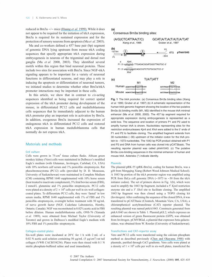
Fig. 1 The trkA promoter. (a) Consensus Brn3a binding sites (Xiang
et al. 1995; Gruber et al. 1997) (b) A schematic representation of the
human trkA-genomic fragment showing the location of the two putative
Brn3a (b) binding motifs (M1, M2) identified in the mouse trkA minimal
enhancer (Ma et al. 2000, 2003). The 457 bp segment required for
appropriate expression during embryogenesis is represented as a
solid box. The sequence and location of primers P1 and P2 used to
amplify human trkA is shown. Nucleotides representing sites for the
restriction endonucleases KpnI and XhoI were added to the 5¢ ends of
P1 and P2 to facilitate cloning. The amplified fragment extends from
30 nucleotides ()30) upstream of the initiator codon for the trkA pro-
tein to )1073 nucleotides. The 1043 bp PCR product obtained with P1
and P2 and DNA from human cells was cloned into pCAT3basic. The
resulting reporter plasmid was called ptrkA1043. (c) The putative
Brn3a core-binding sequences in the minimal enhancer of human and
mouse trkA. Asterisks (*) indicate identity.
Journal Compilation � 2007 International Society for Neurochemistry, J. Neurochem. (2008) 105, 425–435� 2007 The Authors
426 | X. Valderrama and V. Misra

next day with 5 lg of DNA and harvested 48 h later. PC12 cells
were seeded at a density of 2 · 106 per well (collagen-coated six-
well plates) in CM and incubated overnight at 37�C in a 10% CO2
incubator. The next day, medium was replaced with 1.5 mL/well
RPMI 1640 + 10% normal calf serum + 1% penicillin–streptomy-
cin + 2 mmol/L D-glutamine and incubated for at least 1 h at 37�Cin 5% CO2 incubator. DNA concentrations for transfection were
2 lg of reporter plasmid, 1 lg pCMVBGal and 1.5 lg of pRK-
Brn3a DNA/well. After transfection, cells were incubated for 5 h in
5% CO2 and then treated with glycerol as follows: cells were rinsed
with pre-warmed RPMI and then 0.5 mL of pre-warmed 25%
glycerol in 2· BES buffered saline was added. After 45 s, cells were
rinsed twice with RPMI 1640 and 2 mL of PC12 CM. Low serum
medium (1% FBS) was added instead if cells are going to be treated
with NGF. The plates were returned to the 10% CO2 incubator and
harvested 48 h later. To transfect ONS-76 cells, they were plated at a
density of 2 · 105 cells per well of a six-well plate. The next day,
1.5 lg of DNA and 6 lL of Lipofectamine 2000 (Invitrogen) were
mixed and added to the cells overlayed by 2 mL of OptiMEM
(Invitrogen) with 10% fetal serum and no antibiotics. The cells were
analyzed after overnight incubation.
Chloramphenicol acetyl transferase assaysFor chloramphenicol acetyl transferase assays 250 ng or 1 lg of
pCMVBGal, a plasmid specifying beta-galactosidase, was added to
transfection mixtures in Vero and PC12 cells respectively. Lysates
were assayed for beta-galactosidase (Sambrook and Russell 2001)
and for CAT using an ELISA kit (Roche Applied Science,
Indianapolis, IN, USA). CAT values were adjusted for transfection
efficiency using beta-galactosidase values. In figures showing the
results of the CAT assays, each data point is the average of replicate
transfections with the bar representing the range. The data are
representative of at least two, and usually three, independent
experiments that gave the same results.
Flow cytometryViable cells expressing green fluorescent protein were enumerated
by flow cytometry on a Beckman-Coulter Epics XL flow-cytometer
and analyzed using EXPO32 software.
RNA preparation and Real time PCR (QPCR) analysisTotal RNA was extracted from cells in six-well tissue culture plates
using Trizol (Invitrogen) as suggested by the manufacturer and
dissolved in 20 lL of diethylpyrocarbonate-treated water. Next, 5 lgof total RNA was used for first strand cDNA synthesis using
Superscript II reverse transcriptase (Invitrogen). To detect and
quantitate trkA and other transcripts we used Brilliant SYBR Green
QPCR Master Mix (Stratagene, La Jolla, CA, USA). Samples were
amplified in a Mx3005XP QPCR thermocycler (Stratagene) using the
following thermocycle conditions: samples were heated once at 95�Cfor 10 min, 40 cycles of 95�C for 30 s, 60�C for 1 min and 72�C for
1 min. Data were analyzed using the thermocycler-associated
software. For each sample, threshold values for the assayed transcripts
(trkA and Brn3a) were normalized for total input RNA concentrations
using cycle threshold values for transcripts for the ‘normalizer’ house-
keeping gene, glyceraldehyde 3 phosphate dehydrogenase (GAPDH).
The normalized values in each experiment were compared to a
‘calibrator’ sample to determine the relative increase in the amount of
a transcript. Results were analyzed for significant difference using
Student t-test. The primer sets for transcript amplification were used at
a final concentration of 40 nmol/L. The sequences of the primers were
as follows Brn3a-F: 5¢-tggcgtccatctgcgactc-3¢; Brn3a-R: 5¢-ctcaggtt-gttcattttctc-3¢; trkAcd-F: 5¢-gagggcaaaggctctggactcca-3¢; trkAcd-R:5¢-agactccgaagcgcacgatg-3¢: Gapdh-F: 5¢-gcctcctgcaccaccaactg;Gapdh-R: 5¢-gggccatccacagtcttctgg. Following amplification, the
melting curves for the products were generated to ensure that the
product represented a homogenous species. In addition, the PCR
products were analyzed by electrophoresis to ensure that a product of
the predicted size had been generated. The expected sizes of amplified
products were 300 bp for Brn3a, 300 bp for trkA, and 130 bp for
GAPDH. Amplified products were visualized by gel electrophoresis
and when first optimizing the reactions the sequence of the PCR
products was determined.
Electrophoretic mobility shift assayDouble-stranded oligonucleotides (Table 2) were labeled with 32P
and 1 lL of probe containing 10 000–40 000 cpm were incubated
in 5· gel shift buffer containing 20 mmol/L HEPES-KOH,
0.5 mmol/L dithiothreitol, 50 mmol/L KCL and 5 mmol/L MgCl2.
The 5· gel-shift buffer was mixed with 4% Ficoll 400, and 1 lL of
(1 lg/lL) poly (dI-dC) (Roche), and 1 lL (1.5 lg) purified
glutathione-s-transferase (GST)-Brn3a or GST protein. Finally,
water was added to a total volume of 20 lL. The mix was incubated
on ice for 40 min. For competition assays, 100· of unlabeled
double-stranded oligonucleotides were mixed with radiolabeled
probes. Electrophoresis was performed using 6% non-denaturing
polyacrylamide gels in 0.5· tris-borate-EDTA buffer at 4�C at a
constant voltage (150 V) for 3 h. Gels were then dried and either
autoradiographed on Kodak X-OMATR film or using a Typhoon
Trio (GE Healthcare, Piscataway, NJ, USA) multi-purpose scanner.
Purification of GST-Brn3a fusion proteinThe GST-Brn3a (-13) fusion protein does not include the first 13
amino acids of the Brn3a coding sequence. These amino acids have
been shown not to affect Brn3a’s biological activity. Escherichiacoli BL21 competent cells were transformed with a plasmid
expressing GST-Brn3a. 500 mL of cell culture at OD600 = 0.6
was induced to express the fusion protein with 1 mmol/L
Table 1 Genomic human trkA promoter constructs. Mutant con-
structs were generated from plasmid ptrkA1043. Sequences were
deleted by restriction endonuclease digestion and recloned by ligating
complementary ends or by rendering the ends blunt and then ligating
them. The size of the remaining fragments represent only genomic
sequence
Constructs
Deletion from
1043 bp segment
Size of insert
remaining fragment
ptrkA338 BssHII-XhoI 378
ptrkA470 KpnI-XmaIII / ApaI-XhoI 453
ptrkA+190 KpnI-ApaI 175
ptrkA-190 ApaI-XhoI 868
ptrkAM12()/)) SmaI-SmaI/BssHII-BssHII 650
ptrkAM1 BssHII-BssHII 824
ptrkAM2 SmaI-SmaI 869
� 2007 The AuthorsJournal Compilation � 2007 International Society for Neurochemistry, J. Neurochem. (2008) 105, 425–435
Transcription of human trkA in neurons | 427
isopropylthio-b-D-galactoside for 4 h. The cell pellet was resus-
pended in lysis buffer (Tris, 0.01 mol/L, pH 7.5; EDTA, 0.001 mol/
L, sodium chloride, 0.15 mol/L; 15 Triton X-100, Protease Inhibitor
Cocktail [Roche Molecular Biochemicals]; 0.25 mg egg-white
lysozyme) and disrupted by sonication on ice. The supernatant
was purified using Glutathione-Sepharose 4B beads (GE Health-
care). Cells transformed with pGEX-KG, the control plasmid
expressing only GST, were processed simultaneously.
Antibodies and immunoblots and immuno-fluorescenceThe fusion protein GST-Brn3a (-13) was produced inEscherichia coliBL21 (DE3) (Novagen, Ontario, Canada) and purified by using
glutathione-Sepharose beads (GE Healthcare). Antibodies were
produced at the University of Saskatchewan Animal Resources
Centre by immunizing rabbits with about 150 lg of protein in
Freund’s complete adjuvant. The anti-Brn3a serum specifically
detects Brn3a in immunoblots blots of in vitro-synthesized Brn3a
(TnT, Promega, Madison, WI, USA) and lysates of transfected
mammalian cells (data not shown). Procedures for immunoblotting
were as previously described (Akhova et al. 2005). Monoclonal
antibody clone NN18 (Sigma–Aldrich, Ontario, Canada), which
stains a 160 000 molecular weight neurofilament protein, was used in
some immunoblots. For immuno-staining (Lu andMisra 2000; Raggo
et al. 2002), cells were fixed in 1% paraformaldehyde, permeablized
with 0.2% Triton X-100 and treated with diluted rabbit anti-Brn3a
serum and Alexa546 labeled goat anti-rabbit antibodies (Invitrogen).
Results
The 457 bp segment of DNA identified by Ma et al. (2000)as sufficient for appropriate trkA expression during mouse
embryogenesis contains binding sites for many transcriptionfactors. Within this segment the authors identified two sitesfor the binding of Brn3a (Ma et al. 2003). Since NGF-trkAsignaling appears to be important for a variety of neuronalfunctions in differentiated neurons and may play a role ininducing the apoptosis or differentiation of neuronal tumors,we initiated studies to determine whether other Brn3a/trkApromoter interactions may be important in these cells.
Using PCR we amplified a 1043 bp region of DNA thatlies upstream from the coding sequences for human trkA.This fragment contains regions homologous to the 457 bpDNA segment important in mice, including the putativeBrn3a-binding motifs (Fig. 1b). An analysis of the sequenceof the amplified fragment showed that it was similar to thecorresponding portion of the mouse genome (66%) withalmost perfect sequence identity for the putative Brn3a-binding sites (Fig. 1c). To assess the ability of the DNAfragment to function as a promoter, we cloned it upstream ofthe coding sequences of the reporter protein CAT inpCAT3Basic. We refer to this construct as ptrkA1043.
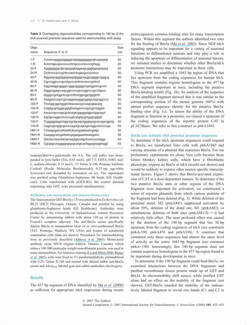
Brn3a can activate trkA proximal promoter sequencesTo determine if the trkA upstream sequences could respondto Brn3a, we transfected Vero cells with ptrkA1043 andvarying amounts of a plasmid that expresses Brn3a. For ourpreliminary experiments we chose Vero cells because theseGreen Monkey kidney cells, which have a fibroblasticphenotype, express no Brn3a or trkA (results not shown) andwould be unlikely to express other neuron specific transcrip-tional factors. Figure 2 shows that Brn3a-activated expres-sion of CAT in a dose-dependent manner. To determine if thetwo putative Brn3a sites or other regions of the DNAfragment were important for activation, we constructed aseries of reporter plasmids from which various portions ofthe fragment had been deleted (Fig. 3). While deletion of theproximal motif, M2 (ptrkAM1) suppressed activation byabout 50%, deletion of the distal site, M1 (ptrkAM2), orsimultaneous deletion of both sites (ptrkAM1/2()/))) hadrelatively little effect. The most profound effect was causedby the deletion of the 190 bp segment that lies 30 bpupstream from the coding sequences of trkA (see constructsptrkA-190, ptrkA470 and ptrkA338). A construct thatcontained only these sequences had almost the same levelof activity as the entire 1043 bp fragment (see constructptrkA+190). Interestingly, this 190 bp segment does notcontain sequences homologous to the 457 bp region found tobe important during development in mice.
To determine if the 190 bp fragment could bind Brn3a, weexamined interactions between the DNA fragments andpurified recombinant fusion protein made up of GST andBrn3a. In electromobility shift assays, while purified GSTalone had no effect on the mobility of the fragment (notshown), GST-Brn3a retarded the mobility of the radioac-tively labeled fragment to reveal two bands (C1 and C2 in
Table 2 Overlapping oligonucleotides corresponding to 190 bp of the
trkA proximal promoter sequence used for electromobility shift assay
Oligo
name Sequence 5¢ to 3¢Size
(nt)
1-F Cctaacaggggagggggcagaggggggggcgtcagagagt 40
1-R Actctctgacgcccccccctctgccccctcccctgttagg 40
24-F Aggggggggcgtcagagagtaggaagcgggtggagaagag 40
24-R Ctcttctccacccgcttcctactctctgacgcccccccct 40
40-F Aggaagcgggtggagaagaggggcaaggcggggccgggcg 40
40-R Cgcccggccccgccttgcccctcttctccacccgcttcct 40
60-F Gggcaaggcggggccgggcgggggccgctggctccgccct 40
60-R Agggcggagccagcggcccccgcccggccccgccttgccc 40
80-F Ggggccgctggctccgccctttcctggcggctgggtcttt 40
80-R Aaagacccagccgccaggaaagggcggagccagcggcccc 40
100-F Ttcctggcggctgggtctttaacaccgcccagcgcacatg 40
100-R Catgtgcgctgggcggtgttaaagacccagccgccaggaa 40
120-F Aacaccgcccagcgcacatgtcgggggaggcctggcagct 40
120-R Agctgccaggcctcccccgacatgtgcgctgggcggtgtt 40
140-F Tcgggggaggcctggcagctgcagctgggagcgcacagacggctg 45
140-R Cagccgtctgtgcgctcccagctgcagctgccaggcctcccccga 45
HM1-F Tctaagagatctattaatttcttcacgaataaatcgatgc 40
HM1-R Caagagcatcgatttattcgtgaagaaattaatagatct 39
HM2-F Gttctacctaacttactacaagtgacatgctcactcccctaggc 44
HM2-R Cgcgtgcctaggggagtgagcatgtcacttggagtaagttaggt 44
Journal Compilation � 2007 International Society for Neurochemistry, J. Neurochem. (2008) 105, 425–435� 2007 The Authors
428 | X. Valderrama and V. Misra
lane 2, Fig. 4a). An excess of unlabeled fragment inhibitedthe formation of these complexes (lane 3) and inclusion ofantiserum against GST-Brn3a retarded their mobility further
(lane 4). These results show that the 190 bp fragmentcontained one and possibly more sites for binding Brn3a.
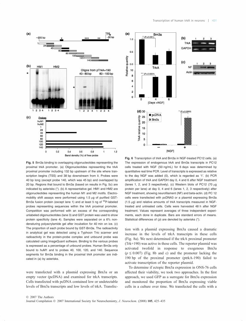
To identify which portion of the 190 bp fragment boundBrn3a, we repeated the electromobility shift assays withoverlapping 40 bp double-stranded oligonucleotides span-ning the 190 bp fragment (Fig. 5a). We also includedoligonucleotides representing HM1 and HM2, humansequences containing homologues of the putative mouseBrn3a binding sites (M1 and M2) described earlier by Maet al. (2000). HM1 specifically bound Brn3a (Fig. 5b lane 2)while there was no specific binding for HM2. Four of theoligonucleotides representing the 190 bp fragment appearedto form complexes with Brn3a. A gel showing analysis ofoligonucleotides 40 and 60 is shown in Fig. 5(b). Using aphosphor-imager and image analysis software we calculatedthe amount of radioactivity in the retarded bands (Brn3a-DNA complexes, * in Fig. 5(b)) from the various oligonu-cleotides expressed as a percentage of unbound radioactivity(Fig. 5c). Our results suggest that Brn3a binds to at least tworegions of the 190 bp fragment (denoted by asterisks inFig. 5a).
Effect of NGF-nduced differentiation and exogenousBrn3a on the expression of endogenous trkA in PC12 cellsTo determine if our observations in Vero cells for Brn3aactivation of the 190 bp fragment were valid for neuronalcells, we examined PC12 cells. These are rat pheochromo-cytoma cells that differentiate into sympathetic neuron-likecells when treated with NGF and low concentrations ofserum. They have been used extensively as an in vitro modelto study ligand-receptor interactions and cellular differenti-ation in response to NGF (Greene and Tischler 1976). Wefirst measured the level of trkA and Brn3a transcripts inPC12 cells over six days of NGF treatment. The levels oftrkA transcripts in these cells increased (Fig. 6a and b day 0,4, 6 in lanes 1, 2, and 3, respectively) in response to NGFwhile levels of Brn3a decreased (Fig. 6a). We confirmedprogressive differentiation of cells by the detection ofincreasing levels of neurofilament, a marker for differentia-tion (Fig. 6c day 0, 4, 6 in lanes 1, 2, and 3, respectively). Wenext transfected NGF-treated and mock-treated PC12 cellswith a plasmid expressing Brn3a. Forty-eight hours aftertreatment, NGF-treated cells (+) expressing exogenous Brn3a(when compared with NGF-treated cells transfected with anempty vector, pcDNA) showed a 25-fold increase in the levelof trkA transcripts (Fig. 6d). There was no such increase inmock-treated cells. NGF-treated cells expressing Brn3a aredesignated as (+) and all transfected with an empty vector as()) in Fig. 6d.
We next examined PC12 cells to determine if the 190 bpproximal trkA promoter was active in these cells. NGF-treated or mock-treated cells were transfected with the emptyreporter plasmid (pCAT3basic) or ptrkA+190. The 190 bpfragment was active in both NGF-treated and mock-treated
Fig. 3 Sequences proximal to trkA coding sequences required for
Brn3a activated transcription in Vero cells. trkA genomic fragments
were generated by restriction enzyme digestion and re-ligation of the
fragment. The striped boxes represent the putative Brn3a binding
motifs (M1, M2). Horizontal lines represent sequences deleted from
original clone. The different reporter constructs (0.25 lg) or pCAT3-
basic were introduced into Vero cells together with 3 lg of a plasmid
specifying Brn3a. A plasmid expressing beta-galactosidase was
included in the reactions to assess transfection efficiency. Cells were
harvested 48 h after transfection and CAT and beta-galactosidase
measured. CAT activity of the samples is expressed relative to the
activity of plasmid ptrkA1043 (100%). Values are the means of those
obtained from two independent experiments, each analyzed in dupli-
cate. Bars indicate the standard deviation from the mean.
Fig. 2 Brn3a activates the trkA promoter in Vero cells. Vero (African
Green Monkey kidney) cells were transfected with ptrkA1043, a
reporter plasmid with the human trkA upstream sequences linked to
the coding sequences for chloramphenicol acetyl transferase (CAT)
and increasing amounts of a plasmid specifying human Brn3a. Each
reaction contained equal amounts of a plasmid coding for beta-
galactosidase and the total DNA concentration of each reaction was
adjusted to 5.5 lg with pcDNA. Cells were harvest 48 h after trans-
fection and CAT and beta-galactosidase activities measured. The CAT
activity in each sample was adjusted for transfection efficiency using
beta-galactosidase values. The CAT activity of samples without Brn3a
expressing plasmid (0) was taken as 1. The results are from a repre-
sentative experiment. The values are the mean of replicates in the
experiment while the bars represent the standard deviation from the
mean.
� 2007 The AuthorsJournal Compilation � 2007 International Society for Neurochemistry, J. Neurochem. (2008) 105, 425–435
Transcription of human trkA in neurons | 429

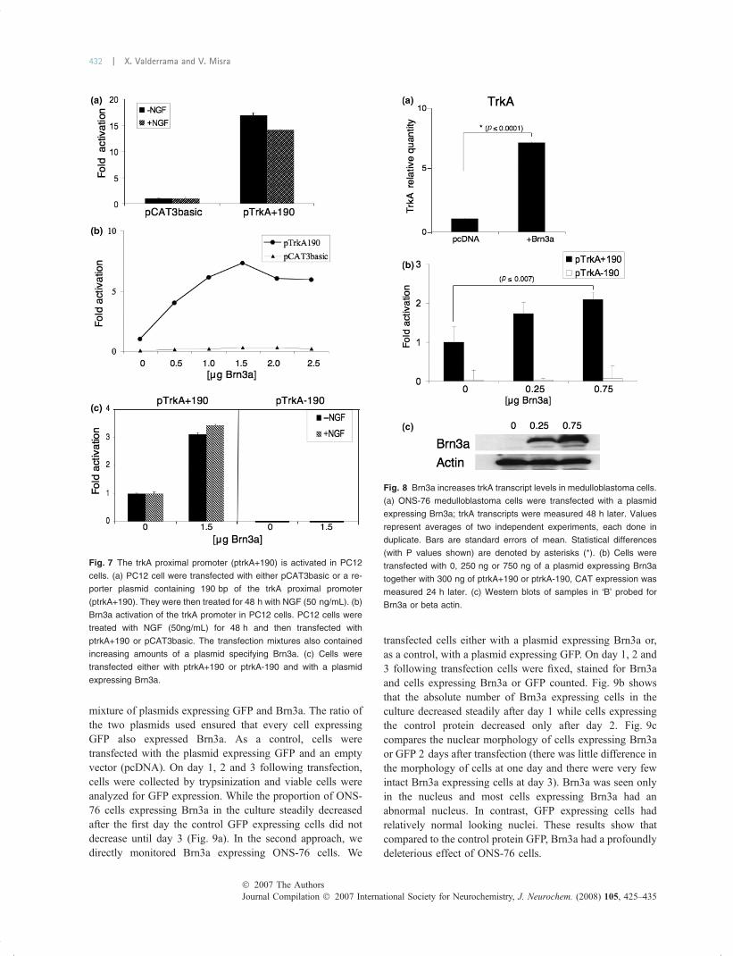
PC12 cells (Fig. 7a), indicating that in these cells the 190 bpfragment has a relatively high level of basal activity. This isin contrast to Vero cells, where in the absence of activatorssuch as exogenous Brn3a, the 190 bp fragment has almost noactivity when compared with the empty reporter plasmid,pCAT3basic (not shown). Despite high the levels of basalactivity in NGF-treated PC12 cells, ptrkA+190 was activatedalmost eightfold by exogenous Brn3a (Fig. 7b). When PC12cells were transfected with a plasmid lacking the proximalpromoter (trkA)190), no activity was observed in thepresence or absence of NGF, indicating that trkA+190 isresponsible for trkA gene activation by Brn3a (Fig. 7c).
Activation of trkA in medulloblastoma cellsMedulloblastomas are childhood neuronal cancers that do notnormally express trkA. However, if medulloblastoma cellsare induced to express trkA they become responsive to NGF-trkA signaling (Muragaki et al. 1997; Chou et al. 2000;Eggert et al. 2000) and this is associated with a favorableprognosis. We examined the ONS-76 medulloblastoma cellline, which was derived from a large cerebellar tumor in a 2-year-old girl (Yamada et al. 1989). The cells express bothglial and neuronal markers and can be induced to differen-tiate into neuron-like cells by treatment with dibutryl cyclicAMP (Moriuchi et al. 1997; Park et al. 1998). ONS-76 cells
Fig. 4 Brn3a binds directly to trkA proximal
promoter sequences. (a) Electromobility
shift assays were performed using 1.5 lg of
purified GST-Brn3a fusion protein and at
least 5 ng of 32P-labeled double stranded
oligonucleotides representing the 190 bp
trkA proximal sequences. Lane 1 contains
probe alone; lane 2, probe + GST-Brn3a;
lane 3, probe + GST-Brn3a + 100-fold
excess of unlabeled competitor; lane 4,
probe + GST-Brn3a + Anti-Brn3a serum.
Samples were analyzed on a 6% non-
denaturing polyacrylamide gel after incu-
bating reaction for 40 min on ice. C1 and C2
represent complexes formed with Brn3a
and the labeled oligonucleotide. Asterisks
(* and **) note the location of bands with
decreased electrophoretic mobility in the
presence of antibodies against Brn3a. (b)
Sequence homology of the trkA 5¢ proximal
promoter region. Nucleotide sequence
homology between species is indicated by
asterisks (*), and sequences that are
missing in the mouse and rat trkA gene are
indicated by dashes (-). Potential Brn3a
binding site depicted by the bold box and
transcription start site (TSS) depicted by a
thin lined box. Start codon ATG is shown in
italics and underlined. (c) Potential binding
site for Brn3a similar to consensus binding
sequences in other promoters (Hill et al.
1997; Smith et al.1997), identical nucleo-
tides at corresponding positions are indi-
cated by asterisks (*).
Journal Compilation � 2007 International Society for Neurochemistry, J. Neurochem. (2008) 105, 425–435� 2007 The Authors
430 | X. Valderrama and V. Misra
were transfected with a plasmid expressing Brn3a or anempty vector (pcDNA) and examined for trkA transcripts.Cells transfected with pcDNA contained low or undetectablelevels of Brn3a transcripts and low levels of trkA. Transfec-
tion with a plasmid expressing Brn3a caused a dramaticincrease in the levels of trkA transcripts in these cells(Fig. 8a). We next determined if the trkA proximal promoter(Trk+190) was active in these cells. The reporter plasmid wasactivated twofold in response to exogenous Brn3a(p £ 0.007) (Fig. 8b and c) and the promoter lacking the190 bp of the proximal promoter (ptrkA-190) failed toactivate transcription of the reporter plasmid.
To determine if ectopic Brn3a expression in ONS-76 cellsaffected their viability, we took two approaches. In the firstapproach, we used GFP as a surrogate for Brn3a expressionand monitored the proportion of Brn3a expressing viablecells in a culture over time. We transfected the cells with a
Fig. 5 Brn3a binding to overlapping oligonucleotides representing the
proximal trkA promoter. (a) Oligonucleotides representing the trkA
proximal promoter including 132 bp upstream of the site where tran-
scription begins (TSS) and 38 bp downstream from it. Probes were
40 bp long (except probe 140, which was 45 bp) and overlapped by
20 bp. Regions that bound to Brn3a (based on results in Fig. 5c) are
indicated by asterisks (*). (b) A representative gel. HM1 and HM2 are
oligonucleotides representing the human M1 and M2 motifs. Electro-
mobility shift assays were performed using 1.5 lg of purified GST-
Brn3a fusion protein (except lane 1) and at least 5 ng of 32P-labeled
probes representing sequences within the trkA proximal promoter.
Competition was performed with an excess of the corresponding
unlabeled oligonucleotides (lane 3) and GST protein was used to show
protein specificity (lane 4). Samples were separated on a 6% non-
denaturing polyacrylamide gel after incubation for 40 min on ice. (c)
The proportion of each probe bound by GST-Brn3a. The radioactivity
in analytical gel was detected using a Typhoon Trio scanner and
radioactivity in the protein-probe complex and unbound probe was
calculated using ImageQuant software. Binding to the various probes
is expressed as a percentage of unbound probes. Human Brn3a only
bound to huM1 and to probes 40, 100, 120, and 140. Sequence
segments for Brn3a binding in the proximal trkA promoter are indi-
cated in (a) by asterisks.
Fig. 6 Transcription of trkA and Brn3a in NGF-treated PC12 cells. (a)
The expression of endogenous trkA and Brn3a transcripts in PC12
cells treated with NGF (50 ng/mL) for 6 days was determined by
quantitative real time PCR. Level of transcripts is expressed as relative
to the day NGF was added (0), which is regarded as ‘1’. (b) PCR
amplification of trkA and GAPDH day 0, 4 and 6 after NGF treatment
(lanes 1, 2, and 3 respectively). (c) Western blots of PC12 (70 lg
protein per lane) at day 0, 4 and 6 (lanes 1, 2, 3 respectively) after
NGF treatment, showing neurofilament (NF) and beta-actin. (d) PC 12
cells were transfected with pcDNA3 or a plasmid expressing Brn3a
(1.5 lg) and relative amounts of trkA transcripts measured in NGF-
treated and untreated cells. Cells were harvested 48 h after NGF
treatment. Values represent averages of three independent experi-
ments, each done in duplicate. Bars are standard errors of means.
Statistical differences of (p) are denoted by asterisks (*).
� 2007 The AuthorsJournal Compilation � 2007 International Society for Neurochemistry, J. Neurochem. (2008) 105, 425–435
Transcription of human trkA in neurons | 431
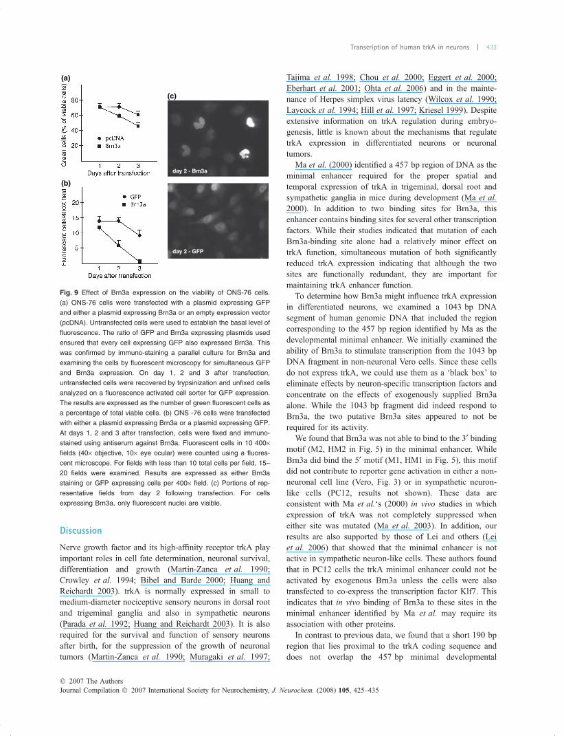
mixture of plasmids expressing GFP and Brn3a. The ratio ofthe two plasmids used ensured that every cell expressingGFP also expressed Brn3a. As a control, cells weretransfected with the plasmid expressing GFP and an emptyvector (pcDNA). On day 1, 2 and 3 following transfection,cells were collected by trypsinization and viable cells wereanalyzed for GFP expression. While the proportion of ONS-76 cells expressing Brn3a in the culture steadily decreasedafter the first day the control GFP expressing cells did notdecrease until day 3 (Fig. 9a). In the second approach, wedirectly monitored Brn3a expressing ONS-76 cells. We
transfected cells either with a plasmid expressing Brn3a or,as a control, with a plasmid expressing GFP. On day 1, 2 and3 following transfection cells were fixed, stained for Brn3aand cells expressing Brn3a or GFP counted. Fig. 9b showsthat the absolute number of Brn3a expressing cells in theculture decreased steadily after day 1 while cells expressingthe control protein decreased only after day 2. Fig. 9ccompares the nuclear morphology of cells expressing Brn3aor GFP 2 days after transfection (there was little difference inthe morphology of cells at one day and there were very fewintact Brn3a expressing cells at day 3). Brn3a was seen onlyin the nucleus and most cells expressing Brn3a had anabnormal nucleus. In contrast, GFP expressing cells hadrelatively normal looking nuclei. These results show thatcompared to the control protein GFP, Brn3a had a profoundlydeleterious effect of ONS-76 cells.
Fig. 7 The trkA proximal promoter (ptrkA+190) is activated in PC12
cells. (a) PC12 cell were transfected with either pCAT3basic or a re-
porter plasmid containing 190 bp of the trkA proximal promoter
(ptrkA+190). They were then treated for 48 h with NGF (50 ng/mL). (b)
Brn3a activation of the trkA promoter in PC12 cells. PC12 cells were
treated with NGF (50ng/mL) for 48 h and then transfected with
ptrkA+190 or pCAT3basic. The transfection mixtures also contained
increasing amounts of a plasmid specifying Brn3a. (c) Cells were
transfected either with ptrkA+190 or ptrkA-190 and with a plasmid
expressing Brn3a.
Fig. 8 Brn3a increases trkA transcript levels in medulloblastoma cells.
(a) ONS-76 medulloblastoma cells were transfected with a plasmid
expressing Brn3a; trkA transcripts were measured 48 h later. Values
represent averages of two independent experiments, each done in
duplicate. Bars are standard errors of mean. Statistical differences
(with P values shown) are denoted by asterisks (*). (b) Cells were
transfected with 0, 250 ng or 750 ng of a plasmid expressing Brn3a
together with 300 ng of ptrkA+190 or ptrkA-190, CAT expression was
measured 24 h later. (c) Western blots of samples in ‘B’ probed for
Brn3a or beta actin.
Journal Compilation � 2007 International Society for Neurochemistry, J. Neurochem. (2008) 105, 425–435� 2007 The Authors
432 | X. Valderrama and V. Misra
Discussion
Nerve growth factor and its high-affinity receptor trkA playimportant roles in cell fate determination, neuronal survival,differentiation and growth (Martin-Zanca et al. 1990;Crowley et al. 1994; Bibel and Barde 2000; Huang andReichardt 2003). trkA is normally expressed in small tomedium-diameter nociceptive sensory neurons in dorsal rootand trigeminal ganglia and also in sympathetic neurons(Parada et al. 1992; Huang and Reichardt 2003). It is alsorequired for the survival and function of sensory neuronsafter birth, for the suppression of the growth of neuronaltumors (Martin-Zanca et al. 1990; Muragaki et al. 1997;
Tajima et al. 1998; Chou et al. 2000; Eggert et al. 2000;Eberhart et al. 2001; Ohta et al. 2006) and in the mainte-nance of Herpes simplex virus latency (Wilcox et al. 1990;Laycock et al. 1994; Hill et al. 1997; Kriesel 1999). Despiteextensive information on trkA regulation during embryo-genesis, little is known about the mechanisms that regulatetrkA expression in differentiated neurons or neuronaltumors.
Ma et al. (2000) identified a 457 bp region of DNA as theminimal enhancer required for the proper spatial andtemporal expression of trkA in trigeminal, dorsal root andsympathetic ganglia in mice during development (Ma et al.2000). In addition to two binding sites for Brn3a, thisenhancer contains binding sites for several other transcriptionfactors. While their studies indicated that mutation of eachBrn3a-binding site alone had a relatively minor effect ontrkA function, simultaneous mutation of both significantlyreduced trkA expression indicating that although the twosites are functionally redundant, they are important formaintaining trkA enhancer function.
To determine how Brn3a might influence trkA expressionin differentiated neurons, we examined a 1043 bp DNAsegment of human genomic DNA that included the regioncorresponding to the 457 bp region identified by Ma as thedevelopmental minimal enhancer. We initially examined theability of Brn3a to stimulate transcription from the 1043 bpDNA fragment in non-neuronal Vero cells. Since these cellsdo not express trkA, we could use them as a ‘black box’ toeliminate effects by neuron-specific transcription factors andconcentrate on the effects of exogenously supplied Brn3aalone. While the 1043 bp fragment did indeed respond toBrn3a, the two putative Brn3a sites appeared to not berequired for its activity.
We found that Brn3a was not able to bind to the 3¢ bindingmotif (M2, HM2 in Fig. 5) in the minimal enhancer. WhileBrn3a did bind the 5¢ motif (M1, HM1 in Fig. 5), this motifdid not contribute to reporter gene activation in either a non-neuronal cell line (Vero, Fig. 3) or in sympathetic neuron-like cells (PC12, results not shown). These data areconsistent with Ma et al.‘s (2000) in vivo studies in whichexpression of trkA was not completely suppressed wheneither site was mutated (Ma et al. 2003). In addition, ourresults are also supported by those of Lei and others (Leiet al. 2006) that showed that the minimal enhancer is notactive in sympathetic neuron-like cells. These authors foundthat in PC12 cells the trkA minimal enhancer could not beactivated by exogenous Brn3a unless the cells were alsotransfected to co-express the transcription factor Klf7. Thisindicates that in vivo binding of Brn3a to these sites in theminimal enhancer identified by Ma et al. may require itsassociation with other proteins.
In contrast to previous data, we found that a short 190 bpregion that lies proximal to the trkA coding sequence anddoes not overlap the 457 bp minimal developmental
(a)
(b)
(c)
day 2 - Bm3a
day 2 - GFP
Fig. 9 Effect of Brn3a expression on the viability of ONS-76 cells.
(a) ONS-76 cells were transfected with a plasmid expressing GFP
and either a plasmid expressing Brn3a or an empty expression vector
(pcDNA). Untransfected cells were used to establish the basal level of
fluorescence. The ratio of GFP and Brn3a expressing plasmids used
ensured that every cell expressing GFP also expressed Brn3a. This
was confirmed by immuno-staining a parallel culture for Brn3a and
examining the cells by fluorescent microscopy for simultaneous GFP
and Brn3a expression. On day 1, 2 and 3 after transfection,
untransfected cells were recovered by trypsinization and unfixed cells
analyzed on a fluorescence activated cell sorter for GFP expression.
The results are expressed as the number of green fluorescent cells as
a percentage of total viable cells. (b) ONS -76 cells were transfected
with either a plasmid expressing Brn3a or a plasmid expressing GFP.
At days 1, 2 and 3 after transfection, cells were fixed and immuno-
stained using antiserum against Brn3a. Fluorescent cells in 10 400·fields (40· objective, 10· eye ocular) were counted using a fluores-
cent microscope. For fields with less than 10 total cells per field, 15–
20 fields were examined. Results are expressed as either Brn3a
staining or GFP expressing cells per 400· field. (c) Portions of rep-
resentative fields from day 2 following transfection. For cells
expressing Brn3a, only fluorescent nuclei are visible.
� 2007 The AuthorsJournal Compilation � 2007 International Society for Neurochemistry, J. Neurochem. (2008) 105, 425–435
Transcription of human trkA in neurons | 433
enhancer, was sufficient for trkA promoter activation byBrn3a in Vero cells (Fig. 3). This region was also sufficientfor Brn3a responsiveness in NGF-differentiated PC12 cells(Fig. 7). At least two portions of the 190 bp fragment bind toBrn3a with an affinity high enough to be detected inelectromobility shift assays (Fig. 5).
Sacristan et al. (1999) and Chang et al. (1998) defined thesite at which transcription of the trkA gene begins (TSS inFig. 4b) and the region of the trkA promoter that is sufficientto direct accurate transcription in trkA. This region encom-passes 150 bp upstream of the transcription start site in themouse and 138 bp upstream of the start of translation fortrkA in humans. These regions are a part of the 190 bpidentified by us as important for Brn3a responsiveness. Inaddition, in our study exogenous Brn3a was only able toactivate the transcription of endogenous trkA in the sympa-thetic-like neuron PC12 (Fig. 8) and not in Vero cellssuggesting that this activation is tissue-specific and requiresother cell type-specific factors. These data are also consistentwith those of Sacristan et al. (1999) and Chang et al. (1998)who found that neuron-specific proteins bound to regions ofthe trkA proximal promoter.
The 190 bp proximal promoter sequence of the humantrkA promoter has a high level of homology (82%) withmouse and rat sequences but it also contains 34 additionalnucleotides (Fig. 4b). The region does not contain theputative motifs for Brn3a found in the trkA minimal enhancerdescribed by Ma et al. (Ma et al. 2000). However, the 190 bpsequence does have a motif with some similarities to Brn3a-DNA binding sequences identified by Xiang et al. (1995) andGruber et al. (1997) (Fig. 4c). These authors defined twoconsensus DNA sequences for Brn3a binding. The sequencesare associated with the activation of anti-apoptotic genes[Bcl-2 and Bcl-xL (Smith et al. 1998)], genes linked todifferentiation [alpha-internexin (Budhrammahadeo et al.1995)], neurofilament (Smith et al. 1997) and synaptic genes[SNAP25 (Lakin et al. 1995)]. The potential Brn3a bindingsequence in the proximal trkA promoter is 40 nt upstream ofthe transcription start site for trkA. It differs by only 2 nt outof 11 nt from the one described by Xiang et al. (1995).
Since nociceptive neurons depend on trkA and NGFduring development and trkA is active in differentiatedsensory neurons, it is likely that Brn3a has a distinctive rolein NGF-trkA signaling in these cells. In addition, Brn3a nullmice not only have aggressive apoptosis of sensory neuronsbut also have reduced trkA transcription.
Because our results showed that Brn3a activates trkAexpression, restoration of which has been shown to inhibitcell-division in medulloblastomas, we determined if Brn3acould induce trkA expression in these cells. As expected,Brn3a was able to increase trkA transcription in ONS-76medulloblastoma cells (Fig. 8) and the 190 bp fragmentactivated reporter gene expression. In addition, ectopicexpression of Brn3a in ONS-76 cells had a deleterious effect
on the viability of the cells (Fig. 9). Although at this stage wedo not know if this effect of Brn3a on medulloblastoma cellswas due to restoration of NGF-TrkA signaling in these cells,our results suggest that pharmacological or gene therapyprocedures that induce Brn3a may lead to regression of thesetumors.
Acknowledgments
This work was supported by a Discovery Grant to VM from the
Natural Sciences and Engineering Research Council of Canada. XV
was supported partially by the NSERC Discovery Grant and
partially by a University of Saskatchewan graduate scholarship.
References
Akhova O., Bainbridge M. and Misra V. (2005) The neuronal host cellfactor-binding protein Zhangfei inhibits herpes simplex virus rep-lication. J. Virol. 79, 14708–14718.
Artinger K. B., Fedtsova N., Rhee J. M., Bronner-Fraser M. and TurnerE. (1998) Placodal origin of Brn-3-expressing cranial sensoryneurons. J. Neurobiol. 36, 572–585.
Bibel M. and Barde Y. A. (2000) Neurotrophins: key regulators of cellfate and cell shape in the vertebrate nervous system. Genes Dev.14, 2919–2937.
Budhrammahadeo V., Morris P. J., Lakin N. D., Theil T., Ching G. Y.,Lillycrop K. A., Moroy T., Liem R. and Latchman D. S. (1995)Activation of the alpha-internexin promoter by the brn-3a tran-scription factor is dependent on the n-terminal region of the pro-tein. J. Biol. Chem. 270, 2853–2858.
Chang B. B., Persengiev S. P., de Diego J. G., Sacristan M. P., Martin-Zanca D. and Kilpatrick D. L. (1998) Proximal promoter sequencesmediate cell-specific and elevated expression of the favorableprognosis marker TrkA in human neuroblastoma cells. J. Biol.Chem. 273, 39–44.
Chen C. A. and Okayama H. (1988) Calcium phosphate-mediated genetransfer: a highly efficient transfection system for stably trans-forming cells with plasmid DNA. Biotechniques. 6, 632–638.
Chou T. T., Trojanowski J. Q. and Lee V. M. (2000) A novel apoptoticpathway induced by nerve growth factor-mediated TrkA activationin medulloblastoma. J. Biol. Chem. 275, 565–570.
Crowley C., Spencer S. D., Nishimura M. C. et al. (1994) Mice lackingnerve growth factor display perinatal loss of sensory and sympa-thetic neurons yet develop basal forebrain cholinergic neurons.Cell. 76, 1001–1011.
Eberhart C. G., Kaufman W. E., Tihan T. and Burger P. C. (2001)Apoptosis, neuronal maturation, and neurotrophin expressionwithin medulloblastoma nodules. J. Neuropathol. Exp. Neurol. 60,462–469.
Eggert A., Ikegaki N., Liu X., Chou T. T., Lee V. M., Trojanowski J. Q.and Brodeur G. M. (2000) Molecular dissection of TrkA signaltransduction pathways mediating differentiation in human neuro-blastoma cells. Oncogene 19, 2043–2051.
Fedtsova N. G. and Turner E. E. (1995) Brn-3.0 expression identifiesearly post-mitotic CNS neurons and sensory neural precursors.Mech. Dev. 53, 291–304.
Gerrero M. R., McEvilly R. J., Turner E., Lin C. R., O’Connell S., JenneK. J., Hobbs M. V. and Rosenfeld M. G. (1993) Brn-3.0: a POU-domain protein expressed in the sensory, immune, and endocrinesystems that functions on elements distinct from known octamermotifs. Proc. Natl Acad. Sci. USA 90, 10841–10845.
Journal Compilation � 2007 International Society for Neurochemistry, J. Neurochem. (2008) 105, 425–435� 2007 The Authors
434 | X. Valderrama and V. Misra
Greene L. A. and Tischler A. S. (1976) Establishment of a noradrenergicclonal line of rat adrenal pheochromocytoma cells which respondto nerve growth factor. Proc. Natl Acad. Sci. USA 73, 2424–2428.
Gruber C. A., Rhee J. M., Gleiberman A. and Turner E. E. (1997) POUdomain factors of the Brn-3 class recognize functional DNA ele-ments which are distinctive, symmetrical, and highly conserved inevolution. Mol. Cell. Biol. 17, 2391–2400.
Hill J. M., Garza H. H. J., Helmy M. F., Cook S. D., Osborne P. A.,Johnson E. M. J., Thompson H. W., Green L. C., O’Callaghan R. J.and Gebhardt B. M. (1997) Nerve growth factor antibody stimu-lates reactivation of ocular herpes simplex virus type 1 in latentlyinfected rabbits. J. Neurovirol. 3, 206–211.
Huang E. J. and Reichardt L. F. (2003) Trk receptors: roles in neuronalsignal transduction. Annu. Rev. Biochem. 72, 609–642.
Huang E. J., Zang K., Schmidt A., Saulys A., Xiang M. and ReichardtL. F. (1999) POU domain factor Brn-3a controls the differentiationand survival of trigeminal neurons by regulating Trk receptorexpression. Development 126, 2869–2882.
Kriesel J. D. (1999) Reactivation of herpes simplex virus: the role ofcytokines and intracellular factors. Curr Opin Infect Dis. 12,235–238.
Lakin N. D., Morris P. J., Theil T., Sato T. N., Moroy T., Wilson M. C.and Latchman D. S. (1995) Regulation of neurite outgrowth andSNAP-25 gene expression by the Brn-3a transcription factor.J. Biol. Chem. 270, 15858–15863.
Laycock K. A., Brady R. H., Lee S. F., Osborne P. A., Johnson E. M. andPepose J. S. (1994) The role of nerve growth factor in modulatingherpes simplex virus reactivation in vivo. Graefes Arch. Clin. Exp.Ophthalmol. 232, 421–425.
Lei L. and Parada L. F. (2007) Transcriptional regulation of Trk familyneurotrophin receptors. Cell Mol. Life Sci. 64, 522–532.
Lei L., Zhou J., Lin L. and Parada L. F. (2006) Brn3a and Klf7 cooperateto control TrkA expression in sensory neurons. Dev. Biol. 300,758–769.
Lu R. and Misra V. (2000) Potential role for Luman, the cellularhomologue of herpes simplex virus VP16 (alpha gene trans-inducing factor), in herpesvirus latency. J. Virol. 74, 934–943.
Ma L., Merenmies J. and Parada L. F. (2000) Molecular characterizationof the TrkA/NGF receptor minimal enhancer reveals regulation bymultiple cis elements to drive embryonic neuron expression.Development 127, 3777–3788.
Ma L., Lei L., Eng S. R., Turner E. and Parada L. F. (2003) Brn3aregulation of TrkA/NGF receptor expression in developing sensoryneurons. Development 130, 3525–3534.
Martin-Zanca D., Barbacid M. and Parada L. F. (1990) Expression of thetrk proto-oncogene is restricted to the sensory cranial and spinalganglia of neural crest origin in mouse development. Genes Dev. 4,683–694.
Moriuchi S., Shimizu K., Miyao Y., Kishima H., Okawa M. andHayakawa T. (1997) Decreased N-myc expression in humanmedulloblastoma cell lines during differentiation. Anticancer Res.17, 301–306.
Muragaki Y., Chou T. T., Kaplan D. R., Trojanowski J. Q. and Lee V. M.(1997) Nerve growth factor induces apoptosis in human medullo-
blastoma cell lines that express TrkA receptors. J. Neurosci. 17,530–542.
Ohta T., Watanabe T., Katayama Y., Kurihara J., Yoshino A., NishimotoH. and Kishimoto H. (2006) TrkA expression is associated with anelevated level of apoptosis in classic medulloblastomas. Neuro-pathology 26, 170–177.
Parada L. F., Tsoulfas P., Tessarollo L., Blair J., Reid S. W. and SoppetD. (1992) The Trk family of tyrosine kinases: receptors for NGF-related neurotrophins. Cold Spring Harb. Symp. Quant. Biol. 57,43–51.
Park K. C., Shimizu K. and Hayakawa T. (1998) Interferon yield andMHC antigen expression of human medulloblastoma cells and itssuppression during dibutyryl cyclic AMP-induced differentiation:do medulloblastoma cells derive from bipotent neuronal and glialprogenitors? Cell. Mol. Neurobiol. 18, 497–507.
Raggo C., Rapin N., Stirling J., Gobeil P., Smith-Windsor E., O’Hare P.and Misra V. (2002) Luman, the cellular counterpart of herpessimplex virus VP16, is processed by regulated intramembraneproteolysis. Mol. Cell. Biol. 22, 5639–5649.
Sacristan M. P., de Diego J. G., Bonilla M. and Martin-Zanca D. (1999)Molecular cloning and characterization of the 5¢ region of themouse trkA proto-oncogene. Oncogene. 18, 5836–5842.
Sambrook J. and Russell D. W. (2001) Molecular Cloning a LaboratoryManual. Cold Spring Harbor Laboratory Press, Cold SpringHarbor, New York.
Smith M. D., Morris P. J., Dawson S. J., Schwartz M. L., SchlaepferW. W. and Latchman D. S. (1997) Coordinate induction ofthe three neurofilament genes by the Brn-3a transcription factor.J. Biol. Chem. 272, 21325–21333.
Smith M. D., Dawson S. J., Boxer L. M. and Latchman D. S. (1998) TheN-terminal domain unique to the long form of the Brn-3a tran-scription factor is essential to protect neuronal cells from apoptosisand for the activation of Bbcl-2 gene expression. Nucleic AcidsRes. 26, 4100–4107.
Tajima Y., Molina Jr R. P., Rorke L. B., Kaplan D. R., Radeke M.,Feinstein S. C., Lee V. M. and Trojanowski J. Q. (1998) Neuro-trophins and neuronal versus glial differentiation in medulloblas-tomas and other pediatric brain tumors. Acta Neuropathol (Berl).95, 325–332.
Wilcox C. L., Smith R. L., Freed C. R. and Johnson E. M. J. (1990)Nerve growth factor-dependence of herpes simplex virus latency inperipheral sympathetic and sensory neurons in vitro. J. Neurosci.10, 1268–1275.
Wyatt S. and Davies A. M. (1993) Regulation of expression of mRNAsencoding the nerve growth factor receptors p75 and trkA indeveloping sensory neurons. Development 119, 635–648.
Xiang M., Zhou L., Macke J. P., Yoshioka T., Hendry S. H., Eddy R. L.,Shows T. B. and Nathans J. (1995) The Brn-3 family of POU-domain factors: primary structure, binding specificity, andexpression in subsets of retinal ganglion cells and somatosensoryneurons. J. Neurosci. 15, 4762–4785.
Yamada M., Shimizu K., Tamura K. et al. (1989) [Establishment andbiological characterization of human medulloblastoma cell lines].No To Shinkei. 41, 695–702.
� 2007 The AuthorsJournal Compilation � 2007 International Society for Neurochemistry, J. Neurochem. (2008) 105, 425–435
Transcription of human trkA in neurons | 435


![Oxidation of the cis,cis-[Ru(bpy)2(OH2)]2O “Blue Dimer”](https://img.dokumen.tips/doc/110x75/6318e3970255356abc080990/oxidation-of-the-ciscis-rubpy2oh22o-blue-dimer.jpg)


