Embed Size (px)
Citation preview

Research Article 2561
IntroductionIn mammals, eyelids are of paramount importance, not only forforming a functional visual system, but also for protecting theocular surface from environmental insults. Eyelid morphogenesisis a dynamic process involving active interactions between theepidermis and the dermis (Findlater et al., 1993). To achieve eyelidclosure in the fetus, a coordinated movement of the neural-crest-derived peri-ocular mesenchymal cells (POMCs) has a pivotal rolein forming the lid-specific structures, including levator smoothmuscle, tarsus and Meibomian glands (Nien et al., 2010).Developmental defects in POMCs during eyelid closure can leadto congenital disorders such as blepharophimosis, ptosis andepicanthus inversus syndrome (BPES).
Human BPES is an autosomal dominant genetic disordercharacterized by craniofacial defects that mainly affect eyeliddevelopment, often in association with premature ovarian failure(POF). Genetic and epidemiological studies have shown thatmutations of the gene encoding the transcription factor forkheadbox L2 (FoxL2) are responsible for BPES, among which, 70%have intragenic mutations, but the remaining 30% have mutationsfound upstream or downstream of the coding region (De Baere etal., 2003). Mice lacking FoxL2 exhibit eyelid open at birth (EOB)and ovarian malformations (Uda et al., 2004; Schmidt et al., 2004).These observations suggest that mutations that lead to qualitativeor quantitative changes of FoxL2 are involved in the pathogenesisof eyelids and ovary in BPES. In the ovary, reactive oxidativestress is the major inducer for upregulating FoxL2, which triggers
the modulation of stress-related target genes such as manganesesuperoxide dismutase (MnSOD) (Benayoun et al., 2009). However,little is known about the effects of mutations upstream anddownstream of FoxL2 during eyelid morphogenesis.
Notch signaling has been shown to have a pivotal role in variouscellular processes, including cell fate determination, differentiation,proliferation, apoptosis, cell–cell adhesion and migration eventsthrough local cell-cell interactions (reviewed by Bolós et al., 2007;Fiúza and Arias, 2007; Gridley, 2007; Borggrefe and Oswald,2009; Kopan and Ilagan, 2009). The Notch receptor exists at thecell surface as a proteolytically cleaved heterodimer consisting ofa large ectodomain and a membrane-tethered intracellular domain.Ligands of the Delta-like (DLL1, DLL3, DLL4) and Jagged (JAG1and JAG2) families interact with receptors of Notch family(NOTCH1–NOTCH4) on an adjacent cell. The binding betweenligand and receptor induces further proteolytic cleavages of Notchthat release the Notch intracellular domain (NICD) from the cellmembrane. The NICD translocates into the nucleus, where it formsa complex with the recombination signal binding protein forimmunoglobulin kappa J region (RBP-J) protein, displacing ahistone deacetylase (HDAc)-co-repressor (CoR) complex from theRBP-J protein. Components of an activation complex,mastermind-like protein 1 (MAML1) and histone acetyltransferases(HAc), are recruited to the NICD–RBP-J complex, leading to thetranscriptional activation of Notch target genes.
Notch signaling has been shown to have pivotal roles in cornealhomeostasis (Ma et al., 2007; Vauclair et al., 2007; Djalilian et al.,
SummaryNotch signaling is pivotal for the morphogenesis and homeostasis of many tissues. We found that aberrant Notch activation in mouseneural-crest-derived periocular mesenchymal cells (POMCs), which contribute to the formation of corneal and eyelid stroma, resultsin blepharophimosis. Compound transgenic mice overexpressing the Notch1 intracellular domain (N1-ICD) in POMCs (POMCN1-ICD)showed relatively minor effects on the cornea, but increased cell apoptosis and decreased cell proliferation during eyelid morphogenesis.Eyelid closure at E15.5 and eyelid formation at birth were incomplete. In further analyses, overexpression of N1-ICD impaired eyelidlevator smooth muscle formation by downregulating the transcription factor FoxL2. This is similar to the effect of haploinsufficiencyof FOXL2 in humans, which results in type II BPES (blepharophimosis, ptosis and epicanthus inversus syndrome). In vitro studiesshowed that FoxL2 expression is augmented by a low dose of N1-ICD but was downregulated by a high dose, depending on the extentof Hes-1 and Hey-1 activation. Moreover, transfection of CMV-FoxL2 enhanced -SMA promoter activity. These data strongly implythat a physiologically low level of Notch1 is crucial for proper FoxL2 expression in POMCs, which is, in turn, essential for Müellermuscle formation and normal eyelid development.
Key words: BPES, FoxL2, Neural crest, Notch signaling, Eyelid, Morphogenesis
Accepted 25 March 2011Journal of Cell Science 124, 000-000© 2011. Published by The Company of Biologists Ltddoi:10.1242/jcs.085001
Notch gain of function in mouse periocularmesenchyme downregulates FoxL2 and impairs eyelidlevator muscle formation, leading to congenitalblepharophimosisYujin Zhang1, Winston W.-Y. Kao1, Emanuele Pelosi2, David Schlessinger2 and Chia-Yang Liu1,*1Edith J. Crawley Vision Research Center/Department of Ophthalmology, College of Medicine, University of Cincinnati, Cincinnati, OH 45267, USA2Laboratory of Genetics, National Institute on Aging, NIH Biomedical Research Center, Baltimore, MD 21224, USA*Author for correspondence ([email protected])
Jour
nal o
f Cel
l Sci
ence

2008; Nakamura et al., 2008), but its function in other ocularsurface tissues such as the eyelid has not been explored. In thepresent study, we took a gain-of-function approach in transgenicmice conditionally misexpressing the Notch1 intracellular domain(N1-ICD) in POMCs during eyelid morphogenesis. As aconsequence, eyelid closure was delayed at embryonic day (E)15.5, resulting in poor lid closure at birth as a result ofdownregulation of FoxL2, the absence of levator muscle, tarsusand Meibomian glands, which together, resembled BPES inhumans. We investigated how activation of Notch1 might serve asthe upstream controller for expression of FoxL2 in periocularmesenchyma cells, which are destined to become levator smoothmuscle cells in the eyelids.
ResultsGeneration and characterization of KR/TC transgenicdriver for POMC gene manipulation in vivoTo manipulate expression of loss-of-function and/or gain-of-function genes at the desired time to study their roles in POMCsduring embryonic development, we first generated a noveltransgenic mouse line called KR which harbors a 1.1 kb mutantreverse tetracycline transactivator (rtTA2S-M2) minigene (Clontech)
driven by a 4.8 kb keratocan gene regulatory cassette (Liu et al.,2000; Holmberg et al., 2004; Hayashi et al., 2005). The KR micewere then crossed with a TetO-Cre (TC) mouse strain (Perl et al.,2002), carrying a Dox-inducible TetO-CMVmin promoter-drivenCre recombinase minigene, to obtain the KR/TC double transgenicmouse strain, which served as a Dox-inducible driver (Fig. 1A).The functionality of the KR/TC strain was tested by crossing withZ/EG, a Cre reporter mouse line (Novak et al., 2000). The resultingKR/TC/Z/EG triple transgenic mouse (Fig. 1, bottom left) wereinduced with Dox chow in the pregnant mother from gestation day12.5 (E12.5) and examined at birth. We found that strong greenfluorescent signals were readily detected in specific regions suchas eyelids, snout, ears and limbs, using dissecting epi-fluorescentmicroscopy (Fig. 1B,D). Such a pattern is consistent with ourpreviously published results in Kerapr3.2--Geo-BpA transgenicmice (Liu et al., 2000). At the cellular level, EGFP was expressedin the stromal but not in the epithelial cells, as shown in a sectionof the eye region. Eyelids and corneal stromal cells displayed astrong EGFP-positive signal, but no positive signal was observedin epithelial or endothelial cells, or in other ocular tissues such asthe sclera, lens and retina (Fig. 1E,F). Thus, the KR/TC mousestrain is a novel transgenic driver to manipulate expression of loss-
2562 Journal of Cell Science 124 (15)
Fig. 1. Functional analysis of the KR/TC double transgenic mice. (A)KR/TC/Z/EG triple transgenic mice were generated by natural crossing between KR, TCand Z/EG mouse lines. EGFP reporter gene expression can be induced by feeding the mouse with Dox. Images from stereomicroscope with (B) and without (C)green fluorescence show EGFP (green) expression pattern in Dox-treated KR/TC/Z/EG transgenic eye at P0. (D)The eye region. (E)A section across the P0 eyeshowing EGFP (green) expression patterns in the ocular tissues. (F)Enlargement of boxed region in E. Co, cornea; Er, ear; Ey, eye; Le, lens; LL, lower lid; Re,retina; s, snout; UL, upper lid.
Jour
nal o
f Cel
l Sci
ence

of-function and/or gain-of-function genes at desired time to studytheir roles in aforementioned tissues including peri-ocularmesenchymal cells during embryonic development.
Aberrant expression of N1-ICD in POMCs interrupts fetaleyelid closureNotch signaling is central to vertebrate development, and analysisof Notch has provided important insights into pathogenicmechanisms in many tissues (Gridley, 2007). However, little isknown about the role of Notch in the development and pathologyof ocular surfaces. To test whether there is potential function ofNotch1 in the specification and differentiation of POMCs forocular surface morphogenesis, we first examined the Notch1expression pattern during eyelid morphogenesis. Immunostainingwith anti-Notch1 antibody clearly showed that Notch1 waspredominantly expressed by the epidermis of the eyelid andmoderately in corneal epithelium (Fig. 2A,B). The expression levelof Notch1 during eyelid closure decreased and the expressionpattern was gradually shifted to the migrating edges at E15.5 andbecame restricted to the eyelid epithelial fusion junction at E17.5(Fig. 2, compare C with B). Likewise, the corneal epitheliumdownregulated Notch1 between E15.5 and E17.5 when the lidswere merged (Fig. 2, compare C with B). However, we noticedthat there was very little Notch1 expression in dermis of the eyelidand corneal stroma during eyelid closure from E13.5–E17.5 (Fig.2A–C). We then asked whether aberrantly expression of N1-ICDin POMCs would have dramatic impact on cell specification anddifferentiation during eyelid morphogenesis. We used tripletransgenic mice KR/TC/R26fN1-ICD in which the rtTA wasconstitutively expressed in the Kera+ POMCs. Upon administrationof Dox, transcriptional activation of TetO-CMVmin promoteryielded the production of Cre recombinase, which functioned as amolecular scissor to delete the loxP flanking sequence and thusactivate the expression of N1-ICD driven by the R26 promoter(Murtaugh et al., 2003) (Fig. 2D).
To examine phenotypic changes caused by the overexpressionof N1-ICD during development, time-mated embryos were obtained
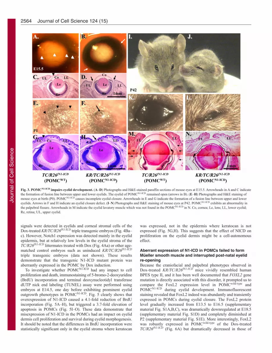
by crossing KR/R26fN1-ICD male and TC/R26fN1-ICD female mouseand Dox chow was administered to the pregnant mouse in the damfrom E12.5 to E15.5 (Fig. 3A–D), day of birth (Fig. 3E–H) andP42 (Fig. 3I–N), respectively. Normally, eyelid closure begins atE13.5–E14 and is complete by E15.5–E16. As expected, eyelidclosure took place and nearly covered the corneal surface at E15.5in control TC/R26fN1-ICD double transgenic mouse embryo (Fig.3A,C); however, it was impaired in Dox-treated KR/TC/R26fN1-ICD
embryos (Fig. 3B,D). At birth, the mouse pups had closed eyelidswith an obvious fusion line between upper and lower eyelids, asseen in control TC/R26fN1-ICD (Fig. 3E,G) and KR/R26fN1-ICD doubletransgenic mice (data not shown). However, there was no obviouseyelid fusion line in Dox-treated KR/TC/R26fN1-ICD mice (Fig.3F,H). Histological examinations revealed that the upper and lowereyelids of POMCN1-ICD did not come close enough to support theformation of this fusion line (Fig. 3H). The mouse eyelid isnormally closed at birth and begins to re-open at P12–14 (Findlateret al., 1993). We have noticed that Dox-induced POMCN1-ICD inthe KR/TC/R26fN1-ICD mice failed to open their eyelid(blepharophimosis) at P15 and P60, and never fully opened (ptosis-like) (data not shown) as compared with their TC/R26fN1-ICD orKR/R26fN1-ICD control littermates. Histological andimmunohistochemical analyses of a Dox-treated KR/TC/R26fN1-ICD
mouse at P42 showed that the eyelid fissures were obviouslynarrower (Fig. 3N) than that of the control TC/R26fN1-ICD littermate(Fig. 3M). This is probably due to the lack of Müeller lavatormuscle in POMCN1-ICD eyelid (Fig. 3, compare N with M). Inaddition to eyelid malformations, these Dox-treated triple transgenicmice also had other defects, including craniofacial malformation,shortened outer ears and forelimb, but they did not have gaitproblems (supplementary material Movie 1) and both genders werefertile. Altogether, our data demonstrate that POMCN1-ICD delayedembryonic eyelid closure and impaired eyelid re-opening aroundP12–P14. The phenotypic manifestations resemble PBES type IIin humans (Crisponi et al., 2001).
Immunofluorescent staining showed that in addition to signalsin eyelid epidermis, strong nuclear Notch1 immunofluorescent
2563Mouse model for blepharophimosis
Fig. 2. Notch1 expression patternduring embryonic eyelid closureand genetic overexpression ofN1-ICD in POMC.(A–C) Immunoflurescence stainingwith anti-Notch1 antibody onsections of mouse eyelids atE13.5~E17.5. Note that Notch1-positive signals (red) are detectedpredominantly in eyelid epidermis,with very few in the dermis.Nuclear counter staining withDAPI is shown in blue.(D)Schematic representation ofN1-ICD expression via Cre-loxPsystem in POMC. The rtTA isconstitutively expressed in Kera-positive POMCs. In the presence ofDox, Cre recombinase is inducedto delete the Neo/Stop cassette,permitting transcription of Notch1ICD. Co, cornea; Le, lens; LL,lower lid; Re, retina; UL, upper lid.
Jour
nal o
f Cel
l Sci
ence
signals were detected in eyelids and corneal stromal cells of theDox-treated KR/TC/R26fN1-ICD triple transgenic embryos (Fig. 4Ba–c). However, Notch1 expression was detected mainly in the eyelidepidermis, but at relatively low levels in the eyelid stroma of theTC/R26fN1-ICD littermates treated with Dox (Fig. 4Aa) or other age-matched control embryos such as uninduced KR/TC/R26fN1-ICD
triple transgenic embryos (data not shown). These resultsdemonstrate that the transgenic N1-ICD mutant protein wasaberrantly expressed in the POMC by Dox induction.
To investigate whether POMCN1-ICD had any impact to cellproliferation and death, immunostaining of 5-bromo-2-deoxyuridine(BrdU) incorporation and terminal deoxynucleotidyl transferasedUTP nick end labeling (TUNEL) assay were performed usingembryos at E14.5, one day before exhibiting prominent eyelidoutgrowth phenotypes in POMCN1-ICD. Fig. 5 clearly shows thatoverexpression of N1-ICD caused a 4.1-fold reduction of BrdUincorporation (Fig. 5A–H), but triggered a 3.7-fold elevation ofapoptosis in POMCs (Fig. 5I–O). These data demonstrate thatmisexpression of N1-ICD in the POMCs had an impact on eyeliddermis cell proliferation and survival during eyelid morphogenesis.It should be noted that the differences in BrdU incorporation werestatistically significant only in the eyelid stroma where keratocan
was expressed, not in the epidermis where keratocan is notexpressed (Fig. 5G,H). This suggests that the effect of NICD onproliferation on the eyelid dermis might be a cell-autonomouseffect.
Aberrant expression of N1-ICD in POMCs failed to formMüeller smooth muscle and interrupted post-natal eyelidre-openingBecause the craniofacial and palpebral phenotypes observed inDox-treated KR/TC/R26fN1-ICD mice vividly resembled humanBPES type II, and it has been well documented that FOXL2 genemutation is directly associated with this disorder, it prompted us tocompare the FoxL2 expression level in POMCwild-type and POMCN1-ICD during eyelid development. Immunofluorescentstaining revealed that FoxL2 indeed was abundantly and transientlyexpressed in POMCs during eyelid closure. The FoxL2 proteinlevel gradually increased from E13.5 to E16.5 (supplementarymaterial Fig. S1A,B,C), was dramatically downregulated at E18.5(supplementary material Fig. S1D) and completely diminished atP2 (supplementary material Fig. S1E). More interestingly, FoxL2was robustly expressed in POMCwild-type of the Dox-treatedTC/R26fN1-ICD (Fig. 6A) but dramatically decreased in those of
2564 Journal of Cell Science 124 (15)
Fig. 3. POMCN1-ICD impairs eyelid development. (A–D) Photographs and H&E-stained paraffin sections of mouse eyes at E15.5. Arrowheads in A and C indicatethe formation of fusion line between upper and lower eyelids. The eyelid of POMCN1-ICD remained open (arrows in B). (E–H) Photographs and H&E staining ofmouse eyes at birth (P0). POMCN1-ICD causes incomplete eyelid closure. Arrowheads in E and G indicate the formation of a fusion line between upper and lowereyelids. Arrows in F and H indicate an eyelid closure defect. (I–N) Photographs and H&E staining of mouse eyes at P42. POMCN1-ICD exhibits an abnormality inthe palpebral fissure. Arrowheads in M indicate the eyelid lavatory muscle which was not found in the POMCN1-ICD in N. Co, cornea; Le, lens; LL, lower eyelid;Re, retina; UL, upper eyelid.
Jour
nal o
f Cel
l Sci
ence

KR/TC/R26fN1-ICD (Fig. 6B) at E15.5. Similarly, a strong -SMA-positive smooth muscle sheet was detected in POMCwild-type of theDox-treated TC/R26fN1-ICD (Fig. 6C), but only a small -SMA-positive cell mass was present in POMCN1-ICD of the Dox-treatedKR/TC/R26fN1-ICD mice (Fig. 6D). Indeed, most of these -SMA-positive signals overlapped with EGFP-positive cells in the eyelidstroma of the KR/TC/Z/EG mice treated with Dox from E12.5 toP0 (supplementary material Fig. S2), suggesting that eyelid levatormuscle was derived from a Kera-positive cell lineage during eyelidmorphogenesis. Moreover, when using anti--SMA antibody toexamine the smooth muscle formation in the eyelid of differentages (P1, P15 and P60), our data showed that unlike in thePOMCwild-type, in which there was a continuous -SMA-positivesmooth muscle sheet detected in eyelids (Fig. 7A–C), the anti--SMA immunoreactivity was either fragmented or missed in thePOMCN1-ICD (Fig. 7D–I), suggesting that they failed to differentiateinto -SMA-positive smooth muscle sheet. Taken together, theseresults suggested that mis-expression of N1-ICD in POMCs resultedin a dramatic downregulation of FoxL2 and -SMA expression inthe developing eyelids. These findings prompt us to ask whetherthe N1-ICD–RBP-J–MAML-1 complex directly binds andregulates FoxL2 promoter activity, or whether it has to go throughactivation of Hes/Hey genes, which in turn regulate FoxL2 proteinexpression.
N1-ICD overexpression downregulates FoxL2 promoteractivityTo understand the molecular mechanism(s) by which aberrantNotch activation resulted in downregulation of FoxL2 in POMCs,we performed chromatin immunoprecipitation (ChIP) assay in anendogenous FoxL2-positive KK1 mouse ovarian granulosa cellline transfected with mN1-ICD–Myc plasmid DNA. Our datashowed that both anti-Myc eptitope tag (for precipitating N1-ICD)and anti-RBP-J antibodies were able to pull-down DNA sequencescorresponding to the RBP-J binding site of the mouse FoxL2promoter region (Fig. 8A,B), suggesting that binding of the N1-ICD–RBP-J complex to the RBP-J site takes place in vivo.Likewise, ChIP assay in KK1 mouse ovarian granulose-derivedcells transfected with HES-1 plasmid showed that anti-Hes-1
antibody could pull-down three of four N-Box Hes-1 binding sites(Fig. 8E). These data suggested that FoxL2 promoter activity isregulated by Notch signaling in vivo. Next, we performed aluciferase assay using the mouse 3.1 kb FoxL2 promoter(mFoxL2pr3.1) by transient transfection of COS-7 cells. Our datashowed that FoxL2 promoter activity was regulated by N1-ICD ina dosage-dependent manner. The mFoxL2pr3.1 activity wasenhanced twofold when co-transfected with low dose (20 ng) N1-ICD; however, it was decreased 3- and 7-fold when the N1-ICDconcentration was elevated to 2 g and 4 g, respectively (Fig.9B). Interestingly, the upregulation of mFoxL2pr3.1 activity by co-transfection of low dosage (20 ng) N1-ICD plasmid was attenuatedby addition of Hes-1 plasmid (Fig. 9C). Western blotting analysisshowed that endogenous expression of Hes-1 and Hey-1 wasupregulated in a dose-dependent manner by transfection ofincreasing amounts of N1-ICD expression vector DNA (Fig. 9D).Indeed, immunofluorescent staining also demonstrated that Hes-1production was increased concomitantly with downregulation ofFoxL2 in POMCN1-ICD at both E14.5 (Fig. 10) and E15.5 (data notshown). However, the pattern of Notch1 expression remainedunchanged in eyelids lacking FoxL2 (supplementary material Fig.S3). Thus, our data argued that Notch signaling activation couldserve as upstream regulator of FoxL2 and aberrant overexpressionof N1-ICD upregulated Hes-1, which, in turn, inhibited FoxL2expression and impaired levator Müeller smooth muscle formation,leading to a BPES-like phenotype.
FoxL2 positively regulates mouse Acta2 promoter activityMagnetic resonance imaging (MRI) study of human BPES eyelidsshowed the absence or hypotrophy of the eyelid superior levatormuscle, resulting in ptosis and suggesting a possible role of FOXL2in the development of this eyelid muscle (Dollfus et al., 2003). Toinvestigate whether FoxL2 can impact on -SMA gene (Acta2)regulation in POMC during eyelid morphogenesis, we first checkedwhether -SMA expression is dependent on the presence of FoxL2.Morphological examinations showed that FoxL2-knockout (FoxL2–/–) mice failed to form eyelids, as revealed by a lack ofconjunctiva located in the inner side of the eyelid, and they exhibitedan EOB phenotype (Uda et al., 2004) (supplementary material Fig.
2565Mouse model for blepharophimosis
Fig. 4. Nuclear N1-ICD misexpression in Dox-treatedKR/TC/R26fN1-ICD/WT (POMCN1-ICD) mice at E15.5.Immunofluorescent staining with anti-Notch1 antibody.Red: Notch positive signal. Blue: DAPI nuclearcounterstain. Note that Notch1-positive signals are rare inPOMCwild-type eye (A,Aa), but abundant in POMCN1-ICD
eyelid and corneal stroma (B,Ba,Bb,Bc). Co, cornea; Le,lens; LL, lower eyelid; Re, retina; UL, upper eyelid.
Jour
nal o
f Cel
l Sci
ence

S4B,D,F). Immunofluorescent staining showed that FoxL2 wasdispensable for corneal epithelial (K12-positive) and stromal(keratocan-positive) differentiation (data not shown). By contrast, -SMA-positive Müeller muscle was absent in FoxL2–/– mice (Fig.11B,D). These data strongly suggest that FoxL2 is required forexpression of -SMA and formation of Müeller muscle duringembryonic eyelid closure. This argument was further strengthenedby ChIP analysis in which anti-FoxL2 antibody clearly broughtdown FoxL2 binding motif in the mouse Acta2 promoter region(Fig. 12A). Moreover, in a cell culture experiment, transfection ofCMV–FoxL2 enhanced mouse Acta2 promoter activity (Fig. 12B).
DiscussionIn this study, we developed a novel Dox-inducible mouse driverstrain, KR/TC, to manipulate expression of loss-of-function and/orgain-of-function genes at the desired time to study their roles in
keratocan-expressing cells, which represent a subset of neural-crest-derived cell lineage destined to become eyelid and ear dermis,corneal stroma and limbs during embryonic development. TheKR/TC driver mouse can complement other neural crest cell lineagedrivers such as Wnt1-Cre (Danielian et al., 1998) and P0-Cre(Feltri et al., 1999) mice because of two facts. First, in the KR/TCsystem, Cre activity is tightly controlled by Dox; however, Wnt1-Cre and P0-Cre are not inducible. Second, in the KR/TC system,the Cre activity is not turned on earlier than E13.5 owing to Kerapromoter activity (Liu et al., 2000). However, the Cre activityderived from the Wnt1-Cre and P0-Cre systems can ‘flox-out’ thegene of interest as early as E8.5, which often causes embryoniclethality before most organogenesis has taken place. Therefore, theKR/TC system is a valuable tool to study the role of signalingmolecules during and following development in thoseaforementioned tissues (Fig. 1).
2566 Journal of Cell Science 124 (15)
Fig. 5. Overexpression of N1-ICD decreases BrdU uptake but triggers apoptosis in POMCs during eyelid morphogenesis at E14.5. (A–F) Immunohistochemistry of anti-BrdU antibody. Sections of the eyelids of TC/R26fN1-ICD (POMCWT) (A, BrdU; B, BrdU/DAPI merged; C, close-up viewfrom B) and KR/TC/R26fN1-ICD (POMCN1-ICD) embryos (D, BrdU; E, BrdU and DAPI merged; F, close-up view from E) labeled with BrdU (red) and nuclearcounterstained with DAPI. (I–N) TUNEL assay. Sections of the eyelids of TC/R26fN1-ICD (POMCWT) (I, TUNEL; J, DAPI; K, TUNEL and DAPI merged) andKR/TC/R26fN1-ICD (POMCN1-ICD) embryos (L, TUNEL; M, DAPI; N, TUNEL and DAPI merged) labeled with Click-iT® TUNEL Assay kit (green) and nuclearcounterstained with DAPI. (G)Quantitative analysis of the percentage of BrdU-labeled cells in eyelid stroma. KR/TC/R26fN1-ICD (POMCN1-ICD, 4.48±0.45%)embryos showed significant reduction in the percentage of BrdU-labeled eyelid stromal cells, compared with the TC/R26fN1-ICD (POMCWT, 18.55±4.34%)littermates in the eyelid mesenchyme. (H)No significant difference of BrdU-labeled eyelid epithelium between POMCN1-ICD (47.54± 7.16%) and POMCWT
(46.84±2.84%). (O) Quantitative analysis of TUNEL-labeled cells per section of eyelid margin. KR/TC/R26fN1-ICD (POMCN1-ICD, 18.31± 2.49%) embryos showedsignificant elevation in TUNEL-labeled cells, compared with the TC/R26fN1-ICD (POMCWT, 4.94±0.51%) littermates in the eyelid stromal mesenchyme. Data arerepresented as mean ± s.d. (n5). *P<0.05. Dashed lines in C, F, K, and N demarcate eyelid epithelium from stromal mesenchyme. Co, cornea; epi, eyelidepithelium; der, eyelid dermis; Le, lens; LL, lower lid; Re, retina; UL, upper lid.
Jour
nal o
f Cel
l Sci
ence

We investigated the role of Notch signaling activation in POMCsduring embryonic eyelid development. Our results showed thatKR/TC/R26fN1-ICD triple transgenic mice administered with Doxexhibited eyelid defects with poor eyelid closure at birth.Interestingly, they did not develop a secondary exposure keratitis,but had a relatively healthy ocular surface. POMCs also contributeto the corneal stroma and are the major source of keratocytes,although there were some quantitative differences in K12 andkeratocan expression but, to our surprise, mis-expression of N1-ICD did not have adverse impact on corneal keratocyte and stromalmorphogenesis (data not shown). Interestingly, however, thePOMCN1-ICD effects caused malformation of Meibomian gland anddysplasia of the Muc5A/C-positive goblet cells in the conjunctivalregion (data not shown). The phenotypes of POMCN1-ICD in corneaand conjunctiva will be reported elsewhere.
In the course of investigating the eyelid development ofPOMCN1-ICD mice, we found that eyelid re-opening was delayedcompared with that in control littermates at P12–P14 and neverreached complete opening, resembling congenital type II BPES(Fig. 3). Therefore, the KR/TC/R26fN1-ICD triple transgenic mousestrain can serve as a novel transgenic mouse model for studyingeyelid morphogenesis and the pathogenesis of human type II BPES.We found that POMCN1-ICD inhibited cell proliferation, whereas ittriggered apoptosis. Although not fully documented in the presentstudy, other cell behavior, such as cell migration and cell fatechanges, might also be involved in the pathogenic progression of POMCN1-ICD in vivo. More intriguing was the fact that POMCN1-ICD downregulated FoxL2 expression, and thus impairedsmooth muscle formation during embryonic eyelid closure. Geneticand epidemiological studies have shown that mutations in FOXL2
2567Mouse model for blepharophimosis
Fig. 6. POMCN1-ICD downregulates FoxL2 and -SMAexpression during eyelid morphogenesis at E15.5.Immunofluorescent staining with anti-FoxL2 (red, A,B) oranti-skeletal muscle myosin (red, C,D) and anti--SMA(green, C,D). Note that FoxL2-positive signals (red) aredownregulated in the POMCN1-ICD (B) compared withPOMCwt (A). Arrows in C indicate -SMA-positivesmooth muscle sheet. Arrowhead in D indicates thatPOMCN1-ICD failed to form -SMA-positive muscle sheet.Other green signals are smooth muscle of the bloodvessels. DAPI nuclear counterstain is shown in blue. Theskeletal muscle expression pattern changed very little. Co, cornea; Le, Lens; LL, lower lid; Re, retina; UL, upper lid.
Fig. 7. Downregulation of -SMAexpression and eyelid levator smoothmuscle malformation in Dox-treatedKR/TC/R26fN1-ICD mice at differentstages. (A–I) Immunofluorescent stainingwith anti-skeletal muscle myosin (red), -SMA (green) and counterstaining withDAPI (blue) at P1 (A,D,G), P15 (B,E,H)and P60 (C,F,I). Arrows indicate -SMA-positive eyelid smooth muscle sheet. Othergreen signals are smooth muscle of theblood vessels. Co, cornea; Le, Lens; LL,lower lid; Re, retina; UL, upper lid.
Jour
nal o
f Cel
l Sci
ence

cause 70% of BPES, but ~30% of people with BPES did not havean identified FOXL2 gene mutation. The cause of BPES in thesepeople is unknown, but regulation of FOXL2 might be altered. Ourstudies strongly support the idea that those cases of the disease notdue to mutation of FOXL2 result from activation of aberrant Notchsignaling and changes in expression of FOXL2.
This result was further strengthened by the ChIP and promoter-luciferase assays concerning FoxL2 regulation by N1-ICD.Sequence analysis showed one RBP-J and four Hes-1 putativebinding (N-Box) sites in the mouse FoxL2 3.1 kb promoter
region. Endogenous Notch signaling was not able to reveal thepresence of N1-ICD–RBP-J complex binding to RBP-J inChIP assay of mouse KK1 cells, but overexpressed N1-ICDclearly formed complexes with endogenous RBP-J within theFoxL2 promoter. Similarly, three out of four N-Box sites wereable to bind to Hes-1 in cells transiently transfected with Hes-1expression vector. Moreover, the mFoxL2pr3.1-luc exhibited afourfold increase in luciferase activity compared with that in thecontrol vector pGL3-basic. Co-transfection with N1-ICD plasmidvector significantly downregulated mFoxL2 promoter activity in
2568 Journal of Cell Science 124 (15)
Fig. 8. Binding of RBP-J and Hes-1 to the mFoxL2 promoter.(A)Predicted RBP-J binding site and Hes-1 binding sites within mFoxL2 5�-flanking regulatory region (~3.1 kb). Arrows are DNA primers used in ChIPassay for RBP-J binding site and Hes-1 binding sites. (B–D) ChIP analysis ofRBP-J binding to mouse FoxL2 promoter region. The binding region of RBP-J to Hes-1 promoter served as ChIP positive control (C) and its non-bindingregion as negative control (D). Notice that both anti-Myc tag and anti-RBP-Jantibodies pull down RBP-J binding region of the mFoxL2 promoter,indicating the binding of the N1-ICD–RBP-J complex to the RBP-J site ofthe mFoxL2 promoter. (E)ChIP analysis of Hes-1 binding to mouse FoxL2promoter region. N-box sites 1, 2 and 4 show evident Hes-1 binding, but site 3does not bind to the mouse FoxL2 promoter region.
Fig. 9. Dosage-dependent N1-ICD regulationof FoxL2 promoter activity in vitro.(A)Schematic drawing of mFoxL2pr3.1-Lucvector, which contains one RBP-J site and fourpotential Hes-1 binding sites in relation to mouseFoxL2 transcription start site (+1). (B)N1-ICDregulates FoxL2 promoter activity. Note that lowdose (20 ng) N1-ICD enhances (lane 3) but N1-ICD greater than 2g downregulates (lanes 4,5)pGL3.0-mFoxL2pr3.1 promoter activity.(C)Hes1 transcriptional inhibition of pGL3.0-mFoxL2pr3.1 promoter activity (compare lane 3with lane 2). Note that pGL3.0-mFoxL2pr3.1promoter activity enhanced by low dose (20ng)N1-ICD is attenuated by co-transfection of CMV-Hes-1 (compare lane 5 with lane 4). (D)Westernblotting analysis of Hes-1 and Hey-1 expressionin NIH3T3 cells transfected with variousamounts of pLIA-mNIC-Myc plasmids.Exogenous N1-ICD upregulates Hes-1 and Hey-1 expression in a dose-dependent manner.Expression level of -actin serves as a proteinloading control.
Jour
nal o
f Cel
l Sci
ence

a dose-dependent manner (Fig. 9B). At low doses, N1-ICDenhanced FoxL2 promoter activity, whereas N1-ICD in highdosage negatively regulated mFoxL2pr3.1 promoter activity,probably through activation of the transcriptional repressorHes/Hey. Indeed, transfection of N1-ICD upregulated Hes-1 andHey-1 protein production (Fig. 9D). We could not detect Hes-5protein by either western blotting analysis using NIH-3T3 cells,or by immunohistochemistry in mouse eyelid (data not shown),so it was hard to judge whether Hes-5 had any role in this regard.Furthermore, co-transfection of the Hes-1 expression vector couldattenuate low-dosage N1-ICD-enhanced FoxL2 promoter activity(Fig. 9C). This result is consistent with the in vivo data that N1-ICD overexpression caused upregulation of Hes-1 (Fig. 10D) anddownregulation of FoxL2 (Fig. 10F) in POMCs during eyelidmorphogenesis. Our data indicated that, depending on itsconcentration, N1-ICD might execute dual functions in theregulation of FoxL2 gene expression. Interestingly, it has alsobeen documented that the transcription factor forkhead box O3a
(FoxO3a) is a key negative transcriptional target of canonicalNotch1 signaling, exerting a protective function in the UVB-induced apoptosis in skin keratinocytes (Mandinova et al., 2008).Similarly, N1-ICD regulates the transcription factor forkhead boxP3 (FOXP3) promoter through RBP-J- and Hes-1-dependentmechanisms in FOXP3+CD4+CD25+ regulatory T cells, or ‘Tregs’(Ou-Yang et al., 2009; Radtke et al., 2010). These data suggestthat FOX-related genes might be a common downstream targetof the canonical Notch signaling in different cell types. However,during embryonic development, Notch1 expression in the mouseeyelid stroma was not altered in the FoxL2–/– mutant, suggestingthat Notch signaling is not regulated by FoxL2 (supplementarymaterial Fig. S3).
In eyelid morphogenesis, FoxL2 expression is specific in POMCsand closely associated with embryonic eyelid closure. Previouspublished data (Crisponi et al., 2001; Uda et al., 2004; Uhlenhautand Treier, 2006) and our results (supplementary material Fig. S1)showed that FoxL2 expression first appears in POMCs at
2569Mouse model for blepharophimosis
Fig. 10. POMCN1-ICD enhances Hes1 expressionconcurrent with FoxL2 downregulation during eyelidclosure at E14.5. Immunofluorescent staining of paraffinsections of TC/R26N1-ICD (POMCWT) (A,C,E) andKR/TC/R26N1-ICD (POMCN1-ICD) (B,D,F) with anti-Notch1 (A,B), anti-Hes-1 (C,D) and anti-FoxL2 (E,F)antibodies. Red color indicates Alexa-Fluor-555-conjugated secondary antibody. Nuclear counterstainingwith DAPI is blue. Misexpression of N1-ICD (B) resultsin an increase of Hes-1 (compare D with C), butdownregulation of FoxL2 (compare F with E) expression.Co, cornea; Le, lens; Ll, lower lid; Re, retina; Ul, upperlid.
Fig. 11. Mice lacking FoxL2 do not form levator(Müeller) smooth muscle at E18.5. Immunofluorescentstaining clearly shows an -SMA-positive stripe (greencolor) indicating the formation of muscle in FoxL2+/–
(A,C) but not in the FoxL2–/– (B,D) mouse. Asterisksindicate -SMA positive blood vessel. Nuclearcounterstaining with DAPI is blue. UL, upper eyelid; LL,lower eyelid; Co, cornea; Le, lens; Re, retina.
Jour
nal o
f Cel
l Sci
ence

E12.5~E14.5 and is restricted to the eyelid dermis at E15.5~E16.5.More interestingly, after morphogenetic eyelid closure is complete,FoxL2 expression was dramatically downregulated at E18.5 andeventually diminished at P2. FoxL2 expression was never detectedin the corneal stroma, although it shared the common ascendant ofPOMC. These data implicated that FoxL2+Kera+ cells mainlycontribute to eyelid stroma, but FoxL2-Kera+ cells become cornealkeratocytes. Most of these FoxL2+Kera+ POMCs should normallydifferentiate into levator Müeller smooth muscle, but fail to do soif the expression of N1-ICD is aberrantly high. We found thatFoxL2 was able to directly bind to and activate mouse Acta2promoter activity (Fig. 12). In addition, expression of Acta-2 wasabsent in the FoxL2-knockout mouse. Taken together, our transgenicmice and cell culture data argue that a physiologically low level ofnotch activation might be very critical for the control of FoxL2expression, which in turn ensures levator muscle differentiationand normal eyelid morphogenesis. By contrast, aberrantly sustainedNotch activation inhibits FoxL2 expression and impairs levatorMüeller smooth muscle formation, leading to eyelid malformationand BPES-like phenotypes (see model in Fig. 13).
It remains unknown whether the effects of POMCN1-ICD onlevator Müeller smooth muscle formation during eyelidmorphogenesis depend on RBP-J and MAML-1 (canonicalpathway) or go through non-canonical pathways (Perumalsamy etal., 2009; Sanalkumar et al., 2010). Notch loss-of-functionapproaches by crossing RBP-Jfloxed/floxed or RosadnMAML–1 mouselines with POMCwild-type and POMCN1-ICD, respectively, will allowelucidation of the molecular pathway(s) of Notch activation inPOMCs, which lead to eyelid malformation and BPES-likephenotypes. Moreover, endogenous factors that fine-tune theappropriate Notch signaling during eyelid morphogenesis remainunknown. It has been recently reported that Notch can bephosphorylated on multiple sites by Nemo-like kinase (NLK), andthe density of NLK phosphorylation sites in Notch serves as amolecular rheostat to fine-tune Notch activity. Knockdown of NLKleads to hyperactivation of Notch signalling, and consequentlydecreases neurogenesis in zebrafish (Ishitani et al., 2010). It wouldbe interesting to know whether this mode of regulation in theNotch signaling pathway also exists in mammalian eyelidmorphogenesis.
Materials and MethodsConstruction and generation of Kerapr3.2-rtTA2S-M2 (KR) transgenic miceTo generate the transgenic mice of C57BL/6 background, a rtTA2S-M2 DNA fragment(1.1 kb) was excised from pTet-ON-advanced plasmid vector (Clontech) with EcoRIand HindIII digestion and subcloned into the EcoRI and HindIII sites of thepKerapr3.2-int-BpA plasmid vector (Liu et al., 2000). The resulting plasmid wasdesignated pKerapr3.2-int-rtTA2S-M2-BpA from which a NotI and SalI digestedDNA fragment containing Kerapr3.2-rtTA2S-M2-SV40 poly A (~6.2 kb) was purifiedand microinjected into fertilized eggs to generate transgenic mice. Thirty-fourtransgenic pups were obtained in which 16 transgenic founders carrying the transgenewere identified by tail DNA polymerase chain reaction (PCR) genotyping withprimers: Forward, 5�-TCAGCCATCGCTATGACTCAGTTC-3� and Reverse, 5�-TTGTTCTTCACGTGCCAGTACAGG-3�. The initial identification of functionalKR mice was carried out by outbreeding each founder line with a rtTA transgenicreporter mouse line called PTR which harbors a bi-directional TetO-EGFP and TetO-TRII (Frugier et al., 2005). The resulting KR/PTR double transgenic pups wereinduced with 1 g per kg doxycycline (dox) chow (Bio-Serve, Laurel, MD) throughpregnant females from E12.5 to the date of birth. Newborn mice were examinedunder a Zeiss stereomicroscope with epi-fluorescence. The founder lines displayingeither weak or no enhanced green fluorescent protein (EGFP) signal were no longerstudied. Two founder lines designated KR4 and KR5 exhibited identical strong EGFP
2570 Journal of Cell Science 124 (15)
Fig. 12. FoxL2 binds to and upregulates mouse Acta2promoter activity. (A)FoxL2 binds to mActa2promoter in ChIP assay. Predicted FoxL2 binding siteswithin the mActa2 5�-flanking regulatory region (~1.1kb). Arrows indicate DNA primers used in ChIP assayfor FoxL2 binding site. A 235 bp PCR-amplifiedproduct was generated from ChIP with anti-Myc tagantibody (lane 3) but not from mouse IgG (lane 4).(B)FoxL2 enhances mActa2 promoter luciferaseactivity in vitro. Diagram of mActa2 promoter-luciferaseconstruct. Co-transfection of pcDNA3.1-mFoxL2plasmid enhances luciferase activity threefold.
Fig. 13. A proposed model of POMCN1-ICD effects that impair levatormuscle formation during eyelid development, leading to BPES-likephenotypes. Aberrantly high expression of N1-ICD causes activation oftranscriptional repressor Hes-1 expression, which suppresses FoxL2expression, which, in turn, downregulates -SMA and thus leads to impairedlevator muscle formation.
Jour
nal o
f Cel
l Sci
ence

signals in eyelid, snout and limb (data not shown) and were used in the present study.Other mouse strains such as TetO-Cre (Perl et al., 2002), Z/EG (Novak et al., 2000),R26fN1-ICD (Murtaugh et al., 2003) are commercially available from the Jacksonlaboratory. FoxL2-knockout mice have been described previously (Crisponi et al.,2001). All the mice except FoxL2 knockouts were bred at the Animal Facility of theUniversity of Cincinnati Medical Center. They were housed in a room maintainedon a 12 hour/12 hour light/dark cycle (light on at 7 a.m.) with a continuous supplyof food and water. Experimental procedures for handling the mice were approved bythe Institutional Animal Care and Utilization Committee, University of CincinnatiCollege of Medicine.
Cell culturesNIH3T3 mouse fibroblast cell line and COS-7 monkey kidney cell line werepurchased from American Type Culture Collection (ATCC). KK1 mouse granulosatumour cell line was obtained from Kenneth W. Escudero (Escudero et al., 2010).3T3 cells were maintained in DMEM supplemented with 10% FBS and 100 U/mlpenicillin-streptomycin solution (Hyclone). KK1 and COS-7 cells were maintainedin DMEM/F12 (1:1) advanced medium supplemented with 5% FBS and 100 U/mlpenicillin-streptomycin solution (Hyclone). All cells were cultured at 37°C in a 5%CO2 incubator.
AntibodiesSee supplementary material Table S1 for the antibodies used for immunofluorescence,western blotting and chromatin immunoprecipitation (ChIP).
Plasmid constructionPCR was used to generate mouse FoxL2 full-length cDNA from one BAC clone(RP24-225E14, BACPAC) which was subcloned into the CMV-driven eukaryoticexpression vector pcDNA3.1/Myc-His(–) (Invitrogen). PCR condition was as follows:95°C for 2 minutes, 35 cycles of 94°C for 30 seconds, 58°C for 30 seconds, 72°Cfor 1 minute, followed by 72°C for 10 minutes as a final extension. Mouse FoxL2promoter region (–2887 to +244) generated by PCR from the same BAC clone wassubcloned into pGL3.0 luciferase basic vector (Promega). Mouse -smooth muscleactin promoter (mActa-2pr) region (–1146 to –1) was generated by PCR from oneBAC clone (RP24-329P13; BACPAC) and cloned into pGL4.10 vector (Promega).PCR conditions were same as above except longer extension time at 72°C. All PCR-amplified DNA were verified by DNA sequencing. Supplementary material Table S2lists the primers used to generate the constructs.
Histological analysisMouse samples were fixed overnight in 4% paraformaldehyde (PFA) in 1�phosphate-buffered saline (PBS; pH 7.2), followed by paraffin embedding. De-paraffinized sections (5 m) were stained with hematoxylin and eosin (H&E) orperiodic acid Schiff reagent (PAS).
Immunofluorescence and immunohistochemistryTissue sections (5 m) were de-paraffinized, rehydrated and subjected to antigenretrieval in sodium citrate buffer (10 mM sodium citrate, 0.05% Tween, pH 6.0) atboiling temperature for 30 minutes. Eyelid sections were then blocked with 3% BSAin PBS containing 0.025% Nonidet P-40 for 1 h at room temperature, then incubatedovernight at 4°C with the primary antibodies diluted in the same buffer. After threewashes with PBST (PBS, 0.1% Tween 20), slides were incubated at room temperaturefor 1 hour with Alexa-Fluor-488- or Alexa-Fluor-555-conjugated secondary antibodies(Invitrogen) and 1 g/ml DAPI (4�,6-diamidino-2-phenylindole) as a nuclearcounterstain, washed with PBST again, and mounted with Mowiol (Sanofi-Aventis).Sections were examined and photographed using a Zeiss microscope equipped witha camera (Axiocam Mrm; Carl Zeiss). For data acquisition, we used the Axiovision4.6 software (Carl Zeiss).
Detection of cell proliferationFor detection of cell proliferation in the developing eyelids, timed pregnant femalemice were injected intraperitoneally on gestational day 14.5 with BrdU (Sigma) (80g/g body weight). Two hours after injection, embryos were dissected, fixed in 4%PFA in PBS, dehydrated through graded alcohols, embedded in paraffin and sectionedin the coronal plane at 5 m thickness. De-paraffinized and rehydrated sections weretreated with 3N HCl in double distilled water for 15 minutes at room temperaturefollowed by three washes with PBS. Immunodetection of BrdU was performed usingmouse anti-BrdU monoclonal antibody followed by Alexa-Fluor-555-labeled rabbitanti-mouse IgG and the sections were counterstained with DAPI. The total numberof cell nuclei (DAPI-positive) as well as the number of BrdU-labeled nuclei onsections through the middle of the eyelids were counted and recorded for themesenchyme and epidermis, respectively, from five adjacent sections. The cellproliferation index was calculated as the percentage of cell nuclei with BrdU labeling.Student’s t-test was used to analyze the significance of difference and a P value lessthan 0.05 was considered statistically significant.
Detection of apoptosis in situEmbryos at E14.5 were obtained through time-mated females. Cell apoptosis statusin Dox-treated and non-treated groups were subjected to TUNEL assay using Click-iT® TUNEL Assay kit (Invitrogen) according the manufacturer’s protocol.
Western blot analysisCells were lysed in lysis buffer (50 mM Tris-HCl, pH 7.5, 150 mM NaCl, 0.1% SDS,1% NP-40, 0.5% deoxycholate). Frozen tissue was homogenized in a buffercontaining phosphatase and protease inhibitors. Protein lysates (20 g) from eachsample were separated on a 4–20% linear gradient Tris-HCl denaturingpolyacrylamide Ready Gel® (Bio-Rad) and transferred to nitrocellulose membranes(Whatman). Antibody incubations were performed in 5% non-fat dry milk in TBS-T (10 mM Tris-HCl, pH 8.0, 150 mM NaCl, and 0.05% Tween-20) or in 5% BSAin TBS-T, depending on the requirement of the antibody.
Chromatin immunoprecipitation (ChIP) assayKK1 cells were transfected with 20 g pLIA-mNIC-myc plasmid (Addgene CC#341)(Bao and Cepko, 1997) or pCMV-SPORT6-mouse Hes-1 cDNA clone(Openbiosystems MMMM1013-9200434 using GeneJammer transfection reagent(Invitrogen). Forty-eight hours post transfection, cells were crosslinked with 1%formaldehyde at 37°C for 10 minutes and subjected to ChIP assay with antibodiesagainst Myc epitope tag (5 g/ml; RDI) and RBP-J (6 g/ml; Santa Cruz) usingthe ChIP assay Kit (Millipore) following the manufacturer’s instructions. PurifiedDNAs after ChIP were used as templates for PCR to verify the interaction betweenDNA promoter and protein. ChIP assay for FoxL2 binding in mouse Acta2 promoter,NIH3T3 cells were transfected with 20 g pcDNA3.1-mFoxL2. PCR primers arelisted in supplementary material Table S3.
Promoter luciferase assayCOS-7 cells seeded in six-well plates at 80% confluence were transiently transfectedwith a mixture of three different types of plasmids: (1) prhL-TK served as transfectionefficiency control vector (Promega); (2) empty vector (pGL3.0 or pGL4.10) orrecombinant plasmids harboring mouse FoxL2 promoter (pGL3.0-mFoxL2pr3.1) or-smooth muscle actin promoter (pGL4.0-Acta2pr) and (3) cDNA expressionplasmids (pLIA mNIC myc, or mouse Hes-1 cDNA clone or pcDNA3.1-mFoxL2) atdifferent dosages as indicated. 36 hours after transfection, cells were collected with1� PassiveLysis buffer (200 l), luciferase assay was conducted using DualLuciferase® Reporter (DLR) Assay System (Promega) according to the manufacturer’srecommendations. Luminescence was measured using a Synergy2 plate readerluminescence module (BioTek).
Statistical analysisTwo-tailed Student’s t-test (Excel, Microsoft, Redmond, WA) was used to analyzethe percentage of BrdU-positive and the number of TUNEL-positive cells. Allquantification data are presented as mean ± s.d.
We thank the reviewers for their instructive comments andsuggestions. We thank Nadean Brown (Cincinnati Childrens Hospital)for providing the anti-Hes-1 antibody. This work was supported bygrants from NIH/NEI RO1 # EY12486 (C.Y.L.) and EY13755(W.W.K.), Research Prevent Blindness, Ohio Lions Foundation forEye Research, and the Intramural Research Program of the NIH,National Institute on Aging. Deposited in PMC for release after 12months.
Supplementary material available online athttp://jcs.biologists.org/cgi/content/full/124/15/2561/DC1
ReferencesBao, Z. Z. and Cepko, C. L. (1997). The expression and function of Notch pathway genes
in the developing rat eye. J. Neurosci. 17, 1425-1434.Benayoun, B. A., Batista, F., Auer, J., Dipietromaria, A., L’Hôte, D., De Baere, E. and
Veitia, R. A. (2009). Positive and negative feedback regulates the transcription factorFOXL2 in response to cell stress: evidence for a regulatory imbalance induced bydisease-causing mutations. Hum. Mol. Genet. 18, 632-644.
Bolós, V., Grego-Bessa, J. and de la Pompa, J. L. (2007). Notch signaling in developmentand cancer. Endocr. Rev. 28, 339-363.
Borggrefe, T. and Oswald, F. (2009). The Notch signaling pathway: transcriptionalregulation at Notch target genes. Cell. Mol. Life Sci. 66, 1631-1646.
Crisponi, L., Deiana, M., Loi, A., Chiappe, F., Uda, M., Amati, P., Bisceglia, L.,Zelante, L., Nagaraja, R., Porcu, S. et al. (2001). The putative forkhead transcriptionfactor FOXL2 is mutated in blepharophimosis/ptosis/epicanthus inversus syndrome.Nat. Genet. 27, 159-166.
Danielian, P. S., Muccino, D., Rowitch, D. H., Michael, S. K. and McMahon, A. P.(1998). Modification of gene activity in mouse embryos in utero by a tamoxifen-inducible form of Cre recombinase. Curr. Biol. 8, 1323-1326.
De Baere, E., Beysen, D., Oley, C., Lorenz, B., Cocquet, J., De Sutter, P., Devriendt,K., Dixon, M., Fellous, M., Fryns, J. P. et al. (2003). FOXL2 and BPES: mutational
2571Mouse model for blepharophimosis
Jour
nal o
f Cel
l Sci
ence

hotspots, phenotypic variability, and revision of the genotype-phenotype correlation.Am. J. Hum. Genet. 72, 478-487.
Djalilian, A. R., Namavari, A., Ito, A., Balali, S., Afshar, A., Lavker, R. M. and Yue,B. Y. (2008). Down-regulation of Notch signaling during corneal epithelial proliferation.Mol. Vis. 14, 1041-1049.
Dollfus, H., Stoetzel, C., Riehm, S., Lahlou Boukoffa, W., Bediard Boulaneb, F.,Quillet, R., Abu-Eid, M., Speeg-Schatz, C., Francfort, J. J., Flament, J. et al.(2003). Sporadic and familial blepharophimosis-ptosis-epicanthus inversus syndrome:FOXL2 mutation screen and MRI study of the superior levator eyelid muscle. Clin.Genet. 63, 117-120.
Escudero, J. M., Haller, J. L., Clay, C. M. and Escudero, K. W. (2010). Microarrayanalysis of Foxl2 mediated gene regulation in the mouse ovary derived KK1 granulosacell line: over-expression of Foxl2 leads to activation of the gonadotropin releasinghormone receptor gene promoter. J. Ovarian Res. 3, 4.
Feltri, M. L., D’Antonio, M., Previtali, S., Fasolini, M., Messing, A. and Wrabetz, L.(1999). P0-Cre transgenic mice for inactivation of adhesion molecules in Schwanncells. Ann. N. Y. Acad. Sci. 883, 116-123.
Findlater, G., McDougall, R. and Kaufman, M. (1993). Eyelid development, fusion andsubsequent reopening in the mouse. J. Anat. 183, 121-129.
Fiúza, U. M. and Arias, A. M. (2007). Cell and molecular biology of Notch. J. Endocrinol.194, 459-474.
Frugier, T., Koishi, K., Matthaei, K. I. and McLennan, I. S. (2005). Transgenic micecarrying a tetracycline-inducible, truncated transforming growth factor beta receptor(TbetaRII). Genesis 42, 1-5.
Gridley, T. (2007). Notch signaling in vascular development and physiology. Development134, 2709-2718.
Hayashi, Y., Liu, C. Y., Jester, J. J., Hayashi, M., Wang, I. J., Funderburgh, J. L.,Saika, S., Roughley, P. J., Kao, C. W. and Kao, W. W. (2005). Excess biglycan causeseyelid malformation by perturbing muscle development and TGF-alpha signaling. Dev.Biol. 277, 222-234.
Holmberg, J., Liu, C. Y. and Hjalt, T. A. (2004). PITX2 gain-of-function in Riegersyndrome eye model. Am. J. Pathol. 165, 1633-1641.
Ishitani, T., Hirao, T., Suzuki, M., Isoda, M., Ishitani, S., Harigaya, K., Kitagawa, M.,Matsumoto, K. and Itoh, M. (2010). Nemo-like kinase suppresses Notch signallingby interfering with formation of the Notch active transcriptional complex. Nat. CellBiol. 12, 278-285.
Kopan, R. and Ilagan, M. X. (2009). The canonical Notch signaling pathway: unfoldingthe activation mechanism. Cell 137, 216-233.
Liu, C. Y., Arar, H., Kao, C. and Kao, W. W. (2000). Identification of a 3.2 kb 5�-flanking region of the murine keratocan gene that directs beta-galactosidase expressionin the adult corneal stroma of transgenic mice. Gene 250, 85-96.
Ma, A., Boulton, M., Zhao, B., Connon, C., Cai, J. and Albon, J. (2007). A role fornotch signaling in human corneal epithelial cell differentiation and proliferation. Invest.Ophthalmol. Vis. Sci. 48, 3576-3585.
Mandinova, A., Lefort, K., Tommasi di Vignano, A., Stonely, W., Ostano, P., Chiorino,G., Iwaki, H., Nakanishi, J. and Dotto, G. P. (2008). The FoxO3a gene is a keynegative target of canonical Notch signalling in the keratinocyte UVB response. EMBOJ. 27, 1243-1254.
Murtaugh, L. C., Stanger, B. Z., Kwan, K. M. and Melton, D. A. (2003). Notchsignaling controls multiple steps of pancreatic differentiation. Proc. Natl. Acad. Sci.USA 100, 14920-14925.
Nakamura, T., Ohtsuka, T., Sekiyama, E., Cooper, L. J., Kokubu, H., Fullwood, N.J., Barrandon, Y., Kageyama, R. and Kinoshita, S. (2008). Hes1 regulates cornealdevelopment and the function of corneal epithelial stem/progenitor cells. Stem Cells 26,1265-1274.
Nien, C. J., Massei, S., Lin, G., Liu, H., Paugh, J. R., Liu, C. Y., Kao, W. W., Brown,D. J. and Jester, J. V. (2010). The development of meibomian glands in mice. Mol.Vis. 16, 1132-1140.
Novak, A., Guo, C., Yang, W., Nagy, A. and Lobe, C. G. (2000). Z/EG, a double reportermouse line that expresses enhanced green fluorescent protein upon Cre-mediatedexcision. Genesis 28, 147-155.
Ou-Yang, H. F., Zhang, H. W., Wu, C. G., Zhang, P., Zhang, J., Li, J.-C., Hou, L.-H.,He, F., Ti, X.-Y., Song, L.-Q. et al. (2009). Notch signaling regulates the FOXP3promoter through RBP-J- and Hes1-dependent mechanisms. Mol. Cell. Biochem. 320,109-114.
Perl, A. K., Wert, S. E., Nagy, A., Lobe, C. G. and Whitsett, J. A. (2002). Earlyrestriction of peripheral and proximal cell lineages during formation of the lung. Proc.Natl. Acad. Sci. USA 99, 10482-10487.
Perumalsamy, L. R., Nagala, M., Banerjee, P. and Sarin, A. (2009). A hierarchicalcascade activated by non-canonical Notch signaling and the mTOR-Rictor complexregulates neglect-induced death in mammalian cells. Cell Death Differ. 16, 879-889.
Radtke, F., Fasnacht, N. and Macdonald, H. R. (2010). Notch signaling in the immunesystem. Immunity 32, 14-27.
Sanalkumar, R., Dhanesh, S. B. and James, J. (2010). Non-canonical activation ofNotch signaling/target genes in vertebrates. Cell. Mol. Life Sci. 67, 2957-2968.
Schmidt, D., Ovitt, C. E., Anlag, K., Fehsenfeld, S., Gredsted, L., Treier, A.-C. andTreier, M. (2004). The murine winged-helix transcription factor Foxl2 is required forgranulosa cell differentiation and ovary maintenance. Development 131, 933-942.
Uda, M., Ottolenghi, C., Crisponi, L., Garcia, J. E., Deiana, M., Kimber, W., Forabosco,A., Cao, A., Schlessinger, D. and Pilia, G. (2004). Foxl2 disruption causes mouseovarian failure by pervasive blockage of follicle development. Hum. Mol. Genet. 13,1171-1181.
Uhlenhaut, N. H. and Treier, M. (2006). Foxl2 function in ovarian development. Mol.Genet. Metab. 88, 225-234.
Vauclair, S., Majo, F., Durham, A. D., Ghyselinck, N. B., Barrandon, Y. and Radtke,F. (2007). Corneal epithelial cell fate is maintained during repair by Notch1 signalingvia the regulation of vitamin A metabolism. Dev. Cell 13, 242-253.
2572 Journal of Cell Science 124 (15)
Jour
nal o
f Cel
l Sci
ence


