Embed Size (px)
Citation preview
Journal of Invertebrate Pathology 91 (2006) 105–114
www.elsevier.com/locate/yjipa
Nosema chrysorrhoeae n. sp. (Microsporidia), isolated from browntail moth (Euproctis chrysorrhoea L.) (Lepidoptera, Lymantriidae) in Bulgaria: Characterization and phylogenetic relationships
Miroslav Hylin a,¤, Daniela K. Pilarska b, Miroslav Oborník c,d, Jilí Vávra a,c, Leellen F. Solter e, Jaroslav Weiser f, Andreas Linde g, Michael L. McManus h
a Department of Parasitology and Laboratory of Electron Microscopy, Faculty of Science, Charles University, Prague, Czech Republicb Bulgarian Academy of Sciences, Institute of Zoology, 1 Tsar Osvoboditel Blvd. 1000 SoWa, Bulgaria
c Institute of Parasitology, Academy of Sciences of the Czech Republic, Beské Bud5jovice, Czech Republicd University of South Bohemia, Faculty of Biological Sciences, Beské Bud5jovice, Czech Republic
e Illinois Natural History Survey, 607 E. Peabody Dr., Champaign, IL 61820, USAf Institute of Entomology, Academy of Sciences of the Czech Republic, Beské Bud5jovice, Czech Republic
g Fachhochschule Eberswalde, Alfred Moeller Str. 1, Eberswalde, D-16225, Germanyh USDA Forest Service, 51 Millpond Road, Hamden, CT 06514, USA
Received 14 July 2005; accepted 29 November 2005Available online 10 January 2006
Abstract
A new microsporidian parasite Nosema chrysorrhoeae n. sp., isolated in Bulgaria from the browntail moth (Euproctis chrysorrhoea L.),is described. Its life cycle includes two sequential developmental cycles that are similar to the general developmental cycles of the Nosema-like microsporidia and are indistinguishable from those of two Nosema spp. from Lymantria dispar. The primary cycle takes place in themidgut tissues and produces binucleate primary spores. The secondary developmental cycle takes place exclusively in the silk glands andproduces binucleate environmental spores. N. chrysorrhoeae is speciWc to the browntail moth. Phylogenetic analysis based on the ssurRNA gene sequence places N. chrysorrhoeae in the Nosema/Vairimorpha clade, with the microsporidia from lymantriid and hymenop-teran hosts. Partial sequences of the lsu rRNA gene and ITS of related species Nosema kovacevici (Purrini K., Weiser J., 1975. NatürlicheFeinde des Goldafters, Euproctis chrysorrhoea L., im Gebiet von Kosovo, FSR Jugoslawien. Anzeiger fuer Schädlingskunde, PXanzen-Umweltschutz, 48, 11–12), Nosema serbica Weiser, 1963 and Nosema sp. from Lymantria monacha was obtained and compared withN. chrysorrhoeae. The molecular data indicate the necessity of future taxonomic reevaluation of the genera Nosema and Vairimorpha. 2005 Elsevier Inc. All rights reserved.
Keywords: Microsporidia; Nosema chrysorrhoeae; Nosema kovacevici; Nosema serbica; Ultrastructure; Taxonomy; rDNA phylogeny; Euproctischrysorrhoea
1. Introduction
The browntail moth (Euproctis chrysorrhoea L.) occursin Central and Eastern Europe in periodic local outbreaks.It is a serious pest of orchards and broadleaf forests in theseregions. E. chrysorrhoea also elicits contact dermatitis andstrong allergic reactions in some humans. Although occa-
* Corresponding author.E-mail address: [email protected] (M. Hylin).
0022-2011/$ - see front matter 2005 Elsevier Inc. All rights reserved.doi:10.1016/j.jip.2005.11.006
sionally outbreaks are reduced by nuclear polyhedrosisvirus and pathogenic fungi, microsporidia are importantregulators of the population dynamics of E. chrysorrhoea(Sterling and Speight, 1989).
Several microsporidian species have been described fromthe browntail moth. Endoreticulatus (formerly Pleistophora)schubergi was described by Zwölfer in 1927. Later, Lipa(1964) recovered a Nosema sp. in a browntail moth popula-tion in Poland, and Sidor et al. (1975) found Nosema sp.infections in Macedonia. Purrini and Weiser (1975) described
106 M. Hylin et al. / Journal of Invertebrate Pathology 91 (2006) 105–114
Nosema kovacevici from E. chrysorrhoea in Kosovo in formerYugoslavia. No data on ultrastructure, primary spore cyclewere provided in these publications and rDNA sequencingwas not available.
In Bulgaria, there have been several reports of microspo-ridian infections in E. chrysorrhoea (Pilarska et al., 2001,2002; Solter et al., 2000). These authors recovered severalmicrosporidian isolates and conducted light and transmis-sion electron microscopy studies, (Pilarska et al., 2001,2002), however, no species descriptions were published.This paper describes a microsporidium found in an E. chry-sorrhoea population in Central Bulgaria.
2. Materials and methods
2.1. Origin of isolate, comparative isolates, and laboratory hosts
The microsporidium was recovered from E. chrysor-rhoea larvae collected in 2001 from Quercus cerris L. in thevicinity of Karlovo, Central Bulgaria. Three of 40 collectedlarvae were infected; the species description is based onmaterial obtained from one of these larvae. Infected silkglands were homogenized in a glass tissue grinder in dis-tilled water, the spore suspension was washed twice by suc-cessive centrifugation in distilled water and the cleanedspores used for infection experiments.
Morphological comparisons of N. kovacevici (Purriniand Weiser, 1975), Nosema serbica Weiser, 1963, andNosema sp. from Lymantria monacha were made from syn-type slides in the collection of Jaroslav Weiser, deposited inthe Natural History Museum in Vienna, Austria.
Eggs of E. chrysorrhoea L. and larvae of the gypsy moth,Lymantria dispar L. were obtained from BIOLA, Chelbice,Czech Republic. Larvae of Mamestra brassicae L. and Spo-doptera littoralis (Boisd.) were obtained from the labora-tory colony maintained at the Institute of Crop Production,Prague, Czech Republic.
The GenBank accession numbers of sequences for N.chrysorrhoeae, N. serbica, N. kovacevici and Nosema sp. fromL. monacha and of other microsporidian species used forphylogenetic analysis are the following: Amblyospora califor-nica (U68473), Amblyospora cinerei (AY090057), Amblyos-pora sp. (A. salinarius) (L28960), Amblyospora stimuli(AF027685), Amblyospora weiseri (AY090048), Amesonmichaelis (L20293), Brachiola algerae (AF069063), Brachiolaalgerae (AF069063), Brachiola algerae (AY230191), Brachi-ola algerae (L28961), Bryonosema plumatellae (AF484690),Encephalitozoon cuniculi (AJ005581), Glugea anomala(AF044391), Gurleya daphniae (AF439320), Gurleya vavrai(AF394526), Heterosporis anguillarum (AF387331), Larsso-nia obtusa (AF394527), Loma embiotocia (AF320310),Nosema apis (U26534), Nosema apis (U97150), Nosemabombi (AY008373), Nosema bombycis (AY259631), Nosemabombycis (L39111), Nosema carpocapsae (AF426104),Nosema ceranae (U26533), Nosema disstriae (L28963),Nosema epilachnae (L28964), Nosema furnacalis (U26532),
Nosema granulosis (AJ011833), Nosema chrysorrhoeae(AY940656), Nosema chrysorrhoeae (AY940657), Nosemakovacevici (AY940658), Nosema oulemae (U27359), Nosemaportugal (AF033316), Nosema serbica (AY940659), Nosemasp. (L. monacha) (AY940660), Nosema spodopterae(AY211390), Nosema tyriae (AJ012606), Nosema vespula(U11047), Nucleospora salmonis (U78176), Ordospora collig-ata (AF394529), Parathelohania anophelis (L28969), Pleisto-phora typicalis (AF044387), Pseudoloma neurophilia(AF322654), Pseudonosema cristatellae (AF484694), Tubuli-nosema kingi (L28966), Vairimorpha ephestiae (L28972),Vairimorpha heterosporum (L28973), Vairimorpha cheracis(AF327408), Vairimorpha imperfecta (isolate 1.) (AJ131645),Vairimorpha imperfecta (isolate 2.) (AJ131646), Vairimorphalymantriae1 (AF033315), Vairimorpha lymantriae (isolateLeviste) (AF141129), Vairimorpha lymantriae (L28974),Vairimorpha necatrix (L28975), Vairimorpha necatrix(Y00266), Vairimorpha sp. (S.richteri) (AF031539), Vairimor-pha sp. (P. xylostella) (AF124331).2
2.2. Laboratory infections of E. chrysorrhoea and other lepidopteran larvae
Second instar E. chrysorrhoea larvae were infected byspreading spore suspension on Salix cf. babylonica L. leaves;larvae of the other laboratory hosts were fed small cubes ofthe artiWcial diet (Bell et al., 1981) inoculated with spores.Dosages were 103–104 �l per larva. The larvae were starvedfor 24 h prior to the treatment and readily consumed the con-taminated diet. After treatment, E. chrysorrhoea larvae werereared on Salix leaves washed with 3% hydrogen peroxide,and other laboratory hosts were reared on artiWcial diet. Alllarvae were reared in growth chambers at 24 °C, 16/8 L/D.
2.3. Examination of infected host tissues; transmission electron microscopy
Fresh tissues and tissue smears stained with Giemsa(Sigma Diagnostic Accustain) were examined for the pres-ence of developmental stages and spores using light micros-copy. Spores were examined using the agar sporeimmobilization method (Vávra, 1964), measured with anImage Splitting Eyepiece (Vickers Instruments Ltd) andphotographed on a Olympus BX 51 microscope.
For transmission electron microscopy (TEM), infectedsilk glands were Wxed for 24 h in 2.5% glutaraldehyde in a0.1 M cacodylate buVer (pH 7.2) and postWxed in 2% OsO4.Fixed tissues were dehydrated through an ascending etha-nol and acetone series and embedded in Epon-Araldite.
1 Vairimorpha lymantriae is not a valid species name and is being rede-scribed as Vairimorpha disparis (Vávra et al., 2006).
2 For phylogenetic analysis based on ITS and LSU rRNA gene sequenc-es Vairimorpha lymantriae L28974 ( D Vairimorpha sp.3 from Baker et al.,1994) was used; other Vairimorpha lymantriae isolates: Vairimorpha sp. 4L28976 and Vairimorpha sp. 5 L28970 (Baker et al., 1994) and ITS regionof Nosema portugal AF033316 (Maddox et al., 1999) were included incomparison of nucleotide diVerences.
M. Hylin et al. / Journal of Invertebrate Pathology 91 (2006) 105–114 107
Thin sections were cut on a Reichert–Jung Ultracut Eultramicrotome and stained using uranyl acetate and leadcitrate. Sections were examined and photographed using aJeol JEM-1010 electron microscope.
2.4. rDNA sequences, phylogenetic analysis
DNA was isolated from fresh puriWed spores ofN. chrysorrhoeae, and from the Giemsa-stained the syn-type slides of N. kovacevici, N. serbica and Nosema sp.from L. monacha (Hylin et al., 2005). Material was pre-pared for DNA isolation using the bead beating method(Baker et al., 1994), followed by the DNeasy Tissue Kit(QIAGEN, Germantown, Maryland, USA) procedure. Aset of primers ss530f:ls580r for N. chrysorrhoeae, andss1537f:ls580r for N. kovacevici, N. serbica and Nosema
sp. were used to amplify conserved and variable regions ofsmall and large subunit rDNA (ss530f GTG CCA GCAGCC GCG G; ss1537f GAA CCA GCA GCA GGA TCATAA; ls580r GGT CCG TGT TTC AAG ACG G)(slightly modiWed primers of Weiss and Vossbrinck, 1999).The PCR reactions (95 °C for 2 min, 30 cycles of 94 °C for1 min, 50 °C for 1 min, 72 °C for 2 min, and 72 °C for10 min) were conducted in a total volume of 25 �l with 50–100 ng of DNA, 25 pmol of each primer, 1 unit Taq poly-merase (TAKARA BIO, Otsu, Shiga, Japan) and buVer/dNTP (TaKaRa) according to manufactured instructions.PCR products were separated using 1% agarose gel elec-trophoresis, extracted from the gel, puriWed using theDNeasy Tissue Kit (QIAGEN, Germantown, Maryland,USA), cloned (TOPO TA Cloning Kits, Invitrogen, Carlsbad,California, USA) and sequenced on an automatic
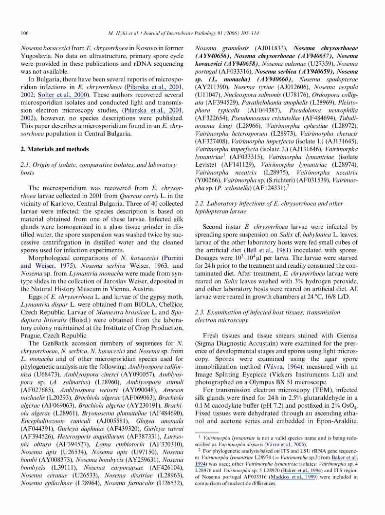
Figs. 1–9. Developmental stages of the primary developmental cycle of Nosema chrysorrhoeae, as seen in the Giemsa-stained smears of the midgut epithe-lial cells of Euproctis chrysorrhoea. The total volume of the nuclei is hardly visible, the dark spots are the chromatin. (Bars D 5�m.). Figs. 1 and 2. Early dip-lokaryotic meronts. Fig. 3. Binucleate meront at the onset of the Wrst nuclear division (there is some asynchrony in the division of the diplokaryonmembers). Fig. 4. Tetranucleate meront. Fig. 5. Probable early sporont issued from the division of a late tetranucleate meront. Figs. 6 and 7. Late sporonts
before the second nuclear division. Fig. 8. Late sporont dividing in two sporoblasts ( D disporoblastic sporogony). Fig. 9. Binucleate primary spores showlarge posterior vacuole and distinctly stained posterosomes (arrows). Late sporonts are at arrowheads.
108 M. Hylin et al. / Journal of Invertebrate Pathology 91 (2006) 105–114
sequencer (Beckman CEQ 2000 XL). The sequences werealigned using ClustalX program (Thompson et al., 1997),gaps and ambiguously aligned regions were omitted fromfurther analyses. Maximum parsimony (MP) trees wereconstructed using PAUP 4b10 (SwoVord, 2000) with TBRas a branch-swapping method and 1000 bootstrap repli-cates. Maximum likelihood trees were constructed byPHYML program (Guindon and Gascuel, 2003), usingGTR model for nucleotide substitutions with discretegamma distribution in 8 + 1 categories; all parameters(gamma shape, proportion of invariants) were estimatedfrom dataset. Multiple datasets for ML bootstrap analy-ses were prepared using SeqBoot (PHYLIP 3.6.3; Felsen-stein, 2001). ML bootstrap support was computed in 500replicates using PHYML program with HKY85 model fornucleotide substitutions and one category of sites with TI/TV ratio estimated from the data set.
3. Results
3.1. Life cycle
Our observations demonstrate that the life cycle ofNosema chrysorrhoeae consists of primary and secondarydevelopmental cycles, which diVer in time and tissue spec-iWcity in the host organism and the type of spore pro-duced. Each cycle consists of merogony and sporogonytypical of microsporidia in the genus Nosema.
3.2. Primary merogonic and sporogonic cycles
Primary merogony and sporogony occur in midgut epi-thelial cells and longitudinal and circular midgut muscle cells.The Wrst stages observed in Giemsa smears of the midgut tis-sue were small meronts, with two nuclei in diplokaryoticarrangement (Fig. 1). Later merogonial stages were largercells with two or four nuclei (Figs. 2–4). Clearly distinct dip-lokaryotic nuclei were apparent in some Giemsa stained mer-onts (Fig. 5), but in others the chromatin distribution wassuch that it was diYcult to decide if there was a diplokaryonor if the two nuclei had fused (Fig. 6). Sporonts were elongateand contained two or four nuclei arranged as diplokarya(Figs. 7 and 8). The division of sporonts produced diplokary-otic sporoblasts and immature primary spores (Fig. 9).Mature primary spores occurred in midgut epithelial cellsand muscles at about 48 h post inoculation. The primaryspores were characterized by large posterior vacuoles. Theposterior vacuole and the two nuclei were also visible inGiemsa stained smears (Fig. 9). Stained primary spores mea-sured 4.1 (4.5–5.2)£1.9 (2.1–2.2) �m (nD25).
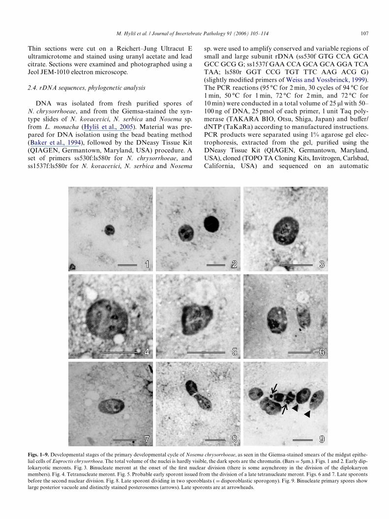
At the ultrastructural level, the developmental stages ofthe primary cycle proved to be diYcult to preserveadequately. Meronts, enveloped by a single cytoplasmicmembrane containing two or four nuclei arranged as diplok-arya, ribosomes, and traces of endoplasmic reticulum wereobserved (Figs. 10 and 11). The beginning of sporogony wasmarked by the thickening of the plasmalemma and the for-
Figs. 10–13. Nosema chrysorrhoeae primary developmental stages in the midgut epithelial cells of Euproctis chrysorrhoea as seen in the electron micro-scope. Figs. 10 and 11. Two- and tetranucleate meronts with nuclei in the diplokaryotic arrangement. The cytoplasm documents the diYculty in obtainingsatisfactory Wxation of these stages. Only some ribosomes, traces of Golgi vesicles (g) and of endoplasmic reticulum (arrow) are shown (Bar D 1�m).
Fig. 12. Primary spore showing the large posterior vacuole (p), thin endospore (arrow) and six polar Wlament coils (pf). Polyribosome aggregates are at ra.(Bar D 1 �m.). Fig. 13. Empty shells of primary spores left after their Wring. (Bar D 2 �m.)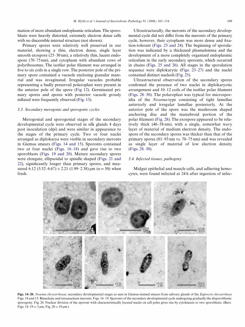
M. Hylin et al. / Journal of Invertebrate Pathology 91 (2006) 105–114 109
mation of more abundant endoplasmic reticulum. The sporo-blasts were heavily distorted, extremely electron dense cellswith no discernible internal structure (not shown).
Primary spores were relatively well preserved in ourmaterial, showing a thin, electron dense, single layersmooth exospore (25–30 nm), a relatively thin, lucent endo-spore (70–75 nm), and cytoplasm with abundant rows ofpolyribosomes. The isoWlar polar Wlament was arranged inWve to six coils in a single row. The posterior pole of the pri-mary spore contained a vacuole enclosing granular mate-rial and was invaginated. Irregular vacuoles probablerepresenting a badly preserved polaroplast were present inthe anterior pole of the spore (Fig. 12). Germinated pri-mary spores and spores with posterior vacuole grosslyinXated were frequently observed (Fig. 13).
3.3. Secondary merogonic and sporogonic cycles
Merogonial and sporogonial stages of the secondarydevelopmental cycle were observed in silk glands 4 dayspost inoculation (dpi) and were similar in appearance tothe stages of the primary cycle. Two or four nucleiarranged as diplokarya were visible in secondary merontsin Giemsa smears (Figs. 14 and 15). Sporonts containedtwo or four nuclei (Figs. 16–18) and gave rise to twosporoblasts (Figs. 19 and 20). Mature secondary sporeswere elongate, ellipsoidal to spindle shaped (Figs. 21 and22), signiWcantly longer than primary spores, and mea-sured 6.12 (5.52–6.67) £ 2.21 (1.99–2.38) �m (n D 50) whenfresh.
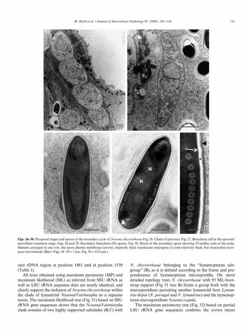
Ultrastructurally, the meronts of the secondary develop-mental cycle did not diVer from the meronts of the primarycycle, however, their cytoplasm was more dense and Wxa-tion-tolerant (Figs. 23 and 24). The beginning of sporula-tion was indicated by a thickened plasmalemma and thedevelopment of a more completely organized endoplasmicreticulum in the early secondary sporonts, which occurredin chains (Figs. 25 and 26). All stages in the sporulationsequence were diplokarytic (Figs. 23–27) and the nucleicontained distinct nucleoli (Fig. 25).
Ultrastructural observation of the secondary sporesconWrmed the presence of two nuclei in diplokaryoticarrangement and 10–12 coils of the isoWlar polar Wlament(Figs. 28–30). The polaroplast was typical for microspor-idia of the Nosema-type consisting of tight lamellaeanteriorly and irregular lamellae posteriorly. At theanterior pole of the spore was the mushroom shapedanchoring disc and the manubroid portion of thepolar Wlament (Fig. 28). The exospore appeared to be rela-tively thick (46–58 nm), with a single, somewhat wavylayer of material of medium electron density. The endo-spore of the secondary spores was thicker than that of theprimary spores (81–93 nm vs. 70–75 nm) and was revealedas single layer of material of low electron density(Figs. 28–30).
3.4. Infected tissues, pathogeny
Midgut epithelial and muscle cells, and adhering hemo-cytes, were found infected at 24 h after ingestion of infec-
Figs. 14–20. Nosema chrysorrhoeae, secondary developmental stages as seen in Giemsa-stained smears from salivary glands of the Euproctis chrysorrhoeaFigs. 14 and 15. Binucleate and tetranucleate meronts. Figs. 16–19. Sporonts of the secondary developmental cycle undergoing gradually the disporoblasticsporogony. Fig. 20. Nuclear division of the sporont with characteristically located nuclei on cell poles gives rise by cytokinesis to two sporoblasts. (Bars:
Figs. 14–19 D 5 �m, Fig. 20 D 10 �m.)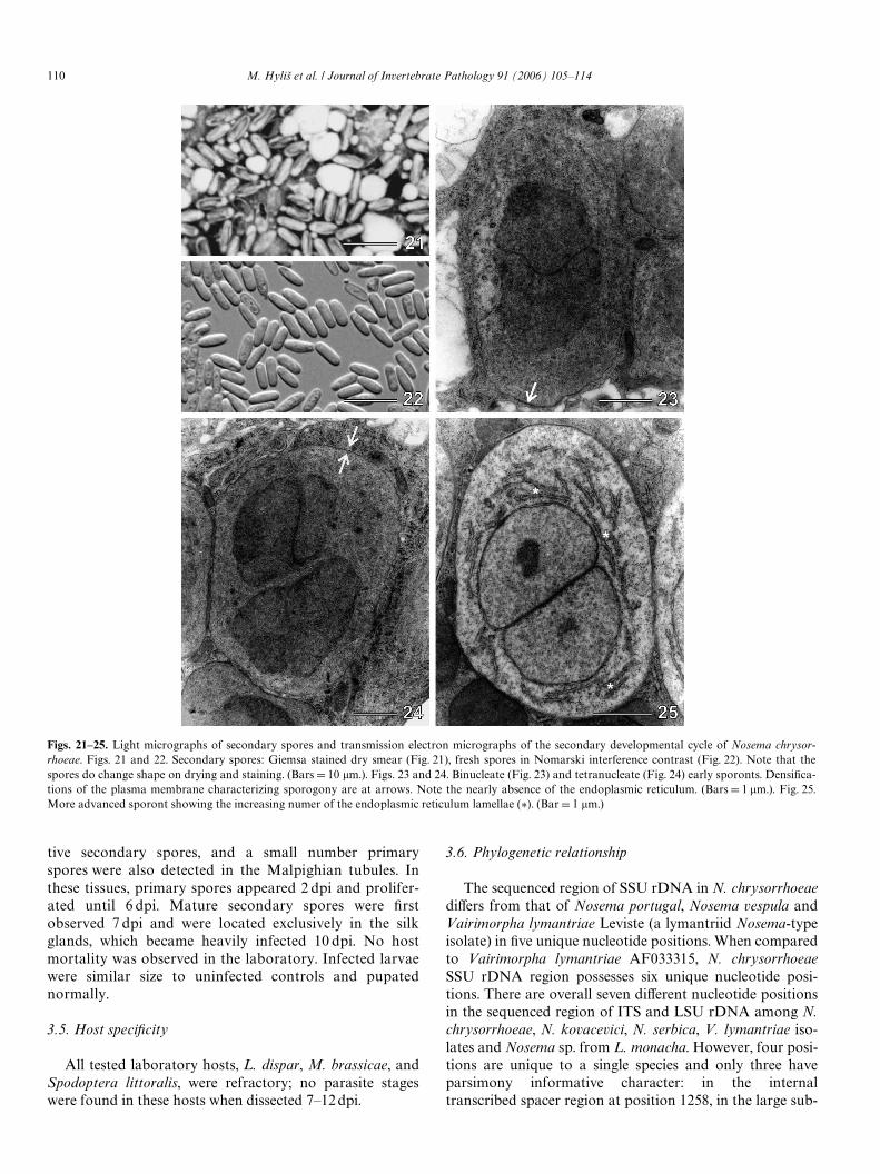
110 M. Hylin et al. / Journal of Invertebrate Pathology 91 (2006) 105–114
tive secondary spores, and a small number primaryspores were also detected in the Malpighian tubules. Inthese tissues, primary spores appeared 2 dpi and prolifer-ated until 6 dpi. Mature secondary spores were Wrstobserved 7 dpi and were located exclusively in the silkglands, which became heavily infected 10 dpi. No hostmortality was observed in the laboratory. Infected larvaewere similar size to uninfected controls and pupatednormally.
3.5. Host speciWcity
All tested laboratory hosts, L. dispar, M. brassicae, andSpodoptera littoralis, were refractory; no parasite stageswere found in these hosts when dissected 7–12 dpi.
3.6. Phylogenetic relationship
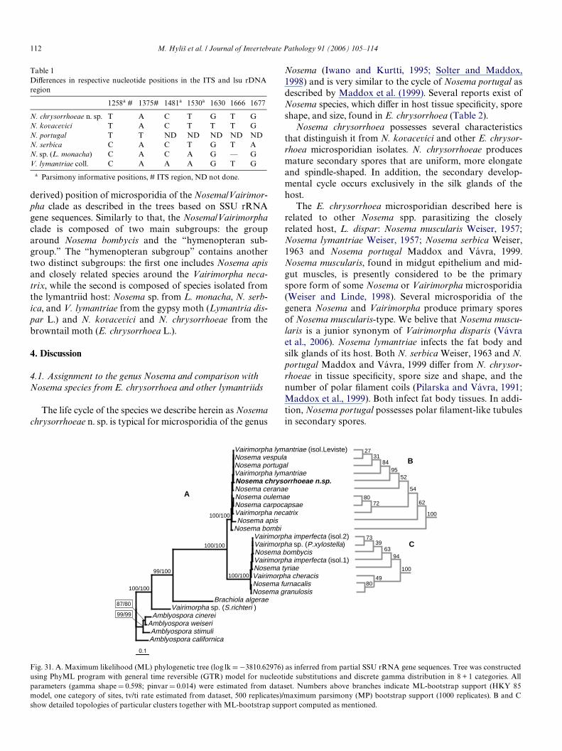
The sequenced region of SSU rDNA in N. chrysorrhoeaediVers from that of Nosema portugal, Nosema vespula andVairimorpha lymantriae Leviste (a lymantriid Nosema-typeisolate) in Wve unique nucleotide positions. When comparedto Vairimorpha lymantriae AF033315, N. chrysorrhoeaeSSU rDNA region possesses six unique nucleotide posi-tions. There are overall seven diVerent nucleotide positionsin the sequenced region of ITS and LSU rDNA among N.chrysorrhoeae, N. kovacevici, N. serbica, V. lymantriae iso-lates and Nosema sp. from L. monacha. However, four posi-tions are unique to a single species and only three haveparsimony informative character: in the internaltranscribed spacer region at position 1258, in the large sub-
Figs. 21–25. Light micrographs of secondary spores and transmission electron micrographs of the secondary developmental cycle of Nosema chrysor-rhoeae. Figs. 21 and 22. Secondary spores: Giemsa stained dry smear (Fig. 21), fresh spores in Nomarski interference contrast (Fig. 22). Note that thespores do change shape on drying and staining. (Bars D 10 �m.). Figs. 23 and 24. Binucleate (Fig. 23) and tetranucleate (Fig. 24) early sporonts. DensiWca-tions of the plasma membrane characterizing sporogony are at arrows. Note the nearly absence of the endoplasmic reticulum. (Bars D 1 �m.). Fig. 25.More advanced sporont showing the increasing numer of the endoplasmic reticulum lamellae (¤). (Bar D 1 �m.)
M. Hylin et al. / Journal of Invertebrate Pathology 91 (2006) 105–114 111
unit rDNA region at position 1481 and at position 1530(Table 1).
All trees obtained using maximum parsimony (MP) andmaximum likelihood (ML) as inferred from SSU rRNA aswell as LSU rRNA sequence data are nearly identical, andclearly support the inclusion of Nosema chrysorrhoeae withinthe clade of lymantriid Nosema/Vairimorpha as a separatetaxon. The maximum likelihood tree (Fig. 31) based on SSUrRNA gene sequences shows that the Nosema/Vairimorphaclade consists of two highly supported subclades (B,C) with
N. chrysorrhoeae belonging to the “hymenopteran sub-group” (B), as it is deWned according to the frame and pre-ponderance of hymenopteran microsporidia. On moredetailed topology trees N. chrysorrhoeae with 95 ML-boot-strap support (Fig. 31 tree B) forms a group both with themicrosporidians parasiting another lymantriid host Lyman-tria dispar (N. portugal and V. lymantriae) and the hymenop-teran microsporidium Nosema vespula.
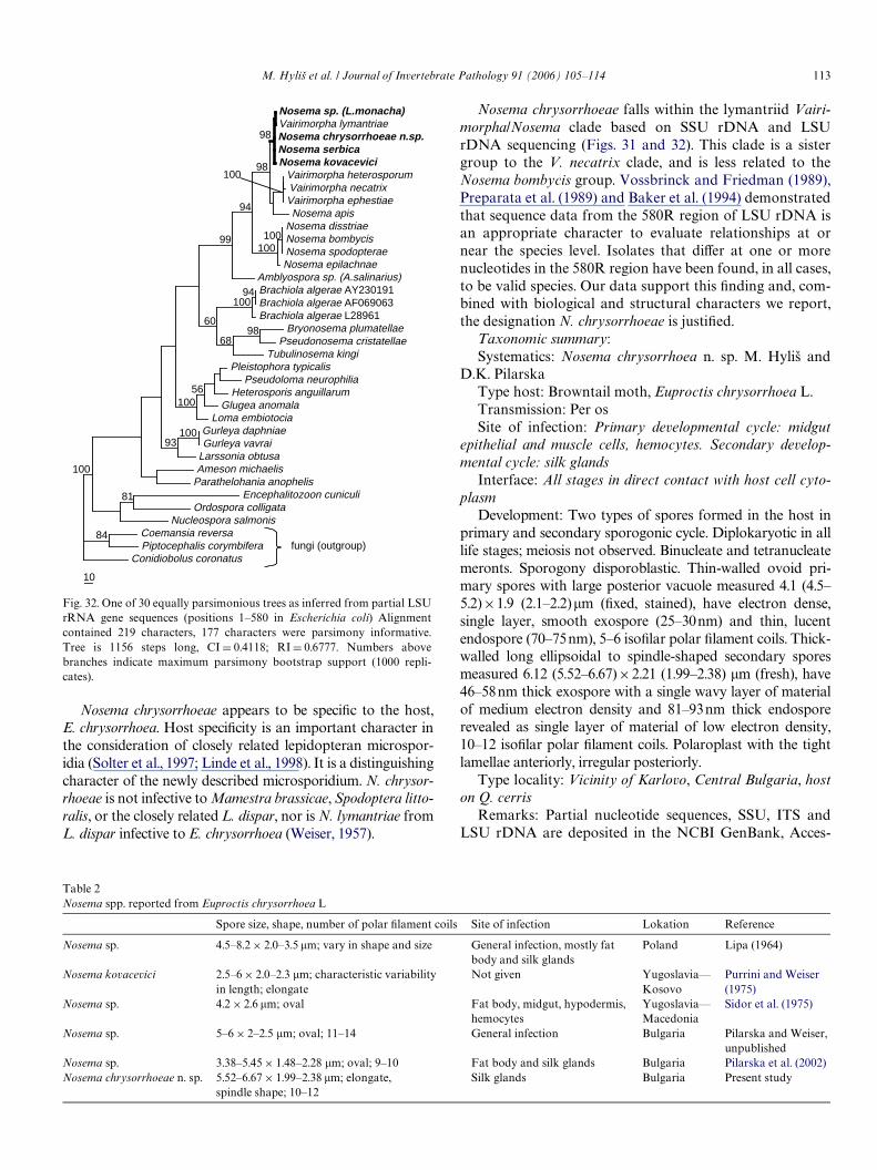
The maximum parsimony tree (Fig. 32) based on partialLSU rRNA gene sequences conWrms the crown (most
Figs. 26–30. Presporal stages and spores of the secondary cycle of Nosema chrysorrhoeae Fig. 26. Chain of sporonts. Fig. 27. Binucleate cell at the sporont/sporoblast transition stage. Figs. 28 and 29. Secondary binucleate (N) spores. Fig. 30. Detail of the secondary spore showing 10 isoWlar coils of the polarWlament arranged in one row, the spore plasma membrane (arrow), relatively thick translucent endospore (¤) and relatively thick, but structurless exos-pore (arrowhead). (Bars: Figs. 26–29 D 1 �m, Fig. 30 D 0.25 �m.)
112 M. Hylin et al. / Journal of Invertebrate Pathology 91 (2006) 105–114
derived) position of microsporidia of the Nosema/Vairimor-pha clade as described in the trees based on SSU rRNAgene sequences. Similarly to that, the Nosema/Vairimorphaclade is composed of two main subgroups: the grouparound Nosema bombycis and the “hymenopteran sub-group.” The “hymenopteran subgroup” contains anothertwo distinct subgroups: the Wrst one includes Nosema apisand closely related species around the Vairimorpha neca-trix, while the second is composed of species isolated fromthe lymantriid host: Nosema sp. from L. monacha, N. serb-ica, and V. lymantriae from the gypsy moth (Lymantria dis-par L.) and N. kovacevici and N. chrysorrhoeae from thebrowntail moth (E. chrysorrhoea L.).
4. Discussion
4.1. Assignment to the genus Nosema and comparison with Nosema species from E. chrysorrhoea and other lymantriids
The life cycle of the species we describe herein as Nosemachrysorrhoeae n. sp. is typical for microsporidia of the genus
Table 1DiVerences in respective nucleotide positions in the ITS and lsu rDNAregion
a Parsimony informative positions, # ITS region, ND not done.
1258a # 1375# 1481a 1530a 1630 1666 1677
N. chrysorrhoeae n. sp. T A C T G T GN. kovacevici T A C T T T GN. portugal T T ND ND ND ND NDN. serbica C A C T G T AN. sp. (L. monacha) C A C A G — GV. lymantriae coll. C A A A G T G
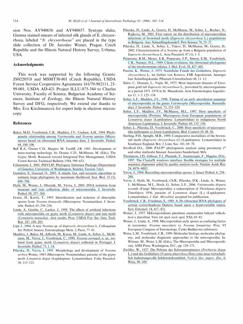
Nosema (Iwano and Kurtti, 1995; Solter and Maddox,1998) and is very similar to the cycle of Nosema portugal asdescribed by Maddox et al. (1999). Several reports exist ofNosema species, which diVer in host tissue speciWcity, sporeshape, and size, found in E. chrysorrhoea (Table 2).
Nosema chrysorrhoea possesses several characteristicsthat distinguish it from N. kovacevici and other E. chrysor-rhoea microsporidian isolates. N. chrysorrhoeae producesmature secondary spores that are uniform, more elongateand spindle-shaped. In addition, the secondary develop-mental cycle occurs exclusively in the silk glands of thehost.
The E. chrysorrhoea microsporidian described here isrelated to other Nosema spp. parasitizing the closelyrelated host, L. dispar: Nosema muscularis Weiser, 1957;Nosema lymantriae Weiser, 1957; Nosema serbica Weiser,1963 and Nosema portugal Maddox and Vávra, 1999.Nosema muscularis, found in midgut epithelium and mid-gut muscles, is presently considered to be the primaryspore form of some Nosema or Vairimorpha microsporidia(Weiser and Linde, 1998). Several microsporidia of thegenera Nosema and Vairimorpha produce primary sporesof Nosema muscularis-type. We belive that Nosema muscu-laris is a junior synonym of Vairimorpha disparis (Vávraet al., 2006). Nosema lymantriae infects the fat body andsilk glands of its host. Both N. serbica Weiser, 1963 and N.portugal Maddox and Vávra, 1999 diVer from N. chrysor-rhoeae in tissue speciWcity, spore size and shape, and thenumber of polar Wlament coils (Pilarska and Vávra, 1991;Maddox et al., 1999). Both infect fat body tissues. In addi-tion, Nosema portugal possesses polar Wlament-like tubulesin secondary spores.
Fig. 31. A. Maximum likelihood (ML) phylogenetic tree (log lk D ¡3810.62976) as inferred from partial SSU rRNA gene sequences. Tree was constructedusing PhyML program with general time reversible (GTR) model for nucleotide substitutions and discrete gamma distribution in 8 + 1 categories. Allparameters (gamma shape D 0.598; pinvar D 0.014) were estimated from dataset. Numbers above branches indicate ML-bootstrap support (HKY 85model, one category of sites, tv/ti rate estimated from dataset, 500 replicates)/maximum parsimony (MP) bootstrap support (1000 replicates). B and C
Vairimorpha lymantriae (isol.Leviste)Nosema vespulaNosema portugal Vairimorpha lymantriaeNosema chrysorrhoeae n.sp.Nosema ceranae Nosema oulemae Nosema carpocapsae Vairimorpha necatrix Nosema apis
Nosema bombi Vairimorpha imperfecta (isol.2)Vairimorpha sp. (P.xylostella)Nosema bombycis Vairimorpha imperfecta (isol.1)Nosema tyriae Vairimorpha cheracis Nosema furnacalis Nosema granulosis
Brachiola algerae Vairimorpha sp. (S.richteri )
Amblyospora cinerei Amblyospora weiseri Amblyospora stimuliAmblyospora californica
100/100
99/100
100/100
99/99
100/100
100/100
2731
8495
52
54
7280
62
100
7339
6394
100
4980
A
B
C
87/80
show detailed topologies of particular clusters together with ML-bootstrap support computed as mentioned.
M. Hylin et al. / Journal of Invertebrate Pathology 91 (2006) 105–114 113
Nosema chrysorrhoeae appears to be speciWc to the host,E. chrysorrhoea. Host speciWcity is an important character inthe consideration of closely related lepidopteran microspor-idia (Solter et al., 1997; Linde et al., 1998). It is a distinguishingcharacter of the newly described microsporidium. N. chrysor-rhoeae is not infective to Mamestra brassicae, Spodoptera litto-ralis, or the closely related L. dispar, nor is N. lymantriae fromL. dispar infective to E. chrysorrhoea (Weiser, 1957).
Fig. 32. One of 30 equally parsimonious trees as inferred from partial LSUrRNA gene sequences (positions 1–580 in Escherichia coli) Alignmentcontained 219 characters, 177 characters were parsimony informative.Tree is 1156 steps long, CI D 0.4118; RI D 0.6777. Numbers abovebranches indicate maximum parsimony bootstrap support (1000 repli-cates).
10
Nosema sp. (L.monacha)Vairimorpha lymantriaeNosema chrysorrhoeae n.sp.Nosema serbicaNosema kovacevici
Vairimorpha heterosporumVairimorpha necatrixVairimorpha ephestiaeNosema apis
Nosema disstriaeNosema bombycisNosema spodopteraeNosema epilachnae
Amblyospora sp. (A.salinarius)Brachiola algerae AY230191Brachiola algerae AF069063Brachiola algerae L28961
Bryonosema plumatellaePseudonosema cristatellae
Tubulinosema kingiPleistophora typicalis
Pseudoloma neurophiliaHeterosporis anguillarum
Glugea anomalaLoma embiotocia
Gurleya daphniaeGurleya vavrai
Larssonia obtusaAmeson michaelis
Parathelohania anophelisEncephalitozoon cuniculi
Ordospora colligataNucleospora salmonis
Coemansia reversaPiptocephalis corymbifera
Conidiobolus coronatus
98
98
94
100
99100100
10056
10093
10094
60
6898
100
84
81
fungi (outgroup)
Nosema chrysorrhoeae falls within the lymantriid Vairi-morpha/Nosema clade based on SSU rDNA and LSUrDNA sequencing (Figs. 31 and 32). This clade is a sistergroup to the V. necatrix clade, and is less related to theNosema bombycis group. Vossbrinck and Friedman (1989),Preparata et al. (1989) and Baker et al. (1994) demonstratedthat sequence data from the 580R region of LSU rDNA isan appropriate character to evaluate relationships at ornear the species level. Isolates that diVer at one or morenucleotides in the 580R region have been found, in all cases,to be valid species. Our data support this Wnding and, com-bined with biological and structural characters we report,the designation N. chrysorrhoeae is justiWed.
Taxonomic summary:Systematics: Nosema chrysorrhoea n. sp. M. Hylin and
D.K. PilarskaType host: Browntail moth, Euproctis chrysorrhoea L.Transmission: Per osSite of infection: Primary developmental cycle: midgut
epithelial and muscle cells, hemocytes. Secondary develop-mental cycle: silk glands
Interface: All stages in direct contact with host cell cyto-plasm
Development: Two types of spores formed in the host inprimary and secondary sporogonic cycle. Diplokaryotic in alllife stages; meiosis not observed. Binucleate and tetranucleatemeronts. Sporogony disporoblastic. Thin-walled ovoid pri-mary spores with large posterior vacuole measured 4.1 (4.5–5.2)£1.9 (2.1–2.2)�m (Wxed, stained), have electron dense,single layer, smooth exospore (25–30nm) and thin, lucentendospore (70–75nm), 5–6 isoWlar polar Wlament coils. Thick-walled long ellipsoidal to spindle-shaped secondary sporesmeasured 6.12 (5.52–6.67)£2.21 (1.99–2.38) �m (fresh), have46–58nm thick exospore with a single wavy layer of materialof medium electron density and 81–93nm thick endosporerevealed as single layer of material of low electron density,10–12 isoWlar polar Wlament coils. Polaroplast with the tightlamellae anteriorly, irregular posteriorly.
Type locality: Vicinity of Karlovo, Central Bulgaria, hoston Q. cerris
Remarks: Partial nucleotide sequences, SSU, ITS andLSU rDNA are deposited in the NCBI GenBank, Acces-
Table 2Nosema spp. reported from Euproctis chrysorrhoea L
Spore size, shape, number of polar Wlament coils Site of infection Lokation Reference
Nosema sp. 4.5–8.2 £ 2.0–3.5 �m; vary in shape and size General infection, mostly fat body and silk glands
Poland Lipa (1964)
Nosema kovacevici 2.5–6 £ 2.0–2.3 �m; characteristic variability in length; elongate
Not given Yugoslavia—Kosovo
Purrini and Weiser (1975)
Nosema sp. 4.2 £ 2.6 �m; oval Fat body, midgut, hypodermis, hemocytes
Yugoslavia—Macedonia
Sidor et al. (1975)
Nosema sp. 5–6 £ 2–2.5 �m; oval; 11–14 General infection Bulgaria Pilarska and Weiser, unpublished
Nosema sp. 3.38–5.45 £ 1.48–2.28 �m; oval; 9–10 Fat body and silk glands Bulgaria Pilarska et al. (2002)Nosema chrysorrhoeae n. sp. 5.52–6.67 £ 1.99–2.38 �m; elongate,
spindle shape; 10–12Silk glands Bulgaria Present study
114 M. Hylin et al. / Journal of Invertebrate Pathology 91 (2006) 105–114
sion Nos. AY940656 and AY940657. Syntype slides,Giemsa stained smears of infected silk glands of E. chrysor-rhoea, labeled “N. chrysorrhoeae” are placed in the typeslide collection of Dr. Jaroslav Weiser, Prague, CzechRepublic and the Illinois Natural History Survey, Urbana,USA.
Acknowledgments
This work was supported by the following Grants:Z60220518 and MSMT30-801 (Czech Republic), USDAForest Service Cooperative Agreements 161/79-982111, 23-99-001, USDA AD-421 Project ILLU-875-344 to CharlesUniversity, Faculty of Science, Bulgarian Academy of Sci-ences, Institute of Zoology, the Illinois Natural HistorySurvey and DFG, respectively. We extend our thanks toMrs. Eva Kirchmanová for expert help in electron micros-copy.
References
Baker, M.D., Vossbrinck, C.R., Maddox, J.V., Undeen, A.H., 1994. Phylo-genetic relationship among Vairimorpha and Nosema species (Micro-spora) based on ribosomal RNA sequence data. J. Invertebr. Pathol.64, 100–106.
Bell R.A., Owens C.D., Shapiro M. TardiV, J.R. 1981. Development ofmass-rearing technology. In: Doane, C.D., McManus, M. (Eds.), TheGypsy Moth: Research toward Integrated Pest Management, USDAForest Service Technical Bulletin 1584, 599–633.
Felsenstein J., 2001. PHYLIP, Phylogeny Inference Package (Departmentof Genetics, University of Washington, Seattle), Version 3.6a3.
Guindon, S., Gascuel, O., 2003. A simple, fast, and accurate algorithm toestimate large phylogenies by maximum likelihood. Syst. Biol. 52 (5),696–704.
Hylin, M., Weiser, J., Oborník, M., Vávra, J., 2005. DNA isolation frommuseum and type collection slides of microsporidia. J. Invertebr.Pathol. 88, 257–260.
Iwano, H., Kurtti, T., 1995. IdentiWcation and isolation of dimorphicspores from Nosema furnacalis (Microspora: Nosematidae). J. Inver-tebr. Pathol. 65, 230–236.
Linde, A., Genthe, C., Lacker, J., 1998. The eVects of artiWcial infectionswith microsporidia on gypsy moth (Lymantria dispar) and nun moth(Lymantria monacha)—Wrst results. Proc. USDA For. Ser. Gen. Tech.Rpt. 247, 198–205.
Lipa J., 1964. A new Nosema sp. of Euproctis chrysorrhoea L. ColloquiumInt. Pathol. Insects, Entomophaga Mem. 2, Paris, 77–81.
Maddox, J., Baker, M., JeVords, M., Kuras, M., Linde, A., Solter, L., McM-anus, M., Vávra, J., Vossbrinck, C., 1999. Nosema portugal, n. sp., iso-lated from gypsy moth (Lymantria dispar) collected in Portugal. J.Invertebr. Pathol. 73, 1–14.
Pilarska, D., Vávra, J., 1991. Morphology and development of Nosemaserbica Weiser, 1963 (Microspora: Nosematidae), parasite of the gypsymoth Lymantria dispar (Lepidoptera: Lymantriidae). Folia Parasitol.38, 115–121.
Pilarska, D., Linde, A., Goertz, D., McManus, M., Solter, L., Bochev, N.,Rajkova, M., 2001. First report on the distribution of microsporidianinfections of browntail moth (Euproctis chrysorrhoea L.) populationsin Bulgaria. Anz. Schaedlingskunde/J. Pest Science 74, 33–37.
Pilarska, D., Linde, A., Solter, L., Takov, D., McManus, M., Goertz, D.,2002. Characterization of a Nosema sp. from a Bulgaria population ofEuproctis chrysorrhoea L. Acta Parasitol. 47 (1), 1–5.
Preparata, R.M., Meyer, E.B., Preparata, F.P., Simon, E.M., Vossbrinck,C.R., Nanney, D.L., 1989. Ciliate evolution: the ribosomal phylogeniesof the tetrahymenine ciliates. J. Mol. Evol. 28, 427–441.
Purrini, K., Weiser, J., 1975. Natürliche Feinde des Goldafters, Euproctischrysorrhoea L., im Gebiet von Kosovo, FSR Jugoslawien. Anzeigerfuer Schädlingskunde, PXanzen-Umweltschutz 48, 11–12.
Sidor, C., Dusanic, L., Vujin, M., 1975. Most important diseases of Euro-pean gold tail Euproctis chrysorrhoea L., provoked by microorganismsin a period 1971–1974 in Sr. Macedonia. Acta Entomologica Jugoslav-ica 11, 1–2. 125–134.
Solter, L.F., Maddox, J.V., 1998. Timing of an early sporulation sequenceof microsporidia in the genus Vairimorpha (Microsporidia: Burenelli-dae). J. Invertebr. Pathol. 72, 323–329.
Solter, L.F., Maddox, J.V., McManus, M.L., 1997. Host speciWcity ofmicrosporidia (Protista: Microspora) from European populations ofLymantria dispar (Lepidoptera: Lymantriidae) to indigenous NorthAmerican Lepidoptera. J. Invertebr. Pathol. 69, 135–150.
Solter, L., Pilarska, D., Vossbrinck, C., 2000. Host speciWcity of microspor-idia pathogenic to forest Lepidoptera. Biol. Control 19, 48–56.
Sterling, P.H., Speight, M.R., 1989. Comparative mortalities of the brown-tail moth (Euproctis chrysorrhoea (l.) (Lepidoptera: Lymantriidae) inSoutheast England. Bot. J. Linn. Soc. 101, 69–78.
SwoVord D.L., 2000. PAUP* phylogenetic analysis using parsimony (*and other methods) Sinauer Associates Sunderland, MA.
Thompson, J.D., Gibson, T.J., Plesniak, F., Jeanmougin, F., Higgins, D.G.,1997. The ClustalX windows interface: Xexible strategies for multiplesequence alignment aided by quality analysis tools. Nucleic Acids Res.24, 4876–4882.
Vávra, J., 1964. Recording microsporidian spores. J. Insect Pathol. 6, 258–260.
Vávra, J., Hylin, M., Vossbrinck, Ch.R., Pilarska, D.K., Linde, A., Weiser,J., McManus, M.L., Hoch, G., Solter, L.F., 2006. Vairimorpha disparisn.comb. (Fungi: Microsporidia): a redescription of Thelohania disparisTimofejeva 1956, parasite of Lymantria dispar (L.) (Lepidoptera:Lymantriidae). J. Euk. Microbiol. accepted for publication.
Vossbrinck, C.R., Friedman, S., 1989. A 28s ribosomal RNA phylogeny ofcertain cyclorrhaphous Diptera based upon a hypervariable region.Syst. Entomol. 14, 417–431.
Weiser, J., 1957. Mikrosporidiemi p4sobená onemocn5ní bekyn5 velkoh-lavé a zlatolitné. V5st. bsl. spol. zool. spol. XXI, 65–82.
Weiser, J., Linde, A., 1998. Microsporidian early spores as confusing factorin taxonomy: Nosema muscularis vs. Nosema lymantriae. Proc. VIEuropean Congress of Entomology, Beske Bud5jovice (abstract).
Weiss, L.M., Vossbrinck, C.R., 1999. Molecular biology, molecular phylog-eny, and molecular diagnostic approaches to the microsporidia. In:Wittner, M., Weiss, L.M. (Eds.), The Microsporidia and Microsporidi-osis. AMS Press, Washington, D.C., pp. 129–171.
Zwölfer, W., 1927. Die Pebrine des Schwammspinners (Porthetria disparL.) und des Goldafters (Nygmia phaerrhoea Don.) eine neue wirtschaft-lich bedeutungsvolle Infektionskrankheit. Verh.d. Ges. angew. Ent., 6.Wien, 98–109.


