Embed Size (px)
Citation preview

Nondestructive Detection of White Root Rot Disease in Avocado Rootstocksby Leaf Chlorophyll Fluorescence
E. Martınez-Ferri, A. Zumaquero, M. T. Ariza, A. Barcelo, and C. Pliego, IFAPA, Centro de Churriana, Cortijo de la Cruz s/n, 29140Churriana, Malaga, Spain
Abstract
Martınez-Ferri, E., Zumaquero, A., Ariza, M. T., Barcelo, A., and Pliego, C. 2016. Nondestructive detection of white root rot disease in avocado root-stocks by leaf chlorophyll fluorescence. Plant Dis. 100:49-58.
White root rot (WRR) disease caused by Rosellinia necatrix is one of themost important threats affecting avocado orchards in temperate regions.In this study, we monitored the progression of WRR disease at the leafand root levels by the combination of nondestructive chlorophyll fluores-cence measurements and confocal laser-scanning microscopy on avo-cado genotypes susceptible to R. necatrix. Leaf photochemistry wasaffected at early stages of disease development prior to the appearanceof aboveground symptoms, made evident as significant decreases inthe trapping efficiency of photosystem-II (Fv¢/Fm¢) and in the steady-state of chlorophyll fluorescence yield (Fs) normalized to the minimal
fluorescence yield (F0) (Fs/F0). Decreases in Fv¢/Fm¢ and Fs/F0 were as-sociated with different degrees of fungal penetration, primarily in the lat-eral roots but not in areas next to the main root collar. Abovegroundsymptoms were observed only when the fungus reached the root collar.Leaf physiology was also tracked in a tolerant genotype where nochanges were observed during disease progression despite the presenceof the fungus in the root system. These results highlight the usefulnessof this technique for the early detection of fungal infection and the rapidremoval of highly susceptible genotypes in rootstock avocado-breedingprograms
Diseases associated with fungal root invasion are relevant onwoody crops (Pliego et al. 2012). Avocado (Persea americanaMill.)is a member of the Lauraceae family and an important commercialfruit crop in over 50 countries. One of the most important diseasesaffecting avocado orchards in temperate regions is white root rot(WRR), caused by the fungus Rosellinia necatrix Berl. ex Prill.(Freeman and Sztejnberg 1992; Pliego et al. 2012). Affected treesshow root rotting followed by leaf wilting, eventually resulting indeath of the trees after the appearance of the first foliar symptoms.WRR disease diagnosis is a major problem for nurseries and com-
mercial orchards. It requires high-throughput genetic techniques,such as those based on real-time Scorpion polymerase chain reaction(PCR; Ruano-Rosa et al. 2007; Schena and Ippolito 2003; Schenaet al. 2002) that are not fully reliable in practice and require highlyskilled personnel.Even with an effective diagnosis, control of avocado WRR is a
complex and difficult task as a consequence of the pathogen’sfeatures, including resistance to drought, survival capacity in acidicsoils, colonization of numerous hosts, deep penetration into the soil,and immunity to various common fungicides. Current control ap-proaches involve the use of physical (Lopez-Herrera et al. 1999; Rajand Sharma 2009) and chemical (Lopez-Herrera and Zea-Bonilla2007; Sugimoto 2002) methods as well as biocontrol strategies(ten Hoopen and Krauss 2006), although none of these methodshas proven to be fully effective; hence, the development of morereliable tools to control this disease is needed (Pliego et al. 2012).As in other woody crops, the use of disease-tolerant or -resistantrootstocks would constitute a method for widespread control pathogen(Broadbent and Gollnow 1993; Dadmal et al. 2002; Trapero et al.2008; Verdejo-Lucas et al. 2003). Until recently, no commercial root-stocks showing tolerance to R. necatrix have been available (Pliegoet al. 2012), although several promising selections are being evaluated(Barcelo-Muñoz et al. 2007) (A. Barcelo-Muñoz, IFAPA, Centro de
Churriana, Malaga, Spain, personal communication). However, suc-cess of breeding programs is greatly limited by the time-consumingand difficult process of working with woody plants. In this sense,the development of rapid and sensitive methods for R. necatrix de-tection would permit substantial advances in breeding for tolerantrootstocks.Understanding the physiological changes in the avocado–R.
necatrix pathosystem during the first stages of disease developmentis necessary for the development of a rapid and comprehensive detec-tion method for WRR disease (Bauriegel and Herppich 2014). Somespecialized root pathogens, such as Phytophthora spp., cause rootrot and leaf physiology impairments prior to wilting (Fleischmannet al. 2002, 2004). These studies also observed that the fungal inva-sion of the plant root system impairs water uptake and flow, induc-ing water stress; which, in part, is counteracted through stomatalclosure at the expense of limiting photosynthesis. Infection by somesoilborne pathogens triggered a systemic effect on photosynthesisassociated with the release of toxic mobile metabolites or hormonalimbalances (Brummer et al. 2002).R. necatrix simultaneously invades avocado at several points
within the roots, and invasion is followed by the proliferation ofhyphal strands in one or several directions, invading both epidermaland cortical cells and, finally, collapsing the vascular system of theplant (Pliego et al. 2009). R. necatrix produces several phytotoxiccompounds in vitro, which have been associated with pathogenicityand virulence (Edwards et al. 2001; Kimura et al. 1989; Kshirsagaret al. 2001). However, it is not clear whether WRR symptomatologyresults from these phytotoxic components or the mycelial invasion ofvascular root tissues (Pliego et al. 2012). Thus, it is reasonable toexpect that R. necatrix would induce a response at the leaf level asthe disease progresses. As for other pathogens, nondestructive tech-niques based on measurements of chlorophyll fluorescence and chlo-rophyll content (SPAD) could be useful for the early detection of thisdisease (Bauriegel and Herppich 2014; Berger et al. 2007a; Calderonet al. 2014).The main aims of the present study were to (i) assess whether the
response of susceptible avocado plants at early stages of R. necatrixroot infection were associated with changes at the physiological leaflevel prior to the appearance of any visible symptoms and, if true,(ii) determine whether this response occurs in genotypes with differ-ent degrees of tolerance.
Corresponding author: C. Pliego; E-mail: [email protected]
Accepted for publication 25 June 2015.
http://dx.doi.org/10.1094/PDIS-01-15-0062-RE© 2016 The American Phytopathological Society
Plant Disease / January 2016 49

The physiological changes in avocado leaves during the first stagesof R. necatrix infection were evaluated using a combination of nonde-structive chlorophyll fluorescence measurements and confocal lasermicroscopy to visualize the fungal invasion of the plant root system.
Material and MethodsFungal isolates and culture conditions. The fungal strains used
in this study were the virulent CH53 strain, isolated in an avocadoorchard at Almuñecar (Granada, Spain) in 1991 (Lopez-Herrera et al.1999), and the virulent derivative strain CH53-gfp, expressing the greenfluorescent protein (GFP), obtained through protoplast transformation(Pliego et al. 2009). Fungal strains were grown at 25°C on potatodextrose agar (PDA; Difco Laboratories). The routine growth ofthe gfp-derivate strain was performed on media containing hygrom-icine (HygB) at 50 mg/ml.Plant material and pathogenicity test in avocado plants.
Pathogenicity tests were performed in avocado genotypes differingin their disease reaction to R. necatrix. Three complementary exper-iments were carried out.Experiment 1. Plantlets of three different ‘Topa-Topa’ avocado
genotypes (TT-16, TT-21, and TT-31) were tested for assessing theearly response toR. necatrix infection. These genotypes are commonlyused as susceptible to R. necatrix in the ongoing breeding program forselection of tolerant rootstocks. The three genotypes were establishedin vitro and micropropagated according to the protocol of Pliego-Alfaro et al. (2013). In vitro rooted shoots from each plantlet were ac-climated in seedbeds filled with a mixture of peat, coconut fiber, andperlite at a ratio of 10:10:1 and 10ml of Osmocote under 100% humid-ity. After 45 days, the plants were potted in 1.5-liter plastic pots con-taining the same substrate and placed in a lathhouse for 6 months.Prior to inoculation, plantlets were potted in 3-liter plastic pots
(14 cm in diameter by 20 cm high) and transferred to controlled green-house conditions (approximately 20°C and 60% relative humidity[RH]). The plants were placed on benches and distributed into six traysof eight plants per genotype and allowed to establish. Five clonal plantsper genotype were used as noninoculated control plants.From a total of eight inoculated plants for each genotype, five
plants were inoculated with R. necatrix wild-type strain and the threeremaining plants were inoculated with the R. necatrix-gfp derivativestrain.For genotype characterization, plant size measurements (plant
height, stem diameter, and number of leaves) were obtained for con-trol plants prior to inoculation (25 October 2013) and 9 weeks post-inoculation (when most of the inoculated plants displayed aerialsymptoms; 9 January 2014). In these plants, the biomass partitioningvariables, including root/shoot ratio (R/S; root dry weight per shootdry weight), root weight ratio (RWR; root dry weight per plant dryweight), and leaf weight ratio (LWR; leaf dry weight per plant dryweight) were calculated by weighing the stems, leaves, and rootsof each plant separately before and after oven drying at 65°C for48 h. The shoot moisture content was estimated from the differencebetween the fresh and dry weights. The leaf fresh and dry weightswere used to calculate the leaf water content (LWC) on a fresh weightbasis ([fresh weight − dry weight]/fresh weight).During this experiment, the temperature (T) and RH in the green-
house were recorded every 30 min using an U23-001 HOBBO* Prov2 logger (Onset Computer Corporation). The diurnal air T rangedfrom 20.9 ± 0.6°C to 10.0 ± 0.7°C, and the mean RH was 61.2 ±1.8% during the course of the experiment. The plantlets were handirrigated twice a week to ensure soil wetness.Experiment 2. To validate results of experiment 1, plants of ‘Dusa’
avocado genotype, previously described as susceptible to R. necatrix,were tested for assessing the early response to R. necatrix infection.Vegetatively propagated plants (9 months old) of the commercial‘Dusa’ rootstock (Brokaw nursery, Spain) were grown under con-trolled greenhouse conditions (approximately 20°C and approximately60%RH) in 3-liter plastic pots (14 cm in diameter by 20 cm high) con-taining the same substratemix described above. In total, 26 plants wereused, but plants were distributed in two sets of 13 plants to conduct twotrials (March to April 2014 and May to June 2014). In each trial, four
plants were used as noninoculated (control plants) and the remainingplants were inoculated withR. necatrix. For genotype characterization,plant size measurements (plant height, stem diameter, and number ofleaves) were recorded in all plants prior to first inoculation (20March).Biomass partitioning variables were also estimated in control plants atthe end of the experiment, as described above. Environmental condi-tions during the experimentation (T and RH) were also recorded witha U23-001 HOBBO* Pro v2 logger (Onset Computer Corporation).Diurnal air T ranged from 19.8 ± 0.3 to 11.7 ± 0.3°C in trial 1 and from23.3 ± 0.3 to 16.7 ± 0.4°C in trial 2.Mean RHwas 66.7 ± 1.5 and 65.0 ±1.3% in the two trials, respectively. Plantlets were hand irrigated evenlyto ensure soil wetness.Experiment 3. To test the photochemical response to R. necatrix
infection on a genotype described as tolerant to this pathogen inthe ongoing avocado rootstock breeding program, plants of BG83selection were used. Explants were established in vitro and micropro-pagated according to the protocol of Pliego-Alfaro et al. (2013). Onceplants were acclimated in seedbeds, they were transferred to a lath-house. When plants were one and a half years old, they were pottedin 9-liter plastic pots (24 cm in diameter by 20 cm high) using thesame substrate described above and transferred to controlled green-house conditions for inoculation. In total, 26 plants were tested intwo trials conducted in March to May 2014 and May to July 2014.On each trial, four plants were used as noninoculated (control plants)and nine plants were inoculated with R. necatrix. Plant size measure-ments were recorded in all plants prior to the first inoculation (20March) and biomass partitioning variables were also estimated incontrol plants at the end of the experiment.Environmental conditions during the experimentation (T and RH)
were also recorded with a U23-001 HOBBO* Pro v2 logger (OnsetComputer Corporation). Diurnal air T ranged from 20.9 ± 0.3 to12.6 ± 0.3°C in trial 1 and from 24.1 ± 0.2 to 17.8 ± 0.3°C in trial2. Mean RH was 65.5 ± 1.0% in the two trials. Plants were handirrigated evenly to ensure soil wetness.Pathogenicity assays were performed according to Sztejnberg and
Madar (1980). The plants were inoculated with 3.75 g of colonizedwheat seed per liter of substrate and monitored during diseaseprogression. To assure the spread of the inoculum, it was placedon eight points scattered around the stem (approximately 3.5 cmapart) and introduced at two depths (at approximately 5 and 15 cm,respectively). The aerial symptoms were observed and assessed dailyaccording to a scale of 1 = healthy plant, 2 = mild wilting, 3 = wilting,4 = desiccated, and 5 = death.Chlorophyll a fluorescence measurements. In vivo chlorophyll
a fluorescence signals were measured in all plants at midmorning(1000 to 1100 h) daily or every 2 days in experiment 1 or twice aweek in experiments 2 and 3. Measurements were taken using a por-table fluorimeter PAM-2100 (Heinz Walz) equipped with leaf-clipholders to monitor incident light (photon flux density) and leaf T.On each plant, one (experiments 1 and 2) or two (in experiment 3)fully expanded mature leaves were labeled at the onset of the exper-iments for repeated measurements throughout the experimental courseson the same leaves. The so-called saturation pulse method was used forthe determination of all fluorescence parameters (Schreiber et al. 1994).The dark-adapted parameters were determined at predawn (0600 to0700 h) prior to inoculation with R. necatrix. To assess the initialminimal fluorescence (F0) in the dark or the steady-state fluores-cence (Fs) in the light (at approximately 450 mmol quanta m−2 s−1),the leaf samples were exposed to a weaklymodulatedmeasuring beam.Subsequently, a saturating flash of light (12,000 mmol quanta m−2 s−1)was administered for 0.8 s to assess the maximal fluorescence levels,either in the dark, when photosystem II (PSII) centers are closed(Fm), or under light conditions (Fm¢). The leaves were immediatelydarkened after every saturation pulse and subsequently exposed tofar-red light for 5.5 s to determine the minimal fluorescence yield ofthe preilluminated sample (F0¢). Measurements of Fm and F0 wereobtained at predawn to calculate the maximal photochemical efficiencyof PSII (Fv/Fm = [Fm − F0]/Fm) and the extent of Stern-Volmer non-photochemical fluorescence quenching (NPQ = [Fm − Fm¢]/[Fm¢])(Bilger and Bjorkman 1990). The relative quantum yield of PSII
50 Plant Disease /Vol. 100 No. 1

photochemistry (FPSII = [Fm¢ − Fs]/Fm¢), photochemical quenching(qP = [Fm¢ − Fs]/[Fm¢ − F0¢]), and maximum photochemical efficiencyof the open reaction centers of PSII (Fv¢/Fm¢ = [Fm¢ − F0¢]/Fm¢) werecalculated according to Genty et al. (1989).The steady-state fluorescence yield (Fs) was normalized to dark-
adapted fluorescence yields (F0). Fs has been described as a functionof the competition between photochemical and nonphotochemicalde-excitation of the energy absorbed by light-harvesting complexes(Schreiber et al. 1998).SPAD index. A hand-held chlorophyll meter (SPAD 502;
Minolta) was used to measure leaf greenness according to a unitlessSPAD index ranging from 0 to 100. This index has previously beenused as an estimation of the chlorophyll content in the leaves (Jifonet al. 2005;Markwell et al. 1995; Uddling et al. 2007) associated withthe degree of chlorosis (Tavakkoli et al. 2010). The measurementsare based on the ratio of chlorophyll absorbance in two wavebands(red = 650 nm, peak of chlorophyll absorbance, and infrared =approximately 940 nm, nonchlorophyll absorbance). The averageSPAD values were calculated from three readings per leaf taken onthe same leaves as for chlorophyll fluorescence measurements.Confocal microscopy.Microscopy was assessed in experiment 1.
The roots of three avocado plants of each genotype were infectedwith R. necatrix CH53-gfp as described above. The infected rootsof each plant were carefully removed after a sustained decrease inthe parameter Fv¢/Fm¢ or Fs/F0 was observed, although the plantappeared to be healthy (Fig. 1, stage 1), and once the disease was wellestablished (Fig. 1, stages 2 and 3). The roots were gently swirled insterile water to wash away the sand particles. To assess myceliumcolonization along the surface of avocado roots, the root segmentswere placed directly into petri dishes and observed under a fluores-cence microscope (Leica Microsystems) equipped with filter blockswith spectral properties matching those of enhanced GFP (488 nmexcitation and emission from 500 to 560 nm).To examine the internal proliferation and tissue invasion of R.
necatrix, the avocado root fragments were fixed in agarose accordingto Alvarez et al. (2006). The samples were fixed overnight in a 2.5%paraformaldehyde solution with an increasing sucrose gradient of 10,20, and 30% for 20, 20, and 30 min, respectively. After treatment,samples were embedded in 7% low-melting-point agarose gel andstored at 4°C. The samples were subsequently sliced using a Leica2000 microtome with freezing. Ultimately, 30- to 90-mm sectionswere obtained and observed using confocal laser-scanning micros-copy (CLSM; model TCS NT; Leica).Statistical analyses. The data were analyzed using STATISTICA
7.0 analytical software (Statsoft Inc.). Differences in plant size andbiomass partitioning variables among genotypes were evaluatedusing one-way analysis of variance (ANOVA). In experiment 1, toevaluate the differences among treatments and genotypes in chloro-phyll fluorescence variables and SPAD index throughout the exper-imental course, two-way repeated measures ANOVA (r-ANOVA)was performed. In this analysis, “genotype” and “treatment” were
the between-subjects factors and “date” was the within-subjects fac-tor. Genotypes that were not significantly different were joined toevaluate the treatment effect using one-way r-ANOVA. In experiments2 and 3, treatment effects were also evaluated by one-way r-ANOVA.The pattern of disease response observed in the two trials of each exper-iment was similar and data from the two trials were analyzed eitherseparately or jointly. To analyze single sampling dates, two-wayANOVA was used. Significant differences were considered at the5% probability level, unless otherwise stated. Prior to ANOVA,normality and homogeneity assumptions were tested using theKolmogorov-Smirnov and Cochran’s C tests, respectively. Whensignificant differences were observed, Fisher’s least significant dif-ference test was used to compare the mean values.
ResultsPlant characterization. Plant height, number of leaves, and stem
diameter of all genotypes were measured at the beginning of eachexperiment. In experiment 1, these measurements were also takenat the end. In experiment 1, no significant differences between geno-types were observed in the stem diameter and number of leaves(Table 1). However, plant height was significantly lower in TT-21than in TT-31 (P = 0.045; Table 1), although these differences werenot observed at the end of the experiment, likely resulting from afaster growth rate in TT-21 plants, indicated as a significantly higherincrease in the stem diameter and number of leaves (Table 1). Plantdry weight, biomass allocation pattern, and leaf water content weresimilar in all plants from the three Topa-Topa genotypes. The rootsand leaves represented approximately 26 and 55%, respectively, ofthe total plant dry weight. Plants ages in experiments 2 and 3 weredifferent and, therefore, direct comparisons in size are not appropri-ate. However, plant size measurements indicated a different architec-ture in the tolerant genotype (i.e., more leaves per stem diameter;Table 1). Biomass allocation to roots was comparatively higher inBG83 than in the susceptible genotypes, which resulted in higherroot/shoot ratios (Table 1).Postinfestation development of aerial andphysiological symptoms.
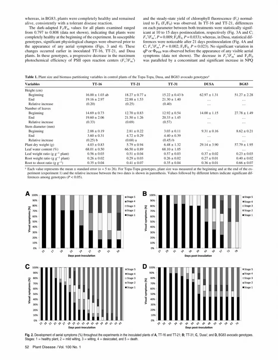
The initial visible aboveground WRR symptoms (Fig. 1) wereobserved around 3 weeks after inoculation in TT-16, TT-21 (ex-periment 1), and Dusa (experiment 2) genotypes (Fig. 2A andC). At this time, only 20 to 30% of the plants were at stage 2(mild wilting; Fig. 1, stage 2) although, in 1 week, the symptomsprogressed to advanced stages of desiccation (Fig. 1, stages 3and 4). Five weeks after inoculation, more than 60% of the plantswere completely desiccated or dead (Fig. 1, stages 4 and 5). Incontrast, initial disease symptoms in TT-31 (experiment 1) andBG83 plants (experiment 3) were delayed to more than 4 weeksafter inoculation, and advanced stages of the disease appearedprogressively within 8 weeks. Approximately more than 50%of plants were dead after 11 weeks postinoculation (Fig. 2Band D). At this time, plants that were alive in TT-31 displayedwilting symptoms (stage 3) and finally died in two more weeks
Fig. 1. Stages of aerial symptoms on avocado caused by white root rot: 1 = healthy plant, 2 = mild wilting, 3 = wilting, 4 = desiccated, and 5 = death.
Plant Disease / January 2016 51
whereas, in BG83, plants were completely healthy and remainedalive, consistently with a tolerant disease reaction.The dark-adapted Fv/Fm values for all plants examined ranged
from 0.797 to 0.808 (data not shown), indicating that plants werecompletely healthy at the beginning of the experiment. In susceptiblegenotypes, significant physiological changes were observed prior tothe appearance of any aerial symptoms (Figs. 3 and 4). Thesechanges occurred earlier in inoculated TT-16, TT-21, and Dusaplants. In these genotypes, a progressive decrease in the maximumphotochemical efficiency of PSII open reaction centers (Fv¢/Fm¢)
and the steady-state yield of chlorophyll fluorescence (Fs) normal-ized to F0 (Fs/F0) was observed. In TT-16 and TT-21, differenceson each parameter between both treatments were statistically signif-icant at 10 to 15 days postinoculation, respectively (Fig. 3A and C;Fv¢/Fm¢, P = 0.009; Fs/F0, P = 0.033); whereas, in Dusa, statistical dif-ferences were noticeable after 21 days postinoculation (Fig. 4A andC; Fv¢/Fm¢, P = 0.002; Fs/F0, P = 0.023). No significant variation inqP or FPSII was observed before the appearance of any visible aerialsymptoms (data not shown). The decrease in Fv¢/Fm¢ and Fs/F0was paralleled by a concomitant and significant increase in NPQ
Table 1. Plant size and biomass partitioning variables in control plants of the Topa-Topa, Dusa, and BG83 avocado genotypesz
Variables TT-16 TT-21 TT-31 DUSA BG83
Height (cm)Beginning 16.00 ± 1.03 ab 18.27 ± 0.77 a 15.22 ± 0.43 b 62.97 ± 1.31 51.27 ± 2.28End 19.16 ± 2.97 22.88 ± 1.53 21.30 ± 1.40 … …
Relative increase (0.20) (0.25) (0.40) … …
Number of leavesBeginning 14.69 ± 0.73 12.70 ± 0.83 12.92 ± 0.54 14.00 ± 1.15 27.78 ± 1.49End 19.60 ± 2.06 21.50 ± 1.26 20.33 ± 1.45 … …
Relative increase (0.33) (0.69) (0.57) … …
Stem diameter (mm)Beginning 2.88 ± 0.19 2.81 ± 0.22 3.03 ± 0.11 9.31 ± 0.16 8.62 ± 0.21End 3.60 ± 0.31 4.72 ± 0.29 4.40 ± 0.39 … …
Relative increase (0.25) b (0.68) a (0.45) b … …
Plant dry weight (g) 4.03 ± 0.83 5.79 ± 0.94 6.48 ± 1.32 29.14 ± 3.90 57.79 ± 1.95Leaf water content (%) 68.01 ± 0.50 66.50 ± 0.89 68.10 ± 1.05 … …
Leaf weight ratio (g g−1 plant) 0.56 ± 0.03 0.51 ± 0.04 0.57 ± 0.03 0.37 ± 0.02 0.23 ± 0.03Root weight ratio (g g−1 plant) 0.26 ± 0.02 0.29 ± 0.03 0.26 ± 0.02 0.27 ± 0.01 0.40 ± 0.02Root to shoot ratio (g g−1) 0.35 ± 0.04 0.41 ± 0.07 0.35 ± 0.04 0.36 ± 0.01 0.66 ± 0.07
z Each value represents the mean ± standard error (n = 5 to 26). For Topa-Topa genotypes, plant size was measured at the beginning and at the end of the ex-periment (experiment 1) and the relative increase between the two dates is shown in parenthesis. Values followed by different letters indicate significant dif-ferences among genotypes (P < 0.05).
Fig. 2. Development of aerial symptoms (%) throughout the experiments in the inoculated plants of A, TT-16 and TT-21; B; TT-31; C, ‘Dusa’; and D, BG83 avocado genotypes.Stages: 1 = healthy plant, 2 = mild wilting, 3 = wilting, 4 = desiccated, and 5 = death.
52 Plant Disease /Vol. 100 No. 1
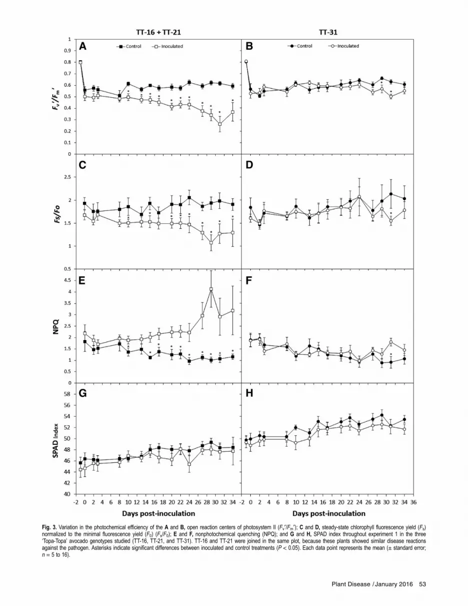
Fig. 3. Variation in the photochemical efficiency of the A and B, open reaction centers of photosystem II (Fv¢/Fm¢); C and D, steady-state chlorophyll fluorescence yield (Fs)normalized to the minimal fluorescence yield (F0) (Fs/F0); E and F, nonphotochemical quenching (NPQ); and G and H, SPAD index throughout experiment 1 in the three‘Topa-Topa’ avocado genotypes studied (TT-16, TT-21, and TT-31). TT-16 and TT-21 were joined in the same plot, because these plants showed similar disease reactionsagainst the pathogen. Asterisks indicate significant differences between inoculated and control treatments (P < 0.05). Each data point represents the mean (± standard error;n = 5 to 16).
Plant Disease / January 2016 53
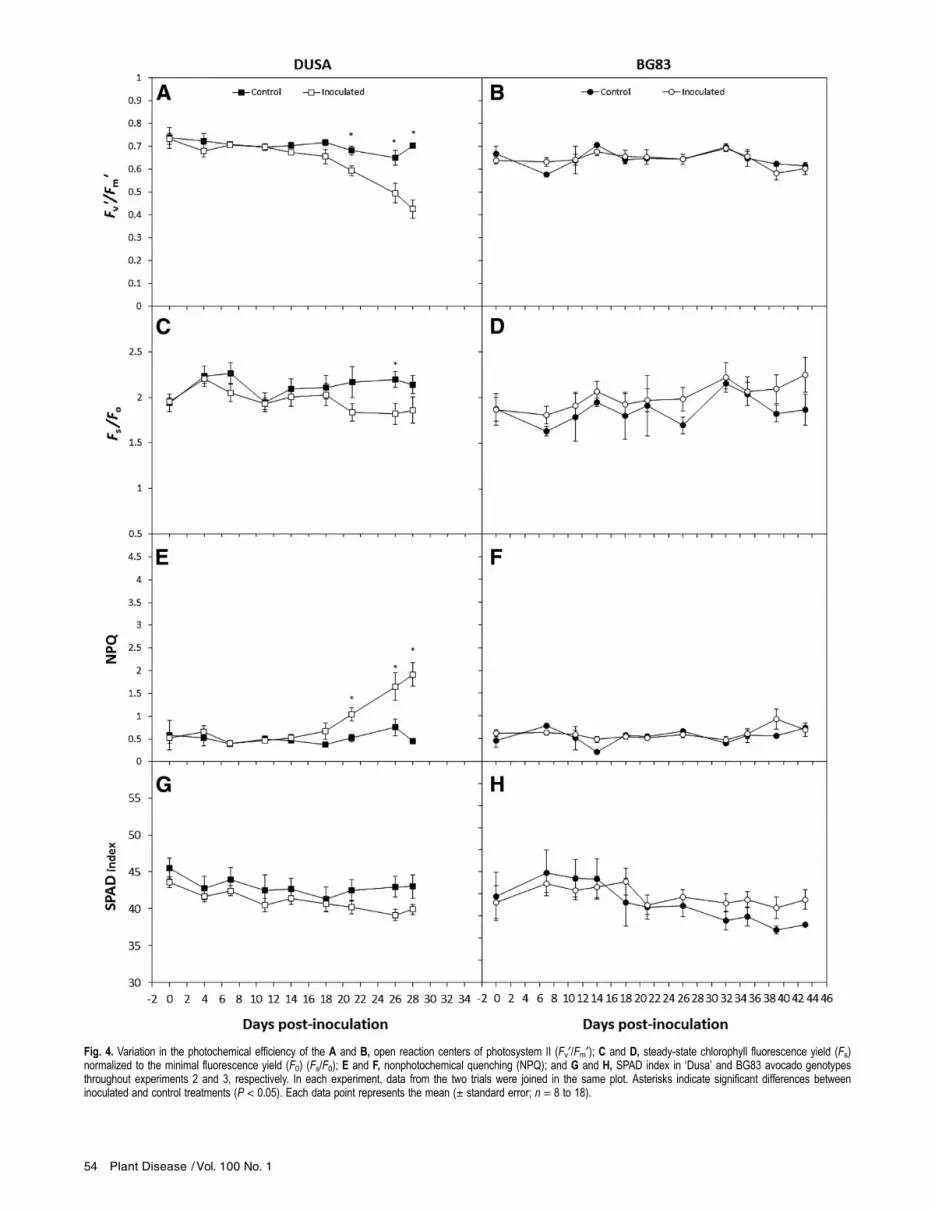
Fig. 4. Variation in the photochemical efficiency of the A and B, open reaction centers of photosystem II (Fv¢/Fm¢); C and D, steady-state chlorophyll fluorescence yield (Fs)normalized to the minimal fluorescence yield (F0) (Fs/F0); E and F, nonphotochemical quenching (NPQ); and G and H, SPAD index in ‘Dusa’ and BG83 avocado genotypesthroughout experiments 2 and 3, respectively. In each experiment, data from the two trials were joined in the same plot. Asterisks indicate significant differences betweeninoculated and control treatments (P < 0.05). Each data point represents the mean (± standard error; n = 8 to 18).
54 Plant Disease /Vol. 100 No. 1
(TT-16 and TT-21, P = 0.001; Dusa, P = 0.006), showing maximumlevels close to 1 month after inoculation (Figs. 3E and 4E). In TT-31plants, the physiological parameters displayed tendencies similar tothose described for TT-16 and TT-21 genotypes but with a 2-weekdelay (Fig. 3B, D, and F). In the tolerant genotype BG83, no signif-icant changes in any of the chlorophyll fluorescence parameters wereobserved during disease progression (Fig. 4B, D, and F). No symp-toms of leaf chlorosis were observed in any of the genotypes studiedunder either treatment, made evident as a lack of significant differ-ences in SPAD index between treatments and among genotypes(Figs. 3G and H and 4G and H) throughout the experimental courses.Imaging GFP-tagged R. necatrix strain to visualize
aboveground symptoms. Roots of infected plants at stage 1 show-ing a reduction of Fv¢/Fm¢ (i.e., 10 to 15 days postinoculation)
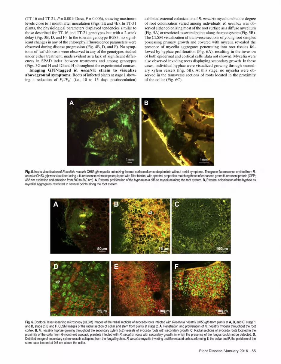
exhibited external colonization of R. necatrixmycelium but the degreeof root colonization varied among individuals. R. necatrix was ob-served either colonizing most of the root surface as a diffuse mycelium(Fig. 5A) or restricted to several points along the root system (Fig. 5B).The CLSM visualization of transverse sections of young root samplespossessing primary growth and covered with mycelia revealed thepresence of mycelia aggregates penetrating into root tissues fol-lowed by hyphae proliferation (Fig. 6A), resulting in the invasionof both epidermal and cortical cells (data not shown). Mycelia werealso observed invading roots displaying secondary growth. In thesecases, individual hyphae were visualized growing through second-ary xylem vessels (Fig. 6B). At this stage, no mycelia were ob-served in the transverse sections of roots located in the proximityof the collar (Fig. 6C).
Fig. 5. In situ visualization of Rosellinia necatrix CH53-gfpmycelia colonizing the root surface of avocado plantlets without aerial symptoms. The green fluorescence emitted from R.necatrix CH53-gfp was visualized using a fluorescence microscope equipped with filter blocks, with spectral properties matching those of enhanced green fluorescent protein (GFP;488 nm excitation and emission from 500 to 560 nm). A, External proliferation of the hyphae as a diffuse mycelium along the root system. B, External colonization of the hyphae asmycelial aggregates restricted to several points along the root system.
Fig. 6. Confocal laser-scanning microscopy (CLSM) images of the radial sections of avocado roots infected with Rosellinia necatrix CH53-gfp from plants at A, B, and C, stage 1and D, stage 2. E and F, CLSM images of the radial section of collar and stem from plants at stage 2. A, Penetration and proliferation of R. necatrix mycelia throughout the rootcortex. B, R. necatrix hyphae growing throughout the secondary xylem (×2) vessels of avocado roots with secondary growth. C, Radial sections of avocado roots located in theproximity of the collar from 6-month-old avocado plantlets infected with R. necatrix; roots with secondary growth, in which the presence of the fungus could not be detected. D,Detailed image of secondary xylem vessels collapsed from the fungal hyphae. R. necatrixmycelia invading undifferentiated cells conforming E, the collar and F, the periderm of thestem base located at 0.5 cm above the collar.
Plant Disease / January 2016 55

To compare R. necatrix root invasion at stage 1 (Fig. 1) withadvanced stages at which aerial symptoms were observed (stage 2and 3; Fig. 1), the roots of plants at stages 2 and 3 (i.e., 22 and27 days postinoculation, respectively) were collected and the areasat the proximity of the root collar were examined. Microscopicvisualization of transverse sections through CLSM revealed greenfluorescent mycelial aggregations invading most of the cork tissueand the phelloderm, reaching the pith. Hyphal strands were alsoobserved collapsing xylem vessels (Fig. 6E). The visualization oftransverse collar sections and stem sections at 0.5 cm above the col-lar revealed the invasion of undifferentiated cells on the collar(Fig. 6D) and the profuse invasion of the R. necatrix hyphal net-work into the periderm of the stem (Fig. 6F). However, in this case,the fungus was not observed colonizing areas bellow the cambium,such as xylem vessels and the pith. No penetration structures, suchas mycelial aggregates, were observed on the stem surface.
DiscussionHerein, we provide the first analysis combining nondestructive
chlorophyll fluorescence measurements and CLSM techniques todescribe the relationship between the physiological changes in avo-cado leaves during the first stages of infection and the degree ofR. necatrix root invasion.One of the earliest plant responses following pathogen attack is the
production of reactive oxygen species in the chloroplast (Berger et al.2007b; Bolwell et al. 2002; Torres et al. 2006) and the uncoupling orinhibition of the photosystem machinery (Berger et al. 2007b). In thepresent study, we showed that leaf photochemistry was affected earlyafter root infection with R. necatrix, as reflected by the decrease in thetrapping efficiency of PSII (Fv¢/Fm¢) before any visible symptomswere observed. This parameter reflects the changes in nonphoto-chemical processes affecting maximum PSII efficiency (Baker et al.2007), which likely involves some degree of photo-inactivation ofthe PSII reaction centers (Havaux et al. 1991; Martınez-Ferri et al.2000).These changes could be associated with the localized effects of
fungal phytotoxic metabolites on the roots or phytotoxic effects onthe leaves induced through mobile signals, as reported for otherwoody plants (Fleischmann et al. 2005; Grimmer et al. 2012).R. necatrix strains produce phytotoxic metabolites such as cytocha-lasin E (Chen 1964; Cole and Cox 1981; Kimura et al. 1989; Kshirsagaret al. 2001), rosellichalasin (Kimura et al. 1989), rosellinic acid, ros-necatrone, and diketopiperazines (Edwards et al. 2001), althoughroles for these compounds in pathogenicity remain unclear. Diseaseprogression was not affected in plants inoculated with R. necatrixmutants not producing cytochalasin E, suggesting that this toxic com-pound is not a major R. necatrix pathogenicity factor (Kanematsuet al. 1997).Previous studies have reported that the primary mechanisms of
woody plants in response to root pathogen infections are associatedwith the impairment of water relations and the limitation of photo-synthesis (Clemenz et al. 2008; Fleischmann et al. 2005). In the pre-sent study, we observed an early and significant decrease in Fs/F0
ratio before any aerial symptoms were observed. Studies have previ-ously shown that Fs/F0 is an excellent indicator of declining stomatalconductance, CO2 assimilation, and NPQ generation during mild wa-ter stress, thereby providing an adequate method for the early detectionof water stress (Flexas et al. 2002). The Fs/F0 parameter consolidatesthe cascade of consequences derived from stomatal limitations of pho-tosynthesis. Thus, the sustained decrease in Fs/F0 associated with theincrease in NPQ under nonlimiting water availability suggests that R.necatrix infection could be also associated with stomatal limitations ofphotosynthesis in avocado plantlets before aerial symptoms are evi-dent (Fig. 1, stage 1). CLSM images revealed that these changes wereconcomitant with the initial invasion of the root vascular system,which might affect water flow toward the aerial ground parts of theplant. These results are consistent with previous studies on avocadoplants infected with the root pathogen Phytophthora cinnamomiRands, in which root infection impaired water transport between rootsand leaves (Sterne et al. 1978). However, further studies, including the
measurement of water relations (i.e., plant water status, gas exchange,and stomatal conductance), are necessary to clarify the relative contri-bution of stomatal limitations of photosynthesis in WRR diseasedevelopment.Notably, during the early stages of infection, different degrees of
root colonization were observed but no mycelia were detected atthe root collar or at the roots located in the proximity of the collar.These differences in the degree of mycelial colonization along theroot did not translate into proportional differences in chlorophyllfluorescence parameters, suggesting that photosynthetic impairmentis not dependent on the degree of mycelial invasion of the lateral rootsystem. This result also demonstrates the reduced contribution of in-fected lateral roots to the transpirational stream compared with theroots close to the root collar and suggests a counteracting effect ofnoninvaded roots.Despite the presence of the fungus along the root system, the de-
crease in chlorophyll fluorescence parameters observed in TT-31was delayed 2 weeks compared with TT-16 and TT-21. These resultsindicate that TT-31 might have a lower susceptibility to R. necatrix,which would not be associated with constitutive differences in thegrowth or biomass partitioning among genotypes. This responseseems to be exclusive to susceptible genotypes because no variationin the photosynthetic parameters was observed in the tolerant geno-type (BG83). Thus, the results of the present study show that chloro-phyll fluorescence may potentially represent a useful, nondestructivetechnique for disease diagnosis which could support the discarding ofhighly susceptible genotypes in breeding programs in a short periodof time. With this method, we would discard TT-16 and TT-21 aspossible candidates for the selection program at 10 to 15 dayspostinoculation.The absence of changes at the photosynthetic level in the tolerant
genotype (BG83) despite of the presence of aerial symptoms suggeststhat avocado tolerance to R. necatrix could be associated with mecha-nisms conferring a higher robustness to the photosynthetic machinery.Moreover, the comparatively higher root investment observed inBG83 could indicate a fast root growth rate or root turnover, whichcan counteract the necrotic effects of R. necatrix in the root system,as has been reported in apple genotypes tolerant to replant disease(Atucha et al. 2014). To explore these issues in more depth, furtherstudies with tolerant genotypes are necessary.Once the aerial symptoms became obvious (Fig. 1, stage 2), the ob-
served wilting suggested the appearance of water stress, which wasconcomitant with pronounced physiological differences in suscepti-ble genotypes. These changes were associated with the profuse inva-sion of the root vascular system and the fungal colonization of theroot collar, obstructing water flow to the aerial parts of the plant.Taken together, these results suggest that visible symptoms ofWRR are associated with a mechanical effect on the main vascularsystem.Plant desiccation and death occurred within a few days after the
appearance of wilting symptoms in all genotypes. The lack of chlo-rosis, as shown by the SPAD index, might indicate that the potentialeffects of toxins during disease progression did not result in lossesof leaf integrity and chlorophyll photo-oxidation (Bolwell et al.2002) in contrast to the effects reported for other fungal toxins(Kim et al. 2010).In conclusion, this study is the first to provide information on the
physiological changes that occur during the initial stages ofR. necatrixinfection on avocado roots prior to the appearance of aboveground vis-ible symptoms in susceptible and tolerant genotypes. This knowledgeis essential for further studies of the avocado–R. necatrix interactionsystem. Thus, studies examining avocado gene expression profilesin contrasting disease reaction genotypes would be addressed to obtaina better understanding of the molecular interaction between the hostand pathogen, which is crucial for the development of new tools tocontrol disease. Moreover, the detection of R. necatrix infection onroots before aerial symptoms are observed would facilitate the applica-tion of control strategies during the early stages of the infection, avoid-ing tree death and further disease spread, which are major problemsto overcome when managing WRR disease. However, the use of
56 Plant Disease /Vol. 100 No. 1

chlorophyll fluorescence as a diagnosis tool in the field needs furtherinvestigation to test its validity. Under field conditions, leaf photo-chemistry can be affected by several environmental factors (i.e., water,T, and nutrients) and it would be necessary to assure that any othertype of stress is not operating in plants under evaluation.
AcknowledgmentsThis research was supported through funding from the AVA201301.13 project
co-financed through the Junta de Andalucıa and European Union (FEDER). M. C.Pliego and M. T. Ariza are currently supported by IFAPA Junta de Andalucıa(20%) and by the Programa Operativo Fondo Social Europeo (FSE) de Andalucıa2007-2013 (80%) under the topic “Andalucıa se mueve con Europa”. We thankD. Navas for his technical support in CLSM studies, M. Carrera for her technicalassistance, and three anonymous referees for their critical comments on a previousdraft of this manuscript.
Literature CitedAlvarez, J. P., Pekker, I., Goldshmidt, A., Blum, E., Amsellem, Z., and Eshed, Y.
2006. Endogenous and synthetic microRNAs stimulate simultaneous, efficient,and localized regulation of multiple targets in diverse species. Plant Cell 18:1134-1151.
Atucha, A., Emmett, B., and Bauerle, T. L. 2014. Growth rate of fine root systemsinfluences rootstock tolerance to replant disease. Plant Soil 376:337-346.
Baker, N. R., Harbinson, J., and Kramer, D. M. 2007. Determining the limitationsand regulation of photosynthetic energy transduction in leaves. Plant CellEnviron. 30:1107-1125.
Barcelo-Muñoz, A., Zea-Bonilla, T., Jurado-Valle, I., Imbroda-Solano, I.,Vidoy-Mercado, I., Pliego-Alfaro, F., and Lopez-Herrera, C. J. 2007.Programa de seleccion de portainjertos de aguacate tolerantes a la podredumbreblanca causada por Rosellinia necatrix en el Sur de España (1999-2007).Pages 537-541 in: Proc. VI World Avocado Congress, Viña del Mar, CL.
Bauriegel, E., and Herppich, W. B. 2014. Hyperspectral and chlorophyllfluorescence imaging for early detection of plant diseases, with specialreference to Fusarium spp. infections on wheat. Agriculture 4:32-57.
Berger, S., Benediktyova, Z., Matous, K., Bonfig, K. B., Mueller, M. J., Nedbal,L., and Roitsch, T. 2007a. Visualization of dynamics of plant-pathogeninteraction by novel combination of chlorophyll fluorescence imaging andstatistical analysis: Differential effects of virulent and avirulent strains ofP. syringae and of oxylipins on A. thaliana. J. Exp. Bot. 58:797-806.
Berger, S., Sinha, A., Thomas, K., and Roitsch, T. 2007b. Plant physiology meetsphytopathology: Plant primary metabolism and plant-pathogen interactions.J. Exp. Bot. 58:4019-4026.
Bilger, W., and Bjorkman, O. 1990. Role of the xanthophyll cycle inphotoprotection elucidated by measurements of light-induced absorbancechanges, fluorescence and photosynthesis in leaves of Hedera canariensis.Photosynth. Res. 25:173-185.
Bolwell, G. P., Bindschedler, L. V., Blee, K. A., Butt, V. S., Davies, D. R.,Gardner, S. L., Gerrish, C., and Minibayeva, F. 2002. The apoplasticoxidative burst in response to biotic stress in plants: A three-componentsystem. J. Exp. Bot. 53:1367-1376.
Broadbent, P., and Gollnow, B. I. 1993. Selecting disease-resistant citrusrootstocks. Aust. J. Exp. Agric. 33:775-780.
Brummer, M., Arend, M., Fromm, J., Schlenzig, A., and Oßwald, W. F. 2002.Ultrastructural changes and immunocytochemical localization of the elicitinquercinin in Quercus robur L. roots infected with Phytophthora quercina.Physiol. Mol. Plant Pathol. 61:109-120.
Calderon, R., Lucena, C., Trapero-Casas, J. L., Zarco-Tejada, P. J., and Navas-Cortes, J. A. 2014. Soil temperature determines the reaction of olive cultivarsto Verticillium dahliae pathotypes. PLoS One 9(10):e110664, doi:10.1371/journal.pone.0110664.
Chen, Y. 1964. Studies on the metabolic products of Rosellinia necatrix Berlesse.Part II. The structure of rosellinic acid. Agric. Biol. Chem. 28:431-435.
Clemenz, C., Fleischmann, F., Haberle, K. H., Matyssek, R., and Oßwald, W.2008. Photosynthetic and leaf water potential responses of Alnus glutinosasaplings to stem-base inoculation with Phytophthora alni subsp. alni. TreePhysiol. 28:1703-1711.
Cole, R. J., and Cox, R. H. 1981. The cytochalasins. Pages 264-343 in: Handbookof Toxic Fungal Metabolites. R. J. Cole, ed. Academic Press, London.
Dadmal, S. M., Pawar, N. P., Kale, K. B., and Shivankar, S. K. 2002. Screening ofcitrus rootstock against leaf miner Phyllocnistis citrella Stainton. InsectEnviron. 8:143-144.
Edwards, R. L.,Maitland, D. J., Scowen, I. J., Teixeira de Sousa, A. J., andWhalley,A. J. S. 2001. Metabolites of the higher fungi. Part 32. Rosnecatrone, aphytotoxic bicyclo[4.1.0]hept-3-en-2-one from the fungus Rosellinia necatrixPrill. J. Chem. Soc. Perk. T. 1:537-542.
Fleischmann, F., Gottlein, A., Rodenkirchen, H., Lutz, C., and Oßwald, W. F. 2004.Biomass, nutrient and pigment content of beech (Fagus sylvatica) saplingsinfected with Phytophthora citricola, P. cambivora, P. pseudosyringae andP. undulata. For. Pathol. 34:79-92.
Fleischmann, F., Koehl, J., Portz, R., Beltrame, A. B., and Oßwald, W. 2005.Physiological changes of Fagus sylvatica seedlings infected with Phytophthora
citricola and the contribution of its elicitin ‘citricolin’ to pathogenesis. PlantBiol. 7:650-658.
Fleischmann, F., Schneider, D., Matyssek, R., and Oßwald, W. F. 2002.Investigations on net CO2 assimilation, transpiration and root growth ofFagus sylvatica infested with four different Phytophthora species. Plant Biol.4:144-152.
Flexas, J., Escalona, J. M., Evain, S., Gulıas, J., Moya, I., Osmond, C. B., andMedrano, H. 2002. Steady-state chlorophyll fluorescence (Fs) measurementsas a tool to follow variations of net CO2 assimilation and stomatalconductance during water‐stress in C3 plants. Physiol. Plant. 114:231-240.
Freeman, S., and Sztejnberg, A. 1992. Rosellinia. Pages 71-73 in: Methodsfor Research on Soilborne Phytopathogenic Fungi. L. L. Singleton, J. D.Mihail, and C. M. Rush, eds. American Phytopathological Society Press, St.Paul, MN.
Genty, B., Briantais, J., and Baker, N. 1989. The relationship between the quantumyield of photosynthetic electron transport and photochemical quenching ofchlorophyll fluorescence. Biochim. Biophys. Acta 990:87-92.
Grimmer, M. K., Foulkes, M. J., and Paveley, N. D. 2012. Foliar pathogenesis andplant water relations; a review. J. Exp. Bot. 63:4321-4331.
Havaux, M., Strasser, R. J., and Greppin, H. 1991. A theoretical and experimentalanalysis of the qP and qN coefficients of chlorophyll fluorescence quenchingand their relation to photochemical and nonphotochemical events.Photosynth. Res. 27:41-55.
Jifon, J. L., Syvertsen, J. P., and Whaley, E. 2005. Growth environment and leafanatomy affect nondestructive estimates of chlorophyll and nitrogen in Citrusspp. leaves. J. Am. Soc. Hortic. Sci. 130:152-158.
Kanematsu, S., Hayashi, T., and Kudo, A. 1997. Isolation of Rosellinia necatrixmutants with impaired cytochalasin E production and its pathogenicity. Jpn.J. Phytopathol. 63:425-431.
Kim, Y. M., Bouras, N., Kav, N. N. V., and Strelkov, S. E. 2010. Inhibition ofphotosynthesis and modification of the wheat leaf proteome by Ptr ToxB: Ahost-specific toxin from the fungal pathogen Pyrenophora tritici-repentis.Proteomics 10:2911-2926.
Kimura, Y., Nakajima, H., and Hamasaki, T. 1989. Structure of Rosellichalasin, anew metabolite produced by Rosellinia necatrix. Agric. Biol. Chem. 53:1699-1701.
Kshirsagar, A., Reid, A. J., McColl, S. M., Saunders, V. A., Whalley, A. J. S., andEvans, E. H. 2001. The effect of fungal metabolites on leaves as detected bychlorophyll fluorescence. New Phytol. 151:451-457.
Lopez-Herrera, C. J., Perez-Jimenez, R. M., Basallote-Ureba, M. J., Zea-Bonilla,T., and Melero-Vara, J. M. 1999. Loss of viability of Dematophora necatrixin solarized soils. Eur. J. Plant Pathol. 105:571-576.
Lopez-Herrera, C. J., and Zea-Bonilla, T. 2007. Effects of benomil, carbendazim,fluazinan and thiophanate methyl on white root rot of avocado. Crop Prot. 26:1186-1192.
Markwell, J., Ostermann, J. C., andMitchell, J. L. 1995. Calibration of the MinoltaSPAD-502 leaf chlorophyll meter. Photosynth. Res. 46:467-472.
Martınez-Ferri, E., Balaguer, L., Valladares, F., Chico, J. M., and Manrique, E.2000. Energy dissipation in drought-avoiding and drought-tolerant treespecies at midday during theMediterranean summer. Tree Physiol. 20:131-138.
Pliego, C., Kanematsu, S., Ruano-Rosa, D., De Vicente, A., Lopez-Herrera, C.,Cazorla, F. M., and Ramos, C. 2009. GFP sheds light on the infection process ofavocado roots by Rosellinia necatrix. Fungal Genet. Biol. 46:137-145.
Pliego, C., Lopez-Herrera, C., Ramos, C., and Cazorla, F. M. 2012. Developingtools to unravel the biological secrets of Rosellinia necatrix, an emergentthreat to woody crops. Mol. Plant Pathol. 13:226-239.
Pliego-Alfaro, F., Barcelo-Muñoz, A., Lopez-Gomez, R., Ibarra-Laclette, L.,Herrera-Estrella, L., Palomo-Rıos, E., Mercado, J. A., and Litz, R. E. 2013.Biotechnology. Pages 268-300 in: The Avocado: Botany, Production andUses, 2nd ed. A. W. Whiley, B. Schaffer, and B. N. Wolstenholme. eds.CABI Publishing, Oxon, UK.
Raj, H., and Sharma, S. D. 2009. Integration of soil solarization and chemicalsterilization with beneficial microorganisms for control of white root rot andgrowth of nursery apple. Sci. Hortic. (Amsterdam) 119:126-131.
Ruano-Rosa, D., Schena, L., Ippolito, A., and Lopez-Herrera, C. J. 2007. Comparisonof conventional and molecular methods for the detection of Rosellinia necatrix inavocado orchards in southern Spain. Plant Pathol. 56:251-256.
Schena, L., and Ippolito, A. 2003. Rapid and sensitive detection of Rosellinianecatrix in roots and soils by real time Scorpion-PCR. J. Plant Pathol. 85:15-25.
Schena, L., Nigro, F., and Ippolito, A. 2002. Identification and detection ofRosellinia necatrix by conventional and real-time Scorpion-PCR. Eur. J.Plant Pathol. 108:355-366.
Schreiber, U., Bilger, W., Hormann, H., and Neubauer, C. 1998. Chlorophyllfluorescence as a diagnostic tool: The basics and some aspects of practicalrelevance. Pages 320-326 in: Photosynthesis: A Comprehensive Treatise.A. S. Raghavendra, ed. Cambridge University Press, Cambridge, UK.
Schreiber, U., Bilger, W., and Neubauer, C. 1994. Chlorophyll fluorescence as anon-intrusive indicator for rapid assessment of in vivo photosynthesis. Pages49-70 in: Ecophysiology of Photosynthesis. E. D. Schulze and M. M.Caldwell, eds. Springer-Verlag, Berlin.
Sterne, R. E., Kaufmann, M. R., and Zentmyer, G. A. 1978. Effect of Phytophthoraroot rot on water relations of avocado: Interpretation with a water transportmodel. Phytopathology 68:595-602.
Plant Disease / January 2016 57

Sugimoto, K. 2002. Fluazinam (Frowncide�) a novel and effective method ofapplication against white and violet root rot. Agrochem. Jpn. 80:14-16.
Sztejnberg, A., and Madar, Z. 1980. Host range of Dematophora necatrix, thecause of white root rot disease in fruit trees. Plant Dis. 64:662-664.
Tavakkoli, E., Rengasamy, P., and McDonald, G. K. 2010. High concentrations ofNa+ and Cl– ions in soil solution have simultaneous detrimental effects ongrowth of faba bean under salinity stress. J. Exp. Bot. 61:4449-4459.
ten Hoopen, G.M., and Krauss, U. 2006. Biology and control ofRosellinia bunodes,Rosellinia necatrix and Rosellinia pepo: A review. Crop Prot. 25:89-107.
Torres, M. A., Jones, J. D. G., and Dangl, J. L. 2006. Reactive oxygen speciessignaling in response to pathogens. Plant Physiol. 141:373-378.
Trapero Muñoz, S., Hervalejo Garcıa, A., Jimenez Perez, M., Boyero, J. R., Vela,J. M., and Martınez-Ferri, E. 2008. Effects of rootstock and flushing on theincidence of three insects on ‘Clementine de Nules’ citrus trees. Environ.Entomol. 37:1531-1537.
Uddling, J., Gelang-Alfredsson, J., Piikki, K., and Pleijel, H. 2007. Evaluating therelationship between leaf chlorophyll concentration and SPAD-502 chlorophyllmeter readings. Photosynth. Res. 91:37-46.
Verdejo-Lucas, S., Galeano, M., Sorribas, F. J., Forner, J. B., and Alcaide, A. 2003.Effect on resistance to Tylenchulus semipenetrans of hybrid citrus rootstockssubjected to continuous exposure to high population densities of thenematode. Eur. J. Plant Pathol. 109:427-433.
58 Plant Disease /Vol. 100 No. 1


