Embed Size (px)
Citation preview
Neuropathology of Nondemented Aging: Presumptive Evidencefor Preclinical Alzheimer Disease
Joseph L. Price, D.Phila, Daniel W. McKeel Jr, MDb, Virginia D. Buckles, PhDc, Catherine M.Roe, PhDc, Chengjie Xiong, PhDd, Michael Grundman, MDe, Lawrence A. Hansen, MDf,Ronald C. Petersen, MD, PhDg, Joseph E. Parisi, MDh, Dennis W. Dickson, MDi, Charles D.Smith, MDj, Daron G. Davis, MDk, Frederick A. Schmitt, PhDl, William R. Markesbery, MDl,Jeffrey Kaye, MDm, Roger Kurlan, MDn, Christine Hulette, MDo, Brenda F. Kurland, PhDp,Roger Higdon, PhDq, Walter Kukull, PhDr, and John C. Morris, MDsa Department of Anatomy and Neurobiology; Alzheimer’s Disease Research Center; WashingtonUniversity School of Medicine; St. Louis, Missourib Pathology and Immunology (Retired); Alzheimer’s Disease Research Center; WashingtonUniversity School of Medicine; St. Louis, Missouric Department of Neurology; Alzheimer’s Disease Research Center; Washington University Schoolof Medicine; St. Louis, Missourid Division of Biostatistics; Alzheimer’s Disease Research Center; Washington University School ofMedicine; St. Louis, Missourie Elan Pharmaceuticals; San Diego, Californiaf Department of Neuroscience; University of California – San Diego; La Jolla, Californiag Department of Neurology; Mayo Clinic; Rochester, Minnesotah Departments of Anatomic Pathology and Laboratory Medicine and Pathology; Mayo Clinic;Rochester, Minnesotai Neuro-Oncology Program and Department of Laboratory Medicine and Pathology; Mayo Clinic;Jacksonville, Floridaj Department of Neurology; University of Kentucky; Lexington, Kentuckyk Pathology Department; Central Baptist Hospital; Lexington, Kentuckyl Sanders-Brown Center on Aging; Alzheimer’s Disease Center; University of Kentucky, Lexington,KYm Layton Aging & Alzheimer Disease Center; Oregon Health Sciences University; Portland, Oregonn Department of Neurology; University of Rochester School of Medicine; Rochester, New Yorko Department of Pathology and Division of Neurology; Duke University Medical Center; Durham,North Carolina
Corresponding author: Dr. John C. Morris; 4488 Forest Park Avenue; Suite 130; St. Louis, Missouri; 63108; [email protected];Phone: 314-286-2881; Fax: 314-286-2763.Disclosure Statement There are no actual or potential conflicts of interest.Publisher's Disclaimer: This is a PDF file of an unedited manuscript that has been accepted for publication. As a service to our customerswe are providing this early version of the manuscript. The manuscript will undergo copyediting, typesetting, and review of the resultingproof before it is published in its final citable form. Please note that during the production process errors may be discovered which couldaffect the content, and all legal disclaimers that apply to the journal pertain.
NIH Public AccessAuthor ManuscriptNeurobiol Aging. Author manuscript; available in PMC 2010 July 1.
Published in final edited form as:Neurobiol Aging. 2009 July ; 30(7): 1026–1036. doi:10.1016/j.neurobiolaging.2009.04.002.
NIH
-PA Author Manuscript
NIH
-PA Author Manuscript
NIH
-PA Author Manuscript

p Clinical Statistics; Fred Hutchinson Cancer Research Center; Seattle, Washingtonq The BIATECH Institute; Seattle, Washingtonr Department of Epidemiology; University of Washington; Seattle, Washingtons Departments of Neurology, Pathology and Immunology, and Physical Therapy; Alzheimer’sDisease Research Center; Washington University School of Medicine, St. Louis, Missouri
AbstractObjective—To determine the frequency and possible cognitive effect of histological Alzheimer’sdisease (AD) in autopsied older nondemented individuals.
Design—Senile plaques (SPs) and neurofibrillary tangles (NFTs) were assessed quantitatively in97 cases from 7 Alzheimer’s Disease Centers (ADCs). Neuropathological diagnoses of AD (npAD)were also made with four sets of criteria. Adjusted linear mixed models tested differences betweenparticipants with and without npAD on the quantitative neuropathology measures and psychometrictest scores prior to death. Spearman rank-order correlations between AD lesions and psychometricscores at last assessment were calculated for cases with pathology in particular regions.
Setting—Washington University Alzheimer’s Disease Research Center.
Participants—Ninety-seven nondemented participants who were age 60 years or older at death(mean = 84 years).
Results—About 40% of nondemented individuals met at least some level of criteria for npAD;when strict criteria were used, about 20% of cases had npAD. Substantial overlap of Braakneurofibrillary stages occurred between npAD and no-npAD cases. Although there was nomeasurable cognitive impairment prior to death for either the no-npAD or npAD groups, cognitivefunction in nondemented aging appears to be degraded by the presence of NFTs and SPs.
Conclusions—Neuropathological processes related to AD in persons without dementia appear tobe associated with subtle cognitive dysfunction and may represent a preclinical stage of the illness.By age 80–85 years, many nondemented older adults have substantial AD pathology.
Keywordspreclinical Alzheimer’s disease; nondemented aging; neuropathological Alzheimer’s disease
IntroductionSenile plaques (SPs) and neurofibrillary tangles (NFTs), the neuropathological hallmarks ofAlzheimer’s disease (AD),(Khachaturian, 1985) are not limited to individuals with dementiaof the Alzheimer type (DAT) but also may be present in the brains of cognitively normal olderadults.(Tomlinson et al., 1968; Crystal et al., 1988; Katzman et al., 1988; Price et al., 1991;Dickson et al., 1992; Troncoso et al., 1996; Troncoso et al., 1998; Hulette et al., 1998; Schmittet al., 2000; Knopman et al., 2003) Neuropathological criteria for AD rely on densities of SPsand NFTs to discriminate AD from aging, as the distinction between the two conditions hasbeen thought to be quantitative rather than qualitative.(Tomlinson et al., 1970; Arriagada etal., 1992) Alternatively, the presence of SPs and NFTs in nondemented older adults mayrepresent AD at a stage prior to its clinical expression,(Morris et al., 1996; Price & Morris,1999; Troncoso et al., 1998; Hulette et al., 1998; Schmitt et al., 2000) in which theneuropathological lesions of AD accumulate over many years before sufficient synaptic andneuronal damage occurs to produce the symptoms of AD. This notion of a preclinical(presymptomatic) stage of AD is consistent with similar latent stages in otherneurodegenerative disorders.(Dickson et al., 2008) Although it is possible to explain the
Price et al. Page 2
Neurobiol Aging. Author manuscript; available in PMC 2010 July 1.
NIH
-PA Author Manuscript
NIH
-PA Author Manuscript
NIH
-PA Author Manuscript

presence of SPs and NFTs without apparent dementia in other ways(Berlau et al., 2007), acontinuous neuropathologic process for AD, regardless of clinical status, is supported byobservations that the mechanisms responsible for SPs and NFTs in nondemented older adultsappear identical to those found in AD(Haroutunian et al., 1998; Haroutunian et al., 1999) andthe distribution of SPs and NFTs in nondemented older adults corresponds with the hierarchicaltopographical progression associated with symptomatic AD.(Arriagada et al., 1992; Price &Morris, 1999; Price et al., 1991)
Previous studies of neuropathological AD in nondemented aging have been based on casescollected at a single site(Price & Morris, 1999; Galvin et al., 2005; Bennett et al., 2006), andcomparison of results has been difficult because neuropathological methods and criteriadiffered in these studies. Such methodological differences, combined with relatively smallsample sizes in all but one study,(Bennett et al., 2006) may contribute to conflicting results onthe relationships between SPs or NFTs and cognition in nondemented aging. Some studieshave reported a negative effect for episodic memory only,(Hulette et al., 1998; Schmitt et al.,2000; Bennett et al., 2006) without effects in other cognitive domains, while other studies foundno relationship between AD pathology and any cognitive measure.(Knopman et al., 2003;Driscoll et al., 2006) We therefore sought to provide a uniform assessment of neuropathologicalmarkers of AD in a large sample of nondemented older adults whose cognitive status had beenevaluated antemortem at several Alzheimer’s Disease Centers (ADCs) and to evaluaterelationships between these markers and cognitive function.
Material and MethodsParticipants
This study capitalized on the National Alzheimer Coordinating Center-supportedNeuropsychological Database Initiative (NDI; M Grundman, PI) that merged longitudinalclinical and cognitive data from more than 4,000 cognitively normal older ADC participants.Individuals in the NDI who came to autopsy at 7 participating ADCs, with a Clinical DementiaRating (CDR)(Morris, 1993) of 0 (indicating the absence of dementia and of mild cognitiveimpairment) within 2 years of death, and were at least 60 years of age at death were eligiblefor this study. Ninety-seven cases met these criteria; 20 from the University of Kentucky, 18from Washington University, 16 each from Mayo Clinic and Duke University, 14 from theUniversity of California, San Diego, 9 from Oregon Health Sciences University, and 4 fromthe University of Rochester. The mean interval from last ADC assessment to death in the 97cases was 0.7 +/− 0.5 years. Clinicians at each ADC verified the nondemented status for eachcase (the neuropathological diagnoses generated at the ADC performing the autopsy were notincluded in the eligibility criteria).
In addition, 11 cognitively impaired cases with CDR greater than 0 were submitted from 6 ofthe 7 ADCs. These 11 cases were assessed together with the nondemented cases, and all caseswere coded so that the central laboratory was blind to the cognitive status of each case. Datafrom cases with CDR > 0 are not reported here because they did not provide an adequatelysized sample.
Clinical and Psychometric MeasuresClinical variables available for this study included age at death, gender, educational attainment,CDR, and Mini Mental Status Examination (MMSE(Folstein et al., 1975)) scores at last ADCassessment. Apolipoprotein E genotypes were not available. Cognitive data included in theNDI database were available for Logical Memory–Delayed from the Wechsler Memory Scale-Revised;(Wechsler, 1987) Category Fluency (animals);(Goodglass & Kaplan, 1983) BostonNaming;(Goodglass & Kaplan, 1983) Trailmaking A and B;(Armitage, 1946) and Digit
Price et al. Page 3
Neurobiol Aging. Author manuscript; available in PMC 2010 July 1.
NIH
-PA Author Manuscript
NIH
-PA Author Manuscript
NIH
-PA Author Manuscript
Symbol from the Wechsler Adult Intelligence Scale.(Wechsler, 1997) These tests measureepisodic memory, semantic knowledge, attention, speeded psychomotor performance, andexecutive function. Not all individuals completed all measures.
Neuropathological ProceduresEach ADC provided a series of 10 unstained paraffin sections (10μm) of formalin-fixed tissuefrom each of five cerebral regions for each case: midfrontal cortex, superior/middle temporalgyrus, occipital cortex (Brodmann area 17, 18, or 19), medial temporal lobe (one block throughthe entorhinal cortex and rostral hippocampus, and a second, optional block through theamygdala), and midbrain. These regions were based on those proposed for the standardneuropathological assessment of AD.(Mirra et al., 1991) The Neuropathology Core (JLP,DWM) of Washington University’s Alzheimer’s Disease Research Center served as the“central laboratory” to provide a standard and uniform neuropathological assessment of eachcase and avoid the variability introduced by different methods, criteria, and interpretations usedat individual ADCs.(Mirra et al., 1994; Alafuzoff et al., 2006) The central laboratory’s stainingmethods included hematoxylin and eosin, modified Bielschowsky, Gallyas, and Nissl stains,as well as immunohistochemical procedures with anti-amyloid-beta (Aβ; monoclonal antibody“10D5”, gift of Athena Neurosciences and Elan) and anti-paired helical filaments (PHF;monoclonal antibody “PHF-1”, gift of Drs Sharon Greenberg and Peter Davies, Albert EinsteinMedical School, Bronx, NY). The anti-Aβ and anti-PHF stains were done both separately ondifferent sections (Vectastain ABC peroxidase kit, DAB substrate) and as a double stain onthe same sections, using different secondary antibody kits and chromogens so that Aβ wasstained red (Vectastain ABC alkaline phosphatase kit, Vector red substrate) and PHF stainedblack (Vectastain ABC peroxidase kit, DAB-Ni substrate).
The central laboratory generated independent neuropathological diagnoses for each of the 97nondemented cases. The diagnoses were done by joint examination of each case by DWM andJLP, using a double-headed microscope to arrive at a consensus neuropathological diagnosis.[Note: the neuropathological diagnoses generated by the central laboratory did not alwayscoincide with the diagnoses generated at the individual sites]. Both observers were “blind” tothe clinical and CDR status of the cases and to the neuropathological findings determined bythe ADCs contributing the cases.
Each case was assessed in accordance with 4 published criteria for the neuropathologicaldiagnosis of AD. Consensus criteria reported by Khachaturian(Khachaturian, 1985) are basedon the highest density of total SPs (diffuse and neuritic) in any cortical field, adjusted for age(the greater the age of the individual, the greater the density required for diagnosis). TheWashington University criteria(Berg et al., 1998; McKeel et al., 2004) modify the Khachaturiancriteria in three ways: 1) rather than relying on the highest SP density in a single field, averageSP density across 10 adjacent neocortical fields (1 mm2) is used; 2) SP densities are not adjustedfor age; and 3) at least one neocortical NFT is required. Both Khachaturian and WashingtonUniversity criteria yield dichotomous outcomes (a diagnosis of either npAD or no-npAD) andare the only criteria that consider diffuse SPs. The Consortium to Establish a Registry forAlzheimer’s Disease (CERAD)(Mirra et al., 1991) criteria use a semiquantitative assessmentof neuritic SP density and are age-adjusted to stipulate three levels of certainty that dementiais explained by the neuropathological diagnosis: Possible, Probable, and Definite AD. Finally,criteria developed at a conference sponsored by the National Institute of Aging and the ReaganInstitute of the Alzheimer’s Association (NIA-Reagan)(National Institute on Aging & ReaganInstitute Working Group on Diagnostic Criteria for the Neuropathological Assessment ofAlzheimer’s Disease, 1997) combine CERAD criteria for neuritic SPs and the neurofibrillarystaging system proposed by Braak and Braak(Braak & Braak, 1991) to characterize individualsin terms of three probabilistic diagnoses (Low, Intermediate, and High) that dementia during
Price et al. Page 4
Neurobiol Aging. Author manuscript; available in PMC 2010 July 1.
NIH
-PA Author Manuscript
NIH
-PA Author Manuscript
NIH
-PA Author Manuscript
life was due to AD. The NIA-Reagan criteria were not intended to characterize nondementedindividuals or those with incipient dementia.(National Institute on Aging & Reagan InstituteWorking Group on Diagnostic Criteria for the Neuropathological Assessment of Alzheimer’sDisease, 1997) In this report, the Washington University criteria were considered “primary”because they were based on discriminative analyses of neuropathological data.(McKeel et al.,2004) In practice and research, however, the CERAD and NIA-Reagan criteria are much morecommonly used.(Schmitt et al., 2000; Knopman et al., 2003; Bennett et al., 2006)
Braak neurofibrillary staging was determined for all cases. Briefly, Braak Stage 0 correspondsto absence of NFTs, Stages I–II correspond to NFTs limited to entorhinal-perirhinal cortex,Stages III–IV correspond to NFTs additionally in hippocampus and inferior temporal cortexand Stages V–VI correspond to NFTs distributed in wider neocortical areas.
Quantitative AssessmentThe central laboratory assessed SPs and NFTs quantitatively with the use of computerizedsampling and the Computer Assisted Stereological Toolbox (CAST) system (OlympusAmerica, Center Valley, PA 18034-0610). Analysis was based on Bielschowsky-stainedsections because they were more consistent, given the variable state of fixation of the materialfrom different sources, although immunohistochemical sections (singly stained for Aβ or PHF,or double stained for both Aβ and PHF) were routinely examined and compared with the silverstained sections. Diffuse SPs had a distinct concentration of Aβ staining without neuriticpathology; neuritic plaques had dystrophic neurites (with PHF) within an Aβ plaque and/orhad a dense amyloid core (Figure 1). Variables yielded by the quantitative analysis includedNFT density, diffuse SP density, neuritic SP density, and percent area occupied by SPs (plaqueburden) in limbic structures (entorhinal and perirhinal cortex, field CA1 of the hippocampus,and the amygdala) and neocortical areas (middle frontal gyrus, superior temporal gyrus, andoccipital cortex). Because the number of sections through each region was limited, totalnumbers of lesions were not obtained; only the density was measured for each marker (otherthan plaque burden). Although density measures may be affected by tissue shrinkage, theuniform treatment of the tissue (formalin fixation and paraffin embedding) and uniformstaining in the central laboratory reduced variability. Plaque burden was defined as the percentarea occupied by plaques in relation to the total area sampled and is unaffected by tissueshrinkage. Plaque burden and NFT density were least dependent on fixation differencesbetween different cases.
Statistical AnalysesAnalyses were performed using SAS statistical software (SAS Institute, Cary, NC). Generallinear mixed models (PROC MIXED) were used to test differences between two groups ofcases, one meeting neuropathological criteria for AD (npAD) and the other withoutneuropathological AD (no-npAD) on age at death, time between last assessment and death,and years of education while adjusting for an ADC effect by including that effect as a randomvariable. General linear mixed models were also used to test differences between the npADand no-npAD groups after adjustment for age, sex, interval between last assessment and death,and education on the quantitative neuropathology variables, and the psychometric test scoresat last assessment.
The ability of diffuse SPs in the neocortical and limbic areas to predict a neuropathologicaldiagnosis of AD as determined by the CERAD and NIA-Reagan criteria (which do notincorporate diffuse SPs) was examined by calculating the area under the Receiver OperatingCharacteristic (ROC) curve, and computing sensitivity values (holding specificity fixed at80%) for each lesion type. Subsequent comparisons of areas under the ROC curves were doneusing Fisher’s z-transformation in order to examine the predictive power of all lesions.
Price et al. Page 5
Neurobiol Aging. Author manuscript; available in PMC 2010 July 1.
NIH
-PA Author Manuscript
NIH
-PA Author Manuscript
NIH
-PA Author Manuscript

To assess whether neuropathological lesions are associated with cognitive performance,Spearman rank-order correlations between average lesion densities and psychometric scoresat last assessment were calculated for cases with lesions in the specified brain regions. In thegeneral linear mixed models, the ROC analyses, and calculation of sensitivity values, thequantitative neuropathology variables were first transformed by addition of 0.5 to each value(to account for cases with no pathology) and then conversion to the natural log.
ResultsThe 97 autopsied nondemented individuals had an average age ± SD at death of 84.3±8.6 years.There were 55 women, the mean years of education was 15.4±2.9, and the mean MMSE scoreat last assessment was 28.1±2.1.
Neuropathological AssessmentA substantial fraction of cases had lesions consistent with a neuropathological diagnosis of ADregardless of the criteria used. Figure 2 shows the percentage of cases meeting the dichotomousoutcomes for npAD with the Khachaturian and Washington University criteria and thepercentage of cases in each of the 3 levels denoting AD lesions with the CERAD and NIA-Reagan criteria. The least strict of the 4 sets of criteria, those proposed by Khachaturian,resulted in a neuropathological diagnosis of AD in 46 of the 97 cases (47%). The WashingtonUniversity criteria yielded a neuropathological diagnosis of AD in 38 cases (39%), identicalto the number of cases meeting any of the 3 levels of npAD using CERAD criteria (19%Possible, 19% Probable, and 2% Definite AD; 39% with all levels combined,) and any of the3 levels of the NIA-Reagan criteria (20% Low, 16% Intermediate, and 3% High; 39%combined). [Note: Many investigators reserve the diagnosis of npAD only for cases meeting“Probable” and “Definite” AD with CERAD criteria or “Intermediate” and “High” probabilitywith NIA-Reagan criteria; using these definitions, 21% were diagnosed with npAD by theCERAD Probable/Definite criteria, and 19% by the NIA-Reagan Intermediate/High criteria.]The WU, CERAD and NIA-Reagan criteria identified the same npAD cases, except for 2 casesdiagnosed as npAD with the WU criteria that were not recognized by CERAD or NIA-Reagan,and 2 cases recognized by CERAD and NIA-Reagan that were not diagnosed as npAD by WUcriteria.
Braak neurofibrillary stages in the 97 cases ranged from 0 (no NFTs observed) to V (widespreadneocortical tangles), with a median value of Stage II (Stage 0, n=4; Stage I, n=31; Stage II,n=36; Stage III, n=15; Stage IV, n=8; Stage V, n=3). No cases met criteria for stage VI (verypronounced neurofibrillary changes throughout all parts of cortex and in many subcorticalstructures). The 4 stage 0 cases were relatively young, aged 66, 69, 72, and 72 years at death.Older age was significantly correlated with higher Braak stages (Spearman r=.372, p<.001),consistent with observations that NFTs increase with age, even in the absence of SPs.(Arriagada et al., 1992; Price & Morris, 1999)
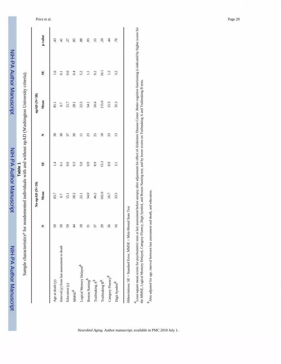
Comparison of npAD and no-npAD cases—There were no differences in mean age atdeath, interval between last clinical assessment and death, education, or psychometricperformance at last assessment before death between cases with npAD versus those withoutnpAD using any of the 4 criteria (even when restricted to Probable and Definite CERAD andIntermediate and High NIA-Reagan criteria) (Table 1).
Association with Braak Neurofibrillary Stage—Although a greater likelihood of npADwas associated with higher Braak stage scores for all 4 criteria (chi-square, p<0.003), therewas substantial overlap of the Braak stages between the npAD and no-npAD cases (Figure 3).
Price et al. Page 6
Neurobiol Aging. Author manuscript; available in PMC 2010 July 1.
NIH
-PA Author Manuscript
NIH
-PA Author Manuscript
NIH
-PA Author Manuscript

There was virtually no difference between the Braak stages of cases at comparable levels ofthe CERAD and NIA-Reagan criteria (Figure 3B,C).
Quantitative Neuropathological AssessmentROC Analysis—To avoid the circularity inherent in using measures as outcomes when thosesame measures were used in classification, this analysis was limited to the CERAD and NIA-Reagan criteria, which do not incorporate diffuse SPs. ROC analyses indicated that the densityof neocortical diffuse SPs best discriminated between npAD and no-npAD (defined bycombination of all three diagnostic levels of either criteria), followed by the density of diffuseSPs in limbic areas. For completeness, the area under the curve and sensitivity for the otherassessed neuropathological lesions (neuritic SPs, NFTs) also are shown in Table 3 althoughthese data are confounded by the circularity noted above. The density of NFTs in limbic areaswas the least discriminative (Table 2). Spearman correlations between the quantitativeneuropathological measures and all outcomes of CERAD criteria (no AD or Possible, Probable,or Definite) and all outcomes of NIA-Reagan criteria (no AD or Low, Intermediate, or High)yielded almost the identical rank order for association with diagnostic level (Table 2).
Correlation with Age—NFT density increased with age in limbic areas (entorhinal andperirhinal cortex, amygdala, and hippocampus field CA1) (Figure 4A; Spearmancorrelation=0.437, p<.001). In contrast, plaque burden was not significantly correlated withage (Figure 4B; Spearman correlation=0.089, p=0.40).
Relation of plaque burden and tangle density—Neocortical plaque burden and limbicNFT density in nondemented cases were weakly correlated (Spearman correlation=0.196, p=.07, Figure 5).
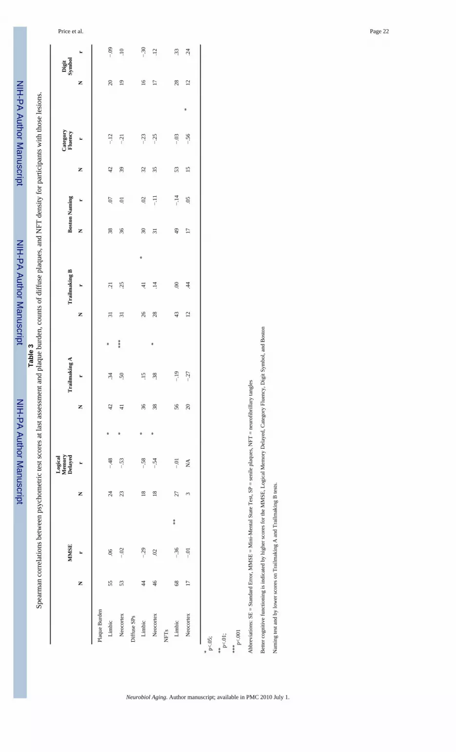
Correlation with psychometric tests—Although none of the participants in this studyhad clinically detectable dementia or cognitive impairment that was measurable on anindividual basis, a significant correlation of some psychometric test scores was found with thedensity of AD lesions across the group, indicating that cognitive function in nondementedaging may decline with increasing numbers of NFTs or SPs, including diffuse SPs (Table 3).In all cases, greater pathology was associated with poorer cognitive performance at lastassessment on measures of episodic memory (Logical Memory Delayed), semantic knowledge(Category Fluency), visuospatial ability (Trailmaking A), and executive function (TrailmakingB) as well as on a general measure, the MMSE. The correlations between the psychometrictest scores and plaque burden, and between the test scores and diffuse SPs, were similar afteradjustment for NFTs in the same brain region using partial correlation coefficients. Likewise,results were similar for the correlation of NFTs with the psychometric test scores afteradjustment for plaque burden within the same region. Adjusting for age at death, all correlationsreported in Table 3 remain significant except for that between limbic diffuse SPs andTrailmaking B (Spearman correlation=0.29, p=.16); conversely, the correlation becomesignificant between limbic NFTs for Trailmaking A (Spearman correlation=−0.39, p=.003)and Digit Symbol (Spearman correlation=0.39, p=.04). Adjusting for education, allcorrelations reported in Table 3 still remain significant except for these between limbic diffuseSPs and Trailmaking B (Spearman correlation=0.36, p=.08) and between neocortical diffuseSPs and Logical Memory Delayed (Spearman correlation=−0.46, p=.06).
As noted in the methods section, the above analysis was limited to cases with lesions in thespecified brain regions. Inclusion of all cases in the analyses, regardless of whether theydemonstrated AD lesions, attenuated the correlations, as expected. Nonetheless, neocorticalplaque burden remained significantly correlated with Logical Memory-Delayed (Spearmancorrelation=−0.42, p=0.03). Limbic NFTs also remained significantly correlated with MMSE
Price et al. Page 7
Neurobiol Aging. Author manuscript; available in PMC 2010 July 1.
NIH
-PA Author Manuscript
NIH
-PA Author Manuscript
NIH
-PA Author Manuscript

(Spearman correlation=−0.38, p<.001), and neocortical NFTs were significantly correlatedwith MMSE (Spearman correlation=−0.26, p=0.03). Another limitation was missing data insome cases, although there were almost no statistical differences for age at death, years ofeducation, and gender between participants missing data for the last psychometric tests beforedeath and those completing the tests. The only exception was Trailmaking A, where the meanyears of education was 16.0 for participants with data vs. 14.4 for participants with missingdata (p=0.008). Interpretative caution still is indicated, however, because of the small numbersof individuals that had both neuropathological lesions and available test results for somemeasures.
DiscussionThere are three major findings from this study. First, consistent with previous reports, asubstantial proportion of nondemented older adults were found to have npAD, with thefrequency ranging from about 20% (when criteria are restricted to Probable/Definite CERADand Intermediate/High NIA-Reagan) to about 40% (all levels of criteria). Second, diffuse SPsare a prominent feature of npAD in nondemented cases and discriminate cases with and withoutnpAD. Third, AD lesions (including diffuse SPs) in nondemented aging are associated with adeleterious effect on cognitive performance when examined across the group and hence areunlikely to be benign.
The finding that up to 40% of nondemented individuals with a mean age of death of 84 yearshave npAD corresponds with some estimates of the prevalence of clinically apparent DAT inthis age group.(Hebert et al., 2003) Earlier neuropathological studies also reported similarfrequency of npAD in nondemented cases, including one reporting that 37.3% of 134cognitively normal older adults, autopsied at a mean age between 82–85 years, met“Intermediate/High” NIA-Reagan criteria for npAD.(Bennett et al., 2006) Although it isimpossible to know whether autopsied cases with npAD would have developed dementia hadthey lived longer, these findings are consistent with the hypothesis that AD is characterized bya preclinical stage that is unaccompanied by sufficient cognitive impairment to yield adiagnosis of mild cognitive impairment, very mild dementia, or any other stage of clinical AD.(Price & Morris, 1999; Goldman et al., 2001; Galvin et al., 2005; Bennett et al., 2006) It ispossible that factors such as pathological aging,(Dickson et al., 1992) cognitive and brainreserve,(Stern, 2006) or others yet to be identified may delay or mask the appearance ofdementia symptoms in individuals with npAD. However, SPs and NFTs characterize theneuropathological substrate of AD and synaptic and neuronal loss developing from these orother lesions appear to be critical for the expression of cognitive symptoms.(Masliah et al.,2001; Price et al., 2001)
The individuals with npAD in this study did not differ in age from those without npAD,suggesting that age alone cannot account for the frequency of npAD. Neuronal loss in theentorhinal cortex or hippocampal field CA1 has been shown to be absent in preclinical npADcompared with substantial cell loss in cases with equivalent neuropathological lesion burdenbut with symptomatic very mild DAT (CDR = 0.5).(Gomez-Isla et al., 1996; Price et al.,2001; West et al., 2004) Presumably the absence of dementia in preclinical npAD reflects adelay between the appearance of Aβ and neurofibrillary lesions and the subsequent neuronalor synaptic degeneration that results in clinical symptoms of dementia. An apparenthypertrophy of cortical neurons has recently been described in “asymptomatic AD” that mayrepresent an early reaction of the cells to amyloid or neurofibrillary changes prior to neuronaldegeneration.(Riudavets et al., 2007; Iacono et al., 2008)
The results suggest several limitations of current criteria for diagnosing npAD, especially innondemented individuals. Exclusion of diffuse SPs from CERAD and NIA-Reagan criteria
Price et al. Page 8
Neurobiol Aging. Author manuscript; available in PMC 2010 July 1.
NIH
-PA Author Manuscript
NIH
-PA Author Manuscript
NIH
-PA Author Manuscript
may omit an important indicator of npAD. Diffuse SPs and consequently plaque burden appearinitially in the neocortex and best discriminate npAD and no-npAD in nondemented aging.Senile plaques may not be ubiquitous with age;(Braak & Braak, 1997; den Dunnen et al.,2008) many of the nondemented older adults in this sample, including the eldest at age 106years, had few if any SPs (Figure 4,B). This observation suggests the possibility that Aβdeposition in the cerebral cortex may represent a qualitative difference between cases with andwithout npAD.
In contrast, NFTs initially develop in limbic areas, and later extend into neocortical areas; theyincrease as a function of age even in the absence of SPs.(Price & Morris, 1999; Bennett et al.,2004) NFTs were present in almost all of the cases in this series, although 4 of the youngestcases had few if any (Braak Stage 0). Age-related neurofibrillary change may accelerate in thepresence of Aβ lesions.(Price & Morris, 1999) Nondemented older adults have a wide rangeof Braak stages, even in individuals with high SP densities. Mismatches between NFT and SPdensities are well-recognized(Dickson, 1995) and can present difficulties in categorization withNIA-Reagan criteria, which assume a strong correlation between NP density (reflected in theCERAD score) and NFT distribution (reflected in the Braak stage).(Geddes et al., 1997)
Once present in the brain, both SPs and NFTs occur in a continuous manner, without a clearboundary between “npAD” and “no-npAD” (Figure 5). The working group that formulated theNIA-Reagan criteria stated that the appearance of even one SP or NFT in the brain representsa pathological event, (National Institute on Aging & Reagan Institute Working Group onDiagnostic Criteria for the Neuropathological Assessment of Alzheimer’s Disease, 1997)suggesting that any set of neuropathological criteria using quantitative levels of SPs and NFTslikely establishes an artificial diagnostic boundary. Given the potentially arbitrary distinctionbetween npAD and no-npAD, neuropathological criteria for AD will need objective, data-driven assessments of their validity.
This is the first report, to our knowledge, that associates deficits in multiple cognitive domains,including episodic memory, semantic knowledge, visuospatial ability, and executive functionwith the neuropathological lesions of AD in nondemented aging. Although the small samplesizes impose interpretative caution, we found preliminary evidence for subtle cognitiveimpairment during the preclinical stage of npAD that correlates with NFT densities, plaqueburden, and SP densities among individuals with those lesions. Although not detectable inindividual subjects, and insufficient to produce dementia, these deficits nonetheless suggestthat the neurofibrillary and Aβ lesions, including diffuse SPs, exert deleterious effects. Subtlecognitive deficits can be detected many years before AD diagnosis(Elias et al., 2000; Kawaset al., 2003; Saxton et al., 2004; Tierney et al., 2005) but have not previously been correlatedwith AD lesions in nondemented individuals. The pathobiological relevance of even diffuseSPs for AD is consistent with evidence that soluble Aβ dimers and oligomers, prior to the stageof SPs, have neurotoxic effects(Hung et al., 2008) and are associated with decreases in longterm potentiation, dendritic spine density, and memory performance in experimental models.(Shankar et al., 2008)
This study had several strengths. First, it involved brain tissue obtained from several ADCs.Second, all participants had been carefully studied longitudinally with clinical andneuropsychological instruments to exclude contamination of the sample by individuals withunrecognized very mild dementia or mild cognitive impairment. Third, because there remainsinter-laboratory variation in assessing AD lesions and consequently in neuropathologicaldiagnosis,(Alafuzoff et al., 2006) the contributed sections were processed uniformly andinterpreted at one center. Fourth, we compared 4 sets of neuropathological criteria for AD.Finally, the sample of 97 individuals was sufficiently large to permit preliminary analyses ofthe cognitive correlates of AD lesions in the absence of dementia.
Price et al. Page 9
Neurobiol Aging. Author manuscript; available in PMC 2010 July 1.
NIH
-PA Author Manuscript
NIH
-PA Author Manuscript
NIH
-PA Author Manuscript

Limitations of the study include the use of a convenience sample, inability to assess a genotypeeffect, small sample sizes for some of the cognitive associations, and variability in initialfixation and tissue processing at individual ADCs that may have influencedimmunohistochemical staining and quantitative measurement of neuritic SPs. Anotherlimitation is that SPs and NFTs may be downstream consequences of the pathophysiologicalprocesses responsible for the synaptic and neuronal degeneration that produces cognitivedeficits in AD.(Shankar et al., 2008) This study did not address the early protein changes thatlead to SPs and NFTs or the later synaptic or neuronal loss and other potential factors (e.g.,vascular lesions; synucleinopathy) that could contribute to cognitive dysfunction; for example,SPs and NFTs may elicit a host response that contributes to the neurotoxicity of AD.(Castellaniet al., 2008) An important caveat for the observations reported here is that they are fromnondemented individuals and thus may not extrapolate to cases in which dementia already ismanifest. For example, neurofibrillary lesions may have greater relevance for cognitiveimpairment once the clinical symptoms of AD are expressed.(Bennett et al., 2004)
The findings of this study are consistent with (but do not prove) the concept of preclinical AD.It may not be possible to recognize preclinical AD with standard clinical and cognitivemeasures, even in expert settings (e.g., ADCs). The detection of preclinical AD likely willrequire the development of validated biomarker, imaging, and other pre-mortem indicators ofAlzheimer pathobiology.(Klunk et al., 2004; Mintun et al., 2006; Fagan et al., 2007; Ray etal., 2007; Ertekin-Taner et al., 2008; Fotenos et al., 2008) As effective treatments for ADbecome available, this may be increasingly important. Because there appears to be littleneuronal degeneration in preclinical AD,(Gomez-Isla et al., 1996; Price et al., 2001; West etal., 2004) this stage of the illness could be an attractive target for treatment with agentsdeveloped to modify or arrest the AD process.
AcknowledgmentsThis study was supported by a National Institute of Aging (NIA) grant to the National Alzheimer Coordinating Center,(U01AG16976; PI, JC Morris), by NIA grants to individual Alzheimer Disease Centers (P50AG05681, P01AG03991,P50AG016574, P30AG028383, P30AG028377, P50AG005131, P30AG008017, and P30AG008665), by thePostdoctoral Program of 1UL1RR024992-01 from the National Center for Research Resources, and by the Charlesand Joanne Knight Alzheimer Research Initiative of Washington University’s Alzheimer’s Disease Research Center(ADRC). At Washington University in St Louis, Hieu Van Luu, Javier Agraz, Jessica Church, Debra Carter andKymberli Sykes provided excellent technical contributions, and Nigel Cairns PhD and James E. Galvin MD providedhelpful comments on the manuscript. Geoffrey Murdoch MD, PhD of the University of Pittsburgh School of Medicineprovided neuropathological assistance when he was at the Oregon Health Sciences University.
Reference ListAlafuzoff I, Pikkarainen M, Al-Sarraj S, Arzberger T, Bell J, Bodi I, Bogdanovic N, Budka H, Bugiani
O, Ferrer I, Gelpi E, Giaccone G, Graeber MB, Hauw J, Kammphorts W, King A, Kopp N,Korkolopoulou P, Kovacs GG, Meyronet D, Parchi P, Patsouris E, Preusser M, Ravid R, RoggendorfW, Seilhean D, Streichenberger N, Thal DR, Kretzschmar H. Interlaboratory comparison ofassessments of Alzheimer disease-related lestions: A study of the BrainNet Europe Consortium. JNeuropathol Exp Neurol 2006;65:740–757. [PubMed: 16896308]
Armitage SG. An analysis of certain psychological tests used in the evaluation of brain injury. PsychMono 1946;60:1–48.
Arriagada PV, Marzloff K, Hyman BT. Distribution of Alzheimer-type pathologic changes innondemented elderly individuals matches the pattern in Alzheimer’s disease. Neurology1992;42:1681–1688. [PubMed: 1307688]
Bennett DA, Schneider JA, Wilson RS, Bienias JL, Arnold SE. Neurofibrillary tangles mediate theassociation of amyloid load with clinical Alzheimer disease and level of cognitive function. ArchNeurol 2004;61:378–384. [PubMed: 15023815]
Price et al. Page 10
Neurobiol Aging. Author manuscript; available in PMC 2010 July 1.
NIH
-PA Author Manuscript
NIH
-PA Author Manuscript
NIH
-PA Author Manuscript

Bennett DA, Schneider JA, Arvanitakis Z, Kelly JF, Aggarwal NT, Shah RC, Wilson RS. Neuropathologyof older persons without cognitive impairment from two community-based studies. Neurology2006;66:1837–1844. [PubMed: 16801647]
Berg L, McKeel DW Jr, Miller JP, Storandt M, Rubin EH, Morris JC, Baty J, Coats M, Norton J, GoateAM, Price JL, Gearing M, Mirra SS, Saunders AM. Clinicopathologic studies in cognitively healthyaging and Alzheimer disease: Relation of histologic markers to dementia severity, age, sex, andapolipoprotein E genotype. Arch Neurol 1998;55:326–335. [PubMed: 9520006]
Berlau DJ, Kahle-Wrobleski K, Head E, Goodus M, Kim R, Kawas C. Dissociation of neuropathologicalfindings and cognition; Case report of an Apolipoprotein E å2/å2 Genotype. Archives of Neurology2007;64:1193–1196. [PubMed: 17698712]
Braak H, Braak E. Neuropathological stageing of Alzheimer-related changes. Acta Neuropathol1991;82:239–259. [PubMed: 1759558]
Braak H, Braak E. Frequency of stages of Alzheimer-related lesions in different age categories. NeurobiolAging 1997;18:351–357. [PubMed: 9330961]
Castellani RJ, Lee HG, Zhu X, Perry G, Smith MA. Alzheimer disease pathology as a host response. JNeuropathol Exp Neurol 2008;67:523–531. [PubMed: 18520771]
Crystal H, Dickson D, Fuld P, Masur D, Scott R, Mehler M, Masdeu J, Kawas C, Aronson M, WolfsonL. Clinico-pathologic studies in dementia: Nondemented subjects with pathologically confirmedAlzheimer’s disease. Neurology 1988;38:1682–1687. [PubMed: 3185902]
den Dunnen WFA, Brouwer WH, Bijlard E, Kamphuis J, Linschoten KV, Essens-Meijer E, Holstege G.No disease in the brain of a 115 year old woman. Neurobiol Aging 2008;29:1127–1132. [PubMed:18534718]
Dickson D, Crystal HA, Mattiace LA, Masur DM, Blau AD, Davies P, Yen SH, Aronson MK.Identification of normal and pathological aging in prospectively studied nondemented elderlyhumans. Neurobiol Aging 1992;13:179–189. [PubMed: 1311804]
Dickson DW. Mismatch between plaques and tangles in staging Alzheimer pathology. Neurobiol Aging1995;16:283–284.
Dickson DW, Fujishiro H, DelleDonne A, Menke J, Ahmed Z, Klos KJ, Josephs KA, Frigerio R, BurnettM, Parisi JE, Ahlskog JE. Evidence that incidental Lewy body disease is pre-symptomaticParkinson’s disease. Acta Neuropathol 2008;115:437–444. [PubMed: 18264713]
Driscoll I, Resnick SM, Troncoso JC, An Y, O’Brien R, Zonderman AB. Impact of Alzheimer’s pathologyon cognitive trajectories in nondemented elderly. Ann Neurol 2006;60:688–695. [PubMed:17192929]
Elias MF, Beiser A, Wolf PA, Au R, White RF, D’Agostino RB. The preclinical phase of Alzheimerdisease. A 22-year prospective study of the Famingham cohort. Archives of Neurology 2000;57:808–813. [PubMed: 10867777]
Ertekin-Taner N, Younkin LH, Yager DM, Parfitt F, Baker MC, Asthana S, Hutton ML, Younkin SG,Graff-Radford NR. Plasma amyloid beta protein is elevated in late-onset Alzheimer disease families.Neurology 2008;70:596–606. [PubMed: 17914065]
Fagan AM, Roe CM, Xiong C, Mintun MA, Morris JC, Holtzman DM. Cerebrospinal fluid tau/β-amyloid42 ratio as a prediction of cognitive decline in nondemented older adults. Arch Neurol 2007;64:343–349. [PubMed: 17210801]
Folstein MF, Folstein SE, McHugh PR. Mini-mental State: A practical method for grading the cognitivestate of patients for the clinicians. J Psychiatr Res 1975;12:189–198. [PubMed: 1202204]
Fotenos AF, Mintun MA, Snyder AZ, Morris JC, Buckner RL. Brain volume decline in aging: evidencefor a relationship between socioeconomic status, preclinical Alzheimer’s disease, and reserve. ArchNeurol 2008;65:113–120. [PubMed: 18195148]
Galvin JE, Powlishta KK, Wilkins K, McKeel D, Xiong C, Grant E, Storandt M, Morris JC. Predictorsof preclinical Alzheimer’s disease and dementia: A clinicopathologic study. Arch Neurol2005;62:758–765. [PubMed: 15883263]
Geddes JW, Tekirian TL, Soultanian NS, Ashford JW, Davis DG, Markesbery WR. Comparison ofneuropathologic criteria for the diagnosis of Alzheimer’s disease. Neurobiol Aging 1997;18:S99–S105. [PubMed: 9330997]
Price et al. Page 11
Neurobiol Aging. Author manuscript; available in PMC 2010 July 1.
NIH
-PA Author Manuscript
NIH
-PA Author Manuscript
NIH
-PA Author Manuscript

Goldman WP, Price JL, Storandt M, Grant EA, McKeel DW Jr, Rubin EH, Morris JC. Absence ofcognitive impairment or decline in preclinical Alzheimer’s disease. Neurology 2001;56:361–367.[PubMed: 11171902]
Gomez-Isla T, Price JL, McKeel DW, Morris JC, Growdon JH, Hyman BT. Profound loss of layer IIentorhinal cortex neurons occurs in very mild Alzheimer’s disease. J Neurosci 1996;16:4491–4500.[PubMed: 8699259]
Goodglass, H.; Kaplan, E. The assessment of aphasia and related disorders. Vol. 2. Philadelphia: Lea &Febiger; 1983.
Haroutunian V, Perl DP, Purohit DP, Marin D, Khalid K, Lantz M, Davis KL, Mohs RC. Regionaldistribution of neuritic plaques in the nondemented elderly and subjects with very mild Alzheimerdisease. Arch Neurol 1998;55:1185–1191. [PubMed: 9740112]
Haroutunian V, Purohit DP, Perl DP, Marin D, Khan K, Lantz M, Davis KL, Mohs RC. Neurofibrillarytangles in nondemented elderly subjects and mild Alzheimer disease. Arch Neurol 1999;56:713–718.[PubMed: 10369312]
Hebert LE, Scherr PA, Bienias JL, Bennett DA, Evans DA. Alzheimer disease in the US population. ArchNeurol 2003;60:1119–1122. [PubMed: 12925369]
Hulette CM, Welsh-Bohmer KA, Murray MG, Saunders AM, Mash DC, McIntyre NJ. Neuropathologicaland neuropsychological changes in “normal” aging: Evidence for preclinical Alzheimer disease incognitively normal individuals. J Neuropathol Exp Neurol 1998;57:1168–1174. [PubMed: 9862640]
Hung LW, Ciccotosto G, Giannakis E, Tew DJ, Perez K, Masters CL, Cappai R, Wade JD, Barnham KJ.Amyloid-B peptide (AB) neurotoxicity is modulated by the rate of peptide aggregation: AB dimersand trimers correlate with neurotoxicity. The Journal of Neuroscience 2008;28:11950–11958.[PubMed: 19005060]
Iacono D, O’Brien R, Resnick SM, Zonderman AB, Pletnikova O, Rudow G, An Y, West MJ, Crain B,Troncoso JC. Neuronal hypertropy in asymptomatic Alzheimer disease. J Neuropathol Exp Neurol2008;67:578–589. [PubMed: 18520776]
Katzman R, Terry R, DeTeresa R, Brown T, Davies P, Fuld P, Renbing X, Peck A. Clinical, pathological,and neurochemical changes in dementia: A subgroup with preserved mental status and numerousneocortical plaques. Ann Neurol 1988;23:138–144. [PubMed: 2897823]
Kawas CH, Corrada MM, Brookmeyer R, Morrison A, Resnick SM, Zonderman AB, Arenberg D. Visualmemory predicts Alzheimer’s disease more than a decade before diagnosis. Neurology2003;60:1089–1093. [PubMed: 12682311]
Khachaturian ZS. Diagnosis of Alzheimer’s disease. Arch Neurol 1985;42:1097–1105. [PubMed:2864910]
Klunk WE, Engler H, Nordberg A, Wang Y, Blomqvist G, Holt DP, Bergström M, Savitcheva I, HuangGF, Estrada S, Ausén B, Debnath ML, Barletta J, Price JC, Sandell J, Lopresti BJ, Wall A, KoivistoP, Antoni G, Mathis CA, Långström B. Imaging brain amyloid in Alzheimer’s disease with PittsburghCompound-B. Ann Neurol 2004;55:306–319. [PubMed: 14991808]
Knopman DS, Parisi JE, Salviati A, Floriach-Robert M, Boeve BF, Ivnik RJ, Smith GE, Dickson DW,Johnson KA, Petersen LE, McDonald WC, Braak H, Petersen RC. Neuropathology of cognitivelynormal elderly. J Neuropathol Exp Neurol 2003;62:1087–1095. [PubMed: 14656067]
Masliah E, Mallory M, Alford M, DeTeresa R, Hansen LA, McKeel DW, Morris JC. Altered expressionof synaptic proteins occurs early during progression of Alzheimer’s disease. Neurology2001;56:127–129. [PubMed: 11148253]
McKeel DW, Price JL, Miller JP, Grant EA, Xiong C, Berg L, Morris JC. Neuropathologic criteria fordiagnosing Alzheimer Disease in persons with pure dementia of Alzheimer type. J Neuropathol ExpNeurol 2004;63:1028–1037. [PubMed: 15535130]
Mintun MA, LaRossa GN, Sheline YI, Dence CS, Lee SY, Mach RH, Klunk WE, Mathis CA, DeKoskyST, Morris JC. [11C] PIB in a nondemented population: Potential antecedent marker of Alzheimerdisease. Neurology 2006;67:446–452. [PubMed: 16894106]
Mirra SS, Gearing M, McKeel DW Jr, Crain BJ, Hughes JP, van Belle G, Heyman A. Interlaboratorycomparison of neuropathology assessments in Alzheimer’s disease: A study of the Consortium toEstablish a Registry for Alzheimer’s Disease (CERAD). J Neuropathol Exp Neurol 1994;53:303–315. [PubMed: 8176413]
Price et al. Page 12
Neurobiol Aging. Author manuscript; available in PMC 2010 July 1.
NIH
-PA Author Manuscript
NIH
-PA Author Manuscript
NIH
-PA Author Manuscript
Mirra SS, Heyman A, McKeel DW, Sumi SM, Crain BJ, Brownlee LM, Vogel FS, Hughes JP, van BelleG, Berg L, CERAD neuropathologists participating. The Consortium to Establish a Registry forAlzheimer’s Disease (CERAD). Part II Standardization of the neuropathologic assessment ofAlzheimer’s disease. Neurology 1991;41:479–486. [PubMed: 2011243]
Morris JC. The Clinical Dementia Rating (CDR): Current version and scoring rules. Neurology1993;43:2412–2414. [PubMed: 8232972]
Morris JC, Storandt M, McKeel DW, Rubin EH, Price JL, Grant EA, Berg L. Cerebral amyloid depositionand diffuse plaques in “normal” aging: Evidence for presymptomatic and very mild Alzheimer’sdisease. Neurology 1996;46:707–719. [PubMed: 8618671]
National Institute on Aging and Reagan Institute Working Group on Diagnostic Criteria for theNeuropathological Assessment of Alzheimer’s Disease. Consensus recommendations for thepostmortem diagnosis of Alzheimer’s disease. Neurobiol Aging 1997;18:S1–S2. [PubMed: 9330978]
Price JL, Davis PB, Morris JC, White DL. The distribution of tangles, plaques and relatedimmunohistochemical markers in healthy aging and Alzheimer’s disease. Neurobiol Aging1991;12:295–312. [PubMed: 1961359]
Price JL, Ko AI, Wade MJ, Tsou SK, McKeel DW Jr, Morris JC. Neuron number in the entorhinal cortexand CA1 in preclinical Alzheimer disease. Arch Neurol 2001;58:1395–1402. [PubMed: 11559310]
Price JL, Morris JC. Tangles and plaques in nondemented aging and “preclinical” Alzheimer’s disease.Ann Neurol 1999;45:358–368. [PubMed: 10072051]
Ray S, Britschgi M, Herbert C, Takeda-Uchimura Y, Boxer A, Blennow K, Friedman LF, Galasko DR,Jutel M, Karydas A, Kaye JA, Leszek J, Miller BL, Minthon L, Quinn JF, Rabinovici GD, RobinsonWH, Sabbagh MN, So YT, Sparks DL, Tabaton M, Tinklenberg J, Yesavage JA, Tibshirani R, Wyss-Coray T. Classification and prediction of clinical Alzheimer’s diagnosis based on plasma signalingproteins. Nat Med 2007;13:1359–1362. [PubMed: 17934472]
Riudavets MA, Iacono D, Resnick SM, O’Brien R, Zonderman AB, Martin LJ, Rudow G, Pletnikova O,Troncoso JC. Resistance to Alzheimer’s pathology is associated with nuclear hypertrophy in neurons.Neurobiol Aging 2007;28:1484–1492. [PubMed: 17599696]
Saxton J, Lopez OL, Ratcliff G, Dulberg C, Fried LP, Carlson MC, Newman AB, Kuller L. PreclinicalAlzheimer disease; neuropsychological test performance 1.5 to 8 years prior to onset. Neurology2004;63:2341–2347. [PubMed: 15623697]
Schmitt FA, Davis DG, Wekstein DR, Smith CD, Ashford JW, Markesbery WR. Preclinical AD revisited.Neuropathology of cognitively normal older adults. Neurology 2000;55:370–376. [PubMed:10932270]
Shankar GM, Li S, Mehta TH, Garcia-Munoz A, Shepardson NE, Smith I, Brett FM, Farrell MA, RowanMJ, Lemere CA, Regan CM, Walsh DM, Sabatini BL, Selkoe DJ. Amyloid-B protein dimers isolateddirectly from Alzheimer’s brains impair synaptic plasticity and memory. Nature Medicine 2008:837–842.
Stern Y. Cognitive reserve and Alzheimer disease. Alz Dis Assoc Disord 2006;20:112–117.Tierney MC, Yao C, Kiss A, McDowell I. Neurospsychological tests accurately predict incident
Alzheimer disease after 5 and 10 years. Neurology 2005;64:1853–1859. [PubMed: 15955933]Tomlinson BE, Blessed G, Roth M. Observations on the brains of non-demented old people. J Neurol
Sci 1968;7:331–356. [PubMed: 5707082]Tomlinson BE, Blessed G, Roth M. Observations on the brains of demented old people. J Neurol Sci
1970;11:205–242. [PubMed: 5505685]Troncoso JC, Cataldo M, Nixon RA, Barnett JL, Lee MK, Checler F, Fowler DR, Smialek JE, Crain B,
Martin LJ, Kawas CH. Neuropathology of preclinical and clinical late-onset Alzheimer’s disease.Ann Neurol 1998;43:673–676. [PubMed: 9585365]
Troncoso JC, Martin LJ, Dal Forno G, Kawas CH. Neuropathology in controls and demented subjectsfrom the Baltimore Longitudinal Study of Aging. Neurobiol Aging 1996;17:365–371. [PubMed:8725897]
Wechsler, D. Manual: Wechsler Memory Scale-Revised. San Antonio, Texas: PsychologicalCorporation; 1987.
Wechsler, D. Administration and scoring manual: Wechsler Adult Intelligence Scale. Vol. 3. San Antonio,TX: Harcourt Brace; 1997.
Price et al. Page 13
Neurobiol Aging. Author manuscript; available in PMC 2010 July 1.
NIH
-PA Author Manuscript
NIH
-PA Author Manuscript
NIH
-PA Author Manuscript

West MJ, Kawas CH, Stewart WF, Rudow GL, Troncoso JC. Hippocampal neurons in pre-clinicalAlzheimer’s disease. Neurobiol Aging 2004;25:1205–1212. [PubMed: 15312966]
Price et al. Page 14
Neurobiol Aging. Author manuscript; available in PMC 2010 July 1.
NIH
-PA Author Manuscript
NIH
-PA Author Manuscript
NIH
-PA Author Manuscript

Figure 1.Photomicrographs of neuritic (A, double arrow) and diffuse plaques (B, single arrows), fromadjacent areas of the temporal cortex of a case that was rated npAD with the Khachaturian andWashington University criteria, probable AD with CERAD criteria, and intermediateprobability of AD with NIA-Reagan criteria. Stained with a double immunohistochemicalprocedure in which Aβ is stained red and paired helical filaments are stained black.
Price et al. Page 15
Neurobiol Aging. Author manuscript; available in PMC 2010 July 1.
NIH
-PA Author Manuscript
NIH
-PA Author Manuscript
NIH
-PA Author Manuscript

Figure 2.Classification of cases by four neuropathological criteria (Khachaturian, WashingtonUniversity, CERAD, NIA-Reagan), N=97.
Price et al. Page 16
Neurobiol Aging. Author manuscript; available in PMC 2010 July 1.
NIH
-PA Author Manuscript
NIH
-PA Author Manuscript
NIH
-PA Author Manuscript

Figure 3.A. Number of npAD and no-npAD participants (defined by the Washington University criteria)at each Braak neurofibrillary stage. B. & C. Number of cases rated at each level of the CERAD(B), and NIA-Reagan (C) criteria, for each Braak neurofibrillary stage. No cases were identifiedat Braak neurofibrillary stage VI.
Price et al. Page 17
Neurobiol Aging. Author manuscript; available in PMC 2010 July 1.
NIH
-PA Author Manuscript
NIH
-PA Author Manuscript
NIH
-PA Author Manuscript

Figure 4.The correlation of age with tangle density in limbic structures (A) and with plaque burden inneocortical areas (B). There is a significant increase in tangle density with age, but no age-related increase in plaque burden.
Price et al. Page 18
Neurobiol Aging. Author manuscript; available in PMC 2010 July 1.
NIH
-PA Author Manuscript
NIH
-PA Author Manuscript
NIH
-PA Author Manuscript

Figure 5.There is no relation between neocortical plaque burden and limbic NFT density for 97nondemented cases, sorted by increasing NFT density (A) and by increasing plaque burden(B). Once present in the brain, both NFT density and plaque burden vary continuously fromlow to high values.
Price et al. Page 19
Neurobiol Aging. Author manuscript; available in PMC 2010 July 1.
NIH
-PA Author Manuscript
NIH
-PA Author Manuscript
NIH
-PA Author Manuscript
NIH
-PA Author Manuscript
NIH
-PA Author Manuscript
NIH
-PA Author Manuscript
Price et al. Page 20Ta
ble
1Sa
mpl
e ch
arac
teris
ticsa f
or n
onde
men
ted
indi
vidu
als w
ith a
nd w
ithou
t npA
D (W
ashi
ngto
n U
nive
rsity
crit
eria
).
No-
npA
D (N
=59)
npA
D (N
=38)
NM
ean
SEN
Mea
nSE
p-va
lue
Age
at d
eath
(y)
5983
.71.
438
85.1
1.6
.43
Inte
rval
(y) f
rom
last
ass
essm
ent t
o de
ath
590.
70.
138
0.7
0.1
.41
Educ
atio
n (y
)59
15.1
0.6
3715
.70.
6.2
7
MM
SEb
4428
.20.
330
28.1
0.4
.85
Logi
cal M
emor
y D
elay
edb
1822
.15.
011
22.5
5.2
.88
Bos
ton
Nam
ingb
3154
.00.
923
54.1
1.1
.95
Trai
lmak
ing
Ab
3746
.28.
925
50.6
9.2
.55
Trai
lmak
ing
Bb
2910
2.0
15.3
1811
5.0
16.1
.29
Cat
egor
y Fl
uenc
yb36
16.7
0.9
2315
.51.
2.4
4
Dig
it Sy
mbo
lb16
33.5
3.1
1335
.33.
2.7
0
Abb
revi
atio
ns: S
E =
Stan
dard
Err
or, M
MSE
= M
ini-M
enta
l Sta
te T
est
a Leas
t-squ
are
mea
n sc
ores
for p
sych
omet
ric te
sts a
t las
t ass
essm
ent b
efor
e au
tops
y af
ter a
djus
tmen
t for
eff
ect o
f Alz
heim
er D
isea
se C
ente
r. B
ette
r cog
nitiv
e fu
nctio
ning
is in
dica
ted
by h
ighe
r sco
res f
orth
e M
MSE
, Log
ical
Mem
ory
Del
ayed
, Cat
egor
y Fl
uenc
y, D
igit
Sym
bol,
and
Bos
ton
Nam
ing
test
; and
by
low
er sc
ores
on
Trai
lmak
ing
A a
nd T
railm
akin
g B
test
s.
b Als
o ad
just
ed fo
r age
, int
erva
l bet
wee
n la
st a
sses
smen
t and
dea
th, a
nd e
duca
tion.
Neurobiol Aging. Author manuscript; available in PMC 2010 July 1.

NIH
-PA Author Manuscript
NIH
-PA Author Manuscript
NIH
-PA Author Manuscript
Price et al. Page 21
Table 2Results of ROC analyses testing the ability of the neuropathological lesions to predict no-npAD vs. npAD cases, andcorrelations of the neuropathological lesions with all levels of CERAD and NIA-Reagan criteria.
ROC Analyses(CERAD or NIA-Reagan Criteria)
Spearman Correlations
AUC (SE) Sensitivity CERAD Criteria NIA-Reagan Criteria
Neocortical diffuse plaques 0.91 (0.028) a 0.879 0.712 a 0.707 a
Limbic diffuse plaques 0.86(0.039) a,b 0.755 0.601 a,b 0.604 a,b
Neocortical plaque burden 0.76(0.050) b, c 0.596 0.625 a,b, c 0.627 a,b, c
Limbic plaque burden 0.79(0.053) b, c, d 0.693 0.578 a,b, c, d 0.574 b, c, d
Neocortical neuritic plaques 0.69(0.058) c, d, e 0.530 0.465 b, d, e 0.479 b, d, e
Limbic neuritic plaques 0.73(0.054) c, d, e 0.577 0.512 b, c, d, e 0.509 b, c, d, e
Neocortical tangle density 0.65(0.060) d, e 0.543 0.259 e,f 0.271 e,f
Limbic tangle density 0.63(0.056) c, e 0.337 0.237 f 0.259 e,f
Within a column, identical superscript letters indicate no significant differences between AUCs or correlations.
Specificity was fixed at 80% in the ROC analyses.
Abbreviations: ROC = Receiver Operating Characteristic Curve, AUC = Area Under the ROC Curve, NIA = National Institute on Aging, CERAD =Consortium to Establish a Registry for Alzheimer’s Disease, SE = Standard Error.
Note: the shaded values, presented for completeness, potentially are confounded by their use both in the classification of neuropathological AD and inthe outcomes shown here.
Neurobiol Aging. Author manuscript; available in PMC 2010 July 1.
NIH
-PA Author Manuscript
NIH
-PA Author Manuscript
NIH
-PA Author Manuscript
Price et al. Page 22Ta
ble
3Sp
earm
an c
orre
latio
ns b
etw
een
psyc
hom
etric
test
scor
es a
t las
t ass
essm
ent a
nd p
laqu
e bu
rden
, cou
nts o
f diff
use
plaq
ues,
and
NFT
den
sity
for p
artic
ipan
ts w
ith th
ose
lesi
ons.
MM
SE
Log
ical
Mem
ory
Del
ayed
Tra
ilmak
ing
AT
railm
akin
g B
Bos
ton
Nam
ing
Cat
egor
yFl
uenc
yD
igit
Sym
bol
Nr
Nr
Nr
Nr
Nr
Nr
Nr
Plaq
ue B
urde
n
Li
mbi
c55
.06
24−.
48*
42.3
4*
31.2
138
.07
42−.
1220
−.09
N
eoco
rtex
53−.
0223
−.53
*41
.50
***
31.2
536
.01
39−.
2119
.10
Diff
use
SPs
Li
mbi
c44
−.29
18−.
58*
36.1
526
.41
*30
.02
32−.
2316
−.30
N
eoco
rtex
46.0
218
−.54
*38
.38
*28
.14
31−.
1135
−.25
17.1
2
NFT
s
Li
mbi
c68
−.36
**27
−.01
56−.
1943
.00
49−.
1453
−.03
28.3
3
N
eoco
rtex
17−.
013
NA
20−.
2712
.44
17.0
515
−.56
*12
.24
* p<.0
5;
**p<
.01;
*** p<
.001
Abb
revi
atio
ns: S
E =
Stan
dard
Err
or, M
MSE
= M
ini-M
enta
l Sta
te T
est,
SP =
seni
le p
laqu
es, N
FT =
neu
rofib
rilla
ry ta
ngle
s
Bet
ter c
ogni
tive
func
tioni
ng is
indi
cate
d by
hig
her s
core
s for
the
MM
SE, L
ogic
al M
emor
y D
elay
ed, C
ateg
ory
Flue
ncy,
Dig
it Sy
mbo
l, an
d B
osto
n
Nam
ing
test
and
by
low
er sc
ores
on
Trai
lmak
ing
A a
nd T
railm
akin
g B
test
s.
Neurobiol Aging. Author manuscript; available in PMC 2010 July 1.


![[11C]PIB in a nondemented population: Potential antecedent marker of Alzheimer disease](https://img.dokumen.tips/doc/110x75/6360676a0f34b5e422068b59/11cpib-in-a-nondemented-population-potential-antecedent-marker-of-alzheimer-disease.jpg)