Embed Size (px)
Citation preview

Neurochemical Coding of Enteric Neurons in Adultand Embryonic Zebrafish (Danio rerio)
Leen Uyttebroek,1 Iain T. Shepherd,2 Fernand Harrisson,1 Guy Hubens,3 Ronny Blust,4
Jean-Pierre Timmermans,5 and Luc Van Nassauw3,5*1Laboratory of Human Anatomy and Embryology, Department of Biomedical Sciences, University of Antwerp,
2020 Antwerpen, Belgium2Department of Biology, Emory University, Atlanta, Georgia 303223Laboratory of Anatomy and Embryology, Faculty of Medicine, University of Antwerp, 2020 Antwerpen, Belgium4Laboratory of Ecophysiology, Biochemistry and Toxicology, Departement of Biology, University of Antwerp,
2020 Antwerpen, Belgium5Laboratory of Cell Biology and Histology, Department of Veterinary Sciences, University of Antwerp, 2020 Antwerpen, Belgium
ABSTRACTAlthough the morphology and development of the
zebrafish enteric nervous system have been extensively
studied, the precise neurochemical coding of enteric
neurons and their proportional enteric distribution are
currently not known. By using immunohistochemistry,
we determined the proportional expression and coex-
pression of neurochemical markers in the embryonic
and adult zebrafish intestine. Tyrosine hydroxylase (TH),
vasoactive intestinal peptide (VIP), and pituitary adenyl-
ate cyclase-activating peptide (PACAP) were observed
only in nerve fibers, whereas other markers were also
detected in neuronal cell bodies. Calretinin and calbin-
din had similar distributions. In embryos, all markers,
except for choline acetyltransferase (ChAT) and TH,
were present from 72 hours postfertilization. Nitrergic
neurons, evenly distributed and remaining constant in
time, constituted the major neuronal subpopulation.
The neuronal proportions of the other markers
increased during development and were characterized
by regional differences. In the adult, all markers exam-
ined were expressed in the enteric nervous system. A
large percentage of enteric neurons displayed calbindin
and calretinin, and serotonin was the only marker show-
ing significant distribution differences in the three intes-
tinal regions. Colocalization studies showed that
serotonin was not coexpressed with any of the other
markers. At least five neuronal subpopulations were
determined: a serotonergic, a nitrergic noncholinergic,
two cholinergic nonnitrergic subpopulations along with
one subpopulation expressing both ChAT and neuronal
nitric oxide synthase. Analysis of nerve fibers revealed
that nitrergic neurons coexpress VIP and PACAP, and
that nitrergic neurons innervate the tunica muscularis,
whereas serotonergic and cholinergic nonnitrergic neu-
rons innervate the lamina propria and the tunica mus-
cularis. J. Comp. Neurol. 518:4419–4438, 2010.
VC 2010 Wiley-Liss, Inc.
INDEXING TERMS: zebrafish; enteric neurons; neurochemical coding
The zebrafish, Danio rerio, is a teleost fish belonging to
the family of Cyprinidae along with goldfish and carp.
Zebrafish are small (63–4 cm) tropical freshwater fish
native to the Southern region of Asia and popular in
aquariums (Spence et al., 2008). Over the past 15 years,
the zebrafish has emerged as a leading model organism
in biomedical research because of its genetic and experi-
mental strengths (for review see Grunwald and Eisen,
2002).
The intestine of the zebrafish closely resembles the
mammalian intestine. It is a large, convoluted tube that
can be subdivided into three regions. Zebrafish, just like
other cyprinids, lack a stomach; instead, the proximal
intestine (PI), also known as the intestinal bulb, is
believed to have an analogous function. The middle intes-
tine (MI) is thought to absorb nutrients and plays a role in
Grant sponsor: Special Research Fund of the University of Antwerp;Grant number: KP2974 (to L.V.N.); Grant sponsor: National Institutes ofHealth; Grant number: R01 DK067285 (to I.T.S.).
*CORRESPONDENCE TO: Luc Van Nassauw, Laboratory of Anatomy andEmbryology, University of Antwerp Faculty of Medicine, Groenenborgerlaan171, 2020 Antwerpen, Belgium. E-mail: [email protected]
VC 2010 Wiley-Liss, Inc.
Received October 16, 2009; Revised April 12, 2010; Accepted June 29,2010
DOI 10.1002/cne.22464
Published online July 26, 2010 in Wiley Online Library (wileyonlinelibrary.com)
The Journal of Comparative Neurology | Research in Systems Neuroscience 518:4419–4438 (2010) 4419
RESEARCH ARTICLE

mucosal immunity, and the distal part of the intestine (DI)
is analogous to the colon (Ng et al., 2005). Although the
wall of the gastrointestinal tract in zebrafish shows a high
degree of similarity to that of mammals, the zebrafish
intestine displays some differences. The zebrafish intes-
tine lacks the muscularis mucosa and tela submucosa
present in higher vertebrates and contains epithelial folds
rather than villi. Furthermore, the organization of the
zebrafish enteric nervous system (ENS) is less complex
compared with mammals. No ganglia are observed;
instead, single enteric neurons form a myenteric plexus
between the longitudinal and the circular smooth muscle
layer (for review see Wallace et al., 2005; Olsson, 2009).
The development of the zebrafish ENS has been stud-
ied extensively (for review see Wallace et al., 2005; Olden
et al., 2008; Olsson et al., 2008) and is regulated by con-
served molecular programs (for review see Burzynski
et al., 2009). In zebrafish, the enteric neuron progenitors
are derived from the vagal neural crest. At 30 hours post-
fertilization (hpf), the first precursor cells enter the ante-
rior part of the gut primordium (Bisgrove et al., 1997;
Shepherd et al., 2004). They migrate caudally as two
chains of cells on either side of the gut, proliferating while
they migrate and reaching the end of the intestine at
about 60 hpf (Shepherd et al., 2004; Elworthy et al.,
2005). After this migratory phase, the precursor cells
continue to proliferate and differentiate. By 96 hpf, en-
teric neurons have circumferentially colonized the entire
gut wall (Shepherd et al., 2004). A significant increase in
the number of enteric neurons occurs between 93 and 98
hpf, during which 640% of the total number of enteric
neurons in the zebrafish are formed (Olden et al., 2008).
Regular spontaneous propagating contractions are
observed in the zebrafish larvae before the onset of feed-
ing, and these contractions have been shown to be under
neuronal control (Holmberg et al., 2004, 2006). There-
fore, it can be assumed that the enteric innervation is
well developed at the onset of feeding, which occurs at
about day 5 postfertilization (dpf; Olsson et al., 2008).
The ENS is the major system controlling gastrointesti-
nal functions. To understand how it operates, it is neces-
sary to identify the different functional classes of enteric
neurons. One way to characterise these different classes
is by determining the combinations of neurochemicals
(neurotransmitters, neuropeptides, enzymes, calcium-
binding proteins (CaBP), carrier proteins, neuroendocrine
markers) expressed by the enteric neurons. The neuro-
chemical coding of enteric neurons has already been
extensively studied in several mammalian species. The
most detailed knowledge about the diverse sets of func-
tional enteric neuron classes has been determined in the
guinea pig ileum, for which 14 major neuron classes have
been identified (for review see Timmermans et al., 1997;
Furness, 2000, 2006; Brookes, 2001). Previous studies
investigating the development of the intrinsic enteric
innervation of the gut in zebrafish have reported the
expression of a number of neurochemicals in the somata
and/or fibers of enteric neurons, including the tachyki-
nins, substance P and neurokinin A, vasoactive intestinal
peptide (VIP), pituitary adenylate cyclase-activating pep-
tide (PACAP), serotonin (5-hydroxytryptamine; 5HT), cal-
retinin (CR), calbindin (CB), neuronal nitric oxide synthase
(nNOS), and calcitonin gene-related peptide (Poon et al.,
2003; Holmberg et al., 2004, 2006; Holmqvist et al.,
2004; Levanti et al., 2008; Nazir et al., 2008; Olden et al.,
2008; Olsson et al., 2008). Enteric nitrergic neurons can
be detected at 2 dpf (Holmqvist et al., 2004; Holmberg
et al., 2006), whereas other markers appear later during
embryogenesis (Olden et al., 2008; Olsson et al., 2008).
However, specific data on the proportions of enteric neu-
rons expressing a specific neurochemical marker and
neurochemical codings linked to specific enteric neuron
types have not been reported. Furthermore, there is
some ambiguity with regard to the proportion of seroto-
nergic neurons in the developing zebrafish ENS. Olsson
and coworkers (2008) reported that 5HT-expressing neu-
rons make up �25% of the total enteric neuron popula-
tion at 5 dpf, but a quantitative study performed by Olden
et al. (2008) indicated that only �12% of the enteric neu-
rons express 5HT at 5 dpf.
The aim of our study was to determine the proportional
distribution of enteric neurons expressing specific neuro-
chemical markers during the development of the zebra-
fish ENS, specifically, in embryos at 72 hpf (when the first
enteric neuron progenitors begin to differentiate), 96 hpf
(when the entire gut wall has been colonized with neuron
progenitors), and 120 hpf (at the onset of feeding), and in
adult animals. In addition, coexpression patterns of neu-
rochemical markers were determined in order to identify
different neuron subtypes in the zebrafish ENS.
MATERIALS AND METHODS
Fish stockAdult zebrafish (Danio rerio) of a domestic aquarium
strain, AB wild-type, were purchased from a local supplier
and kept at the Laboratory of Human Anatomy and Em-
bryology, University of Antwerp. Zebrafish maintenance
and breeding were performed as previously described
(Matthews et al., 2002; Westerfield, 2007). Briefly, the
animals were kept in colonies in aerated fresh water at
26–28.5�C on a 14/10-hour light/dark cycle. The water
was continuously filtered both chemically and biologi-
cally. Fish were fed daily with commercial aquarium fish
food. The animals were allowed to breed spontaneously
in breeding containers placed in the aquariums. Eggs
Uyttebroek et al.
4420 The Journal of Comparative Neurology |Research in Systems Neuroscience

were collected in the morning shortly after spawning and
fertilization (0 dpf) and transferred to small containers
containing embryo medium (0.99 mM MgSO4.7H2O, 0.15
mM KH2PO4, 0.04 mM Na2HPO4�2H2O, 0.99 mM
CaCl2.2H2O, 0.50 mM KCl, NaCl, 7.14 mM NaHCO3 in
dH2O) at 28.5�C containing 0.003% 1-phenyl-2-thiourea
(Sigma-Aldrich, St. Louis, MO) to inhibit melanin pigment
formation. The embryos were staged by hpf and by mor-
phological criteria (Kimmel et al., 1995). At 5 dpf, feeding
of the embryos was started. All experimental procedures
were approved by the ethical committee of the University
of Antwerp.
Tissue preparationEmbryos were collected at 72, 96, or 120 hpf and killed
with 0.04% MS222 (ethyl-m-aminobenzoate methane-
sulphonate; Western Chemical Inc., Ferndale, WA) in
embryo medium. Next, the embryos were fixed in 4%
paraformaldehyde (PFA) in 10 mM phosphate-buffered
saline (PBS; pH 7.4) for 1–4 hours at room temperature.
After immersion in PBS containing 0.5% Triton X-100 for
10 minutes, the embryos were stored at 4�C in PBS con-
taining 0.01% sodium azide until further use.
Adult zebrafish (6–8 months old) were killed in 0.04%
MS222 and decapitated, and the entire intestine was dis-
sected out. The brain tissue of a few animals was also
removed and further processed for immunoblotting. The
intestine was opened along the mesenteric border and
pinned out in a Sylgard-lined Petri dish filled with 4% PFA
in PBS for 2 hours at room temperature. After fixation,
whole mounts were thoroughly washed in PBS and further
processed for optimal immunocytochemical staining
according to a modified procedure of Llewellyn-Smith
et al. (1985). Briefly, after rinsing in PBS, the whole
mounts were incubated for 1 hour in PBS containing
0.05% thimerosal. Next, after washing in PBS, they were
immersed for 30 minutes in PBS containing 0.1%
NaBH3CN. Finally, they were rinsed in PBS and stored at
4�C in PBS containing 0.01% sodium azide. The intestine
and brain tissue of some animals were isolated and fur-
ther processed for cryosectioning. First, these tissues
were immersed in 4% PFA for 16 hours at 4�C. They werethen rinsed in PBS, transferred to PBS containing 25%
sucrose, and stored at 4�C overnight. Next, they were
embedded in Tissue-Tek O.C.T. Compound (Sakura Fine-
tek, Zoeterwoude, The Netherlands), sectioned in a cryo-
stat at 10 lm, and thaw mounted on SuperFrost Plus glass
slides (Microm International GmbH, Walldorf, Germany).
ImmunohistochemistryDouble immunostainings of embryos, adult intestine
whole mounts, and cryosections were carried out at room
temperature. The samples were rinsed in PBS between
each incubation step unless indicated otherwise. The pri-
mary and secondary antibodies used in this study as well
as their dilutions are listed in Table 1. The antigens in the
double-labeling experiments, with primary antisera raised
in different species, were simultaneously detected by
means of indirect immunofluorescence. By using two pri-
mary antisera raised in the same species, a sequential im-
munostaining technique was performed according to the
method of Negoescu et al. (1994). In this procedure, a flu-
orochrome-conjugated polyclonal monovalent Fab frag-
ment was used to visualize the first primary antibody.
These Fab fragments prevent the secondary antibodies,
used to detect the second primary antibody, from binding
to the first primary antibody. The monovalence of the Fab
fragments also eliminates the possibility of linking the pri-
mary antibody from the second part of the sequential im-
munostaining procedure. After detection of the first anti-
gen, the samples were washed prior to incubation for 2–4
hours with unlabeled Fab fragments, diluted in PBS,
directed against the first primary antibody to block resid-
ual binding sites on the first primary antibodies (Lewis
Carl et al., 1993). Next, they were rinsed in PBS, and the
second antigen was detected and visualized with a stand-
ard fluorophore-labeled secondary antibody.
Embryos were processed as previously described (Rai-
ble and Kruse, 2000). They were rinsed three times with
double-distilled water for 1 hour and transferred for 1
hour to a blocking solution to reduce nonspecific staining.
The blocking solution was PBS containing 0.5% Triton X-
100 (Sigma-Aldrich), 0.2% bovine serum albumin (BSA;
Jackson Immunoresearch, West Grove, PA), 1% dimethyl-
sulphoxide, 0.02% sodium azide, and 5% normal horse se-
rum (NHS; Jackson Immunoresearch). Next, the embryos
were incubated for 16–18 hours in primary antibodies
diluted in the blocking solution. After a 3 � 30 minutes
washing step with PBS containing 0.1% Triton X-100, the
embryos were immersed for 16–18 hours in the appropri-
ate secondary antibodies diluted in the blocking solution.
Subsequently, the embryos were rinsed with PBS contain-
ing 0.1% Triton X-100 and stored at 4�C in PBS containing
50% glycerol and 0.02% sodium azide. To improve visual-
ization of the labeled antigens and to perform quantitative
analyzes, the intestines were dissected out and mounted
in a glycerol-PBS solution, CitiFluor AF1 (CitiFluor Ltd.,
London, United Kingdom).
The whole mounts of the adult intestines were rinsed in
PBS and pretreated for 30 minutes with PBS containing
50 mM glycine to block free aldehyde groups. To reduce
nonspecific immunostaining and to increase tissue per-
meability, the whole mounts were consecutively trans-
ferred for 1 hour to PBS containing 5% NHS, 0.5% BSA,
and 5% Triton X-100, prior to incubation for 16–18 hours
in primary antibodies diluted in PBS containing 1% NHS,
Chemical coding of enteric neurons in zebrafish
The Journal of Comparative Neurology | Research in Systems Neuroscience 4421

0.1% BSA, and 1% Triton X-100. After rinsing with PBS,
the tissues were incubated for 2 hours in the appropriate
secondary antibodies diluted in PBS. Next, the whole
mounts were thoroughly rinsed with PBS and mounted in
CitiFluor AF1 after carefully scraping off the mucosa.
Cryosections were dried at 37�C and consecutively
rinsed in PBS. Next, they were pretreated with PBS con-
taining 0.01% sodium azide, 10% NHS, 0.1% BSA, 0.05%
thimerosal, and 0.5% Triton X-100, before incubation for
18 hours in primary antibodies diluted in PBS containing
0.01% sodium azide, 10% NHS, 0.1% BSA, and 0.05% thi-
merosal. After being rinsed with PBS, the sections were
incubated for 2 hours in the appropriate secondary anti-
bodies diluted in PBS. Finally, the cryosections were
washed with PBS and mounted in CitiFluor AF1.
To improve immunostaining with primary antibodies
raised in goat, BSA in blocking and dilution solutions was
replaced by acetylated BSA (BSA-c; Aurion, Wageningen,
The Netherlands). The specificity of secondary antibodies
was tested by omitting the primary antisera separately
and together in each protocol used. No aspecific staining
was observed. Interference control stainings were per-
formed as described by Negoescu et al. (1994). These
stainings showed no linking of the second-step secondary
antibody with the first-step primary antibody or of the
second-step primary antibody with the first-step second-
ary antibody. Cryosections of brain tissue were used as
positive controls.
Primary antibodiesAll primary antibodies were chosen on the basis of pre-
vious morphological studies performed in zebrafish. We
used two pan-neuronal markers, an antibody directed
against human neuron-specific HuC/HuD proteins (Hu)
and an antibody directed against acetylated a-tubulin (a-
TABLE 1.
Primary and Secondary Antibodies
Primary antibodies
Antigen Host Dilution Company Catalog No.
a-tub; acetylated tubulin (sea urchin) Mouse 1/2,000 Sigma-Aldrich T6793CB; recombinant calbindin D-28K (rat) Rabbit 1/5,000 SWant, Belinzona, Switzerland CB-38aChAT; placental choline acetyltransferase (human) Goat 1/50–1/100 Chemicon International Inc.,
Temecula, CAAB114P
CR; recombinant calretinin (rat) Rabbit 1/2,000–1/4,000 Chemicon International Inc. AB5054Hu; synthetic peptide QAQRFRLDNLLN from humanneuronal protein D (amino acid 240–251)
Mouse 1/50–1/500 Molecular Probes Inc.,Eugene, OR
A-21271
nNOS; purified peptide from the N-terminus ofhuman neuronal nitric oxide synthase (aminoacid 37–56)
Rabbit 1/100–1/2,000 Santa Cruz Biotechnology,Santa Cruz, CA
sc-1025
PACAP; full-length mammalian pituitary adenylatecyclase-activating peptide (38 amino acids)
Rabbit 1/15,000–1/40,000 Bachem AG, Bubendorf,Switzerland
T-4473.0050
TH; tyrosine hydroxylase purified from PC12 cells Mouse 1/200–1/500 Chemicon International Inc. MAB3185HT; serotonin coupled to bovine serumalbumin with paraformaldehyde
Rabbit 1/10,000–1/20,000 Immunostar, Hudson, WI 20080
VIP; synthetic vasoactive intestinal peptide coupled tobovine thyroglobulin with a carbodiimide linker
Rabbit 1/2,000–1/4,000 Immunostar 20077
Secondary antibodies
Antigen Fluorophore or enzyme Host Dilution Company Catalog No.
Mouse IgG DyLight 488 Donkey 1/200 Jackson Immunoresearch, West Grove, PA 715-485-150Mouse IgG FITC Goat 1/200 Jackson Immunoresearch 115-095-003Rabbit IgG FITC Donkey 1/200 Jackson Immunoresearch 711-095-152Mouse IgG FITC Donkey 1/200 Jackson Immunoresearch 715-095-150Rabbit IgG FITC Goat 1/200 Jackson Immunoresearch 111-095-003Rabbit Fab IgG FITC Goat 1/50 Jackson Immunoresearch 111-097-003Mouse Fab IgG FITC Goat 1/50 Jackson Immunoresearch 115-097-003Mouse IgG Cy3 Goat 1/400–1/800 Jackson Immunoresearch 115-167-003Rabbit Fab IgG Cy3 Goat 1/200 Jackson Immunoresearch 111-167-003Mouse Fab IgG Cy3 Goat 1/200 Jackson Immunoresearch 115-167-003Rabbit Fab IgG Cy3 Donkey 1/200 Jackson Immunoresearch 711-167-003Goat IgG Cy3 Donkey 1/400–1/1,000 Jackson Immunoresearch 705-165-147Rabbit IgG Cy3 Donkey 1/800–1/1,000 Jackson Immunoresearch 771-165-152Rabbit IgG HRP Donkey 1/10,000 GE Healthcare, Munich, Germany NA934VMouse Fab IgG — Goat 1/50 Jackson Immunoresearch 115-007-003Rabbit Fab IgG — Goat 1/50 Jackson Immunoresearch 111-007-003
Uyttebroek et al.
4422 The Journal of Comparative Neurology |Research in Systems Neuroscience

tub). Hu proteins belong to the embryonic lethal abnormal
visual system family of RNA-binding proteins and are
involved in many posttranscriptional mechanisms for the
development and maintenance of the nervous system.
Three members, i.e., HuB, HuC, and HuD, are neuron-spe-
cific proteins that are present in the cytoplasm and nu-
cleus of neurons (Fornaro et al., 2007). The anti-Hu anti-
body has been previously shown to label specifically
neuronal cell bodies in zebrafish (Olsson et al., 2008; Ols-
son, 2009) and is considered the most appropriate anti-
body in mammals with which to obtain reliable estimates
of total numbers of enteric neurons (Lin et al., 2002). The
anti-a-tub antibody specifically detects neuronal fibers
and, to some extent, neuronal cell bodies in zebrafish
(Wallace et al., 2005; Olsson, 2009). Staining of brain cry-
osections produced a pattern of immunoreactivity (IR)
that was identical to previous descriptions for a-tub (Pel-
legrini et al., 2007) and Hu (Zupanc et al., 2005) proteins.
The characterization, specificity, and reliability of the
antibodies directed against PACAP, nNOS, and choline
acetyltransferase (ChAT) and their application in morpho-
logical studies of the zebrafish ENS have already been
described in detail by Olsson et al. (2008). Preabsorption
of these primary antisera according the guidelines of the
suppliers led to complete elimination of the immunostain-
ing in all cases. Immunostaining of brain cryosections
showed a distribution of IR similar to that previously
detected for PACAP (Mathieu et al., 2004), ChAT (Clem-
ente et al., 2004), and nNOS (Holmqvist et al., 2000) in
zebrafish brain.
The specificity of the anti-5HT antibody is assumed to
be similar in all vertebrate species, because the neuro-
transmitter 5HT is identical in all vertebrate species (Ols-
son et al., 2008). Moreover, this antibody has already
been used to detect serotonergic neurons in the ENS of
mutant and wild-type zebrafish embryos (Pietsch et al.,
2006). We preincubated the anti-5HT antibody with 20
lg/ml 5HT/BSA conjugate (Immunostar, Hudson, WI),
which completely blocked immunostaining. Furthermore,
IR observed in cryosections of zebrafish brain with this
anti-5HT antibody was similar to previous stainings in the
zebrafish brain (Kaslin and Panula, 2001).
The monoclonal antibody directed against TH recog-
nizes an epitope on the outside of the regulatory N-termi-
nus of TH purified from PC12 cells that shows 70% homol-
ogy with the zebrafish TH. This antibody has been used to
detect catecholaminergic neurons in the central and pe-
ripheral nervous system of the zebrafish (Chen et al.,
2009). Moreover, the band labelled by this antibody in im-
munoblots of the zebrafish brain is similar in size (�62
kD) to that in immunoblots of the rainbow trout (Linard
et al., 1998). Staining of brain cryosections gave a pattern
of IR similar to previous observations (Kaslin and Panula,
2001). The immunogen was not commercially available to
perform a preabsorption experiment.
According to the supplier, the anti-VIP antibody
directed against synthetic VIP coupled to bovine thyroglo-
bulin with a carbodiimide linker detects VIP in the zebra-
fish. The antibody has been used to reveal peptidergic
neurons in the wall of the zebrafish swim bladder (Finney
et al., 2006). VIP, just like PACAP, is highly conserved in
vertebrates (Olsson and Holmgren, 2001). Furthermore,
the distribution of VIP IR in cryosections of brain was sim-
ilar to that observed in a previous study in zebrafish
(Mathieu et al., 2001). It was also shown that preabsorp-
tion of the antiserum with 20 lg/ml synthetic VIP
(Sigma-Aldrich) eliminated IR, whereas preabsorption
with 20 lg/ml bovine thyroglobulin (AbD Serotec, Oxford,
United Kingdom) had no effect on the immunostaining.
The anti-CR antibody is directed against recombinant
rat CR showing 71% homology with zebrafish CR. This
antibody has previously been used to label amacrine cells
in the zebrafish retina (Yeo et al., 2009). The anti-CB anti-
body has also been used in previous studies in zebrafish
and other fish species to label neurons in the central
nervous system (CNS; Zupanc et al., 2005; Ikenaga et al.,
2006; Grana et al., 2008) and the ENS (Olsson, 2008),
although at present only a predicted protein sequence is
available for zebrafish (XP 001341604; http://
www.ncbi.nlm.nih.gov). The immunostaining of both
CaBPs in brain tissue was similar to that in previous stud-
ies performed in zebrafish (Zupanc et al., 2005; Castro
et al., 2006). All CB IR was abolished when the primary
antibody was preabsorbed with 5 lg/ml recombinant rat
CB (SWant, Belinzona, Switzerland). No effect on the im-
munostaining was observed when the CR antibody was
preincubated with recombinant rat CB. Furthermore, the
specificity of both antibodies was tested by immunoblot-
ting. Homogenates of zebrafish brains were made accord-
ing to Levanti et al. (2008). Briefly, the brain tissue was
immersed for 30 minutes in an ice-cold lysis buffer, T-PER
Tissue Protein Extraction Reagent (Thermo Fisher Scien-
tific Inc., Waltham, MA) containing Halt Protease Inhibitor
Single Use Cocktail (Thermo Fisher Scientific Inc.) and 3%
ethylenediaminetetraacetic acid, prior to homogenization
using mechanical grinding and small ceramic beads in the
homogenator Precellys 24 (Bertin Technologies, Mon-
tigny, France). Homogenates were centrifuged at 14,000
rpm during 60 minutes at 4�C, and the supernatants werestored at –20�C until further use. Next, the supernatants
were thawed, diluted in the Lane Marker Reducing Sam-
ple Buffer (Thermo Fisher Scientific Inc.) and heated for 5
minutes at 95�C. Samples were then analyzed by SDS-
polyacrylamide gel electrophoresis on gradient gels
(8–16% Precise Protein Gels; Thermo Fisher Scientific
Inc.) and transferred to a nitrocellulose membrane
Chemical coding of enteric neurons in zebrafish
The Journal of Comparative Neurology | Research in Systems Neuroscience 4423

(Hybond ECL; GE Healthcare, Munich, Germany). These
membranes were immersed in a blocking buffer, Super-
Block T20 (Thermo Fisher Scientific Inc.), and consecu-
tively incubated overnight at room temperature with a
primary antibody diluted in SuperBlock T20. After wash-
ing in PBS containing 0.05% Tween20, they were incu-
bated for 2 hours at room temperature with a horseradish
peroxidase (HRP)-conjugated secondary antibody (Table
1) diluted in PBS. Binding was visualized by using the
SuperSignal West Pico kit (Thermo Fisher Scientific Inc.).
Images were taken with the gel documentation system
G:Box (Syngene Europe, Cambridge, United Kingdom)
and analyzed with GeneTools analysis software (Syngene
Europe). In immunoblots of zebrafish brain, bands of
appropriate size (i.e., �28 kD for CR and �26 kD for CB)
were observed (Fig. 1).
Analysis and imagingThe immunostained embryos and whole mounts were
evaluated by using a Zeiss AxioPhot epifluorescence
microscope (Carl Zeiss Microimaging GmbH, Gottingen,
Germany) equipped with an Olympus DP70 digital camera
system (Olympus Belgium NV, Aartselaar, Belgium) and
the laser scanning microscope LSM510 Meta equipped
with an argon-ion and helium-neon laser system (Carl
Zeiss Microimaging GmbH). The images shown in this pa-
per are composites or projection views from a series of
optical sections made with the LSM510 Meta. Contrast
and brightness were adjusted in LSM Image Browser
Release 4.2 (Carl Zeiss Microimaging GmbH), and mon-
tages were made in Adobe Photoshop 7.0 (Adobe Sys-
tems Inc., San Jose, CA).
For the quantitative analysis, immunoreactive nerve
cell bodies in the myenteric plexus were counted and
sized per visual frame (8.2 � 104 lm2) in whole-mount
preparations or embryonic intestines in the software
Cell^P (Olympus Soft Imaging Solutions, Munster, Ger-
many). Ten randomly chosen frames in each of the three
regions of the adult intestine were analyzed. In embryonic
intestines, the three regions were delineated using mor-
phological features previously described by Ng and co-
workers (2005), and each region was completely ana-
lyzed. The number of immunoreactive neurons was
calculated, and the proportional expression of a neuro-
chemical marker in enteric neurons was estimated based
on the number of Hu-expressing neurons. In addition, the
proportional coexpression of neurochemical markers was
determined in the adult intestine. For each experiment,
eight embryonic intestines for each developmental stage
(n ¼ 8) and three adult intestines (n ¼ 3) were analyzed.
The results are expressed as means 6 SD, and were ana-
lyzed by ANOVA followed by a Student-Newman-Keuls
test when appropriate. Significance was assumed at P <
0.05.
RESULTS
Zebrafish embryosSimultaneous detection of Hu and a-tub revealed that
the number and density of enteric neurons increased in
the embryonic intestine from 72 hpf to 120 hpf. At each
embryonic stage, enteric neurons were present in each
region of the intestine, with approximately 50% located in
the MI. Examination of the distribution of the neurochemi-
cal markers in the zebrafish ENS at the three embryonic
stages revealed only nNOS, 5HT, CR, and CB IR in neuro-
nal cell bodies. Quantitative data on the proportional dis-
tribution of these four neurochemical markers in enteric
neurons during embryonic development are depicted in
Figures 2 and 3. The mean size of the enteric neurons’
soma (72 hpf: 24 6 8 lm2; 96 hpf: 24 6 8 lm2; 120 hpf:
226 8 lm2) did not significantly change between the dif-
ferent points in time measured, between neurons with a
different neurochemical content, or between the intesti-
nal regions.
The neurochemical marker expressed predominantly in
the enteric neurons was nNOS (Fig. 4a–c). More than
40% of the enteric neurons expressed nNOS IR at 72 hpf,
and this proportion remained constant during further em-
bryonic development. The PI contained a significant lower
proportion of nitrergic neurons than the two other
Figure 1. Western blot analysis of CaBP in zebrafish. Lane A: Bio-
tinylated protein ladder; lane B: blot of brain homogenate immu-
nostained with the anti-CR antibody revealing a band of 628 kD;
lane C: blot of brain homogenate immunostained with the anti-CB
antibody revealing a band of �26 kD.
Uyttebroek et al.
4424 The Journal of Comparative Neurology |Research in Systems Neuroscience

intestinal regions (pooled values PI 36% 6 7%; MI 44% 69%; DI 48%6 11%).
The other three markers, 5HT, CB, and CR, were also
expressed in enteric neurons at 72 hpf, and their propor-
tion significantly increased in the later stages. 5HT IR was
present mainly in neuronal cell bodies. The highest num-
ber of 5HT-immunoreactive neurons was found in the MI,
and the highest proportion of 5HT-expressing neurons
was detected in the PI. The percentage of 5HT-positive
neurons significantly decreased from the PI to the DI (72
hpf: PI 20% 6 10%; MI 10% 6 3%; DI 6% 6 6%; 96 hpf: PI
27% 6 7%; MI 20% 6 5%; DI 14% 6 3%; 120 hpf: PI 31%
6 15%; MI 28% 6 9%; DI 23% 6 8%) at each embryonic
stage examined. We also observed, in the intestinal wall,
5HT-positive Hu-negative cells, which are 5HT-producing
enteroendocrine cells as previously described (Olsson
et al., 2008; Fig. 4d–f).
The proportional expression of both CaBP in the intesti-
nal segments was similar at each of the embryonic stages
examined. The highest proportion of CR-expressing neu-
rons was observed in the MI at each embryonic stage (72
hpf: 10% 6 3%; 96 hpf: 20% 6 5%; 120 hpf: 24% 6 8%;
Fig. 4g–i). At 72 hpf, the PI and DI contained a very low
percentage of CR-positive neurons (values: PI: 4% 6 5%;
DI: 3% 6 4%). In some fish, CR-positive neurons were
even absent in the PI and DI. At 96 hpf, a significant
increase was observed in the PI (13%6 5%), whereas CR-
expressing neurons in the DI (2% 6 3%) were still rare or
completely absent. Although the proportion of CR-
expressing neurons in the entire intestine had increased
Figure 2. Average proportional distribution of CR, CB, nNOS, and
5HT in the zebrafish ENS during development between hatching
(72 hpf) and onset of oral feeding (120 hpf). *P < 0.05.
Figure 3. Average proportional distribution of enteric neurons expressing nNOS (a), 5HT (b), CR (c), or CB (d) in the proximal (PI), middle
(MI), and distal (DI) parts of the zebrafish intestine during embryonic development between hatching 72 hpf and onset of oral feeding
(120 hpf).
Chemical coding of enteric neurons in zebrafish
The Journal of Comparative Neurology | Research in Systems Neuroscience 4425

by 120 hpf, a significantly lower percentage of these neu-
rons was observed in the DI compared with the other in-
testinal regions (PI: 20% 6 8%; MI: 24% 6 8%; DI: 10% 6
9%). The expression pattern of CB in the ENS was similar
to that of CR; i.e., the highest percentage of CB-positive
neurons was found in the MI (72 hpf: 8% 6 5%; 96 hpf:
20% 6 6%; 120 hpf: 27% 6 5%), and a significantly lower
proportion was seen in the DI (72 hpf: 3% 6 4%; 96 hpf:
3%6 3%; 120 hpf: 12%6 5%; Fig. 5a–c).
At 72 hpf, VIP and PACAP IR were also detected in the
ENS. However, no immunostaining was observed in neu-
ronal cell bodies for either marker; instead, IR was pres-
ent in varicosity-like structures along nerve fibers (Fig.
5d–i). VIP- or PACAP-expressing cell bodies were
detected in the intestine, but these cells did not express
Hu IR and were not located in the same focal plane as the
Hu-positive neurons. These cells were likely to be enter-
oendocrine cells (Olsson et al., 2008). Although ChAT IR
Figure 4. Confocal images of embryonic zebrafish intestines. a–c: Double immunolabeling with antibodies directed against Hu (a; green)
and nNOS (b; magenta) revealing nitrergic neurons (c; merged) in the MI at 96 hpf. d–f: Double immunostaining with antibodies directed
against Hu (d; green) and 5HT (e; magenta) revealing serotonergic neurons (f; merged) in the DI at 96 hpf. Arrowheads, 5HT-immunoreac-
tive enteroendocrine cells. g–i: CR IR (h; magenta) was observed in enteric neuronal cell bodies (i; merged) immunostained for Hu
(g; green) in the MI at 96 hpf. O!A: From oral to aboral side of the intestine. Scale bars ¼ 50 lm.
Uyttebroek et al.
4426 The Journal of Comparative Neurology |Research in Systems Neuroscience

was observed in the CNS at each of the embryonic stages
examined, no ChAT-expressing neuronal cell bodies or
fibers were observed in the intestine. Furthermore, no TH
IR was detected in any of the three embryonic stages in
the CNS and ENS.
Adult zebrafishSimultaneous detection of Hu and a-tub in the intes-
tine of adult zebrafish revealed that the density of enteric
neurons significantly increased from the PI to the DI
(Figs. 6, 7a). Moreover, just as in the embryonic intestine,
VIP and PACAP IR were not detected in neuronal cell
bodies, whereas IR was present in varicosity-like struc-
tures along enteric nerve fibers and in some epithelial
enteroendocrine cells (Fig. 7b,c). Furthermore, in the
myenteric plexus, TH IR was observed in nerve fibers. In
the PI, Hu-positive TH-expressing cells were found at the
serosal side of the intestinal wall (Fig. 7d).
Figure 5. Confocal images of embryonic zebrafish intestines. a–c: Double immunolabeling with antibodies directed against Hu (a; green)
and CB (b; magenta) showing CB-expressing neurons (c; merged) in the PI at 96 hpf. d–f: Double immunolabeling with antibodies directed
against Hu (d; green) and VIP (e; magenta) showing VIP IR (f; merged) in nerve fibers in the MI at 96 hpf. g–i: Double immunostaining for
Hu (g; green) and PACAP (h; magenta) showing PACAP-immunoreactive nerve fibers (i; merged) in the MI at 120 hpf. Arrowheads, PACAP-
expressing epithelial cells. O!A: From oral to aboral side of the intestine. Scale bars ¼ 50 lm.
Chemical coding of enteric neurons in zebrafish
The Journal of Comparative Neurology | Research in Systems Neuroscience 4427

Immunostaining of the other neurochemical markers,
ChAT, CB, CR, 5HT, and nNOS, was found in nerve fibers
and in neuronal cell bodies (Fig. 7e–i). The quantitative
data on the proportional distribution of these markers in
the adult zebrafish ENS are presented in Table 2 and
depicted in Figure 8. The mean size of the neuronal cell
bodies in the adult intestine (856 30 lm2) did not signifi-
cantly change between neurons expressing ChAT, CB,
CR, or nNOS. The average size of the 5HT-positive neu-
rons’ somas (101 6 21 lm2) was significantly greater
than the mean size of the neuronal cell bodies expressing
other neurochemical markers. Moreover, in the DI, the av-
erage size of the 5HT-positive somata (766 17 lm2) was
significantly lower than in the PI (112 6 32 lm2) or the
MI (114 6 29 lm2). No significant changes between the
three intestinal regions were observed for the mean sizes
of neuronal cell bodies expressing ChAT, CB, CR, or
nNOS.
5HT IR was observed not only in neuronal cell bodies
and nerve fibers but also in some epithelial cells (Fig. 7e).
It was also clearly demonstrated, based on the orienta-
tion of the axon hillocks and of the axons, that the major-
ity of the 5HT-positive neurons (52% 6 12%) sent their
axons in the oral direction, whereas 13% 6 2% project
mainly aborally. As also observed in the embryonic intes-
tine, the proportion of serotonergic neurons significantly
decreased from the proximal part (25%6 8%) to the distal
part (11%6 3%) of the intestine.
In contrast to the embryonic intestine, cholinergic neu-
rons were visualized in the adult intestine. ChAT IR was
found in neuronal cell bodies and nerve fibers (Fig. 7f),
and ChAT-positive neurons were equally distributed in the
three intestinal regions (mean percentage of cholinergic
enteric neurons 39%6 6%).
The adult intestine revealed a similar expression pat-
tern for both CB (43% 6 6%) and CR (45% 6 7%) com-
pared with the ENS in the embryonic zebrafish (Fig. 7g,h).
No significant variation was observed in their proportional
distribution among the three intestinal regions. In the MI
and DI, some CB- and CR-expressing epithelial cells were
also found.
The percentage of nitrergic neurons decreased to 23%
6 3% compared with the embryonic intestine (Fig. 7i). In
contrast to the embryonic intestine, nitrergic neurons
were equally distributed in each region of the adult zebra-
fish intestine.
Double immunolabelings were performed to reveal the
coexpression of neurochemical markers in the enteric
neurons. The quantitative results of these stainings are
presented in Table 3 and immunostainings are depicted
in Figures 9 and 10. No regional differences were found
for any of the coexpressions. The most striking results
were that CB and CR were present in the same neurons
(Fig. 9a–c; i.e., colocalization of CB and CR was 100%)
and that 5HT was not coexpressed with any of the other
neurochemical markers (Fig. 9d–i). Furthermore, 84% 611% of the CB-positive neurons and 85% 6 11% of the
CR-positive neurons displayed ChAT IR and, conversely,
93%6 1% of the cholinergic neurons appeared to express
CB IR, whereas 96% 6 5% expressed CR IR (Fig. 10a–c).
In addition, 37% 6 15% of the CB-expressing and and
39% 6 15% of the CR-expressing neurons showed immu-
nostaining for nNOS (Fig. 10d–f). A small number of the
cholinergic neurons (7% 6 4%) expressed nNOS IR,
whereas 11% 6 1% of the nitrergic neurons displayed
ChAT IR (Fig. 10g–i). Based on these data, five enteric
neuronal subpopulations were identified: neurons
expressing 1) CB, CR, ChAT, and nNOS (63% of all enteric
neurons); 2) CB, CR, and nNOS (615% of all enteric neu-
rons); 3) CB, CR, and ChAT (635% of all enteric neurons);
4) only ChAT (65% of all enteric neurons); or 5) only 5HT
(618% of all enteric neurons).
Furthermore, double immunostainings revealed that
VIP and PACAP completely colocalized in enteric nerve
fibers (Fig. 11a–c). In addition, almost all nNOS-immuno-
reactivie nerve fibers coexpressed VIP and PACAP IR (Fig.
11d–f). This result indicated that the previously described
nitrergic neuronal subpopulations also coexpress VIP and
PACAP. However, not all VIP- and PACAP-immunoreactive
nerve fibers coexpressed nNOS IR.
Finally, immunostainings of cryosections clearly
showed that, in each intestinal segment, nerve fibers
expressing nNOS IR were found predominantly in the tun-
ica muscularis (Fig. 11g). In the lamina propria, nitrergic
nerve fibers were observed adjacent to blood vessels.
Serotonergic nerve fibers were detected in the tunica
muscularis but also were abundant in the lamina propria
Figure 6. Number of neurons in different regions of the adult
zebrafish intestine calculated per square millimeter. A graph
showing that the number of Hu-immunoreactive neurons
increased significantly (**P < 0.0001) toward the distal part of
the adult intestine.
Uyttebroek et al.
4428 The Journal of Comparative Neurology |Research in Systems Neuroscience
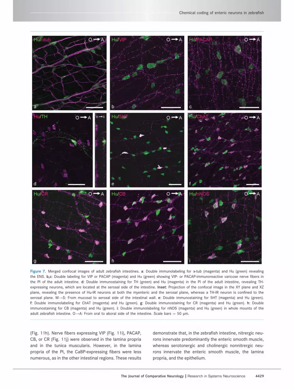
(Fig. 11h). Nerve fibers expressing VIP (Fig. 11i), PACAP,
CB, or CR (Fig. 11j) were observed in the lamina propria
and in the tunica muscularis. However, in the lamina
propria of the PI, the CaBP-expressing fibers were less
numerous, as in the other intestinal regions. These results
demonstrate that, in the zebrafish intestine, nitrergic neu-
rons innervate predominantly the enteric smooth muscle,
whereas serotonergic and cholinergic nonnitrergic neu-
rons innervate the enteric smooth muscle, the lamina
propria, and the epithelium.
Figure 7. Merged confocal images of adult zebrafish intestines. a: Double immunolabeling for a-tub (magenta) and Hu (green) revealing
the ENS. b,c: Double labeling for VIP or PACAP (magenta) and Hu (green) showing VIP- or PACAP-immunoreactive varicose nerve fibers in
the PI of the adult intestine. d: Double immunostaining for TH (green) and Hu (magenta) in the PI of the adult intestine, revealing TH-
expressing neurons, which are located at the serosal side of the intestine. Inset: Projection of the confocal image in the XY plane and XZ
plane, revealing the presence of Hu-IR neurons at both the myenteric and the serosal plane, whereas a TH-IR neuron is confined to the
serosal plane. M!S: From mucosal to serosal side of the intestinal wall. e: Double immunostaining for 5HT (magenta) and Hu (green).
f: Double immunolabeling for ChAT (magenta) and Hu (green). g: Double immunostaining for CR (magenta) and Hu (green). h: Double
immunostaining for CB (magenta) and Hu (green). i: Double immunolabeling for nNOS (magenta) and Hu (green) in whole mounts of the
adult zebrafish intestine. O!A: From oral to aboral side of the intestine. Scale bars ¼ 50 lm.
Chemical coding of enteric neurons in zebrafish
The Journal of Comparative Neurology | Research in Systems Neuroscience 4429

DISCUSSION
The present study sought define the proportional distri-
bution of enteric neurons expressing specific neurochem-
ical markers in the zebrafish during early development
and in the adult. In the embryonic intestine, all the
markers that we investigated were expressed from 72
hpf, except for ChAT and TH. Only nNOS, 5HT, CB, and
CR were detected in neuronal cell bodies, permitting the
proportional distribution of these markers to be deter-
mined, but VIP and PACAP were only found in nerve
fibers. A high percentage of nitrergic neurons was
observed, and their numbers remained constant through-
out embryonic development. By contrast, lower propor-
tions were detected for the other three markers, which
increased at later stages and were also characterized by
regional differences. In the adult intestine, all neurochem-
ical markers were expressed, but only ChAT, CB, CR,
nNOS, and 5HT were present in neuronal cell bodies and
on nerve fibers, whereas VIP, PACAP, and TH were only
observed on nerve fibers. A major proportion of enteric
neurons in the adult intestine displayed CaBP IR, but 5HT
was the only marker to show a significant distribution dif-
ference in the three intestinal regions. Colocalization
studies showed that both CB and CR were expressed in
the same neurons and that 5HT was not coexpressed
with any of the other markers. At least five neuronal sub-
populations were characterized: CBþ/CRþ/ChATþ/nNOSþ-, CBþ/CRþ/nNOSþ/ChAT�-, CBþ/CRþ/ChATþ/nNOS�-, ChATþ/CB�/CR�/nNOS�-, and 5HTþ-expressing neurons. Analysis of immunostaining of nerve
fibers revealed that nitrergic neurons also coexpressed
VIP and PACAP. Moreover, nitrergic neurons innervate
predominantly the enteric smooth muscle, whereas sero-
tonergic and cholinergic nonnitrergic neurons innervate
the enteric smooth muscle, the lamina propria, and the
epithelium.
Although we detected VIP and PACAP in enteric nerve
fibers of the embryonic and adult intestine, no IR was
observed in neuronal cell bodies. Similar observations
have been reported in mammals. To overcome this lack
of VIP and PACAP IR in the mammalian gastrointestinal
tract, gastrointestinal whole mounts, prior to fixation, are
incubated in vitro in a colchicine-containing culture me-
dium to increase IR in nerve cell bodies (Furness et al.,
1984). Colchicine is known to cause derangement of cel-
lular microtubules, which leads to inhibition of axonal
transport and accumulation of axonally transported sub-
stances in the neuronal cell body (CeccatelLi et al., 1991;
Boyer et al., 1994). We attempted to use this approach in
zebrafish, but it did not yield immunostaining of neuronal
cell bodies. However, previous zebrafish studies suggest
that VIP and PACAP coexist in enteric neurons, as
observed in other nonmammalian species (Olsson and
Holmgren, 1994, 2001; Olsson et al., 2008). The present
study clearly demonstrated that VIP and PACAP are coex-
pressed in the ENS of the adult zebrafish. Some of the
VIP- and PACAP-expressing nerve fibers were assumed to
be intrinsic to the intestine (Olsson et al., 2008). In addi-
tion, zebrafish vagal nerve may also account for some of
the VIP- and PACAP-positive nerve fibers in the intestine.
For other teleost species, vagal neurons expressing VIP
and PACAP have been reported (Olsson and Holmgren,
1994). Although we observed VIP- and PACAP-immunore-
active enteric nerve fibers in zebrafish embryos at 72 hpf,
PACAP-containing enteric nerve fibers appear already to
TABLE 2.
Proportional Expression of Neurochemical Markers in
Hu-Labeled Myenteric Neurons of the Adult Zebrafish
PI (%) MI (%) DI (%) Mean (%)
5HT 25 6 8 18 6 4 11 6 3 18nNOS 24 6 2 24 6 3 23 6 3 23ChAT 39 6 6 39 6 6 40 6 7 39CB 41 6 7 46 6 5 40 6 6 43CR 44 6 4 49 6 5 42 6 9 45
Figure 8. Average proportional distribution of enteric neurons
expressing CR, CB, ChAT, nNOS, or 5HT in the proximal (PI), mid-
dle (MI), and distal (DI) parts of the adult intestine. Proportional
distribution for most markers remained constant over the three
intestinal parts, except for 5HT, which significantly decreased (*P
< 0.05) along the length of the intestine.
TABLE 3.
Proportional coexpression of Neurochemical Markers in
Enteric Neurons of the Adult Zebrafish
Colocalizing
with
Neurons expressing
CB (%) CR (%) ChAT (%) nNOS (%) 5HT (%)
CB — 100 93 6 2 100 0CR 100 — 94 6 5 100 0ChAT 86 6 9 85 6 11 — 11 6 1 0nNOS 37 6 14 39 6 14 7 6 4 — 05HT 0 0 0 0 —
Uyttebroek et al.
4430 The Journal of Comparative Neurology |Research in Systems Neuroscience

be present at 48 hpf in the PI and MI (Holmberg et al.,
2004). By using digital motion analysis, an inhibitory
effect of PACAP on the wave frequency of anterogradely
propagating waves has been previously demonstrated at
5 dpf (Holmberg et al., 2004). These data indicate that
the VIP/PACAP inhibitory pathway is already developed
before the onset of feeding. Previous studies in sea bass
also suggest that VIP may play a role in the physiological
maturation of the intestinal epithelium during embryonic
development (Pederzoli et al., 2004).
In agreement with the study of Olsson and colleagues
(2008), we did not find any ChAT IR in the embryonic
intestine, although pharmacological studies in zebrafish
embryos clearly indicated that acetylcholine release may
modulate gut motility from 4 dpf onward (Holmberg et al.,
2004, 2007). The proportional size of the cholinergic
Figure 9. Confocal images showing the coexpression of neurochemical markers in the ENS of the adult zebrafish. a–c: CR (a; green) and
CB (b; magenta) were expressed in the same neurons (c; merged). d–f: No colocalization of CR (d; green) and 5HT (e; magenta) was
detected (f; merged). g–i: No coexpression of 5HT (g; green) and ChAT (h; magenta) was observed (i; merged). O!A: From oral to aboral
side of the intestine. Scale bars ¼ 50 lm.
Chemical coding of enteric neurons in zebrafish
The Journal of Comparative Neurology | Research in Systems Neuroscience 4431

neuronal population in the adult zebrafish intestine
(639%) did not correlate with data obtained from mam-
mals. For mouse ileum, it was found that 60–70% of the
myenteric neurons are cholinergic (Sang and Young,
1998; Qu et al., 2008), whereas, in the human gastroin-
testinal tract, cholinergic neurons account for 50–70% of
all intrinsic neurons (Anlauf et al., 2003).
In agreement with previous studies, we did not observe
TH IR in the embryonic intestine (Guo et al., 1999; Olsson
et al., 2008). However, as in these other studies, we
observed the presence of TH-immunoreactive nerve
fibers in the myenteric plexus and of TH-IR vagal neurons
at the serosal side of the adult intestine. It is assumed
that the TH-positive nerve fibers are of extrinsic origin,
arising from TH-expressing vagal neurons. However, it
cannot be excluded that some of these nerve fibers are
intrinsic to the intestine. TH-expressing neurons have
been described in the submucous and myenteric plexus
Figure 10. Confocal images showing the coexpression of neurochemical markers in the ENS of the adult zebrafish. a–c: Colocalization of
CB (a; green) and ChAT (b, magenta; c, merged). Arrowheads, neurons expressing only CB. d–f: Colocalization of CR (d; green) and nNOS
(e, magenta; f, merged). g–i: Colocalization of nNOS (g; green) and ChAT (h, magenta; i, merged). O!A: From oral to aboral side of the
intestine. Scale bars ¼ 50 lm.
Uyttebroek et al.
4432 The Journal of Comparative Neurology |Research in Systems Neuroscience

in several mammalian species (Schemann et al., 1995;
Sann et al., 1998; Anlauf et al., 2003; Chiocchetti et al.,
2004; Li et al., 2004, 2006; Chevalier et al., 2008; Qu
et al., 2008; Tian et al., 2008; Mongardi Fantaguzzi et al.,
2009). They appear late during perinatal development of
the ENS, and their precise function remains unclear.
Zebrafish enteric neurons expressing nNOS IR appear
first in the DI and MI between 40 and 55 hpf and, 1 day
later, also in the PI of the embryonic zebrafish intestine
(Holmqvist et al., 2004; Holmberg et al., 2006). In agree-
ment with these previous studies, we found that nitrergic
neurons constitute a major neuronal subpopulation in the
embryonic intestine. The percentage of nitrergic neurons
remains constant during the developmental time points
we examined (72–120 hpf), and the proximal part of the
intestine contained a significant lower proportion of
Figure 11. a–f: Confocal images showing the coexpression of VIP (a; green) and PACAP (b; magenta) and of VIP (d; green) and nNOS (e;
magenta) in enteric nerve fibers (c,f: merged). O!A: From oral to aboral side of the intestine. g–j: Merged images of cryosections the MI
of the adult zebrafish. g: Double immunolabeling with antibodies directed against nNOS (magenta) and Hu (green) revealing nitrergic nerve
fibers in the tunica muscularis (M). h: Double immunostaining with antibodies directed against 5HT (magenta) and Hu (green) revealing
serotonergic nerve fibers in the tunica muscularis and the lamina propria (L). i: Double immunolabeling with antibodies directed against
VIP (magenta) and Hu (green) showing VIP-expressing nerve fibers in the tunica muscularis and the lamina propria. j: Double immunostain-
ing for Hu (green) and CR (magenta) showing CR-positive nerve fibers in the tunica muscularis and the lamina propria. Arrowheads, immu-
noreactive nerve fibers in the lamina propria; arrows, immunoreactive epithelial cells. Scale bars ¼ 50 lm.
Chemical coding of enteric neurons in zebrafish
The Journal of Comparative Neurology | Research in Systems Neuroscience 4433

nitrergic neurons than the other two regions of the intes-
tine. Despite the presence of enteric nitrergic neurons at
this early stage, an NO-mediated inhibitory tonus has
been reported to be present only just before or at the
onset of exogenous feeding (Holmberg et al., 2006). This
is similar to what has been suggested for other vertebrate
species (Timmermans et al., 1994; Van Ginneken et al.,
1998, 2001; Bagyanszki et al., 2000; Belai and Burnstock,
2000; Bayram et al., 2002; Pederzoli et al., 2007). Fur-
thermore, an additional role for nNOS has been proposed
in the development and differentiation of the embryonic
zebrafish gut (Holmqvist et al., 2004). In the adult zebra-
fish intestine, the percentage of nitrergic neurons (623%)
was comparable to the proportional values obtained in
the myenteric plexus of the mouse (629%; Qu et al.,
2008) and rat (627%; Belai et al., 1995) ileum.
The main source of 5HT in the body is the gut, where it
is expressed in enterochromaffin cells and in intrinsic
neurons. 5HT acts as an enteric neurotransmitter and as
a mucosal signalling molecule. It is involved in initiating
motility and secretory reflexes. For the embryonic zebra-
fish, we observed that 5HT-expressing enteric neurons
were already present in the intestine at 72 hpf, before the
onset of oral feeding, as has been previously reported by
Olden et al. (2008). The percentage of 5HT-positive neu-
rons significantly decreased from the PI to the DI. The
proportional value obtained at 120 hpf was comparable
to the value estimated by Olsson et al. (2008). Previous
studies in guinea pig revealed that 5HT signaling in the
gut is required for the postnatal development of motility
patterns before the onset of oral intake of nutrition (Bian
et al., 2007). For the adult zebrafish intestine, we
observed a similar distribution pattern of 5HT-expressing
neurons, although the percentage of 5HT-expressing neu-
rons was lower than that in the embryonic intestine. In
contrast to the mammalian gut, 5HT-expressing enteroen-
docrine cells were sparsely distributed in the zebrafish
intestine, which is consistent with previous studies (Ols-
son et al., 2008). This indicates that the enteric neurons
are the main source of 5HT in the zebrafish intestine.
CB and CR are members of the EF-hand (E-helix-loop-
F-helix) family of CaBP, which also includes parvalbumin,
calmodulin, and S100 protein, and are widely distributed
in the central and peripheral nervous systems (Christakos
et al., 1989). Although their putative role in neurons
remains to be elucidated, it is known that they are
involved in the regulation of all aspects of cell function
(Grabarek, 2006). One report has emphasized a role for
CB as a neuroprotective protein (Iacopino et al., 1994). In
the mammalian gut, CR and CB are expressed in several
neuronal subpopulations. In the myenteric plexus of the
mouse and guinea pig ileum, CB has been found exclu-
sively in intrinsic primary afferent or sensory neurons
(IPANs; Timmermans et al., 1997; Furness, 2000, 2006;
Brookes, 2001; Qu et al., 2008; Mongardi Fantaguzzi
et al., 2009). In the sheep ileum, CB is also expressed in
other neuron types (Chiocchetti et al., 2004), whereas, in
the rabbit ileum, CB is expressed predominantly in cholin-
ergic interneurons (Denes and Gabriel, 2004). In the pig
small intestine, CB is coexpressed with 5HT in interneur-
ons and viscerofugal neurons (Timmermans et al., 1997).
The vast majority of IPANs in the human small intestine
express CR and a minority CB (Brehmer et al., 2004a). In
the rat small intestine, IPANs express CR, but CB is
expressed in other neuron types (Sayegh and Ritter,
2003). In mouse, CR is also found in IPANs (Qu et al.,
2008). In the guinea pig ileum, CR is expressed in inter-
neurons, secretomotor neurons, and excitatory motor
neurons, whereas, in the pig small intestine, CR is found
in interneurons and inhibitory motor neurons (Timmer-
mans et al., 1997; Furness, 2000, 2006; Brookes, 2001).
Our results show a similar distribution pattern for CB and
CR in both the embryonic and the adult zebrafish intes-
tine. Similar neuronal colocalization of CB and CR has
been reported for the vagal lobe of the goldfish (Ikenaga
et al., 2006). Partial coexpression of CB and CR in enteric
neurons has been reported for the mouse (Qu et al.,
2008) and sheep (Chiocchetti et al., 2004) ileum and for
the rat small intestine (Sayegh and Ritter, 2003). The per-
centages of enteric neurons displaying CR or CB IR in
zebrafish do not correlated with the proportional values
obtained in mammals for both these CaBP (Chiocchetti
et al., 2004; Qu et al., 2008).
In mammals, coexpression of neurochemical markers
has been used to determine different neuronal classes,
which are functionally classified as motor neurons (exci-
tatory or inhibitory), sensory neurons, interneurons,
secretomotor neurons, vasodilator neurons, and viscero-
fugal neurons (Timmermans et al., 1997, 2001; Furness,
2000, 2006; Brookes, 2001; Qu et al., 2008). Colocaliza-
tion studies of the five neurochemicals, CB, CR, ChAT,
5HT, and nNOS, in the adult zebrafish intestine has
allowed us to distinguish five neuronal subpopulations
covering approximately 75% of all enteric neurons. One
subpopulation contained neurons expressing only 5HT
and was found to project mainly orally. Serotonergic neu-
rons are commonly present in the fish intestine (Watson,
1979; Goodrich et al., 1980; Anderson, 1983; Jensen and
Holmgren, 1985; Anderson and Campbell, 1988; Kiliaan
et al., 1989; Karila et al., 1998; Olden et al., 2008; Olsson
et al., 2008) and are assumed to play a role in the regula-
tion of intestinal motility and in epithelial function (Jensen
and Holmgren, 1985; Kiliaan et al., 1989; Karila et al.,
1998). In mammals, serotonergic neurons are designated
as descending interneurons (Timmermans et al., 1997,
2001; Furness, 2000, 2006; Brookes, 2001; Qu et al.,
Uyttebroek et al.
4434 The Journal of Comparative Neurology |Research in Systems Neuroscience
2008). For fish, it has been suggested that the orally
directed serotonergic neurons are ascending excitatory
neurons and the anally projecting ones are descending
interneurons (Karila et al., 1998).
Our identification of a small group of intrinsic neurons
displaying only ChAT IR requires further investigation in
order to ascribe a specific function to this neuronal sub-
population. The three remaining subpopulations dis-
played CB and CR IR. The smallest subpopulation also
expressed ChAT and nNOS. In mammals, coexpression of
ChAT and nNOS occurred in a small group of descending
interneurons (Timmermans et al., 1997, 2001; Furness,
2000, 2006; Porter et al., 2002; Brehmer et al., 2004b;
Qu et al., 2008). Nonadrenergic, noncholinergic nitrergic
neurons in mammals have been characterized as inhibi-
tory motor neurons, which also express VIP (Porter et al.,
1997; Timmermans et al., 1997; Furness, 2000; Brookes,
2001; Jungbauer et al., 2006; Qu et al., 2008). For the
zebrafish, we have observed that nitrergic nerve fibers
are present predominantly in the tunica muscularis and
coexpress VIP and PACAP. This being the case, we sug-
gest that the CB/CR/nNOS-expressing neurons in the
zebrafish intestine modulate intestinal motility. Coexpres-
sion of CB and CR with ChAT in mammals has been
observed in the murine intestine in some intrinsic sensory
neurons (Qu et al., 2008), in a small population of irregu-
larly shaped enteric neurons of the sheep ileum (Chioc-
chetti et al., 2004), and in putative motor neurons of the
gastric corpus in the guinea pig (Reiche et al., 2001).
Coexpression of both CaBPs in cholinergic neurons has
not been observed in other mammals. As a result, further
neurochemical characterization is needed to determine
the neuron type corresponding to the CB/CR/ChAT-cod-
ing type in zebrafish.
In conclusion, this is the first study with zebrafish to
show the proportional expression of neurochemical
markers in the ENS during embryonic development as
well as the colocalization of neurochemical markers in en-
teric neurons of the adult intestine. From early stages in
embryonic development, nearly all of the neurochemical
markers that we investigated are expressed. During em-
bryonic development, the major proportion of enteric
neurons is nitrergic, and the proportional distributions of
the neurochemical markers that we examined in this
study exhibit regional differences in the developing intes-
tine. Coexpression of neurochemical markers in the adult
intestine revealed a serotonergic, a nitrergic, and two
cholinergic neuronal subpopulations, and one subpopula-
tion expressing both ChAT and nNOS. Further studies are
required on the neuronal function of these different neu-
ronal subpopulations that we have identified in the zebra-
fish intestine. The information provided here will also
serve as a basis for future studies investigating the
effects of mutations, toxic substances, drugs, and inflam-
mation on the development and function of the zebrafish
ENS.
ACKNOWLEDGMENTS
The authors thank Mrs. E. Goeman, Mrs. M. Van Geel,
and Mr. D. De Rijck for their technical assistance.
LITERATURE CITEDAnderson C. 1983. Evidence for 5-HT-containing intrinsic
neurons in the teleost intestine. Cell Tissue Res 230:377–386.
Anderson C, Campbell G. 1988. Immunohistochemical studyof 5-HT-containing neurons in the teleost intestine: rela-tionship to the presence of enterochromaffin cells. CellTissue Res 254:553–559.
Anlauf M, Schafer MK-H, Eiden L, Weihe E. 2003. Chemicalcoding of the human gastrointestinal nervous system:cholinergic, VIPergic, and catecholaminergic phenotypes.J Comp Neurol 459:90–111.
Bagyanszki M, Roman V, Fekete E. 2000. Quantitative distri-bution of NADPH-diaphorase-positive myenteric neurons indifferent segments of the developing chicken small intes-tine and colon. Histochem J 32:679–684.
Bayram Z, Asar M, Cayli S, Demir R. 2002. Immunocytochemi-cal detection of neuronal nitric oxide synthase (nNOS)-IRin embryonic rat stomach between days 13 and 21 of ges-tation. J Histochem Cytochem 50:671–679.
Belai A, Burnstock G. 2000. Pattern of distribution and co-localization of NOS and ATP in the myenteric plexus ofhuman fetal stomach and intestine. Neuroreport 11:5–8.
Belai A, Cooper S, Burnstock G. 1995. Effect of age onNADPH-diaphorase-containing myenteric neurones of rat il-eum and proximal colon. Cell Tissue Res 279:379–383.
Bian X, Patel B, Dai X, Galligan JJ, Swain G. 2007. High muco-sal serotonin availability in neonatal guinea pig ileum isassociated with low serotonin transporter expression. Gas-troenterology 132:2438–2447.
Bisgrove BW, Raible DW, Walter V, Eisen JS, Grunwald DJ.1997. Expression of c-ret in the zebrafish embryo: poten-tial roles in motoneuronal development. J Neurobiol 33:749–768.
Boyer PA, Trembleau A, Leviel V, Arluison M. 1994. Effects ofintranigral injections of colchicine on the expression ofsome neuropeptides in the rat forebrain: an immunohisto-chemical and in situ hybridization study. Brain Res Bull 33:541–560.
Brehmer A, Croner R, Dimmler A, Papadopoulos T, Schrodl F,Neuhuber W. 2004a. Immunohistochemical characteriza-tion of putative primary afferent (sensory) myentericneurons in human small intestine. Auton Neurosci 112:49–59.
Brehmer A, Schrodl F, Neuhuber W, Tooyama I, Kimura H.2004b. Co-expression pattern of neuronal nitric oxide syn-thase and two variants of choline acetyltransferase inmyenteric neurons of porcine ileum. J Chem Neuroanat 27:33–41.
Brookes SJH. 2001. Classes of enteric nerve cells in theguinea-pig small intestine. Anat Rec 262:58–70.
Burzynski G, Shepherd IT, Enomoto H. 2009. Genetic modelsystem studies of the development of the enteric nervoussystem, gut motility and Hirschsprung’s disease. Neurogas-troenterol Motil 21:113–127.
Castro A, Becerra M, Manso MJ, Anadon R. 2006. Calretininimmunoreactivity in the brain of the zebrafish, Danio rerio:
Chemical coding of enteric neurons in zebrafish
The Journal of Comparative Neurology | Research in Systems Neuroscience 4435

distribution and comparison with some neuropeptidesand neurotransmitter-synthesizing enzymes. II. Midbrain,hindbrain, and rostral spinal cord. J Comp Neurol 494:792–814.
Ceccatelli S, Cortes R, Hokfelt T. 1991. Effect of reserpineand colchicine on neuropeptide mRNA levels in the rathypothalamic paraventricular nucleus. Brain Res Mol BrainRes 9:57–69.
Chen Q, Huang N-N, Huang J-T, Chen S, Fan J, Li C, Xie F-K.2009. Sodium benzoate exposure down-regulates theexpression of tyrosine hydroxylase and dopamine trans-porter in dopaminergic neurons in developing zebrafish.Birth Defects Res (B) 86:85–91.
Chevalier J, Derkinderen P, Gomes P, Thinard R, Naveilhan P,Vanden Berghe P, Neunlist M. 2008. Activity-dependentregulation of tyrosine hydroxylase expression in the entericnervous system. J Physiol 586:1963–1975.
Chiocchetti R, Grandis A, Bombardi C, Clavenzani P, Coster-bosa GL, Lucchi ML, Furness JB. 2004. Characterisation ofneurons expressing calbindin immunoreactivity in the ileumof the unweaned and mature sheep. Cell Tissue Res 318:289–303.
Christakos S, Gabrielides C, Rhoten WB. 1989. Vitamin D-de-pendent calcium binding proteins: chemistry, distribution,functional considerations, and molecular biology. EndocrRev 10:3–26.
Clemente D, Porteros A, Weruaga E, Alonso JR, Arenzana FJ,Aijon J, Arevalo R. 2004. Cholinergic elements in thezebrafish central nervous system: histochemical and immu-nohistochemical analysis. J Comp Neurol 474:75–107.
Denes V, Gabriel R. 2004. Calbindin-immunopositive cells arecholinergic interneurons in the myenteric plexus of rabbitileum. Cell Tissue Res 318:465–472.
Elworthy S, Pinto JP, Pittefer A, Cancela ML, Kelsh RN. 2005.Phox2b expression in the enteric nervous system is con-served in zebrafish and is sox10-dependent. Mech Dev122:659–669.
Finney JL, Robertson GN, McGee CAS, Smith FM, Croll RP.2006. Structure and autonomic innervation of the swimbladder in the zebrafish (Danio rerio). J Comp Neurol 495:587–606.
Fornaro M, Raimondo S, Lee JM, Giacobini-Robecchi MG.2007. Neuron-specific Hu proteins sub-cellular localizationin primary sensory neurons. Ann Anat 189:223–228.
Furness JB. 2000. Types of neurons in the enteric nervoussystem. J Auton Nerv Syst 81:87–96.
Furness JB. 2006. The enteric nervous system. Oxford: Black-well Publishing.
Furness JB, Costa M, Keast JR. 1984. Choline acetyltransfer-ase- and peptide immunoreactivity of submucous neuronsin the small intestine of the guinea-pig. Cell Tissue Res237:329–336.
Grabarek Z. 2006. Structural basis for diversity of the EF-hand calcium-binding proteins. J Mol Biol 359:509–525.
Grana P, Anadon R, Yanez J. 2008. Immunocytochemicalstudy of calretinin and calbindin D-28K expression in theretina of three cartilaginous fishes and a clastidian (Poly-pterus). Brain Res Bull 75:375–378.
Goodrich JT, Bernd P, Sherman D, Gershon MD. 1980. Phylog-eny of enteric serotonergic neurons. J Comp Neurol 190:15–28.
Grunwald DJ, Eisen JS. 2002. Headwaters of the zebrafish:emergence of a new model vertebrate. Nat Rev Genet 3:717–724.
Guo S, Wilson SW, Cooke S, Chitnis AB, Driever W, RosenthalA. 1999. Mutations in the zebrafish unmask shared regula-tory pathways controlling the development of catecholami-nergic neurons. Dev Biol 208:473–487.
Holmberg A, Schwerte T, Pelster B, Holmgren S. 2004. Ontog-eny of the gut motility control system in zebrafish Daniorerio embryos and larvae. J Exp Biol 207:4085–4094.
Holmberg A, Olsson C, Holmgren S. 2006. The effects of en-dogenous and exogenous nitric oxide on gut motility inzebrafish Danio rerio embryos and larvae. J Exp Biol 209:2472–2479.
Holmberg A, Olsson C, Hennig GW. 2007. TTX-sensitive andTTX-insensitive control of spontaneous gut motility in thedeveloping zebrafish (Danio rerio) larvae. J Exp Biol 210:1084–1091.
Holmqvist B, Ellingsen B, Alm P, Forsell J, Øyan A-M, GoksøyrA, Fjose A, Seo H-C. 2000. Identification and distributionof nitric oxide synthase in the brain of adult zebrafish.Neurosci Lett 292:119–122.
Holmqvist B, Ellingsen B, Forsell J, Zhdanova I, Alm P. 2004.The early ontogeny of neuronal nitric oxide synthase sys-tems in the zebrafish. J Exp Biol 207:923–935.
Iacopino AM, Quintero EM, Miller EK. 1994. Calbindin-D28K: apotential neuroprotective protein. Neurodegeneration 3:1–20.
Ikenaga T, Huesa G, Finger TE. 2006. Co-occurrence of cal-cium-binding proteins and calcium-permeable glutamatereceptors in the primary gustatory nucleus of goldfish.J Comp Neurol 499:90–105.
Jensen J, Holmgren S. 1985. Neurotransmitters in the intes-tine of the Atlantic cod, Gadus morhua. Comp BiochemPhysiol 82C:81–89.
Jungbauer C, Lindig TM, Schrodl F, Neuhuber W, Brehmer A.2006. Chemical coding of myenteric neurons with differentaxonal projection patterns in the porcine ileum. J Anat209:733–743.
Karila P, Shahbazi F, Jensen J, Holmgren S. 1998. Projectionsand actions of tachykininergic, cholinergic, and serotoner-gic neurones in the intestine of the Atlantic cod. Cell Tis-sue Res 291:403–413.
Kaslin J, Panula P. 2001. Comparative anatomy of the histami-nergic and other aminergic systems in zebrafish (Daniorerio). J Comp Neurol 440:342–377.
Kiliaan AJ, Joosten HWJ, Bakker R, Dekker K, Groot JA. 1989.Serotonergic neurons in the intestine of two teleosts, Car-assius auratus and Oreochromis mossambicus, and theeffect of serotonin on transepithelial ion-selectivity andmuscle tension. Neuroscience 31:817–824.
Kimmel CB, Ballard WM, Kimmel SR, Ullmann B, Schilling TF.1995. Stages of embryonic development of the zebrafish.Dev Dyn 203:253–310.
Levanti MB, Montalbano G, Laura R, Ciriaco E, Cobo T, Gar-cia-Suarez O, Germana A, Vega JA. 2008. Calretinin in theperipheral nervous system of the adult zebrafish. J Anat212:67–71.
Lewis Carl SA, Gilette-Ferguson I, Ferguson DG. 1993. Anindirect immunofluorescence procedure for staining thesame cryostat section with two mouse monoclonal primaryantibodies. J Histochem Cytochem 41:1273–1278.
Li ZS, Pham TD, Tamir H, Chen JJ, Gershon MD. 2004. Entericdopaminergic neurons: definition, developmental lineage,and effects of extrinsic denervation. J Neurosci 24:1330–1339.
Li ZS, Schmauss C, Cuenca A, Ratcliffe E, Gershon MD. 2006.Physiological modulation of intestinal motility by entericdopaminergic neurons and the D2 receptor: analysis of do-pamine receptor expression, location, development, andfunction in wild-type and knock-out mice. J Neurosci 26:2798–2807.
Lin Z, Gao N, Hu H-Z, Liu S, Gao C, Kim G, Ren J, Xia Y,Peck OC, Wood JD. 2002. Immunoreactivity of Hu pro-teins facilitates identification of myenteric neurones in
Uyttebroek et al.
4436 The Journal of Comparative Neurology |Research in Systems Neuroscience

guinea-pig small intestine. Neurogastroenterol Motil 14:197–204.
Linard B, Pakdel F, Marmignon MH, Saligaut C. 1998. Cloningof a cDNA coding for active tyrosine hydroxylase in therainbow trout (Oncorhynchus mykiss): comparison withother hydroxylases and enzymatic expression. J Neuro-chem 71:920–928.
Llewelyn-Smith IJ, Costa M, Furness JB. 1985. Light and elec-tron microscopic immunocytochemistry of the same nervesfrom whole mount preparations. J Histochem Cytochem 9:857–866.
Mathieu M, Tagliafierro G, Angelini C, Vallarino M. 2001. Orga-nization of vasoactive intestinal peptide-like immunoreac-tive system in the brain, olfactory organ and retina of thezebrafish, Danio rerio, during development. Brain Res 888:235–247.
Mathieu M, Ciarlo M, Trucco N, Griffero F, Damonte G, SalisA, Vallarino M. 2004. Pituitary adenylate cyclase-activatingpolypeptide in the brain, spinal cord and sensory organs ofthe zebrafish, Danio rerio, during development. Brain ResDev Brain Res 151:169–185.
Matthews M, Trevarrow B, Matthews J. 2002. A virtual tour ofthe guide for zebrafish users. Lab Anim 31:34–40.
Mongardi Fantaguzzi C, Thacker M, Chiocchetti R, FurnessJB. 2009. Identification of neuron types in the sub-mucosal ganglia of the mouse ileum. Cell Tissue Res336:179–189.
Nazir A, Polynice M, Chant-Haphavong R, Elha II, BuchholzBM, Stuckenholz C, Bahary N, Bauer AJ. 2008. The adultzebrafish (Danio rerio) as a model of gastrointestinal motil-ity. Neurogastroenterol Motil 20(Suppl 1):14.
Negoescu A, Labat-Moleur F, Lorimier P, Lamarcq L, Guiller-met C, Chambaz E, Brambilla E. 1994. F(ab) secondaryantibodies: a general method for double immunolabelingwith primary antisera from the same species. Efficiencycontrol by chemiluminescence. J Histochem Cytochem 42:443–437.
Ng ANY, de Jong-Curtain TA, Mawdsley DJ, White SJ, Shin J,Appel B, Dong PD, Stainier DY, Heatj JK. 2005. Formationof the digestive system in zebrafish: III. Intestinal epithe-lium morphogenesis. Dev Biol 286:114–135.
Olden T, Akhtar T, Beckman SA, Wallace KN. 2008. Differena-tion of the zebrafish enteric nervous system and intestinalsmooth muscle. Genesis 46:484–498.
Olsson C. 2008. Calbindin-immunoreactive nerve cells in thezebrafish gut. Neurogastroenterol Motil 20(Suppl 1):44.
Olsson C. 2009. Autonomic innervation of the fish gut. ActaHistochem 111:185–195.
Olsson C, Holmgren S. 1994. Distribution of PACAP (pituitaryadenylate cyclase-activating polypeptide)-like and helo-spectin-like peptides in the teleost gut. Cell Tissue Res277:539–547.
Olsson C, Holmgren S. 2001. The control of gut motility.Comp Biochem Physiol A 128:481–503.
Olsson C, Holmberg A, Holmgren S. 2008. Development of en-teric and vagal innervation of the zebrafish (Danio rerio)gut. J Comp Neurol 508:756–770.
Pederzoli A, Bertacchi I, Gambarelli A, Mola L. 2004. Immuno-localisation of vasoactive intestinal peptide and substanceP in the developing gut of Dicentrarchus labrax (L.). Eur JHistochem 48:179–184.
Pederzoli A, Conte A, Tagliazucchi D, Gambarelli A, Mola L.2007. Occurrence of two NOS isoforms in the developinggut of sea bass Dicentrarchus labrax (L.). Histol Histopa-thol 22:1057–1064.
Pellegrini E, Mouriec K, Anglade I, Menuet A, Le Page Y, Gue-guen M-M, Marmignon M-H, Brion F, Pakdel F, Kah O.2007. Identification of aromatase-positive radial cells as
progenitor cells in the ventricular layer of the forebrain inzebrafish. J Comp Neurol 501:150–167.
Pietsch J, Delalande J-M, Jakaitis B, Stensby JD, Dohle S, Tal-bot WS, Raible DW, Shepherd IT. 2006. lessen Encodes azebrafish trap100 required for enteric nervous system de-velopment. Development 133:395–406.
Poon KL, Richardson M, Lam CS, Khoo HE, Korzh V. 2003.Expression pattern of neuronal nitric oxide synthase in em-bryonic zebrafish. Gene Expr Patterns 3:463–466.
Porter AJ, Wattchow DA, Brookes SJH, Costa M. 1997. Theneurochemical coding and projections of circular musclemotor neurons in the human colon. Gastroenterology 113:1916–1923.
Porter AJ, Wattchow DA, Brookes SJH, Costa M. 2002. Cholin-ergic and nitrergic interneurones in the myenteric plexusof the human colon. Gut 51:70–75.
Qu Z-D, Thacker M, Castelucci P, Bagyanszki M, Epstein ML,Furness JB. 2008. Immunohistochemical analysis of neurontypes in the mouse small intestine. Cell Tissue Res 334:147–161.
Raible DW, Kruse GJ. 2000. Organization of the lateral linesystem in embryonic zebrafish. J Comp Neurol 421:189–198.
Reiche D, Huber K, Hoppe S, Schemann M. 2001. Neuro-chemically distinct myenteric neurone populations contain-ing calbindin have specific distribution patterns around thecircumference of the gastric corpus. Cell Tissue Res 303:319–328.
Sang Q, Young HM. 1998. The identification and chemicalcoding of cholinergic neurons in the small and large intes-tine of the mouse. Anat Rec 251:185–199.
Sann H, Hoppe S, Baldwin L, Grundy D, Schemann M. 1998.Presence of putative neurotransmitters in the myentericplexus of the gastrointestinal tract and in the musculatureof the urinary bladder of the ferret. NeurogastroenterolMotil 10:35–47.
Sayegh AI, Ritter RC. 2003. Morphology and distribution of ni-tric oxide synthase-, neurokinin-1 receptor-, calretinin-, cal-bindin-, and neurofilament-M-immunoreactive neurons inthe myenteric and submucosal plexuses of the rat smallintestine. Anat Rec 271A:209–216.
Schemann M, Schaaf C, Mader M. 1995. Neurochemical cod-ing of enteric neurons in the guinea pig stomach. J CompNeurol 353:161–178.
Shepherd IT, Pietsch J, Elworthy S, Kelsh RN, Raible DW.2004. Roles for GFRa1 receptors in zebrafish enteric nerv-ous system development. Development 131:241–249.
Spence R, Gerlach G, Lawrence C, Smith C. 2008. The behav-iour and ecology of the zebrafish, Danio rerio. Biol RevCamb Philos Soc 83:13–34.
Tian Y-M, Chen X, Luo D-Z, Zhang X-H, Xue H, Zheng L-F, YangN, Wang X-M, Zhu J-X. 2008. Alteration of dopaminergicmarkers in gastrointestinal tract of different rodent modelsof Parkingson’s disease. Neuroscience 153:634–644.
Timmermans J-P, Barbiers M, Scheuermann DW, Bogers JJ,Adriaensen D, Fekete E, Mayer B, Van Marck EA, DeGroodt-Lasseel MHA. 1994. Nitric oxide synthase immuno-reactivity in the enteric nervous system of the developinghuman digestive tract. Cell Tissue Res. 275:235–245.
Timmermans J-P, Adriaensen D, Cornelissen W, ScheuermannDW. 1997. Structural organization and neuropeptide distri-bution in the mammalian enteric nervous system, with spe-cial attention to those components involved in mucosalreflexes. Comp Biochem Physiol 118A:331–340.
Timmermans J-P, Hens J, Adriaensen D. 2001. Outer submu-cous plexus: an intrinsic nerve network involved in bothsecretory and motility processes in the intestine of largemammals and humans. Anat Rec 262:71–78.
Chemical coding of enteric neurons in zebrafish
The Journal of Comparative Neurology | Research in Systems Neuroscience 4437

Van Ginneken C, Van Meir F, Sommereyns G, Sys S, WeynsA. 1998. Nitric oxide synthase expression in enteric neu-rons during development in the pig duodenum. AnatEmbryol 198:399–408.
Van Ginneken C, Van Meir F, Sys S, Weyns A. 2001. Stereo-logic description of the changing expression of constitutivenitric oxide synthase and heme oxygenase in the entericplexuses of the pig small intestine during development.J Comp Neurol 437:118–128.
Wallace KN, Akhter S, Smith EM, Lorent K, Pack M. 2005. In-testinal growth and differentiation in zebrafish. Mech Dev122:157–173.
Watson AH. 1979. Fluorescent histochemistry of the teleostgut: evidence for the presence of serotonergic neurones.Cell Tissue Res 197:155–164.
Westerfield M. 2007. The zebrafish book, 5th ed. A guide forthe laboratory use of zebrafish (Danio rerio). Eugene, OR:University of Oregon Press.
Yeo J-Y, Lee E-S, Jeon C-J. 2009. Parvalbumin-immunoreactiveneurons in the inner nuclear layer of zebrafish retina. ExpEye Res 88:553–560.
Zupanc GKH, Hinsch K, Gage FH. 2005. Proliferation, migration,neuronal differentiation, and long-term survival of new cellsin the adult zebrafish brain. J Comp Neurol 488:290–319.
Uyttebroek et al.
4438 The Journal of Comparative Neurology |Research in Systems Neuroscience


