Embed Size (px)
Citation preview

Neuroblastoma: an enigmatic disease
Josee Brossard*, Mark L Bernstein^ and Bernard Lemieux** Department of Pediatrics, Centre universitaire de sante de I'Estrie, University of Sherbrooke;^Departments of Pediatrics and Oncology, Montreal Children's Hospital, McGill University,Quebec, Canada
Neuroblastoma is the most common extra-cranial solid tumor of childhood1. Itoriginates in cells of the neural crest, and so can be found anywhere alongthe paravertebral sympathetic chain or in the adrenal gland. In the last 15years, new developments in the genetics and biology of neuroblastoma, haveled to a better understanding of the natural history and prognostic features ofthis cancer. The presence of identifying biochemical markers detectable in theurine of patients with neuroblastoma, as well as the remarkably inferiorsurvival of children diagnosed at more than 12 months of age, have led somegroups to screen infants for neuroblastoma, in the hope of decreasing bothoverall mortality, as well as the incidence of advanced stage disease. Thisarticle reviews some clinical aspects of neuroblastoma, but emphasizes thegenetic and biologic features in relation to prognosis and treatment. Finally,we discuss the different screening experiences for this disease, in particularfrom the Quebec Neuroblastoma Screening Project.
Correspondence fo:Or Josee Brossard,
Department ofPediatrics,
Centre untverafa/re desante de I'Estrie, 3001,
12th Avenue North,Sherbroofee, Quebec,
Canada J1H 5N4
Neuroblastoma affects approximately 1/7,000 children under the age of5 years2, and is very rarely diagnosed beyond the age of 10 years (36% ofpatients being less than 1 year of age, 79% less than 4 years and only 3%being diagnosed after the age of 10)1. This neural crest-derived tumor hasmany unique aspects, and shows great heterogeneity. Neuroblastomaoccasionally spontaneously regresses, or differentiates into a benignlesion. Microscopic neuroblastic nodules, histologically identical toneuroblastoma, are found in the adrenal gland of a high percentage offetuses and young infants dying of other causes3-4. These findings suggestthat these nodules represent a normal embryonic stage in the develop-ment of the adrenal gland. Their failure to regress or mature may result inthe development of malignant neuroblastoma. In contrast to thespontaneous regression which may occur, neuroblastoma may alsobehave as a very aggressive malignant tumor.
British Mtdtcal Buff«hn 199622 (No. 4):787-801 ©The Britiih Council 1996
Dow
nloaded from https://academ
ic.oup.com/bm
b/article/52/4/787/327658 by guest on 17 July 2022

Cancer in children
Biology
The biologic characteristics and clinical heterogeneity of neuroblastomamake it unique among pediatric tumors. Based on our currentknowledge, it is likely that neuroblastoma represents at least two distinctclinical/biologic entities5. Recent research at the cellular and molecularlevel has helped to clarify this distinction6.
One of the most characteristic and consistent cytogenetic anomaliesidentified in neuroblastoma is the deletion of the short arm ofchromosome 1 (loss of heterozygosity, LOH) resulting in partial lpmonosoirry6, thought to represent the loss of a suppressor gene in partresponsible for development of neuroblastoma1'6. This allelic loss ofchromosome lp has been found to be strongly prognostic both inlocalized and disseminated neuroblastoma, in both younger (^ 12months of age) and older (> 12 months of age) children7. Some groupshave also demonstrated the occurrence of loss of heterozygosity onchromosome 14q in some cases of neuroblastoma, suggesting apossibility of genetic heterogeneity in this tumor6.
About 25-30% of children with neuroblastoma have N-myc geneamplification in their tumors6. Initial cytogenetic analyses of humanneuroblastomas had identified the presence of extrachromosomal double-minute chromatin bodies (dmin) and/or homogeneously staining regionson different chromosomes. Those two abnormalities were later found torepresent N-myc gene amplification8. Amplification of N-myc is found inmore than 30% of patients with advanced stages but in only 5-10% ofpatients with earlier stages6. It is associated with rapidly progressivedisease and poor prognosis, regardless of the clinical stage9"11. Moreover,studies by Brodeur et al. demonstrated that the N-myc copy number in agiven tumor is usually consistent at different tumor sites and also overtime12. These findings suggest that the presence or absence of N-mycamplification is an intrinsic biologic property of the tumor. Finally, thereis a correlation between the presence of N-myc amplification and the lossof heterozygosity of chromosome lp, with the two findings beingstrongly predictive of poor prognosis6-7. Allelic loss of chromosome lpmay select a larger population of poor-risk patients, with the N-mycamplified tumors being a particularly unfavorable subset7.
The DNA content of neuroblastoma tumor has also proven to be ofgreat help in predicting the outcome of patients and their response totherapy, especially in infants less than 1 year of age with advanced stagedisease. Look et a/.13 were the first to report the analysis of DNA contentin the tumor tissue of 35 infants with neuroblastoma. They found acorrelation between abnormally high DNA content and low stage tumorswith a good response to chemotherapy. Further studies confirmed theprognostic significance of flow-cytometric analysis of DNA-index
788 Bn'KshMecfcaf Bulletin 1996£2 (No. 4)
Dow
nloaded from https://academ
ic.oup.com/bm
b/article/52/4/787/327658 by guest on 17 July 2022
Neuroblastoma: an enigmatic disease
(DI)10>14. Hyperdiploidy [especially near triploidy, DI(1,25-1,75)] pre-dicts a favorable outcome and good response to chemotherapy even inunresectable disease, as opposed to near-diploidy (or near tetraploidy)which is more likely to be associated with an adverse outcome. Again,analysis of multiple samples of tumor for DNA content showed greatconsistency within different areas of the same tumor and in tumorsampled at different times during the course of therapy15. Finally, there isgenerally a good correlation between N-tnyc amplification and near-diploidy in a given tumor10'15.
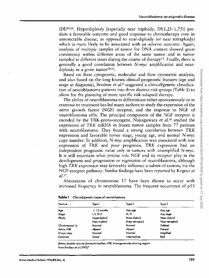
Based on these cytogenetic, molecular and flow cytometric analysis,and also based on the long known clinical prognostic features (age andstage at diagnosis), Brodeur et al.6 suggested a clinical/genetic classifica-tion of neuroblastoma patients into three distinct risk-groups (Table 1) toallow for the planning of more specific risk-adapted therapy.
The ability of neuroblastoma to differentiate either spontaneously or inresponse to treatment has led many authors to study the expression of thenerve growth factor (NGF) receptor, and the response to NGF ofneuroblastoma cells. The principal component of the NGF receptor isencoded by the TRK-proto-oncogene. Nakagawara et al.i6 studied theexpression of TRK mRNA in frozen tumor samples from 77 patientswith neuroblastoma. They found a strong correlation between TRKexpression and favorable tumor stage, young age, and normal N-tnyccopy number. In addition, N-myc amplification was associated with lowexpression of TRK and poor prognosis. TRK expression had anindependent prognostic value only in tumors with unamplified N-myc.It is still uncertain what precise role NGF and its receptor play in thedevelopment and progression or regression of neuroblastoma, althoughhigh TRK expression may favorably influence a subset of tumors, via theNGF-receptor pathway. Similar findings have been reported by Kogner etalP.
Aberrations of chromosome 17 have been shown to occur withincreased frequency in neuroblastoma. The frequent occurrence of p53
Table 1 Clinical/genetic types of neuroblastoma
Feature
AgeStageHoidy
Chromosome 1pdmins, HSRN-myc copyOutcome
Typei
< 12 months1, II, IV-SHyperdiploidNear trip!oidNormalAbsentNormalGood
Typ.2
Any ageIII, IVNeardiploidNeartvtraploidNormalAbsentNormalIntermediate
Type 3
Any ageAny stageNeardiploidNeartetraploidDeletedPresentAmpfintdBod
dmins, doubU-minute chromatin bodies; HSR, homogeneously staining regionFrom Brodtur »> al (1992)'
British MeaW BuHohn 1996;52 (No. 4) 789
Dow
nloaded from https://academ
ic.oup.com/bm
b/article/52/4/787/327658 by guest on 17 July 2022
Cancer in children
mutations in many human neoplasms, together with the location of thep53 tumor suppressor gene on chromosome 17, led Vogan et al.18 fromthe Quebec Neuroblastoma Screening Study19 to examine 38 primarytumors for the presence of p53 mutations. They found no p53abnormality and concluded that the chromosome 17 alterations inneuroblastoma involve other genes.
The multidrug resistance (MDR) gene product P-glycoprotein has beenfound by some groups to be of prognostic importance in neuro-blastoma20. More recently, another protein leading to decreasedintracelJular concentrations of natural product antineoplastic drugs, themultidrug resistance protein (MRP), has been shown to have prognosticsignificance in neuroblastoma21. MRP may also mediate resistance toagents important in the therapy of neuroblastoma such as cyclophos-phamide and cisplatinum by extrusion of their glutathione conjugatedmetabolites21.
Other biologic variables to consider with respect to prognosis ofneuroblastoma include several serum markers: ferritin, neuron-specificenolase (NSE), ganglioside GD2, and lactic dehydrogenase (LDH). Allhave been and continue to be included in many large neuroblastomastudies. Each has some predictive prognostic value. Their specificity andsensitivity are variable, and their precise independent power in predictingprognosis compared to the clinical and genetic variables alreadydescribed may be limited1'7.
Clinical aspects1
Clinical presentation
The majority of primary tumors (65%) occur in the abdomen, either inthe adrenal, or in the paravertebral sympathetic ganglia. Thoracicprimaries are found in approximately 20% of cases. A minority ofpatients have their primary tumor in the neck (1-5%) or pelvis (2-3%).Rarely, a primary rumor cannot be found or there may be multipleprimaries
Extension of disease can occur via either the lymphatic (regional ordistant lymph nodes) or the hematogenous route. Typical sites ofmetastasis are bone marrow, bone, liver and skin. Low stage disease(INSS22 1,2,4-S) is seen in a larger proportion of infants (^ 1 year) thanolder children, whereas disseminated disease occurs in at least 50% ofpatients older than 1 year of age. Whatever the stage, infants have abetter outcome than older children.
7 9 0 British Madkal BulltHn 1996^2 (No. 4)
Dow
nloaded from https://academ
ic.oup.com/bm
b/article/52/4/787/327658 by guest on 17 July 2022

Neuroblastoma: an enigmatic dijoaie
The signs and symptoms at presentation depend on both the locationof the primary mass and the metastases (if any). Abdominal or pelvicprimaries can cause discomfort, gastrointestinal tract dysfunction orcompression, urologic obstruction, venous and lymphatic compressionsyndromes, and occasionally, renin-mediated hypertension. Younginfants with stage 4-S disease may present with massive liver enlargementleading to respiratory insufficiency and other complications. Neck andthoracic masses may cause mechanical compression resulting inrespiratory distress, Horner's syndrome, or superior vena cava syndrome.Disseminated disease can present with proptosis and periorbitalecchymoses (retrobulbar and orbital infiltration), bone pain andirritability (bone and marrow disease), skin nodules, as well asconstitutional symptoms. Finally, two classic but rare paraneoplasticsyndromes occur in some patients: opsomyoclonus and/or cerebellarataxia, and severe diarrhea secondary to secretion of vasoactive intestinalpeptide (VIP). On the other hand, asymptomatic neuroblastomas mayoccasionally be found by imaging alone for other reasons, eitherpostnatally or antenatally.
Diagnosis and staging
Before 1988, there were no published established uniform criteria for thediagnosis of neuroblastoma, its staging and response to treatment. Aninternational group of experts met in 1986 to reach a consensus on thesedifferent issues and established the International Criteria for Neuroblas-toma Diagnosis, Staging and Response to Treatment22, which were thenrevised and published in 199323. Minimal criteria for the diagnosis ofneuroblastoma are:
1. An unequivocal pathologic diagnosis made from tumor tissue bystandard methods, including immunohistology or electron microscopyif necessary; or
2. Bone marrow containing unequivocal tumor cells (e.g. syncytia orimmunohistologically positive cells) and urine containing increasedurinary catecholamine metabolites (VMA and/or HVA > 3 SD abovethe mean permg creatinine, corrected for age). Minimum tests arerecommended to determine the extent of disease in the abdomen,chest, bone and bone marrow (see23 for details).
With the growing evidence that many biologic and genetic features oftumor tissue are very informative with respect to prognosis, it is stronglyrecommended that such analysis be performed at diagnosis. DNA index,
British Medical Bulktin 1996;52 (No. 4) 791
Dow
nloaded from https://academ
ic.oup.com/bm
b/article/52/4/787/327658 by guest on 17 July 2022

Cancer in children
N-myc copy number, chromosome 1 deletion and TRK-A expressionshould be sought whenever possible. Tumor histology and, to someextent, certain serum factors (ferritin, NSE, LDH and others) also possesssome prognostic significance and should be analysed in most patients.
Many different systems have been used for staging neuroblastoma. TheInternational Neuroblastoma Staging System (INSS)22 uses the mostimportant elements of each previous system. Table 2 compares differentsurgicopathologic staging systems with the INSS.
Management general principles and controversies
The management of neuroblastoma relies on three treatment modalities:surgery, chemotherapy, and radiotherapy. The indications for radio-therapy have considerably decreased with time as chemotherapy/surgeryregimens have improved. Until recently, risk-grouping was based onstage and age. The low-risk group of patients included all patients with
Table 2 Comparison of staging systems for neuroblastoma
CCSG system POG system International (INSS)
Stage I. Tumor confined to the organ or structure
of origin
Stage II. Tumor extending in continuity beyond
the organ or structure of origin, but not crossing
the midiine. Regional lymph nodes on the
ipsiiateral side may be involved
Stage III. Tumor extending in continuity beyond
th« mkJIine. Regional lymph nodes may be
involved bdate rally
Stage IV. Remote disease involving the skeleton,
bone marrow, soft tissue and distant lymph node
groups (see stage IV-S)
Stage IV-S. As defined in stage I or II, except for
the presence of remote disease confined to the
liver, skin, or marrow (without bone metastases)
Stage A. Complete gross resection of the
primary tumor, with or without microscopic
residual disease. Intracavitary lymph nodes not
adhered to the primary tumor must be
histologjcally free of tumor. Nodes adhered to
the surface of or within the primary may be
positive
Stage B. Grossly unresected primary tumor.
Nodes and nodules the same as in stage A
Stage C. Complete or incomplete resection of
primary. Introcavitary nodes not adhered to
primary must be hbtologicaOy positive for
tumor. Liver as in stage A
Stage D. Dissemination of disease beyond
introcavitary nodes (i.e., extracavrtary nodes,
fiver, skin, bone marrow, bone, etc)
Stage D-S. Infants < 1 year of age with stage
IV-S disease (see CCSG)
Stage 1. Localized tumor confined to the ana of
origin; complete gross excision, with or without
microscopic residual disease; identfiable ipsilaterd
and contralaterd lymph nodes negative
microscopically
Stage 2A. Unilateral tumor with incomplete gross
excision; identifiable ispilateral and contralateral
lymph nodes negative microscopically.
Stage 2B. Unilateral tumor with complete of
incomplete gross excision; with positive ipsibterd
regional lymph nodes; identifiable contralateral
lymph nodes negative microscopically
Stage 3. Tumor infiltrating across the midlme with or
without regional lymph node involvement; or,
unilateral tumor with contrcdateral regional lymph
node involvement; or, midline tumor with bilateral
lymph node involvement
Stage 4. Dissemination of tumor to distant lymph
nodes, bone, bone marrow, liver and/or other
organs (except at defined in stage 4-S)
Stage 4-S. Localized primary tumor as defined for
stage 1 or 2 with dissemination limited to liver, skin
and/or bone marrow
From Brodeur and Castleberry (1993)1
792 British Mmdical Bulletin 1996^2 (No. 4)
Dow
nloaded from https://academ
ic.oup.com/bm
b/article/52/4/787/327658 by guest on 17 July 2022
Neuroblastoma: an enigmatic disease
ENSS stages 1 and 2A and infants (^ 1 year) with stages 2B, 3 and 4-S.Patients with stage 1 have an excellent 90% disease-free survival with nofurther treatment after surgical resection. Children with stage 2A diseaseand infants with stages 2B/3 may benefit from low dose and short coursechemotherapy after surgery, and have been reported to achieveapproximately an 85% disease-free survival. Also still debated is theoverall treatment, if any, of infants with 4-S disease who have anextremely varied and unpredictable course. Regardless of the manage-ment, survival rates for this subset of patients vary from 57-90% indifferent reports1.
The intermediate clinical risk-group included children (> 1 year) withstages 2B/3 and infants with stage 4 disease. These patients have beentreated by different groups with moderately aggressive chemotherapy(e.g. cyclophosphamide/doxorubicin + cisplatin/teniposide). Adjuvantradiotherapy has improved the outcome for children with 2B and 3stages. Different reported survival rates for this subgroup of patientsrange between 59-75%1. More recently, the need for additional therapy,beyond surgery in this favorable group of patients (in the absence ofunfavorable biologic features) has been questioned23a.
The prognosis of disseminated neuroblastoma in children over 1 yearremains dismal despite the intensification of treatment regimens and theuse of colony-stimulating factors. Even the use of bone marrowtransplantation for this highest risk-group has been disappointing. Thesedifferent strategies seem able to prolong remission duration, but the longterm cure rate remains very low (10-30%)24. Recently, McCowage etal.ls reported impressive results using autologous bone marrowtransplantation (ABMT) for 17 patients with stage 4 disease in remissionafter intensive conventional chemotherapy, surgery, and tumor-bedirradiation (5 year disease-free survival of 87%). These results comparevery favorably with results from previous studies. However, the selectionof the patients on the basis of response to induction therapy may haveintroduced a positive bias, and the number of patients is too small topermit any firm conclusion. Also, as the authors state in their conclusion:'an unresolved question concerning the use of ABMT for neuroblastomais whether such therapy increases the ultimate cure rate'.
With the hope of improving the survival rate and cure of the subset ofpatients with poor prognosis neuroblastoma, while at the same timeminimizing toxicity for good prognosis patients, better definition ofdifferent risk-groups is necessary. Thus, most current studies considervarious biologic features in addition to stage and age (DNA index in infantsand N-tnyc copy number in all patients, histology, and serum markers,among others). Brodeur and Castleberry1 propose a prognostic classifica-
• tion for neuroblastoma therapy, based on both the clinical characteristics ofthe patients, as well as the most accepted biologic tumor features (Table 3).
British AW/col Bulletin 1996;52 (No. 4) 793
Dow
nloaded from https://academ
ic.oup.com/bm
b/article/52/4/787/327658 by guest on 17 July 2022
Cancer in children
T a b l e 3 Propoted prognotKc strata for neuroblastoma therapy bassd on clinical and biologic tumor
fsatures
Risk
category
Low
1 ntormoai ot»
High
Unclear
Patient
oge (y*an)
=S 1> 1
=*1> 1
=S 1> 1> 1
< 1> 1
INSS stage
1,2A,2B,3,4,4-S
1,2A
2A,2*JAA-S2B,3
2A,2B,3,4,4-S
42A,2B,3,4
11
N-myccopy
111> 1> 11> 1> 1> 1
DNA index
> 1NA1NANANANA
and/or 1
NA
NA = not applicable. From Brodeur and Cosrieberry (1993)'
Other newer therapeutic avenues under investigation for neuroblastomainclude immunotherapy with monoclonal antibodies against the GD2ganglioside26 or with interleukin-227. Also under investigation are theinteresting differentiating effect of retinoic acid and the cytotoxic andmaturational effects of deferoxamine on neuroblastoma, as well as novel(but still myelosuppressive) cytotoxic compounds such as topotecan, andthe combination of topotecan with cyclophosphamide.
Screening
The initial arguments for the interest in neuroblastoma screening indifferent parts of the world were numerous. Briefly, approximately 90%of patients with neuroblastoma excrete one or both catecholaminemetabolites, homovanillic acid (HVA), a product of dopamine metabo-lism, or vanillylmandelic acid (VMA), a product of norepinephrine andepinephrine metabolism. The hope was that the detection of pre-clinicalneuroblastoma in infancy by urinary screening would improve theoutcome by decreasing the incidence of more advanced disease in olderchildren, so that screening would decrease the overall mortality rate.
The Japanese were the first group to institute experimental screeningprograms in 197328. The initial screening experience in Kyoto wasrapidly expanded to eight other Japanese regions, forming the JapaneseMass Screening Study Group (MSSG)29. In 1981, mass screening wasstarted in Sapporo city with an improvement in the urinary screeningtechnique (from qualitative to quantitative analysis of filter paper urineby high-performance liquid chromatography-HPLC). Encouraging re-sults from the MSSG along with the apparent dramatic effect onmortality in Sapporo city30, and eventually in the entire island of
794 British Medfco/Bu//etinl996;52 (No. 4)
Dow
nloaded from https://academ
ic.oup.com/bm
b/article/52/4/787/327658 by guest on 17 July 2022

Neuroblastoma: an enigmatic disease
Hokkaido31, led to the introduction of a nationwide mass screeningsystem for 6 month-old babies in 1985.
Later analysis of the early Japanese data as well as more recentexperience and reports from Japan have tempered the early enthusiasm.Epidemiologic analysis showed that the incidence of neuroblastoma hasat least doubled since the beginning of screening in Sapporo city, with anunchanged incidence in the unscreened area. Importantly, there has beenno significant change in the number of diagnoses in older children32"34,which suggests that mass screening possibly identifies tumors that wouldeither never have presented clinically, or would have regressedspontaneously. None of the Japanese programs are population-based.Many authors affirm the central importance of studying population-based incidence and mortality rates, comparing screened and unscreenedpopulations32'33'35, in the evaluation of a screening program.
Moreover, several reviews of the biology of neuroblastoma detectedthrough mass screening programs provide strong evidence that almost allof the tumors had favorable biologic features, and would therefore haveconferred a very good prognosis36-37 even if detected clinically at a laterage. Children bearing such favorable tumors may therefore beovertreated38. On the other hand, advanced stage disease occurring inolder children has persisted at a stable rate, with a dismal prognosis. It isunclear if screening at a later age39-40 would influence the outcome ofthese patients since the pre-clinical stage of their tumor and the durationof the pre-clinical period are unknown. Finally, the overall decrease inmortality from neuroblastoma in Japan from the prescreening to thepostscreening era simply parallels a generalized phenomenon also notedin other countries without screening programs41. In conclusion, althoughthe Japanese are well recognized for their pioneering work inneuroblastoma screening35, the lack of controlled population-basedstudies precludes any conclusion regarding the effect of screening onmortality rate from this disease.
The Quebec Neuroblastoma Screening Project (QNS) was initiated in1989 to study the impact of screening a large birth cohort of infants onthe population-based mortality from this tumor, comparing population-based incidence and mortality rates with 4 other prospective controlpopulations (the states of Minnesota and Florida, the Greater DelawareValley, and the province of Ontario) and with retrospective data2 fromthe Greater Delaware Valley and the province of Quebec (prescreeningdata). This screening study was introduced after several preliminary andfeasibility studies42^*4, as well as statistical calculations concerning thesize of a study required to demonstrate a significant benefit of screening.Screening was offered at 3 weeks and at 6 months of age. As a first step, asemi-quantitative thin-layer chromatography analysis45 was performed inSherbrooke, with borderline or abnormal samples sent to Minneapolis
British M«£a>lBufU'n1996;52 (No. 4) 795
Dow
nloaded from https://academ
ic.oup.com/bm
b/article/52/4/787/327658 by guest on 17 July 2022
Cancer in children
for a quantitative test using capillary-gas chromatography and massspectroscopy (GC-MS)46. If the urinary excretion of VMA or HVA waselevated by GC-MS, a second urine sample was requested. Children withconfirmed abnormal results were referred to one of four Quebec pediatriccancer centers for clinical and radiologic evaluation, as well as a repeattest for VMA and HVA. Children found to have disease were uniformlystaged and treated according to the contemporary Pediatric OncologyGroup (POG) protocols.
QNS-results
All children born between May 1, 1989 and April 30, 1994 in theProvince of Quebec were offered urinary screening for neuroblastoma at3 weeks and at 6 months of age. There were 470,229 births during thisperiod of time; 430,000 samples were analysed for the 3 week screen(91% compliance) and 344,133 at 6 months (74% compliance). Overallspecificity and predictive value of the combined screening techniqueswere 99,99% and 52% respectively. Thus, approximately half of thepatients referred for evaluation after 2 positive urine screens were foundto have neuroblastoma.
Through December 31, 1995 (20-80 months follow-up of the birthcohort), 123 cases of neuroblastoma were diagnosed. 45 patients weredetected preclinically by screening, 18 at the 3 week screen and 27 at 6months. The remaining patients (78 cases) were diagnosed clinically,having been missed by screening (54 cases), diagnosed before the 3 weekscreen (20 cases) or never screened (4 cases). Of the 54 cases missed byscreening, 20 were 'non-secretors' at the time of diagnosis and 34 werecatecholamine positive at diagnosis. Only 3 cases were found to be 'truefalse-negative', i.e. their urine tested positive by GC-MS on retrospectivere-testing of the frozen stored filter paper. None of the 3 samples wasextremely positive, either because of only mild elevation of thecatecholamine metabolites, or because of the dilute nature of the sampleobtained, as shown by a low creatinine concentration.
The screening program in the province of Quebec has created a verysignificant increase in the overall incidence of neuroblastoma, and morespecifically in infants under 1 year of age47. From statistical analysisavailable through July 31, 1995, observed cases compared to theexpected number of cases (using incidence data from the Surveillance,Epidemiology, and End Results (SEER) program of the US NationalCancer Institute) results in a standardized incidence ratio (SIR) for thewhole cohort of 2.17 (95% confidence interval (CI) 1.79-2.57,P < 0.0001 )47. The SIR for two of the 4 control groups (Ontario and
796 Br.h.h Medical Bol/.fm 1996£2 (No. 4)
Dow
nloaded from https://academ
ic.oup.com/bm
b/article/52/4/787/327658 by guest on 17 July 2022
Neuroblastoma: an enigmatic disease
Minnesota) with more rapid ascertainment of cases was within theexpected range. The SIR for the group under the age of one was verysignificantly elevated in Quebec: 2.86 (95% CI 2.26-3.50) butunfortunately, cases over 1 year of age continued to be diagnosed withno decrease in the incidence compared to that expected (SIR 1.42).
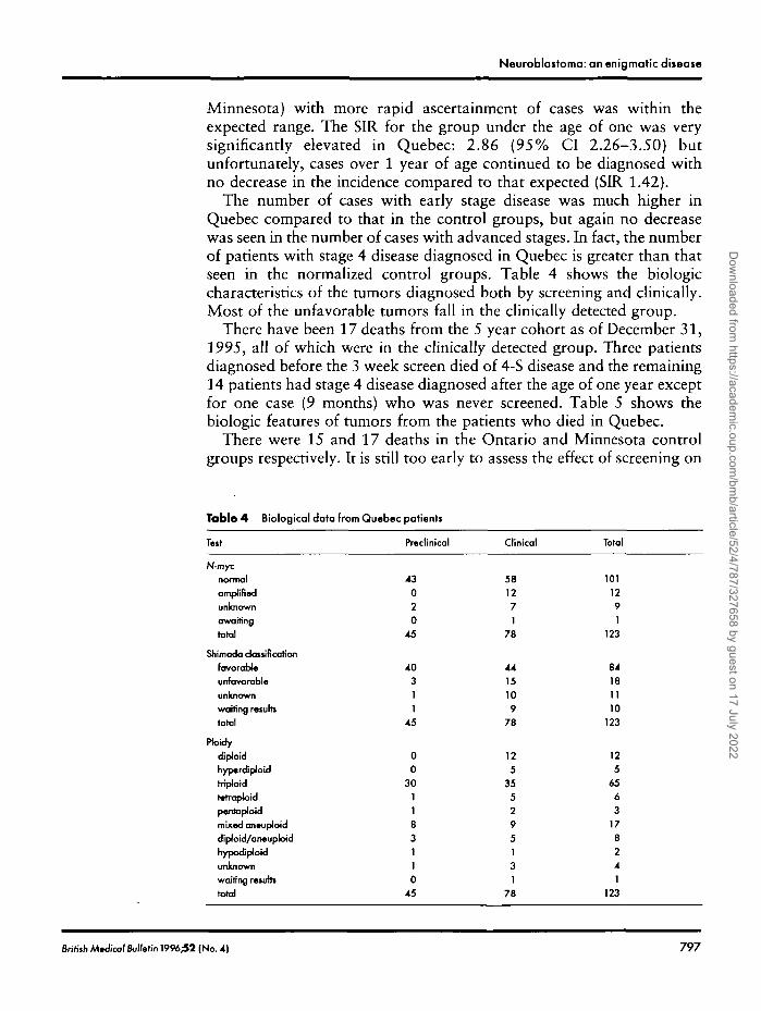
The number of cases with early stage disease was much higher inQuebec compared to that in the control groups, but again no decreasewas seen in the number of cases with advanced stages. In fact, the numberof patients with stage 4 disease diagnosed in Quebec is greater than thatseen in the normalized control groups. Table 4 shows the biologiccharacteristics of the tumors diagnosed both by screening and clinically.Most of the unfavorable tumors fall in the clinically detected group.
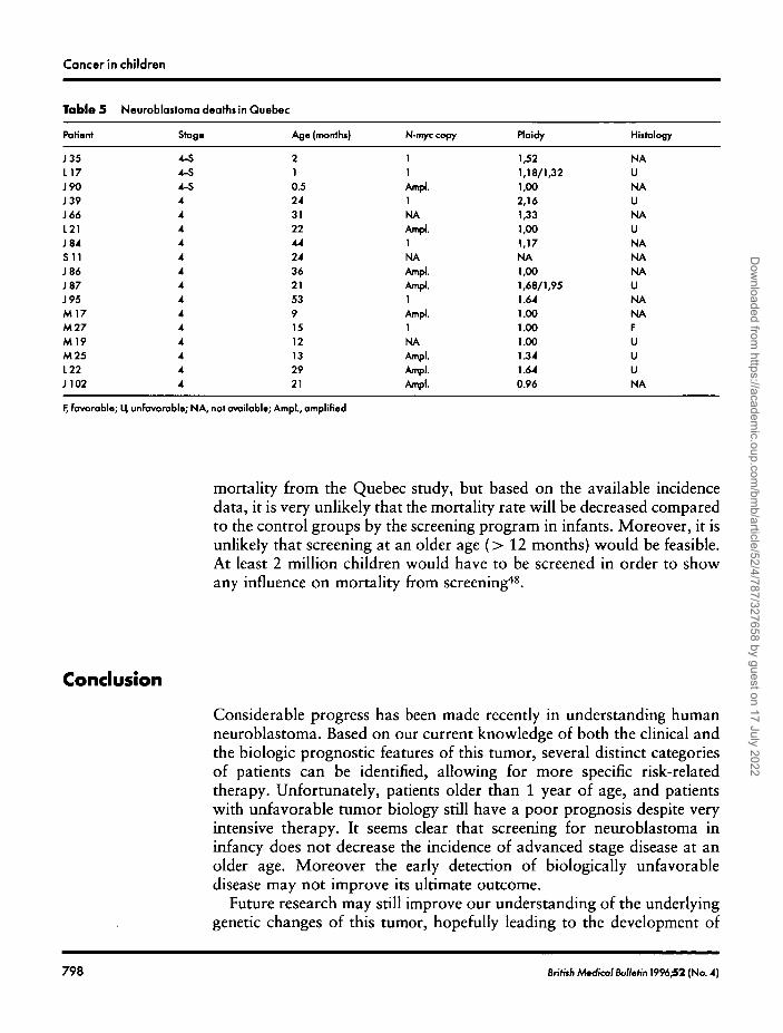
There have been 17 deaths from the 5 year cohort as of December 31,1995, all of which were in the clinically detected group. Three patientsdiagnosed before the 3 week screen died of 4-S disease and the remaining14 patients had stage 4 disease diagnosed after the age of one year exceptfor one case (9 months) who was never screened. Table 5 shows thebiologic features of tumors from the patients who died in Quebec.
There were 15 and 17 deaths in the Ontario and Minnesota controlgroups respectively. It is still too early to assess the effect of screening on
Table 4 Biological data from Quebec patients
Tut
N-mycnormalamplifiedunknownawaitingtotal
Shim ado classificationfavorableunfavorableunknownwaiting resultstotal
PtoidydjploidhypcrdiploidtrrpJoidtetrapJoidpentaploidmixed an•upJoiddlploid/aneuploidhypodJploidunknownwafting remitstotal
Preclinical
43020
45
40311
45
00
301183110
45
Clinical
581271
78
4415109
78
125
355295131
78
Total
1011291
123
84181110
123
125
6563
178241
123
British Mtdical Bulletin 1996;52 (No. 4) 797
Dow
nloaded from https://academ
ic.oup.com/bm
b/article/52/4/787/327658 by guest on 17 July 2022
Cancer in children
Table 5
Patient
J35L17J90J39J66L21J84S11J86J87J95M17M27M l ?M25L22J102
Neuroblaitoma deaths in Quebec
Stag*
4-S4-S4-S44444444444444
Age (monthi)
210.5243122442436215391512132921
N-myccopy
11Ampl.i :NAAmpl.]
NA IAmpl.Ampl.1Ampl.1NAAmpl.Ampl.
loldy
,52,18/1,32,00
2,16,33,00,17
MA,00,68/1,95.64.00.00.00.34.64
Ampl. 0.96
Hiitology
NAUNAUNAUNANANAUNANAFUUUNA
F, favorable; U unfavorable; NA, not available; Ampl., amplified
mortality from the Quebec study, but based on the available incidencedata, it is very unlikely that the mortality rate will be decreased comparedto the control groups by the screening program in infants. Moreover, it isunlikely that screening at an older age (> 12 months) would be feasible.At least 2 million children would have to be screened in order to showany influence on mortality from screening48.
Conclusion
Considerable progress has been made recently in understanding humanneuroblastoma. Based on our current knowledge of both the clinical andthe biologic prognostic features of this tumor, several distinct categoriesof patients can be identified, allowing for more specific risk-relatedtherapy. Unfortunately, patients older than 1 year of age, and patientswith unfavorable tumor biology still have a poor prognosis despite veryintensive therapy. It seems clear that screening for neuroblastoma ininfancy does not decrease the incidence of advanced stage disease at anolder age. Moreover the early detection of biologically unfavorabledisease may not improve its ultimate outcome.
Future research may still improve our understanding of the underlyinggenetic changes of this tumor, hopefully leading to the development of
798 British M*oW Bulletin 1996^2 (No. 4)
Dow
nloaded from https://academ
ic.oup.com/bm
b/article/52/4/787/327658 by guest on 17 July 2022

Neuroblastoma: an enigmatic disease
new approaches in the diagnosis and tumor-specific therapy ofneuroblastoma.
Acknowledgement
Supported in part by Grant 46907 from The United States NationalInstitutes of Health, the National Cancer Institute of Canada (Grant#2691), and the Quebec Network for Genetic Medicine.
References
1 Brodeur GM, Castleberry RP. Neuroblastoma. In: Pizzo PA, Poplack DG, eds. Principles andPractice of Pediatric Oncology. 2nd edn. Philadelphia: JB Lippincott, 1993; 739-68
2 Bernstein ML, Leclerc JM, Bunin G et al. A population based study of neuroblastoma incidence,survival, and mortality in North America. / Clm Oncol 1992; 10: 323-9
3 Beclcwith JB, Perrin EV. In situ neuroblastoma: a contribution to the natural history of neuralcrest tumors. Am ] Pathol 1963; 43: 1089-104
4 Turkel SB, Itabashi HH. The natural history of neuroblastoma cells in the fetal adrenal gland.Am } Pathol 1974; 76: 225-44
5 Woods WG, Lemieux B, Tuchman M. Neuroblastoma represents distinct clinical-biologicentities: a review and perspective from the Quebec Neuroblastoma Screening Project, Pediatrics1992; 89: 114-8
6 Brodeur GM, Azar C, Brother M et al. Neuroblastoma: effect of genetic factors on prognosisand treatment. Cancer 1992; 70 suppL: 1685-94
7 Caron H, van Sluis P, de Kraker J et al: Allelic loss of chromosome lp as a predictor ofunfavorable outcome in patients with neuroblastoma. N Engl} Med 1996; 334: 225-30
8 Schwab M, Alitalo K, Klempnauer KH et al. Amplified DNA with limited homology to myccellular oncogene is shared by human neuroblastoma cell lines and a neuroblastoma tumor.Nature 1983; 305: 245-8
9 Seeger RC, Brodeur GM, Sather H et al. Association of multiple copies of the N-myc oncogenewith rapid progression of neuroblastoma. N Engl] Med 1985; 313: 1111-6
10 Look AT, Hayes FA, Shuster JJ et al. Clinical relevance of tumor cell ploidy and N-myc geneamplification in childhood neuroblastoma: a Pediatric Oncology Group Study. / Clin Oncol1990; 9: 581-91
11 Bourhis J, Dominici C, McDowell H et al. N-myc genomic content and DNA ploidy in stage IV-S neuroblastoma. / Clm Oncol 1991; 9: 1371-5
12 Brodeur GM, Hayes FA, Green AA et al. Consistent N-myc copy number in simultaneous orconsecutive neuroblastoma samples from sixty individual patients. Cancer Res 1987; 47: 4248—53
13 Look AT, Hayes FA, Nitschke R, McWilliams NB, Green AA. Cellular DNA content aspredictor of response to chemotherapy in infants with unresectable neuroblastoma. N Engl JMed 1984; 311: 231-5
14 Brenner DW, Barranco SC, Winslow BH, Shaeffer J. Flow cytometric analysis of DNA contentin children with neuroblastoma. / Pediatr Surg 1989; 24: 204-7
15 Taylor SR, Blatt J, Constantino JP, Roederer M, Murphy RF. Flow cytometric DNA analysis ofneuroblastoma and ganglioneuroma: a 10-year retrospective study. Cancer 1988; 62: 749-54
16 Nakagawara A, Arima-Nakagawara M, Scavarda NJ, Azar CG, Cantor AB, Brodeur GM.Association between high levels of expression of the TRK gene and favorable outcome in humanneuroblastoma. N Engl J Med 1993; 328: 847-54
6ritithM»<6cal Bulletin 1996^2 (No. 4) 7 9 9
Dow
nloaded from https://academ
ic.oup.com/bm
b/article/52/4/787/327658 by guest on 17 July 2022
Cancer in children
17 Kogner P, Barbany G, Dominici C, Castello MA, Raschella G, Persson H. Coexpression ofmessenger RNA for TRK protooncogene and low affinity nerve growth factor in neuroblastomawith favorable prognosis. Cancer Res 1993; 53: 2044-50
18 Vogan K, Bernstein ML, Leclerc JM et al. Absence of p53 mutations in primary neuroblastomas.Cancer Res 1993; 53: 5269-73
19 Tuchman M, Lerrueux B, Auray-Blais C et al. Screening for neuroblastoma at 3 weeks of age:methods and preliminary results from the Quebec Neuroblastoma Screening Project. Pediatrics1990; 86: 765-73
20 Chan HSL, Haddad G, Thorner PS et al. P-glycoprotein expression as a predictor of theoutcome of therapy for neuroblastoma. N EnglJ Med 1991; 325: 1608-14
21 Norris MD, Bordow SB, Marshall GM et al. Expression of the gene for multidrug resistance-associated protein and outcome in patients with neuroblastoma. N Engl] Med 1996; 334: 231-8
22 Brodeur GM, Seeger RC, Barrett A et al. International criteria for diagnosis, staging, andresponse to treatment in patients with neuroblastoma. / Clin Oncol 1988; 6: 1874—81
23 Brodeur GM, Prichard J, Berthold F et al. Revisions of the international criteria forneuroblastoma diagnosis, staging, and response to treatment. / Clin Oncol 1993; 11: 1466-77
23a Kushner BH, Cheung NKV, La Quaglia MP et al. Survival from locally invasive or widespreadneuroblastoma without cytotoxic therapy./ Clm Oncol 1996; 14: 373—81
24 Pinkerton CR. Intensive chemotherapy with stem cell support-experience in paediatnc solidtumours. Bull Cancer 1995; 82 suppl. 1: 61-5
25 McCowage GB, Vowels MR, Shaw PJ, Lockwood L, Mameghan H. Autologous bone marrowtransplantation for advanced neuroblastoma using teniposide, doxorubicin, melphalan,cisplatin, and total-body irradiation. / Clin Oncol 1995; 13: 2789-95
26 Handgretinger R, Anderson K, Lang P et al. A phase 1 study of human/mouse chimeric anti-ganglioside GDSOO24T2SOOOOT antibody ch 14.18 in patients with neuroblastoma. Eur JCancer 1995; 31A: 261-7
27 Bauer M, Reaman GH, Hank JA et al. A phase 2 trial of human recombinant interleukin-2administered as a 4-day continuous infusion for children with refractory neuroblastoma, non-Hodgkin's lymphoma, sarcoma, renal cell carcinoma, and malignant melanoma. A Children'sCancer Group Study. Cancer 1995; 75: 2959-65
28 Sawada T, Todo S, Fujita K, Lino S, Imashuku S, Kusunoki T. Mass screening of neuroblastomain infancy. Am ] Dis Child 1982; 136: 710-2
29 Sawada T, Nakata T, Takasugi N et al. Mass screening for neuroblastoma in infants in Japan:interim report of a mass screening study group. Lancet 1984; ii: 271—3
30 Nishi M, Miyake H, Takeda T et al. Effects of the mass screening of neuroblastoma in Sapporocity. Cancer 1987; 60: 433-6
31 Naito H, Sasaki M, Yamashiro K et al. Improvement in prognosis of neuroblastoma throughmass population screening. / Pediatr Surg 1990; 25: 245—8
32 Goodman S. Neuroblastoma screening data: an epidemiologic analysis. Am J Dis Child 1991;145: 1415-22
33 Craft AW, Parker L. Poor prognosis neuroblastoma: is screening the answer? Br J Cancer 1992;66 suppl. XVm: S96-S101
34 Bessho F, Hashizume K, Nakajo T, Kamoshita S. Mass screening in Japan increased thedetection of infants with neuroblastoma without a decrease in cases in older children. / Pediatr1991; 119: 237^tl
35 Murphy SB, Cohn SL, Craft AW et al. Do children benefit from mass screening forneuroblastoma? Consensus statement from the American Cancer Society Workshop onneuroblastoma screening. Lancet 1991; 337: 344—6
36 Nakagawara A, Zaizen Y, Ikeda K et al. Different genomic and metabolic patterns betweenmass screening-positive and mass screening-negative later-presenting neuroblastomas. Cancer1991; 9: 2037-44
37 Hachitanda Y, Ishimoto K, Hata J, Shimada H. One hundred neuroblastomas detected througha mass screening system in Japan. Cancer 1994; 74: 3223—6
38 Suita S, Zaizen Y, Yano H et al. How to deal with advanced cases of neuroblastoma detected bymass screening: a report from the pediatric oncology study group of the Kyushu area of Japan. /Pediatr Surg 1994; 29: 599-603
800 British AWicol Bullmbn 1996£2 (No. A)
Dow
nloaded from https://academ
ic.oup.com/bm
b/article/52/4/787/327658 by guest on 17 July 2022
Neuroblastoma: an enigmatic disease
39 Ishimoto K, Kiyokawa N, Fujita H et al. Problems of mass screening for neuroblastoma: analysisof false-negative cases. / Pediatr Surg 1990; 25: 398-401
40 Hayashi Y, Ohi R, Yaoita S et al. Problems of neuroblastoma screening for 6 month olds andresults of second screening for 18 month olds. / Pediatr Surg 1995; 30: 467-70
41 Bernstein ML, Woods WG. Screening for neuroblastoma: reporting for the QuebecNeuroblastoma Screening Project, In: Miller A, ed. Advances in Cancer Screening, 1996; Inpress
42 Woods WG, Tuchman M. Neuroblastoma: the case for screening infants in North America.Pediatrics 1987; 79: 869-73
43 Tuchman M, Auray-Blais C, Ramnaraine MLR, Neglia J, Krivit W, Lemieux B. Determinationof urinary homovanilhc and vamllylmandelic acid from dried filter paper samples: assessment ofpotential methods for neuroblastoma screening. Clin Biochem 1987; 20: 173—7
44 Scriver CR, Gregory D, Bernstein ML et al. Feasibility of chemical screening of urine forneuroblastoma case finding in infancy in Quebec. CMAJ 1987; 136: 952-6
45 Auray-Blais C, Gigu£re R, Lemieux B. Thin-layer chromatography of urinary homovanillic acidand vanillylmandelic acid for large-scale neuroblastoma mass screening. Med Pediatr Oncol1989; 17: 364-7
46 Tuchman M, Crippin PJ, Krivit W. Capillary gas-chromatographic determination of urinaryhomovanillic acid and vanillylmandelic acid. Clin Chem 1983; 29: 828-31
47 Woods WG, Tuchman M, Robison LL et al. Screening for neuroblastoma markedly increasesthe incidence in infants without reducing the incidence of unfavorable advanced stage disease inolder children. Submitted, 1996
48 Esteve J. Some remarks on power calculation for neuroblastoma screening. Presented at theThird International Symposium on Neuroblastoma Screening, Kyoto, Japan, 1993
Bntiih Mnfical BuIMn 1996^2 (No. 4) 801
Dow
nloaded from https://academ
ic.oup.com/bm
b/article/52/4/787/327658 by guest on 17 July 2022


