Embed Size (px)
Citation preview

NeuroImage 16, 873–882 (2002)doi:10.1006/nimg.2002.1181
Neural Correlates of Visual-Motion Perceptionas Object- or Self-motion
Andreas Kleinschmidt,* Kai V. Thilo,† Christian Buchel,‡ Michael A. Gresty,§Adolfo M. Bronstein,§ and Richard S. J. Frackowiak¶
*Department of Neurology, Johann Wolfgang Goethe-University, D-60590 Frankfurt, Germany; †Laboratory of Physiology, OxfordUniversity, Oxford, United Kingdom; ‡Department of Neurology, Hamburg University, D-20246 Hamburg, Germany; §Academic
Department of Neuro-otology, Division of Neuroscience and Psychological Medicine, Imperial College, London, United Kingdom; and¶Wellcome Department of Imaging Neuroscience, University College London, London, United Kingdom
em
Both self-motion and objects moving in our visualfield generate visual motion by displacing images onthe retina. Resolving this ambiguity may seem effort-less but large-field visual-motion stimuli can yield per-ceptual rivalry between the real percept of object-motion and the illusory percept of self-motion(vection). We used functional magnetic resonance im-aging to record brain activity in human observers ex-posed to constant-velocity roll-motion. This stimulusinduced responses in areas reaching from calcarineto parieto-occipital and to ventral and lateral tem-poro-occipital cortex and the anterior insula. Duringvection, early motion-sensitive visual areas and ves-tibular parieto-insular cortex deactivated, whereashigher-order parieto- and temporo-occipital areasknown to respond to optical flow retained identicalactivity levels. Within this sustained response, theselatter areas displayed transient activations in re-sponse to each perceptual switch as identified inevent-related analyses. Our results thus show thatthese areas are responsive to the type of visual motionstimulus and highly sensitive to its perceptual bi-stability. The only region to be more active duringperceived self-motion was in, or close to, the cerebel-lar nodulus. This activation may correspond to thegain increase of torsional optokinetic nystagmus dur-ing vection and/or to changes in sensory processingrelated to the rotational percept. In conclusion, weidentified neural correlates of perceiving self-motionfrom vision alone, i.e., in the absence of confirmatoryvestibular or proprioceptive input. These functionalproperties preserve the organism’s ability to move ac-curately in its environment by relying on visual cuesunder conditions when the other spatial senses fail toprovide such information. © 2002 Elsevier Science (USA)
Key Words: visual system; circular vection; ego-mo-tion; vestibular cortex; perceptual ambiguity; visualawareness; functional magnetic resonance imaging.
INTRODUCTION
Visual motion processing serves several perceptualpurposes (Movshon et al., 1985; Nakayama, 1985; Al-bright and Stoner, 1995). It is one of the most powerfulcues for binding objects (“form-from-motion”). Speed-and direction-sensitive motion detectors covering thevisual field will respond in synergy to object-motion,thus defining an object form, e.g., by coherence of dis-placement that its parts display against a background.Wide visual field coverage by the object and/or impov-erished information from the stationary background,however, also permit a different perceptual judgement,namely that retinal shifting of an object is caused byobserver motion (Mach, 1875; Helmholtz, 1896; Gib-son, 1954; Warren, 1995; Lappe et al., 1999). Whilevisual-motion reflects only relative motion informationbetween object and observer, additional cues usuallyhelp to disambiguate object- from self-motion. Vestib-ular and proprioceptive sensory feedback and efferencecopies of body, head, and eye movements can preventus from mistaking the origin of visual-motion (Wer-theim, 1994; Wexler et al., 2001). However, when visu-al-motion is the sole informative cue for reconstructingself-motion a perceptual ambiguity may arise and in-duce the intermittent illusion of self-motion (Dichgansand Brandt, 1978), e.g., when a sensation of self-mo-tion (vection) is evoked by a train slowly moving on aneighboring track as we watch from a stationarywagon.
To identify brain systems processing self-motion wechose the approach of using one constant visual-motionstimulus that was made sufficiently ambiguous to yieldboth percepts, i.e., object- and self-motion, in sponta-neous alternation. With functional magnetic resonanceimaging we recorded brain activity in human subjectswho, exposed to this stimulus, fluctuated between per-ceiving a rotating object and feeling themselves rotatearound the nearly earth-vertical axis of gaze while
Received Nov
8
ber 30, 2001
73
1053-8119/02 $35.00© 2002 Elsevier Science (USA)All rights reserved.

looking at an apparently stationary object (circularvection; see Fig. 1 and Materials and Methods). Thus,we addressed the distinction of object- and self-motionat the level of perceptual awareness rather than that ofspecialized visual feature processing. Any brain activ-ity change correlating with either of the percepts can-not be accounted for by the stimulus properties per se.This avoids the confound of previous studies that gen-erated illusory self-motion but compared related brainactivity with conditions differing in physical stimulusproperties, e.g., coherence of speed or direction (deJong et al., 1994; Brandt et al., 1998).
MATERIALS AND METHODS
Functional Imaging
Data from eight healthy subjects (written informedconsent, one female, seven males, age 22–48 years)were acquired on a 2T magnetic resonance imager(Siemens Vision, Erlangen, Germany; head coil), ob-taining a structural (T1-weighted) scan and then seriesof blood-oxygenation-sensitive (T2*-weighted) echopla-nar image volumes every 4.1 s (image repetition time/echo time � 80.7/40 ms; 48 contiguous transverseslices, voxel size � 3 � 3 � 3 mm3).
Visual Stimulation
Prior to each scanning series (118 image volumes, 8min duration, three runs), subjects started looking atthe rotational center of a disk with a windmill pattern(12 fluorescent radial stripes alternating with blackstripes of equivalent width subtending approximatelyradially 45° of the visual field) in an otherwise dimmedscanner room. The disk was mounted above the headcoil (approximately 12 cm viewing distance to center)and mechanically controlled by a cogwheel connectionto a propylene rod that was driven by a motor in theconsole room. For the first 18 image volumes the diskremained stationary and then started rotating aboutits center at a constant speed (45°/s). We used bothclockwise and counter-clockwise rotation in differentsessions. Since no significant brain activity differenceswere found between these conditions, the data werepooled for the other analyses. Previous measurementswith an almost identical stimulus under video-oculog-raphy (Thilo et al., 1999) established that perception isbistable and that subjects can maintain fixation overthe length of time chosen for the runs.
Bistable Perception
Key presses with the right index finger indicated theonset of visual-motion stimulation and of epochs withperceived object-motion. Middle finger key presses in-dicated the onset of illusory self-motion, i.e., circularvection. Thus, a sequence of alternating key presseswas recorded, defining the subjects’ bistable perception
of the stimulus (see Fig. 1). All subjects experiencedintermittent circular vection, the first epoch after amean latency of 9 s (�4 s SD) after onset of diskrotation and ensuing epochs of perceived self- and ob-ject-motion with mean durations of 16 and 19s, respec-tively. Object- and self-motion percepts were mutuallyexclusive; i.e., during vection the disk was perceived asstationary and when disk rotation was perceived therewas no simultaneous impression of self-motion. Noneof the subjects suffered from nausea. Subjects werefamiliarized with the stimulus and the percepts that itevokes and with the task prior to the experimentalruns during functional imaging.
Data Analysis
Data processing and analysis used statistical para-metric mapping (http://www.fil.ion.ucl.ac.uk/spm). Af-ter discarding the initial eight, all image volumes werethree-dimensionally realigned to the first volume,coregistered with the subject’s corresponding anatom-ical (T1-weighted) images, nonlinearly normalized intostandard stereotactic space (template provided cour-tesy of the Montreal Neurological Institute), andsmoothed using a 9-mm full-width at half maximumGaussian kernel. We removed low-frequency fluctua-tions by a temporal high-pass filter with a cutoff timeconstant at 70 s and analyzed two types of responses,sustained (perceptual state) and transient (perceptualswitch). These were modeled as boxcar functions (sus-tained) and delta functions (transient) that were con-volved with a synthetic hemodynamic response func-tion. Durations of states (percepts) and timing ofevents (switches) were derived from the subjects’ keypresses (see above).
We analyzed neural activity changes in relation tostimulation as a first and perception as a second levelof experimental variable. Statistical comparisons wereperformed by contrasting parameter estimates for themodeled sustained responses during visual-motionstimulation (across both percepts) with the stationarybaseline and, as an embedded comparison within on-going constant visual-motion stimulation, by contrast-ing those responses during perceived object-motionwith those during perceived self-motion and vice versa(see Fig. 1). For event-related responses, we analyzedthe evoked transient hemodynamic responses, i.e., theevents at real-time resolution convolved with a canon-ical hemodynamic response function, both as changesagainst baseline activity and against each other (Klein-schmidt et al., 1998). The three event types reported bykey presses (see above) were the onset of visual-motion,perceived self-motion, and perceived object-motion.
These procedures resulted in statistical parametricmaps for every voxel showing sustained or transientactivity differences related to the stimulus and to ei-ther of the percepts. Statistical inferences were cor-rected for multiple nonindependent comparisons by us-
874 KLEINSCHMIDT ET AL.

ing Gaussian random field theory. Unless statedotherwise we applied a significance threshold of P �0.05, corrected.
Cortical Localization
We assigned tentative area labels to group analysisresponse foci that were in part supported by functionalresponses determined here, i.e., sensitivity to opticalflow (Figs. 2 and 3), or by stereotactic coordinates forlocalized functional response properties reported in theliterature. In particular, we related activations in cal-carine cortex to V1, in the posterior fusiform gyrus toV4, in the lateral occipito-temporal cortex to V5/MT,and in the posterior insula to vestibular cortex. Thejoint label of V3/V3a reflects that these areas arerather thin stripes and variable in position even inrelation to sulcal landmarks. We chose the anatomi-cally descriptive labels of dorso-medial cortex (DM) andsuperior temporal cortex (ST) to avoid implying a sin-gle visual area and merely conclude that the functionalbehavior in these foci suggests homologies with ma-caque areas V6/PO and STPa, respectively. Finally, theputative identification of area V5a/MST and PIVC isdiscussed below.
RESULTS
Regions Responsive to the Visual-Motion Stimulus
We determined responsiveness to the visual-motionstimulus (coherent wide-field stimulation) by contrast-ing activity between the period of rotation and thepreceding stationary phase (see Fig. 1 and Materialsand Methods). The spatial distribution of regional ac-tivations found here largely conforms with previousneuroimaging studies mapping the responses to visual-motion (Watson et al., 1993; Zeki et al., 1993; Dupont etal., 1994; de Jong et al., 1994; Tootell et al., 1995, 1997;Cheng et al., 1995; McCarthy et al., 1995; McKeefry etal., 1997, Dieterich et al., 1998; Greenlee, 2000; Previcet al., 2000). Since the response to the stimulus per sewas not the effect of interest in this study we presentonly an overview of the cortical response topography(Fig. 2).
Regions Responsive to Perceptual State
The experimental parameter of interest in our studywas that, over time, all subjects perceived object-mo-tion and circular vection in irregular alternation (Fig.1). Reports of these perceptual switches enabled us toassign our data to one or other percept (see Materialsand Methods). Hence, we determined activity levelsduring the two percepts in motion-sensitive candidateregions that animal electrophysiology, human lesionstudies (Straube and Brandt, 1987; Heide et al., 1990;Vaina, 1998), and neuroimaging studies had suggestedas important for optical flow processing. This included
DM (comprising cuneus and parieto-occipital cortex;see Richer et al. (1991) and Lee et al. (2000) for elec-trostimulation studies), the anterior portion (putative“V5a/MST”) of the human motion complex (“V5/MTcomplex”) at the occipito-temporal junction (Duffy andWurtz, 1991; Orban, 1997; Tanaka, 1998; Wurtz,1998), an area of ST (see Bruce et al., 1981; Andersonand Siegel, 1999), and posterior (intra-) parietal cortex(see Siegel and Read, 1997). All these regions re-
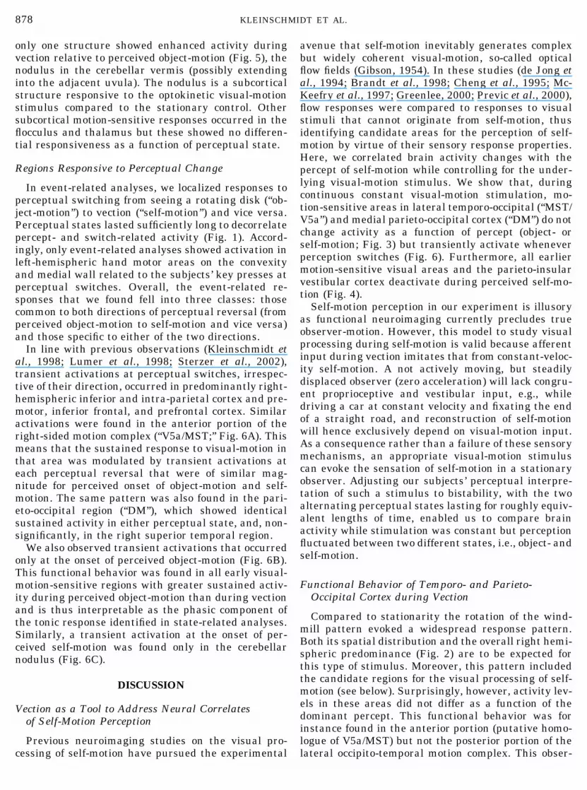
FIG. 1. Experimental design and perceptual behavior. (A) Afteran initial baseline period, a stationary disk with a windmill patternstarts rotating. While fixating its center of rotation, subjects switchbetween the percept of object-motion and that of self-motion, i.e.,circular vection. Data obtained while scanning one of the participat-ing subjects are shown as a time course alternating between the twopercepts of object-motion and self-motion (vection). (B) Distributionof durations for each percept for the same data as shown in A. Theepoch length histograms were approximated by gamma functions(bin width 8.2 s, i.e., equivalent to two sampled image volumes), atemporal switch distribution typical of bistable perception (Klein-schmidt et al., 1998; Sterzer et al., 2002).
875OBJECT- OR SELF-MOTION

sponded to our stimulus compared to the stationarybaseline (see Fig. 2) but showed no differential activa-tion in relation to the two different perceptual states(Fig. 3). For these comparisons, we used a more sensi-tive threshold (P � 0.001, uncorrected) that took ac-count of the greatly reduced number of multiple com-parisons involved and analyzed single subjects. Thismeans that our negative result was not accounted forby overly conservative statistical thresholds or byblurred localization due to group analysis.
In the next step, we tested our data for an influenceof perceptual state on regional activity levels. This wasdone by mapping activity differences between imagesreflecting the percept of object-motion and those re-flecting the percept of self-motion and vice versa. Whencontrasting perceived object- with self-motion (Fig. 4),activity in a subset of the motion-sensitive brain areas
FIG. 4. Brain areas activated during perceptual dominance ofobject-motion. The statistical parametric maps on the left were ob-tained from a group analysis testing for greater activity duringperceived object-motion than during circular vection (visualised atP � 0.001, uncorrected and superimposed onto individual structuralsections). Response foci are highlighted by green circles and weretentatively labeled (cf. Fig. 3; V1–V5, visual areas 1–5; PIVC, pari-eto-insular vestibular cortex). Correspondingly, activity levels in themaximally percept-sensitive voxels in these foci (coordinates in-serted) are plotted in the adjacent panels for each percept relative tobaseline. Note differences in y axis scaling for the plots. All differ-ences shown were significant at the level of P � 0.05, corrected.
FIG. 2. Brain areas responsive to optokinetic visual-motion. Sta-tistical parametric maps showing areas with greater activity duringvisual-motion (disk rotation) than during the preceding baseline (diskstationary). To illustrate the overall cortical response pattern in a groupanalysis (three repetitions per subject of the stimulation protocol shownin Fig. 1A), the results are displayed by color-coding and rendered ontoan anatomical template image (visualized at P � 0.05, corrected).
FIG. 3. Motion-sensitive brain areas equally responsive during per-ceived object-motion and self-motion. Statistical parametric maps (left)from a group analysis of areas responsive to visual-motion (disk rota-tion) compared to baseline (disk stationary). These maps are derivedfrom the same statistical comparison as in Fig. 2, color-coded, andsuperimposed onto sections from an individual structural dataset of aparticipating subject (visualised at P � 0.001, uncorrected). The loca-tion of the significance maxima contained in green circles is given interms of stereotactic coordinates and with tentative area labels (seeCortical Localization in Materials and Methods; DM, dorsomedial cor-tex; ST, superior temporal area; V5a/MST, accessory V5/medial supe-rior temporal area). The graphs on the right plot response strength inthese foci (in the circled maximally significant responsive voxels at thecoordinates given) for perceived object-motion and self-motion relativeto the stationary phase (parameter estimates of percentage signalchange averaged across runs and subjects; note differences in y axisscaling). There was no significant difference in activity levels across thetwo percepts (P � 0.001, uncorrected). Black vertical bars indicatestandard error of the mean, i.e., topographical and functional intersub-ject variability, and are not related to response identification (which isrepresented by the statistical parametrical maps).
876 KLEINSCHMIDT ET AL.

shown in Fig. 2 showed greater activity during per-ceived object-motion and less activity during perceivedself-motion. The “earliest” percept-sensitive cortical ac-
tivity change occurred in primary (calcarine) visualcortex (“V1”) and extended over an intermediate levelin the superior occipital gyrus (“V3/V3A”) into ventraloccipital cortex (fusiform gyrus, “V4”) and onto theconvexity (occipito-temporal junction, posterior “V5/MT”). These activity differences were significant in thegroup analysis even at thresholds corrected for multi-ple comparisons but, again, were not significant whenanalyzed in single subjects.
A significant activity difference also occurred in anarea not activated by the visual-motion stimulus,namely, the posterior parieto-insular cortex, a regionproposed to be a human homologue of a vestibularcortex (Fig. 4, bottom). Compared to activity levelsduring the stationary stimulus, this difference resultsfrom a deactivation during perceived self-motion.Hence, while activity levels were not significantly dif-ferent between the stationary and the rotating stimulias long as the latter was perceived as object-motion,there was a significant activity decrease during illu-sory self-motion.
In contrast to the large set of motion-sensitive areaswith greater activity during perceived object-motion,
FIG. 5. The response focus for perceptual dominance of self-motion.Statistical parametric map from a group analysis testing for greateractivity during perceived self-motion than object-motion showing thecerebellar nodulus and plot of the associated parameter estimates (sta-tistical comparison opposite to that in Fig. 4; same procedure, signifi-cance, and visualization thresholds). Note that all individual structuralscans of the participating subjects were inspected at this location to ruleout an underlying or adjacent large vascular structure that might forma potential nonspecific source of contrast change.
FIG. 6. Results from a group analysis of transient activations during perceptual reversals. (A) The regions responsive to both theperceived onset of self-motion and that of object-motion are shown by rendering them onto a surface reconstruction of the right hemisphere(regions responding to vection onset and to perceived object-motion onset, both thresholded at P � 0.001; the latter enacted by using aninclusive mask option). Note that unlike the motion-sensitive areas shown in Fig. 2 there is predominant activation in a fronto-parietalsystem, whereas effects in visual areas are restricted to dorsomedial parieto-occipital cortex (not shown) and the anterior pole of thetemporo-occipital motion complex. The modeled responses (solid line) and their standard error (hatched) in the peak voxel from that latterarea (green circle) are plotted below in response to the three event types at time 0 (for use of colors for event types see bottom). (B) Theresponses at the perceived onset of object-motion. The result of the comparison of greater responses during the onset of perceivedobject-motion than of self-motion (at P � 0.001) are displayed on a rendering of the ventral brain surface (cerebellum removed) with theunderlying modeled responses (from peak voxel in the green circle) to the three event types plotted below (for colors see bottom). (C) Theresponses at the perceived onset of self-motion. The result of the comparison of greater responses during the onset of perceived self-motionthan of object-motion (at P � 0.001) is displayed on a medio-sagittal section of the structural brain scan of one of the participating subjectsand the underlying modeled responses (solid line) for the three different event types are plotted below (for colors see bottom).
877OBJECT- OR SELF-MOTION
only one structure showed enhanced activity duringvection relative to perceived object-motion (Fig. 5), thenodulus in the cerebellar vermis (possibly extendinginto the adjacent uvula). The nodulus is a subcorticalstructure responsive to the optokinetic visual-motionstimulus compared to the stationary control. Othersubcortical motion-sensitive responses occurred in theflocculus and thalamus but these showed no differen-tial responsiveness as a function of perceptual state.
Regions Responsive to Perceptual Change
In event-related analyses, we localized responses toperceptual switching from seeing a rotating disk (“ob-ject-motion”) to vection (“self-motion”) and vice versa.Perceptual states lasted sufficiently long to decorrelatepercept- and switch-related activity (Fig. 1). Accord-ingly, only event-related analyses showed activation inleft-hemispheric hand motor areas on the convexityand medial wall related to the subjects’ key presses atperceptual switches. Overall, the event-related re-sponses that we found fell into three classes: thosecommon to both directions of perceptual reversal (fromperceived object-motion to self-motion and vice versa)and those specific to either of the two directions.
In line with previous observations (Kleinschmidt etal., 1998; Lumer et al., 1998; Sterzer et al., 2002),transient activations at perceptual switches, irrespec-tive of their direction, occurred in predominantly right-hemispheric inferior and intra-parietal cortex and pre-motor, inferior frontal, and prefrontal cortex. Similaractivations were found in the anterior portion of theright-sided motion complex (“V5a/MST;” Fig. 6A). Thismeans that the sustained response to visual-motion inthat area was modulated by transient activations ateach perceptual reversal that were of similar mag-nitude for perceived onset of object-motion and self-motion. The same pattern was also found in the pari-eto-occipital region (“DM”), which showed identicalsustained activity in either perceptual state, and, non-significantly, in the right superior temporal region.
We also observed transient activations that occurredonly at the onset of perceived object-motion (Fig. 6B).This functional behavior was found in all early visual-motion-sensitive regions with greater sustained activ-ity during perceived object-motion than during vectionand is thus interpretable as the phasic component ofthe tonic response identified in state-related analyses.Similarly, a transient activation at the onset of per-ceived self-motion was found only in the cerebellarnodulus (Fig. 6C).
DISCUSSION
Vection as a Tool to Address Neural Correlatesof Self-Motion Perception
Previous neuroimaging studies on the visual pro-cessing of self-motion have pursued the experimental
avenue that self-motion inevitably generates complexbut widely coherent visual-motion, so-called opticalflow fields (Gibson, 1954). In these studies (de Jong etal., 1994; Brandt et al., 1998; Cheng et al., 1995; Mc-Keefry et al., 1997; Greenlee, 2000; Previc et al., 2000),flow responses were compared to responses to visualstimuli that cannot originate from self-motion, thusidentifying candidate areas for the perception of self-motion by virtue of their sensory response properties.Here, we correlated brain activity changes with thepercept of self-motion while controlling for the under-lying visual-motion stimulus. We show that, duringcontinuous constant visual-motion stimulation, mo-tion-sensitive areas in lateral temporo-occipital (“MST/V5a”) and medial parieto-occipital cortex (“DM”) do notchange activity as a function of percept (object- orself-motion; Fig. 3) but transiently activate wheneverperception switches (Fig. 6). Furthermore, all earliermotion-sensitive visual areas and the parieto-insularvestibular cortex deactivate during perceived self-mo-tion (Fig. 4).
Self-motion perception in our experiment is illusoryas functional neuroimaging currently precludes trueobserver-motion. However, this model to study visualprocessing during self-motion is valid because afferentinput during vection imitates that from constant-veloc-ity self-motion. A not actively moving, but steadilydisplaced observer (zero acceleration) will lack congru-ent proprioceptive and vestibular input, e.g., whiledriving a car at constant velocity and fixating the endof a straight road, and reconstruction of self-motionwill hence exclusively depend on visual-motion input.As a consequence rather than a failure of these sensorymechanisms, an appropriate visual-motion stimuluscan evoke the sensation of self-motion in a stationaryobserver. Adjusting our subjects’ perceptual interpre-tation of such a stimulus to bistability, with the twoalternating perceptual states lasting for roughly equiv-alent lengths of time, enabled us to compare brainactivity while stimulation was constant but perceptionfluctuated between two different states, i.e., object- andself-motion.
Functional Behavior of Temporo- and Parieto-Occipital Cortex during Vection
Compared to stationarity the rotation of the wind-mill pattern evoked a widespread response pattern.Both its spatial distribution and the overall right hemi-spheric predominance (Fig. 2) are to be expected forthis type of stimulus. Moreover, this pattern includedthe candidate regions for the visual processing of self-motion (see below). Surprisingly, however, activity lev-els in these areas did not differ as a function of thedominant percept. This functional behavior was forinstance found in the anterior portion (putative homo-logue of V5a/MST) but not the posterior portion of thelateral occipito-temporal motion complex. This obser-
878 KLEINSCHMIDT ET AL.

vation suggests a functional subdivision of the humanmotion complex, with the posterior portion putativelyreflecting the functional behavior of area V5/MT andthe anterior that of area V5a/MST(d). This interpreta-tion is compatible with a recent fMRI study that com-pared the responses to circular and radial flow to thoseto translational visual-motion and identified a specifi-cally flow-sensitive area within the human motioncomplex V5/MT as a putative human homologue ofMSTd (Morrone et al., 2000). Stereotactic coordinatesreported for that area relative to responses to transla-tional motion were at the anterior (and ventral) end ofthe motion complex.
Electrophysiological recordings in the dorsal medialsuperior temporal area (MSTd) of nonhuman primateshave shown preferential responses to flow stimuli(Duffy and Wurtz, 1991; Orban, 1997; Tanaka, 1998)and congruent vestibular input with real self-motion(Duffy, 1998; Bremmer et al., 1999). These neuronalresponse properties suggest that this area is ideallysuited to contribute to the reconstruction of self-motion(Wurtz, 1998; Andersen et al., 2000) although otherareas, which have been less well characterized by sin-gle-unit recordings, may also play an important role.Our findings support the notion that this area is notthe only one relevant for self-motion perception be-cause a similar functional behavior was found in sev-eral other areas including a dorsomedial parieto-occip-ital region (DM, putatively corresponding to area V6/PO; see Galletti et al., 1999; Rosa and Tweedale, 2001)but also in superior and intra-parietal foci and superiortemporal foci (putatively corresponding to the anteriorsuperior temporal polysensory area described in non-human primates; see Bruce et al., 1981; Cusick, 1997;Anderson and Siegel, 1999).
The question with regard to these candidate areas,however, is whether identical (instead of increased)activity levels during perceived self-motion (as com-pared to object-motion) are compatible with a func-tional role of these areas in the perception of self-motion. As a visual stimulus, self-motion is only one ofthe multiple cases of complex visual-motion. Any vi-sual area implicated in self-motion processing will in-evitably also display response properties suitable forother functional contexts, and processing of these con-texts may interact (Probst et al., 1984; Geesaman andAndersen, 1996; Recanzone et al., 1997; Zemel andSejnowski, 1998; but see also Royden and Hildreth,1999). We therefore propose that the lateral temporo-occipital (“MST/V5a”) and medial parieto-occipital ar-eas (“DM”), where activity remained constantly ele-vated during both percepts, process the visual-motioninput arising from self-motion but also other types ofcoherent visual-motion.
This interpretation is compatible with the sustainedresponses that we found in the candidate areas forself-motion perception. However, an area that is sen-sitive to the perceptual consequence of a visual stimu-
lus should also display transient signal changes when-ever the perceptual interpretation changes. Usingambiguous visual stimuli that maintain a constantsensory input, event-related activations during percep-tual switches have been found in several extrastriateareas and their localization conforms with the sensoryresponse properties of those areas. Hence, perceptualchanges related to objects and faces were accompaniedby fusiform activations (Kleinschmidt et al., 1998) andchanges related to motion direction by activations intemporo-occipital visual-motion areas (Sterzer et al.,2002).
Our event-related analyses here show that, over andabove a continuous response to the visual-motion stim-ulus, lateral temporo-occipital (“MST/V5a”) and medialparieto-occipital areas (“DM”) activated transientlyeach time perception changed from one possible inter-pretation to the other. Different from other areas,these responses were equivalent for both directions ofperceptual change, as were the sustained activity lev-els for both percepts. Interestingly, in the aforemen-tioned study by Morrone et al. (2000), the best re-sponses in putative MST/V5a were elicited by changesin optic flow direction rather than constant flow whichis also in line with electrophysiological findings in MST(Paolini et al., 2000; see also Duffy and Wurtz, 1997).We also observed transient activations in other brainareas that seem related to more general aspects ofperceptual synthesis and selection (Kleinschmidt et al.,1998; Lumer et al., 1998; Sterzer et al., 2002) andactivations during only one direction of perceptualchange that occurred in regions sensitive to one type ofpercept but not the other, as discussed in the followingsection.
Brain Activity Changes Related to the Perceptof Object- or Self-Motion
Comparing sustained activity levels in the two per-ceptual states, we found greater activity during per-ceived object-motion in a set of areas reaching fromcalcarine cortex (“V1”) to ventral occipital cortex (“V4”)and lateral temporo-occipital cortex (“V5”). The latterfocus corresponds to the posterior portion of the motioncomplex; the former was in the fusiform gyrus. Thesetwo functional ramifications of the visual system aredifferentially engaged by color, form, and motion, andtheir activation during perceived object-motion mightindicate integrative segregation by construction of“form-from-motion” (Lamme et al., 1993; Van Essenand Gallant, 1994).
The alternative interpretation for differential activ-ity in the two percepts is a deactivation in early mo-tion-sensitive areas during vection. We had no eye-movement recording facilities available in this studybut the optokinetic stimulus that we used reliably elic-its a torsional nystagmus (Brecher, 1934; Finke andHeld, 1978; Cheung and Howard, 1991) which fails
879OBJECT- OR SELF-MOTION
almost completely to cancel retinal slip. Its gain (ratiobetween stimulus velocity and eye velocity) is �0.1 butincreases by �40%, from 0.08 to 0.12, during vection(Thilo et al., 1999). For a stimulus of 45°/s this trans-lates into less than 2°/s retinal stimulation differencebetween the two percepts—too little to account for theprofound activity difference found in multiple visualareas.
The influence of extraretinal signals related to thesmall but significant changes in the slow and fast (sac-cadic) components of nystagmus during self-motionperception is more difficult to evaluate. Saccades (andvestibularly elicited saccades) suppress activity inearly visual cortex (Paus et al., 1995; Wenzel et al.,1996; Gallant et al., 1998) but in extrastriate visualareas of the parvo-dominated ventral stream perisac-cadic modulation either is absent or is even excitatory(Leopold and Logothetis, 1998). Similarly, the modula-tory influence of slow-pursuit eye movements on activ-ity in the human motion complex seems to be, if any-thing, excitatory (Freitag et al., 1998). It thereforeappears unlikely that the widespread deactivations inseveral visual areas (and lack of activation in candi-date self-motion perception areas) during vectionmerely reflect a slight percept-dependent gain changeof the associated nystagmus.
We did find enhanced activity during vection in thecerebellar nodulus (and maybe the adjacent uvula).The nodulus is a critical site both for visuo-vestibularinteraction (Precht et al., 1976) and for torsional opto-kinetic nystagmus (Angelaki and Hess, 1994). Nodulusactivation during vection could therefore be accountedfor by the oculomotor gain increase or by changes insensory processing related to the rotational percept(reviewed in Cohen et al., 1999). Yet, nystagmus facil-itation is only one of several functionally plausibleneuronal adjustments to self-motion. In fact, deactiva-tions in early motion-sensitive areas, nystagmus facil-itation, and saccadic suppression may all share a com-mon functional purpose. Greater nystagmus gainduring observer-motion decreases retinal slippage ofthe environment and perisaccadic suppression of re-lated retinal slip stabilizes visual perception againstthe effects of eye movements. Yet, a functional benefitfrom suppressing observer-induced retinal slip wouldalso apply to head and body movements (Crowell et al.,1998). A more general suppressive mechanism duringself-motion could thus ensure that despite retinal slip-page due to eye, head, and body movements, ongoingvisual processing remains undisturbed (Probst et al.,1984; Wexler et al., 2001). During self-motion such asuppression would prove helpful only if it selectivelyspares those visual cortical areas specialized for theflow fields arising from self-motion. This is the case inareas that maintain a veridical visual-motion activityprofile across both percepts (Fig. 3). Even though theydo not activate further during vection (compared toobject-motion) they nevertheless remain activated
(Fig. 3) as opposed to the deactivation in earlier mo-tion-sensitive areas (Fig. 4).
Visuo-Vestibular Interactions during Vection
In addition to perceptually driven visuo-visual inter-actions (between cortices processing object-motion andself-motion) we found corresponding visuo-vestibularinteractions. It was shown previously that a parieto-insular region identified as a human “vestibular” cor-tex (Bottini et al., 1994; see also Guldin and Grusser,1998) is deactivated by visual-motion stimuli that gen-erate vection (Brandt et al., 1998). The critical findingin our study is that this deactivation results not fromthe stimulus per se but from its perceptual interpreta-tion (Fig. 4, bottom). Hence, during a constant visual-motion stimulus, the percept of self-motion is accom-panied by deactivations of structures that processobject-motion and vestibular information, i.e., poten-tial distracters to the visual analysis of steady self-motion (Brandt, 1999). In accordance with this view,the neural signature of self-motion perception is notthe activity of a single “self-motion center” but a dis-tributed activity pattern of interconnected areas thatprocess spatial sensory cues such as retinal and ves-tibular input.
CONCLUSION
Our study addressed how the human brain processestwo alternative perceptual results of an ambiguousvisual-motion stimulation that differ profoundly infunctional meaning. The illusion of self-motion, inter-mittently generated by our stimulus, provides a modelof how the brain deals with retinal stimulation thatresults from our own actions. While there is a generalsuppression of activity in visual and vestibular cortexduring vection, the activity levels in advanced visualprocessing stages remain unaffected and thus presum-ably allow for continued reconstruction of self-motion.The effects that we observed can be functionallyframed as mechanisms that allow us to stabilize ourperception of the visual environment and to use ourvisual sense to guide our locomotion.
ACKNOWLEDGMENTS
We thank Mr. William Cameron from the MRC for constructingthe stimulus setup and the unit’s physics and methods teams forcontinuous support. A.K. and C.B. are supported by the Volkswagen-Stiftung, K.V.T by the Dix Foundation, M.A.G. and A.M.B. by theMedical Research Council, and R.S.J.F. by the Wellcome Trust.
REFERENCES
Albright, T. D., and Stoner, G. R. 1995. Visual-motion perception.Proc. Natl. Acad. Sci. USA 92: 2433–2440.
Andersen, R. A., Shenoy, K. V., Crowell, J. A., and Bradley, D. C.2000. Neural mechanisms for self-motion perception in area MST.Int. Rev. Neurobiol. 44: 219–233.
880 KLEINSCHMIDT ET AL.
Anderson, K. C., and Siegel, R. M. 1999. Optic flow sensitivity in theanterior superior temporal polysensory area, STPa, of the behav-ing monkey. J. Neurosci. 19: 2681–2692.
Angelaki, D. E., and Hess, B. J. M. 1994. The cerebellar nodulus andventral uvula control the torsional vestibuloocular reflex. J. Neu-rophysiol. 72: 1443–1447.
Bottini, G., Sterzi, R., Paulesu, E., Vallar, G., Cappa, S. F., Erminio,F., Passingham, R. E., Frith, C. D., and Frackowiak, R. S. F. 1994.Identification of the central vestibular projections in man: Apositron emission tomography activation study. Exp. Brain Res.99: 164–169.
Brandt, T. 1999. Cortical visual-vestibular interaction for spatialorientation and self-motion perception. Curr. Opin. Neurol. 12:1–4.
Brandt, T., Bartenstein, P., Janek, A., and Dieterich, M. 1998. Re-ciprocal inhibitory visual-vestibular interaction: Visual motionstimulation deactivates the parieto-insular vestibular cortex.Brain 121: 1749–1758.
Brecher, G. A. 1934. Die optokinetische Auslosung von Augenrollungund rotatorischem Nystagmus. Pflug. Arch. Ges. Physiol. 234:13–28.
Bremmer, F., Kubischik, M., Pekel, M., Lappe, M., and Hoffmann,K. P. 1999. Linear vestibular self-motion signals in monkey medialsuperior temporal area. Ann. N. Y. Acad. Sci. 871: 272–281.
Bruce, C. J., Desimone, R., and Gross, C. G. 1981. Visual propertiesof neurons in a polysensory area in superior temporal sulcus of themacaque. J. Neurophysiol. 46: 369–384.
Cheng, K., Fujita, H., Kanno, I., Miura, S., and Tanaka, K. 1995.Human cortical regions activated by wide-field visual motion: AnH215O PET study. J. Neurophysiol. 74: 413–427.
Cheung, B. S., and Howard, I .P. 1991. Optokinetic torsion: Dynam-ics and relation to circularvection. Vis. Res. 31: 1327–1335.
Cohen, B., Wearne, S., Dai, M., and Raphan, T. 1999. Spatial orien-tation of the angular vestibulo-ocular reflex. J. Vestib. Res. 9:163–172.
Crowell, J. A., Banks, M. S., Shenoy, K. V., and Andersen, R. A. 1998.Visual self-motion perception during head turns. Nat. Neurosci. 1:732–737.
Cusick, C. G. 1997. The superior temporal polysensory region inmonkeys. In Cerebral Cortex, Vol. 12: Extrastriate Cortex (K. S.Rockland, J. H. Kaas, and A. Peters, Eds.), pp. 435–468. Plenum,New York.
de Jong, B. M., Shipp, S., Skidmore, B., Frackowiak, R. S. J., andZeki, S. 1994. The cerebral activity related to the visual perceptionof forward motion in depth. Brain 117: 1039–1054.
Dichgans, J., and Brandt, T. 1978. Visual-vestibular interaction:Effects on self-motion perception and postural control. In Hand-book of Sensory Physiology (R. Held, H. W. Leibowitz, and H. L.Teuber, Eds.), Vol. 8, pp. 755–804. Springer-Verlag, Heidelberg.
Dieterich, M., Bucher, S. F., Seelos, K. C., and Brandt, T. 1998.Horizontal or vertical optokinetic stimulation activates visual mo-tion-sensitive, ocular motor and vestibular cortex areas with righthemispheric dominance. Brain 121: 1479–1495.
Duffy, C. J. 1998. MST neurons respond to optic flow and transla-tional movement. J. Neurophysiol. 80: 1816–1827.
Duffy, C. J., and Wurtz, R. H. 1991. Sensitivity of MST neurons tooptic flow stimuli. I. A continuum of response selectivity to large-field stimuli. J. Neurophysiol. 65: 1329–1345.
Duffy, C. J., and Wurtz, R. H. 1997. Multiple temporal components ofoptic flow responses in MST neurons. Exp. Brain Res. 114: 472–482.
Dupont, P., Orban, G. A., de Bruyn, B., Verbruggen, A., and Mortel-mans, L. 1994. Many areas in the human brain respond to visualmotion. J. Neurophysiol. 72: 1420–1424.
Finke, R. A., and Held, R. 1978. State reversals of optically inducedtilt and torsional eye movements. Percept. Psychophys. 23: 337–340.
Freitag, P., Greenlee, M. W., Lacina, T., Scheffler, K., and Radu,E. W. 1998. Effect of eye movements on the magnitude of func-tional magnetic resonance imaging responses in extrastriate cor-tex during visual motion perception. Exp. Brain Res. 119: 409–414.
Gallant, J. L., Connor, C. E., and Van Essen, D. C. 1998. Neuralactivity in areas V1, V2 and V4 during free viewing of naturalscenes compared to controlled viewing. NeuroReport 9: 1673–1678.
Galletti, C., Fattori, P., Gamberini, M., and Kutz, D. F. 1999. Thecortical visual area V6: Brain location and visual topography. Eur.J. Neurosci. 11: 3922–3936.
Geesaman, B. J., and Andersen, R. A. 1996. The analysis of complexmotion patterns by form/cue inavariant MSTd neurons. J. Neuro-sci. 16: 4716–4732.
Gibson, J. J. 1954. The visual perception of objective motion andsubjective movement. Psychol. Rev. 61: 304–314.
Greenlee, M. W. 2000. Human cortical areas underlying the percep-tion of optic flow: Brain imaging studies. Int. Rev. Neurobiol. 44:269–292.
Guldin, W. O., and Grusser, O. J. 1998. Is there a vestibular cortex?Trends Neurosci. 21: 254–259.
Heide, W., Koenig, E., and Dichgans, J. 1990. Optokinetic nystag-mus, self-motion sensation and their aftereffects in patients withoccipito-parietal lesions. Clin. Vis. Sci. 5: 145–156.
Helmholtz, H. 1896. Handbuch der Physiologischen Optik. Voss,Leipzig.
Kleinschmidt, A., Buchel, C., Zeki, S., and Frackowiak, R. S. J. 1998.Human brain activity during spontaneously reversing perceptionof ambiguous pictures. Proc. R. Soc. Lond. B 265: 2427–2433.
Lamme, V. A. F., Vandijk, B. W., and Spekreijse, H. 1993. Contourfrom motion processing occurs in primary visual cortex. Nature363: 541–543.
Lappe, M., Bremmer, F., and van den Berg, A. V. 1999. Perception ofself-motion from visual flow. Trends Cogn. Sci. 3: 329–336.
Lee, H. W., Hong, S. B., Seo, D. W., Tae, W. S., and Hong, S. C. 2000.Mapping of functional organization in human visual cortex: Elec-trical cortical stimulation. Neurology 54: 849–854.
Leopold, D. A., and Logothetis, N. K. 1998. Microsaccades differen-tially modulate neural activity in the striate and extrastriate vi-sual cortex. Exp. Brain Res. 123: 341–345.
Lumer, E. D., Friston, K. J., and Rees, G. 1998. Neural correlates ofperceptual rivalry in the human brain. Science 280: 1930–1934.
Mach, E. 1875. Grundlinie der Lehre von den Bewegungsempfindun-gen. Engelmann, Leipzig.
McCarthy, G., Spicer, M., Adrignolo, A., Luby, M., Gore, J., andAllison, T. 1995. Brain activation associated with visual motionstudied by functional magnetic resonance imaging in humans.Hum. Brain Mapp. 2: 234–243.
McKeefry, D. J., Watson, J. D. G., Frackowiak, R. S. J., and Zeki, S.1997. The activity in human areas V1/V2, V3, and V5 during theperception of coherent and incoherent motion. NeuroImage 5:1–12.
Morrone, M. C., Tosetti, M., Montanaro, D., Fiorentini, A., Cioni, G.,and Burr, D. C. 2000. A cortical area that responds specifically tooptic flow, revealed by fMRI. Nat. Neurosci. 3: 1322–1328.
Movshon, J. A., Adelson, E. H., Gizzi, M. S., and Newsome, W. T.1985. The analysis of moving visual patterns. Exp. Brain Res.Suppl. 11: 117–151.
Nakayama, K. 1985. Biological image motion processing. Vision Res.25: 625–660.
Orban, G. A. 1997. Visual processing in macaque area MT/V5 and itssatellites (MSTd and MSTv). In Cerebral Cortex, Vol. 12: Extra-
881OBJECT- OR SELF-MOTION

striate Cortex (K. S. Rockland, J. H. Kaas, and A. Peters, Eds.), pp.359–434. Plenum, New York.
Paolini, M., Distler, C., Bremmer, F., Lappe, M., and Hoffmann, K. P.2000. Responses to continuously changing optic flow in area MST.J. Neurophysiol. 84: 730–743.
Paus, T., Marrett, S., Worsley, K. J., and Evans, A. C. 1995. Ex-traretinal modulation of cerebral blood flow in the human visualcortex—Implications for saccadic suppression. J. Neurophysiol. 74:2179–2183.
Precht, W., Simpson, J. I., and Llinas, R. 1976. Responses of Purkinjecells in rabbit nodulus and uvula to natural vestibular and visualstimuli. Pflugers Arch. 367: 1–6.
Previc, F. H., Liotti, M., Blakemore, C., Beer, J., and Fox, P. 2000.Functional imaging of brain areas involved in the processing ofcoherent and incoherent wide field-of-view visual motion. Exp.Brain Res. 131: 393–405.
Probst, T., Krafczyk, S., Brandt, T., and Wist, E. R. 1984. Interactionbetween perceived self-motion and object-motion impairs vehicleguidance. Science 225: 536–538.
Recanzone, G. H., Wurtz, R. H., and Schwarz, U. 1997. Responses ofMT and MST neurons to one and two moving objects in the recep-tive field. J. Neurophysiol. 78: 2904–2915.
Richer, F., Martinez, M., Cohen, H., and Saint-Hilaire, J. M. 1991.Visual-motion perception from stimulation of the human medialparietooccipital cortex. Exp. Brain Res. 87: 649–652.
Rosa, M. G. P., and Tweedale, R. 2001. The dorsomedial visual areasin new world and old world monkeys: Homology and function. Eur.J. Neurosci. 13: 421–427.
Royden, C. S., and Hildreth, E. C. 1999. Differential effects of sharedattention on perception of heading and 3-D object motion. Percept.Psychophys. 61: 120–133.
Siegel, R. M., and Read, H. L. 1997. Analysis of optic flow in themonkey parietal area 7a. Cereb. Cortex 7: 327–346.
Statistical parametric mapping [Online]. Wellcome Department ofCognitive Neurology, University College London, U.K. http://www.fil.ion.ucl.ac.uk/spm.
Sterzer, P., Russ, M. O., Preibisch, C., and Kleinschmidt, A. 2002.Neural correlates of spontaneous direction reversals in ambiguousapparent visual-motion. NeuroImage 15: 908–916.
Straube, A., and Brandt, T. 1987. Importance of the visual andvestibular cortex for self-motion perception in man (circular vec-tion). Hum. Neurobiol. 6: 211–218.
Tanaka, K. 1998. Representation of visual motion in the extrastriatevisual cortex. In High-Level Motion Processing (T. Watanabe, Ed.),pp. 295–313. MIT Press, Cambridge, MA.
Thilo, K. V., Probst, T., Bronstein, A. M., Ito, Y., and Gresty, M. A.1999. Torsional eye movements are facilitated during perception ofself-motion. Exp. Brain Res. 126: 495–500.
Tootell, R. B. H., Reppas, J. B., Kwong, K. K., Malach, R., Born, R. T.,Brady, T. J., Rosen, B. R., and Belliveau, J. W. 1995. Functionalanalysis of human MT and related visual cortical areas usingmagnetic resonance imaging. J. Neurosci. 15: 3215–3230.
Tootell, R. B. H., Mendola, J. D., Hadjikhani, N. K., Ledden, P. J.,Liu, A. K., Reppas, J. B., Sereno, M. I., and Dale, A. M. 1997.Functional analysis of V3A and related areas in human visualcortex. J. Neurosci. 17: 7060–7078.
Vaina, L. M. 1998. Complex motion perception and its deficits. Curr.Opin. Neurobiol. 8: 494–502.
Van Essen, D. C., and Gallant, J. L. 1994. Neural mechanisms ofform and motion processing in the primate visual system. Neuron13: 1–10.
Warren, W. H. 1995. Self-Motion: Visual Perception and Visual Con-trol. Academic Press, New York.
Watson, J. D. G., Myers, R., Frackowiak, R. S. J., Hajnal, J. V.,Woods, R. P., Mazziotta, J. C., Shipp, S., and Zeki, S. 1993. Area V5of the human brain: Evidence from a combined study usingpositron emission tomography and magnetic resonance imaging.Cereb. Cortex 3: 79–94.
Wenzel, R., Bartenstein, P., Dieterich, M., Danek, A., Weindl, A.,Minoshima, S., Ziegler, S., Schwaiger, M., and Brandt, T. 1996.Deactivation of human visual cortex during involuntary ocularoscillations—A PET activation study. Brain 119: 101–110.
Wertheim, A. 1994. Motion perception during self-motion—The di-rect versus inferential controversy revisited. Behav. Brain Sci. 17:293–311.
Wexler, M., Panerai, F., Lamouret, I., and Droulez, J. 2001. Self-motion and the perception of stationary objects. Nature 409: 85–88.
Wurtz, R. H. 1998. Optic flow: A brain region devoted to optic flowanalysis. Curr. Biol. 8: R554–R556.
Zeki, S., Watson, J. D., and Frackowiak, R. S. J. 1993. Going beyondthe information given: The relation of illusory visual motion tobrain activity. Proc. R. Soc. Lond. B 252: 215–222.
Zemel, R. S., and Sejnowski, T. J. 1998. A model for encoding mul-tiple object motions and self-motion in area MST of primate visualcortex. J. Neurosci. 18: 531–547.
882 KLEINSCHMIDT ET AL.


