Embed Size (px)
Citation preview

INVITED REVIEW
Nectar and pollination drops: how different are they?
Massimo Nepi1,*, Patrick von Aderkas2, Rebecca Wagner2, Serena Mugnaini1, Andrea Coulter2
and Ettore Pacini1
1Department of Environmental Sciences ‘G. Sarfatti’, University of Siena, Via Mattioli 4, 53100 Siena, Italy and 2GraduateCentre for Forest Biology, Department of Biology, University of Victoria, V8W 3N5 Victoria, BC, Canada
Received: 9 January 2009 Returned for revision: 5 March 2009 Accepted: 21 April 2009 Published electronically: 28 May 2009
† Background Pollination drops and nectars (floral nectars) are secretions related to plant reproduction. The pol-lination drop is the landing site for the majority of gymnosperm pollen, whereas nectar of angiosperm flowersrepresents a common nutritional resource for a large variety of pollinators. Extrafloral nectars also are knownfrom all vascular plants, although among the gymnosperms they are restricted to the Gnetales. Extrafloralnectars are not generally involved in reproduction but serve as ‘reward’ for ants defending plants against herbi-vores (indirect defence).† Scope Although very different in their task, nectars and pollination drops share some features, e.g. basic chemi-cal composition and eventual consumption by animals. This has led some authors to call these secretions collec-tively nectar. Modern techniques that permit chemical analysis and protein characterization have very recentlyadded important information about these sugary secretions that appear to be much more than a ‘reward’ for pol-linating (floral nectar) and defending animals (extrafloral nectar) or a landing site for pollen (pollination drop).† Conclusions Nectar and pollination drops contain sugars as the main components, but the total concentrationand the relative proportions are different. They also contain amino acids, of which proline is frequently themost abundant. Proteomic studies have revealed the presence of common functional classes of proteins suchas invertases and defence-related proteins in nectar (floral and extrafloral) and pollination drops. Invertasesallow for dynamic rearrangement of sugar composition following secretion. Defence-related proteins provide pro-tection from invasion by fungi and bacteria. Currently, only few species have been studied in any depth. Thechemical composition of the pollination drop must be investigated in a larger number of species if eventual phy-logenetic relationships are to be revealed. Much more information can be provided from further proteomic studiesof both nectar and pollination drop that will contribute to the study of plant reproduction and evolution.
Key words: Nectar, pollination drop, ovular secretion, plant reproduction, proteins, sugars, gymnosperms,angiosperms, plant–animal interaction.
INTRODUCTION
A large variety of animals rely on sugary secretions for theirnutrition. This is why sugary secretions are largely involved inmutualistic interactions. Sugary secretions can be produced byanimals, fungi and plants. Aphids (Hymenoptera: Aphididae)suck the sap of the plants and produce sugary droplets,called honeydew, eagerly collected by ants that defend theaphids from predators. Caterpillars of some species of blues(Lepidoptera: Lycaenidae) produce sugary droplets by adorsal gland; these droplets also attract ants that protect thecaterpillar from its enemies (Wackers, 2002). Rust fungi(Basidiomycetes; Uredinales) produce sugar droplets amongthe maturing spores; insects (especially flies) feed on these dro-plets and disperses the spores (Wackers, 2002). Sugary secretionsare much more common in higher plants (pteridophytes, gym-nosperms, angiosperms) where generally they maintain thisdouble function: protection against predators by attracting ants(Hymenoptera: Formicidae) or attraction of animals thatmediate the dispersal of spores or pollen. With the exceptionof the secretions by members of the Gnetales, gymnospermoussugary secretions are not generally involved in such mutualisticrelationships. In most gymnosperms a sugary secretion, the
so-called pollination drop, is the landing site for pollen grains.This secretion is produced by ovules, and it protrudes beyondthe micropylar terminus (Gelbart and von Aderkas, 2002).After pollen lands, the pollination drop is withdrawn, thus trans-porting the pollen on to the surface of the nucellus (Tomlinsonet al., 1997; Mugnaini et al., 2007, and references therein)where it germinates. According to Lloyd and Wells (1992), amajor step in angiosperm carpel evolution was a change in thelanding site for pollen from a pollination drop on the nakedovule to a wet stigma on the outside of a closed carpel (seealso Heslop-Harrison and Shivanna, 1977). Another importantreproductive feature that characterizes early angiosperms is theinteraction with insects for pollen dispersal. Insects visitingflowers in search of a food reward unintentionally mediated thetransfer of pollen to receptive surfaces, thereby favouring cross-pollination (Faegri and van der Pijl, 1979; Crepet, 1983; Crepetand Friis, 1987). Entomophily is considered a plesiomorphiccharacter in angiosperms but it is apomorphic in gymnosperms,i.e. the gnetophytes (Lloyd and Wells, 1992). The majority ofgymnosperms are anemophilous, entomophily having evolvedin Cycadales and Gnetales (or Chlamydospermae) and a fewextint lineages (Crepet, 1974; Labandeira et al., 2007). In eachof these groups the types of insects that are attracted are differentand how they carry out pollination also differs.
* For correspondence. E-mail [email protected]
# The Author 2009. Published by Oxford University Press on behalf of the Annals of Botany Company. All rights reserved.
For Permissions, please email: [email protected]
Annals of Botany 104: 205–219, 2009
doi:10.1093/aob/mcp124, available online at www.aob.oxfordjournals.org

The reward to pollinating insects in early angiospermflowers was most probably floral secretions (Endress, 1994).The pollination drop on the ovular micropyle and later the stig-matic secretion may have served as reward for pollinators, atfirst mainly flies (Diptera), micropterigid moths and beetles(Coleoptera) (Lloyd and Wells, 1992; Endress, 1994). Someauthors (Lloyd and Wells, 1992; Endress, 1994) called thesesecretions ‘nectar’, focusing on their ecological meaning.They created some confusion. Real nectar, i.e. a sugarysecretion produced by a defined organ that is the nectary,first appeared, according to the fossil record, in lateCretaceous flowers (Friis and Endress, 1990). Although themain function of the pollination drop and nectar is completelydifferent, some similarities are reported in the literature, bothin the chemical composition, as well as in the anatomy ofsecretory tissues (Owens et al., 1987; Bernardello, 2007).
The recent increase of information about the molecularbiology and proteomics of the two secretion types (O’Learyet al., 2004; Poulis et al., 2005; Carter et al., 2007;Thornburg, 2007; Wagner et al., 2007) has added new insightsinto their function. New similarities have also been discovered.In this review, an updated comparison concerning the cytolo-gical, ecological and biochemical features of these two typesof secretions is provided with the aim to stimulate furtherstudies and discussion about their chemical, biochemical andfunctional complexity that still remains widely unknown.
SUGARY EXUDATES: PHYLOGENETIC ANDSYSTEMATIC CONSIDERATIONS
The evolution of more specialized interactions between plantsand animals frequently transformed their inter-relationshipfrom predation to mutualism in which both ‘partners’
benefit: the animal is generally rewarded with food whilethe plant benefits in terms of both propagule dispersal(spores, pollens and/or seeds) and defence against herbivores(Pacini et al., 2008). Among the various tissues of higherplants, secretory tissues are most often involved in suchinteractions (Pacini et al., 2008). Many arthropods feed oncarbohydrate-rich products elaborated directly by plants(most frequently nectar), or indirectly through insectssucking phloem sap (honeydew) (Wackers, 2002).
Ferns
In the phylogeny of plants, nectar-like secretions appear forthe first time in pteridophytes (Table 1). Extant genera of nectar-secreting pteridophytes include: Angiopteris, Cyathea,Drynaria, Hemitelia, Holostachyum, Merinthosorus,Photinopteris, Platycerium, Polypodium, Polybotra andPteridium (see Koptur et al., 1982). Secretions are abundantlyrich in sugar and insects can exploit them as food sources.The history of sugary exudates has resulted in nectar that isnot produced by flowers being called ‘extrafloral nectar’,which in the case of pteridophytes represents an unfortunate ter-minological anachronism, as fern sugar secretions were the firstoccurrence among all vascular plants. According to Schmid(1988) it is better to regard the pteridophytes nectaries asfoliar nectaries. There are conflicting reports in the literatureabout the function of these nectaries. Most probably theyreward ants that protect the plant from herbivore attack,although this function was not always confirmed (Tempel,1983; Heads and Lawton, 1985; Cooper-Driver, 1990, and refer-ences therein). Ants are not the only arthropod species that havebeen observed visiting these nectaries; parasitic wasps, cocci-nellid species, flies and spiders have also been observed
TABLE 1. Systematic distribution and chemical, functional and ecological characters of pollination drop and nectar (floral andextrafloral)
Character Pollination drop Extrafloral nectar Floral nectar
DistributionPteridophytes Some speciesGymnospems Most species GnetophytesAngiosperms Widespread Widespread
Main functions Pollen capture Rewards animals (ants) that protect frompredators
Rewards animals involved in pollination
Selective pollen rehydration andgermination
Defence against microbes and fungi Defence against microbes and fungi
Pollen transport to ovuleDefence against microbes andfungi
Secretion Holocrine Merocrine . holocrine Merocrine . holocrineReabsorption Obligated for pollen transport Not demonstrated Facultative and present especially when nectar
volume is highMain dissolvedsubstances
Carbohydrates* (G, F, S), aminoacids, proteins, Ca
Carbohydrates* (G, F, S), amino acids,proteins
Carbohydrates* (G, F, S), amino acids, proteins
Presentation From few hours to few days From few days to some months. Herbivoryincreases nectar production
From few hours to several days
Secreted again after natural orartificial removal
Natural or artificial removal may increase orreduce secretion
Pollination stops secretion Pollination may decrease or stops secretionPredation Known in some species Predated by adults and larvae of insects as
aphids and also mitesPrimary and secondary predation by insects andrarely vertebrates in many species
* G, Glucose; F, fructose; S, sucrose. The dominant sugar in the majority of the species is shown in bold font.
Nepi et al. — Nectar and pollination drops206

(Tempel, 1983). Although nectaries on both developing andmature fronds attract and help maintain an ant-guard system,there may be other explanations. Foliar nectaries, especiallythose in close proximity to spore-producing structures such assori, may also attract insects that will serve as spore dispersalagents (Koptur et al., 1982; Tryon, 1985; Walker, 1985).
Gymnosperms
Although gymnospermous nectaries are not a commonphenomenon, currently being entirely restricted to the gneto-phytes, sugar secretion is widespread, particularly in the formof pollination drops (Gelbart and von Aderkas, 2002) (Fig. 1and Table 1). Pollination drops appeared very early in their phy-logeny. According to Doyle (1945) and Rothwell (1977) theywere common among pteridosperms (seed ferns) and early gym-nosperms. This mechanism of pollen capture was widespread
during the Carboniferous (Taylor and Millay, 1979) and hasremained so to this day. Only one family – Araucariaceae, afamily that has some primitive reproductive features such as recal-citrant pollen grains with 15 prothallial cells (Singh, 1978) –lacks a pollination drop system (Gelbart and von Aderkas, 2002).
The groups that are most controversial in gymnosperm phylo-genetic studies – the three genera of Gnetales (Ephedra,Gnetum and Welwitschia) – all possess pollination drops.These genera are all dioecious with particular features. Somespecies have strobili in which sterile ovules are regularly associ-ated with stamens in a morphologically bisexual, flower-likestructures (Bino et al., 1984a; Lloyd and Wells, 1992, and refer-ences therein). More remarkably, in all three genera, sterileovules of male plants produce pollination drops that resemblethose produced by ovules of female plants (Lloyd and Wells,1992, and references therein; Kato et al., 1995). Severalinsects (ants, bees, wasps, flies, moths and some Heteroptera)
CD
A B
FI G. 1. Female reproductive structures and pollination drops in gymnosperms: (A) the modified scale of Ginkgo biloba (Ginkgoaceae) contains only one ovulethat produces a small pollination drop; (B) female cone of Taxus baccata (Taxaceae) containing one ovule that secrets a large pollination drop; (C) female cone ofJuniperus communis (Cupressaceae) bearing three ovules, each one with its own pollination drops; (D) female cone of Cupressus sempervirens (Cupressaceae)
with numerous small pollination drops each one secreted by an ovule. Scale bars ¼ 1 mm.
Nepi et al. — Nectar and pollination drops 207

have been reported feeding on pollination drops of both maleand female plants of all the three genera. Insect pollinationhas also been demonstrated in Gnetales (Lloyd and Wells,1992; Kato et al. 1995; Wetsching and Depisch, 1999, and refer-ences therein). In addition to these ovular secretions, someEphedra nectaries have been found not only on the bracts ofmale and female plants, but on integuments of the femaleplants (Bino et al., 1984b). This is the only case of the coexis-tence of both nectar and pollination drop secretions. Anotherinteresting case is offered by Gnetum cuspidatum. Althoughmale strobili lack sterile ovules in this species, they neverthelesssecrete nectar between and on collars, which are vegetativestructures associated with male strobili (Kato et al., 1995).
Angiosperms
The new condition of enveloped ovules that characterizesangiosperms brings about a complication because theprimary source of sugary exudates (i.e. the pollination drop)disappears. Insect mouthpart structures and plant reproductivefeatures indicate that insect feeding on plant exudates far ante-dates the origin of angiosperms extending to the earlierMesozoic and into the late Paleozoic (Meeuse, 1978; Crepetand Friis, 1987). Thus insects were already pre-adapted tofeed on nectar-like liquids at the moment of angiosperm rise,and a strong selective pressure must probably have operatedon provision of a sugary exudate (nectar) in the flowers ofvery early angiosperms. Another way to think about theproblem of nectar tissue occurrence in early angiosperms isone in which flower petals and sepals are, from a developmen-tal viewpoint, considered to be modified leaves. Convergentevolution of nectar production occurred in widely differentspecies, whose leaves developed the ability to produce extra-floral nectar. Assembling modified leaves into a flower, itwas only a matter of time before extrafloral nectaries ceasedbeing extrafloral.
Nectar is present in a great number of unrelated angiospermtaxa (Fig. 2 and Table 1). It is believed that both floral andextrafloral nectaries independently arose a number of times(Brown, 1938; Meeuse, 1978; Friis and Endress, 1990).Unlike other floral structures whose relative positions are con-served throughout the angiosperms, the nectary is not locatedin the same position in all plants and can be easily lost oracquired within a lineage (Bernardello, 2007). The molecularbasis for such great variability was recently discovered byBaum et al. (2001): the nectary is independent of the ABCfloral homeotic genes that are responsible for floral organ iden-tity specification according to their position. Thus the nectaryis ‘free’ to move about the flower and plant during evolution inresponse to selection imposed by interactions with the environ-ment and pollinators. The position of the nectar inside theflower has an ecological meaning and the place of nectar pres-entation can be different from the site of nectar production(secondary presentation) (Pacini et al., 2003). This findingreinforces the statement of Meeuse (1978) that‘. . . even thepresence of nectaries in corresponding parts of the flowerdoes not necessarily mean that they are homologous; inother words, the presence of nectariferous organs of seeminglythe same origin does not necessarily imply that there is alwaysa close taxonomic relationship’.
Extrafloral nectaries are also present in angiosperms wherethey maintain the function of ‘indirect defence’ (Heil,2008a) by means of attraction of predatory animals (generallyants) that defend the plant from phytophagous insects (seesection entitled ‘Functions’).
HISTOLOGY, CYTOLOGY AND THE SECRETIONPROCESS
Nucellus: angiosperm versus gymnosperm
Structures that secrete water (e.g. hydathodes), proteins (e.g.digestive glands) or carbohydrates (nectaries) are consideredpart of a continuum of secretory structures (Fahn, 1979). Anectary, by definition, is a carbohydrate secretory structure, amore or less defined organ that is specialized for this function(Nepi, 2007). The diversity of anatomical organization andlocalization of the nectaries inside the flowers is incrediblybroad, such that several morphological classifications wereproposed (Fahn, 1979; Schmid, 1988; Smets and Cresens,1988; Bernardello, 2007).
The association of nectaries and pollination drops withreproductive structures shows strong differences. Althoughgymnosperm reproduction is simpler than angiosperm repro-duction, and pollination drops are secreted from simple nucel-lar tissue, quite unlike the anatomically specialized nectariesof flowering plants, the biochemical composition of thesedrops is similarly complex and rich. A more comparableorgan would be the angiosperm ovule. Although there issome evidence for ovular secretion in some angiosperms(Franssen-Verheijen and Willemse, 1993, and referencestherein), the nucellus does not play an active secretory role.Experimental and histological evidence shows that the gym-nosperm pollination drop is produced by the nucellus (Tison,1911; O’Leary et al., 2004). The nucellus of gymnospermsand that of angiosperms is quite different in some functions.A nucellus is a sporogenous tissue giving rise to the megaga-metophyte in the case of gymnosperms and the embryo sac inthe case of angiosperms. By the time female tissues are recep-tive, the surrounding nucelli are quite different in size: gym-nosperms have a thick nucellus but angiosperm nucellus canbe only one or two cell layers thick, and therefore muchreduced. The larger gymnosperm nucellus has many morefunctions that change during development. In some species,such as Picea spp., it may breakdown to form a pollinationchamber (Singh, 1978). Whether it forms such a chamber ornot, it is able to secrete a pollination drop in all generaexcept Araucaria, Abies and Thuja. After pollen has been cap-tured, the nucellus may be long-lived, in the case of Pinus andat least 13 other genera in which there is a very long period(approx. 1 year) before the pollen tube finally penetrates thenucellus to enter the egg and bring about fertilization. Afterfertilization the role of the nucellus is to build up pectin andwax layers. By the time that the seed is mature and ready tobe released, the nucellus is already a dead organ. In spite ofa loss of vitality, it continues to play a physiologically impor-tant role. In many species, the nucellus is impermeable towater. In this way, the dead layer provides a barrier effectivelyforcing water movement along selective pathways into thegerminating embryo (Tillmann-Sutela and Kauppi, 1995a, b,
Nepi et al. — Nectar and pollination drops208
1998; Tersikh et al., 2005). The angiosperm nucellus, by com-parison, does not have a post-fertilization role similar to thatdescribed in gymnosperms. This adds some evidence that thenucellus performs many functions in gymnosperms that havebeen distributed among other tissues of the angiosperm ovule.
Gymnosperm nucellar tissue is polarized. Cytologicalstudies have revealed that in some aspects the nucellar tipclosely resembles certain types of nectaries in ontogeny, ultra-structure and secretory products. A group of small, starch-filledmeristematic-like cells develop ultrastructural modificationsassociated with pollination drop secretion (Owens et al.,1987; Takaso and Owens, 1995). The dynamics of organelledevelopment before and during the secretory stage – abundantdictyosomes in early stages, abundant endoplasmic reticula,the formation of many small vesicles and then large vacuoles– are similar to those found in some nectaries (Nepi, 2007). Itis plausible that the secretory process in the nucellar tip is verysimilar at the cellular level to what occurs in the nectary.
Accumulation of starch before secretion is a characteristiccommon to both nucellus and nectary. Starch hydrolysis pro-vides the soluble sugars of the secretion (Carafa et al., 1992;Nepi, 2007). Cell degeneration at the tip of the nucellus mayoccur at the moment of secretion or soon thereafter (Owenset al., 1987; Carafa et al., 1992; Takaso and Owens, 1995),which is similar to holocrine secretion events in some nec-taries (Vesprini et al., 1999; Horner et al., 2003; Vespriniet al., 2008). The merocrinal secretion, in which cellssurvive after releasing the secretory product, is much morecommon for nectar secretion (Pacini and Nepi, 2007). Aftersecretion, programmed cell death has been shown to character-ize degeneration in both nectary and nucellus (Li et al., 2003;Zhu and Hu, 2002). Another similarity is that the cuticle layercan show modifications, especially interruptions, at themoment of secretion. It becomes interrupted or stretched inthe nectary (Nepi, 2007) and is almost absent from the nucellarapex (Takaso and Owens, 1995).
A B
DC
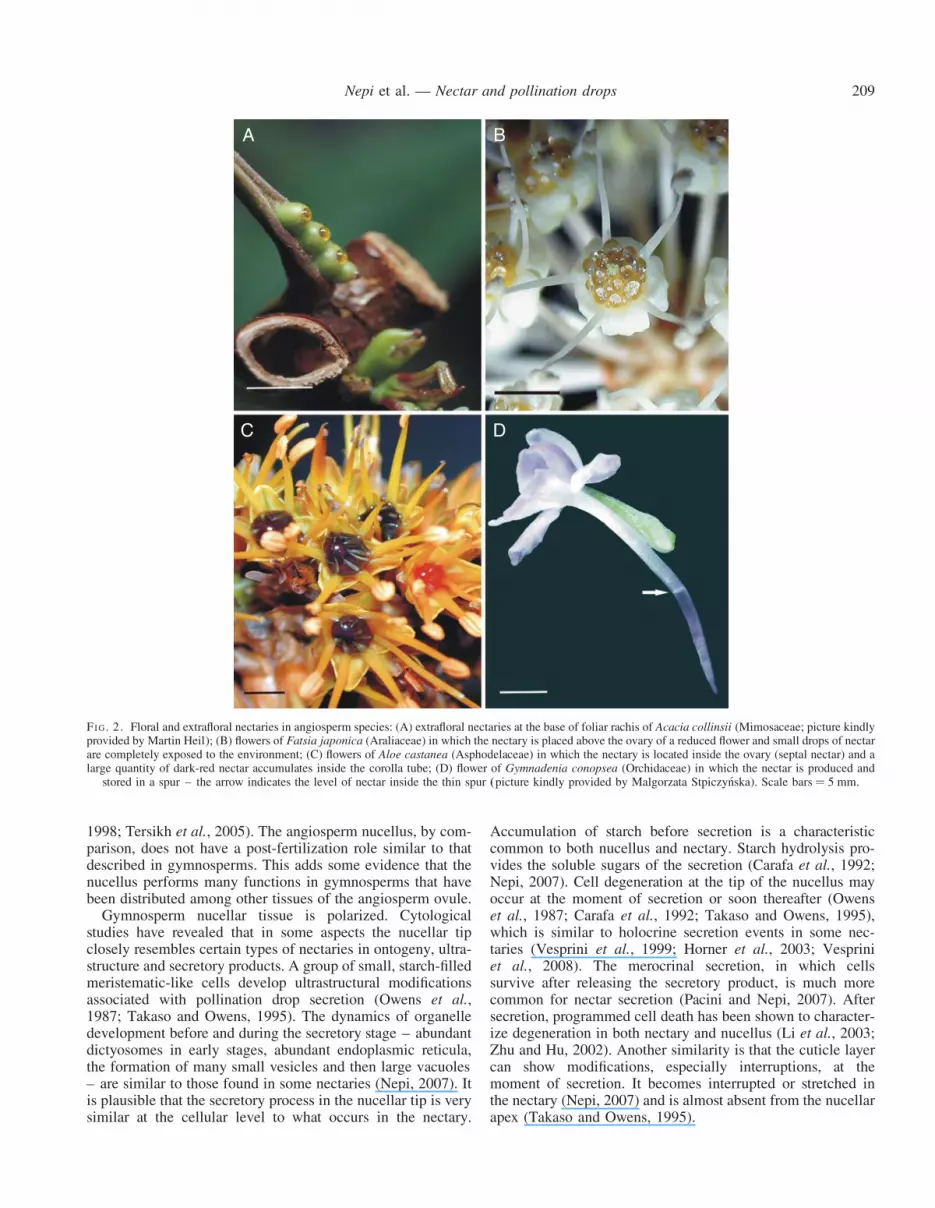
FI G. 2. Floral and extrafloral nectaries in angiosperm species: (A) extrafloral nectaries at the base of foliar rachis of Acacia collinsii (Mimosaceae; picture kindlyprovided by Martin Heil); (B) flowers of Fatsia japonica (Araliaceae) in which the nectary is placed above the ovary of a reduced flower and small drops of nectarare completely exposed to the environment; (C) flowers of Aloe castanea (Asphodelaceae) in which the nectary is located inside the ovary (septal nectar) and alarge quantity of dark-red nectar accumulates inside the corolla tube; (D) flower of Gymnadenia conopsea (Orchidaceae) in which the nectar is produced and
stored in a spur – the arrow indicates the level of nectar inside the thin spur (picture kindly provided by Malgorzata Stpiczynska). Scale bars ¼ 5 mm.
Nepi et al. — Nectar and pollination drops 209

A difference between the two secreting structures concernsthe vascular tissue. In general, the gymnosperm nucellus isnot directly vascularized whereas the functional efficiency ofthe nectary relies on the presence of or proximity to xylemand/or phloem vessels (Nepi, 2007). In spite of such obviousdifferences, a recent systematic survey of floral nectaries byBernardello (2007) considered the nucellar tip of Gnetales tobe ovular, non-structural nectaries according to Schmid’sbroad topographical classification of nectaries (Schmid,1988). However, it seems that the presence of such kinds ofovular nectaries and flower-like reproductive structures in theGnetales does not mean necessarily a phylogenetic relation-ship with angiosperms, as previously stated, according toclassic cladistic analysis based on morphological data(Crane, 1985; Loconte and Stevenson, 1990). More recentmolecular work on the phylogeny of seed plants has groupedthe Gnetales with the conifers, and placed the ancestor ofthe angiosperms among ancient gymnosperms (Chaw et al.,1997; Bowe et al., 2000; Donoghue and Doyle, 2000;Magallon and Sanderson, 2002; Willis and McElwaine, 2002.)
Nectar resorption and pollination drop withdrawal
Plants not only secrete sugary exudates, they can also reab-sorb them. The ability of the plant to reabsorb sugarysecretions previously produced may allow the plant torecoup part of the energy involved in the secretion process.This energy can be used elsewhere in the plant for other func-tions. Nectar resorption has been demonstrated in several butnot all the studied species (Pacini and Nepi, 2007, and refer-ences therein; Nepi and Stpiczynska, 2008). Two principalfunctions can be recognized: the recovery of resources investedin nectar production and a homeostatic mechanism duringnectar secretion and presentation (Nepi and Stpiczynska,2008). The first function can be strongly involved in the regu-lation of the energy balance of the plant. In fact, nectarsecretion is an expensive process; Southwick (1984) estimatedthat from 4 % to 37 % of daily photosynthate assimilated byAsclepias syriaca during blooming was secreted as nectarsugar. He also reported that the energy invested in nectar pro-duction by Medicago sativa is twice the energy invested inseeds. Recovery of resources is therefore an important advan-tage for plants that reuse this source of carbohydrates left overas uncollected nectar. The homeostatic mechanism has animportant ecological function, as it is utilized to maintainnectar characteristics, such as volume and concentration, in arange suitable for the foraging pollinators. The resorbedmaterial can be transferred from the nectary tissue to otherfloral or vegetative parts (Pedersen et al., 1958; Nepi andStpiczynska, 2007). Developing ovaries have been recognizedas the main sink of such resorbed substances, at least inPlatanthera chlorantha (Orchidaceae) and Cucurbita pepo(Cucurbitaceae) (Nepi and Stpiczynska, 2008). The epidermisand the parenchyma of the nectary are mainly involved in theresorption process, although other floral parts (petals andovary) can also play a role. To date, a mechanism for nectarresorption has not been elucidated. The mechanism most prob-ably involves changes in cell turgor that, in turn, respondrapidly to changes in osmolality (Castellanos et al., 2002;Nepi et al., 2007).
Even less is known about pollination drop withdrawal. In thePodocarpaceae, the process is merely physical, attributable topassive evaporation independent of the presence of pollen.However, in most gymnosperms, such as Pinus, Callitris,Chamaecyparis, Cryptomeria, Thuja and Juniperus, withdra-wal seems to be a metabolic process that is triggered by thepresence of pollen that also stops drop secretion (Tomlinsonet al., 1997, and references therein; Gelbart and vonAderkas, 2002; Mugnaini et al., 2007). When triggered bypollen, drop withdrawal is generally very quick, so that aresorption process can be hypothesized. The resorption mech-anism is not understood, nor is it clear which tissue is perform-ing the resorption. It is for these reasons that the term dropwithdrawal is generally used instead of drop resorption. Theactual stimulus for withdrawal–resorption is obscure.Recently, it was pointed out that it may depend on the sizeof the deposited particles. In addition, an unidentified biologi-cal stimulus carried by viable pollen plays a role in theresponse (Mugnaini et al., 2007). It is interesting to notethat, in some cases of particular angiosperm species, nectarresorption is also stimulated by pollination (Koopowitz andMarchant, 1998; Luyt and Johnson, 2002). Regardless of themechanism responsible for the drop withdrawal, the majorfunction of the drop is to transport pollen grains to the nucellussurface. Pollination drop withdrawal is essential in the pollina-tion mechanism of gymnosperms, whereas nectar resorptionappears to be a facultative phenomenon (Table 1) (Burquezand Corbet, 1991; Nepi and Stpiczynska, 2008).
Volume and composition of the secretion
The volume of the secretion per nectary or per pollinationdrop is widely variable but, generally speaking, the volumeof nectar is higher (especially flower nectar) than that of thepollination drop. Pollination drop volume is 60 nL onaverage (Seridi-Benkaddour and Chesnoy, 1988), reaching250 nL in the single-ovuled cone of Taxus baccata(Fig. 1B). For nectar, the range of variability is from about50 nL, as in single florets of Asteraceae (Wist and Davis,2006) to 9.4 mL in Ochroma lagopus (Bombacaceae, bat pol-linated) (Opler, 1983). Variability of volume in extrafloralnectar is in the lower range of floral nectar. The variabilityof floral nectar volume was analysed at the intraspecific leveland can be correlated with the age of the flower, the positionof the flower in the plant or in the inflorescence, environmentalparameters (temperature and relative humidity) and type andbody size of pollinator (Opler, 1983; Pacini and Nepi, 2007).More uncertain and less studied were the reasons for the varia-bility of pollination drop volume. Putatively, volume variabil-ity can be related to the number of ovules per cone,environmental parameters during the period of drop pro-duction, the mean number and dimension of the pollen arriv-ing on the drop, but currently no detailed study has beencarried out.
Nectar and pollination drops have a similar basic chemistry:the main components are sugars, amino acids and proteins(Table 1). Nectar is a complex mixture of a surprisingvariety of substances: sugars, inorganic ions, amino acids, pro-teins, lipids, organic acids, phenolics, alkaloids, terpenoids,etc. (Nicolson and Thornburg, 2007). Generally sweet, its
Nepi et al. — Nectar and pollination drops210

high sugar content was recognized early. The first quotedanalysis of individual sugars appeared in the second half ofthe nineteenth century (see Baker and Baker, 1983a). It wasnot until the middle of the twentieth century that it was rea-lized that many other substances besides sugars can bepresent in nectar. Sugars are the best studied componentbecause there is a relationship between the relative proportionsof the three main sugars found in nectar (sucrose, glucose andfructose) and the type of animal that is attracted (Baker andBaker, 1983b). Nectar amino acids also have similar ecologicalimportance (Baker and Baker, 1986). Sugar and/or amino acidcomposition of nectar has been reported for numerous species.
Ovular secretions in conifers were first described in the lit-erature by Vaucher in (1841). However, the ephemeral natureand small quantity of the secretions has significantly limitedthe ability to study the constituents of the drop. As a result,there have been very few reports on composition. Earlystudies by Fujii (1903) reported the presence of glucose,amino acids, malic acid and calcium in Taxus baccata.Similar results were found in Cupressus funebris by Tison(1911): glucose, calcium and malic acid. Since then, fructose,sucrose, mixed carbohydrate polymers, organic acids, aminoacids and proteins have been identified in the ovular secretionsof a handful of conifer species (Table 2; Gelbart and vonAderkas, 2002). More recently, plant-defence proteins havebeen found in the ovular secretions of a number of gymnos-perms (O’Leary et al., 2007; Wagner et al., 2007; Table 3).
In both nectar and pollination drop it was demonstrated thattheir chemical composition may reflect phylogenetic relation-ships (Nicolson and Thornburg, 2007, and references therein;Wagner et al., 2007), although nectar chemical compositioncan also be driven by adaptation to specific pollinators(Nicolson and Thornburg, 2007).
Sugars
Fructose, glucose and sucrose are the main sugars present inboth the pollination drops and nectar (Table 1). Total sugar con-centration of the pollination drop ranges from 1–2 % in anemo-philous conifers (e.g. Pinus; McWilliam, 1958) to 10–80 % inentomophilious Gnetales (Bino et al., 1984a, b). Sugar contentof nectar varies more or less in the same range (8–80 %)(Nicolson and Thornburg, 2007), but a high variability ofnectar concentration was found in individual angiosperms andis related mainly to flower age and microclimatic effects(Nicolson and Nepi, 2005; Pacini and Nepi, 2007).
Sucrose is the preferred compound for carbon transfer inplants (Akazawa and Okamoto, 1980) and both developingnectary and ovules act as sinks for sucrose. Sucrose is very fre-quently found in angiosperm floral nectar, although it is not ubi-quitous. In an extensive study (765 species) of nectar sugarcomposition, Baker and Baker (1983b) found that sucrose waspresent in the 89 % of the species. Almost all studies of carbo-hydrate composition of gymnosperm pollination drops foundthat sucrose is not the dominant sugar, if it is present at all(see Gelbart and von Aderkas, 2002). There is only one excep-tion: the pollination drop of Ephedra helvetica has high sucroseconcentrations (approx. 25 %; Ziegler, 1959). It is interesting tonote that extrafloral nectars from several Polypodium species(Pteridophyta; Koptur et al., 1982) as well as angiosperm extra-floral nectars are generally sucrose-poor (Koptur, 1994).
Pollination drops are a poor food source for insects for the fol-lowing general reasons: they possess low concentration ofsugars, of which some, such as xylose, are unattractive to polli-nators such as hymenopterans, and those sugars that are attrac-tive, such as sucrose are present in low concentrations, if they arepresent at all (Baker and Baker, 1983b). Xylose was recentlyfound in the pollination drop of Larix and Pseudotsuga
TABLE 2. Non-proteinaceous components of the gymnosperm ovular secretion
Species Compound* Reference
Cephalotaxus drupacea Fructose, glucose Seridi-Benkaddour and Chesnoy, 1988Galacturonic acidPolymer comprised of galactose, arabinose, glucose, rhamnose, mannoseUnidentified phenolic compoundPro, Asp, Glu, Ala, Ser, Leu†, Iso†, Thr†, Gln†, Asp†
Ephedra helvetica Sucrose Ziegler, 1959PhospateGln, Glu, Thr, Ala, Met, Val, Tyr, Gly
Juniperus communis Glucose, fructose, mannitol† Mugnaini et al., 2007Pinus elliottii Fructose, glucose, sucrose McWilliam, 1958Pinus engelmannii Fructose, glucose Owens et al., 1987Pinus nigra Fructose, glucose, sucrose McWilliam, 1958Taxus baccata Fructose, glucose, sucrose Ziegler, 1959; Seridi-Benkaddour and Chesnoy, 1988
Galacturonic acid Seridi-Benkaddour and Chesnoy, 1988Inorganic phosphate, malic acid, citric acid Ziegler, 1959Calcium Fuji, 1903Glu, Pro, Ala, Gln, Asp, Lys, Trp, Val, Leu†, Ser† Ziegler, 1959
Thuja orientalis Fructose Seridi-Benkaddour and Chesnoy, 1988Galacturonic acidSer, Gly, Ala, Glu, Phe†, Tyr†, Leu†, Iso†, Thr†, Asp†
Welwitschia mirabilis Glucose, fructose, galactose† Carafa et al., 1992Uronic acidSer, Tyr, Val
* The dominant sugar is shown in bold font.† Trace amounts.
Nepi et al. — Nectar and pollination drops 211

(M. Nepi et al., unpubl. res.). Presence of xylose in angiospermfloral nectars, though rare, was considered to be a deterrent toinsects and birds (Jackson and Nicolson, 2002).
The dominant sugar that was usually found in the pollina-tion drop was fructose (Table 2). In vitro experiments withPinus mugo pollen indicated that during germination pollentakes up fructose preferentially over other sugars (Nygaard,1977). This implies that the chemical composition of the pol-lination drop is likely to be more suited for pollen nutritionrather than for insect consumption.
Uronic and galacturonic acids as well as xylose can be foundin the pollination drops of some species (Table 2). The pres-ence of these substances is due to breakdown of the nucellustip cells, as these molecules form complex polymeric struc-tural components of the plant cell wall and can be dispersedin the secretion when the cell wall ruptures.
Amino acids
Amino acids have been found in foliar nectar of pterido-phytes, gymnosperm pollination drops, and extrafloral andfloral nectar of angiosperms. Serine, glycine, alanine, arginine
and proline are frequently and abundantly detected in all thesesecretions (Keeler, 1977; Koptur et al., 1982; Baker and Baker,1983a; Pate et al., 1985) (for pollination drop see Table 2). Ofthese, proline seems to have a special importance for insectsfor a variety of reasons: it contributes a taste preferred byinsects (Alm et al., 1990), it stimulates the insect’s salt cell,a labellar chemosensory receptor of insects, resulting inincreased feeding behaviour (Hansen et al., 1998), and it canbe metabolized very rapidly. In honeybees, proline is themost abundant amino acid in the haemolymph and is requiredfor egg laying (Nicolson and Thornburg, 2007, and referencestherein). Oxidative proline degradation is a very efficient,short-term burst of energy that can be utilized during theinitial ‘lift’ phase of insect flight, whereas glucose is usedfor extended flight (Carter et al., 2005).
The accumulation of proline in plant tissue is generallytaken as a sign of stress. Drought, salinity and freezingincrease biosynthesis of proline (Carter et al., 2005).Proline dominates the free amino acid composition – it canbe up to 70 % – of angiosperm pollen free amino acids; it ismuch less abundant in gymnosperm pollen (Stanley andLinskens, 1974; Zhang et al., 1982). Proline is used by the
TABLE 3. Proteinaceous components of the gymnosperm ovular secretion
Species Compound Putative function Reference
Chamaecyparislawsoniana
83-kDa subtilisin-like protein Protein cleavage (amino acid mobilization for pollen tube growth) Wagner et al.,2007
62-kDa b-d-glucan exohydrolase Cell-wall elongation (pollen tube growth)47.5-kDa glucan 1,3-b-glucosidase Antifungal (in conjunction with chitinase), cell wall elongation
(pollen tube growth)Two 25-kDa thaumatin-like proteins Antifungal, antifreeze
Juniperus communis 83-kDa subtilisin-like proteinase Protein cleavage (amino acid mobilization for pollen tube growth) Wagner et al.,2007
62-kDa glycosyl hydrolase Cell-wall elongation (pollen tube growth)47.5-kDa glucan 1,3-b-glucosidaseprecursor
Antifungal (in conjunction with chitinase), cell wall elongation(pollen tube growth)
30-kDa chitinase Antifungal, antifreeze25-kDa thaumatin-like protein Antifungal, antifreeze
Juniperus oxycedrus 30-kDa chitinase Antifungal, antifreeze Wagner et al.,2007
25-kDa thaumatin-like protein Antifungal, antifreeze32.5-kDa glucanase-like protein Cell-wall elongation (pollen tube growth)
Pseudotsuga menziesii 90-kDa xylosidase Cell-wall elongation (pollen tube growth) Poulis et al.,2005
65-kDa xylosidase Cell-wall elongation (pollen tube growth)70-kDa invertase Sucrose cleavage (pollen tube growth)50-kDa invertase Sucrose cleavage (pollen tube growth)45-kDa galactosidase Cell-wall elongation (pollen tube growth)29-kDa galactosidase Cell-wall elongation (pollen tube growth)40-kDa aspartyl protease Protein cleavage (amino acid mobilization for pollen tube growth)37-kDa peroxidase Self-incompatibility, antimicrobial33-kDa serine carboxypeptidase-likeprotein
Protein cleavage (amino acid mobilization for pollen tube growth)
27-kDa chitinase Antifungal, antifreezeTaxus � media 28-kDa acidic thaumatin-like protein Antifungal, antifreeze O’Leary et al.,
200725 kDa basic thaumatin-like protein Antifungal, antifreezeArabinogalactan proteins Pollen tube growth, pollen tube guidance O’Leary et al.,
2004Welwitschia mirabilis Acid phosphatase* Carafa et al.,
199225-kDa chitinase Antifungal, antifreeze Wagner et al.,
2007
* Trace amounts.
Nepi et al. — Nectar and pollination drops212
germinating pollen grains as a substrate or in the synthesis ofhydroxyproline-rich wall proteins (Shivanna, 2003). Only asmall proportion of the proline pool is used in this way; themajor pool is required for other functions (Shivanna, 2003).Uptake experiments demonstrated that mature as well germi-nating pollen rapidly take up proline by means of a specifictransporter (Schwacke et al., 1999). This adds furthersupport to the suggestion that abundance of proline in the pol-lination drop appears to be correlated with its interaction withpollen grains, although this function requires experimentalverification.
Proteins
The existence of proteins in floral nectar was reported longago (Beutler, 1935), but only recently was this shown in gym-nospermous pollination drops (Poulis et al., 2005; O’Learyet al., 2007; Wagner et al., 2007). Although different proteinshave been identified in nectar and pollination drop (Tables 3and 4), similar putative functions can be ascribed to some pro-teins. Two general functional classes of proteins that are foundin both nectar and pollination drops include carbohydrate-metabolizing enzymes and defence proteins.
A group of proteins comprising mainly invertases, transfruc-tosidase and transglucosidase are involved in simple sugarmetabolism. Among them invertases have been detected ingymnosperm pollination drops (Poulis et al., 2005), and infloral and extrafloral nectar (Pate et al., 1985; Heil et al.,2005; Nicolson and Thornburg, 2007). Invertases catalysecleavage of sucrose into fructose and glucose. This activitycan rapidly change the basic sugar composition of thesecretions as well as their osmolarity. It has been shown inextrafloral nectar of Vigna unguiculata inflorescence stalksthat invertase activity was osmotically regulated (Pate et al.,1985).
Another group of proteins is involved in defence againstfungi and bacteria. Sugary solutions should be excellentmedia in which to grow fungi and bacteria, especially asthese solutions are openly exposed to the environment.Plants must defend their sugary secretions, protecting these
solutions for their reproductive purposes, while preventingtheir use as a base for microbes to further attack the plant’sreproductive systems. Glucan 1,3-b-d-glucosidases, chitinasesand thaumatin-like proteins are recognized as plantpathogenesis-related proteins. They have been found recentlyin pollination drops of some gymnosperm species (Table 3)(Wagner et al., 2007). Defence proteins are also known fromfloral nectar (Table 4). In Allium nectar, both alliinase and amannose-binding lectin have antibiotic properties (Peumanset al., 1997).
A nectar redox cycle involving five enzymes first discoveredin ornamental tobacco has been found to be more widespread(Carter et al., 1999, 2007; Carter and Thornburg, 2000, 2004).The nectar redox cycle maintains high levels of hydrogen per-oxide in nectar 40 times the level produced by human neutro-phils in response to microbial attack. The nectar redox cycledefends the metabolic-rich nectar from invading micro-organisms carried to the flower by wind or by non-sterile pol-linators (Thornburg et al., 2003).
Defence proteins have also been found in extrafloralnectar (Gonzalez-Teuber et al., 2009). We can state thatthese two functional groups of proteins (carbohydratemetabolism-related proteins and defence proteins) are likelyto be common to all plant sugar secretions.
A third group of proteins restricted typically to gymnospermpollination drops are probably involved in pollen developmentand germination. These enzymes influence plasticity of the cellwall, cell expansion, and use of specific substrates (Table 3).This group of proteins includes 1,3-b-d-glucosidases (a multi-function protein), glycosyl hydrolases, xylosidases andsubtilisin-like proteinases (Wagner et al., 2007).
The protein profiles recently revealed in Juniperus commu-nis, Juniperus oxycedrus, Chamaecyparis lawsoniana andWelwitschia mirabilis seem to reflect phylogenetic relation-ships. In each species of Juniperus nearly all peptidesequences and identified proteins had an identical match inthe other species. There was an overall similarity in proteinprofile for all the species in the Cupressaceae (J. communis,J. oxycedrus and C. lawsoniana), but to a lesser degree thanspecies within Juniperus. There were no obvious similarities
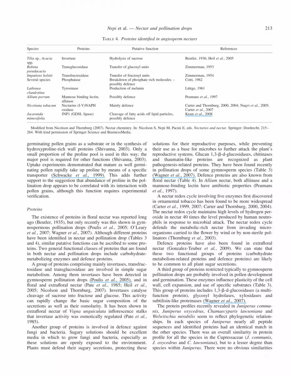
TABLE 4. Proteins identified in angiosperm nectars
Species Proteins Putative function References
Tilia sp., Acaciaspp.
Invertase Hydrolysis of sucrose Beutler, 1936; Heil et al., 2005
Robinapseudacacia
Transglucosidase Transfer of glucosyl units Zimmerman, 1953
Impatiens holstii Transfructosidase Transfer of fructosyl units Zimmerman, 1954Several species Phosphatase Breakdown of phosphate rich molecules –
possibly defenceCotti, 1962
Lathraeaclandestina
Tyrosinase Production of melanin Luttge, 1961
Allium porrum Mannose binding lectin,allinase
Possibly defence Peumans et al., 1997
Nicotiana tabacum Nectarins (I-V)NAPHoxidase
Mainly defence Carter and Thornburg, 2000, 2004; Naqvi et al., 2005;Carter et al., 2007
Jacarandamimosifolia
JNP1 (GDSL lipase) Cleavage of fatty acids off lipid particles,possibly defence
Kram et al., 2008
Modified from Nicolson and Thornburg (2007). Nectar chemistry. In: Nicolson S, Nepi M, Pacini E, eds. Nectaries and nectar. Springer: Dordrecht, 215–264. With kind permission of Springer Science and BusinessMedia.
Nepi et al. — Nectar and pollination drops 213

between W. mirabilis and all the other species studied (Wagneret al., 2007).
Inorganic ions
Pollination drops contain calcium (Fujii, 1903; Tison,1911). Atomic absorption spectroscopy of pollination dropshas shown calcium concentration differences between coniferspecies (K. Gill and P. von Aderkas, unpubl. res.), implyingthat drops vary in this element, one that is critical for pollengermination (Brewbaker and Kwack, 1963). Such differencesbetween species and genera may confer a germination advan-tage to homospecific pollen over heterospecific pollen landingon the same pollination drop.
Very few studies have looked at ion concentrations ofnectar. The most abundant ions detected were Kþ and Naþ,while Ca2þ and Mg2þ were present in lower concentration(Hiebert and Calder, 1983; Heinrich, 1989;Kronestedt-Robards et al., 1989). Findings by Hiebert andCalder (1989) suggested a phylogenetic component to patternsof nectar ion composition. On the other hand, Barclay (2002)demonstrated that nectar ion composition may reflect adap-tation to specific pollinators.
Functions
Nectar is considered to be the most common floral foodresource that is exploitable by the largest variety of animals,in particular animals that can fly. The sugar content is con-sidered to be a ready-to use energy resource that can be usedby feeding animals generally to power their flight (Nicolson,2007). Nectar amino acids, besides contributing to the tasteof nectar, are also an important source of nutrients foranimals, especially for those that are exclusively dependenton nectar for their nutrition, such as butterflies. Insect prefer-ences for single sugars or for particular mixtures of sugars andamino acids are known (Baker and Baker, 1983b; Bluthgenand Fiedler, 2006). Although floral nectar production rep-resents a high cost for the plant (Southwick, 1984; Pyke,1991), it ensures a higher possibility of seed set and thushigher reproductive success and fitness (Neiland andWilcock, 1998).
Extrafloral nectar predates floral nectar by many hundreds ofmillions of years. Extrafloral nectaries are known from .700genera of plants (about 4000 species), including pteridophytes,gymnosperms and angiosperms (see http://www.biosci.unl.edu/emeriti/keeler/extrafloral/worldlistfamilies.htm). In manycases, extrafloral nectaries are a mutualistic response ofplants to ants. Ants are the most common small-sized predatorin the animal kingdom; it would appear that to rid themselvesof herbivores, plants have recruited carnivores. Extrafloral nec-taries are not the only reward – indeed they supply only abiased diet. Myrmecophytes also produce Beltian bodies anddomatia to accommodate the ants, who, in return, providethe plant with a rapid defence (Heil, 2008a). In this way,sugary exudates are part of a tritrophic innate defence, whichmay be composed of a number of elements, including oneor more of the following: (a) herbivore feeding inducesgreater formation of Beltian bodies (food bodies), domatiaand/or extrafloral nectaries, which, in turn, attract herbivore
predators; and (b) herbivore feeding induces release – by theplant – of volatile organic chemicals, which attract predators.The plant therefore plays a significant role in communicatingto carnivores that herbivores are present. For the predators,there is a rich protein diet of herbivores supplemented witheither fat-rich Beltian bodies or sugar-rich extrafloral nectarysecretions. Although studies have clearly shown the advantagethat plants reap in their fitness when they attract ants as protec-tors, other such relationships between insect trophic levels andplants require experimental dissection in the context of fitness.
In the fossil record, extrafloral nectaries appeared longbefore ants (early Cretaceous; Ward, 2007). Thus the extra-floral nectar initially had a function not involving ants.According to De la Barrera and Nobel (2004) nectar may orig-inally have developed independently of any interaction withanimals. Because extrafloral nectaries are frequently associatedwith developing organs (stems or leaves), the ‘leaky phloem’hypothesis of the origin of nectar (De la Barrera and Nobel,2004) can be applied to extrafloral nectaries. According tothis hypothesis, nectar secretion may thus have originated asleakage of phloem solution, resulting from the structural weak-ness of developing tissues exposed to high pressure in thephloem. Interaction of extrafloral nectaries with ants mayhave evolved after the latter’s appearance in the earlyCretaceous (Ward, 2007). Since a symbiotic relationshipwith ants increased plant fitness, acting as an indirectdefence against herbivores, it may have been established andthen reinforced by differentiation of other ant-attracting struc-tures, such as domatia and food bodies.
It is now clear that the classical definition of angiospermfloral nectar as simply a food reward for pollinating or defend-ing animals is outdated. As Pedersen et al. (1958) stated somedecades ago ‘nectar is not a static product remaining outsidethe plant once produced but is in close contact with theplant system’ and the substances present in the nectar can bedynamically exchanged with the nectary itself in response toecological and physiological constraints. This exchange main-tains a relatively constant nectar concentration to ensure insectvisits (nectar homeostasis) and reallocates resources,especially during development of the ovules and ovary afterfertilization (Nepi and Stpiczynska, 2008). Although nectaris much more than a simple reward for visiting insects, thepresence of nectaries, in either reproductive (floral nectar) orvegetative parts of a plant (extrafloral nectar), represents abasic interaction between plants and animals. This is not appli-cable to the pollination drop of most gymnosperms, with thenotable exception of the Gnetales.
Pollination drops have a major function in regulating pollenbehaviour, which is not the case with nectar. Here lies the mostsignificant functional difference. Pollen requires water fromthe female counterpart to germinate. It also requires calciumand carbohydrates, as has been shown in many in vitrostudies (Stanley and Linskens, 1977; Shivanna, 2003). Theindisputable function of the pollination drop is that of alanding site and germination medium for pollen grains(Table 1). The drop’s resorption serves to transport pollengrains inside the ovule, putting them within close proximityto the megagametophyte and its gametes. A number of compo-sitional features of the pollination drop point to its role in nour-ishing pollen. These include the following: its sugar content,
Nepi et al. — Nectar and pollination drops214

which is too low a concentration to benefit insects (with theexception of Gnetales pollination drops); the presence ofcalcium (Fujii, 1903), proline (see Table 2) and specific pro-teins related to pollen germination and tube growth (Wagneret al., 2007; Table 3). Although phylogenetic relationshipscan be recognized, the basic chemical composition of pollina-tion drops is different from species to species. These differ-ences may represent a direct method of homospecific pollenselection. For example, pollen that enters the ovule of itsown species will encounter a favourable environment, bothosmotically and chemically. This environment is very differentin heterospecific ovules (Gelbart and von Aderkas, 2002).
The sucrose composition of pollination drops of gymnos-perms may regulate pollen behaviour. Gnetales have highsucrose concentrations, whereas that of cycads (Tang, 1987)and conifers (McWilliam, 1958) is low. Sucrose concentrationcan affect germination as has been shown in vitro studies ofmany conifers (Dumont-BeBoux et al., 1999, 2000).Furthermore, in some conifers, proteomic profiling hasrevealed the presence of secreted invertases, which arethought to be responsible for the general absence of sucroseand the presence of fructose and glucose in the drop (Pouliset al., 2005). It is postulated that invertases, at least not func-tional ones, are unlikely to be found in the Gnetales.
Interestingly, differences in sucrose concentration betweenGnetales (high sucrose) and cycads (low sucrose) may berelated to differences in the foraging behaviour of pollinators:Gnetales are pollinated by insects that use the pollination dropsas a reward, while cycads are pollinated by insects that do notfeed on pollination drops. High levels of sucrose in sugarysecretions may be maintained in gymnosperms and angios-perms under a selection regime of insect pollination.
Did pollination drops provide a source of nutrition to insectspresent or of the past? In a sweeping review of pollinationdrops and possible insect pollination in gymnosperms duringthe Mesozoic era that immediately preceded angiosperm radi-ation, Labandeira et al. (2007) make a compelling case thatmecopteroids and brachyceran dipterans had siphonate probos-cides type mouthparts adapted to uptake of nectar and sugar-rich pollination drops. The authors contend that the mostplausible food sources would have been the hidden micropylarsecretions of gymnosperms. Two extinct plant species –Frenelopsis alata and Cycadeoidea dacotensis – were usedto illustrate paleobotanical and paleoentomological evidencefor insect consumption of pollen and pollination drops. Theevidence for Mesozoic insect feeding on the former is muchstronger than the latter, which is mainly indirect. With the radi-ation of angiosperms and their associated insects, novel groupssuch as lepidopterans and new types of hymenopteransappeared, and thus, the types of insects that feed on gymnos-perm pollination drops have changed.
FUTURE PERSPECTIVES
Despite a significant body of literature about sugar compo-sition of nectar, there is a notable lack of studies characterizingthe presence of proteins. Although the presence of proteins innectar has been long reported (Beutler, 1935), only threespecies [Allium porrum (Liliaceae), Nicotiana tabacum(Solanaceae) and Jacaranda mimosifolia (Bignoniaceae)]
have been studied closely (Peumans, 1997; Carter et al.,1999, 2000, 2004, 2007; Kram et al., 2008). Nectar proteincharacterization is in its infancy and nectar protein profilingcan be very complex. As an example, .30 proteins havebeen separated by 2D electrophoresis in Cucurbita pepo(Nepi et al., 1996). Several studies have been attempted todemonstrate whether nectar sugar composition is shaped byphylogenetic or ecological constraints (Nicolson, 2007, andreferences therein). Although results are not always consistent,it seems that phylogenetic history is the primary determinantof nectar chemistry, with pollinators having a secondaryeffect (Nicolson, 2007). It appears also that phylogenetic con-straints can be more or less relaxed in different taxonomicalgroups (Kromer et al., 2008).
On the other hand, there is no research that has taken intoconsideration the phylogenetic relationships of nectar proteins.
Our knowledge of pollination drop composition is still rela-tively incomplete for both sugars and proteins. A limitednumber of species have had their sugar composition character-ized (Table 2) and even fewer (seven) species have had theirproteins studied (Table 3).
Proteomic analysis of pollination drops from four species ofgymnosperms recently revealed a phylogenetic pattern(Wagner et al., 2007). Results of protein and sugar analysesare generally difficult to compare because of the limitednumber of species sampled, and the lack of uniform methodsof analysis. These prevent us from drawing any furthergeneral conclusion about phylogenetic relationships in thecomposition of the pollination drops. We can only hypothesizethat since pollination drops are not generally utilized byanimals, the composition must be shaped predominantly byphylogenetic constraints and less by ecological ones.
Defence proteins look to be widespread in carbohydratesecretions of gymnosperms and angiosperms. Chitinases, asa functional class of proteins, seem to be conservativeamong the gymnosperms (Wagner et al., 2007) but nothingis known about their presence in angiosperm nectar as wellas in foliar nectar of pteridophytes. Are some defence proteinsconservative throughout the carbohydrate secretions of vascu-lar plants?
When leaves of plants are attacked by fungi, they mayrespond with apoplastic pH signalling, as has been found inbarley leaves (Felle et al., 2004). Such apoplastic responseshave yet to be studied in nectar of angiosperms or in pollina-tion drops of gymnosperms, as we are unaware of anystudies on the response of these drops to microbial attack. Isthe function of proteins of pollination drops and nectar tocleave oligosaccharides from microbes or fungi, and thatthese, in turn, act as signals to up-regulate defence genes inthe nucellus or in the nectary? Little is known at present.
Another field open to research is the interaction betweenproteins of pollen grains and proteins in the pollination dropsand the nucellus of gymnosperms. Particularly important inthis respect is the report of arabinogalactan proteins in the pol-lination drop and in the micropylar region of the nucellus inTaxus � media (O’Leary et al., 2004). Arabinogalactan pro-teins are proteins expressed at the cell surface and in the inter-cellular spaces and are present throughout the plant kingdom.Arabinogalactan proteins were found abundantly in pistil exu-dates of angiosperm species where they have been recognized
Nepi et al. — Nectar and pollination drops 215

as adhesive, nutritional, protective and chemotropic agentsduring pollen–pistil interactions (Showalter, 2001). Theirdetection in gymnosperm pollination drops and the nucellusreveals biochemical adaptation during pre-zygotic events incommon with angiosperms. How taxonomically widespreadarabinogalactan proteins are in pollination drops and nucellusof gymnosperm is unknown at present.
It is clear that both nectar and pollination drop, althoughbeing simple liquid secretions, hide a biochemical and physio-logical complexity that requires further study for clarification.Results from these studies may reveal new information aboutthe evolution of vascular plants, their phylogenetic relation-ships, their reproduction and animal associates.
CONCLUDING REMARKS
Pollination drops of gymnosperms and the nectar of angios-perms are analogous but not homologous. They are analogousin several aspects: their basic chemical composition (sugars,amino acids, proteins) exhibits similarity, the secretoryprocess involves similar cell biological processes, and bothtypes of secretion are known to be reabsorbed in somespecies. They are not homologous since they are producedby ontogenetically unrelated organs. Pollination drops aresecreted by the nucellus, providing evidence that reproductiveorgans are able to secrete a wide range of proteins, fromcarbohydrate-modifying invertases to pathogenesis-relatedproteins and proteins interacting with pollen development. Inangiosperms, this ability to produce proteins during reproduc-tion did not disappear with the evolutionary loss of the polli-nation drop. From the genetic analysis of Arabidopsis, it isclear that proteins are secreted from the filiform apparatus,e.g. MYB98 (a transcription factor) that regulates geneexpression in cells of the embryo sac, in particular, synergidcells (Punwani et al., 2007). This triggers gene networksresponsible for guiding pollen tubes toward the egg(Franssen-Verhaijen and Willemse, 1993; Fortescue andTurner, 2005). Due to the presence of the gynoecium, thesesecretions are essentially internal.
Nectar and pollination drops share a number of classes ofcompounds. These classes show qualitative and quantitativedifferences in their composition. Sugar composition variesand protein profiles differ. This reflects the different functionsof the two types of secretions. Pollination drops act as landingsites for gymnosperm pollen. Afterwards, the drop provideswater for pollen hydration, carbohydrates and amino acidsduring germination. In contrast, nectar is a food source for pol-linating or defending animals. In addition, pollination dropshave cations, mainly Ca2þ, as well as specific proteins ableto promote pollen development and germination(1,3-b-d-glucosidases, glycosyl hydrolases, xylosidases,subtilisin-like proteinases).
The differences between nectar and pollination drop func-tion are reflected in the different ranges of sugar concen-trations. Pollination drop sugar concentrations range from 5 %to 10 %, whereas nectar sugar concentration is almostwithout exception much higher. Gymnosperm pollen culti-vated in vitro absorbs fructose preferentially; fructose wasalso found to be the more abundant sugar of pollinationdrops in several gymnosperm species (Table 2). The total
sugar concentration is higher in nectar. Of the three mostcommon sugars – glucose, fructose and sucrose – sucrose isthe most common form found in nectar, but least common inpollination drops. The first appearance in the fossil record ofadvanced pollinator groups, including many Hymenopteraand glossate Lepidoptera, dates approximately to 140 millionyears ago, which is very close to the first appearance of angios-perms (Willis and McElwain, 2002). The food preferences ofthese two groups of insects – especially Lepidoptera – forsucrose-rich nectars (Baker and Baker, 1983b) may havedriven the selection of individual plants producing increasedcarbohydrate rewards.
Some chemical components are functionally very similar inboth nectar and pollination drops. Defence proteins show con-vergent evolution: they are necessary for defence of exposedsugary secretions from fungi and microbial attack. Sugar sol-utions are perfect media for growth of fungal and bacterialspores. Microbes can cause profound changes in the physico-chemical properties of nectar and pollination drops, to the det-riment of pollinators and pollen, respectively.
Other components common to these various secretions areenzymes involved in sugar metabolism. Among these, inver-tase is a key enzyme in sugar metabolism. Invertase hydrolysessucrose to form glucose plus fructose. Its presence in nectars(both floral and extrafloral) and pollination drops indicatesthat these secretions may be able to alter their compositionand osmolarity after they are produced. Previously thought tobe static, these secretions may prove to be more dynamic.The presence of these enzymes may also offer a way inwhich osmolarity may be regulated in response to variableenvironmental parameters such as temperature and relativehumidity allowing a certain degree of homeostasis.
There is fossil evidence that some groups of insects able tofeed on sugary secretions, such as pollination drops, alreadyexisted in the Jurassic period (Labandeira et al., 2007; Heil,2008b). These associations are specialized relationships thatpre-date those occurring later in angiosperms (Labandeiraet al., 2007). In the context of these gymnosperm–insectassociations, the presence of proteins in pollination dropsmay be an exaptation (Gould and Vrba, 1982) for co-optinginsects into feeding on these secretions and providing themwith a source of carbon (sugars) and nitrogen (proteins).
To return to the question presented in the title, we can saythat functionally and ontogenetically the nectar and pollinationdrops are very different, but chemically they show strong simi-larities with significant differences. Sugars, although present indifferent concentration and composition, are strictly necessaryfor visiting animals’ diet and for pollen germination. Defenceproteins and carbohydrate metabolism-related proteins protectfrom changes in composition and osmolarity imposed byfactors both biotic (fungi and bacteria) or abiotic (changes inenvironmental parameters such as temperature and relativehumidity).
ACKNOWLEDGEMENTS
We are grateful to Martin Heil (Department of GeneticEngineering, CINVENSTAV—Irapuato, Mexico) andMalgorzata Stpiczynska (Department of Botany, LublinAgricultural University, Lublin, Poland) for providing
Nepi et al. — Nectar and pollination drops216

Fig. 2A and D, respectively. We thank two anonymous refer-ees for the helpful review of the manuscript. Researchrelated to this review was supported by PAR (University ofSiena) and by PRIN (MIUR, Italian Ministry for Universityand Research to authors M.N., S.M. and E.P.) as well as theNational Research Council of Canada (to authors P.v.A.,A.C. and R.W.).
LITERATURE CITED
Akazawa T, Okamoto K. 1980. Biosynthesis and metabolism of sucrose. In:Stumpf PK, Conn EE. eds. The biochemistry of plants, a comprehensivetreatise. New York, NY: Academic Press, 199–220.
Alm J, Ohnmeiss TE, Lanza J, Vriesenga L. 1990. Preference of cabbagewhite butterflies and honey bees for nectar that contains amino acids.Oecologia 84: 53–57.
Baker HG, Baker I. 1983a. A brief historical review of the chemistry of floralnectar. In: Bentley B, Elias T. eds. The biology of nectaries. New York,NY: Columbia University Press, 126–151.
Baker HG, Baker I. 1983b. Floral nectar sugar constituents in relation to pol-linator type. In: Little RJ, Jones CE. eds. Handbook of pollinationbiology. New York, NY: Scientific and Academic Editions, 117–141.
Baker HG, Baker I. 1986. The occurrence and significance of amino acids infloral nectar. Plant Systematics and Evolution 151: 175–186.
Barclay RMR. 2002. Do plants pollinated by flying fox bats (Megachiroptera)provide an extra calcium reward in their nectar? Biotropica: 34: 168–171.
Baum SF, Eshed Y, Bowman JL. 2001. The Arabidopsis nectary is anABC-independent floral structure. Development 128: 4657–4667.
Bernardello G. 2007. A systematic survey of floral nectaries. In: NicolsonSW, Nepi M, Pacini E. eds. Nectaries and nectar. Dordrecht: Springer,19–128.
Beutler R. 1935. Nectar. Bee World 24: 106–162.Bino RJ, Dafni A, Meeuse ADJ. 1984a. Entomophily in the dioecious gym-
nosperm Ephedra aphylla Fork. (¼ E. alte A. Mey.), with some notes onE. campylopoda C.A. Mey. I. Verhandelingen der KoninklijkeNederlandse Akademie van Wetenschappen Amsterdam, Series C 87:1–13.
Bino RJ, Devente N, Meeuse ADJ. 1984b. Entomophily in the dioeciousgymnosperm Ephedra aphylla Fork. (¼ E. alte A. Mey.), with somenotes on E. campylopoda C.A. Mey. II. Pollination droplets, nectaries,and nectarial secretion in Ephedra. Verhandelingen der KoninklijkeNederlandse Akademie van Wetenschappen Amsterdam, Series C 87:15–24.
Bluthgen N, Fiedler K. 2006. Competition for composition: lessons fromnectar-feeding ant communities. Ecology 85: 1479–1485.
Bowe LM, Coat G, de Pamphilis W. 2000. Phylogeny of seed plants on allthree genomic compartments: extant gymnosperms are monophyleticand Gnetales’ closest relatives are conifers. Proccedings of the NationalAcademy of Science of the USA 97: 4092–4097.
Brewbaker JL, Kwack BH. 1963. The essential role of calcium ion in pollengermination and pollen tube growth. American Journal of Botany 50:859–865.
Brown WH. 1938. The bearing of nectaries on the phylogeny of floweringplants. Proceedings of the American Philosophical Society 79: 549–595.
Burquez A, Corbet SA. 1991. Do flowers reabsorb nectar? FunctionalEcology 5: 369–379.
Carafa AM, Carratu G, Pizzolongo P. 1992. Anatomical observations on thenucellar apex of Wellwitschia mirabilis and the chemical composition ofthe micropylar drop. Sexual Plant Reproduction 5: 275–279.
Carter C, Thornburg RW. 2000. Tobacco Nectarin I: purification andcharacterization as a germin-like, manganese superoxide dismutase impli-cated in the defence of floral reproductive tissues. Journal of BiologicalChemistry 275: 36726–36733.
Carter C, Thornburg RW. 2004. Is the nectar redox cycle a floral defenceagainst microbial attack? Trends in Plant Science 9: 320–324.
Carter C, Graham R, Thornburg RW. 1999. Nectarin I is a novel, solublegermin-like protein expressed in the nectar of Nicotiana sp. PlantMolecular Biology 41: 207–216.
Carter C, Shafir S, Yehonatan L, Palmer R, Thornburg RW. 2005. A novelrole for proline in plant floral nectars. Naturwissenschaften 93: 72–79.
Carter C, Healy R, O’Tool NM, et al. 2007. Tobacco nectaries express anovel NADPH oxidase implicated in the defence of floral reproductivetissues against microorganisms. Plant Physiology 143: 389–399.
Castellanos MC, Wilson P, Thomson JD. 2002. Dynamic nectar replenish-ment in flowers of Penstemon (Scrophulariaceae). American Journal ofBotany 89: 111–118.
Chaw S-M, Zharkikh A, Sung W-M, Lau T-C, Li W-H. 1997. Molecularphylogeny of extant gymnosperms and seed plant evolution: analysis ofnuclear 18S rRNA sequences. Molecular Biology and Evolution 14:56–68.
Cooper-Driver GA. 1990. Defence strategies in bracken, Pteridium aquilinum(L.) Kuhn. Annals of the Missouri Botanical Garden 77: 281–286.
Cotti T. 1962. Ueber die quantitative Messung der Phosphataseaktivitaet inNektarien. Berichte der Schweizerischen Botanischen Gesellschaft 72:306–331.
Crane PR. 1985. Phylogenetic analysis of seed plants and the origin ofangiosperms. Annals of Missouri Botanical Gardens 72: 716–793.
Crepet WL. 1974. Investigations of North American cycadeoids: the repro-ductive biology of Cycadeoidea. Paleontographica 148B: 144–169.
Crepet WL. 1983. The role of insect pollination in the evolution of the angios-perms. In: Real L. ed. Pollination biology. New York, NY: AcademicPress, 29–50.
Crepet WL, Friis EM. 1987. The evolution of insect pollination mechanismsin angiosperms. In: Friis EM, Chaloner WG, Crane PR. eds. The origin ofangiosperms and their biological consequences. Cambridge: CambridgeUniversity Press, 181–201.
De la Barrera E, Nobel PS. 2004. Nectar: properties, floral aspects, andspeculations on origin. Trends in Plant Science 9: 65–69.
Donoghue MJ, Doyle JA. 2000. Seed plant phylogeny: demise of the antho-phyte hypothesis? Current Biology 10: R106–R109.
Doyle J. 1945. Developmental lines in pollination mechanisms in theConiferales. Scientific Proceedings of the Royal Dublin Society 24:43–62.
Dumont-BeBoux N, Anholt B, von Aderkas P. 1999. In vitro Douglas-firpollen germination. Annals of Forest Science 99: 11–18.
Dumont-BeBoux N, Anholt B, von Aderkas P. 2000. In vitro germination ofwestern larch pollen. Canadian Journal of Forest Research 30: 329–332.
Endress P.K. 1994. Floral structure and evolution of primitive angiosperms:recent advances. Plant Systematics and Evolution 192: 79–97.
Faegri K, van der Pijl L. 1979. The principles of pollination biology, 3rd rev.edn. Oxford: Pergamon Press.
Fahn A. 1979. Secretory tissues in plants. London: Academic Press.Felle HH, Herrmann A, Hanstein S, Huckelhoven R, Kogel K-H. 2004.
Apoplastic pH signalling in barley leaves attacked by the powderymildew fungus Blumeria graminis f. sp. hordei. Molecular Plant–Microbe Interactions 17: 118–123.
Fortescue JA, Turner DW. 2005. The occurrence of a micropylar exudate inMusa and Ensete (Musaceae). Scientia Horticulturae 104: 445–461.
Franssen-Verheijen MAW, Willemse MTM. 1993. Micropylar exudate inGasteria (Aloaceae) and its possible function in pollen tube growth.American Journal of Botany 80: 253–262.
Friis EM, Endress PK. 1990. Origin and evolution of angiosperm flowers.Advances in Botanical Research 17: 99–162.
Fujii K. 1903. Uber die Besaubungstropfen der Gymnospermen. Berichte derDeutschen Botanischen Gesellschaft 21: 211–217.
Gelbart G, von Aderkas P. 2002. Ovular secretions as part of pollinationmechanisms in conifers. Annals of Forest Science 59: 345–357.
Gould SJ, Vrba ES. 1982. Exaptation – a missing term in the science ofform. Paleobiology 8: 4–15.
Gonzalez-Teuber M, Eilmus S, Muck A, Svatos A, Heil M. 2009.Pathogenesis-related proteins protect extrafloral nectar from microbialinfestation. Plant Journal 58: 464–473.
Hansen K, Wacht S, Seebauer H, Schnuch M. 1998. New aspects of che-moreception in flies. Annals of the New York Academy of Sciences 855:143–147.
Heads PA, Lawton JH. 1985. Bracken, ants and extrafloral nectaries. III. Howinsect herbivores avoid ant predation. Ecological Entomology 10: 29–42.
Heil M. 2008a. Indirect defence – recent developments and open questions.In: Luttke U, Beyschlag W, Murata J. eds. Progress in botany, Vol. 69.Berlin: Springer, 360–395.
Heil M. 2008b. Indirect defence via tritrophic interactions. New Phytologist78: 41–61.
Nepi et al. — Nectar and pollination drops 217

Heil M, Rattke J, Boland W. 2005. Postsecretory hydrolysis of nectar sucroseand specialization in ant/plant mutualism. Science 308: 560–563.
Heinrich G. 1989. Analysis of cations in nectars by means of a laser microp-robe mass analyser (LAMMA). Beitrage zur Biologie der Pflanzen 64:293–308.
Heslop-Harrison J, Shivanna KR. 1977. The receptive surface of the angios-perm stigma. Annals of Botany 50: 831–842.
Hiebert SM, Calder WA. 1983. Sodium, potassium, and chloride in floralnectars: energy-free contributions to refractive index and salt balance.Ecology 64: 399–440.
Horner HT, Healy RA, Cervantes-Martinez T, Palmer R. 2003. Floralnectary fine structure and development in Glycine max L. (Fabaceae).International Journal of Plant Sciences 164: 675–690.
Jackson S, Nicolson SW. 2002. Xylose as nectar sugar: from biochemistry toecology. Comparative Biochemistry and Physiology 131: 613–620.
Kato M, Inoue T, Nagamitsu T. 1995. Pollination biology of Gnetum(Gnetaceae) in a lowland mixed dipterocarp forest in Sarawak.American Journal of Botany 82: 862–868.
Keeler KH. 1977. The extrafloral nectaries of Ipomoea carnea(Convolvulaceae). American Journal of Botany 64: 1282–1288.
Koopowitz H, Marchant TA. 1998. Postpollination nectar reabsorption in theAfrican epiphyte Aerangis verdickii (Orchidaceae). American Journal ofBotany 85: 508–512.
Koptur S. 1994. Floral and extrafloral nectars of Costa Rican Inga trees: acomparison of their constituents and composition. Biotropica 26:276–284.
Koptur S, Smith AR, Baker I. 1982. Nectaries in some neotropical species ofPolypodium (Polypodiaceae): preliminary observations and analysis.Biotropica 14: 108–113.
Kram B, Bainbridge E, Perera M, Carter C. 2008. Identification, cloningand characterization of a GDSL lipase secreted into the nectar ofJacaranda mimosifolia. Plant Molecular Biology 68: 173–183.
Kromer T, Kessler M, Lohaus G, Schmidt-Lebuhn AN. 2008. Nectar sugarcomposition and concentration in relation to pollination syndromes inBromeliaceae. Plant Biology 10: 502–511.
Kronestedt-Robards EC, Geeger M, Robards AW. 1989. The nectar ofStrelitzia reginae flowers. Physiologia Plantarum 77: 341–346.
Labandeira CC, Kvacek J, Mostovski MB. 2007. Pollination drops, pollen,and insect pollination in Mesozoic gymnosperms. Taxon 56: 663–695.
Li D-H, Yang X, Cui K-M, Li Z-L. 2003. Morphological changes in nucellarcells undergoing programmed cell death (PCD) during pollen chamberformation in Ginkgo biloba. Acta Botanica Sinica 45: 53–63.
Lloyd DG, Wells MS. 1992. Reproductive biology of a primitive angiosperm,Pseudowintera colorata (Winteraceae), and the evolution of pollinationsystems in the Anthophyta. Plant Systematics and Evolution 181: 77–95.
Loconte H, Stevenson DW. 1990. Cladistics of the Spermatophyta. Brittonia42: 197–211.
Luttge U. 1961. Uber die Zusammensetzung des Nektars und denMechanismus seiner Sekretion. I. Planta 56: 189–212.
Luyt R, Johnson SD. 2002. Postpollination nectar reabsorption and its impli-cations for fruit quality in an epiphytic orchid. Biotropica 34: 442–446.
McWilliam JR. 1958. The role of the micropyle in the pollination of Pinus.Botanical Gazette 120: 109–117.
Magallon S, Sanderson MJ. 2002. Relationships among seed plants inferredfrom highly conserved genes: sorting conflicting phylogenetic signalsamong ancient lineages. American Journal of Botany 89: 1991–2006.
Meeuse ADJ. 1978. Nectarial secretion, floral evolution, and the pollinationsyndrome in early angiosperm. Verhandelingen der KoninklijkeNederlandse Akademie van Wetenschappen Amsterdam, Series C 81:300–326.
Mugnaini S. 2007. The pollination mechanism in Juniperus communis andJ. oxycedrus: physiological and ecological aspects. PhD thesis,University of Siena, Italy.
Mugnaini S, Nepi M, Guarnieri M, Piotto B, Pacini E. 2007. Pollinationdrop in Juniperus communis: response to deposited material. Annals ofBotany 100: 1475–1481.
Naqvi S, Harper A, Carter C, et al. 2005. Tobacco Nectarin IV is a specificinhibitor of fungal xylosidases secreted into the nectar of ornamentaltobacco plants. Plant Physiology 139: 1389–1400.
Neiland MRM, Wilcock CC. 1998. Fruit set, nectar reward, and rarity in theOrchidaceae. American Journal of Botany 85: 1657–1671.
Nepi M. 2007. Nectary structure and ultrastructure. In: Nicolson S, Nepi M,Pacini E. eds. Nectaries and nectar. Dordrecht: Springer, 129–166.
Nepi M, Stpiczynska M. 2007. Nectar resorption and translocation inCucurbita pepo L. and Platanthera chlorantha Custer (Rchb.). PlantBiology 9: 93–100.
Nepi M, Stpiczynska M. 2008. The complexity of nectar: secretion andresorption dynamically regulate nectar features. Naturwissenschaften95: 177–184.
Nepi M, Pacini E, Willemse MTM. 1996. Nectary biology of Cucurbitapepo: ecophysiological aspects. Acta Botanica Neerlandica 45: 41–54.
Nepi M, Guarnieri M, Artese D, Cresti L, Pacini E, Stpiczynska M. 2007.Dynamics of nectar: new insights from Cucurbita pepo flowers. In:Gardner CAC, Harris MA, Hellmich RW, et al. eds. 9th InternationalPollination Symposium on Plant–Pollinator Relationships. Ames: IowaState University, 34–35.
Nicolson SW. 2007. Nectar consumers. In: Nicolson S, Nepi M, Pacini E eds.Nectaries and nectar. Dordrecht: Springer, 289–342.
Nicolson SW, Nepi M. 2005. Dilute nectar in dry atmosphere: nectar secretionpatterns in Aloe castanea (Asphodelaceae). International Journal of PlantSciences 166: 227–233.
Nicolson SW, Thornburg RW. 2007. Nectar chemistry. In: Nicolson S, NepiM, Pacini E eds. Nectaries and nectar. Dordrecht: Springer, 215–264.
Nygaard P. 1977. Utilization of exogenous carbohydrates for tube growth andstarch synthesis in pine pollen suspension cultures. PhysiologiaPlantarum 39: 206–210.
O’Leary SJ, Joseph C, von Aderkas P. 2004. Origin of arabinogalactan pro-teins in the pollination drop of Taxus � media. Austrian Journal of ForestScience 121: 35–46.
O’Leary SJB, Poulis BAD, von Aderkas P. 2007. The identification of twothaumatin-like proteins (TLPs) in the pollination drop of hybrid yew thatmay play a role in pathogen defence during pollen collection. TreePhysiology 27: 1649–1659.
Opler PA. 1983. Nectar production in tropical ecosystem. In: Bentley B, EliasT. eds. The biology of nectaries. New York, NY, Columbia UniversityPress, 30–79.
Owens JN, Simpson SJ, Caron GE. 1987. The pollination mechanism ofEngelmann spruce (Picea Engelmannii). Canadian Journal of Botany65: 1439–1450.
Pacini E, Nepi M, Vesprini JL. 2003. Nectary biodiversity: a short review.Plant Systematics and Evolution 238: 7–21.
Pacini E, Nepi M. 2007. Nectar production and presentation. In: Nicolson S,Nepi M, Pacini E. eds. Nectaries and nectar. Dordrecht: Springer,167–214.
Pacini E, Viegi L, Franchi GG. 2008. Types, evolution and significance ofplant–animal interactions. Rendiconti Licei 19: 75–101.
Pate JS, Peoples MB, Storer PJ, Atkins CA. 1985. The extrafloral nectariesof cowpea (Vigna unguiculata (L.) Walp.). II. Nectar composition, originof solutes, and nectary functioning. Planta 166: 28–38.
Pedersen MW, LeFevre CW, Wiebe HH. 1958. Absorption of C14-labelledsucrose by alfalfa nectaries. Science 127: 758–759.
Peumans WJ, Smeets K, Van Nerum K, Van Leuven E, Van Damme EJM.1997. Lectin and alliinase are the predominant proteins in nectar fromleek (Allium porrum L.) flowers. Planta 201: 298–302.
Poulis BAD, O’Leary SJB, Haddow JD, von Aderkas P. 2005. Identificationof proteins present in the Douglas fir ovular secretion: an insight intoconifer pollen selection and development. International Journal ofPlant Sciences 166:733–739.
Punwani JA, Rabiger DS, Drews GN. 2007. MYB98 positively regulates abattery of synergid-expressed genes encoding filiform apparatus-localizedproteins. The Plant Cell 19: 2557–2568.
Pyke GH. 1991. What does it cost a plant to produce floral nectar? Nature 350:58–59.
Rothwell G. 1977. Evidence for a pollination-drop mechanism in Paleozoicpteridosperms. Science 198: 1251–1252.
Schmid R. 1988. Reproductive versus extra-reproductive nectaries-historicalperspective and terminological recommendations. Botanical Review 54:179–232.
Schwacke R, Grallath S, Breitkreutz KE, et al. 1999. LeProT1, a transporterfor proline, glycine, betaine, and g-amino butyric acid in tomato pollen.The Plant Cell 11: 377–391.
Seridi-Benkaddour R, Chesnoy L. 1988. Secretion and composition of thepollination drop of Cephalotaxus drupacea (Gymnosperm,Cephalotaxaceae). In: Cresti M, Gori P, Pacini E. eds. Sexual reproduc-tion in higher plants. Berlin: Springer Verlag, 345–350.
Nepi et al. — Nectar and pollination drops218
Shivanna KR. 2003. Pollen biology and biotechnology. Enfield, NH: SciencePublishers.
Showalter AM. 2001. Arabinogalactan-proteins: structure, expression andfunction. Cellular and Molecular Life Science 58: 1399–1417.
Singh H. 1978. Embryology of gymnosperms. Stuttgart: Gebruder Borntraeger.Smets EF, Cresens EM. 1988. Types of floral nectaries and the concept of
‘character’ and ‘character state’ – a reconsideration. Acta BotanicaNeerlandica 37: 121–128.
Southwick EE. 1984. Photosynthate allocation to floral nectar: a neglectedenergy investment. Ecology 65: 1775–1779.
Stanley RG, Linskens HF. 1974. Pollen – biology biochemistry management.New York, NY: Springer.
Takaso T, Owens JN. 1995. Pollination drop and microdrop secretions inCedrus. International Journal of Plant Sciences 156: 640–649.
Tang W. 1987. Insect pollination in the cycad Zamia pumila (Zamiaceae).American Journal of Botany 74: 90–99.
Taylor TN, Millay MA. 1979. Pollination biology and reproduction in earlyseed plants. Review of Paleobotany and Palynology 27: 329–358.
Tempel AS. 1983. Bracken fern (Pteridium aquilinum) and nectar-feedingants: a non-mutualistic interaction. Ecology 64: 1411–1422.
Tersikh VV, Ferutado JA, Ren C, Abrams SR, Kermode AR. 2005. Wateruptake and oil distribution during imbibition of seeds of western whitepine (Pinus monticola Dougl. ex D. Don) monitored in vivo using mag-netic resonance imaging. Planta 221: 17–27.
Thornburg RW. 2007. Molecular biology of the Nicotiana floral nectary. In:Nicolson S, Nepi M, Pacini E. eds. Nectaries and nectar. Dordrecht:Springer, 265–288.
Thornburg RW, Carter C, Powell A, Rizhsky L, Mittler R, Horner HT.2003. A major function of the tobacco floral nectary is defence againstmicrobial attack. Plant Systematics and Evolution 238: 211–218.
Tillmann-Sutela E, Kauppi A. 1995a. The morphological background toimbibition in seeds of Pinus sylvestris L. of different provenances.Trees 9: 123–133.
Tillmann-Sutela E, Kauppi A. 1995b. The significance of structure for imbi-bition in seeds of Norway spruce, Picea abies (L.) Karst. Trees 9:269–278.
Tillmann-Sutela E, Kauppi A. 1998. Structures restricting passage of waterin the mature seeds of yellow-cedar (Chamaecyparis nootkatensis).Canadian Journal of Botany 76: 1458–1466.
Tison A. 1911. Remarques sur les gouttelettes collectrices des ovules des con-iferes. Memoires de la Societe Linneene de Normandie 23: 51–64.
Tomlinson PB, Braggins JE, Rattenbury JA. 1997. Contrasted pollencapture mechanisms in Phyllocladaceae and certain Podocarpaceae(Coniferales). American Journal of Botany 84: 214–223.
Tryon AF. 1985. Spores of myrmecophytic ferns. Proceedings of the RoyalSociety of Edinburgh 86B: 105–110.
Vaucher JP. 1841. Histoire physiologique des plantes d’Europe, Vol. 4. Paris:Marc Aurel Freres.
Vesprini JL, Nepi M, Pacini E. 1999. Nectary structure, nectar secretion pat-terns and nectar composition in two Helleborus species. Plant Biology 1:560–568.
Vesprini JL, Nepi M, Ciampolini F, Pacini E. 2008. Holocrine secretion andcytoplasmic content of Helleborus foetidus L. (Ranunculaceae). PlantBiology 10: 268–271.
Wackers F. 2002. It pays to be sweet: sugars in mutualistic interactions.Biologist 49: 165–169.
Wagner R, Mugnaini S, Sniezko R, et al. 2007. Proteomic evaluation ofgymnosperm pollination drop proteins indicates highly conserved andcomplex biological functions. Sexual Plant Reproduction 20: 181–189.
Walker TG. 1985. Spore filaments in the ant-fern Lecanopteris mirabilis – analternative viewpoint. Proceedings of the Royal Society of Edinburgh86B: 111–114.
Ward PS. 2007. Phylogeny, classification, and species-level taxonomy of ants(Hymenoptera: Formicidae). Zootaxa 1668: 549–563.
Wetsching W, Depisch B. 1999. Pollination biology of Welwitschia mirabilisHook. Phyton 39: 167–183.
Willis KJ, McElwain JC. 2002. The evolution of plants. Oxford: OxfordUniversity Press.
Wist TJ, Davis AR. 2006. Floral nectar production and nectary anatomy andultrastructure of Echinacea purpurea (Asteraceae). Annals of Botany 97:177–193.
Zhang H, Croes A, Linskens H. 1982. Protein synthesis in germinating pollenof Petunia: the role of proline. Planta 154: 199–203.
Zhu J, Hu ZH. 2002. Cytological studies on the development of sieve elementand floral nectary tissue in Arabidopsis thaliana. Acta Botanica Sinica 44:9–14.
Ziegler H. 1959. Uber die zusammensetzung des ‘bestaubungstropfens’ undmechanismus seiner secretion. Planta 52: 587–599.
Zimmerman JG. 1954. Uber die Sekretion saccharosespaltendertrans-Glucosidasen in pflanzlichem Nektar. Experientia 9: 145–149.
Zimmerman M. 1953. Papierchromatographische Untersuchungen uber diepflanzliche Zuckersekretion. Berichte der Schweizerischen BotanischenGesellschaft 63: 402–429.
Nepi et al. — Nectar and pollination drops 219


