Embed Size (px)
Citation preview

MOLECULAR AND CELLULAR BIOLOGY, Sept. 2007, p. 6093–6102 Vol. 27, No. 170270-7306/07/$08.00�0 doi:10.1128/MCB.00789-07Copyright © 2007, American Society for Microbiology. All Rights Reserved.
Nap1l2 Promotes Histone Acetylation Activity duringNeuronal Differentiation�
Mikael Attia,1 Christophe Rachez,2 Antoine De Pauw,1† Philip Avner,1 and Ute Christine Rogner1*Unite de Genetique Moleculaire Murine1 and Unite Postulante de Regulation Epigenetique,2 CNRS URA 2578,
Institut Pasteur, 25 rue du Docteur Roux, 75724 Paris Cedex 15, France
Received 4 May 2007/Returned for modification 11 June 2007/Accepted 13 June 2007
The deletion of the neuronal Nap1l2 (nucleosome assembly protein 1-like 2) gene in mice causes neural tubedefects. We demonstrate here that this phenotype correlates with deficiencies in differentiation and increasedmaintenance of the neural stem cell stage. Nap1l2 associates with chromatin and interacts with histones H3and H4. Loss of Nap1l2 results in decreased histone acetylation activity, leading to transcriptional changes indifferentiating neurons, which include the marked downregulation of the Cdkn1c (cyclin-dependent kinaseinhibitor 1c) gene. Cdkn1c expression normally increases during neuronal differentiation, and this correlateswith the specific recruitment of the Nap1l2 protein and an increase in acetylated histone H3K9/14 at the siteof Cdkn1c transcription. These results lead us to suggest that the Nap1l2 protein plays an important role inregulating transcription in developing neurons via the control of histone acetylation. Our data support the ideathat neuronal nucleosome assembly proteins mediate cell-type-specific mechanisms of establishment/modifi-cation of a chromatin-permissive state that can affect neurogenesis and neuronal survival.
Mammals possess three neuron-specific nucleosome assem-bly protein (NAP)-encoding genes, NAP1L2 (29), NAP1L3(39), and NAP1L5 (37), all of which are both intronless andmonoallelically expressed. While the role of the ubiquitouslyexpressed NAPs, NAP1 (NAP1L1) (15, 17, 19, 25, 35) andNAP2 (NAP1L4) (14, 26), in the assembly of nucleosomes andtransport of histones has been extensively characterized, littleis known about the role and functioning of neuron-specificNAP1-like proteins.
We previously showed that targeted deletion of the murineneuron-specific Nap1l2 (nucleosome assembly protein 1-like 2)gene leads to embryonic lethality from mid-gestation phaseonwards, with surviving mutant chimeric embryos showing ex-tensive surface ectoderm defects and open neural tubes similarto those observed in humans with spina bifida and anenceph-aly. These developmental defects were attributed to the over-proliferation of neural precursor cells that is thought to beassociated with the absence of Nap1l2 activity (27). Thepresent study aimed to understand the cellular and molecularmechanisms underlying this knockout phenotype.
Here we show by ex vivo differentiation studies of embryonicstem (ES) cells from which Nap1l2 was deleted that Nap1l2regulates the kinetics of neuronal differentiation. In the ab-sence of Nap1l2, neural precursors exhibit a diminished capac-ity for differentiation, an increase in proliferation, and in-creased levels of apoptosis. These effects of Nap1l2 deletionare associated with a global decrease in cellular levels of his-tone acetyltransferase (HAT) activity. This finding is sup-ported by observations that the highly acidic Nap1l2 protein
colocalizes to the chromatin in the neuronal nucleus, binds tohistones H3 and H4 in vitro, and increases HAT activity. Lossof Nap1l2 results in extensive changes in the transcriptionalprofile of neural precursor cells, with genes such as Cdkn1c,involved in neuronal differentiation, being affected. Not onlycan Nap1l2 be recovered in association with the transcriptionstart site of Cdkn1c but also the deletion of Nap1l2 reduceshistone H3 acetylation at the Cdkn1c promoter. Our datasuggest strongly that Nap1l2 is implicated in the epigeneticregulation of gene expression occurring during neuronal dif-ferentiation and provide novel insights into the functions oftissue-specific members of the NAP family.
MATERIALS AND METHODS
Knockout construction. The 46C ES cell line was kindly provided by AustinSmith (43). A 10-kb genomic DNA fragment containing the entire Nap1l2 genewas cloned into pBluescript SK(�) (Stratagene) by using the restriction enzymesNotI and XhoI (27). A herpes simplex virus thymidine kinase cassette wasinserted into the XhoI site, and a loxP site was inserted into the PmlI site 5� ofthe Nap1l2 promoter. A hygromycin resistance gene flanked by two loxP sites wasinserted into the PacI site 3� of the Nap1l2 gene, which is located 5� and outsideof the last exon of the Ppnx gene (9). The construct was then inserted into the46C ES cell line by homologous recombination. The hygromycin resistancecassette, alone or together with the Nap1l2 gene, was removed by transienttransfection with the pCre-Pac plasmid (38). The correct integration of theconstruct and loxP sites and absence of the Cre-expressing plasmid in ES cellclones were verified by PCR and Southern blotting. Unless otherwise stated,three independent 46C�Nap1l2 clones, termed C8, B2, and E2, were comparedto the 46CloxPNap1l2loxP clone E1 for phenotypic analysis. The clones withNap1l2 deleted and the Nap1l2-floxed clones were used at similar cell passagenumbers and were differentiated in parallel.
ES cell culture and differentiation into neurons. N2B27 medium was preparedas originally described (43). Differentiation was initiated in bacterial dishes, with5 � 106 cells per B10 petri dish. Starting with embryoid body formation insuspension culture, 8 to 12 days of culture were required to enrich the culturesin 60% to 70% of neural stem cells. These cultures could be further enriched (upto 90%) by selection with 1 �g puromycin/ml for at least 4 days. Embryoid bodieswere dissociated at the point of maximal expression of Sox1-green fluorescentprotein (GFP) using Accumax (PAA Laboratories) and 5 � 106 neural precursorcells attached to poly-D-lysine (13.3 �g/ml phosphate-buffered saline [PBS] for1 h)-pretreated 25-cm2 cell culture dishes. When required, 20 �g/ml AraC
* Corresponding author. Mailing address: Unite Genetique Molecu-laire Murine, CNRS URA 2578, Institut Pasteur, 25 rue du DocteurRoux, 75724 Paris Cedex 15, France. Phone: 33145688602. Fax:33145688656. E-mail: [email protected].
† Present address: Genetique Oncologique, Institut Curie, 26, rued’Ulm, 75248 Paris Cedex 5, France.
� Published ahead of print on 25 June 2007.
6093
on February 11, 2016 by guest
http://mcb.asm
.org/D
ownloaded from

(Sigma) was added for 48 h 5 days after attachment of the cells to eliminateproliferative cells from the cultures. The differentiated cells were almost exclu-sively neuronal with only a few astrocytes (glial fibrillary acidic protein [GFAP]-positive) detectable 5 days after attachment of the cultures. Neurons could bemaintained for at least 2 weeks after attachment. In some experiments, untreatedembryoid bodies were maintained in suspension culture for about 3 weeks. Whenappropriate, neural stem cells were treated with 100 ng/ml trichostatin A (TSA)for 24 h. The increase in HAT activity was monitored by Western blot analysisusing an anti-acetyl histone H3 antibody.
Immunofluorescence. Subconfluent cells (70 to 80%) were grown directly on9-cm2 slide flasks (Nunclon). Cells were fixed using 2% paraformaldehyde in PBSfor 20 min at room temperature (RT), then treated with 0.1% Triton X-100 inPBS for 2 min, and rinsed once with PBS. First and secondary antibodies werediluted 1:200 to 1:400 in PBS and incubated for 1 h at RT. Three 5-min washeswith PBS were carried out after each incubation step. The cells were embeddedin Vectashield with DAPI (4�,6�-diamidino-2-phenylindole; Vector) and visual-ized using a fluorescence microscope (Zeiss) equipped with SmartCapture soft-ware (Vysis).
Fluorescence-activated cell sorter (FACS) analyses were performed using aFACSscan (BD Bioscience). Percentages of GFP-positive cells were determinedafter acquisition of 10,000 live cells using the Cell Quest 3.3 software (BDBioscience). Cell tracer labeling was done using CellTrace Far Red DDAO-SE(Molecular Probes) at 25 �M in PBS–1% bovine serum albumin (BSA) for 10min at 37°C. The reaction was stopped by the adding a threefold excess ofice-cold medium containing 20% fetal calf serum, and the cells were washed withPBS before FACS acquisition. The analysis was carried out on 10,000 Sox1-GFP-positive cells. DNA content analysis was carried out on 2 � 106 puromycin-selected neural precursor cells. Single-cell suspensions were incubated overnightat 4°C in 1 ml of 0.1% trisodium citrate dihydrate containing 0.1% Triton X-100,100 �g/ml RNase, and 25 �g/ml propidium iodide (Sigma). FACS acquisitionand analysis were performed on 30,000 live cells. Apoptosis assays were carriedout using the annexin V-PE apoptosis detection kit I (BD Bioscience) accordingto the manufacturer’s instructions. The FACS analysis was carried out on 10,000cells.
RNA preparation and cDNA synthesis. Total RNA was prepared by usingRNABle (Eurobio) and RNA quality examined using an Agilent 2100 bioana-lyzer (Agilent). Random cDNA synthesis was carried out on 10 �g total RNA byusing Moloney murine leukemia virus reverse transcriptase (Invitrogen) accord-ing to the manufacturer’s conditions.
Quantitative PCR was performed using an ABIPRISM 7700 sequence detec-tor and SYBR green PCR master mix (PE Biosystems) according to the manu-facturer’s conditions. Primers were designed using PrimerExpress software andused at optimal concentrations. Quantification of the amplification product wasperformed using the comparative threshold cycle method and the Arpo (acidicribosomal phosphoprotein PO) gene as the gene expression reporter. We foundthat Arpo expression was stable during neuronal ex vivo differentiation andindependent of Nap1l2 expression. Sequences of the oligonucleotides used wereas follows: for Nap1l2, 5�CAGACCGTCCAAAAGGACTTA3�(forward) and5�AGTAAGGGTTGGTACATTTCA3�(reverse); for Oct4, 5�CTCACCCTGGGCGTTCTCT3�(forward) and 5�GGCCGCAGCTTACACATGTT3�(reverse);for Nestin, 5�CTCCTGTGACAGCCTTTCTGAAG3� (forward) and 5�AGGAT-AGGGAGCCTCAGACATAGG3� (reverse); for the ß-III-tubulin gene, 5�CCCCATTTTAGCCACCTCTGT3�(forward) and 5�TACCCCTCCCCCCGAATAA3�(reverse); for Cdkn1c, 5�AGAACCGCTGGGACTTCAACT3�(forward) and5�GTAGAAGGCGGGCACAGACT3�(reverse); and for Arpo, 5�TCCAGAGGCACCATTGAAATT3�(forward) and 5�TCGCTGGCTCCCACCTT3�(reverse).
Microarray analysis was carried out using Agilent 8k mouse cDNA and Agilent22k mouse development oligonucleotide arrays. Ten micrograms of total ARNwas directly transcribed and Cy3 or Cy5 labeled and hybridized as indicated bythe manufacturer. Data from scans were analyzed using Feature Extraction(version 7) and Rosetta software and annotated using SOURCE software (pro-vided by the Genetics Department of Stanford University).
Expression constructs. The Nap1l2 coding sequences were PCR amplifiedfrom genomic DNA and cloned into the BamHI and BglII sites of pEGFP-C1(Clontech) or into the XhoI and XbaI sites of pcDNA3.1/HISA (Invitrogen), orinto BglII and XbaI sites of p3xFLAG-CMV24 (Sigma). The HA-p300 pCMVvector was obtained from Upstate.
Transient transfection of neurons with plasmid DNA was carried out usingLipofectamine Plus (Gibco) according to the manufacturer’s instructions. Alter-natively, neurons were transfected with a construct containing the entire FLAG-tagged Nap1l2 gene (see “Knockout construction” above) by using the Nucleo-fection method (Program C20, Nucleofector kit V; Amaxa). HeLa cells werecultured in Dulbecco’s modified Eagle’s medium (Gibco) supplemented with
10% fetal calf serum (Sigma). Transfections of p3xFLAG expression vectorswere carried out in six-well plates by using polyethyleneimine (Exgen500;Euromedex) according to the manufacturer’s instructions.
Antibodies. Polyclonal antibodies were raised in rabbits against Nap1l2-spe-cific peptide N-terminal sequence Ala-Glu-Ser-Val-Asp-His-Lys-Glu-Leu-Ser-Glu-Ser-Asn-Gln-Glu (NeoMPS SA, Strasbourg, France) and were purifiedagainst the peptide by use of the Aminolink kit from Pierce. Specificity of theanti-Nap1l2 antibodies was tested by Western blotting using whole tissue extractsfrom brains and from COS7 cells overexpressing the 52-kDa Nap1l2 protein.Specific competition assays for the 1:1,000 diluted antisera were performed using10 �g/ml of the appropriate peptide and a 30-min preincubation on ice. Theanti-Nestin antibody Rat401 was from DSHB, anti-�-III-tubulin and anti-micro-tubule-associated protein 2 (anti-MAP2) antibodies were from Sigma, anti-GFAP antibody was from DAKO, while the anti-histone H3 and anti-p300antibodies were all from Upstate. Anti-HA.11 antibodies were from COVANCE,and anti-FLAG antibodies were from Sigma.
Western blotting. Cytosolic and chromatin-containing nuclear extracts wereprepared as described previously (21). Cellular extracts were separated on de-naturing 12% polyacrylamide gels and blotted on Hybond C pure (Amersham)membranes. After transfer, the blots were blocked with 5% skim milk in water orin PBS containing 0.05% Tween 20 (PBS-Tween 20) for 30 min at RT and rinsedthree times with PBS-Tween 20. Primary antibodies were diluted according tothe manufacturer’s indications. Polyclonal antisera against Nap1l2 were diluted1:1,000 in PBS-Tween 20. After a one-hour incubation and three washes inPBS-Tween 20, the secondary peroxidase-coupled antibody (Sigma) was appliedat a 1:2,000 dilution for 1 h at RT. After three washes in PBS-Tween 20, the blotswere revealed using the ECL� kit (Amersham).
Far-Western analysis was performed using 3 �g of purified histones (Rocheand Upstate) separated on denaturing 15% polyacrylamide gels. After transfer toHybond C pure nitrocellulose (Amersham), membranes were regenerated inPBS, 5% BSA, 0.05% Tween 20, and 1% fetal calf serum. Nap1l2 or luciferasewas translated in vitro by using the coupled TnT kit (Promega) in the presenceof [35S]methionine (Amersham) and pcDNA-Nap1l2 or a luciferase expressionvector. The radiolabeled proteins were then incubated with the membrane for2 h at RT in the same buffer. After the membranes were washed, the signals wereanalyzed using a phosphorimager and Storm scanner control software. Signalswere quantified using ImageQuant software (Amersham).
HAT assays were performed for 30 min at 30°C in 30 �l of 100 mM Tris-HCl,pH 8, 5% glycerol, 0.1 mM EDTA, 50 mM KCl, 1 mM dithiothreitol, 10 mMsodium butyrate, and 1 mM phenylmethylsulfonyl fluoride (HAT buffer) con-taining 0.25 �Ci of [3H]acetyl coenzyme A (MP Biomedicals) and 10 �g ofhistones (Roche). Depending on the experiment, the reaction mix was supple-mented with immunoprecipitated Nap1l2 and/or p300 (each with 300 ng proteinas the input), 2 �g of recombinant p300 (HAT domain; Upstate) or 2 �g ofcellular extract, corresponding to the supernatant of cells incubated for 1 h at 4°Cin HAT buffer containing 150 mM KCl and 0.5% NP-40. Protein concentrationswere determined by Bradford assay (Bio-Rad). Fifteen microliters of the reac-tion mix was applied to P81 paper (Whatman). Scintillation counting of theair-dried membranes was performed for 1 min after three washes in 50 mMNa2CO3-NaHCO3 buffer at pH 9.2 and one wash in acetone.
Immunoprecipitation assays. Extracts of transfected HeLa cells were pre-pared in 0.25 ml of lysis buffer containing 10 mM Tris-HCl, pH 7.5, 150 mMNaCl, 3 mM MgCl2, 0.5% NP-40, protease inhibitors (Roche), and 0.1 U DNaseI (Sigma). After centrifugation, the cleared extracts were diluted five times inlysis buffer without NP-40 and incubated at 4°C for 3 h with 40 �l of a 50%suspension of anti-FLAG M2 affinity resin (Sigma) equilibrated in lysis bufferwith 0.1% NP-40. After four washes with lysis buffer containing 0.15% NP-40,samples were resolved on a 10% sodium dodecyl sulfate-polyacrylamide gel anddetected by Western blotting.
Chromatin immunoprecipitation (ChIP) assays were done as previously de-scribed (22). Primers for quantitative PCR were as follows: for Cdkn1c positionA, 5�AAGAACTCTGGGCTTCGGCT3�(forward) and 5�TCCGGTTCCTGCTACATGAAC3�(reverse); for Cdkn1c position B, 5�GGCAGATACTTAGGCCTGGGT3�(forward) and 5�GGCAGTACCGTGGCTGGAT3�(reverse); forCdkn1c position C, 5�CCCTCCTCCTCTCCCCTCT3�(forward) and 5�CAGCGAGAAAGAAGGGAACG3�(reverse); for Cdkn1c position D, 5�TCCTCCCGTTCCCTTCTTTC3�(forward) and 5�GGTCGAAGGCTGTGCAAA3�(reverse);for Cdkn1c position E, 5�TCACCTTCTCGACTCCCTGC3�(forward) and 5�TACCGCCGCCAAAAGGA3�(reverse); for Arpo, 5�CCAATAGGCATGGACGACGT3�(UP2) and 5�CCCGCGTGTGCCTTTTATAG3�(LO2); for the ß-actingene, 5�CCGTTCCGAAAGTTGCCTT3�(UP3) and 5�CGCCGCCGGGTTTTATA3�(LO3); and for Nanog, 5�GAGGATGCCCCCTAAGCTTTCCCTCCC3�(UP2) and 5�CCTCCTACCCTACCCACCCCCTATTCTCCC3�(LO2).
6094 ATTIA ET AL. MOL. CELL. BIOL.
on February 11, 2016 by guest
http://mcb.asm
.org/D
ownloaded from
Microarray data accession number. Raw experimental data from the microar-ray experiments have been submitted to the MIAMExpress database underaccession number E-MEXP-1002.
RESULTS
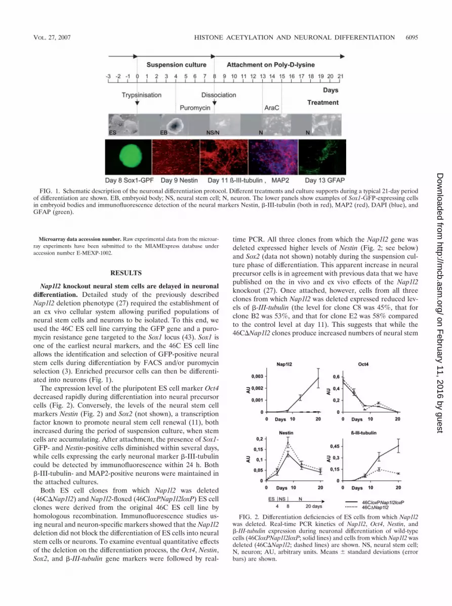
Nap1l2 knockout neural stem cells are delayed in neuronaldifferentiation. Detailed study of the previously describedNap1l2 deletion phenotype (27) required the establishment ofan ex vivo cellular system allowing purified populations ofneural stem cells and neurons to be isolated. To this end, weused the 46C ES cell line carrying the GFP gene and a puro-mycin resistance gene targeted to the Sox1 locus (43). Sox1 isone of the earliest neural markers, and the 46C ES cell lineallows the identification and selection of GFP-positive neuralstem cells during differentiation by FACS and/or puromycinselection (3). Enriched precursor cells can then be differenti-ated into neurons (Fig. 1).
The expression level of the pluripotent ES cell marker Oct4decreased rapidly during differentiation into neural precursorcells (Fig. 2). Conversely, the levels of the neural stem cellmarkers Nestin (Fig. 2) and Sox2 (not shown), a transcriptionfactor known to promote neural stem cell renewal (11), bothincreased during the period of suspension culture, when stemcells are accumulating. After attachment, the presence of Sox1-GFP- and Nestin-positive cells diminished within several days,while cells expressing the early neuronal marker �-III-tubulincould be detected by immunofluorescence within 24 h. Both�-III-tubulin- and MAP2-positive neurons were maintained inthe attached cultures.
Both ES cell clones from which Nap1l2 was deleted(46C�Nap1l2) and Nap1l2-floxed (46CloxPNap1l2loxP) ES cellclones were derived from the original 46C ES cell line byhomologous recombination. Immunofluorescence studies us-ing neural and neuron-specific markers showed that the Nap1l2deletion did not block the differentiation of ES cells into neuralstem cells or neurons. To examine eventual quantitative effectsof the deletion on the differentiation process, the Oct4, Nestin,Sox2, and �-III-tubulin gene markers were followed by real-
time PCR. All three clones from which the Nap1l2 gene wasdeleted expressed higher levels of Nestin (Fig. 2; see below)and Sox2 (data not shown) notably during the suspension cul-ture phase of differentiation. This apparent increase in neuralprecursor cells is in agreement with previous data that we havepublished on the in vivo and ex vivo effects of the Nap1l2knockout (27). Once attached, however, cells from all threeclones from which Nap1l2 was deleted expressed reduced lev-els of �-III-tubulin (the level for clone C8 was 45%, that forclone B2 was 53%, and that for clone E2 was 58% comparedto the control level at day 11). This suggests that while the46C�Nap1l2 clones produce increased numbers of neural stem
FIG. 1. Schematic description of the neuronal differentiation protocol. Different treatments and culture supports during a typical 21-day periodof differentiation are shown. EB, embryoid body; NS, neural stem cell; N, neuron. The lower panels show examples of Sox1-GFP-expressing cellsin embryoid bodies and immunofluorescence detection of the neural markers Nestin, �-III-tubulin (both in red), MAP2 (red), DAPI (blue), andGFAP (green).
FIG. 2. Differentiation deficiencies of ES cells from which Nap1l2was deleted. Real-time PCR kinetics of Nap1l2, Oct4, Nestin, and�-III-tubulin expression during neuronal differentiation of wild-typecells (46CloxPNap1l2loxP; solid lines) and cells from which Nap1l2 wasdeleted (46C�Nap1l2; dashed lines) are shown. NS, neural stem cell;N, neuron; AU, arbitrary units. Means � standard deviations (errorbars) are shown.
VOL. 27, 2007 HISTONE ACETYLATION AND NEURONAL DIFFERENTIATION 6095
on February 11, 2016 by guest
http://mcb.asm
.org/D
ownloaded from

cells, their differentiation into neurons is less efficient. FACSanalysis using an anti-�-III-tubulin antibody performed oneday after cell attachment showed some 5% less �-III-tubulin-positive cells in cultures of the clones with deletions than thecontrol culture, which contained some 40% �-III-tubulin-pos-itive Sox1-GFP-negative cells. We conclude that Nap1l2 dele-tion leads to less efficient differentiation of neural stem cellsinto neurons.
Proliferative changes in Nap1l2 knockout neural precursorcells. We next turned our attention to estimating the survival ofneural stem cells by analyzing the number of Sox1-GFP-ex-pressing cells in suspension cultures. The control46CloxPNap1l2loxP cell line and all three 46C�Nap1l2 celllines produced between 60% and 70% of neural precursor cellswithin 8 days of the initiation of differentiation. Typically incontrol cultures, the numbers of GFP-positive cells then de-creased, dropping below 50% by day 21. However, in the46C�Nap1l2 cultures, the numbers of neural stem cells con-tinued to increase until day 16 (P � 0.03 in unpaired Student’st test), and the clones with deletions showed a small but con-sistent increase (7%) in the number of precursor cells that wasstill detectable at day 21 (Fig. 3A and B). This suggests thatdeletion of the Nap1l2 gene improves the capacity for neuralstem cell renewal.
To test whether the cells from which Nap1l2 was deleted hadan enhanced capacity for cell division, we performed mem-
brane-labeling experiments at day 10 of differentiation in sus-pension culture. The fluorescence intensity of the CellTracedye, which is covalently bound to the cell membrane, dimin-ishes by half at each cellular division. FACS analysis of non-synchronized GFP-positive neural precursor cultures revealedthat the 46C�Nap1l2 cells lost fluorescence more rapidly dur-ing the initial 24 h of culture after labeling than control cellsdid, indicating that they had undergone more cellular divisionsthan the nonmutant cells or that they contained more dividingcells (Fig. 3C). We noted that, concomitant with these results,the level of Ki67, a marker of cycling cells, in the 46C�Nap1l2neural precursor cultures showed a twofold increase (data notshown), while the expression of Cdkn1c, which promotes cellcycle exit, was markedly decreased (see below).
We tested the hypothesis that the increase in neural stemcell numbers in the mutant cultures was associated withchanges in their cell cycle by use of FACS based on a compar-ative DNA content analysis of Sox1-GFP-positive cells. Whiletwo independent experiments showed a slight expression levelincrease in G1 (or G0) phase (for mutant and wild-type cells,respectively, 59% versus 62% of 30,000 propidium iodide-stained viable neural precursor cells per experiment) and de-creases in the S (for mutant and wild-type cells, respectively,17% versus 16.5%) and G2/M phases (for mutant and wild-type cells, respectively, 18.5% versus 16.5%), overall, the cellcycle was not significantly altered in the neural stem cells with
FIG. 3. Maintenance, proliferation, and apoptosis of neural precursors from which Nap1l2 was deleted. (A) The percentages of Sox1-GFP-positive cells in suspension culture were analyzed by FACS. The data summarize three independent experiments using three different 46C�Nap1l2ES cell clones. Error bars represent standard errors of the means. (B) GFP expression in embryoid bodies of 46CloxPNap1l2loxP (WT) and46C�Nap1l2 cells (D) at day 21 is shown. (C) Sox1-GFP-positive cells were labeled using CellTrace Far Red DDAO-SE at day 10 of suspensionculture, and the 24-h Far Red (FR) kinetics was analyzed by FACS. Results for wild-type cells are represented by dark lines, and results for mutantcells are represented by light-gray lines. Data are representative of two experiments. (D) Relative quantification in arbitrary units (AU) andstandard deviations of Par-4 expression by real-time PCR (bars) and determination of percentages of annexin V-positive cells by FACS (lines),given as the means � standard errors of the means of two independent experiments, are shown for 46CloxPNap1l2loxP (dark bars and solid line)and 46C�Nap1l2 cells (light bars and dashed line) at different stages of neuronal differentiation. NS, neural stem cell; N, neuron.
6096 ATTIA ET AL. MOL. CELL. BIOL.
on February 11, 2016 by guest
http://mcb.asm
.org/D
ownloaded from

Nap1l2 deleted compared to control cells (paired Student’s ttest). This result suggests that the increase in neural stem cellpresence associated with the Nap1l2 deletion is most likely dueto increased stem cell renewal linked to a prolongation of theneural stem cell stage and a reduction in the differentiationcapacity of the 46C�Nap1l2 neural stem cells.
We next addressed the question of why the increase in neu-ral precursor cells seen for clones with the Nap1l2 deletion didnot lead to an increased number of neurons. One possibilitywas that this could be associated with a concomitant increase inapoptosis of some fraction of the neural precursor cell popu-lation, for example, due to increased removal of damaged ormisspecified cells that failed to undergo differentiation (42). Toevaluate this possibility, we first tested expression of the Par-4(Prostate apoptosis response 4) gene. Par-4 is an inhibitor ofthe antiapoptotic caspase and is known to regulate neuralprecursor cell death during division through a mechanism in-volving the asymmetric distribution of Par-4 to cells that willundergo apoptosis (5). As expected, Par-4 showed maximalexpression in neural precursor cells (Fig. 3D) with 46C�Nap1l2 cells showing significantly higher expression of thismarker, indicative of increased levels of apoptosis. To confirmthis observation, we performed an apoptosis assay using7-amino-actinomycin D and annexin V-phycoerythrin stain-ing. The FACS analysis, carried out in two independent dif-ferentiation experiments, revealed a 7 to 12% increase in thenumber of annexin V-positive cells undergoing apoptosis in theSox1-GFP-positive 46C�Nap1l2 population (Fig. 3D). We con-clude that Nap1l2 deletion both increases renewal of cells withneural stem cell characteristics and hampers via apoptoticmechanisms the passage of neural stem cells to form differen-tiated neurons.
Nap1l2 deletion leads to transcriptional changes in neuralprecursor cells. To assess the effects of Nap1l2 deletion ongene expression, we performed microarray experiments usingthree independent preparations of puromycin-selected neuralprecursor cells. In each set of experiments, the control cellline 46CloxPNap1l2loxP was compared against one of the 46C�Nap1l2 cell lines. All three experiments included dye swaps tominimize possible technical bias. Primary data were analyzedand normalized using Feature Extraction software and Rosettasoftware set to a P value smaller than 0.1. We found that 97genes were upregulated and 128 downregulated in the mutantcell lines in all three experiments. This corresponds to a de-regulation of some 3% of the analyzed gene complement, with2.3% showing P values of less than 0.05 by Student’s t test. Theextension of the analysis to a mouse development oligonucle-
otide array carrying 20,000 probes has confirmed these results,with 309 genes shown to be downregulated and 474 genesupregulated (3.6%). Average changes (n-fold) were modest(�5), and no chromosomal or domain clustering was apparentfor the deregulated genes. While the upregulation of certainneuronal markers, such as Nestin (1.7-fold) and Sox2 (1.5-foldin the mutant), was confirmed, annotation of deregulatedgenes revealed that they were associated with a wide range ofcellular functions rather than being restricted to neuron-spe-cific expression and function. We conclude that the absence ofNap1l2 in neural stem cells does not lead to a specific dereg-ulation of neural-specific genes.
Only six known genes showed expression levels that werealtered threefold or greater (Table 1). We identified amongthese downregulated genes Cdkn1c (p57kip2), a gene requiredfor cell cycle exit and for postmitotic differentiation of neurons.Rothschild et al. (28) have previously shown both ex vivo andin vivo that Cdkn1c is expressed in differentiating neurons.Mice lacking the Cdkn1c gene exhibit altered cell proliferation,apoptosis, and differentiation (44). In agreement with this,Joseph et al. (16) and Park et al. (24) demonstrated by trans-fection experiments that Cdkn1c promotes cell cycle exit andneuronal development. Detailed analysis of the Cdkn1c ex-pression in our system has confirmed that its expression wasconcomitant with the onset of both neuronal differentiationand Nap1l2 expression (see Fig. 5A and B), suggesting that thedownregulation of Cdkn1c might be of particular interest withrespect to the Nap1l2 deletion phenotype. We therefore testedwhether Cdkn1c could be a direct target of Nap1l2.
Nap1l2 binds to the Cdkn1c target gene and increases theacetylation status of chromatin bound histone H3. Nap1l2 ismainly expressed in postmitotic neurons. Both, immunofluo-rescence studies using an anti-Nap1l2 antibody (Fig. 4A and B)and transient transfection with GFP-Nap1l2-expressing con-structs showed that the Nap1l2 protein is enriched in the nu-clear compartment of �-III-tubulin-positive neurons (Fig. 4B,C, and D). This suggests that the function of the Nap1l2 pro-tein in neurons might be related to a nuclear, chromatin-associated activity. We turned to ChIP to study whetherNap1l2 binds effectively to chromatin using as the putativetarget gene Cdkn1c (p57kip2). Our studies showed that Nap1l2is specifically recruited to the transcription start site of Cdkn1cin differentiating neurons (Fig. 5C and D). This binding of theNap1l2 protein in differentiating neurons is associated withincreased levels of acetylated histone H3K9/14 around thetranscription start site of Cdkn1c (Fig. 5C and E). Since thishistone modification effect was not associated with general
TABLE 1. Summary of microarray results for genes strongly deregulated in the Nap1l2 knockout neural precursorsa
GenBankaccession no. Gene product Gene Chromosomeb Fold change in
expression
AA437922 Neuronatin Nnat 2 (88.0 cM) �3,200AI509951 Calcium channel, voltage-dependent, T type, alpha 1G subunit Cacna1g 11 (D band) �3,867AA014727 Pleckstrin homology-like domain, family A, member 2 Phlda2 7 (69.5 cM) 3,000AA671166 Cyclin-dependent kinase inhibitor 1c (p57) Cdkn1c 7 (69.49 cM) 4,733AA874599 Host cell factor C1 Hcfc1 X (29.54 cM) 3,667AI322387 Mouse insulin-like growth factor II Igf2 7 (69.09 cM) 4,767
a Genes with an average change in expression level (n-fold) greater than 3 are listed.b cM, centimorgans.
VOL. 27, 2007 HISTONE ACETYLATION AND NEURONAL DIFFERENTIATION 6097
on February 11, 2016 by guest
http://mcb.asm
.org/D
ownloaded from

changes in histone H3 levels (data not shown) and Nap1l2itself is bound to Cdkn1c, it seems likely that these changes inH3 histone modifications were directly dependent on the pres-ence of Nap1l2. We conclude that one mechanism by whichNap1l2 may regulate expression of target genes, such asCdkn1c, is via regulation of histone acetylation.
The Cdkn1c gene is known to be subject to epigenetic reg-ulation and can be activated by after TSA treatment (31).Histone acetylation has also been suggested as an epigeneticfactor promoting neuronal differentiation (4, 13), and this cor-relates with an increase in the global pool of chromatin-boundacetylated histone H3 in differentiated neurons compared toneural stem cells and ES cells (Fig. 6A).
To test whether increased HAT activity could rescue Cdkn1cexpression deficiency and the Nap1l2 deletion phenotype dur-ing neuronal differentiation, we treated neural stem cells withthe histone deacetylase inhibitor TSA for 24 h. QuantitativePCR results showed that Cdkn1c expression was strongly in-creased by TSA treatment in both the wild-type and mutant
cells (Fig. 6B). Neural stem cells with Nap1l2 deleted appearedto be more sensitive to TSA treatment than wild-type cellswere. TSA also appeared to increase neuronal differentiationas shown by the expression of the marker �-III-tubulin gene(Fig. 6D). Interestingly, after TSA treatment, the �-III-tubulinlevels of cells from which Nap1l2 was deleted were even higherthan those of nonmutant cells. This inverted kinetics may beinterpreted as the consequence of the simultaneous differen-tiation of a larger number of neural stem cells in the mutant,which correlates with the observation that the previously de-scribed differences in Nestin expression (Fig. 2 and 6C) wereabolished by TSA treatment (Fig. 6C). Our data suggest thatthe delay in differentiation associated with the Nap1l2 deletioncan be rescued by an increase in HAT activity. Our data arecompatible with the idea that the Nap1l2 deletion phenotypecould be related to altered levels of HAT activity.
Nap1l2 binds to histones H3 and H4. We turned to ex vivoand in vitro studies to try to understand the basis for theNap1l2-associated activities on HAT. By use of far-Westernblotting and an in vitro-translated 35S-radiolabeled Nap1l2protein as a probe, Nap1l2 could be shown to interact with corehistones, most strongly with histone H3 and to some extentalso with histone H4 (Fig. 7A) but not with histones H2A andH2B or linker histones. We performed similar experiments
FIG. 4. Localization of Nap1l2 in neurons. (A) Western blot anal-ysis using extracts from Nap1l2 (arrow)-expressing cells and the anti-Nap1l2 antibody (left) or the anti-Nap1l2 antibody preincubated withthe Nap1l2 N-terminal peptide (right). (B) Immunofluorescence usingan anti-Nap1l2 (green) and an anti-�-III-tubulin (�IIItub; red) anti-body on differentiated neurons. DAPI staining is shown in blue.(C) Immunofluorescence of GFP-Nap1l2 (green)-transfected neuronswith an anti-acetyl-H3K9/14 antibody (H3Ac; red). (D) Western blotanalysis using fractions from FLAG-Nap1l2-transfected neurons andanti-FLAG, anti-histone H3, and anti-RAR antibodies. SC, solublecytoplasmic extract; SN, soluble nuclear extract.
FIG. 5. Cdkn1c expression, Nap1l2 binding and histone H3 acety-lation. Relative mRNA concentrations of Nap1l2 (A) and Cdkn1c(B) were measured by quantitative real-time PCR (QRT-PCR). NS,neural stem cells; N, neurons. Values for controls are indicated bydark-gray bars, and values for mutant cells are indicated by light-graybars. The experiment was repeated three times. (C) The relative po-sitions of primer pairs A, B, C, D, and E in the Cdkn1c gene are shownwith the transcription start indicated by an arrow and exons by blackboxes. The lower panels show the specific binding of Nap1l2 to theCdkn1c gene in wild-type neurons (summary of three independentexperiment � standard errors of the means [error bars]) (D), the gainin H3K9/14 acetylation on Cdkn1c in differentiated 46CloxPNap1l2loxPneurons compared to 46C�Nap1l2 neurons (summary of two indepen-dent experiments � standard errors of the means [error bars]) (E).The primers used for the control Arpo, �-actin, and Nanog genes arelocated at the transcription start.
6098 ATTIA ET AL. MOL. CELL. BIOL.
on February 11, 2016 by guest
http://mcb.asm
.org/D
ownloaded from

using substrate pools enriched in acetylated histones, purifiedfrom butyrate-treated cells. In these experiments, Nap1l2could be shown to bind preferentially to the acetylated formsof histones H3 and H4 (Fig. 7A and B).
Nap1l2 increases HAT activity. The increase in expression ofNap1l2 parallels the increase in the global pool of chromatin-bound acetylated histone H3 in differentiating neurons (Fig.6A). The preferential binding of Nap1l2 to histone H3 and itsin vivo effect on the acetylation status of chromatin-boundhistone H3 led us to examine whether Nap1l2 could be directlyinvolved in the process of histone acetylation.
Using histone acetyltransferase assays of cellular extractsfrom differentiated neurons, at a stage when Nap1l2 is highlyexpressed, for a comparison of the Nap1l2-floxed control withNap1l2 knockout cells showed that extracts lacking Nap1l2 hadlowered HAT activity (Fig. 8A). In another experiment inwhich either GFP or GFP plus Nap1l2 was overexpressed inneurons (Fig. 8B), overexpression of Nap1l2 again appeared toenhance the global HAT activity of the cellular extract. Similarobservations were made using HeLa cells transfected with aNap1l2-expressing construct (Fig. 8C). It is therefore likelythat Nap1l2 participates in HAT complexes or facilitates accessof such complexes to histones.
Other members of the NAP family, NAP1 (NAP1L1) and
NAP2 (NAP1L4), are known to be functionally importantcomponents of the p300 coactivator complex suggesting thatNAPs may serve as a point of integration between transcrip-tional coactivators and chromatin (2, 15, 34). Like other mem-bers of the NAP family, Nap1l2 contains a SET/TAF-1� on-coprotein domain, which has been shown to interact with p300and to influence its HAT activity (33). Our preliminary resultssuggest that immunoprecipitated p300 from 46CloxPNap1l2loxPcontrol neurons has a higher level of HAT activity than p300-containing complexes immunoprecipitated from neurons withNap1l2 deleted. This putative activation of p300 in the pres-ence of Nap1l2 was confirmed by in vitro assays using immu-noprecipitated Nap1l2 and both immunoprecipitated andrecombinant p300 (data not shown). By use of coimmunopre-cipitation assays using HeLa cells overexpressing Nap1l2 andp300, we were able to show that these proteins interact (datanot shown). ChIP experiments using anti-p300 antibodies didnot, however, provide any evidence that binding of p300 itselfat the site of Cdkn1c transcription was dependent on Nap1l2 inneurons (data not shown). We conclude that Nap1l2 orNap1l2-containing complexes stimulate HAT activity in neu-rons and have preliminary evidence that p300 may be part ofthese complexes.
DISCUSSION
We have shown that Nap1l2 is implicated in the neural stemdifferentiation pathway and that deletion of Nap1l2 leads toboth delayed neuronal differentiation and increased neuralprecursor renewal, maintenance, and apoptosis. It appearslikely that these cellular phenotypes are, at least in part, me-diated by the effects of Nap1l2 downstream target genes show-ing altered transcriptional activity in cells with Nap1l2 deleted.Among genes downregulated in neural precursors and neuronsfrom which Nap1l2 was deleted was the Cdkn1c gene, a nega-
FIG. 6. Neuronal differentiation, histone acetylation, and Cdkn1cexpression. (A) Kinetics of histone H3K9/14 acetylation on Cdkn1c(for primer positions, see Fig. 5) and Arpo during neuronal differen-tiation was determined by ChIP experiments. NS, neural stem cells; N,neurons. Means � SD of two measurements are shown. Neural stemcells derived from 46CloxPNap1l2loxP (dark bars) and 46C�Nap1l2(light bars) cells were treated with 100 ng/ml TSA for 24 h. The relativemRNA levels for Cdkn1c (B), Nestin (C), and the �-III-tubulin genes(D) of all samples were determined 48 h after TSA treatment. Thedata are representative of two independent experiments. Results aregiven in arbitrary units (AU) � SD.
FIG. 7. Nap1l2 interaction with histones. (A) Far-Western blotanalysis using a 35S-radiolabeled Nap1l2 protein. Ponceau S staining(PS) and autoradiogram analysis reveal the interactions with histonesH3 and H4 (C) and stronger interaction with acetylated histones H3and H4 (Ac); results of one of three independent experiments areshown. Control Western blots (WB) for this experiment using anti-H3and anti-acetyl-H3 antibodies are shown below. The calculated ratiosof the far-Western signals for H3 against the Ponceau S signals (FW/P)are shown at the bottom of panel B.
VOL. 27, 2007 HISTONE ACETYLATION AND NEURONAL DIFFERENTIATION 6099
on February 11, 2016 by guest
http://mcb.asm
.org/D
ownloaded from

tive regulator of cell proliferation that promotes neuronal dif-ferentiation (16, 24, 28). The contribution of the altered ex-pression of other candidate genes, such as Igf2 (Insulin-likegrowth factor II) and Phlda2 (Pleckstrin homology-like do-main, family A, member 2), both of which have been impli-cated in growth control (12, 30), to the Nap1l2 deletion phe-notype remains to be ascertained. Interestingly, Cdkn1c acts asa cyclin kinase inhibitor in mitotic progenitor cells and isknown to play a distinct role in neuronal differentiation (16, 24,28). Our study shows that Cdkn1c is increasingly expressedduring neuronal differentiation and that its upregulation isassociated with increased histone H3K9/14 acetylation at itstranscription start site. We have also shown that the Nap1l2protein binds to the transcription start site of Cdkn1c and thatin neurons from which Nap1l2 was deleted, Cdkn1c becomesboth downregulated and depleted in histone H3 acetylation.Our data suggest that Nap1l2 is involved in regulating Cdkn1cexpression in neurons via regulation of histone acetylation.The functional importance of histone acetylation for Cdkn1c/CDKN1C expression, which is known to be regulated by vari-ous epigenetic marks (18, 31, 40), has also been suggested bythe finding that the gene is silenced by histone deacetylation(18) and the increased expression of the gene in the presenceof the deacetylase inhibitor TSA (36).
The role of the Nap1l2 protein at the Cdkn1c locus is likelya reflection of a more extensive role for Nap1l2 within theneuronal nucleus. The rather subtle transcriptional downregu-
lation of Cdkn1c may indicate a role of Nap1l2 in the epige-netic modulation of basal transcription levels rather than in thespecific activation of a subset of neurally expressed genes. Sucha role would correlate with our finding that Nap1l2 interactsdirectly with the core histones H3 and H4 and increases bothglobal and site-specific histone acetylation. Since the Nap1l2protein itself has no intrinsic HAT activity (data not shown),one possibility is that it acts through activation of histoneacetyltransferases such as p300 (7, 10), a protein which has thecapacity to acetylate histone H3 and H4 both in vitro and invivo (32). The previous finding of binding sites for the tumorsuppressor p300 on the human CDKN1C promoter (36) is ofobvious interest in this regard, as is the observation that micenullizygous for p300, similar to Nap1l2 knockout chimeras, diebetween days E9 and E11.5 of gestation and exhibit strongdefects in neurulation and cell proliferation (41). It should benoted that the role for the neuron-specific Nap1l2 protein inhistone acetylation that this leads us to propose is the somehowopposite of that described for the ubiquitously expressed NAPand SET proteins, which have been shown to inhibit HATactivity (2, 33). While Nap1l2 also has a particularly strongaffinity for the acetylated forms of histones H3 and H4, possi-bly indicating a role in maintaining the acetylation status ofhistones, we have been unable to demonstrate a direct inhib-itory effect of Nap1l2 on histone deacetylation in vitro (datanot shown). Of relevance may be the fact that while Nap1l2 hastwo homology domains in common with other members of theNAP family (amino acids 96 to 159 and 235 to 377), it alsocontains a specific Glu-rich acidic domain (amino acids 160 to234) that may well contribute to its unique functioning. A roleof Nap1l2 in the establishment of other histone modificationsremains to be shown. We interpret our preliminary evidencefor a reduction in repressive histone H3 marks such as trim-ethyl H3K27 at the Cdkn1c locus in the presence of Nap1l2(data not shown) as a likely secondary consequence of higherH3 acetylation levels.
The acetylation status of histones is thought to act as ageneral regulator of chromatin structure that mediates theremoval of epigenetically controlled repression and enhancestranscriptional activity. This is reflected by the finding thatacetylated K9/14 of histone H3 is found to be highly localizedto the 5� regions of transcriptionally active human genes (20).Gene expression states depend on the binding of sequence-specific transcription factors and the recruitment of chromatin-remodeling complexes and can be modified during DNA rep-lication and transcription (8). While studies provide evidencefor general alterations in histone modification, such as theincreased histone H3 and H4 acetylation occurring during neu-ronal differentiation (13), postmitotic neurons can no longerrely on a replication-dependent machinery to renew or modifytheir chromatin-bound histone pool. Chromatin modification-dependent gene expression patterns can, however, be estab-lished and maintained in such cells by certain histone variants,such as the hyperacetylated variant histone H3.3 (6), that canbe incorporated into nucleosomes in a DNA replication-inde-pendent manner (1). During mitosis, histone modification andvariant incorporation combine to set stable epigenetic marksfor the memory of active transcriptional states (8).
Our findings suggest that Nap1l2 is involved in the dynamicsof histone modification at its target loci during differentiation.
FIG. 8. HAT activity in the presence of Nap1l2. HAT assays using2 �g of crude extract from control (WT) and �Nap1l2 neurons(D) (panel A) or WT neurons overexpressing Nap1l2 and GFP or GFPalone (panel B). (C) HAT assays using HeLa cell extracts transfectedwith an empty 3� FLAG vector (E C) or with the 3� FLAG-Nap1l2expression vector (E N). The result of the Western blot analysis (WB)using an anti-3� FLAG antibody is shown below. Dark-gray columnsrefer to cpm for the histone samples, and light-gray columns refer tocpm for the BSA control samples.
6100 ATTIA ET AL. MOL. CELL. BIOL.
on February 11, 2016 by guest
http://mcb.asm
.org/D
ownloaded from

One possible mechanism could be that Nap1l2 facilitates theaccessibility of HATs, such as p300, to histones, although thepossibility that Nap1l2 might participate directly in the selec-tive incorporation or exchange of hyperacetylated histones re-mains to be excluded (23). Irrespective of the precise mecha-nism, the resulting modulation in gene expression regulates thekinetics of neuronal differentiation, which is central to theprocess of neurulation. Our findings add another dimension ofcomplexity to the potential role of the Nap1l2 molecule andreinforce the likelihood that other members of the mammalianneuronal NAP1-like proteins may be involved in a possiblycomplementary manner in regulating cellular activity via his-tone modifications. It remains to be shown whether such find-ings for Nap1l2 can be extended to the ubiquitous NAPs thatare present not only in neurons but also in all other cell types.
ACKNOWLEDGMENTS
We are grateful to Marie Christine Wagner for helping with theFACS analysis and to Robert Olaso for advice on the microarrayanalysis. We thank Delphine Bohl for help with immunofluorescence,Claire Rougeulle and Pablo Navarro for the introduction to ChIPanalysis, and Genevieve Almouzni, Angelita Rebollo, Matthieu Gerard,and Nicolas Zarjevski for critical reading of the manuscript.
This work was supported by grants from the ARC, AFM, and theEU Epigenome program (CE LSHG CT 2004 503433) and by recur-rent funding from the CNRS, INSERM, and the Pasteur Institute(GPH7SP2). M.A. is recipient of a fellowship from the Canceropole,Ile de France.
REFERENCES
1. Ahmad, K., and S. Henikoff. 2002. The histone variant H3.3 marks activechromatin by replication-independent nucleosome assembly. Mol. Cell9:1191–1200.
2. Asahara, H., S. Tartare-Deckert, T. Nakagawa, T. Ikehara, F. Hirose, T.Hunter, T. Ito, and M. Montminy. 2002. Dual roles of p300 in chromatinassembly and transcriptional activation in cooperation with nucleosome as-sembly protein 1 in vitro. Mol. Cell. Biol. 22:2974–2983.
3. Aubert, J., M. P. Stavridis, S. Tweedie, M. O’Reilly, K. Vierlinger, M. Li, P.Ghazal, T. Pratt, J. O. Mason, D. Roy, and A. Smith. 2003. Screening formammalian neural genes via fluorescence-activated cell sorter purification ofneural precursors from Sox1-gfp knock-in mice. Proc. Natl. Acad. Sci. USA100(Suppl. 1):11836–11841.
4. Balasubramaniyan, V., E. Boddeke, R. Bakels, B. Kust, S. Kooistra, A.Veneman, and S. Copray. 2006. Effects of histone deacetylation inhibition onneuronal differentiation of embryonic mouse neural stem cells. Neuroscience143:939–951.
5. Bieberich, E., S. MacKinnon, J. Silva, S. Noggle, and B. G. Condie. 2003.Regulation of cell death in mitotic neural progenitor cells by asymmetricdistribution of prostate apoptosis response 4 (PAR-4) and simultaneouselevation of endogenous ceramide. J. Cell Biol. 162:469–479.
6. Bosch, A., and P. Suau. 1995. Changes in core histone variant composition indifferentiating neurons: the roles of differential turnover and synthesis rates.Eur. J. Cell Biol. 68:220–225.
7. Chan, H. M., and N. B. La Thangue. 2001. p300/CBP proteins: HATs fortranscriptional bridges and scaffolds. J. Cell Sci. 114:2363–2373.
8. Chow, C. M., A. Georgiou, H. Szutorisz, A. Maia e Silva, A. Pombo, I.Barahona, E. Dargelos, C. Canzonetta, and N. Dillon. 2005. Variant histoneH3.3 marks promoters of transcriptionally active genes during mammaliancell division. EMBO Rep. 6:354–360.
9. Chureau, C., M. Prissette, A. Bourdet, V. Barbe, L. Cattolico, L. Jones, A.Eggen, P. Avner, and L. Duret. 2002. Comparative sequence analysis of theX-inactivation center region in mouse, human, and bovine. Genome Res.12:894–908.
10. Giordano, A., and M. L. Avantaggiati. 1999. p300 and CBP: partners for lifeand death. J. Cell. Physiol. 181:218–230.
11. Graham, V., J. Khudyakov, P. Ellis, and L. Pevny. 2003. SOX2 functions tomaintain neural progenitor identity. Neuron 39:749–765.
12. Hartmann, W., A. Koch, H. Brune, A. Waha, U. Schuller, I. Dani, D.Denkhaus, W. Langmann, U. Bode, O. D. Wiestler, K. Schilling, and T.Pietsch. 2005. Insulin-like growth factor II is involved in the proliferationcontrol of medulloblastoma and its cerebellar precursor cells. Am. J. Pathol.166:1153–1162.
13. Hsieh, J., K. Nakashima, T. Kuwabara, E. Mejia, and F. H. Gage. 2004.
Histone deacetylase inhibition-mediated neuronal differentiation of multi-potent adult neural progenitor cells. Proc. Natl. Acad. Sci. USA 101:16659–16664.
14. Hu, R. J., M. P. Lee, L. A. Johnson, and A. P. Feinberg. 1996. A novel humanhomologue of yeast nucleosome assembly protein, 65 kb centromeric to thep57KIP2 gene, is biallelically expressed in fetal and adult tissues. Hum. Mol.Genet. 5:1743–1748.
15. Ito, T., T. Ikehara, T. Nakagawa, W. L. Kraus, M. Muramatsu, M. Gos, andA. Szpecht-Potocka. 2000. p300-mediated acetylation facilitates the transferof histone H2A-H2B dimers from nucleosomes to a histone chaperone.Genes Dev. 14:1899–1907.
16. Joseph, B., A. Wallen-Mackenzie, G. Benoit, T. Murata, E. Joodmardi, S.Okret, and T. Perlmann. 2003. p57(Kip2) cooperates with Nurr1 in devel-oping dopamine cells. Proc. Natl. Acad. Sci. USA 100:15619–15624.
17. Kellogg, D. R., A. Kikuchi, T. Fujii-Nakata, C. W. Turck, and A. W. Murray.1995. Members of the NAP/SET family of proteins interact specifically withB-type cyclins. J. Cell Biol. 130:661–673.
18. Kikuchi, T., M. Toyota, F. Itoh, H. Suzuki, T. Obata, H. Yamamoto, H.Kakiuchi, M. Kusano, J. P. Issa, T. Tokino, and K. Imai. 2002. Inactivationof p57KIP2 by regional promoter hypermethylation and histone deacetyla-tion in human tumors. Oncogene 21:2741–2749.
19. Laskey, R. A., B. M. Honda, A. D. Mills, and J. T. Finch. 1978. Nucleosomesare assembled by an acidic protein which binds histones and transfers themto DNA. Nature 275:416–420.
20. Liang, G., J. C. Lin, V. Wei, C. Yoo, J. C. Cheng, C. T. Nguyen, D. J.Weisenberger, G. Egger, D. Takai, F. A. Gonzales, and P. A. Jones. 2004.Distinct localization of histone H3 acetylation and H3–K4 methylation to thetranscription start sites in the human genome. Proc. Natl. Acad. Sci. USA101:7357–7362.
21. Mendez, J., and B. Stillman. 2000. Chromatin association of human originrecognition complex, Cdc6, and minichromosome maintenance proteins dur-ing the cell cycle: assembly of prereplication complexes in late mitosis. Mol.Cell. Biol. 20:8602–8612.
22. Navarro, P., S. Pichard, C. Ciaudo, P. Avner, and C. Rougeulle. 2005. Tsixtranscription across the Xist gene alters chromatin conformation withoutaffecting Xist transcription: implications for X-chromosome inactivation.Genes Dev. 19:1474–1484.
23. Okuwaki, M., K. Kato, H. Shimahara, S. Tate, and K. Nagata. 2005. As-sembly and disassembly of nucleosome core particles containing histonevariants by human nucleosome assembly protein I. Mol. Cell. Biol. 25:10639–10651.
24. Park, H. C., J. Boyce, J. Shin, and B. Appel. 2005. Oligodendrocyte specifi-cation in zebrafish requires notch-regulated cyclin-dependent kinase inhib-itor function. J. Neurosci. 25:6836–6844.
25. Park, Y. J., and K. Luger. 2006. The structure of nucleosome assemblyprotein 1. Proc. Natl. Acad. Sci. USA 103:1248–1253.
26. Rodriguez, P., J. Pelletier, G. B. Price, and M. Zannis-Hadjopoulos. 2000.NAP-2: histone chaperone function and phosphorylation state through thecell cycle. J. Mol. Biol. 298:225–238.
27. Rogner, U. C., D. D. Spyropoulos, N. Le Novere, J. P. Changeux, and P.Avner. 2000. Control of neurulation by the nucleosome assembly protein-1-like 2. Nat. Genet. 25:431–435.
28. Rothschild, G., X. Zhao, A. Iavarone, and A. Lasorella. 2006. E proteins andId2 converge on p57Kip2 to regulate cell cycle in neural cells. Mol. Cell. Biol.26:4351–4361.
29. Rougeulle, C., and P. Avner. 1996. Cloning and characterization of a murinebrain specific gene Bpx and its human homologue lying within the Xiccandidate region. Hum. Mol. Genet. 5:41–49.
30. Salas, M., R. John, A. Saxena, S. Barton, D. Frank, G. Fitzpatrick, M. J.Higgins, and B. Tycko. 2004. Placental growth retardation due to loss ofimprinting of Phlda2. Mech. Dev. 121:1199–1210.
31. Sato, N., H. Matsubayashi, T. Abe, N. Fukushima, and M. Goggins. 2005.Epigenetic down-regulation of CDKN1C/p57KIP2 in pancreatic ductal neo-plasms identified by gene expression profiling. Clin. Cancer Res. 11:4681–4688.
32. Schiltz, R. L., C. A. Mizzen, A. Vassilev, R. G. Cook, C. D. Allis, and Y.Nakatani. 1999. Overlapping but distinct patterns of histone acetylation bythe human coactivators p300 and PCAF within nucleosomal substrates.J. Biol. Chem. 274:1189–1192.
33. Seo, S. B., P. McNamara, S. Heo, A. Turner, W. S. Lane, and D. Chakravarti.2001. Regulation of histone acetylation and transcription by INHAT, ahuman cellular complex containing the set oncoprotein. Cell 104:119–130.
34. Shikama, N., H. M. Chan, M. Krstic-Demonacos, L. Smith, C. W. Lee, W.Cairns, and N. B. La Thangue. 2000. Functional interaction between nu-cleosome assembly proteins and p300/CREB-binding protein family coacti-vators. Mol. Cell. Biol. 20:8933–8943.
35. Simon, H. U., G. B. Mills, M. Kozlowski, D. Hogg, D. Branch, Y. Ishimi, andK. A. Siminovitch. 1994. Molecular characterization of hNRP, a cDNAencoding a human nucleosome-assembly-protein-I-related gene product in-volved in the induction of cell proliferation. Biochem. J. 297:389–397.
36. Smith, J. L., W. J. Freebern, I. Collins, A. De Siervi, I. Montano, C. M.Haggerty, M. C. McNutt, W. G. Butscher, I. Dzekunova, D. W. Petersen, E.
VOL. 27, 2007 HISTONE ACETYLATION AND NEURONAL DIFFERENTIATION 6101
on February 11, 2016 by guest
http://mcb.asm
.org/D
ownloaded from

Kawasaki, J. L. Merchant, and K. Gardner. 2004. Kinetic profiles of p300occupancy in vivo predict common features of promoter structure and co-activator recruitment. Proc. Natl. Acad. Sci. USA 101:11554–11559.
37. Smith, R. J., W. Dean, G. Konfortova, and G. Kelsey. 2003. Identification ofnovel imprinted genes in a genome-wide screen for maternal methylation.Genome Res. 13:558–569.
38. Taniguchi, M., M. Sanbo, S. Watanabe, I. Naruse, M. Mishina, and T. Yagi.1998. Efficient production of Cre-mediated site-directed recombinantsthrough the utilization of the puromycin resistance gene, pac: a transientgene-integration marker for ES cells. Nucleic Acids Res. 26:679–680.
39. Watanabe, T. K., T. Fujiwara, Y. Nakamura, Y. Hirai, H. Maekawa, and E.Takahashi. 1996. Cloning, expression pattern and mapping to Xq ofNAP1L3, a gene encoding a peptide homologous to human and yeast nu-cleosome assembly proteins. Cytogenet. Cell Genet. 74:281–285.
40. Yang, H., K. Hoshino, B. Sanchez-Gonzalez, H. Kantarjian, and G. Garcia-
Manero. 2005. Antileukemia activity of the combination of 5-aza-2�-deoxy-cytidine with valproic acid. Leuk. Res. 29:739–748.
41. Yao, T. P., S. P. Oh, M. Fuchs, N. D. Zhou, L. E. Ch’ng, D. Newsome, R. T.Bronson, E. Li, D. M. Livingston, and R. Eckner. 1998. Gene dosage-dependent embryonic development and proliferation defects in mice lackingthe transcriptional integrator p300. Cell 93:361–372.
42. Yeo, W., and J. Gautier. 2004. Early neural cell death: dying to becomeneurons. Dev. Biol. 274:233–244.
43. Ying, Q. L., M. Stavridis, D. Griffiths, M. Li, and A. Smith. 2003. Conversionof embryonic stem cells into neuroectodermal precursors in adherent mo-noculture. Nat. Biotechnol. 21:183–186.
44. Zhang, P., N. J. Liegeois, C. Wong, M. Finegold, H. Hou, J. C. Thompson, A.Silverman, J. W. Harper, R. A. DePinho, and S. J. Elledge. 1997. Altered celldifferentiation and proliferation in mice lacking p57KIP2 indicates a role inBeckwith-Wiedemann syndrome. Nature 387:151–158.
6102 ATTIA ET AL. MOL. CELL. BIOL.
on February 11, 2016 by guest
http://mcb.asm
.org/D
ownloaded from


