Embed Size (px)
Citation preview

Seediscussions,stats,andauthorprofilesforthispublicationat:https://www.researchgate.net/publication/51149900
MyD88andRetinoicAcidSignalingPathwaysInteracttoModulateGastrointestinalActivitiesofDendriticCells
ARTICLEinGASTROENTEROLOGY·APRIL2011
ImpactFactor:16.72·DOI:10.1053/j.gastro.2011.04.010·Source:PubMed
CITATIONS
38
READS
31
13AUTHORS,INCLUDING:
EduardoJVillablanca
MassachusettsGeneralHospital
33PUBLICATIONS1,008CITATIONS
SEEPROFILE
JaimeDeCalisto
UniversidadMayor
14PUBLICATIONS440CITATIONS
SEEPROFILE
RuneBlomhoff
UniversityofOslo
314PUBLICATIONS11,855CITATIONS
SEEPROFILE
MariaRosaBono
UniversityofChile
89PUBLICATIONS2,276CITATIONS
SEEPROFILE
Availablefrom:JaimeDeCalisto
Retrievedon:03February2016

MyD88 and Retinoic Acid Signaling Pathways Interact toModulate Gastrointestinal Activities of Dendritic Cells
Eduardo J. Villablanca1, Sen Wang1, Jaime De Calisto1,2, Daniel C. O. Gomes3, Maureen A.Kane4, Joseph L. Napoli4, William S. Blaner5, Hiroyuki Kagechika6, Rune Blomhoff7, MarioRosemblatt2,8, Maria Rosa Bono2, Ulrich H. von Andrian9, and J. Rodrigo Mora1,*
1 Gastrointestinal Unit, Massachusetts General Hospital, Harvard Medical School, Boston, MA,USA2 Facultad de Ciencias, Universidad de Chile, Santiago, Chile3 Universidade Federal do Rio de Janeiro, Brazil4 University of California, Berkeley, CA, USA5 Columbia University, New York, NY, USA6 Tokyo Medical and Dental University, Tokyo, Japan7 Institute of Basic Medical Sciences, University of Oslo, Oslo, Norway8 Fundacion Ciencia para la Vida & Universidad Andres Bello, Santiago, Chile9 Immune Disease Institute, Harvard Medical School, Boston, MA, USA
AbstractBACKGROUND & AIMS—Gut-associated dendritic cells (DC) metabolize vitamin A into all-trans retinoic acid (RA), which is required to induce lymphocytes to localize to the gastrointestinal(GI) tract and promotes the differentiation of Foxp3+ regulatory T cells (TREG) andimmunoglobulin (Ig)A antibody-secreting cells (IgA-ASC). We investigated whether RAfunctions in a positive-feedback loop, via DC, to induce its own synthesis.
METHODS—We measured levels of retinoids in intestine tissues from mice and assessed the roleof RA in activities of gut-associated DC in cell cultures and mice. We used pharmacologicantagonists to determine the signaling pathways involved in regulation of DC and used MyD88−/−mice to determine the contribution of Toll-like receptor (TLR) signaling in RA-mediated activitiesof DC.
RESULTS—The concentration of retinoids decreased in a proximal-to-distal gradient along theintestine, which correlated with the activity of gut-specific DC. Importantly, RA regulated the
© 2011 The American Gastroenterological Association. Published by Elsevier Inc. All rights reserved.*Correspondence and requests for materials should be addressed to J. Rodrigo Mora ([email protected]).Author’s contributions: EJV, SW, JDC and DCG designed and performed experiments. MAK and JLN performed the retinoidmeasurements. HK, WSB and RB provided reagents and mice. MR, MRB and UHvA, provided valuable suggestions and advise onexperiments and on the manuscript. EJV and JRM develop the conceptual framework for the study, supervised the project, designedexperiments, analyzed data and wrote the paper.Conflicts of interest: The Authors disclose no conflictsPublisher's Disclaimer: This is a PDF file of an unedited manuscript that has been accepted for publication. As a service to ourcustomers we are providing this early version of the manuscript. The manuscript will undergo copyediting, typesetting, and review ofthe resulting proof before it is published in its final citable form. Please note that during the production process errors may bediscovered which could affect the content, and all legal disclaimers that apply to the journal pertain.
NIH Public AccessAuthor ManuscriptGastroenterology. Author manuscript; available in PMC 2012 July 1.
Published in final edited form as:Gastroenterology. 2011 July ; 141(1): 176–185. doi:10.1053/j.gastro.2011.04.010.
NIH
-PA Author Manuscript
NIH
-PA Author Manuscript
NIH
-PA Author Manuscript

ability of gut-associated DC to produce RA, induce T cells to localize to the GI tract, and generateTREG and IgA secreting cells. RA was sufficient to induce its own production by extra-intestinalDC, in vitro and in vivo. RA-mediated regulation of DC required signaling through the mitogen-activated protein kinase signaling pathway and unexpectedly required MyD88, which has beenassociated with TLR, interleukin (IL)-1, and IL-18 signaling.
CONCLUSIONS—RA is necessary and sufficient to induce DC to regulate T-cell localization tothe GI tract and IgA secretion. These findings indicate crosstalk between the RA receptor andMyD88-dependent TLR signaling pathways.
KeywordsInflammation; IBD; immune response; mouse model
Lymphocyte migration is a key event during intestinal inflammation 1. Therefore, it iscritical to understand how T and B cells migrate to the intestine and how their migrationpatterns could be manipulated for therapeutic purposes. The gut-homing receptors integrinα4β7 and chemokine receptor CCR9 are required for T and B lymphocyte migration to thegut mucosa in the steady state and also during intestinal inflammation in mice andhumans 1, 2, thus acting as molecular “zip codes” by controlling lymphocyte migration in atissue-specific fashion.
DC from mesenteric lymph nodes (MLN-DC), Peyer’s patches (PP-DC) and small intestinelamina propria (gut-associated DC), but not DC from extra-intestinal sites, induce a highexpression of gut-homing receptors on lymphocytes 3–6, which is explained by their abilityto metabolize vitamin A (retinol) into all-trans retinoic acid (RA). RA is necessary toimprint gut-homing lymphocytes 5–7, to promote differentiation of IgA-ASC 5 and to controlthe balance between TREG and Th17 cells in the gut mucosa 8, 9. Therefore, given theessential role of RA in intestinal immune homeostasis, it is important to understand how thesynthesis of RA is modulated in the gut mucosa.
Here we show that RA controls RA-synthesizing capacity in DC in vitro and in vivo, henceinducing a positive feedback loop on its own synthesis and conferring DC with gut-specificimprinting properties. In addition, we found that RA-mediated DC education requires theexpression of the intracellular adaptor MyD88, which is conventionally associated with TLRand IL-1/IL-18 signaling 11, 12, suggesting a novel crosstalk between RA- and MyD88-dependent pathways.
ResultsDC ability to imprint gut homing correlates with retinoid levels in the gut
RA and its precursors retinol and retinyl esters were readily detected in the intestinal mucosaand their concentrations followed a gradient from proximal to distal, with the highestconcentrations being found in the duodenum and jejunum (Fig. 1A). Interestingly, retinoidlevels correlated with DC imprinting ability. PP-DC or lamina propria DC (LP-DC) fromduodenum and jejunum induced higher levels of gut-homing receptors (α4β7 and CCR9)and Foxp3+ T cells as compared to their counterparts in ileum, colon or spleen (Fig. 1B, Cand Fig. S1A). Reciprocally, the skin-homing receptors E- and P-selectin ligands were moreefficiently induced by ileum PP-DC/LP-DC, colon LP-DC and Spleen-DC than byduodenum or jejunum PP-DC/LP-DC (Fig. S1B), consistent with the notion that theinduction of these receptors occurs as a default pathway in T cells activated without RA 7.Consistent with their higher gut-homing imprinting capacity, PP-DC and LP-DC fromduodenum and jejunum exhibited higher Raldh activity (Aldefluor staining 17) than ileum or
Villablanca et al. Page 2
Gastroenterology. Author manuscript; available in PMC 2012 July 1.
NIH
-PA Author Manuscript
NIH
-PA Author Manuscript
NIH
-PA Author Manuscript
colon DC (Fig. 1D), which was not explained by different proportions of CD103+ DC (Fig.S1C). Of note, the expression of Aldh1a2 mRNA (encoding Raldh2) was not significantlydifferent among DC from different intestinal segments and their relative mRNA levels werelower than those found in MLN-DC (Fig. S1D), suggesting that Aldh1a2 mRNA levels inDC do not always fully correlate with their Raldh activity or gut-homing imprintingcapacity. Whether this dissociation between Aldh1a2 mRNA and Raldh activity reflectslocal differences in Raldh protein expression or stability remains to be determined. Inaddition, using DR5-luciferase reporter mice, in which luciferase is controlled by a promoterwith RA response elements (RARE) 13, we determined that all tested DC have the capacityto respond to RA ex vivo (Fig. S1E). However, consistent with their exposure to high levelsof RA, DC from the proximal small intestine exhibited higher luciferase activity than distalDC (Fig. 1E).
RA is necessary in vivo for gut-associated DC educationWe depleted mice of the RA precursor retinol by feeding them a vitamin A-deficient (VAD)diet, as described 5, 7. Since vitamin A is abundantly stored in the liver it is difficult to attaincomplete vitamin A depletion, even when using a VAD diet for several months 14. Toaddress this shortcoming we used mice deficient in lecithin:retinol acyltransferase (LRAT),which cannot store retinol in the liver 14. LRAT−/− mice develop normally when maintainedon a vitamin A-sufficient diet, but they become vitamin A depleted after only 2–4 weeks ona VAD diet, with the additional advantage of avoiding potential unwanted effects of chronicvitamin A depletion 14. In agreement with a critical role of RA in gut-associated DCeducation, PP-DC and MLN-DC from VAD mice induced lower levels of gut-homingreceptors on T cells as compared to their counterparts from mice on a vitamin A-sufficientdiet (Fig. 2A). In addition, MLN-DC and PP-DC from VAD mice were impaired in inducingFoxp3+ T cells (Fig. 2B) and IgA-ASC (Fig. 2C), respectively. Moreover, MLN-DC fromVAD mice showed a marked reduction in Aldh1a2 mRNA expression (Fig. 2D) and animpaired Raldh activity in total and CD103+ MLN-DC (Fig. 2E), which was rescued by oralRA supplementation (Fig. 2F). Thus, RA is necessary in vivo to educate gut-associated DCwith critical gut-specific imprinting properties.
RA is sufficient to confer DC with RA synthesizing capacity in vitro and in vivoMurine Spleen DC pre-treated with RA (RA-DC) upregulated Aldh1a2 mRNA (Fig. 3A andFig. S2A, B). This effect was not limited to RA, as other natural and synthetic RA receptor(RAR-RXR) agonists (Am80, 9-cis RA, 13-cis RA) also induced Aldh1a2 in Spleen-DC. Incontrast, agonists for RXR-RXR (HX630, PA024), PPARγ-RXR (Rosiglitazone), PPARβ/δ-RXR (GW0742), PXR-RXR (Lithocholic Acid), LXR-RXR (TO901317) or AHR (ITE)nuclear receptors did not induce Aldh1a2 mRNA. RA also induced Aldh1a2 in murineperipheral lymph node (PLN)-DC and bone marrow (BM)-derived DC (Fig S2C) as well asALDH1A2 mRNA in human monocyte-derived DC (Mo-DC) (Fig. S2D), suggesting that ourfindings could be extrapolated, at least in part, to human DC.
Consistent with their higher expression of Aldh1a2 mRNA, RA-DC displayed higher Raldhactivity as compared to untreated Spleen-DC (UT-DC) (Fig. 3B), which was also observedwhen treating PLN-DC and BM-DC with RA (Fig. S2E), indicating that RA-DC acquiredRA-synthesizing capacity. Importantly, RA-DC induced significantly higher levels of gut-homing receptors α4β7 and CCR9 on activated T cells as compared to UT-DC, which wasabrogated when T cells were activated in retinol-free media (Fig. 3C) or in the presence ofthe Raldh inhibitor diethylaminobenzaldehyde (DEAB) 17 (Fig. 3D), implying that theinduction of gut-homing receptors by RA-DC required active RA synthesis and was not dueto RA carry-over, which happens when DC are incubated with high concentrations of RA 18.Similarly, human monocyte-derived DC (Mo-DC) pre-treated with RA induced higher levels
Villablanca et al. Page 3
Gastroenterology. Author manuscript; available in PMC 2012 July 1.
NIH
-PA Author Manuscript
NIH
-PA Author Manuscript
NIH
-PA Author Manuscript

of gut-homing receptors on polyclonally activated human T cells than untreated Mo-DC(Fig. S2F). Adding the RAR-inhibitor LE540 during the co-culture abrogated gut-homingimprinting by RA-DC (Fig. S2G), indicating that gut-homing imprinting by RA-DC requiresRA activity via RAR nuclear receptors. Consistent with their higher expression of gut-homing receptors, T cells activated with RA-DC migrated significantly more to the smallintestine as compared to those activated with UT-DC (Fig. 3E). In addition, analogous togut-associated DC 8, 9, RA-DC also promoted the induction of Foxp3+ TREG (Fig. 3F).
To obtain mechanistic clues on Aldh1a2 induction by RA in DC, we incubated Spleen-DCfrom DR5-luciferase mice with RA in the presence or absence of the transcription inhibitoractinomycin-D or the translation inhibitor cycloheximide as described 19. RA-treatedSpleen-DC exhibited increased Raldh and luciferase activities (induction of Aldh1a2 mRNAand a RAR-dependent reporter by RA, respectively), which were abrogated by actinomycin-D or cycloheximide treatment (Fig. S3A, B), suggesting that RA-mediated DC education toexpress Raldh and synthesize RA requires de novo transcription and protein synthesis.Moreover, actinomycin-D inhibited RA-mediated induction of Aldh1a2 mRNA and RarbmRNA (encoding RARβ, a known RA-induced gene) (Fig. S3C), indicating that theirincrease is mostly due to de novo transcription rather than enhanced mRNA stability.Interestingly, cycloheximide blocked Aldh1a2 but not Rarb mRNA induction, suggestingthat the increased Aldh1a2 mRNA transcription in RA-treated Spleen-DC requires newlysynthesized proteins, whereas Rarb appears to be directly induced by RA without requiringde novo protein synthesis. This finding is consistent with their distinct transcription kinetics,as Rarb mRNA was clearly induced as quickly as 1 hour post RA treatment, whereasAldh1a2 mRNA exhibited a more delayed kinetics (Fig. S3D). In sum, RA-induction ofAldh1a2 mRNA in Spleen-DC appears to occur indirectly in a process that requires prior denovo protein synthesis.
Of note, although Spleen-DC acquired Raldh activity when pre-treated with RA for 24hours, this was not observed when DC were exposed to RA for 30 min (i.e., only during theAldefluor assay) (Fig S3E). Moreover, MLN-DC maintained the same high Raldh activityregardless of whether the mice were supplemented or not with oral RA (Fig S3F). Thus, RAdoes not appear to directly induce, enhance or otherwise affect Raldh enzymatic activity,suggesting that its main effect on DC education involves de novo Aldh1a2 mRNA and Raldhprotein induction.
To assess whether RA can also educate extra-intestinal DC in vivo, we treated mice with RAvia oral gavage and then analyzed PLN-DC for their expression of Aldh1a2 mRNA, Raldhactivity and gut-homing imprinting capacity (Fig. 4A). As expected, PLN-DC from controlmice expressed much lower levels of Aldh1a2 mRNA and Raldh activity as compared toMLN-DC (Fig. 4B, C and Fig. S4A). By contrast, PLN-DC from mice treated with oral RAexhibited significantly higher levels of Aldh1a2 mRNA and Raldh activity than PLN-DCfrom control mice. Moreover, PLN-DC from RA-treated mice were able to induce gut-homing receptors on activated T cells, an effect that was dependent on the presence ofretinol and Raldh activity (Fig. 4D), demonstrating that it required active RA biosynthesis.
We did not detect RA in PLN from untreated animals, even using the most sensitivemethods 16, whereas RA was readily detected in PLN from RA-treated animals with levelsapproaching those found in MLN from untreated animals (Fig. S4B). Moreover, luciferaseactivity in PLN cells from RA-treated DR5-luciferase mice was similar to that in MLN orPP cells from control animals (Fig. S4C).
Of note, RA supplementation was associated with increased proportions of activated CCR9+
CD8 T cells in PLN and spleen upon s.c. immunization, reaching levels comparable to
Villablanca et al. Page 4
Gastroenterology. Author manuscript; available in PMC 2012 July 1.
NIH
-PA Author Manuscript
NIH
-PA Author Manuscript
NIH
-PA Author Manuscript

control MLN (Fig. S4D). Thus, oral RA treatment increases RA concentration and RA-dependent activity in PLN to levels comparable to those in the normal MLN environment,suggesting that RA or other RAR agonists, such as 13-cis RA (isotretinoin) (Fig. 3A), couldbe used as “mucosal adjuvants” to increase gut-associated immune responses in peripheralcompartments.
In summary, our data show that RA is sufficient to induce a positive feedback loop in extra-intestinal DC, conferring them with the capacity to synthesize RA and to generate gut-homing T cells in vitro and in vivo.
RA-mediated DC education requires ERK/MAPK signalingTo obtain further mechanistic insights on how RA educates DC we tested the role ofcanonical signaling pathways in RA-mediated DC education by adding pharmacologicalinhibitors concomitant with RA treatment. Blockade of p38/MAPK (SB203580), JNK/MAPK (SP600125) or NF-κB (SN50) pathways 3 did not affect RA-mediated DC education(Fig. 5A). However, inhibition of ERK/MAPK (U0126) 3 during RA treatment of DCsignificantly decreased their capacity to induce gut-homing receptors on T cells. Moreover,blocking ERK completely abolished Aldh1a2 mRNA induction in RA-DC (Fig. 5B),indicating that ERK signaling is required for RA-mediated DC education. Interestingly,ERK inhibition in Spleen-DC isolated from DR5-luciferase mice abrogated RA-mediatedluciferase induction (Fig. 5C), suggesting that ERK signaling might be required for generalRAR-dependent effects on DC.
To assess whether ERK signaling plays a physiological role in gut-associated DC education,we treated mice with PD0325901, an orally bioavailable ERK inhibitor that has beenrecently used in vivo 15. Consistent with a physiological role of ERK signaling in gut-associated DC education, treatment with PD0325901 significantly decreased Raldh activityin endogenous CD103+ MLN-DC (Fig. 5D), without decreasing the frequency of CD103+
DC in MLN (data not shown).
Thus, ERK signaling is needed for RA-mediated DC education and is also required in vivofor endowing gut-associated DC with RA-synthesizing capacity.
MyD88 is required for RA-mediated DC educationGiven that some TLR-agonists can induce Raldh enzymes in DC in an ERK-dependentmanner 20 and that RA-mediated DC education also requires ERK signaling, we exploredwhether TLR signals might modulate RA effects on DC 11. Consistent with this possibility,Spleen-DC from mice lacking MyD88 (essential for most TLR signaling 11) were impairedin their RA-mediated DC education. MyD88−/− RA-DC induced lower levels of gut-homingreceptors on T cells as compared to wild type RA-DC (Fig. 6A) and the induction ofAldh1a2 mRNA was completely abrogated in MyD88−/− RA-DC (Fig. 6B). Similarly, Tgm2mRNA (encoding tissue transglutaminase, a well-known RA target gene 21) was not inducedin MyD88−/− RA-DC, whereas it was readily induced in wild type RA-DC (Fig. 6B),suggesting that MyD88 fulfills a more general role in RA-mediated effects on DC. Of note,DC subset composition and maturation status were comparable in MyD88−/− and wild typeSpleen-DC (data not shown).
Next, we asked whether MyD88 was required in vivo for RA-mediated DC education.MyD88−/− or wild type mice were orally supplemented with RA. Spleen-DC from RA-treated mice were used to activate naïve CD8 T cells. PLN-DC isolated from RA-treatedMyD88−/− mice exhibited lower Raldh activity than PLN-DC from RA-treated wild typemice (Fig. 6C and Fig. S5A). Moreover, Spleen-DC from RA-treated MyD88−/− miceinduced lower levels of α4β7 and CCR9 on T cells as compared to Spleen-DC from RA-
Villablanca et al. Page 5
Gastroenterology. Author manuscript; available in PMC 2012 July 1.
NIH
-PA Author Manuscript
NIH
-PA Author Manuscript
NIH
-PA Author Manuscript

treated wild type mice (Fig. S5B). In addition, the expression of the RA-induced gene Tgm2was lower in MyD88−/− PP-DC as compared to wild type PP-DC, whereas Fabp4 mRNA, aPPARγ-RXR target gene 10, was expressed at similar levels in MyD88−/− and wild type PP-DC (Fig. S5C). These data suggest that MyD88 expression is necessary for RAR-RXRmediated effects on DC, but is not generally required for the activity of other nuclearreceptors sharing the RXR nuclear receptor partner.
Wild type and MyD88−/− Spleen-DC did not differ in the expression of CrabpII and Fabp5mRNA, which encode proteins that modulate RA signaling by channeling RA to eitherRAR-RXR or PPARβ/δ-RXR, respectively 22 (Fig. S5D).
Interestingly, RA induced Tlr1 and Tlr2 mRNA (Fig. S5E). Thus, RA might contribute tosensitizing DC to TLR1/2 ligands, suggesting a potential intersection point between TLR-and RA-mediated effects on DC. In addition, RA induces Rarb mRNA in DC (Fig. S3C, Dand Fig. S5E), hence exerting a positive feedback loop in its signaling.
We found that RA-mediated education was ERK dependent (Fig. 5) and it has been shownthat TLR engagement results in ERK activation in DC 20. Moreover, RA has been shown torapidly induce ERK phosphorylation 23, suggesting that ERK signaling might representanother potential intersection point between TLR- and RA-mediated signaling in DC.Therefore, we assessed whether RA can induce ERK phosphorylation in Spleen-DC fromwild-type and MyD88−/− mice. Interestingly, although RA induced ERK1/2phosphorylation in Spleen-DC, this effect was decreased on MyD88−/− DC (Fig. S5F, G).Of note, ERK phosphorylation was not affected in TLR4−/− Spleen-DC, hence excluding aneffect due to LPS contamination. These results suggest that RA induces ERK1/2phosphorylation, an effect that requires MyD88 expression in Spleen-DC.
The mechanism by which lack of MyD88 impairs RA-mediated ERK1/2 phosphorylationremains undefined, although we hypothesize that it might be related, at least in part, todecreased RA-mediated signaling. In fact, whereas wild type and MyD88−/− Spleen-DC didnot differ in the expression of most RAR and RXR isoforms, Rarb mRNA (encoding RARβ)was not detected in MyD88−/− Spleen-DC (Fig. 6D), suggesting that lack of RARβ mightexplain, at least in part, the decreased RA-mediated education on MyD88−/− DC. Inagreement with this possibility, LE540, a predominantly RARβ inhibitor 24, completelyabrogated RA-mediated Aldh1a2 induction (Fig. 6E) and RA-induced luciferase activity inSpleen-DC from DR5-luciferase mice (Fig. 6F). Thus, the mechanism by which MyD88controls RA-mediated effects on DC might involve RARβ modulation.
TLR signals contribute to RA-mediated DC educationSince our data suggest that MyD88 is critical for RA-mediated DC education, wehypothesized that TLR-agonists contribute to DC education. Consistent with this possibility,human Mo-DC treated with RA and/or a TLR1/2-agonist (Pam3CSK4) exhibited higherRaldh activity as compared to untreated Mo-DC (Fig. 7A). The increased Raldh activity inMo-DC correlated with their ability to induce Foxp3 in human CD4 T cells (Fig. 7B).Moreover, mouse Spleen-DC or human Mo-DC treated with α4β7+ and CCR9+ T cells ascompared to DC RA plus Pam3CSK4 induced higher proportions of treated with either RAor Pam3CSK4 alone (Fig. 7C, D). Moreover, TLR1/2 stimulation and RA synergized toconfer Spleen-DC with the capacity to induce IgA-ASC upon B cell activation (Fig. 7E), aneffect that correlated with the upregulation of April, Baff, and Nos2 in DC (encoding APRIL,BAFF and iNOS, respectively) (Fig. S6), all of which are important factors for inducingIgA-ASC 25. Of note, RA-treated human Mo-DC did not enhance IgA production bypolyclonally activated human B cells (data not shown), suggesting that Mo-DC(differentiated with IL-4 and GM-CSF) are not permissive for IgA class-switching or that
Villablanca et al. Page 6
Gastroenterology. Author manuscript; available in PMC 2012 July 1.
NIH
-PA Author Manuscript
NIH
-PA Author Manuscript
NIH
-PA Author Manuscript
there are some species-specific differences in this regard. Therefore, in addition tomodulating RA synthesis and gut-homing imprinting, RA- and MyD88-dependent pathwayssynergize to confer DC with the capacity to induce Foxp3 T cells and IgA-ASC, which arehallmarks of mucosal-specific imprinting.
DiscussionRA is synthesized from all-trans retinol, which is obtained from the food as all-trans retinol,retinyl esters or β-carotenes (provitamin A) 26. Interestingly, we found that retinoids followa proximal-to-distal gradient in the intestine, with the highest levels being found in theduodenum and jejunum. This retinoid gradient might be explained, at least in part, bydifferences in the absorption and/or synthesis of retinoids along the intestinal tract, which issupported by the higher levels of carotenoid receptor SI-BI (scavenger receptor type B, classI) and carotenoid metabolizing enzyme BCMO1 (β,β-carotene-15,15′-monooxygenase 1)found in the proximal intestine as compared to ileum 27. A similar proximal-to-distalexpression gradient has been described for cellular retinol binding protein (CRBP)-II andLRAT 28, which might contribute to further enhance the pool of retinol and retinyl estersfound in the proximal intestine (see also Supp. Text-2).
A model in which RA is physiologically required to induce its own synthesis implies theexistence of another (primary) source of RA to educate DC. In this regard, it has been shownthat intestinal epithelial cells (IEC) express high levels of Raldh1 30, 31 and can produceRA 7, 32. In fact, in contrast to Raldh2 which is decreased in MLN-DC from VAD mice (ourdata and ref. 17), Raldh1 expression in IEC does not require RA 32, 33, suggesting that IECmight be either “hard-wired” to synthesize RA or that their RA-synthesizing capacitydepends on other environmental factors (e.g., TLR signals, microbiota). Thus, IEC mightprovide, at least in part, a primary supply of RA in the gut mucosa, which in turn inducesRaldh2 in gut-associated DC, conferring them with RA-synthesizing capacity (see alsoSupp. Text-2).
A recent report suggested that GM-CSF is physiologically required for gut-associated DCeducation and that RA might be important for DC education by inducing GM-CSF-expressing macrophages in MLN 17. However, we did not observe impairment in Aldh1a2mRNA expression, Raldh activity or gut-homing imprinting capacity in gut-associated DCfrom GM-CSF−/− mice (data not shown), suggesting that GM-CSF may not be essential forgut-associated DC education in vivo. These discrepancies could be partially explained by thefact that we used DC from mice deficient only in GM-CSF, whereas Yokota et al. isolatedDC from mice deficient in the common β-c subunit, which is shared by GM-CSF, IL-3 andIL-5 receptors 17.
Similar to TLR2 stimulation 20, RA-mediated DC education also required signaling viaERK/MAPK. This finding is in line with previous studies showing that some RA-mediatedeffects depend on ERK/MAPK, but not on p38/MAPK or JNK/MAPK signaling 35.However, despite the fact that RA share some signaling mechanisms with TLR2 stimulation,including ERK/MAPK and MyD88, these pathways are not redundant, because we observedadditive and even synergistic effects on DC education when pre-treating DC with RA and aTLR1/2-agonist (see also Supp. Text-3).
We show that RA is a critical factor in gut-associated DC education, which is necessary andsufficient to confer DC with key gut-specific imprinting properties, including the capacity tosynthesize RA, imprint gut-tropic lymphocytes and TREG, as well as promote IgA-ASCdifferentiation (Fig. S7). Nevertheless, some of our results in murine DC were not
Villablanca et al. Page 7
Gastroenterology. Author manuscript; available in PMC 2012 July 1.
NIH
-PA Author Manuscript
NIH
-PA Author Manuscript
NIH
-PA Author Manuscript

completely reproduced using human Mo-DC and further investigation will be needed todetermine whether there are some species-specific differences in this regard.
In conclusion, our data highlight an unexpected link between RAR nuclear receptor andMyD88/TLR-dependent pathways, indicating that gut-associated DC specializationintegrates signals derived from both dietary components acting on nuclear receptors, andfrom the intestinal microbiota acting via pathogen-recognition receptors. These signals couldbe eventually manipulated to improve vaccination strategies aimed at enhancing intestinalimmunity.
Supplementary MaterialRefer to Web version on PubMed Central for supplementary material.
AcknowledgmentsFunding: EJV was supported by grants from Crohn’s & Colitis Foundation of America (CCFA). MRB and MRwere supported by Fondecyt Grants 1100557 and 1100448 and PFB16 from Conicyt. UHvA was supported bygrants from NIH. JRM was supported by grants from Crohn’s & Colitis Foundation of America (CCFA), CancerResearch Institute (CRI), Howard H. Goodman (MGH), Massachusetts Life Science Center (MLSC) and NIH DP22009A054301.
We thank Drs. Barbara Cassani, Oscar Pello and Scott Snapper for comments on this manuscript. JRM is indebtedto Ingrid Ramos for constant support.
References1. Mora JR. Homing imprinting and immunomodulation in the gut: role of dendritic cells and retinoids.
Inflamm Bowel Dis. 2008; 14:275–89. [PubMed: 17924560]2. Eksteen B, Adams DH. GSK-1605786, a selective small-molecule antagonist of the CCR9
chemokine receptor for the treatment of Crohn’s disease. IDrugs. 2010; 13:472–781. [PubMed:20582872]
3. Mora JR, Bono MR, Manjunath N, Weninger W, Cavanagh LL, Rosemblatt M, von Andrian UH.Selective imprinting of gut-homing T cells by Peyer’s patch dendritic cells. Nature. 2003; 424:88–93. [PubMed: 12840763]
4. Johansson-Lindbom B, Svensson M, Wurbel MA, Malissen B, Marquez G, Agace W. Selectivegeneration of gut tropic T cells in gut-associated lymphoid tissue (GALT): requirement for GALTdendritic cells and adjuvant. J Exp Med. 2003; 198:963–969. [PubMed: 12963696]
5. Mora JR, Iwata M, Eksteen B, Song SY, Junt T, Senman B, Otipoby KL, Yokota A, Takeuchi H,Ricciardi-Castagnoli P, Rajewsky K, Adams DH, von Andrian UH. Generation of gut-homing IgA-secreting B cells by intestinal dendritic cells. Science. 2006; 314:1157–60. [PubMed: 17110582]
6. Eksteen B, Mora JR, Haughton EL, Henderson NC, Turner LL, Villablanca EJ, Curbishley SM,Aspinall AI, von Andrian UH, Adams DH. Gut Homing Receptors on CD8 T Cells Are RetinoicAcid Dependent and Not Maintained by Liver Dendritic or Stellate Cells. Gastroenterology. 2009;137:320–329. [PubMed: 19233184]
7. Iwata M, Hirakiyama A, Eshima Y, Kagechika H, Kato C, Song SY. Retinoic acid imprints gut-homing specificity on T cells. Immunity. 2004; 21:527–38. [PubMed: 15485630]
8. Mucida D, Park Y, Kim G, Turovskaya O, Scott I, Kronenberg M, Cheroutre H. Reciprocal TH17and regulatory T cell differentiation mediated by retinoic acid. Science. 2007; 317:256–60.[PubMed: 17569825]
9. Sun CM, Hall JA, Blank RB, Bouladoux N, Oukka M, Mora JR, Belkaid Y. Small intestine laminapropria dendritic cells promote de novo generation of Foxp3 T reg cells via retinoic acid. J ExpMed. 2007; 204:1775–1785. [PubMed: 17620362]
10. Szatmari I, Pap A, Ruhl R, Ma JX, Illarionov PA, Besra GS, Rajnavolgyi E, Dezso B, Nagy L.PPARgamma controls CD1d expression by turning on retinoic acid synthesis in developing humandendritic cells. J Exp Med. 2006; 203:2351–62. [PubMed: 16982809]
Villablanca et al. Page 8
Gastroenterology. Author manuscript; available in PMC 2012 July 1.
NIH
-PA Author Manuscript
NIH
-PA Author Manuscript
NIH
-PA Author Manuscript

11. Kawai T, Akira S. TLR signaling. Semin Immunol. 2007; 19:24–32. [PubMed: 17275323]12. Adachi O, Kawai T, Takeda K, Matsumoto M, Tsutsui H, Sakagami M, Nakanishi K, Akira S.
Targeted disruption of the MyD88 gene results in loss of IL-1- and IL-18-mediated function.Immunity. 1998; 9:143–50. [PubMed: 9697844]
13. Svensson M, Johansson-Lindbom BFZ, Jaenssona E, Austenaa LM, Blomhoff R, Agace WW.Retinoic acid receptor signaling levels and antigen dose regulate gut homing receptor expressionon CD8+ T cells. Mucosal Immunology. 2008; 1:38–48. [PubMed: 19079159]
14. O’Byrne SM, Wongsiriroj N, Libien J, Vogel S, Goldberg IJ, Baehr W, Palczewski K, Blaner WS.Retinoid absorption and storage is impaired in mice lacking lecithin:retinol acyltransferase(LRAT). J Biol Chem. 2005; 280:35647–57. [PubMed: 16115871]
15. Lee SH, Hu LL, Gonzalez-Navajas J, Seo GS, Shen C, Brick J, Herdman S, Varki N, Corr M, LeeJ, Raz E. ERK activation drives intestinal tumorigenesis in Apc(min/+) mice. Nat Med. 2010;16:665–70. [PubMed: 20473309]
16. Kane MA, Napoli JL. Quantification of endogenous retinoids. Methods Mol Biol. 2010; 652:1–54.[PubMed: 20552420]
17. Yokota A, Takeuchi H, Maeda N, Ohoka Y, Kato C, Song SY, Iwata M. GM-CSF and IL-4synergistically trigger dendritic cells to acquire retinoic acid-producing capacity. Int Immunol.2009; 21:361–77. [PubMed: 19190084]
18. Saurer L, McCullough KC, Summerfield A. In vitro induction of mucosa-type dendritic cells byall-trans retinoic Acid. J Immunol. 2007; 179:3504–14. [PubMed: 17785784]
19. Sun D, Ding A. MyD88-mediated stabilization of interferon-gamma-induced cytokine andchemokine mRNA. Nature immunology. 2006; 7:375–81. [PubMed: 16491077]
20. Manicassamy S, Ravindran R, Deng J, Oluoch H, Denning TL, Kasturi SP, Rosenthal KM,Evavold BD, Pulendran B. Toll-like receptor 2-dependent induction of vitamin A-metabolizingenzymes in dendritic cells promotes T regulatory responses and inhibits autoimmunity. Nat Med.2009; 15:401–9. [PubMed: 19252500]
21. Moore WT Jr, Murtaugh MP, Davies PJ. Retinoic acid-induced expression of tissuetransglutaminase in mouse peritoneal macrophages. J Biol Chem. 1984; 259:12794–802.[PubMed: 6149218]
22. Schug TT, Berry DC, Shaw NS, Travis SN, Noy N. Opposing effects of retinoic acid on cellgrowth result from alternate activation of two different nuclear receptors. Cell. 2007; 129:723–33.[PubMed: 17512406]
23. Canon E, Cosgaya JM, Scsucova S, Aranda A. Rapid effects of retinoic acid on CREB and ERKphosphorylation in neuronal cells. Mol Biol Cell. 2004; 15:5583–92. [PubMed: 15371543]
24. Li Y, Hashimoto Y, Agadir A, Kagechika H, Zhang X. Identification of a novel class of retinoicacid receptor beta-selective retinoid antagonists and their inhibitory effects on AP-1 activity andretinoic acid-induced apoptosis in human breast cancer cells. J Biol Chem. 1999; 274:15360–6.[PubMed: 10336422]
25. Tezuka H, Abe Y, Iwata M, Takeuchi H, Ishikawa H, Matsushita M, Shiohara T, Akira S, OhtekiT. Regulation of IgA production by naturally occurring TNF/iNOS-producing dendritic cells.Nature. 2007; 448:929–33. [PubMed: 17713535]
26. Mora JR, Iwata M, von Andrian UH. Vitamin effects on the immune system: vitamins A and Dtake centre stage. Nat Rev Immunol. 2008
27. von Lintig J. Colors With Functions: Elucidating the Biochemical and Molecular Basis ofCarotenoid Metabolism. Annu Rev Nutr. 2010; 30:35–56. [PubMed: 20415581]
28. Herr FM, Wardlaw SA, Kakkad B, Albrecht A, Quick TC, Ong DE. Intestinal vitamin Ametabolism: coordinate distribution of enzymes and CRBP(II). J Lipid Res. 1993; 34:1545–54.[PubMed: 8228637]
29. Stenstad H, Svensson M, Cucak H, Kotarsky K, Agace WW. Differential homing mechanismsregulate regionalized effector CD8alphabeta+ T cell accumulation within the small intestine. ProcNatl Acad Sci U S A. 2007; 104:10122–7. [PubMed: 17551016]
30. Lampen A, Meyer S, Arnhold T, Nau H. Metabolism of vitamin A and its active metabolite all-trans-retinoic acid in small intestinal enterocytes. J Pharmacol Exp Ther. 2000; 295:979–85.[PubMed: 11082432]
Villablanca et al. Page 9
Gastroenterology. Author manuscript; available in PMC 2012 July 1.
NIH
-PA Author Manuscript
NIH
-PA Author Manuscript
NIH
-PA Author Manuscript
31. Iliev ID, Mileti E, Matteoli G, Chieppa M, Rescigno M. Intestinal epithelial cells promote colitis-protective regulatory T-cell differentiation through dendritic cell conditioning. Mucosal Immunol.2009
32. Frota-Ruchon A, Marcinkiewicz M, Bhat PV. Localization of retinal dehydrogenase type 1 in thestomach and intestine. Cell Tissue Res. 2000; 302:397–400. [PubMed: 11151452]
33. Bhat PV. Retinal dehydrogenase gene expression in stomach and small intestine of rats duringpostnatal development and in vitamin A deficiency. FEBS Lett. 1998; 426:260–2. [PubMed:9599020]
34. Edele F, Molenaar R, Gutle D, Dudda JC, Jakob T, Homey B, Mebius R, Hornef M, Martin SF.Cutting edge: instructive role of peripheral tissue cells in the imprinting of T cell homing receptorpatterns. J Immunol. 2008; 181:3745–9. [PubMed: 18768825]
35. Battle TE, Roberson MS, Zhang T, Varvayanis S, Yen A. Retinoic acid-induced blr1 expressionrequires RARalpha, RXR, and MAPK activation and uses ERK2 but not JNK/SAPK to acceleratecell differentiation. Eur J Cell Biol. 2001; 80:59–67. [PubMed: 11211936]
36. Suzuki K, Maruya M, Kawamoto S, Sitnik K, Kitamura H, Agace WW, Fagarasan S. The sensingof environmental stimuli by follicular dendritic cells promotes immunoglobulin A generation inthe gut. Immunity. 2010; 33:71–83. [PubMed: 20643338]
37. Zou F, Liu Y, Liu L, Wu K, Wei W, Zhu Y, Wu J. Retinoic acid activates human inducible nitricoxide synthase gene through binding of RARalpha/RXRalpha heterodimer to a novel retinoic acidresponse element in the promoter. Biochem Biophys Res Commun. 2007; 355:494–500. [PubMed:17306764]
Villablanca et al. Page 10
Gastroenterology. Author manuscript; available in PMC 2012 July 1.
NIH
-PA Author Manuscript
NIH
-PA Author Manuscript
NIH
-PA Author Manuscript

Figure 1. DC ability to imprint gut homing correlates with retinoid levels in the gut(A) Quantification of retinyl esters, retinol and RA in duodenum (duo), jejunum (jej), ileum(ile) and colon (n=6). (B) LP-DC or PP-DC from duo, jej, ile, colon were used to activatenaïve CD8 T cells. After 4–5 days T cells were analyzed for α4β7 and CCR9 expression(n=5). (C) DC were used to activate naïve CD4 T cells. After 4 days T cells were analyzedfor Foxp3 expression. FACS plots are representative of two independent experiments. (D)Raldh activity in DC (n=4). (E) Luciferase activity in DC from DR5-luciferase mice (n=4).Mean ± SEM, *p<0.05, **p<0.01, ***p<0.001
Villablanca et al. Page 11
Gastroenterology. Author manuscript; available in PMC 2012 July 1.
NIH
-PA Author Manuscript
NIH
-PA Author Manuscript
NIH
-PA Author Manuscript
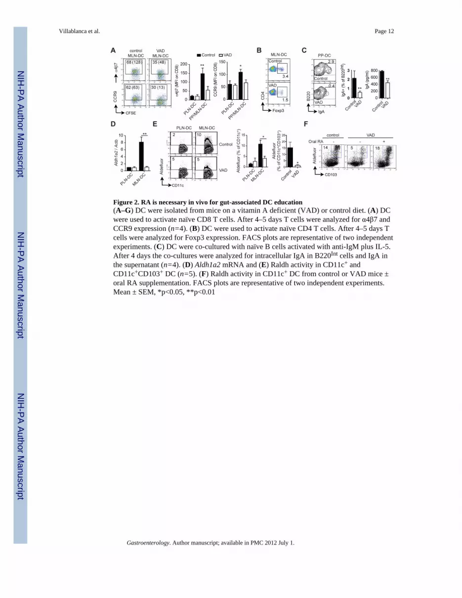
Figure 2. RA is necessary in vivo for gut-associated DC education(A–G) DC were isolated from mice on a vitamin A deficient (VAD) or control diet. (A) DCwere used to activate naïve CD8 T cells. After 4–5 days T cells were analyzed for α4β7 andCCR9 expression (n=4). (B) DC were used to activate naïve CD4 T cells. After 4–5 days Tcells were analyzed for Foxp3 expression. FACS plots are representative of two independentexperiments. (C) DC were co-cultured with naïve B cells activated with anti-IgM plus IL-5.After 4 days the co-cultures were analyzed for intracellular IgA in B220Int cells and IgA inthe supernatant (n=4). (D) Aldh1a2 mRNA and (E) Raldh activity in CD11c+ andCD11c+CD103+ DC (n=5). (F) Raldh activity in CD11c+ DC from control or VAD mice ±oral RA supplementation. FACS plots are representative of two independent experiments.Mean ± SEM, *p<0.05, **p<0.01
Villablanca et al. Page 12
Gastroenterology. Author manuscript; available in PMC 2012 July 1.
NIH
-PA Author Manuscript
NIH
-PA Author Manuscript
NIH
-PA Author Manuscript

Figure 3. RA is sufficient to confer DC with RA synthesizing capacity in vitro(A) Aldh1a2 mRNA in Spleen-DC incubated for 24 h with 100 nM all-trans RA (RA) or100 nM of the indicated nuclear receptor agonists. HX630 and PA024 were used at 1 or 10μM, with similar results (n=5–10). (B–F) Spleen-DC were incubated for 24 h with orwithout 100 nM RA (RA-DC and UT-DC, respectively) and analyzed for (B) Raldh activity(n=5) or (C) washed, pulsed with antigen and used to activate naïve CD8 T cells from TCRtransgenic OT-1xRAG2−/− mice in FBS-free media ± 50 nM retinol. After 4–5 days T cellswere analyzed for α4β7 and CCR9 expression (n=7). (D) Co-cultures were performed asdescribed in (C) either in the presence or absence of the Raldh inhibitor DEAB (n=7). (E)Competitive homing experiment between CD8 T cells activated with RA-DC or UT-DC(n=5). (F) Naïve CD4 T cells from OT-1xRAG2−/− mice were activated with UT-DC orRA-DC. Five days later CD4 T cells were analyzed for Foxp3 expression (n=3).
Villablanca et al. Page 13
Gastroenterology. Author manuscript; available in PMC 2012 July 1.
NIH
-PA Author Manuscript
NIH
-PA Author Manuscript
NIH
-PA Author Manuscript

Figure 4. RA is sufficient to confer DC with RA synthesizing capacity in vivo(A) Wild type mice were supplemented orally with RA (400 μg/dose) every other day for 6days. After that, MLN-DC and PLN-DC were isolated and analyzed for the expression of(B) Aldh1a2 mRNA and (C) Raldh activity (n=5). (D) PLN-DC from control or RA-treatedmice were used to activate naïve CD8 T cells in FBS-free media ± retinol and either in thepresence or absence of DEAB. FACS plots are representative of two independentexperiments. Mean ± SEM, *p<0.05, **p<0.01, ***p<0.001
Villablanca et al. Page 14
Gastroenterology. Author manuscript; available in PMC 2012 July 1.
NIH
-PA Author Manuscript
NIH
-PA Author Manuscript
NIH
-PA Author Manuscript

Figure 5. DC education requires ERK/MAPK signaling(A–C) Spleen-DC were incubated for 24 h with or without 100 nM RA and in the presenceor the absence of inhibitors for p38 (SB203580, 10 μM), ERK (U0126, 10 μM), NF-κB(SN50, 50 μM) or JNK (SP600125, 50 μM). (A) DC were used to activate naïve CD8 Tcells. After 4–5 days T cells were analyzed for α4β7 and CCR9 expression (n=9). (B)Aldh1a2 mRNA expression in DC (n=3). (C) Spleen-DC from DR5-luciferase mice wereincubated for 24 h with or without RA and in the presence or absence of the ERK inhibitor(U0126, 10 μM) and analyzed for their luciferase activity (n=3). (D) Wild type mice wereorally treated with the ERK inhibitor PD0325901 (25 μg/g/dose) every other day for 6 days.After that, CD11c+ MLN-DC were analyzed for CD103 expression and Raldh activity(n=5). Mean ± SEM, *p<0.05, **p<0.01
Villablanca et al. Page 15
Gastroenterology. Author manuscript; available in PMC 2012 July 1.
NIH
-PA Author Manuscript
NIH
-PA Author Manuscript
NIH
-PA Author Manuscript

Figure 6. MyD88 is required for RA-mediated DC education(A–B) Wild type or MyD88−/− Spleen-DC were incubated with 100 nM RA for 24 h. (A)DC were used to activate naïve CD8 T cells. After 4–5 days T cells were analyzed for α4β7and CCR9 expression (n=5). (B) Aldh1a2 (n=4) or Tgm2 (n=5) mRNA expression in DC.(C) Wild type or MyD88−/− mice were supplemented orally with RA (400 μg/dose) everyother day for 6 days and PLN-DC were analyzed for Raldh activity (n=7). (D) Spleen-DCfrom wild type or MyD88−/− mice were analyzed for their expression of Rxra, Rxrb. Rxrg,Rara, Rarb and Rarg mRNA (n=4–6). ND: not detected. (E) Spleen-DC were incubated for24 h with or without 100 nM RA and either in the presence or absence of the RAR®inhibitor LE540 (1 μM) and then analyzed for Aldh1a2 mRNA expression (n=3). (F)Spleen-DC from DR5-luciferase mice were incubated for 24 h with or without 100 nM RAand either in the presence or absence of LE540 and then analyzed for their luciferase activity(n=3). Mean ± SEM, *p<0.05, ***p<0.001
Villablanca et al. Page 16
Gastroenterology. Author manuscript; available in PMC 2012 July 1.
NIH
-PA Author Manuscript
NIH
-PA Author Manuscript
NIH
-PA Author Manuscript

Figure 7. TLR signals contribute to RA-mediated human and murine DC education(A) Monocyte-derived DC (Mo-DC) from healthy donors were treated with either RA (100nM), Pam3CSK4 (0.5 μg/ml) or both from day 3 of differentiation. Raldh activity wasanalyzed at day 6 in CD11c+ cells (n=4). FACS plots show representative results in Mo-DCfrom two donors. (B) Mo-DC pretreated with either RA, Pam3CSK4 or both were washedand cultured with allogenic CD4 T cells activated with anti-CD3 plus anti-CD28 and hTGF-β1 (2 ng/ml). After 4 days, CD4 T cells were analyzed for Foxp3 expression. FACS plotsshow one representative experiment out of two. (C) Spleen-DC were incubated for 24 h inthe presence of 100 nM of RA, Pam3CSK4 (0.5 μg/ml) or both, washed and used to activatenaive CD8 T cells. After 4 days, T cells were analyzed for their expression of α4β7 andCCR9. FACS plots are representative of three experiments. (D) Human Mo-DC were treatedat day 6 with or without 100 nM of RA, Pam3CSK4 (0.5 μg/ml) or both for 24 h, washedand co-cultured with total human T cells activated with plate-bound anti-CD3 plus anti-CD28 antibodies. After 6 days, CD4 and CD8 T cells were analyzed for their expression ofα4β7 (n=3). (E) Spleen-DC were incubated for 24 h in the presence of 100 nM of RA,Pam3CSK4 (0.5 μg/ml) or both, washed and co-cultured with naïve B cells activated withanti-IgM plus IL-5. After 4 days IgA levels were measured in the culture supernatants(n=7). Mean ± SEM, *p<0.05, ***p<0.001
Villablanca et al. Page 17
Gastroenterology. Author manuscript; available in PMC 2012 July 1.
NIH
-PA Author Manuscript
NIH
-PA Author Manuscript
NIH
-PA Author Manuscript


