Embed Size (px)
Citation preview
Mycobacterium tuberculosis Rv2224c modulatesinnate immune responsesJyothi Rengarajan*†, Elissa Murphy*, Arnold Park*, Cassandra L. Krone*, Erik C. Hett*, Barry R. Bloom*,Laurie H. Glimcher*‡§, and Eric J. Rubin*
*Department of Immunology and Infectious Diseases, Harvard School of Public Health, 665 Huntington Avenue, Boston, MA 02115; and ‡Department ofMedicine, Harvard Medical School, Boston, MA 02115
Contributed by Laurie H. Glimcher, November 15, 2007 (sent for review October 17, 2007)
Tuberculosis remains a major global health problem that kills upto 2 million people annually. Central to the success of Myco-bacterium tuberculosis (Mtb) as a pathogen is its ability to evadehost immunity and to establish a chronic infection. Although itsprimary intracellular niche is within macrophages, the underly-ing molecular mechanisms are poorly understood. Here we showthat Rv2224c, a cell envelope-associated predicted protease, iscritical for Mtb virulence. Disruption of Rv2224c led to prolongedsurvival of infected mice and highly reduced lung pathology.Absence of Rv2224c enhanced host innate immune responses,compromised the intracellular survival of Mtb in macrophages,and increased its susceptibility to lysozyme. We provide insightsinto the molecular basis for Rv2224c function by showing thatRv2224c activity promotes processing and extracellular releaseof the Mtb protein, GroEL2. Inhibition of Rv2224c and itstargets offers opportunities for therapeutic interventions andimmune-modulatory strategies.
macrophages � pathogen � intracellular � cell envelope
Mycobacterium tuberculosis (Mtb) is a highly successful humanpathogen that elicits strong innate and adaptive immune
responses. Tuberculosis (TB) is characterized by an inflammatoryresponse that leads to containment, but not eradication, of bacteriawithin granulomas in the lung (1). The Th1-type response elicitedby Mtb contributes to the extensive immunopathology, tissue ne-crosis, and lung damage associated with TB (2). Macrophages arecentral immune cells linking innate and adaptive immunity bypromoting lymphocyte activation and recruitment (1). They alsoserve as the primary niche in vivo, because Mtb actively replicateswithin macrophages. Mtb inhibits fusion of phagosomes with lyso-somes and interferes with antigen presentation, IFN-�-mediatedsignaling pathways, and transcriptional responses (3). Thus, theability of Mtb to subvert macrophage microbicidal functions likelycontributes to the hallmark delayed T cell response in TB, but thebacterial factors that mediate these processes are poorly understood(2, 3). In recent genome-wide studies, we comprehensively identi-fied the bacterial genes necessary for Mtb survival in vivo and inmacrophages (4, 5). Our data predicted that one of these genes,Rv2224c, was required for both in vivo growth in mice and intra-cellular replication in macrophages (4). In this study, we show thatRv2224c is indeed critical for Mtb virulence in vivo, promotessurvival in macrophages, and modulates innate immune control ofinfection.
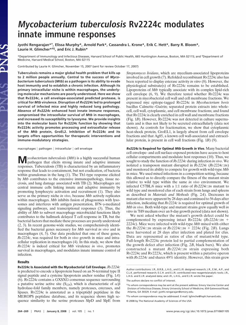
ResultsRv2224c Is Associated with the Mycobacterial Cell Envelope. Rv2224cis predicted to encode a lipoprotein based on an N-terminal type IIsignal peptide and a cysteine lipoprotein anchor residue (Fig. 1A)(6). Rv2224c contains a GXSXG consensus sequence surroundinga putative serine active site (S228), which is characteristic of �/�hydrolase-fold family members, namely proteases, esterases, andlipases. Rv2224c is annotated as a tripeptidyl peptidase in theMEROPS peptidase database, and its sequence shows high se-quence similarity to the serine proteases SlpD and SlpE from
Streptomyces lividans, which are mycelium-associated lipoproteinsinvolved in cell growth (7). Refolded recombinant Rv2224c also hasbeen reported to display esterase activity in vitro (8). However, thephysiological substrate(s) of Rv2224c remains to be established.Lipoproteins of Mtb typically associate with its complex lipid-richcell envelope (6, 9). We therefore tested whether Rv2224c waspresent in mycobacterial cell wall and cell membrane fractions. Weexpressed myc epitope-tagged Rv2224c in Mycobacterium bovisbacillus Calmette–Guerin; separated protein extracts into whole-cell, cell wall, cytoplasmic, and cell membrane fractions; and foundthat Rv2224c is clearly enriched in cell wall and membrane fractions(Fig. 1B). However, Rv2224c was not detected in culture superna-tants and is thus not likely to be secreted extracellularly (data notshown). As controls for fractionation, we show that cytoplasmicheat-shock protein, GroEL1, is largely absent from cell envelopefractions and that Ag85, a known cell wall-associated and extracel-lular protein, is present in cell wall fractions (Fig. 1B) (9).
Rv2224c Is Required for Optimal Mtb Growth in Vivo. Many bacterialcell envelope- and cell surface-exposed proteins have access to hostcellular compartments and modulate host responses (10). Thus, wesought to study the function of Rv2224c during infection in vivo. Weisolated a transposon mutant disrupted in Rv2224c (Rv2224c::tn)and monitored its ability to compete for growth with wild-type Mtbin mice. We used mixed infections in a competition setting, becausethis allowed us to directly compare the fitness of the mutant strainrelative to wild type within the same in vivo environment. Weinfected C57BL/6 mice with a 1:1 ratio of Rv2224c::tn mutant towild type and monitored cfus of each strain from lungs and spleensas described in Materials and Methods (Fig. 2A). Decreases inmutant cfus were apparent by 28 days and continued to 56 days afterinfection, indicating that Rv2224c is required for optimal growth ofMtb in vivo. Both wild-type and mutant strains grew equally well invitro in 7H9 medium over an 8-day growth period (data not shown).
We next asked whether the mutant’s growth defect could becomplemented by expressing intact Rv2224c (Rv2224c::tn �2224c). Mice were infected with wild-type Mtb mixed with eitherthe Rv2224c::tn strain or Rv2224c::tn � 2224c (Fig. 2B). Lungswere harvested at 28 days after infection and plated for cfus.Data are represented as ratios of cfus of mutant/wild type.Full-length Rv2224c protein led to partial complementation ofthe growth defect after infection (Fig. 2B, black bars). We alsoconstructed a mutant Rv2224c::tn strain expressing bothRv2224c and Rv2223c, which is present within a putative operonwith Rv2224c and shares 49% identity. However, this strain grew
Author contributions: J.R., B.R.B., L.H.G., and E.J.R. designed research; J.R., E.M., A.P., andC.L.K. performed research; E.C.H. and E.J.R. contributed new reagents/analytic tools; J.R.,L.H.G. and E.J.R. analyzed data; and J.R., L.H.G., and E.J.R. wrote the paper.
The authors declare no conflict of interest.
†To whom correspondence may be sent at the present address: Emory Vaccine Center andDivision of Infectious Disease, Emory University School of Medicine, 654 Gatewood Road,Atlanta, GA 30329. E-mail: [email protected].
§To whom correspondence may be addressed. E-mail: [email protected].
© 2008 by The National Academy of Sciences of the USA
264–269 � PNAS � January 8, 2008 � vol. 105 � no. 1 www.pnas.org�cgi�doi�10.1073�pnas.0710601105
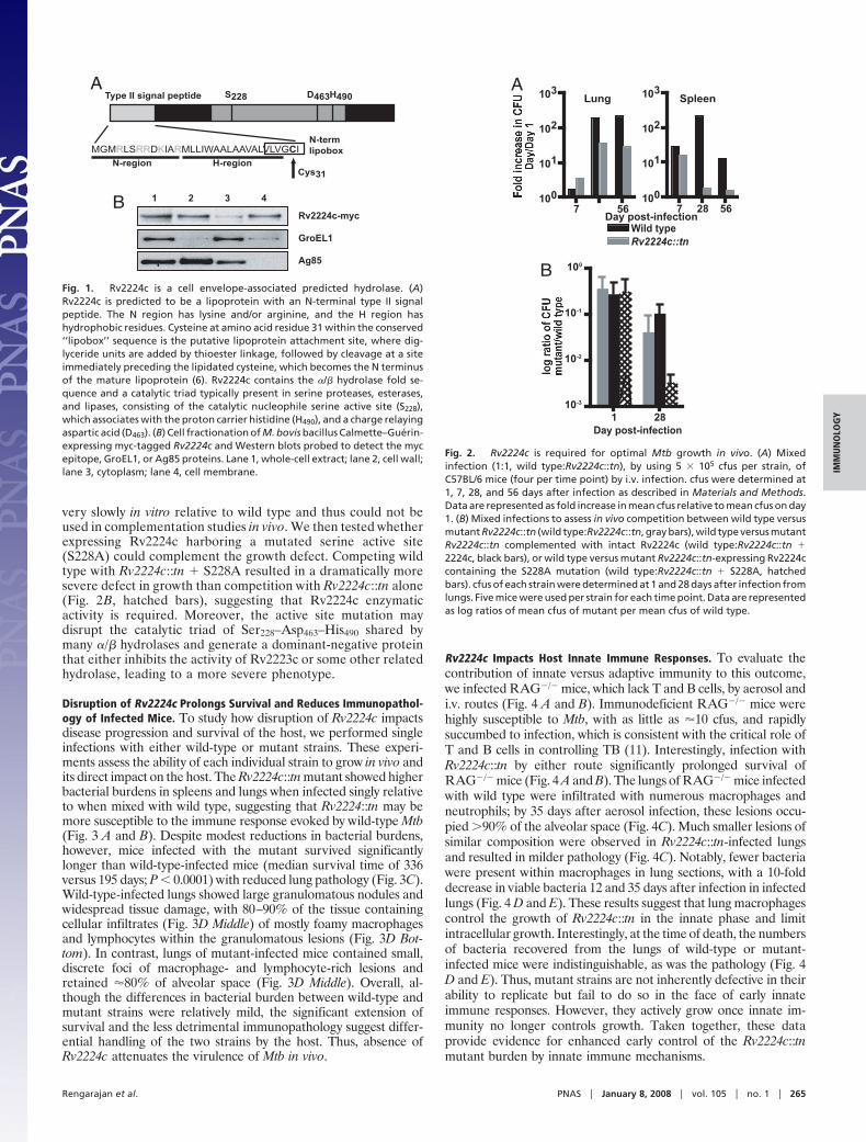
very slowly in vitro relative to wild type and thus could not beused in complementation studies in vivo. We then tested whetherexpressing Rv2224c harboring a mutated serine active site(S228A) could complement the growth defect. Competing wildtype with Rv2224c::tn � S228A resulted in a dramatically moresevere defect in growth than competition with Rv2224c::tn alone(Fig. 2B, hatched bars), suggesting that Rv2224c enzymaticactivity is required. Moreover, the active site mutation maydisrupt the catalytic triad of Ser228–Asp463–His490 shared bymany �/� hydrolases and generate a dominant-negative proteinthat either inhibits the activity of Rv2223c or some other relatedhydrolase, leading to a more severe phenotype.
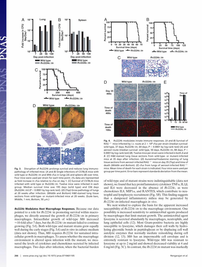
Disruption of Rv2224c Prolongs Survival and Reduces Immunopathol-ogy of Infected Mice. To study how disruption of Rv2224c impactsdisease progression and survival of the host, we performed singleinfections with either wild-type or mutant strains. These experi-ments assess the ability of each individual strain to grow in vivo andits direct impact on the host. The Rv2224c::tn mutant showed higherbacterial burdens in spleens and lungs when infected singly relativeto when mixed with wild type, suggesting that Rv2224::tn may bemore susceptible to the immune response evoked by wild-type Mtb(Fig. 3 A and B). Despite modest reductions in bacterial burdens,however, mice infected with the mutant survived significantlylonger than wild-type-infected mice (median survival time of 336versus 195 days; P � 0.0001) with reduced lung pathology (Fig. 3C).Wild-type-infected lungs showed large granulomatous nodules andwidespread tissue damage, with 80–90% of the tissue containingcellular infiltrates (Fig. 3D Middle) of mostly foamy macrophagesand lymphocytes within the granulomatous lesions (Fig. 3D Bot-tom). In contrast, lungs of mutant-infected mice contained small,discrete foci of macrophage- and lymphocyte-rich lesions andretained �80% of alveolar space (Fig. 3D Middle). Overall, al-though the differences in bacterial burden between wild-type andmutant strains were relatively mild, the significant extension ofsurvival and the less detrimental immunopathology suggest differ-ential handling of the two strains by the host. Thus, absence ofRv2224c attenuates the virulence of Mtb in vivo.
Rv2224c Impacts Host Innate Immune Responses. To evaluate thecontribution of innate versus adaptive immunity to this outcome,we infected RAG�/� mice, which lack T and B cells, by aerosol andi.v. routes (Fig. 4 A and B). Immunodeficient RAG�/� mice werehighly susceptible to Mtb, with as little as �10 cfus, and rapidlysuccumbed to infection, which is consistent with the critical role ofT and B cells in controlling TB (11). Interestingly, infection withRv2224c::tn by either route significantly prolonged survival ofRAG�/� mice (Fig. 4 A and B). The lungs of RAG�/� mice infectedwith wild type were infiltrated with numerous macrophages andneutrophils; by 35 days after aerosol infection, these lesions occu-pied �90% of the alveolar space (Fig. 4C). Much smaller lesions ofsimilar composition were observed in Rv2224c::tn-infected lungsand resulted in milder pathology (Fig. 4C). Notably, fewer bacteriawere present within macrophages in lung sections, with a 10-folddecrease in viable bacteria 12 and 35 days after infection in infectedlungs (Fig. 4 D and E). These results suggest that lung macrophagescontrol the growth of Rv2224c::tn in the innate phase and limitintracellular growth. Interestingly, at the time of death, the numbersof bacteria recovered from the lungs of wild-type or mutant-infected mice were indistinguishable, as was the pathology (Fig. 4D and E). Thus, mutant strains are not inherently defective in theirability to replicate but fail to do so in the face of early innateimmune responses. However, they actively grow once innate im-munity no longer controls growth. Taken together, these dataprovide evidence for enhanced early control of the Rv2224c::tnmutant burden by innate immune mechanisms.
Type II signal peptide S228 D463H490
MGMRLSRRDKIARMLLIWAALAAVALVLVGCIN-termlipobox
Cys31N-region H-region
Rv2224c-myc
GroEL1
Ag85
1 2 3 4B
A
Fig. 1. Rv2224c is a cell envelope-associated predicted hydrolase. (A)Rv2224c is predicted to be a lipoprotein with an N-terminal type II signalpeptide. The N region has lysine and/or arginine, and the H region hashydrophobic residues. Cysteine at amino acid residue 31 within the conserved‘‘lipobox’’ sequence is the putative lipoprotein attachment site, where dig-lyceride units are added by thioester linkage, followed by cleavage at a siteimmediately preceding the lipidated cysteine, which becomes the N terminusof the mature lipoprotein (6). Rv2224c contains the �/� hydrolase fold se-quence and a catalytic triad typically present in serine proteases, esterases,and lipases, consisting of the catalytic nucleophile serine active site (S228),which associates with the proton carrier histidine (H490), and a charge relayingaspartic acid (D463). (B) Cell fractionation of M. bovis bacillus Calmette–Guerin-expressing myc-tagged Rv2224c and Western blots probed to detect the mycepitope, GroEL1, or Ag85 proteins. Lane 1, whole-cell extract; lane 2, cell wall;lane 3, cytoplasm; lane 4, cell membrane.
B
1 2810-3
10-2
10-1
100
Day post-infection
Lung Spleen
7 28 56100
101
102
103
Wild typeRv2224c::tn
7 56100
101
102
103
Day post-infection
A
Fig. 2. Rv2224c is required for optimal Mtb growth in vivo. (A) Mixedinfection (1:1, wild type:Rv2224c::tn), by using 5 � 105 cfus per strain, ofC57BL/6 mice (four per time point) by i.v. infection. cfus were determined at1, 7, 28, and 56 days after infection as described in Materials and Methods.Data are represented as fold increase in mean cfus relative to mean cfus on day1. (B) Mixed infections to assess in vivo competition between wild type versusmutant Rv2224c::tn (wild type:Rv2224c::tn, gray bars), wild type versus mutantRv2224c::tn complemented with intact Rv2224c (wild type:Rv2224c::tn �2224c, black bars), or wild type versus mutant Rv2224c::tn-expressing Rv2224ccontaining the S228A mutation (wild type:Rv2224c::tn � S228A, hatchedbars). cfus of each strain were determined at 1 and 28 days after infection fromlungs. Five mice were used per strain for each time point. Data are representedas log ratios of mean cfus of mutant per mean cfus of wild type.
Rengarajan et al. PNAS � January 8, 2008 � vol. 105 � no. 1 � 265
IMM
UN
OLO
GY
Rv2224c Modulates Host Macrophage Responses. Because our datapointed to a role for Rv2224c in promoting survival within macro-phages, we directly assessed the growth of Rv2224c::tn in primarymacrophages. Intracellular growth of wild-type Mtb increased�10-fold after 7 days, but the Rv2224c::tn mutant failed to continuegrowing (Fig. 5A). Both wild-type and mutant strains grew equallywell during the early stages (Fig. 5A) and in vitro in culture medium(data not shown). Thus, Mtb requires Rv2224c for sustained intra-cellular growth in macrophages. To assess whether the macrophageenvironment is altered upon infection with the mutant, we mea-sured the levels of cytokines and chemokines secreted by infectedmacrophages. Two days after infection, when the bacterial burden
of wild-type and of mutant strains were indistinguishable (data notshown), we found that key proinflammatory cytokines TNF�, IL1�,and IL6 were decreased in the absence of Rv2224c, as werechemokines IL8, MIP1�, and RANTES, which contribute to neu-trophil and lymphocyte recruitment (Fig. 5B). This finding suggeststhat a dampened inflammatory milieu may be generated byRv2224c::tn-infected macrophages in vivo.
We next wished to explore the basis for the apparent increasedsusceptibility of Rv2224c::tn to the macrophage environment. Onepossibility is increased sensitivity to antimicrobial agents producedby macrophages that limit mutant growth. The antimicrobial agentlysozyme is secreted abundantly by macrophages, neutrophils, andlung epithelial cells (12). Most Gram-positive bacteria are highlysusceptible to lysozyme, which damages their cell walls by hydro-lyzing glycosidic bonds in peptidoglycan or by displacing cell wallautolytic enzymes that normally mediate remodeling during celldivision (12, 13). Mtb has an impermeable cell envelope and isextremely resistant to lysozyme. Wild-type Mtb was resistant tolysozyme at up to 2 mg/ml and showed decreased viability at 4 and6 mg/ml (Fig. 5C). In contrast, the Rv2224::tn mutant was markedly
A B
7 28 56 140100
101
102
103
Wild typeRv2224c::tn
Day post-infection7 28 56 140
100
101
102
Wild typeRv2224c::tn
Day post-infection
Lung Spleen
0 200 400 6000
25
50
75
100
Wild type Rv2224c::tn
Survival time (days)
C
Wild type Rv2224c::tnD
Fig. 3. Disruption of Rv2224c prolongs survival and reduces lung immuno-pathology of infected mice. (A and B) Single infections of C57BL/6 mice withwild type or Rv2224c::tn and Mtb cfus in lungs (A) and spleens (B) over time.Four mice were used per strain for each time point. cfu data are representedas fold increase in cfus relative to cfus on day 1. (C) Survival of C57BL/6 miceinfected with wild type or Rv2224c::tn. Twelve mice were infected in eachgroup. Median survival time was 195 days (wild type) and 336 days(Rv2224c::tn) (P � 0.0001 by log-rank test). (D) (Top) Gross pathology of lungsat 20 weeks after infection. (Middle and Bottom) H&E-stained lung tissuesections from wild-type- or mutant-infected mice at 20 weeks. (Scale bars,Middle, 1 mm; Bottom, 50 �m.)
Wild type Rv2224c::tnC
Rv2224c::tnWild type
Day 35 400x
Time of death 400x
Time of death 100x
1 12 35 50 89101102103104105106107108109
Wild type
Time of Death
Rv2224c::tn
D E
A
0 25 50 75 100 1250
25
50
75
100
Wild type Rv2224c::tn
Survival time (days)
B
0 10 20 30 40 50 60 700
25
50
75
100
Wild typeSurvival time (days)
Rv2224c::tn
Fig. 4. Rv2224c modulates innate immune responses. (A and B) Survival ofRAG�/� mice infected by i.v. route at 2 � 106 cfus per strain (median survival:wild type, 37 days; Rv2224c::tn, 64 days; P � 0.0001 by log-rank test) (A) andaerosol route (median survival: wild type, 50 days; Rv2224c::tn, 84 days; P �0.0001 by log-rank test) (B). Twelve mice per strain were infected in both A andB. (C) H&E-stained lung tissue sections from wild-type- or mutant-infectedmice at 35 days after infection. (D) Auramine/rhodamine staining of lungtissue sections from aerosol-infected RAG�/� mice on day 35 (Top) and time ofdeath (Middle and Bottom). (E) cfus from lungs of aerosol-infected RAG�/�
mice. Mean time of death for each strain is indicated. Four mice were used pergroup per time point. Error bars represent standards deviation from the mean.
266 � www.pnas.org�cgi�doi�10.1073�pnas.0710601105 Rengarajan et al.
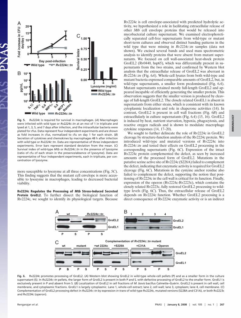
more susceptible to lysozyme at all three concentrations (Fig. 5C).This finding suggests that the mutant cell envelope is more acces-sible to lysozyme in macrophages, leading to decreased bacterialviability.
Rv2224c Regulates the Processing of Mtb Stress-Induced SecretedProtein GroEL2. To further dissect the biological function ofRv2224c, we sought to identify its physiological targets. Because
Rv2224c is cell envelope-associated with predicted hydrolytic ac-tivity, we hypothesized a role in facilitating extracellular release ofother Mtb cell envelope proteins that would be released intomycobacterial culture supernatant. We examined electrophoreti-cally separated cell-free supernatants from wild-type or mutantshort-term cultures and observed distinct banding patterns in thewild type that were missing in Rv2224c::tn samples (data notshown). We excised several bands and used mass spectrometricanalysis to identify proteins that were absent from mutant super-natants. We focused on cell wall-associated heat-shock proteinGroEL2 (Rv0440, hsp65), which was differentially present in su-pernatants from the two strains, and confirmed by Western blotanalysis that the extracellular release of GroEL2 was aberrant inRv2224c::tn (Fig. 6A). Whole-cell lysates from both wild-type andmutant bacteria expressed comparable amounts of GroEL2, but, inwild-type supernatants, a smaller form predominated (Fig. 6A).Mutant supernatants retained mostly full-length GroEL2 and ap-peared incapable of efficiently generating the smaller protein. Thisobservation suggests that the smaller version is produced by cleav-age of full-length GroEL2. The closely related GroEL1 is absent insupernatants from either strain, which is consistent with its knowncytoplasmic localization and role in chaperone activities (14). Incontrast, GroEL2 is present in cell wall fractions (Fig. 6B) andextracellularly in culture supernatants (Fig. 6A) (15, 16). GroEL2is induced by heat, nutrient starvation, hypoxia, phagocytosis, andreactive oxygen radicals and is shown to modulate macrophagecytokine responses (14, 17–20).
We sought to further delineate the role of Rv2224c in GroEL2cleavage by structure-function analysis of the Rv2224c protein. Weintroduced wild-type and mutated versions of Rv2224c intoRv2224c::tn and tested their effects on GroEL2 processing in thecorresponding supernatants (Fig. 6C). Expression of the intactRv2224c protein complemented the defect, as seen by increasedamounts of the processed form of GroEL2. Mutations in theputative serine active site of Rv2224c (S228A) failed to complementthe defect, indicating that enzymatic activity is required for GroEL2cleavage (Fig. 6C). Mutations in the cysteine anchor residue alsofailed to complement the defect, supporting the notion that posi-tioning of Rv2224c in the cell wall is critical for its function. Finally,expression of the operon (Rv2224c-Rv2223c), which contains theclosely related Rv2223c, fully restored GroEL2 processing to wild-type levels (Fig. 6C). Thus, the extracellular release of GroEL2depends on Rv2224c function. Whether GroEL2 processing is adirect consequence of Rv2224c enzymatic activity or is an indirect
A
3 5 70
5
10
15
20
Wild type Rv2224c::tnDay post-infection
C
2 4 60.00
0.25
0.50
0.75
1.00
Wild typeRv2224c::tn
Lysozyme (mg/ml)
B
IL1β100
101
102
103
104
Wild type Rv2224c::tnTNFα IL6
MIP1αIL8
MCP1
Rantes
Fig. 5. Rv2224c is required for survival in macrophages. (A) Macrophageswere infected with wild type or Rv2224c::tn at an moi of 1 in triplicate andlysed at 1, 3, 5, and 7 days after infection, and the intracellular bacteria wereplated for cfus. Data represent four independent experiments and are shownas fold increases in cfus, normalized to cfu on day 1 for each strain. (B)Secretion of cytokines and chemokines by macrophages 48 h after infectionwith wild type or Rv2224c::tn. Data are representative of three independentexperiments. Error bars represent standard deviation from the mean. (C)Survival index of wild-type Mtb or Rv2224c::tn in the presence of lysozyme(ratio of cfu of each strain in the presence/absence of lysozyme). Data arerepresentative of four independent experiments, each in triplicate, per con-centration of lysozyme.
GroEL2
GroEL1
1 2 3 4
GroEL2
GroEL1
wt mutant +wt 2224c +S228A +C31A +Operon
P S P S P S P SP S P S P S
Complementation of Rv2224c::tn mutant
GroEL2
GroEL1
wt Rv2224c::tn
P S P S
A B
C
Fig. 6. Rv2224c promotes processing of GroEL2. (A) Western blot showing GroEL2 in wild-type whole-cell pellets (P) and as a smaller form in the culturesupernatant (S). In Rv2224c::tn pellets, the larger form of GroEL2 is present in both P and S, with defective processing of GroEL2 to the smaller form. GroEL1 isexclusively present in P and absent from S. (B) Localization of GroEL2 in cell fractions of M. bovis bacillus Calmette–Guerin. GroEL2 is present in cell wall, cellmembrane, and cytoplasmic fractions. GroEL1 is largely cytoplasmic. Lane 1, whole-cell extract; lane 2, cell wall; lane 3, cytoplasm; lane 4, cell membrane. (C)Complementation of GroEL2 processing defect in Rv2224c::tn by expression in trans of wild-type Rv2224c, mutated versions (S228A and C31A), or both Rv2223cand Rv2224c (operon).
Rengarajan et al. PNAS � January 8, 2008 � vol. 105 � no. 1 � 267
IMM
UN
OLO
GY
consequence of regulation of intermediate proteins is unclear andawaits detailed biochemical characterization.
DiscussionInnate immune responses are critical for controlling infection andlaunching adaptive immunity. By avoiding macrophage microbici-dal functions and by replicating intracellularly, Mtb interferes withthe initiation of adaptive immunity (1). In this article, we demon-strate that Rv2224c is critical for Mtb disease progression andintracellular survival and modulates innate immune responses.Interestingly, aerosol infection of RAG�/� mice shows that, in theabsence of Rv2224c, early control of Mtb replication is in factpossible (Fig. 4) and maps to growth limitation in macrophages(Fig. 5A). Although Rv2224c::tn mutant bacteria eventually over-whelm host innate responses, this early control leads to significantextension of survival. Thus, inhibiting Mtb factors such as Rv2224ccould enhance innate immunity to TB. Macrophages infected withRv2224c::tn also show dampened inflammatory milieu, andRv2224c::tn was more susceptible to cell wall damage by lysozyme(Fig. 5 B and C). These results suggest that inhibition of Rv2224ccould help drive the immune response away from its detrimentalaspects and could simultaneously debilitate Mtb, resulting in morefavorable outcomes for the host.
Competition experiments showed that Rv2224c is requiredfor optimal growth in vivo (Fig. 2). Complementation withintact Rv2224c did not fully restore the growth defect (Fig.2C), suggesting that disruption of Rv2224c may affect thefunction of the related Rv2223c. We can detect expression ofRv2223c mRNA in the Rv2224c::tn (data not shown), andtherefore transcription of Rv2223c does not appear to becompromised. However, Rv2224c may be involved in posttran-scriptional regulation of Rv2223c, and the two proteins mayfunction in concert for optimal activity toward their targets.Unfortunately, complementation of the in vivo growth defectby expressing both Rv2224c and Rv2223c was not feasiblebecause ectopic expression of the operon resulted in slowgrowth of the strain in vitro. Speculation that Rv2223c may infact work in conjunction with Rv2224c is supported by theobservation that expressing both genes results in bettercomplementation of the GroEL2-processing defect (Fig. 6C).In addition, mutating the serine active site of Rv2224c resultedin a more severe growth defect in vivo, suggesting thatdisrupting Rv2224c activity may compromise Rv2223c func-tion directly or indirectly by generating a dominant-negativeprotein that interferes with the function of Rv2223c or otherrelated hydrolases.
We have uncovered a role for Rv2224c in extracellular GroEL2release from the cell wall of Mtb. GroEL2 is a highly expressed,immunodominant protein and induced by many physiologicalstresses related to intracellular growth, including phagocytosis,hypoxia, nutrient limitation, and reactive oxygen radicals (14, 21,22). Recent purification of GroEL2 for crystallization studiesrevealed that it eluted as both a full-length and smaller N-terminallyprocessed form, suggesting that physiological proteolysis mightoccur (23). Thus, the cleaved GroEL2 protein may constitute thefunctionally active form, whose release depends on Rv2224c activ-ity. Whether or not GroEL2 is a direct substrate for Rv2224cremains unclear and awaits further biochemical characterization.Although it is unlikely to be the only target of Rv2224c, GroEL2 hasbeen implicated in modulating proinflammatory cytokine re-sponses in macrophages, which is consistent with our observationsof dampened inflammatory milieu (Fig. 5C) (14, 19, 20). A detailedanalysis of Rv2224c enzymatic activity toward GroEL2 and addi-tional physiological substrates should help elucidate the precisebiochemical function of Rv2224c.
Our results implicating Rv2224c in the processing of GroEL2 andthe resistance to lysozyme suggest that other cell envelope com-ponents may be substrates for RV2224c activity. This idea also was
advanced in studies from Flores et al. (24), who isolated a trans-poson mutant with insertions in Mycobacterium smegmatis expA, thehomolog of Mtb Rv2224c, in a screen designed to study hypersus-ceptibility to �-lactam antibiotics, which act on bacterial cell wallpeptidoglycan. Interestingly, M. smegmatis expA mutants showedincreased susceptibility to lysozyme and displayed elongated cellmorphology with swollen termini. The authors speculated thatexpA and expB (the Rv2223c homolog) might be directly involvedin hydrolytic breakdown of cell wall components or indirectlyinvolved by regulating proteins involved in autolysis or cell wallremodeling (24). Rv2224c is classified as an �/� hydrolase in afamily with both esterase and amidase members (25). Its closestbiochemically characterized homolog, the secreted SlpD triamin-opeptidase from S. lividans, was identified by its activity against anamide (7). Protease activity is consistent with the requirement ofRv2224c for correct processing of GroEL2. However, severalproteins related by homology to Rv2224c are carboxyesterases andlipases. Indeed, a recent study showed that refolded recombinantRv2224c can hydrolyze ester compounds, which suggests that cellenvelope lipids may be substrates (8). The chemical similarity ofester and amide hydrolysis is such that most proteases also catalyzethe degradation of ester substrates, although the converse is nottypically true (26). These possibilities will be clarified when phys-iological substrates of Rv2224c are established. Many Mtb proteinsare exported to the cell envelope; some of these are secretedextracellularly by specialized secretion systems and in turn promoteinteractions with host cells (27, 28). Our studies suggest thatcleavage of cell envelope proteins constitutes an additional mech-anism by which Mtb can release critical extracellular factors. En-zymatic regulation of their release in vivo would facilitate rapidadaptation to changing immune environments.
In summary, we have shown that Rv2224c is a key determinantof disease outcome in vivo. Rv2224c modulates innate immuneresponses in macrophages and is required for extracellular releaseof the Mtb stress-induced protein, GroEL2. The cell surface acces-sibility of Rv2224c and its importance in host–pathogen interactionsmake it a target for therapeutics and immune-modulatorystrategies.
Materials and MethodsBacterial Strains and Media. Mtb H37Rv strains and M. bovis bacillus Calmette–Guerin Pasteur were grown at 37°C in Middlebrook 7H9 broth or 7H10 agar asdescribed in ref. 4. For the Rv2224c::tn mutant, 25 �g/ml kanamycin (Sigma–Aldrich)wasadded,and, forcomplementedstrains,50�g/mlhygromycin (Roche)was added. The Mtb transposon library has been described in detail in ref. 29. TheRv2224::tnmutantwasobtainedbysequencingmutants fromthe librarybyusingarbitrary primers as described in ref. 4. The mutant was maintained in thepresence of 25 �g/ml kanamycin throughout.
Construction of Plasmids and Strains. To construct myc-tagged Rv2224c driven byits own promoter, the gene was amplified by 5�-CCTCGGGCGATGGTCTAGATG-CACCATGC-3� and 5�-CGTTTCTTCGCCACTAGTGCACTTGGCG-3� cloned into theXbaIandSpeIsitesofpMV762(hyg)withC-terminalc-myctag.TheC31Smutationwas made with site-directed mutagenesis by using 5�-GTTCTTGTGGGCTCCATC-CGCGTGGTC-3� and S228A 5�-CTACCTGGGCTACGCGTACGGCACC-3�. TheRv2223c-2224c operon was cloned into the XbaI and SpeI sites of pMV762 (hyg)by using 5�-CCTCGGGCGATGGTCTAGATGCACCATGC-3� and 5�-AAGGGGAGT-GCGCGACTAGTCCAGGGCG-3�.
Preparation of Protein Extracts and Western Blotting. Each Mtb strain wasgrown to an OD600 of 0.8, pelleted, washed, and grown in Sautons’ medium(30) plus 0.05% Tween 80 to an OD600 of 0.6, resuspended into Sautons’medium minus Tween 80, and grown for 24 h at 37°C. Supernatants wereconcentrated by using Centricon Plus-70 (Millipore). Each cell pellet wasresuspended in 50 mM Tris, 10 mM NaCl (pH 8.0), and glass beads (Bio101)with bead beating for 45 s, was centrifuged at 16,000 � g for 15 min at 4°C,and 150 �l was removed for protein estimation. Remaining extracts wereboiled with SDS protein-loading buffer for 20 min. Samples were separatedby a 200-V SDS/PAGE by using NuPage 10% [bis(2-hydroxyethyl)amino]tris(hydroxymethyl)methane or 2-[bis(2-hydroxyethyl)amino]-2-(hydroxymethyl)-
268 � www.pnas.org�cgi�doi�10.1073�pnas.0710601105 Rengarajan et al.
1,3-propanediol (Bistris) gels (Invitrogen) and transferred onto nitrocellulose at30 V for 1 h. Antibodies used were mouse anti-GroEL2 (1:2,000 in 1% BSA;Imgenex), mouse anti-Ag85 (1:500 in 1% BSA; Abcam), GroEL1:rabbit anti-His(1:5,000 in 1% BSA; Novus Biologicals), rabbit anti-c-myc [1:10,000 in 3% 50 mMTris�HCl (pH 7.4), 100 mM NaCl, and 5% nonfat dry milk (Blotto); Novus Biologi-cals], and HRP conjugated anti-mouse or anti-rabbit serum (1:10,000; Kirkegaard& Perry Laboratories) as a secondary antibody.
Fractionation of M. bovis Bacillus Calmette–Guerin. Fractionation was per-formed essentially as described in ref. 31. Fractions were loaded onto gels nor-malized for cell numbers proportionally.
Macrophage Survival Assays and Cytokine Assays. Macrophages were derivedfrom bone marrow of C57BL/6 mice as described in ref. 4. Macrophages wereplated onto 24-well plates (2 � 105 per well). Each strain was used for infection (intriplicate per time point) at an moi of 1 as described in ref. 4. Cell-free superna-tants from macrophage monolayers 48 h after infection (moi of 5 in triplicate)wereassayedbyusingaLuminexELISAkitandwereanalyzedbyusingtheBioplex200 Luminex system (Bio-Rad). Error bars represent standard deviation from themean.
Lysozyme Susceptibility Assays. These assays were developed in consultationwith M. Pavelka. We incorporated hen egg lysozyme (MP Biomedicals) directlyinto 7H10 plates and plated serial dilutions of Mtb strains onto plates containing0, 2, 4, or 6 mg/ml lysozyme. Plates were incubated for 4–5 weeks at 37°C andscored for cfus.
Infection of Mice with Mtb. All animal experiments were approved by theInstitutional Animal Care and Use Committee of Harvard University. For aerosolinfections, RAG�/� mice (C67BL/6) were infected by using an aerosol apparatus
manufactured by the College of Engineering Shops at the University of Wiscon-sin, Madison. The mice were then exposed for 40 min, resulting in 10 cfus permouse lung (determined by plating lungs on day 1 after infection; four mice pergroup). For i.v. infections, mice were infected by tail-vein injection, with 5 � 105
cfus of each strain for mixed infections and 2 � 106 cfus for single infections (fourmiceper timepoint).Organswerehomogenized,andserialdilutionswereplatedfor cfus onto 7H10, 7H10 kanamycin (for the mutant Rv2224c::tn), or 7H10kanamycin plus hygromycin (for Rv2224c::tn complemented).
Statistical Analysis. GraphPad Prizm software, Version 4.0, was used for allanalyses.
Histology and Staining of Tissue Sections. Organs were fixed in 10% bufferedformalin and embedded in paraffin, and 6-�m sections were stained with H&E.For detecting Mtb in tissue sections, slides were baked at 60°C and washed twicein xylenes; decreasing concentrations of ethanol were used to dehydrate tissue,and slide sections were incubated in auramine–rhodamine at 37°C for 15 min,washed in 1% HCl/70% EtOH, and counterstained by using Harris’ modifiedhaematoxylin.Excess stainwas removedbysequentialwashing inacidicandbasicsolutions, rehydrated by reversing sequence in ethanol, and cleared in xylenes.Images were captured on a Nikon Eclipse 80i microscope by using a SPOT digitalcamera.
ACKNOWLEDGMENTS. We thank Dr. R. Bronson (Harvard Rodent PathologyCore) for his expertise and insights; Drs. G. Petsko, D. Ringe, J. Naffin, G. Brandt,and S. Sampson for helpful discussions; Dr. C. Sassetti for isolation of theRv2224c:tn mutant; Drs. D. Sarracino, M. Chase, and S. Fortune for their input inmass spectrometry experiments; Ms. I. Breiterene for technical assistance; and Dr.Martin Pavelka for expert advice on lysozyme-susceptibility plate assays. Thiswork was supported by National Institutes of Health grants (to B.R.B., L.H.G., andE.J.R.) and the New York Community Trust Heiser Program (J.R.).
1. Bhatt K, Salgame P (2007) Host innate immune response to Mycobacterium tubercu-losis. J Clin Immunol 27:347–362.
2. Flynn JL (2004) Immunology of tuberculosis and implications in vaccine development.Tuberculosis (Edinburgh) 84:93–101.
3. Houben EN, Nguyen L, Pieters J (2006) Interaction of pathogenic mycobacteria with thehost immune system. Curr Opin Microbiol 9:76–85.
4. Rengarajan J, Bloom BR, Rubin EJ (2005) Genome-wide requirements for Mycobacte-rium tuberculosis adaptation and survival in macrophages. Proc Natl Acad Sci USA102:8327–8332.
5. Sassetti CM, Rubin EJ (2003) Genetic requirements for mycobacterial survival duringinfection. Proc Natl Acad Sci USA 100:12989–12994.
6. Sutcliffe IC, Harrington DJ (2004) Lipoproteins of Mycobacterium tuberculosis: An abun-dant and functionally diverse class of cell envelope components. FEMS Microbiol Rev28:645–659.
7. Binnie C, et al. (1995) Isolation and characterization of two genes encoding proteasesassociated with the mycelium of Streptomyces lividans 66. J Bacteriol 177:6033–6040.
8. Lun S, Bishai WR (2007) Characterization of a novel cell wall-anchored protein withcarboxyesterase activity required for virulence in Mycobacterium tuberculosis. J BiolChem 282:18348–18356.
9. Lee RE, Brennan PJ, Besra GS (1996) Mycobacterium tuberculosis cell envelope. CurrTop Microbiol Immunol 215:1–27.
10. Finlay BB, Falkow S (1997) Common themes in microbial pathogenicity revisited.Microbiol Mol Biol Rev 61:136–169.
11. Flynn JL, Chan J (2001) Immunology of tuberculosis. Annu Rev Immunol 19:93–129.12. Ganz T (2002) Antimicrobial polypeptides in host defense of the respiratory tract. J Clin
Invest 109:693–697.13. Keep NH, Ward JM, Cohen-Gonsaud M, Henderson B (2006) Wake up! Peptidoglycan
lysis and bacterial non-growth states. Trends Microbiol 14:271–276.14. Qamra R, Mande, Coates AR, Henderson B (2005) The unusual chaperonins of Myco-
bacterium tuberculosis. Tuberculosis (Edinburgh) 85:385–394.15. MarquesMA,ChitaleS,BrennanPJ,PessolaniMC(1998)Mappingandidentificationofthe
major cell wall-associated components of Mycobacterium leprae. Infect Immun 66:2625–2631.
16. Rosenkrands I, et al. (2000) Mapping and identification of Mycobacterium tuberculosisproteins by two-dimensional gel electrophoresis, microsequencing and immunode-tection. Electrophoresis 21:935–948.
17. Henderson B, Allan, Coates AR (2006) Stress wars: The direct role of host and bacterialmolecular chaperones in bacterial infection. Infect Immun 74:3693–3706.
18. Stewart GR, et al. (2002) Dissection of the heat-shock response in Mycobacteriumtuberculosis using mutants and microarrays. Microbiology 148:3129–3138.
19. Lewthwaite JC, et al. (2001) Mycobacterium tuberculosis chaperonin 60.1 is a morepotent cytokine stimulator than chaperonin 60.2 (Hsp 65) and contains a CD14-bindingdomain. Infect Immun 69:7349–7355.
20. Zugel U, Kaufmann SH (1999) Role of heat shock proteins in protection from andpathogenesis of infectious diseases. Clin Microbiol Rev 12:19–39.
21. Shinnick TM (1987) The 65-kilodalton antigen of Mycobacterium tuberculosis. J Bac-teriol 169:1080–1088.
22. Rinke de Wit TF, et al. (1992) Mycobacteria contain two groEL genes: The second Myco-bacterium leprae groEL gene is arranged in an operon with groES. Mol Microbiol 6:1995–2007.
23. Qamra R, Mande SC (2004) Crystal structure of the 65-kilodalton heat shockprotein, chaperonin 60.2, of Mycobacterium tuberculosis. J Bacteriol 186:8105–8113.
24. Flores AR, Parsons LM, Pavelka MS, Jr (2005) Characterization of novel Mycobacteriumtuberculosis and Mycobacterium smegmatis mutants hypersusceptible to �-lactamantibiotics. J Bacteriol 187:1892–1900.
25. Koschorreck M, Fischer M, Barth S, Pleiss J (2005) How to find soluble proteins: Acomprehensive analysis of alpha/beta hydrolases for recombinant expression in E. coli.BMC Genomics 6:49.
26. Fujii R, Nakagawa Y, Hiratake J, Sogabe A, Sakata K (2005) Directed evolution ofPseudomonas aeruginosa lipase for improved amide-hydrolyzing activity. Protein EngDes Sel 18:93–101.
27. Gey Van Pittius NC, et al. (2001) The ESAT-6 gene cluster of Mycobacterium tuberculosisand other high G�C Gram-positive bacteria. Genome Biol 2:RESEARCH0044.
28. Pallen MJ (2002) The ESAT-6/WXG100 superfamily–and a new Gram-positive secretionsystem? Trends Microbiol 10:209–212.
29. Sassetti C, Boyd DH, Rubin EJ (2001) Comprehensive identification of conditionallyessential genes in mycobacteria. Proc Natl Acad Sci USA 98:12712–12717.
30. Allen BW (1998) in Mycobacteria Protocols, eds Parish T, Stoker NG (Humana, Totowa,NJ), pp 15–31.
31. Parish T, Wheeler PR (1998) in Mycobacteria Protocols, eds Parish T, Stoker NG (Hu-mana, Totowa, NJ), pp 77–90.
Rengarajan et al. PNAS � January 8, 2008 � vol. 105 � no. 1 � 269
IMM
UN
OLO
GY

![[Comparison of the proteome of Mycobacterium tuberculosis with Bovine mycobacterium by immuno-proteomic technology]](https://img.dokumen.tips/doc/110x75/634c78af738f1906320e20d0/comparison-of-the-proteome-of-mycobacterium-tuberculosis-with-bovine-mycobacterium.jpg)


