Embed Size (px)
Citation preview

REPORT
Mutations in DDX3X Are a Common Causeof Unexplained Intellectual Disabilitywith Gender-Specific Effects on Wnt Signaling
Lot Snijders Blok,1,48 Erik Madsen,2,48 Jane Juusola,3,48 Christian Gilissen,1 Diana Baralle,4
Margot R.F. Reijnders,1 Hanka Venselaar,5 Celine Helsmoortel,6 Megan T. Cho,3 Alexander Hoischen,1
Lisenka E.L.M. Vissers,1 Tom S. Koemans,1 Willemijn Wissink-Lindhout,1 Evan E. Eichler,7,8
Corrado Romano,9 Hilde Van Esch,10 Connie Stumpel,11 Maaike Vreeburg,11 Eric Smeets,11
Karin Oberndorff,12 Bregje W.M. van Bon,1,13 Marie Shaw,13 Jozef Gecz,13 Eric Haan,13,14
Melanie Bienek,15 Corinna Jensen,15 Bart L. Loeys,6 Anke Van Dijck,6 A. Micheil Innes,16
Hilary Racher,16 Sascha Vermeer,17 Nataliya Di Donato,18 Andreas Rump,18 Katrina Tatton-Brown,19
Michael J. Parker,20 Alex Henderson,21 Sally A. Lynch,22 Alan Fryer,23 Alison Ross,24
Pradeep Vasudevan,25 Usha Kini,26 Ruth Newbury-Ecob,27 Kate Chandler,28 Alison Male,29
the DDD Study, Sybe Dijkstra,30
(Author list continued on next page)
Intellectual disability (ID) affects approximately 1%–3% of humans with a gender bias toward males. Previous studies have identified
mutations in more than 100 genes on the X chromosome in males with ID, but there is less evidence for de novo mutations on the
X chromosome causing ID in females. In this study we present 35 unique deleterious de novo mutations in DDX3X identified by whole
exome sequencing in 38 females with ID and various other features including hypotonia, movement disorders, behavior problems,
corpus callosum hypoplasia, and epilepsy. Based on our findings, mutations in DDX3X are one of the more common causes of ID,
accounting for 1%–3% of unexplained ID in females. Although no de novoDDX3Xmutations were identified inmales, we present three
families with segregating missense mutations in DDX3X, suggestive of an X-linked recessive inheritance pattern. In these families, all
males with the DDX3X variant had ID, whereas carrier females were unaffected. To explore the pathogenic mechanisms accounting
for the differences in disease transmission and phenotype between affected females and affected males with DDX3X missense variants,
we used canonicalWnt defects in zebrafish as a surrogatemeasure of DDX3X function in vivo.We demonstrate a consistent loss-of-func-
tion effect of all tested de novo mutations on the Wnt pathway, and we further show a differential effect by gender. The differential
activity possibly reflects a dose-dependent effect of DDX3X expression in the context of functional mosaic females versus one-copy
males, which reflects the complex biological nature of DDX3X mutations.
Intellectual disability (ID) affects approximately 1%–3% of
humans with a gender bias toward males.1–4 It is character-
ized by serious limitations in intellectual functioning and
adaptive behavior, starting before the age of 18 years.5
Though mutations causing monogenic recessive X-linked
intellectual disability (XLID) have been reported in more
than 100 genes,6,7 the identification of conditions caused
by de novo mutations on the X chromosome affecting
females only is limited.8,9
By undertaking a systematic analysis of whole exome
sequencing (WES) data on 820 individuals (461 males,
359 females) with unexplained ID or developmental delay
1Department of Human Genetics, Radboud University Medical Center, 6500 HB
ment of Cell Biology, Duke University Medical Center, Durham, NC 27710, U
Health, Faculty of Medicine, University of Southampton, Southampton SO16
Nijmegen Centre for Molecular Life Sciences, Radboud University Medical Cen
University of Antwerp and University Hospital Antwerp, 2650 Antwerp, Belgium
98195-5065, USA; 8Howard Hughes Medical Institute, Seattle, WA 98195, USA
sima, 94018 Troina, Italy; 10Center for HumanGenetics, University Hospitals Le
for Oncology & Developmental Biology (GROW), Maastricht UMCþ, 6202 AZM
ical Center, 6162 BG Sittard, the Netherlands; 13School of Paediatrics and Repro
Adelaide, SA 5006, Australia; 14South Australian Clinical Genetics Service, SA
Max Planck Institute for Molecular Genetics, 14195 Berlin, Germany; 16Depart
The Amer
(from the Department of Human Genetics Nijmegen, the
Netherlands), we identified de novo variants in DDX3X
(MIM: 300160; GenBank: NM_001356.4) in seven females
(1.9% of females). Exome sequencing and data analysis
were performed essentially as previously described,10 and
sequencing was performed in the probands and their
unaffected parents (trio approach).11 To replicate these
findings, we examined a second cohort of 957 individuals
(543 males, 414 females) with intellectual disability or
developmental delay from GeneDx (sequencing methods
as previously published12) and a third cohort of 4,295 indi-
viduals with developmental disorders (2,409 males, 1,886
Nijmegen, the Netherlands; 2Center for Human Disease Modeling, Depart-
SA; 3GeneDx, Gaithersburg, MD 20877, USA; 4Human Development and
6YD, UK; 5Nijmegen Centre for Molecular and Biomolecular Informatics,
ter, 6500 HB Nijmegen, the Netherlands; 6Department of Medical Genetics,
; 7Department of Genome Sciences, University ofWashington, Seattle, WA
; 9Pediatrics and Medical Genetics, IRCCS Associazione Oasi Maria Santis-
uven, 3000 Leuven, Belgium; 11Department of Clinical Genetics and School
aastricht, the Netherlands; 12Department of Pediatrics, Atrium-Orbis Med-
ductive Health and Robinson Research Institute, The University of Adelaide,
Pathology, Adelaide, SA 5006, Australia; 15Department of Human Genetics,
ment of Medical Genetics and Alberta Children’s Hospital Research Institute
(Affiliations continued on next page)
ican Journal of Human Genetics 97, 343–352, August 6, 2015 343
Jolanda Schieving,31 Jacques Giltay,32 Koen L.I. van Gassen,32 Janneke Schuurs-Hoeijmakers,1
Perciliz L. Tan,2 Igor Pediaditakis,2 Stefan A. Haas,33 Kyle Retterer,3 Patrick Reed,3 Kristin G. Monaghan,3
Eden Haverfield,3 Marvin Natowicz,34 Angela Myers,35 Michael C. Kruer,35,36 Quinn Stein,36
Kevin A. Strauss,37 Karlla W. Brigatti,37 Katherine Keating,38 Barbara K. Burton,38 Katherine H. Kim,38
Joel Charrow,38 Jennifer Norman,39 Audrey Foster-Barber,40 Antonie D. Kline,41 Amy Kimball,41
Elaine Zackai,42 Margaret Harr,42 Joyce Fox,43 Julie McLaughlin,43 Kristin Lindstrom,44 Katrina M. Haude,45
Kees van Roozendaal,11 Han Brunner,1,11 Wendy K. Chung,46 R. Frank Kooy,6 Rolph Pfundt,1
Vera Kalscheuer,15 Sarju G. Mehta,47,49 Nicholas Katsanis,2,49,* and Tjitske Kleefstra1,49,*
females) from the Deciphering Developmental Disorders
(DDD) study (UK).13 In most individuals in these cohorts,
a SNP array or array CGH had been performed as well and,
based on the clinical findings, in several individuals addi-
tional metabolic testing, Fragile X testing, or targeted
Sanger analysis of different genes associated with ID was
completed previously. None of these prior analyses re-
vealed the genetic cause in these individuals. We therefore
refer to the individuals in these cohorts as individuals with
unexplained ID. We identified 12 de novo alleles in
DDX3X in the second cohort (2.9% of females) and 20
de novo alleles in the DDD cohort (1.1% of females).
Consequently, based on our findings, mutations in
DDX3X are one of the more common causes of ID,
accounting for 1%–3% of unexplained ID in females.
Altogether, 39 females with de novo variants in DDX3X
were identified in our three cohorts and no de novo vari-
ants in DDX3X were identified in males (Fisher’s exact
test: p ¼ 4.815 3 10�9).
To further define this neurodevelopmental disorder,
additional females with de novo DDX3X variants were
collected from other clinical and diagnostic centers in
the Netherlands, Belgium, Germany, Italy, and Canada.
In total, we obtained the complete clinical and molecular
details of 38 females from across cohorts, which we present
for Child and Maternal Health, Cumming School of Medicine, University of C
Medical Center Groningen, 9700 RB Groningen, the Netherlands; 18Faculty19St George’s University of London, London SW17 0RE, UK; 20Sheffield Clini
S10 2TH, UK; 21Northern Genetics Service, Newcastle upon Tyne Hospitals NH
for Medical Genetics, Temple Street Children’s Hospital, Crumlin, Dublin 12, I
Alder Hey Children’s Hospital, Liverpool L8 7SS, UK; 24North of Scotland Reg25Department of Clinical Genetics, University Hospitals of Leicester, Leicester R
Oxford University Hospitals NHS Trust, Oxford OX3 7LE, UK; 27Department of
Centre for Genomic Medicine, St. Mary’s Hospital, Manchester Academic H
Thames Regional Genetics Service, Great Ormond Street Hospital, Great Ormo
tellectual Disabilities, 5751 PH Deurne, the Netherlands; 31Department of Chi
Netherlands; 32Department of Medical Genetics, University Medical Center U
Molecular Biology, Max Planck Institute for Molecular Genetics, 14195 Berlin,
stitutes, Cleveland Clinic, Cleveland, OH 44195, USA; 35Sanford Children’s Sp
and Ronald A. Matricaria Institute of Molecular Medicine, Phoenix Children’s
Marshall College, Pennsylvania, PA 17579, USA; 38Division of Genetics, Birth D
Chicago, IL 60611, USA; 39Integris Pediatric Neurology, OklahomaCity, OK 731
San Francisco, CA 94925, USA; 41The Harvey Institute for Human Genetics, Gre
Pediatrics, Division of Human Genetics, Children’s Hospital of Philadelphia,
dren’s Medical Center, New Hyde Park, NY 11040, USA; 44Division of Genetic45University of Rochester Medical Center, Rochester, NY 14642, USA; 46Dep
New York, NY 10032, USA; 47East Anglian Regional Genetics Service, Cambridge
bridge CB2 0QQ, UK48These authors contributed equally to this work49These authors contributed equally to this work
*Correspondence: [email protected] (N.K.), tjitske.kleefstra@radbou
http://dx.doi.org/10.1016/j.ajhg.2015.07.004. �2015 by The American Societ
344 The American Journal of Human Genetics 97, 343–352, August 6
in this study. All legal representatives provided informed
consent for the use of the data and photographs, and
the procedures followed are in accordance with relevant
institutional and national guidelines and regulations.
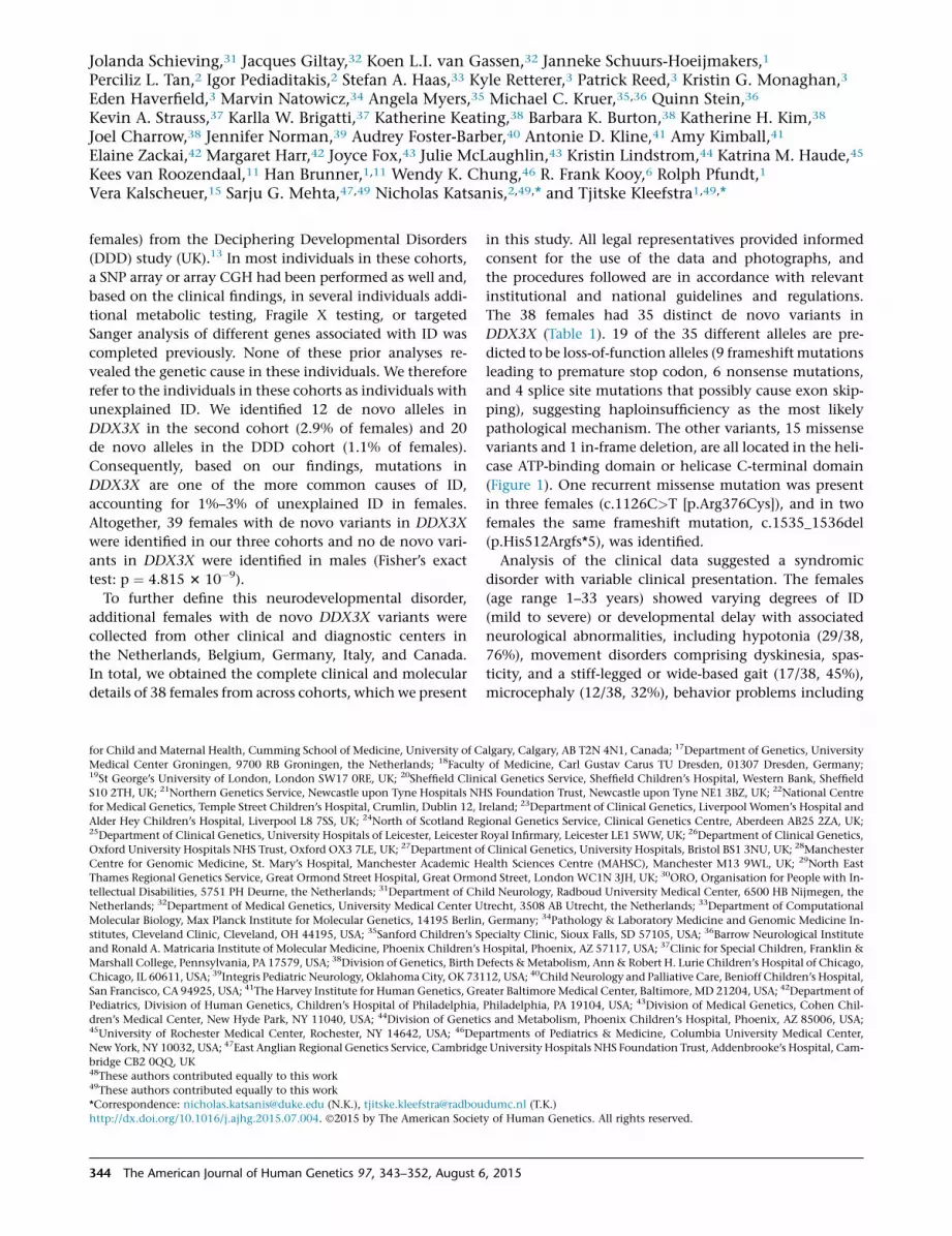
The 38 females had 35 distinct de novo variants in
DDX3X (Table 1). 19 of the 35 different alleles are pre-
dicted to be loss-of-function alleles (9 frameshift mutations
leading to premature stop codon, 6 nonsense mutations,
and 4 splice site mutations that possibly cause exon skip-
ping), suggesting haploinsufficiency as the most likely
pathological mechanism. The other variants, 15 missense
variants and 1 in-frame deletion, are all located in the heli-
case ATP-binding domain or helicase C-terminal domain
(Figure 1). One recurrent missense mutation was present
in three females (c.1126C>T [p.Arg376Cys]), and in two
females the same frameshift mutation, c.1535_1536del
(p.His512Argfs*5), was identified.
Analysis of the clinical data suggested a syndromic
disorder with variable clinical presentation. The females
(age range 1–33 years) showed varying degrees of ID
(mild to severe) or developmental delay with associated
neurological abnormalities, including hypotonia (29/38,
76%), movement disorders comprising dyskinesia, spas-
ticity, and a stiff-legged or wide-based gait (17/38, 45%),
microcephaly (12/38, 32%), behavior problems including
algary, Calgary, AB T2N 4N1, Canada; 17Department of Genetics, University
of Medicine, Carl Gustav Carus TU Dresden, 01307 Dresden, Germany;
cal Genetics Service, Sheffield Children’s Hospital, Western Bank, Sheffield
S Foundation Trust, Newcastle upon Tyne NE1 3BZ, UK; 22National Centre
reland; 23Department of Clinical Genetics, Liverpool Women’s Hospital and
ional Genetics Service, Clinical Genetics Centre, Aberdeen AB25 2ZA, UK;
oyal Infirmary, Leicester LE1 5WW, UK; 26Department of Clinical Genetics,
Clinical Genetics, University Hospitals, Bristol BS1 3NU, UK; 28Manchester
ealth Sciences Centre (MAHSC), Manchester M13 9WL, UK; 29North East
nd Street, LondonWC1N 3JH, UK; 30ORO, Organisation for People with In-
ld Neurology, Radboud University Medical Center, 6500 HB Nijmegen, the
trecht, 3508 AB Utrecht, the Netherlands; 33Department of Computational
Germany; 34Pathology & Laboratory Medicine and Genomic Medicine In-
ecialty Clinic, Sioux Falls, SD 57105, USA; 36Barrow Neurological Institute
Hospital, Phoenix, AZ 57117, USA; 37Clinic for Special Children, Franklin &
efects &Metabolism, Ann & Robert H. Lurie Children’s Hospital of Chicago,
12, USA; 40Child Neurology and Palliative Care, Benioff Children’s Hospital,
ater Baltimore Medical Center, Baltimore, MD 21204, USA; 42Department of
Philadelphia, PA 19104, USA; 43Division of Medical Genetics, Cohen Chil-
s and Metabolism, Phoenix Children’s Hospital, Phoenix, AZ 85006, USA;
artments of Pediatrics & Medicine, Columbia University Medical Center,
University Hospitals NHS Foundation Trust, Addenbrooke’s Hospital, Cam-
dumc.nl (T.K.)
y of Human Genetics. All rights reserved.
, 2015
autism spectrum disorder (ASD), hyperactivity, and aggres-
sion (20/38, 53%), and epilepsy (6/38, 16%). Several
recurrent additional features were noted, including joint
hyperlaxity, skin abnormalities (mosaic-like pigmentary
changes in some females), cleft lip and/or palate, hearing
and visual impairment, and precocious puberty. Abnormal
brain MRIs were reported in various females, with corpus
callosum hypoplasia (13/37, 35%), ventricular enlarge-
ment (13/37, 35%), and evidence of cortical dysplasia
(e.g., polymicrogyria) in four individuals. A summary of
the clinical data is presented in Tables 2 and S1, and facial
profiles of 30 of the 38 females are shown in Figure 2.
Although common dysmorphic features are reported,
there is no consistent recognizable phenotype. Based on
these clinical and molecular data, there is no evidence for
an obvious genotype-phenotype correlation between the
different types of mutations and degree of ID.
DDX3X is known as one of the genes that are able to
escape X inactivation.14,15 X-linked dominant conditions
often show a remarkable variability among affected fe-
males which in particular holds true for genes that escape
X inactivation.9,16 It is known that most of the transcripts
escaping X inactivation are not fully expressed from the
inactivated X chromosome, which means that the escape
is often partial and incomplete.9 Based on this, phenotypic
severity might be influenced by the amount of gene
expression of DDX3X in females, which could be affected
by possibly skewed X inactivation or incompleteness of
the escape. Different expression of DDX3X in different
tissues could also be a contributing factor. To further
explore the possible skewing of X inactivation, we deter-
mined X inactivation via the androgen receptor gene
(AR) methylation assay17 on DNA from lymphocytes in
15 females. We found an almost complete skewing
(>95%) of X inactivation in seven individuals and random
skewing in the remainder. This is more than would be ex-
pected by chance, because it is known that a high degree
of skewing of X inactivation is a statistically rare event in
young women.18 However, in our affected females, there
is no evidence of correlation of skewing of X inactivation
with disease severity.
Given the high frequency (1%–2%) of DDX3X muta-
tions in females with unexplained ID, we sought to
determine whether males carry deleterious alleles. We
identified no de novo variants in DDX3X males in any of
our cohorts. However, upon sequencing of the X chromo-
some (X-exome) of ID-affected families with apparent
X-linked inheritance pattern,7 we identified two families
with segregating missense variants in DDX3X. Moreover,
one additional family was identified by diagnostic whole
exome sequencing in Antwerp, Belgium. In these three
families, males with the DDX3X variant have borderline
to severe ID and carrier females are unaffected. Pedigrees
of these three families are shown in Figure S1, and a sum-
mary of the clinical features of the affected males is pre-
sented in Table S2. All three missense mutations were
predicted to be deleterious by prediction programs Poly-
The Amer
Phen-2 and SIFT (Table 1) and map within the helicase
ATP-binding domain (Figure 2). With three-dimensional
protein analysis, we could not discern any clear difference
between the missense mutations found in affected males
and the de novo mutations found in females that could
possibly explain the gender-specific pathogenicity
(Figure S2, Table S3). Although in the first two families
with affected males the phenotype consisted mainly of in-
tellectual disability, family 3 was more complex. The male
proband had severe ID and various other features such as a
dysplastic pulmonary valve, hypertonia, and strabismus.
In this male a SNP-array analysis was performed, as well
as DNA analysis of PTEN (MIM: 601728) and FMR1
(MIM: 309550) andmethylation studies on Angelman syn-
drome (MIM: 105830), all without abnormalities. With
exome sequencing no other candidate genes were found.
Hismother had recurrentmiscarriages of unknown gender.
A second initially viable pregnancy was terminated
because of ultrasound anomalies that had also been noted
in the proband, including a thickened nuchal fold and ab-
sent nasal bone. After termination of the pregnancy, the
male fetus was tested and found to have the samemissense
mutation in DDX3X as his brother. Sequencing of other
family members demonstrated that the mutation arose
de novo in the proband’s mother. X-inactivation studies
in this mother demonstrated a random X-inactivation
pattern (68/32). X-inactivation studies in female carriers
in the other families showed that in family 1, the obligate
carrier female (II-2) had highly skewed X inactivation
(>95%), whereas X-inactivation studies in family 2 were
not informative.
None of the three DDX3X variants found in these
families with affected males were reported in the ExAC
database or in the Exome Variant Server (ESP), nor was
one of the de novo variants found in females reported in
these databases. Moreover, none of the DDX3X mutations
identified in males were detected in affected females. As far
as we are aware, in addition to the de novo missense muta-
tion identified in family 3, no other de novo mutations
in DDX3X are reported in healthy individuals or control
cohorts. We downloaded all variants from the ExAC data-
base, containing exome data of 60,706 individuals, and
calculated per gene the number of missense and synony-
mous variants. These numbers were then normalized by
dividing through the total number of possible missense
and synonymous variants per gene. The ratio of corrected
missense over synonymous variants was then used as a
measure for tolerance of the gene to normal variation,
similarly as was done previously.19 When genes were
ranked according to their tolerance score, DDX3X was
among the most intolerant genes (1.09% of genes, rank
194 out of 17,856), showing that normal variation in this
gene is extremely rare.
DDX3X encodes a conserved DEAD-box RNA helicase
important in a variety of fundamental cellular processes
that include transcription, splicing, RNA transport, and
translation.20,21 DDX3X has been associated with many
ican Journal of Human Genetics 97, 343–352, August 6, 2015 345

Table 1. Mutation Characteristics
Nucleotide Change(GenBank:NM_001356.4) Amino Acid Change SIFT PolyPhen-2 Cohort
PreviouslyReported
Females
Individual 1 c.1126C>T p.Arg376Cys not tolerated probably damaging Nijmegen –
Individual 2 c.233C>G p.Ser78* NA NA Nijmegen –
Individual 3 c.1126C>T p.Arg376Cys not tolerated probably damaging DDD Study DDD Study13
Individual 4 c.136C>T p.Arg46* NA NA DDD Study DDD Study13
Individual 5 c.1601G>A p.Arg534His not tolerated probably damaging DDD Study DDD Study13
Individual 6 c.641T>C p.Ile214Thr not tolerated probably damaging DDD Study DDD Study13
Individual 7 c.1520T>C p.Ile507Thr not tolerated probably damaging other –
Individual 8 c.977G>A p.Arg326His not tolerated probably damaging other –
Individual 9 c.868del p.Ser290Hisfs*31 NA NA other Rauch et al.41
Individual 10 c.1229_1230dup p.Thr411Leufs*10 NA NA Nijmegen –
Individual 11 c.1105dup p.Thr369Asnfs*14 NA NA Nijmegen –
Individual 12 c.865�2A>G p.? NA NA Nijmegen –
Individual 13 c.1600dup p.Arg534Profs*13 NA NA other –
Individual 14 c.269dup p.Ser90Argfs*8 NA NA Nijmegen –
Individual 15 c.1440A>T p.Arg480Ser not tolerated probably damaging Nijmegen –
Individual 16 c.873C>A p.Tyr291* NA NA Nijmegen –
Individual 17 c.1693C>T p.Gln565* NA NA Nijmegen –
Individual 18 c.1535_1536del p.His512Argfs*5 NA NA DDD Study –
Individual 19 c.766�1G>C p.? NA NA other –
Individual 20 c.599dup p.Tyr200* NA NA USA –
Individual 21 c.1321del p.Asp441Ilefs*3 NA NA USA –
Individual 22 c.1383dup p.Tyr462Ilefs*3 NA NA USA –
Individual 23 c.1384_1385dup p.His463Thrfs*34 NA NA USA –
Individual 24 c.1535_1536del p.His512Argfs*5 NA NA USA –
Individual 25 c.1541T>C p.Ile514Thr not tolerated probably damaging USA –
Individual 26 c.704T>C p.Leu235Pro not tolerated probably damaging USA –
Individual 27 c.1175T>C p.Leu392Pro not tolerated probably damaging USA –
Individual 28 c.1463G>A p.Arg488His not tolerated possibly damaging USA –
Individual 29 c.1126C>T p.Arg376Cys not tolerated probably damaging USA –
Individual 30 c.1250A>C p.Gln417Pro not tolerated probably damaging USA –
Individual 31 c.698C>T p.Ala233Val not tolerated probably damaging DDD Study –
Individual 32 c.931C>T p.Arg311* NA NA DDD Study –
Individual 33 c.46-2A>C p.? NA NA DDD Study –
Individual 34 c.1678_1680del p.Leu560del NA NA DDD Study –
Individual 35 c.1423C>G p.Arg475Gly not tolerated probably damaging DDD Study –
Individual 36 c.46-2A>G p.? NA NA DDD Study –
Individual 37 c.1703C>T p.Pro568Leu not tolerated probably damaging DDD Study –
Individual 38 c.1526A>T p.Asn509Ile not tolerated probably damaging DDD Study –
(Continued on next page)
346 The American Journal of Human Genetics 97, 343–352, August 6, 2015
Table 1. Continued
Nucleotide Change(GenBank:NM_001356.4) Amino Acid Change SIFT PolyPhen-2 Cohort
PreviouslyReported
Males
Family 1 c.1084C>T p.Arg362Cys not tolerated probably damaging – –
Family 2 c.1052G>A p.Arg351Gln not tolerated possibly damaging – Hu et al.7
Family 3 c.898G>T p.Val300Phe not tolerated probably damaging – –
‘‘Other’’ indicates additional females with de novo DDX3Xmutations collected from different diagnostic centers in the Netherlands, Belgium, Germany, Italy, andCanada.
cellular processes, such as cell cycle control, apoptosis, and
tumorigenesis.22,23 It is also thought to be an essential fac-
tor in the RNAi pathway24 and it is a key regulator of the
Wnt/b-catenin pathway, acting as a regulatory subunit of
CSNK1E and stimulating its kinase activity.25
Notably, of the 35 different de novo variants, 19 alleles
are predicted to give a loss-of-function effect. Antisense-
based knockdown of the DDX3X-ortholog PL10 in zebra-
fish (72% identical, 78% similar) is already described and
shows a reduced brain size and head size in zebrafish em-
bryos at 2 days postfertilization (dpf).13 The missense
changes in affected females were located in the same pro-
tein domain as the missense changes in affected males,
so we explored the pathogenic mechanisms accounting
for the differences in disease transmission and phenotype
between affected females and affected males with DDX3X
missense variants. We therefore employed a combination
of in vitro and in vivo assays based on the known role of
DDX3X in the regulation of Wnt/b-catenin signaling.25
Wnt signaling is a critical developmental pathway;
the zebrafish is a tractable model in which to study Wnt
output26–31 and to interrogate alleles relevant to neurocog-
nitive traits.32 We tested several representative missense
variants, including the female-specific de novo vari-
ants c.641T>C (p.Ile214Thr), c.977G>A (p.Arg326His),
c.1126C>T (p.Arg376Cys), c.1520T>C (p.Ile507Thr), and
c.1601G>A (p.Arg534His) as well as the inherited variants
c.898G>T (p.Val300Phe), c.1052G>A (p.Arg351Gln), and
c.1084C>T (p.Arg362Cys) identified in males. A variant
from the Exome Variant Server (c.586G>A [p.Glu196Lys])
The Amer
was chosen as a negative control (rs375996245; MAF <
0.002).
We first tested whether the missense variants identified
in our ID cohort resulted in dominant effects by overex-
pressing either WT or mutant DDX3X human transcript
in zebrafish embryos and examining the phenotype. Ze-
brafish (Danio rerio) were raised and mated as described.33
Embryos from ZDR strain fish were injected into the
yolk with 1 nl of solution containing mRNA at the 1- to
2-cell stage using a Picospritzer III microinjector (Parker).
The wild-type (WT) human DDX3X open reading frame
(ORF) construct was obtained from the Ultimate ORF
Collection (LifeTechnologies; clone ID: IOH13891),
sequenced fully, and sub-cloned into the pCS2þ vector
using Gateway LR clonase II- mediated cloning (LifeTech-
nologies). Capped mRNA was generated using linearized
constructs as a template with the mMessage mMachine
SP6 transcription kit (LifeTechnologies). Injection of 100
pg of WT or mutant DDX3X transcripts did not produce
a discernible phenotype at 36 hr postfertilization (hpf)
(n¼ 50–75 embryos/injection; repeated twice withmasked
scoring; Figure S3A) or at 72 hpf (not shown). To corrobo-
rate these data in a different system, we used a mammalian
cell-based assay of canonical Wnt signaling (TOPFlash).34
In brief, HEK293T cells and mouse L-cells (both control
and WNT3A expressing) were grown in 10% FBS/DMEM.
WNT3A-containing media was prepared by incubating
confluent L-cells with serum-free media for 24 hr and
removing and filtering the media. This was subsequently
used to stimulate transfected HEK293Tcells. HEK293Tcells
Figure 1. Location of Amino Acid Substi-tutions in DDX3XSchematic view of DDX3X with the 15different amino acid substitutions (andone in-frame deletion) found in affectedfemales and the 3 different amino acid sub-stitutions found in affected males. DDX3Xconsists of two subdomains: a helicaseATP-binding domain and a helicase C-ter-minal domain. All amino acid substitu-tions found in affected females are locatedwithin these two protein domains. Thethree amino acid substitutions found inaffected males are all located within thehelicase ATP-binding domain.
ican Journal of Human Genetics 97, 343–352, August 6, 2015 347

Table 2. Clinical Features of Females with De Novo DDX3XMutations
Percentage Number
Development
Intellectual disability or developmental delay 100% 38/38
Mild or mild-moderate disability 26% 10/38
Moderate or moderate-severe disability 26% 10/38
Severe disability 40% 15/38
Developmental delay 8% 3/38
Growth
Low weight 32% 12/38
Microcephaly 32% 12/38
Neurology
Hypotonia 76% 29/38
Epilepsy 16% 6/38
Movement disorder (including spasticity) 45% 17/38
Behavior problems 53% 20/38
Brain MRI
Corpus callosum hypoplasia 35% 13/37
Cortical malformation 11% 4/37
Ventricular enlargement 35% 13/37
Other
Skin abnormalities 37% 14/38
Hyperlaxity 37% 14/38
Visual problems 34% 13/38
Hearing loss 8% 3/38
Cleft lip or palate 8% 3/38
Precocious puberty 13% 5/38
Scoliosis 11% 4/38
at a density of 1 3 104 cells/well on 24-well plates were
transfected with 1.025 mg total of DNA containing 0.5 mg
of pGL4.18 TOPFLASH vector, 25 ng of renilla luciferase
plasmid (pRL-SV40), and 0.5 mg of pCS2þ with or without
DDX3X using XtremeGene9 (Roche). After 24 hr the me-
dia was removed and replaced with serum-free media
collected as above from L-cells with and without WNT3A
(MIM: 606359). After 24 hr, cells were harvested and
luciferase activity measured by the Dual-Luciferase Re-
porter Assay System (Promega). Results were normalized
internally for each well to renilla luciferase activity and
then to the unstimulated wells (conditioned with control
L-cell media). Consistent with the in vivo result, transfec-
tion of expression constructs harboring DDX3X variants
did not differ from WT in the modulation of WNT3A-
b-catenin mediated luciferase activity (Figure S3B; tripli-
cate wells with 3–5 biological replicates). Taken together,
our results suggest that neither the female nor the male
348 The American Journal of Human Genetics 97, 343–352, August 6
DDX3X variants produce detectable dominant changes
in the context of Wnt signaling. These results are in line
with the high number of de novo truncating mutations
in females, suggesting haploinsufficiency as themost likely
pathogenic mechanism. As a consequence, we pursued a
loss-of-function paradigm.
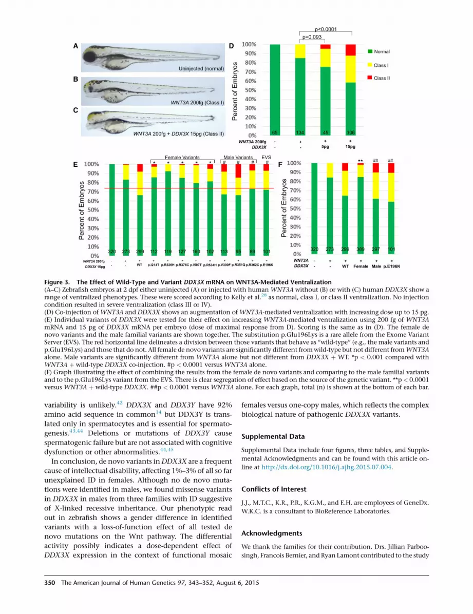
Previous studies have shown that co-expression of a
canonical Wnt ligand and DDX3X is necessary to produce
a phenotype in vivo.25 Expression of either of the canoni-
cal Wnt ligands WNT3A orWNT8A (MIM: 606360) results
in ventralization of zebrafish embryos that ranges from
mild (hypoplasia of the eyes, which we term class I em-
bryos) to moderate (absence of one or both eyes; class II),
severe (loss of all anterior neural structures; class III), and
radialized (class IV) phenotypes at 2 dpf.28 We recapitu-
lated these defects by injecting increasing doses of human
WNT3A or WNT8A mRNA into embryos (Figures 3A
and 3B). Full-length human WNT3A and WNT8A ORFs
were cloned into pCS2þ from the Ultimate ORF Collec-
tion (Clone ID WNT8A: IOH35591; Clone ID WNT3A:
IOH80731). The WNT3A clone required site-directed
mutagenesis to introduce a stop codon using primers
(50-ctgcaaggccgccaggcacTAGGGTGGGCGCGCCGA-30 andits reverse complement). We selected a dose of WNT3A
mRNA (200 fg), which produced a modest effect (15%–
20% class IþII embryos, no class III or IV) and we co-
injected it with progressively increasing concentrations
of human DDX3X mRNA. At 2 dpf, we observed a dose-
response curve concomitant with increasing doses
of DDX3X (Figure 3D). At low doses of mutant DDX3X
(15–20 pg/embryo), the ventralization phenotype pro-
duced by WNT3A is exacerbated, producing 40%–50%
class IþII embryos, with a greater percentage in class II
(10%–15%), indicative of augmented severity (Figure 3D).
Because of the significant (p < 0.0001) augmentation of
WNT3A-mediated ventralization at 15 pg of DDX3X, we
next used this combination of doses to test the effect of
the missense alleles on Wnt signaling. WNT3A mRNA
(200 fg) was co-injected with 15 pg of WT or variant
DDX3X mRNA, and we scored embryos for ventralization
at 2dpf.We founddifferential effects of the variants on ven-
tralization (Figure 3E; triplicate experiments; pooled for the
final result). All de novo variants tested were significantly
different from WT (p < 0.001) and indistinguishable from
WNT3A alone, suggesting that they all confer complete
loss of function toDDX3X. By contrast, the control variant
c.586G>A (p.Glu196Lys) resulted in phenotypes similar
to WT. Notably, all three inherited variants found in the
affected males were also indistinguishable from WT in-
jected and each was statistically different from WNT3A
alone (p < 0.0001). Given this apparent dichotomization
of effect between female denovo andmale inherited alleles,
we tested whether pooling of results from the female versus
male variants would exhibit a ‘‘class effect’’ separation
between the two groups. We found this to be true; there
was significant differential effect of the gender-specific
sets of variants on Wnt signaling (Figure 3F).
, 2015

Individual 1 Individual 2 Individual 3 Individual 4 Individual 5 Individual 7
Individual 8 Individual 9 Individual 10 Individual 11 Individual 12 Individual 13
Individual 14 Individual 15 Individual 16 Individual 17 Individual 18 Individual 19
Individual 20 Individual 22 Individual 23 Individual 24 Individual 26 Individual 27
Individual 31 Individual 32 Individual 33 Individual 34 Individual 35 Individual 37
Figure 2. Facial Profiles of Females withDe Novo DDX3X MutationsFacial features of 30 of 38 females with a denovo variant in DDX3X. Common facialfeatures include a long and/or hypotonicface (e.g., individuals 2, 4, 5, 12, 22, 32),a high and/or broad forehead (e.g., individ-uals 1, 7, 9, 23, 24, 26), a wide nasalbridge and/or bulbous nasal tip (e.g., indi-viduals 11, 13, 15, 16), narrow alae nasiand/or anteverted nostrils (e.g., individ-uals 2, 8, 9, 12, 14, 18, 24, 27, 32, 35),and hypertelorism (e.g., individuals 5,7, 8, 20, 27). Informed consent wasobtained for all 30 individuals shown.The individual numbers correspond to thenumbers mentioned in Tables 1 and S1.
Taken together, our in vivo testing of the nonsynony-
mous DDX3X mutations demonstrated marked alteration
in Wnt-mediated ventralization for changes arising de
novo in affected females. Our data also reinforce the
notion that disruption of b-catenin signaling during
neurodevelopment has profound consequences. Loss of
Wnt signaling inhibits neuroblast migration, neural
differentiation, and suppression of the development of
the forebrain.35–38 Moreover, we previously reported that
mutations in b-catenin contribute to ID in humans,39,40
and mutations in d-catenin, some of which abrogate its
biochemical interaction with b-catenin, can contribute to
severe autism in females,32 strengthening further the link
between Wnt signaling and human neurodevelopmental
disorders.
DDX3X has recently been proposed as a candidate ID
gene.13,41 Our data substantiate this hypothesis and sug-
gest that mutated DDX3X is among the most common
causes of simplex cases of intellectual disability in females.
Our data also suggest that the genetic architecture of
DDX3X-mediated pathology in males is different. All
The American Journal of Human G
detected male alleles were inherited
from unaffected mothers, suggesting
either a gender-specific buffering
effect or a milder effect of those
inherited alleles on protein function.
Both our in vivo and our in vitro
studies indicate that none of the
tested alleles (male or female) confer
dominant effects and that the male
alleles are indistinguishable from WT
in our system. Given that human ge-
netics and computational predictions
support the causality of these alleles
in the families studied, we speculate
that the effect of these alleles is
beyond the dynamic range of our as-
says and thus could not be detected;
a substantially larger assay of embryos
might detect a signal, although the
transient nature of our system will always be limited at
detecting mild allele effects. Alternatively, the mechanism
of the male-derived alleles might be qualitatively different
from the de novo female variants and reflective of the
complex biology of DDX3X. Of note, we found that
increased amounts of wild-type DDX3X ameliorated the
Wnt3a-induced ventralization phenotype (Figure S4B),
an observation that we reproduced with Wnt8a, another
b-catenin-dependent ligand (Figure S4A) and our TOPFlash
reporter assay (Figure S3B). This bimodal activity of
DDX3X has been intimated previously by Cruciat et al.,
in which knockdown of DDX3X caused loss of WNT3A
signaling that was restored by transfection with WT
construct whereas overexpression alone resulted in
decreased WNT3A signaling.25 We reproduced this latter
finding when solely overexpressing DDX3X in HEK293
cells (Figure S3B).
Summarized, these data suggest that DDX3X is dosage
sensitive and might have a differential activity in females
than in males. Contribution of the DDX3X homolog at
the Y chromosome,DDX3Y (MIM: 400010), to phenotypic
enetics 97, 343–352, August 6, 2015 349
A
B
C
E
D
F
Figure 3. The Effect of Wild-Type and Variant DDX3X mRNA on WNT3A-Mediated Ventralization(A–C) Zebrafish embryos at 2 dpf either uninjected (A) or injected with human WNT3Awithout (B) or with (C) human DDX3X show arange of ventralized phenotypes. These were scored according to Kelly et al.28 as normal, class I, or class II ventralization. No injectioncondition resulted in severe ventralization (class III or IV).(D) Co-injection of WNT3A and DDX3X shows an augmentation of WNT3A-mediated ventralization with increasing dose up to 15 pg.(E) Individual variants of DDX3X were tested for their effect on increasing WNT3A-mediated ventralization using 200 fg of WNT3AmRNA and 15 pg of DDX3X mRNA per embryo (dose of maximal response from D). Scoring is the same as in (D). The female denovo variants and the male familial variants are shown together. The substitution p.Glu196Lys is a rare allele from the Exome VariantServer (EVS). The red horizontal line delineates a division between those variants that behave as ‘‘wild-type’’ (e.g., the male variants andp.Glu196Lys) and those that do not. All female de novo variants are significantly different fromwild-type but not different fromWNT3Aalone. Male variants are significantly different from WNT3A alone but not different from DDX3X þ WT. *p < 0.001 compared withWNT3A þ wild-type DDX3X co-injection. #p < 0.0001 versus WNT3A alone.(F) Graph illustrating the effect of combining the results from the female de novo variants and comparing to the male familial variantsand to the p.Glu196Lys variant from the EVS. There is clear segregation of effect based on the source of the genetic variant. **p< 0.0001versus WNT3A þ wild-type DDX3X. ##p < 0.0001 versus WNT3A alone. For each graph, total (n) is shown at the bottom of each bar.
variability is unlikely.42 DDX3X and DDX3Y have 92%
amino acid sequence in common14 but DDX3Y is trans-
lated only in spermatocytes and is essential for spermato-
genesis.43,44 Deletions or mutations of DDX3Y cause
spermatogenic failure but are not associated with cognitive
dysfunction or other abnormalities.44,45
In conclusion, de novo variants inDDX3X are a frequent
cause of intellectual disability, affecting 1%–3% of all so far
unexplained ID in females. Although no de novo muta-
tions were identified in males, we found missense variants
in DDX3X in males from three families with ID suggestive
of X-linked recessive inheritance. Our phenotypic read
out in zebrafish shows a gender difference in identified
variants with a loss-of-function effect of all tested de
novo mutations on the Wnt pathway. The differential
activity possibly indicates a dose-dependent effect of
DDX3X expression in the context of functional mosaic
350 The American Journal of Human Genetics 97, 343–352, August 6
females versus one-copy males, which reflects the complex
biological nature of pathogenic DDX3X variants.
Supplemental Data
Supplemental Data include four figures, three tables, and Supple-
mental Acknowledgments and can be found with this article on-
line at http://dx.doi.org/10.1016/j.ajhg.2015.07.004.
Conflicts of Interest
J.J., M.T.C., K.R., P.R., K.G.M., and E.H. are employees of GeneDx.
W.K.C. is a consultant to BioReference Laboratories.
Acknowledgments
We thank the families for their contribution. Drs. Jillian Parboo-
singh, Francois Bernier, and Ryan Lamont contributed to the study
, 2015

design, data interpretation, and X-inactivation studies of individ-
ual 8. Drs. Concetta Barone and Paolo Bosco were instrumental for
clinical evaluation and X-inactivation studies in individual 13.
Miriam Uppill provided technical assistance with investigations
in family 1. This work was supported by a grant from the
Netherlands Organization for Health Research and Development
(ZonMw grant 907-00-365 to T.K.), the Italian Ministry of
Health and ‘5 per mille’ funding (C.R.), a grant from the Alberta
Children’s Hospital Foundation (A.M.I.), a NIH Training Grant
(5T32HD060558-04 to E.C.M.), German Ministry of Education
and Research (01GS08166 to N.D.D. and A.R.), the EU FP7 project
GENCODYS (grant number 241995 to V.M.K.), Australian
NH&MRC grants 628952 and 1041920 to J.G., support from the
Simons Foundation (W.K.C.), Doris Duke Charitable Foundation
Clinical Scientist Development Award (M.C.K.), a grant from
the Marguerite-Marie Delacroix Foundation (A.V.D.), and FWO-
Flanders (H.V.E. and F.K.). B.L. is a senior clinical investigator
supported by Fund for Scientific Research Flanders and holds
an ERC starting grant. Acknowledgments of the DDD Study are
included in the Supplemental Data.
Received: June 4, 2015
Accepted: July 13, 2015
Published: July 30, 2015
Web Resources
The URLs for data presented herein are as follows:
ExAC Browser, http://exac.broadinstitute.org/
NHLBI Exome Sequencing Project (ESP) Exome Variant Server,
http://evs.gs.washington.edu/EVS/
OMIM, http://www.omim.org/
PolyPhen-2, http://genetics.bwh.harvard.edu/pph2/
RefSeq, http://www.ncbi.nlm.nih.gov/RefSeq
SIFT (v.1.03), http://sift.bii.a-star.edu.sg/
References
1. Roeleveld, N., Zielhuis, G.A., and Gabreels, F. (1997). The
prevalence of mental retardation: a critical review of recent
literature. Dev. Med. Child Neurol. 39, 125–132.
2. Leonard, H., andWen, X. (2002). The epidemiology of mental
retardation: challenges and opportunities in the new millen-
nium. Ment. Retard. Dev. Disabil. Res. Rev. 8, 117–134.
3. Maulik, P.K., Mascarenhas, M.N., Mathers, C.D., Dua, T., and
Saxena, S. (2011). Prevalence of intellectual disability: a
meta-analysis of population-based studies. Res. Dev. Disabil.
32, 419–436.
4. Van Naarden Braun, K., Christensen, D., Doernberg, N.,
Schieve, L., Rice, C., Wiggins, L., Schendel, D., and Yeargin-
Allsopp, M. (2015). Trends in the prevalence of autism
spectrum disorder, cerebral palsy, hearing loss, intellectual
disability, and vision impairment, metropolitan Atlanta,
1991-2010. PLoS ONE 10, e0124120.
5. Schalock, R.L., Borthwick-Duffy, S.A., Bradley, V.J., Buntinx,
W.H.E., Coulter, D.L., Craig, E.M., Gomez, S.C., Lachapelle,
Y., Luckasson, R., Reeve, A., et al. (2010). Intellectual
Disability: Definition, Classification, and Systems of Supports
(American Association on Intellectual and Developmental
Disabilities).
The Amer
6. Piton, A., Redin, C., and Mandel, J.L. (2013). XLID-causing
mutations and associated genes challenged in light of data
from large-scale human exome sequencing. Am. J. Hum.
Genet. 93, 368–383.
7. Hu, H., Haas, S.A., Chelly, J., Van Esch, H., Raynaud, M., de
Brouwer, A.P., Weinert, S., Froyen, G., Frints, S.G., Laumon-
nier, F., et al. (2015). X-exome sequencing of 405 unresolved
families identifies seven novel intellectual disability genes.
Mol. Psychiatry. Published online February 3, 2015. http://
dx.doi.org/10.1038/mp.2014.193.
8. Dobyns, W.B., Filauro, A., Tomson, B.N., Chan, A.S., Ho, A.W.,
Ting, N.T., Oosterwijk, J.C., and Ober, C. (2004). Inheritance
of most X-linked traits is not dominant or recessive, just
X-linked. Am. J. Med. Genet. A. 129A, 136–143.
9. Morleo, M., and Franco, B. (2008). Dosage compensation of
the mammalian X chromosome influences the phenotypic
variability of X-linked dominant male-lethal disorders.
J. Med. Genet. 45, 401–408.
10. Neveling, K., Feenstra, I., Gilissen, C., Hoefsloot, L.H., Kam-
steeg, E.J., Mensenkamp, A.R., Rodenburg, R.J., Yntema,
H.G., Spruijt, L., Vermeer, S., et al. (2013). A post-hoc compar-
ison of the utility of sanger sequencing and exome sequencing
for the diagnosis of heterogeneous diseases. Hum. Mutat. 34,
1721–1726.
11. de Ligt, J., Willemsen, M.H., van Bon, B.W., Kleefstra, T.,
Yntema, H.G., Kroes, T., Vulto-van Silfhout, A.T., Koolen,
D.A., de Vries, P., Gilissen, C., et al. (2012). Diagnostic exome
sequencing in persons with severe intellectual disability.
N. Engl. J. Med. 367, 1921–1929.
12. Retterer, K., Scuffins, J., Schmidt, D., Lewis, R., Pineda-Alvarez,
D., Stafford, A., Schmidt, L., Warren, S., Gibellini, F., Konda-
kova, A., et al. (2014). Assessing copy number from exome
sequencing and exome array CGH based on CNV spectrum
in a large clinical cohort. Genet. Med. Published online
November 6, 2014. http://dx.doi.org/10.1038/gim.2014.160.
13. Deciphering Developmental Disorders Study (2015). Large-
scale discovery of novel genetic causes of developmental dis-
orders. Nature 519, 223–228.
14. Lahn, B.T., and Page, D.C. (1997). Functional coherence of the
human Y chromosome. Science 278, 675–680.
15. Cotton, A.M., Price, E.M., Jones, M.J., Balaton, B.P., Kobor,
M.S., and Brown, C.J. (2015). Landscape of DNA methylation
on the X chromosome reflects CpG density, functional chro-
matin state and X-chromosome inactivation. Hum. Mol.
Genet. 24, 1528–1539.
16. Carrel, L., and Willard, H.F. (2005). X-inactivation profile
reveals extensive variability in X-linked gene expression in
females. Nature 434, 400–404.
17. Allen, R.C., Zoghbi, H.Y., Moseley, A.B., Rosenblatt, H.M., and
Belmont, J.W. (1992). Methylation of HpaII and HhaI sites
near the polymorphic CAG repeat in the human androgen-
receptor gene correlates with X chromosome inactivation.
Am. J. Hum. Genet. 51, 1229–1239.
18. Busque, L., Mio, R., Mattioli, J., Brais, E., Blais, N., Lalonde, Y.,
Maragh, M., and Gilliland, D.G. (1996). Nonrandom X-inacti-
vation patterns in normal females: lyonization ratios vary
with age. Blood 88, 59–65.
19. Gilissen, C., Hehir-Kwa, J.Y., Thung, D.T., van de Vorst, M.,
van Bon, B.W., Willemsen, M.H., Kwint, M., Janssen, I.M.,
Hoischen, A., Schenck, A., et al. (2014). Genome sequencing
identifies major causes of severe intellectual disability. Nature
511, 344–347.
ican Journal of Human Genetics 97, 343–352, August 6, 2015 351

20. Abdelhaleem, M. (2005). RNA helicases: regulators of differen-
tiation. Clin. Biochem. 38, 499–503.
21. Garbelli, A., Beermann, S., Di Cicco, G., Dietrich, U., and
Maga, G. (2011). A motif unique to the human DEAD-box
protein DDX3 is important for nucleic acid binding, ATP hy-
drolysis, RNA/DNA unwinding and HIV-1 replication. PLoS
ONE 6, e19810.
22. Li, Q., Zhang, P., Zhang, C., Wang, Y., Wan, R., Yang, Y., Guo,
X., Huo, R., Lin, M., Zhou, Z., and Sha, J. (2014). DDX3X reg-
ulates cell survival and cell cycle during mouse early embry-
onic development. J. Biomed. Res. 28, 282–291.
23. Schroder, M. (2010). Human DEAD-box protein 3 has multi-
ple functions in gene regulation and cell cycle control and is
a prime target for viral manipulation. Biochem. Pharmacol.
79, 297–306.
24. Kasim, V., Wu, S., Taira, K., andMiyagishi, M. (2013). Determi-
nation of the role of DDX3 a factor involved in mammalian
RNAi pathway using an shRNA-expression library. PLoS ONE
8, e59445.
25. Cruciat, C.M., Dolde, C., de Groot, R.E., Ohkawara, B.,
Reinhard, C., Korswagen, H.C., and Niehrs, C. (2013). RNA
helicase DDX3 is a regulatory subunit of casein kinase 1 in
Wnt-b-catenin signaling. Science 339, 1436–1441.
26. Bellipanni, G., Varga, M., Maegawa, S., Imai, Y., Kelly, C.,
Myers, A.P., Chu, F., Talbot, W.S., and Weinberg, E.S. (2006).
Essential and opposing roles of zebrafish b-catenins in the
formation of dorsal axial structures and neurectoderm. Devel-
opment 133, 1299–1309.
27. Caneparo, L., Huang, Y.-L., Staudt, N., Tada, M., Ahrendt, R.,
Kazanskaya, O., Niehrs, C., and Houart, C. (2007). Dickkopf-
1 regulates gastrulation movements by coordinated modu-
lation of Wnt/b catenin and Wnt/PCP activities, through
interaction with the Dally-like homolog Knypek. Genes
Dev. 21, 465–480.
28. Kelly, C., Chin, A.J., Leatherman, J.L., Kozlowski, D.J., and
Weinberg, E.S. (2000). Maternally controlled (beta)-catenin-
mediated signaling is required for organizer formation in the
zebrafish. Development 127, 3899–3911.
29. Kelly, G.M., Erezyilmaz, D.F., and Moon, R.T. (1995). Induc-
tion of a secondary embryonic axis in zebrafish occurs
following the overexpression of beta-catenin. Mech. Dev. 53,
261–273.
30. Kumar, S., �Zigman, M., Patel, T.R., Trageser, B., Gross, J.C.,
Rahm, K., Boutros, M., Gradl, D., Steinbeisser, H., Holstein,
T., et al. (2014). Molecular dissection ofWnt3a-Frizzled8 inter-
action reveals essential and modulatory determinants of Wnt
signaling activity. BMC Biol. 12, 44.
31. Lekven, A.C., Thorpe, C.J., Waxman, J.S., and Moon, R.T.
(2001). Zebrafish wnt8 encodes two wnt8 proteins on a bicis-
tronic transcript and is required for mesoderm and neurecto-
derm patterning. Dev. Cell 1, 103–114.
32. Turner, T.N., Sharma, K., Oh, E.C., Liu, Y.P., Collins, R.L., Sosa,
M.X., Auer, D.R., Brand, H., Sanders, S.J., Moreno-De-Luca, D.,
et al. (2015). Loss of d-catenin function in severe autism.
Nature 520, 51–56.
352 The American Journal of Human Genetics 97, 343–352, August 6
33. Westerfield, M. (1995). The Zebrafish Book (Eugene, OR:
University of Oregon Press).
34. Wu, W., Glinka, A., Delius, H., and Niehrs, C. (2000). Mutual
antagonism between dickkopf1 and dickkopf2 regulates Wnt/
beta-catenin signalling. Curr. Biol. 10, 1611–1614.
35. Andoniadou, C.L., Signore, M., Young, R.M., Gaston-Massuet,
C., Wilson, S.W., Fuchs, E., and Martinez-Barbera, J.P. (2011).
HESX1- and TCF3-mediated repression of Wnt/b-catenin
targets is required for normal development of the anterior
forebrain. Development 138, 4931–4942.
36. Najm, F.J., Lager, A.M., Zaremba, A., Wyatt, K., Caprariello,
A.V., Factor, D.C., Karl, R.T., Maeda, T., Miller, R.H., and Tesar,
P.J. (2013). Transcription factor-mediated reprogramming
of fibroblasts to expandable, myelinogenic oligodendrocyte
progenitor cells. Nat. Biotechnol. 31, 426–433.
37. Mentink, R.A., Middelkoop, T.C., Rella, L., Ji, N., Tang, C.Y.,
Betist, M.C., van Oudenaarden, A., and Korswagen, H.C.
(2014). Cell intrinsic modulation of Wnt signaling controls
neuroblast migration in C. elegans. Dev. Cell 31, 188–201.
38. Zhang, S., Li, J., Lea, R., Vleminckx, K., and Amaya, E. (2014).
Fezf2 promotes neuronal differentiation through localised
activation ofWnt/b-catenin signalling during forebrain devel-
opment. Development 141, 4794–4805.
39. Kuechler, A., Willemsen, M.H., Albrecht, B., Bacino, C.A., Bar-
tholomew, D.W., van Bokhoven, H., van den Boogaard, M.J.,
Bramswig, N., Buttner, C., Cremer, K., et al. (2015). De novo
mutations in beta-catenin (CTNNB1) appear to be a frequent
cause of intellectual disability: expanding the mutational
and clinical spectrum. Hum. Genet. 134, 97–109.
40. Tucci, V., Kleefstra, T., Hardy, A., Heise, I., Maggi, S., Willem-
sen, M.H., Hilton, H., Esapa, C., Simon, M., Buenavista,
M.T., et al. (2014). Dominant b-catenin mutations cause
intellectual disability with recognizable syndromic features.
J. Clin. Invest. 124, 1468–1482.
41. Rauch, A., Wieczorek, D., Graf, E., Wieland, T., Endele, S.,
Schwarzmayr, T., Albrecht, B., Bartholdi, D., Beygo, J., Di Do-
nato, N., et al. (2012). Range of genetic mutations associated
with severe non-syndromic sporadic intellectual disability:
an exome sequencing study. Lancet 380, 1674–1682.
42. Sekiguchi, T., Iida, H., Fukumura, J., and Nishimoto, T. (2004).
Human DDX3Y, the Y-encoded isoform of RNA helicase
DDX3, rescues a hamster temperature-sensitive ET24 mutant
cell line with a DDX3Xmutation. Exp. Cell Res. 300, 213–222.
43. Ditton, H.J., Zimmer, J., Kamp, C., Rajpert-De Meyts, E., and
Vogt, P.H. (2004). The AZFa gene DBY (DDX3Y) is widely tran-
scribed but the protein is limited to the male germ cells by
translation control. Hum. Mol. Genet. 13, 2333–2341.
44. Foresta, C., Ferlin, A., and Moro, E. (2000). Deletion and
expression analysis of AZFa genes on the human Y chromo-
some revealed a major role for DBY in male infertility. Hum.
Mol. Genet. 9, 1161–1169.
45. Ferlin, A., Moro, E., Rossi, A., Dallapiccola, B., and Foresta, C.
(2003). The human Y chromosome’s azoospermia factor b
(AZFb) region: sequence, structure, and deletion analysis in
infertile men. J. Med. Genet. 40, 18–24.
, 2015

The American Journal of Human Genetics
Supplemental Data
Mutations in DDX3X Are a Common Cause
of Unexplained Intellectual Disability
with Gender-Specific Effects on Wnt Signaling
Lot Snijders Blok, Erik Madsen, Jane Juusola, Christian Gilissen, Diana Baralle, Margot
R.F. Reijnders, Hanka Venselaar, Céline Helsmoortel, Megan T. Cho, Alexander
Hoischen, Lisenka Vissers, Tom S. Koemans, Willemijn Wissink-Lindhout, Evan E.
Eichler, Corrado Romano, Hilde Van Esch, Connie Stumpel, Maaike Vreeburg, Eric
Smeets, Karin Oberndorff, Bregje W.M. van Bon, Marie Shaw, Jozef Gecz, Eric Haan,
Melanie Bienek, Corinna Jensen, Bart L. Loeys, Anke Van Dijck, A. Micheil Innes, Hilary
Racher, Sascha Vermeer, Nataliya Di Donato, Andreas Rump, Katrina Tatton-Brown,
Michael J. Parker, Alex Henderson, Sally A. Lynch, Alan Fryer, Alison Ross, Pradeep
Vasudevan, Usha Kini, Ruth Newbury-Ecob, Kate Chandler, Alison Male, the DDD
Study, Sybe Dijkstra, Jolanda Schieving, Jacques Giltay, Koen L.I. van Gassen,
Janneke Schuurs-Hoeijmakers, Perciliz L. Tan, Igor Pediaditakis, Stefan A. Haas, Kyle
Retterer, Patrick Reed, Kristin G. Monaghan, Eden Haverfield, Marvin Natowicz, Angela
Myers, Michael C. Kruer, Quinn Stein, Kevin A. Strauss, Karlla W. Brigatti, Katherine
Keating, Barbara K. Burton, Katherine H. Kim, Joel Charrow, Jennifer Norman, Audrey
Foster-Barber, Antonie D. Kline, Amy Kimball, Elaine Zackai, Margaret Harr, Joyce Fox,
Julie McLaughlin, Kristin Lindstrom, Katrina M. Haude, Kees van Roozendaal, Han
Brunner, Wendy K. Chung, R. Frank Kooy, Rolph Pfundt, Vera Kalscheuer, Sarju G.
Mehta, Nicholas Katsanis, and Tjitske Kleefstra

Supplemental Acknowledgments
The DDD study presents independent research commissioned by the Health Innovation Challenge Fund (grant number HICF-1009-003), a parallel funding partnership between the Wellcome Trust and the Department of Health, and the Wellcome Trust Sanger Institute (grant number WT098051). The views expressed in this publication are those of the author(s) and not necessarily those of the Wellcome Trust or the Department of Health. The DDD study has UK Research Ethics Committee approval (10/H0305/83 granted by the Cambridge South REC, and GEN/284/12 granted by the Republic of Ireland REC). The research team acknowledges the support of the National Institute for Health Research through the Comprehensive Clinical Research Network.
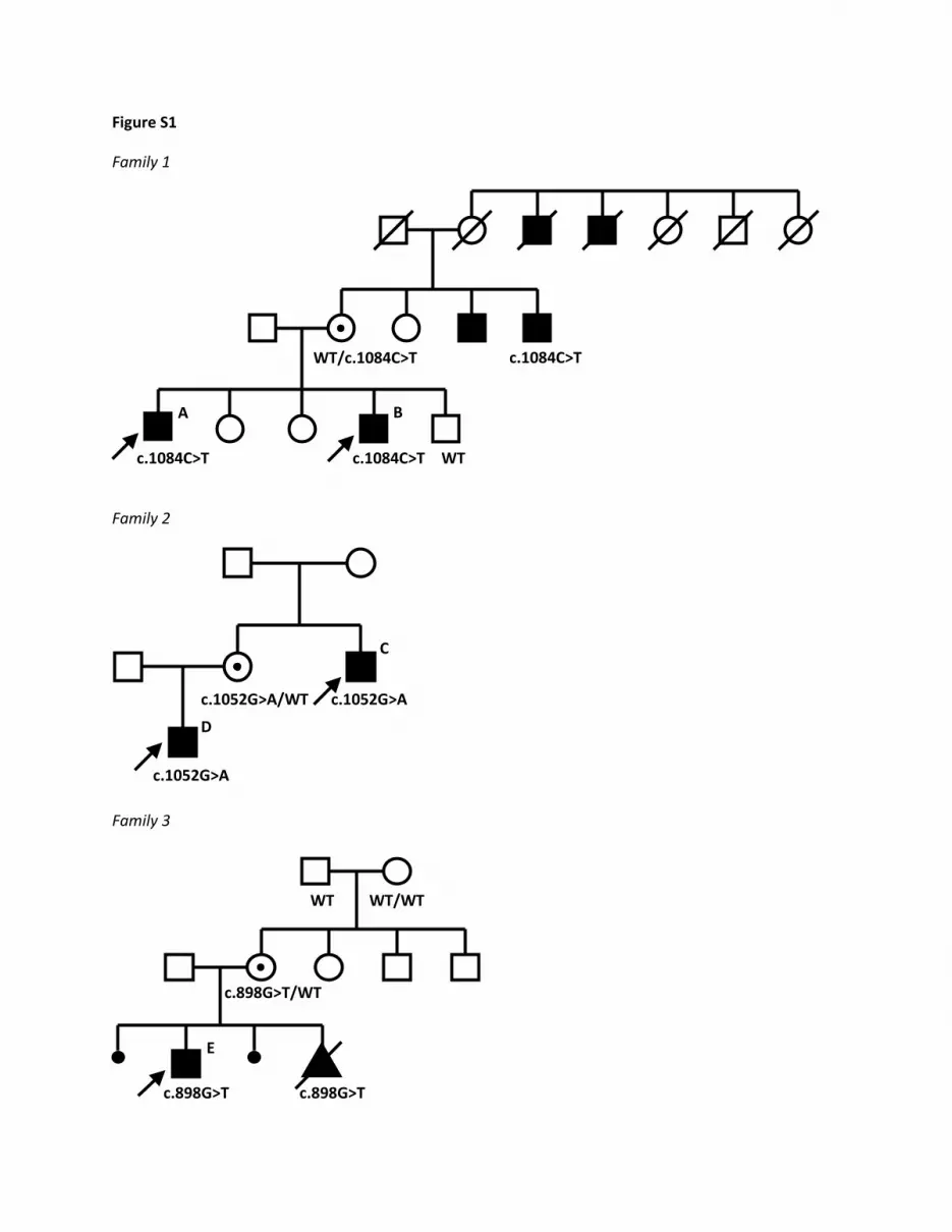
Figure S1
Family 1
Family 2
Family 3
c.1052G>A
c.1052G>A/WT c.1052G>A
WT/c.1084C>T
c.1084C>T
c.1084C>T WT
c.1084C>T
c.898G>T
c.898G>T
c.898G>T/WT
WT WT/WT
A B
C
D
E
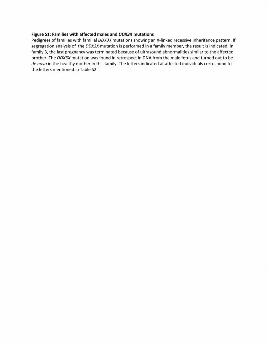
Figure S1: Families with affected males and DDX3X mutations Pedigrees of families with familial DDX3X mutations showing an X-linked recessive inheritance pattern. If segregation analysis of the DDX3X mutation is performed in a family member, the result is indicated. In family 3, the last pregnancy was terminated because of ultrasound abnormalities similar to the affected brother. The DDX3X mutation was found in retrospect in DNA from the male fetus and turned out to be de novo in the healthy mother in this family. The letters indicated at affected individuals correspond to the letters mentioned in Table S2.
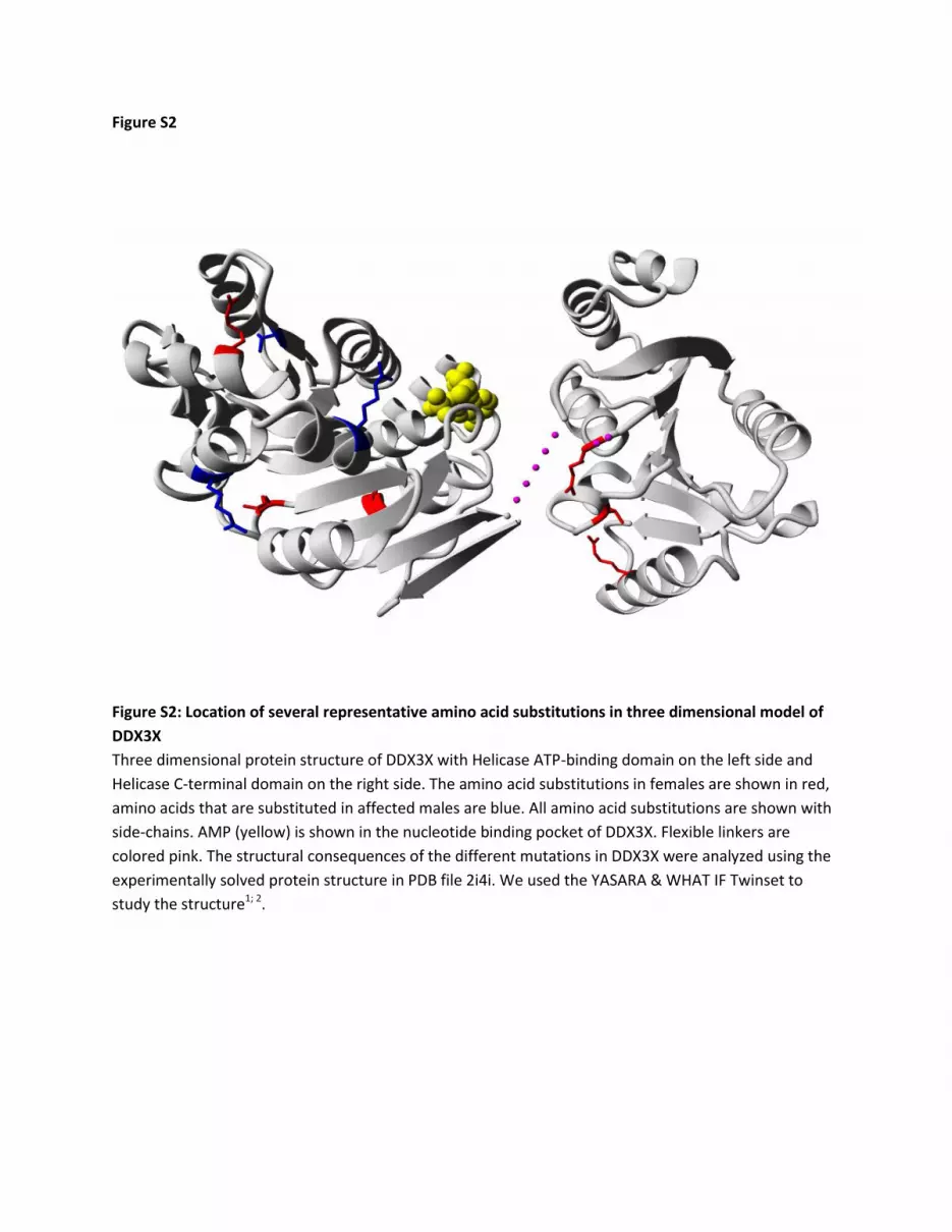
Figure S2
Figure S2: Location of several representative amino acid substitutions in three dimensional model of
DDX3X
Three dimensional protein structure of DDX3X with Helicase ATP-binding domain on the left side and
Helicase C-terminal domain on the right side. The amino acid substitutions in females are shown in red,
amino acids that are substituted in affected males are blue. All amino acid substitutions are shown with
side-chains. AMP (yellow) is shown in the nucleotide binding pocket of DDX3X. Flexible linkers are
colored pink. The structural consequences of the different mutations in DDX3X were analyzed using the
experimentally solved protein structure in PDB file 2i4i. We used the YASARA & WHAT IF Twinset to
study the structure1; 2.
Figure S3
Figure S3: Overexpression of DDX3X variants does not produce alterations in Wnt signaling
(A) Zebrafish embryos were injected with 100pg of DDX3X mRNA containing either the wild-type sequence or
the non-synonymous variants found in the affected individuals. At 36 hours post-fertilization (hpf) the embryos
were phenotyped visually. There were no apparent defects in development either with wild-type or variant
DDX3X (not shown). (B) TOPFLASH assay in HEK293T cells overexpressing DDX3X in pCS2+. Wild-type DDX3X
consistently shows a 50% decrease in WNT3A mediated -catenin activation as demonstrated by decreased
luciferase activity relative to vector-only control. No variant from the affected individuals caused a significant
difference in suppression of WNT3A signaling in this assay. Each bar represents N > 3 biological replicates each
done in triplicate and internally normalized to 1) renilla activity per well and 2) unstimulated transfected cells.
These were then normalized to vector only. Error bars are SEM for the 3 replicates.
A
B
Figure S4
Figure S4: Co-injection of DDX3X and WNT8A produces the same dose-dependent changes in ventralization
as WNT3A
(A) Zebrafish embryos were injected with WNT8A 5pg mRNA without or with DDX3X wild-type mRNA at varying
doses. Embryos were scored at 48 hours post fertilization for degree of ventralization as in Figure 3. The same
augmentation of WNT8A mediated ventralization is seen at low doses of DDX3X which is reversed at high
doses. (B) Dose-response effect of WNT3A and DDX3X at higher doses of DDX3X. The same assay was
performed as in Figure 3 with scoring of ventralization (normal, Class I, Class II). As was found for WNT8A,
DDX3X augments the WNT3A-mediated ventralization at lower doses but ameliorates the phenotype at higher
doses.
A B
Table S1: Clinical details of affected females with DDX3X de novo variants
Age
at
last
vis
it
Gro
wth
Low
we
igh
t
Mic
roce
ph
aly
De
velo
pm
en
t
Inte
llect
ual
dis
abili
ty
Ne
uro
logy
Hyp
oto
nia
Epile
psy
Mo
vem
ent
dis
ord
er (
incl
. sp
asti
city
)
Beh
avio
r p
rob
lem
s
Bra
in M
RI
Co
rpu
s ca
llosu
m h
ypo
pla
sia
Co
rtic
al m
alfo
rmat
ion
Ven
tric
ula
r en
larg
emen
t
Oth
er
Skin
ab
no
rmal
itie
s
Hyp
erla
xity
Vis
ual
pro
ble
ms
Hea
rin
g lo
ss
Cle
ft li
p o
r p
alat
e
Pre
coci
ou
s p
ub
erty
Sco
liosi
s
Individual 1 4y
- -
Severe
+ +/- + -
+ - -
- + + - - - -
Individual 2 8y
- -
Mild-Mod
+ - - -
- - -
- + - - - - -
Individual 3 8y
- -
Severe
- - + +
- - -
+ + - - - + -
Individual 4 8y
+ +
Moderate
- - + -
- - -
- - - - - - - Individual 5 13y
- -
Severe
- - - +
+ - -
+ + - + + - -
Individual 6 2y
- -
Mild-Mod
+ - - -
- - -
+ - - - - - -
Individual 7 3y
+ +
Severe
+ + + -
+ + +
+ - + + - - - Individual 8 13y
- -
Severe
+ - - -
+ + -
- - + - + - +
Individual 9 8y
- -
Moderate
+ - - +
- - -
- - - - - - - Individual 10 9y
- -
Moderate
- - - -
- - +
+ - - - - + -
Individual 11 3y
+ +
Mild-Mod
+ - + -
- + +
- + - - - - - Individual 12 2y
- -
Mild-Mod
- - + +
- - -
- - - - - - -
Individual 13 18y
- -
Mild
- - + +
- - +
- + - - - - + Individual 14 33y
- -
Mild-Mod
+ - + +
- - +
- - - - - - -
Individual 15 14y
+ -
Severe
+ + + +
- - -
+ - + - - - -
Individual 16 13y
- +
Moderate
+ + + +
NP NP NP
+ + - - - - -
Individual 17 9y
- -
Severe
- - + +
+ - +
- - + - - - - Individual 18 18y
- -
Moderate
- - - -
- - -
- + - - - + -
Individual 19 17y
- -
Severe
+ - - +
- - -
- - + - - - - Individual 20 1y
+ +
DD
+ - - NA
+ - -
+ - - - - - -
Individual 21 2y
+ -
DD
+ + - +
- - -
- - + - - - - Individual 22 4y
- +
Moderate
+ - - +
+ + -
- - - - - - -
Individual 23 5y
- -
Moderate
+ - + +
- - -
- - - - - - - Individual 24 10y
- -
Mild
+ - - -
- - +
- - - - - - -
Individual 25 1y
+ +
DD
+ - - NA
+ - -
+ - + - - + - Individual 26 7y
+ -
Severe
+ - - +
- - +
- + + - - - -
Individual 27 2y
+ +
Severe
+ - + -
+ - -
- - + + - - - Individual 28 18y
+ -
Severe
+ - - +
+ - +
+ - - - + - +
Individual 29 3y
- -
Mild
+ - - +
+ - +
- - - - - - - Individual 30 9y
- -
Severe
- - - +
- - +
- - - - - - -
Individual 31 15y - - Mod-Severe + - + + - - + + - - - - - - Individual 32 10y - - Severe + + - + - - - + + - - - + - Individual 33 12y - - Mild-Mod + +/- + - - - - + + - - - - - Individual 34 7y - - Mild + - - - - - - - + + - - - - Individual 35 3y - + Mod-Severe + - - + - - - - - - - - - - Individual 36 6y + + Moderate + - + - + - - + + + - - - - Individual 37 11y + + Severe + + + - + - + - - + - - - + Individual 38 7y - + Severe + - - - - - - - + - - - - -
DD = developmental delay, Mod = moderate, NA = not applicable, NP = not performed
Table S2: Clinical details of affected males with DDX3X variants
X-i
nac
tiva
tio
n in
car
rier
fe
mal
e
Age
at
last
vis
it
Gro
wth
Hea
d c
ircu
mfe
ren
ce
De
velo
pm
en
t
Inte
llect
ual
dis
abili
ty
Ne
uro
logy
Mo
vem
ent
dis
ord
er (
incl
. sp
asti
city
)
Beh
avio
ral p
rob
lem
s
Oth
er
Bra
chyc
eph
aly
Stra
bis
mu
s
Faci
al d
ysm
orp
his
ms
Dys
pla
stic
pu
lmo
nar
y va
lve
Bif
id u
vula
Family 1 Highly skewed (>95%)
Individual A 43 y -1 SD Borderline-mild
- + + - + - -
Individual B 36 y 0 SD Mild - - + - + - -
Family 2 Not informative
Individual C 42 y -4.5 SD Moderate-severe
+ - - + - - -
Individual D 24 y -3.5 SD Moderate-severe
+ - - - - - -
Family 3 Random skewing (68/32)
Individual E 5 y +1.8 SD Severe + - + + + + +

Table S3: Predicted consequences of several amino acid substitutions on protein function and structure
Nucleotide change
Amino acid change
Proposed impact on DDX3X based on protein analysis
Females Individual 1 c.1126C>T Arg376Cys Disrupts local structure, protein regulation could be affected.
Individual 5 c.1601G>A Arg534His Possibly disrupting a functional site within the C-terminal helicase domain.
Individual 6 c.641T>C Ile214Thr Mutation could slightly change the folding of the ligand binding pocket.
Individual 7 c.1520T>C Ile507Thr Mutation is expected to affect the structure and folding of the C-terminal domain.
Individual 8 c.977G>A Arg326His Severe disturbance of local structure, possibly causing problems with protein folding.
Individual 15 c.1440A>T Arg480Ser Mutation might affect the stability and folding of the C-terminal domain.
Males Family 1 c.1084C>T Arg362Cys Disturbance of ionic interactions and hydrogen bonds, possibly affecting ligand binding or regulation.
Family 2 c.1052G>A Arg351Gln No severe effect on protein function is expected.
Family 3 c.898G>T Val300Phe Disrupts stabilizing hydrophobic reactions, possibly affecting regulation or complex formation.
The methods used to predict impact on DDX3X protein structure are shown in Figure S2.

Supplemental references
1. Krieger, E., Koraimann, G., and Vriend, G. (2002). Increasing the precision of comparative models with YASARA NOVA--a self-parameterizing force field. Proteins 47, 393-402.
2. Vriend, G. (1990). WHAT IF: a molecular modeling and drug design program. Journal of molecular graphics 8, 52-56, 29.


