Embed Size (px)
Citation preview

Mutated β-catenin evades a microRNA-dependentregulatory loopAngelo Veronesea,b, Rosa Visonea, Jessica Consiglioa, Mario Acunzoa, Laura Lupinib, Taewan Kima, Manuela Ferracinb,Francesca Lovata, Elena Miottob, Veronica Balattia, Lucilla D’Abundob, Laura Gramantieric, Luigi Bolondic,Yuri Pekarskya, Danilo Perrottia, Massimo Negrinib,1, and Carlo M. Crocea,b,1
aDepartment of Molecular Virology, Immunology and Medical Genetics, Comprehensive Cancer Center, Ohio State University, Columbus, OH 43210;bDipartimento di Medicina Sperimentale e Diagnostica, Università di Ferrara, 44100 Ferrara, Italy; and cDipartimento di Medicina Interna e Gastroenterologia,Università di Bologna, 40138 Bologna, Italy
Contributed by Carlo M. Croce, February 3, 2011 (sent for review December 14, 2010)
hsa-mir-483 is located within intron 2 of the IGF2 gene. We havepreviously shown oncogenic features of miR-483-3p through co-operation with IGF2 or by independently targeting the proapop-totic gene BBC3/PUMA. Here we demonstrate that expression ofmiR-483 can be induced independently of IGF2 by the oncoproteinβ-catenin through an interaction with the basic helix–loop–helixprotein upstream stimulatory transcription factor 1. We also showthat β-catenin itself is a target of miR-483-3p, triggering a negativeregulatory loop that becomes ineffective in cells harboring an ac-tivating mutation of β-catenin. These results provide insights intothe complex regulation of the IGF2/miR-483 locus, revealing play-ers in the β-catenin pathway.
The multifunctional protein β-catenin is involved in cell–celladhesion when it is localized to the cellular membrane (1),
and in transcriptional regulation by translocation into the nu-cleus through the Wnt pathway (2). Wnt signaling is an importantmolecular pathway required for cellular differentiation, tissuehomeostasis, and tissue morphogenesis. Wnt/β-catenin signaling isone of the most commonly activated pathways in cancer, andseveral Wnt signaling-related gene mutations have been described:adenomatous polyposis coli (APC) and protein phosphatase 2regulatory subunit A (PPP2R1B) mutations in colorectal cancer(3), AXIN1 mutation in hepatocarcinoma (4), and WTX genemutations in Wilms’ tumor (5), and β-catenin gene (CTNNB1)itself was shown to be mutated (6–8) in the amino-terminal regionused for degradation by the GSK3β–APC–AXIN–WTX complex(5, 9). These mutations prevent β-catenin degradation and resultin its accumulation in the nucleus, where it acts as a specifictranscriptional coactivator of the DNA-binding T-cell factor/lym-phoid enhancer factor protein family. Among the targets of thisfamily are important genes involved in tumorigenesis such as MYC,CCND1, CJUN, and FRA1 (10, 11).MicroRNAs (miRNAs) are small noncoding RNAs that
modulate gene expression by base pairing to target messengerRNAs (mRNAs) and by inhibiting their translation and/or pro-moting their degradation (12). MicroRNAs play a critical role inthe normal maintenance of fundamental cellular processes, andtheir deregulation in human neoplasm has been proven to affecta large number of molecular pathways related to cancer (13–15).Because the miR-483 locus is dysregulated in tumors involving
the β-catenin pathway (16–18), we investigated their possibleconnection.
ResultsmiR-483-3p Expression Correlates with the Mutational Status of Wnt/β-Catenin Genes in Hepatocarcinoma. The Wnt/β-catenin pathwayis one of the most important pathways dysregulated in hep-atocarcinoma (HCC), colorectal cancer (CRC), and Wilms’ tu-mor (19–21). Because we previously found that miR-483-3p,which is located in intron 2 of the IGF2 gene, is up-regulated inthese cancers as well, we investigated the possible involvement ofWnt/β-catenin in miR-483-3p dysregulation.
We previously found a positive coefficient of correlation (R)between IGF2 and miR-483-3p expression in HCC (R= 0.69, P <0.0001), CRC (R= 0.86, P < 0.0001), andWilms’ tumor (R= 0.9,P < 0.0001) (16), suggesting that miR-483-3p transcription occursfrom the IGF2 promoter. Conversely, some tumor samples fromHCC that have a low coefficient of correlation exhibited a di-vergent expression of IGF2 and miR-483-3p, suggesting alterna-tive mechanisms regulating these two genes. We analyzed themutational status of APC, CTNNB1, and AXIN1 in 24 HCCsamples in which miR-483-3p and IGF2 expression had alreadybeen assessed (16). With an miR-483-3p expression cutoff level of10-fold over the average expression of the controls, we detectedan association between miR-483-3p up-regulation and the mu-tational status of these genes (P = 0.053; Fisher’s exact test)(Table S1), whereas no association between IGF2 expressionand the Wnt/β-catenin mutational status was found (expressioncutoff = 10, P > 0.5; Fisher’s exact test). These data suggest thatβ-catenin may be involved in the regulation of miR-483-3p sepa-rately from IGF2. To prove this point, we calculated the ratiobetweenmiR-483-3p and IGF2 expression levels (median value =1.3) to identify samples in which they were divergent (Table S1). Astrong association between the miR-483-3p/IGF2 ratio and themutational status of the Wnt/β-catenin genes was observed whenthe ratio is greater than 5 (P= 0.015; Fisher’s exact test). Overall,these data strongly suggest that expression of miR-483-3p, butnot that of IGF2 is associated with the mutational status of theWnt/β-catenin pathway.
miR-483 Locus Is Regulated by β-Catenin. Because β-catenin is theprincipal transcriptional mediator of the Wnt/β-catenin pathway,we investigated its involvement in the regulation of the IGF2/miR-483 locus. We cloned the coding sequence of the β-catenin geneinto the expression vector pIRES-Neo2. Then we assessed theexpression level of miR-483-3p in response to β-catenin over-expression in HEK293 cells. A significant increase in miR-483-3pexpression was detected in cells transfected with the pIRES-Neo2-β-catenin vector compared with cells transfected with theempty vector (Fig. 1A). To confirm these results, we transientlyknocked down β-catenin by using short interfering RNA tech-nology (siRNA) in HCT116 cells that exhibits a higher β-cateninnuclear activity, and miR-483-3p expression, compared withHEK293 cells. Quantitative real-time RT-PCR (qRT-PCR) ver-
Author contributions: A.V., R.V., T.K., M.N., and C.M.C. designed research; A.V., R.V., J.C.,M.A., L.L., T.K., M.F., F.L., E.M., V.B., L.D., L.G., L.B., Y.P., D.P., and C.M.C. performedresearch; A.V., R.V., and M.N. analyzed data; and A.V., R.V., Y.P., D.P., M.N., and C.M.C.wrote the paper.
The authors declare no conflict of interest.
Freely available online through the PNAS open access option.1To whom correspondence may be addressed. E-mail: [email protected] or [email protected].
This article contains supporting information online at www.pnas.org/lookup/suppl/doi:10.1073/pnas.1101734108/-/DCSupplemental.
4840–4845 | PNAS | March 22, 2011 | vol. 108 | no. 12 www.pnas.org/cgi/doi/10.1073/pnas.1101734108

ified a significant reduction of miR-483-3p expression only in cellstransfected with β-catenin siRNA (Fig. S1B).Next, we investigated the effect of β-catenin on IGF2 expression.
By qRT-PCR, we evaluated the expression of different DNA seg-ments across the IGF2 gene locus using five sets of primers span-ning the junctions between the IGF2 cDNA sequence and IGF2intron 2. The only DNA segment whose transcription was inducedby β-catenin was within the second intron of IGF2 that includesmiR-483-3p (Fig. 1B). Thus, we concluded that β-catenin activatesan miR-483-3p promoter inside the second intron of IGF2.To further confirm these results, we stabilized β-catenin protein
by treatingHEK293 cells with lithium chloride (LiCl), an inhibitorof GSK3B which is responsible for β-catenin degradation. Fig. 1Cshows that expression of the entire IGF2/miR-483 locus was sig-nificantly activated (three- to fivefold) by LiCl treatment. Con-versely, treatment with β-catenin siRNA resulted in reducedexpression only of miR-483 after LiCl treatment. Taken together,these results suggest that miR-483 locus expression can be drivenby β-catenin independently from IGF2.
Zinc Finger CCCTC-Binding Factor CTCF Represses the Genomic RegionUpstream of themiR-483 Locus. To explore the connection betweenβ-catenin and expression of the miR-483 locus, we cloned fourfragments of different lengths, including the putative miR-483promoter (Fig. 2A) upstream of the luciferase gene, into thepGL4 enhancer vector (pGL4E). Fig. 2B shows LiCl treatmentcauses significant induction of luciferase activity for all fragmentsexcept the small clone pGL4E-6907 (Fig. 2B). Thus, we inferredthat the genomic region responsive to LiCl treatment is locatedbetween positions 6841 and 6907 of the reference sequence(Gene Bank accession number AF517226).Because the luciferase activity in each of these vectors with or
without LiCl treatment was always lower than the control(pGL4E empty vector), we suspected the presence of a repressiveelement within this region. Du et al. have shown that a 151-bpfragment (called IGF2-CBI by the authors) (Fig. 2A), immediatelyupstream of the miR-483 stem loop, has strong insulator activityand binds to the CTCF repressor (22). CTCF is an importantmethyl-sensitive regulator of transcription involved in the epige-netic regulation of genomic imprinted loci such as the IGF2/H19locus in 11p15.5, and is involved in Wilms’ tumor (23, 24), breastcancer (25, 26), and prostate cancer (27). We decided to de-termine whether CTCF is also involved in the repression of theIGF2/mir-483 genomic regions we cloned. Using bioinformaticstools (http://insulatordb.uthsc.edu), we identified two possibleCTCF binding sites (CTCF BS_1 and CTCF BS_2) (Fig. 2A). Bytransfecting the pGL4 vectors with mutated versions of eitherCTCF BS_2 or CTCF BS_1 to prevent CTCF binding, we ob-served a two- or fourfold increase of the luciferase activity com-pared with the wild-type control (Fig. 2C). Because the CTCFrepressor only binds demethylated DNA, we analyzed the meth-ylation status of three CpG dinucleotides close to the CTCFbinding sites in a set of 14 cell lines. We found a significantpositive correlation between the methylation level of the firstCpG (CG_1; Fig. 2A) and miR-483-3p expression (R = 0.682,P = 0.007), whereas the correlation with each of the other twoCpGs was less significant (Table S2). These data indicate thatCTCF is an important regulator of themiR-483 locus and that thisregulation is likely affected by DNA methylation.
Transcription Factor USF1 Serves as a Mediator Between β-Cateninand the miR-483 Locus. The minimal genomic region responsive toLiCl treatment (between nucleotides 6841 and 6907) contains anE-box motif (CACGTG) that could bind the basic helix–loop–helix (bHLH) protein family. Because one of these bHLH pro-teins is the MYC transcription factor, a well-known target of theWnt/β-catenin pathway, it is reasonable to speculate that MYCcould be involved in the LiCl regulation of the miR-483 locus. Totest this hypothesis, we mutated pGL4E-6487 in the E-box siteand also mutated CTCF BS_1 to partially eliminate the repressiveactivity of this region. After cotransfection of β-catenin and thereporter vectors into HEK293 cells, there was a significant in-duction of luciferase activity in the wild-type but not in the E-boxmutant clone (Fig. 2D). This confirmed the data obtained afterLiCl treatment (Fig. 2B) and identified within the E-box motif thesequence responsive to the β-catenin/LiCl stimulation. Similarresults were obtained with the vector clone containing only 69 bparound the E-box motif (pGL4E-6841–6910) (Fig. 2D).Then we determined whether MYC is the β-catenin mediator
for miR-483 transcriptional activation. Because MYC is well-expressed in HEK293 cells, we knocked down its expression usinga specific siRNA and measured the relative expression of theprecursor of miR-483 (pri-miR-483). Unexpectedly, this resultedin an increase in the expression of pri-miR-483 (Fig. S2 A and B).Similar results were obtained by a luciferase assay using thepGL4E-E-box reporter vector in cells cotransfected with siRNAfor MYC. These data indicate that MYC is not responsible for
Fig. 1. Induction ofmiR-483-3p expression by β-catenin. (A) miR-483-3p wasinduced by enforced expression of wild-type CTNNB1. Expression value wasrelated to miRNA expression on empty vector transfected cells (2−ΔΔCt). Theexpression of CTNNB1 was assessed by Western blot (Upper). *P < 0.02. (B)Expression analyses across the IGF2 locus modulated by the enforced ex-pression of β-catenin. Three PCR products were designed to amplify IGF2exon junctions (Ex_1/2, Ex_2/3, and Ex_3/4) and one its 3′UTR. One set ofprimers was located within intron 2 and was used to assess the expression ofmiR-483 precursor (intron_2). qRT-PCR was carried out with SYBR Greentechnologies. RNA was previously treated with DNase to avoid genomiccontamination. Black bars indicate the expression detected in cells trans-fected with the CTNNB1-expressing vector; white bars indicate the expres-sion detected in cells transfected with the empty vector. (C) Expressionanalysis of miR-483-3p, miR-483-5p, pri-miR-483, and IGF2 genes assayed byqRT-PCR using TaqMan probes after LiCl treatment (20 mM for 24 h) withand without siRNA for CTNNB1 gene. IGF2 expression was still induced, al-though more weakly than the control. This IGF2 induction by LiCl has notbeen previously described, and is not necessarily due to β-catenin because ofthe large number of pathways affected by GSK3B inhibition.
Veronese et al. PNAS | March 22, 2011 | vol. 108 | no. 12 | 4841
CELL
BIOLO
GY

the activation of the miR-483 locus after β-catenin activationbecause of its suggested repressive role on miR-483 transcription.Because the E-box element can also bind to the upstream
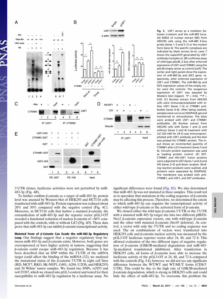
stimulating transcription factor 1 (USF1), we tested this inter-action by electrophoretic mobility shift (EMSA) and supershiftassays. USF1 is an evolutionarily well-conserved and ubiquitoustranscription factor involved in a wide number of cellular activitiessuch as immune response, cell cycle and proliferation, and lipidand glucose metabolism (28). As shown in Fig. 3A, the EMSAgenerates a specific band-shift pattern (complexes b–e, lane 2,Fig. 3A) that disappears in the mutant form of the E-box elementoligonucleotide (lane 4, Fig. 3A). Moreover, by using the anti-USF1 antibody, a supershift complex was generated (complex a,lane 7, Fig. 3A), suggesting that USF1 recognizes the E-box ele-ment upstream of the miR-483 locus.To confirm this result, we cloned the coding sequence of the
USF1 gene into the pCMV-Tag vector and cotransfected it intoHEK293 cells along with pIRES-Neo β-catenin and the reportervector pGL4E-6841–6910 with a wild-type or mutant E box (Fig.3B Left). USF1 overexpression was able to induce luciferase ac-tivity (twofold) that was further increased in the presence of ex-ogenous β-catenin (about threefold) compared with the control.Similar results obtained by qRT-PCR for miR-483-3p showedthat miR-483-3p was weakly induced by exogenous USF1 (P =0.05) but increased about threefold with coexpression of USF1and CTNNB1 (P < 0.02) (Fig. 3B Center). Note that IGF2 ex-pression was unchanged (Fig. 3B Right). To exclude a transcrip-tional regulation of USF1 by β-catenin, we also tested that theUSF1 protein level after β-catenin enforced expression was notchanged (Fig. S3). To further confirm these results, we transientlyknocked down USF1 by siRNA in HepG2 cells that show a veryhigh β-catenin activity and miR-483-3p expression. qRT-PCR
verified a significant reduction of miR-483-3p expression, com-pared with the control, after 72 h from siRNA transfection (Fig.S4). These data suggest USF1 is an important mediator of theregulation of the miR-483 locus driven by β-catenin.
β-Catenin and USF1 Directly Interact. To understand the interplaybetween β-catenin and USF1, we tested the possibility of in-teraction between these two proteins. We coimmunoprecipitatedfrom HEK293 nuclear extract lysate with either an anti-USF1antibody or an anti-β-catenin antibody and then immunoblottedwith either anti-β-catenin or anti-USF1, respectively, and foundthe two proteins coimmunoprecipitated (Fig. 3C). Because LiCltreatment is able to induce miR-483 locus expression, we testedthe ability of β-catenin to coimmunoprecipitate with USF1 withand without LiCl treatment. Western blot analysis revealed anincreased quantity of β-catenin immunoprecipitated with anti-USF1 (22%) compared with the nontreated control (Fig. 3D),where the induction of β-catenin protein level after LiCl treat-ment was increased about 60%. Then, we demonstrated the di-rect interaction by using the purified proteins CTNNB1-GST andUSF1-HIS in pull-down assays (Fig. 3E).
miR-483-3p Reveals a Negative Regulatory Loop by Targeting β-Catenin.Because regulatory feedback loops between microRNAs and theirtargets have been shown in numerous cases (29–31), we inves-tigated the possibility that β-catenin and/or USF1 are targets ofmiR-483-3p or -5p. By in silico analysis (http://targetscan.org), wefound that CTNNB1 is a predicted target of miR-483-3p (Fig. 4A).We tested the direct interaction of miR-483-3p with the
CTNNB1 3′UTR by luciferase assay as described in SI Materialsand Methods. In comparison with the control vector, miR-483-3pcaused a decrease in luciferase activity of about 30% and 50% inHEK293 and HCT116 cells, respectively, whereas in the mutated
Fig. 2. Analysis of the miR-483 minimal promoter region re-sponsive to LiCl/CTNNB1 stimuli. (A) Genomic structure of theIGF2/483 locus from the reference AF517226 genomic se-quence. Exons (black bars), start (ATG_5947) and stop codons(TGA_8745), E-box elements (gray triangle), the two predictedCTCF binding sites (black triangle), three CpG dinucleotidesaround the CTCF binding sites (black circles), and miR-483-3pand miR-483-5p (gray boxes) are shown. The five genomicfragments cloned upstream of the luciferase reporter gene inpGL4E for the analysis of the promoter are indicated at thebottom of the panel. The genomic region with insulator ac-tivity studied by Du et al. (22) is indicated (broken line). (B)Luciferase activity of four genomic fragments cloned in pGL4Ewith and without LiCl treatment; the pGL4E empty vector wasused as control. (C) Luciferase activity of the wild-type pGL4E-6487 (WT) and the mutated pGL4E-6487 in the predicted CTCFbinding sites (CTCF mut1 and CTCF mut2). (D) Analysis of thewild-type E-box element (Ebox wt) and mutant (Ebox mut)by luciferase assay of the pGL4E-6487 CTCF mut1 andpGL4E_Ebox. Firefly luciferase activity was normalized onRenilla luciferase activity of the cotransfected pGL4R vector.*P < 0.02, **P > 0.02.
4842 | www.pnas.org/cgi/doi/10.1073/pnas.1101734108 Veronese et al.
3′UTR clones, luciferase activities were not perturbed by miR-483-3p (Fig. 4B).To further confirm β-catenin as a target of miR-483-3p, protein
level was assessed by Western blot of HEK293 and HCT116 cellstransfected with miR-483-3p. Protein expression was reduced about20% and 50% compared with the negative control (Fig. 4C).Moreover, in HCT116 cells that harbor a mutated β-catenin, thecotransfection of miR-483-3p and the reporter vector pGL3-OTrevealed a functional reduction of nuclear β-catenin of ∼60% com-pared with the controls, with or without LiCl (Fig. 4D). These dataprove that miR-483-3p can inhibit β-catenin transcriptional activity.
Mutated Form of β-Catenin Can Evade the miR-483-3p RegulatoryLoop. Our findings suggest that a negative regulatory loop be-tween miR-483-3p and β-catenin exists. However, both genes areoverexpressed or have higher activity in tumors, suggesting thatβ-catenin could escape miR-483-3p regulation. Because it hasbeen described that an SNP within the 3′UTR of the mRNAtarget could affect the binding of the miRNA (32), we analyzedthe mutational status of the β-catenin 3′UTR in eight cell lines(RD, MCF7, RKO, SK-NEP1, G401, A204, U2OS, and HEK293)and 30 Wilms’ tumor samples. We found two SNPs, rs2953 andrs4135387, which we cloned into pGL3-control and tested for theirsusceptibility to miR-483-3p regulation by a luciferase assay. No
significant differences were found (Fig. S5). We also determinedthat miR-483-3p was not mutated in these samples. This result ledus to speculate that mutations in the coding sequence of β-cateninmay be affecting this process. Therefore, we determined the extentto which miR-483-3p can regulate the transcriptional activity ofeither wild-type β-catenin or the activated form of β-catenin.We cloned either the wild-type β-catenin 3′UTR or the 3′UTR
with a mutated miR-483-3p target site into two different pIRES-Neo2 β-catenin expression vectors, one with wild-type β-cateninand the other with mutated β-catenin (Ser45 deletion). As con-trol, a vector with only the 3′UTR and no coding sequence wasused. The six combinations of vectors were transfected intoHEK293 cells and β-catenin nuclear activity was measured by thepGL3-OT luciferase reporter vector after 24, 48, and 72 h. Thisallowed evaluation of the two different types of negative regula-tion of β-catenin (GSK3B-mediated degradation and miR-483-3p-mediated translational inhibition). After transfection inHEK293 cells, all forms of β-catenin were able to increase theluciferase activity of the pGL3-OT at 24, 48, and 72 h comparedwith the controls (Fig. 5A); however, we did not see any significantdifference between the WT/WT and WT/MUT forms (CDS/3′UTR). This could be due to the high rate of GSK3B-mediatedβ-catenin degradation, which is strong in HEK293 cells and couldhide the effect of miR-483-3p. To overcome this problem the
Fig. 3. USF1 serves as a mediator be-tween β-catenin and the miR-483 locus.(A) EMSA of nuclear extract (NE) fromHEK293 cells using the miR-483 E-boxprobe (lanes 1–3 and 5–7) or the mutantform (lane 4). The specific complexes areindicated by black arrows (b–e). Lane 7shows the supershift generated by USF1antibody (complex a). (B) Luciferase assayof wild-type pGL4E_E box after enforcedexpression of USF1 and CTNNB1 using thepGL4E empty vector as control (Left). Thecenter and right panels show the expres-sion of miR-483-3p and IGF2 gene, re-spectively, after enforced expression ofUSF1 and CTNNB1. The miR-483-3p andIGF2 expression values of the empty vec-tor were the controls. The exogenousexpression of USF1 was assessed byWestern blot (Upper). *P < 0.02, **P >0.02. (C) Nuclear extract from HEK293cells were immunoprecipitated with ei-ther USF1 (lanes 1–3) or CTNNB1 anti-bodies (lanes 4–6). After being washed,sampleswere runonanSDS/PAGEgel andtransferred to nitrocellulose. The blotswere probed with USF1 and CTNNB1antibodies. (D) Nuclear extract fromHEK293 cells with (lanes 1 and 2) andwithout (lanes 3 and 4) treatment withLiCl (20 mM for 24 h) was immunopreci-pitated with USF1 antibody and the blotwas probed for CTNNB1 protein. The in-put shows an incremented quantity ofCTNNB1 after LiCl treatment (lanes 5 and6). Vinculin protein expression was usedas loading protein control. (E) GST-CTNNB1 and HIS-USF1 fusion proteinswere subjected to GST (lanes 1 and 2) andHIS (lanes 3–5) pull-down analysis. Bind-ing reaction products were washed, andproteins were separated by SDS/PAGE.The membrane was probed with anti-CTNNB1, anti-USF1, and GST antibodies.
Veronese et al. PNAS | March 22, 2011 | vol. 108 | no. 12 | 4843
CELL
BIOLO
GY

experiment was conducted in the presence of LiCl, which resultedin a significant increase of activity in the presence of the WT/MUT but not the WT/WT form, suggesting that the wild-type 3′UTR is controlled by miR-483-3p but the mutant form is not (Fig.5 A and B Left).On the other hand, the Ser45 mutant form of β-catenin
showed a significant difference in regulation by miR-483-3p atthe 3′UTR (MUT/WT vs. MUT/MUT) only at 24 h without LiCl(P = 0.036). Because the mutant form is not degraded and thusaccumulates, it is reasonable that at an early time point β-cateninprotein levels are still controlled by miR-483-3p but that thisregulation is lost over time (Fig. 5B Right). Taken together, thesedata support that the activating mutation of β-catenin results inloss of regulation by miR-483-3p.
DiscussionWe identified a molecular mechanism of autoregulation ofβ-catenin activity through miR-483-3p, and a unique interactionbetween the transcription factors USF1 and β-catenin. Aftergenetic analysis of HCC samples, we identified an associationbetween the activation of the β-catenin pathway and the over-expression of miR-483-3p, an association that was stronger ina subset of samples that exhibited a divergent expression betweenIGF2 and miR-483-3p. This observation was supported by thefinding that the miR-483 locus could be up-regulated indepen-dently from its host gene IGF2 by enforced overexpression ofβ-catenin. These findings indicate the existence of at least two
mechanisms responsible for the expression of miR-483-3p: cor-egulation with IGF2 and transcriptional induction by β-catenin.The first mechanism appears to be significantly represented inWilms’ tumors and possibly other pediatric tumors. In these tu-mors, the well-known overexpression and loss of imprinting ofIGF2 may be responsible for most of the miR-483 up-regulation.Through this mechanism, both cell growth and survival can besimultaneously stimulated by IGF2 and miR-483-3p, respectively.The second mechanism may explain the overexpression of miR-483-3p in adult human cancers, where its up-regulation may occurindependently from IGF2 expression.Themediator ofmiR-483 stimulation by β-catenin was identified
as the basic helix–loop–helix upstream stimulating factor USF1.Here we prove that it can directly interact with β-catenin andrecognize the E-box CACGTG element located 400 nucleotidesupstream of themiR-483 locus. The interaction between β-catenin
Fig. 4. β-Catenin is a target of miR-483-3p. (A) Putative binding site of miR-483-3p in CTNNB1 3′UTRs (TargetScan database). Asterisks indicate nucleo-tides substituted in 3′UTR miR-483-3p predicted target site to perform lu-ciferase assay. (B) CTNNB1 3′UTRs regulate luciferase activity dependent onmiR-483-3p in HEK293 and HCT116 cell lines. MUT, mutant; WT, wild type.(C) Western blot analysis of CTNNB1 after miR-483-3p transfection inHEK293 and HCT116 cell lines. Cells were collected 48 h after miRNA trans-fection. (D) Luciferase activity of the reporter vectors pOT and pOF inHCT116 cells cotransfected with miR-483-3p and scramble oligo (NC2) withand without LiCl treatment. *P < 0.02, **P > 0.02.
Fig. 5. Mutated form of β-catenin evades the miR-483-3p regulatory loop.(A) Luciferase activity during the time (24, 48, and 72 h) of the reportervectors pOT and pOF in HEK293 cells cotransfected with the four differentCTNNB1 expression vectors (mutational status of the CDS and 3′UTR miR-483-3p target site of CTNNB1: CDS/3′UTR) with (Right) and without LiCltreatment (Left). As experiment controls, wild-type CTNNB1 3′UTR andmutated mir-483-3p target were cloned in the expression vector pIRESNeo2.E/MUT, empty/MUT; E/WT, empty/WT. (B) P values are indicated andgraphically represented.
4844 | www.pnas.org/cgi/doi/10.1073/pnas.1101734108 Veronese et al.

and another bHLH protein, MyoD, was previously described (33),and here we describe the direct interaction between β-catenin andUSF1. USF1 is a widely expressed transcription factor that playsa crucial role in the regulation of the cell cycle and proliferation(34, 35) and gluco-lipidic metabolism (36, 37). Moreover, it hasbeen shown that USF1 also plays an important role in the main-tenance of a chromatin barrier at the insulator elements of thechicken β-globin gene (38).Our discovery of a negative regulatory loop between miR-483-
3p and β-catenin generates an apparent paradox. To explain thisdiscrepancy, we found that the mutated form of β-catenin is ableto evade miR-483-3p regulation and the mutated protein can stillaccumulate in the cell.In addition to the β-catenin-USF1/miR-483 mechanism, in this
study we found that CTCF can also participate in the regulationof miR-483 expression. Indeed, we confirmed data published byDu et al. (22), who found that the region immediately upstreamof the miR-483 locus binds the multifunctional protein CTCF.We found evidence that CTCF could play an important role inthe regulation of the miR-483 locus as a transcriptional repressorand that repression is indirectly correlated to the methylationstatus of a CpG dinucleotide in the CTCF binding site. CTCFis a well-known regulator of imprinted regions of the genomesuch as the IGF2/H19 locus, where it permits the monoallelicexpression of these two genes (39, 40) by modulating the con-formation of this region, which (41) affects transcriptional ac-
tivity. It is also important to note that the chromosomal regionwhere CTCF is located (16q21) is often deleted in Wilms’ tumor(42, 43). Our finding highlights the role of the miR-483 locus asa point of junction among the most important players in Wilms’tumor (IGF2 locus, Wnt/β-catenin pathway, and CTCF).By unraveling the complex regulation of the miR-483 locus,
the present study provides insight into the Wnt/β-catenin path-way and indicates new targets for anticancer therapy.
Materials and MethodsHEK293 and HCT116 cells were transfected using Lipofectamine 2000 (Invi-trogen). Mutational and DNA methylation analysis was carried out usingprimers indicated in Table S3. DNA constructs were cloned using primerslisted in Table S3. RNA isolation was performed using TRIzol (Invitrogen)according to the manufacturer’s instructions and quantitative real-time re-verse transcription–PCR using SYBR Green and TaqMan technologies (Ap-plied Biosystems). Western blot, EMSA, immunoprecipitation, and pull-downanalyses were carried out using primary antibodies for CTNNB1 (Cell Sig-naling; 9562), USF1 (Santa Cruz Biotechnology; sc-229), and MYC (Cell Sig-naling; 5605). Statistical analysis results are expressed as mean ± SD andsignificance was accepted at a P value <0.05. More detailed information isprovided in SI Materials and Methods.
ACKNOWLEDGMENTS. This work was supported by grants from theAssociazione Italiana per la Ricerca sul Cancro, Ministero dell’Università edella Ricerca, Ministero della Salute and Fondazione Cariplo (ProgettoNOBEL) to M.N. and by National Cancer Institute grants to C.M.C.
1. Perez-Moreno M, Jamora C, Fuchs E (2003) Sticky business: Orchestrating cellularsignals at adherens junctions. Cell 112:535–548.
2. Clevers H (2006) Wnt/β-catenin signaling in development and disease. Cell 127:469–480.
3. Wang SS, et al. (1998) Alterations of the PPP2R1B gene in human lung and coloncancer. Science 282:284–287.
4. Satoh S, et al. (2000) AXIN1 mutations in hepatocellular carcinomas, and growthsuppression in cancer cells by virus-mediated transfer of AXIN1.Nat Genet 24:245–250.
5. Major MB, et al. (2007) Wilms tumor suppressor WTX negatively regulates WNT/β-catenin signaling. Science 316:1043–1046.
6. Morin PJ, et al. (1997) Activation of β-catenin-Tcf signaling in colon cancer bymutations in β-catenin or APC. Science 275:1787–1790.
7. Devereux TR, et al. (2001) CTNNB1 mutations and β-catenin protein accumulation inhuman hepatocellular carcinomas associated with high exposure to aflatoxin B1. MolCarcinog 31:68–73.
8. Kusafuka T, Miao J, Kuroda S, Udatsu Y, Yoneda A (2002) Codon 45 of the β-cateningene, a specific mutational target site of Wilms’ tumor. Int J Mol Med 10:395–399.
9. Rubinfeld B, et al. (1996) Binding of GSK3β to the APC-β-catenin complex andregulation of complex assembly. Science 272:1023–1026.
10. Mann B, et al. (1999) Target genes of β-catenin-T cell-factor/lymphoid-enhancer-factor signaling in human colorectal carcinomas. Proc Natl Acad Sci USA 96:1603–1608.
11. Tetsu O, McCormick F (1999) β-Catenin regulates expression of cyclin D1 in coloncarcinoma cells. Nature 398:422–426.
12. Ambros V (2004) The functions of animal microRNAs. Nature 431:350–355.13. Calin GA, Croce CM (2006) MicroRNA signatures in human cancers. Nat Rev Cancer 6:
857–866.14. Iorio MV, Croce CM (2009) MicroRNAs in cancer: Small molecules with a huge impact.
J Clin Oncol 27:5848–5856.15. Negrini M, Ferracin M, Sabbioni S, Croce CM (2007) MicroRNAs in human cancer: From
research to therapy. J Cell Sci 120:1833–1840.16. Veronese A, et al. (2010) Oncogenic role of miR-483-3p at the IGF2/483 locus. Cancer
Res 70:3140–3149.17. Guled M, et al. (2009) CDKN2A, NF2, and JUN are dysregulated among other genes
by miRNAs in malignant mesothelioma—A miRNA microarray analysis. GenesChromosomes Cancer 48:615–623.
18. Soon PS, et al. (2009) miR-195 and miR-483-5p identified as predictors of poorprognosis in adrenocortical cancer. Clin Cancer Res 15:7684–7692.
19. Nusse R (2007) Cancer. Converging on β-catenin in Wilms tumor. Science 316:988–989.20. Reya T, Clevers H (2005) Wnt signalling in stem cells and cancer. Nature 434:843–850.21. de La Coste A, et al. (1998) Somatic mutations of the β-catenin gene are frequent in
mouse and human hepatocellular carcinomas. Proc Natl Acad Sci USA 95:8847–8851.22. Du M, et al. (2003) Insulator and silencer sequences in the imprinted region of human
chromosome 11p15.5. Hum Mol Genet 12:1927–1939.23. Sparago A, et al. (2007) Mechanisms causing imprinting defects in familial Beckwith-
Wiedemann syndrome with Wilms’ tumour. Hum Mol Genet 16:254–264.24. Prawitt D, et al. (2005) Microdeletion of target sites for insulator protein CTCF in
a chromosome 11p15 imprinting center in Beckwith-Wiedemann syndrome andWilms’ tumor. Proc Natl Acad Sci USA 102:4085–4090.
25. Chan CS, Song JS (2008) CCCTC-binding factor confines the distal action of estrogenreceptor. Cancer Res 68:9041–9049.
26. Butcher DT, Rodenhiser DI (2007) Epigenetic inactivation of BRCA1 is associated withaberrant expression of CTCF and DNA methyltransferase (DNMT3B) in some sporadicbreast tumours. Eur J Cancer 43:210–219.
27. Paradowska A, et al. (2009) Aberrant epigenetic modifications in the CTCF bindingdomain of the IGF2/H19 gene in prostate cancer compared with benign prostatehyperplasia. Int J Oncol 35:87–96.
28. Corre S, Galibert MD (2005) Upstream stimulating factors: Highly versatile stress-responsive transcription factors. Pigment Cell Res 18:337–348.
29. Petrocca F, et al. (2008) E2F1-regulated microRNAs impair TGFβ-dependent cell-cyclearrest and apoptosis in gastric cancer. Cancer Cell 13:272–286.
30. Yang X, et al. (2009) miR-449a and miR-449b are direct transcriptional targets of E2F1and negatively regulate pRb-E2F1 activity through a feedback loop by targetingCDK6 and CDC25A. Genes Dev 23:2388–2393.
31. Xu N, Papagiannakopoulos T, Pan G, Thomson JA, Kosik KS (2009) MicroRNA-145regulates OCT4, SOX2, and KLF4 and represses pluripotency in human embryonicstem cells. Cell 137:647–658.
32. Nicoloso MS, et al. (2010) Single-nucleotide polymorphisms inside microRNA targetsites influence tumor susceptibility. Cancer Res 70:2789–2798.
33. Kim CH, Neiswender H, Baik EJ, XiongWC, Mei L (2008) β-Catenin interacts with MyoDand regulates its transcription activity. Mol Cell Biol 28:2941–2951.
34. North S, et al. (1999) Regulation of cdc2 gene expression by the upstream stimulatoryfactors (USFs). Oncogene 18:1945–1955.
35. Cogswell JP, Godlevski MM, Bonham M, Bisi J, Babiss L (1995) Upstream stimulatoryfactor regulates expression of the cell cycle-dependent cyclin B1 gene promoter. MolCell Biol 15:2782–2790.
36. Read ML, Clark AR, Docherty K (1993) The helix-loop-helix transcription factor USF(upstream stimulating factor) binds to a regulatory sequence of the human insulingene enhancer. Biochem J 295:233–237.
37. Iynedjian PB (1998) Identification of upstream stimulatory factor as transcriptionalactivator of the liver promoter of the glucokinase gene. Biochem J 333:705–712.
38. Huang S, Li X, Yusufzai TM, Qiu Y, Felsenfeld G (2007) USF1 recruits histonemodification complexes and is critical for maintenance of a chromatin barrier. MolCell Biol 27:7991–8002.
39. Murrell A, Heeson S, Reik W (2004) Interaction between differentially methylatedregions partitions the imprinted genes Igf2 and H19 into parent-specific chromatinloops. Nat Genet 36:889–893.
40. Hark AT, et al. (2000) CTCF mediates methylation-sensitive enhancer-blocking activityat the H19/Igf2 locus. Nature 405:486–489.
41. Kurukuti S, et al. (2006) CTCF binding at the H19 imprinting control region mediatesmaternally inherited higher-order chromatin conformation to restrict enhancer accessto Igf2. Proc Natl Acad Sci USA 103:10684–10689.
42. MawMA, et al. (1992) A third Wilms’ tumor locus on chromosome 16q. Cancer Res 52:3094–3098.
43. Yeh A, et al. (2002) Chromosome arm 16q in Wilms tumors: Unbalanced chromosomaltranslocations, loss of heterozygosity, and assessment of the CTCF gene. GenesChromosomes Cancer 35:156–163.
Veronese et al. PNAS | March 22, 2011 | vol. 108 | no. 12 | 4845
CELL
BIOLO
GY


