Embed Size (px)
Citation preview
Multi-Locus Sequence Typing of EnteroaggregativeEscherichia coli Isolates from Nigerian Children UncoversMultiple LineagesIruka N. Okeke1*, Faith Wallace-Gadsden1, Hannah R. Simons1, Nicholas Matthews2, Amy S. Labar1,
Jennifer Hwang1, John Wain2,3
1 Department of Biology, Haverford College, Haverford, Pennsylvania, United States of America, 2 Wellcome Trust Sanger Institute, Genome Campus, Hinxton, Cambridge,
United Kingdom, 3 Health Protection Agency, Colindale, London, United Kingdom
Abstract
Background: Enteroaggregative Escherichia coli (EAEC) are defined by their stacked-brick adherence pattern to humanepithelial cells. There is no all-encompassing genetic marker for EAEC. The category is commonly implicated in diarrhea butresearch is hampered by perplexing heterogeneity.
Methodology/Principal Findings: To identify key EAEC lineages, we applied multilocus sequence typing to 126 E. coliisolates from a Nigerian case-control study that showed aggregative adherence in the HEp-2 adherence assay, and 24 otherEAEC strains from diverse locations. EAEC largely belonged to the A, B1 and D phylogenetic groups and only 7 (4.6%)isolates were in the B2 cluster. As many as 96 sequence types (STs) were identified but 60 (40%) of the EAEC strains belongto or are double locus variants of STs 10, 31, and 394. The remainder did not belong to predominant complexes. The mostcommon ST complex, with predicted ancestor ST10, included 32 (21.3%) of the isolates. Significant age-related distributionsuggests that weaned children in Nigeria are at risk for diarrhea from of ST10-complex EAEC. Phylogenetic group D EAECstrains, predominantly from ST31- and ST394 complexes, represented 38 (25.3%) of all isolates, include genome-sequencedstrain 042, and possessed conserved chromosomal loci.
Conclusions/Significance: We have developed a molecular phylogenetic framework, which demonstrates that althoughgrouped by a shared phenotype, the category of ‘EAEC’ encompasses multiple pathogenic lineages. Principal amongisolates from Nigeria were ST10-complex EAEC that were associated with diarrhea in children over one year and ECOR Dstrains that share horizontally acquired loci.
Citation: Okeke IN, Wallace-Gadsden F, Simons HR, Matthews N, Labar AS, et al. (2010) Multi-Locus Sequence Typing of Enteroaggregative Escherichia coli Isolatesfrom Nigerian Children Uncovers Multiple Lineages. PLoS ONE 5(11): e14093. doi:10.1371/journal.pone.0014093
Editor: Ulrich Dobrindt, University of Munster, Germany
Received June 12, 2010; Accepted November 4, 2010; Published November 23, 2010
Copyright: � 2010 Okeke et al. This is an open-access article distributed under the terms of the Creative Commons Attribution License, which permitsunrestricted use, distribution, and reproduction in any medium, provided the original author and source are credited.
Funding: This study was supported by National Science Foundation (www.nsf.org) grant RUI #0516591, by a Branco Weiss Fellowship from the Society ofScience (http://www.society-in-science.ethz.ch/) to INO and by Wellcome Trust (http://www.wellcome.ac.uk/) support to JW and NM. We thank the HowardHughes Medical Institute for support of Haverford College undergraduate researchers FW-G and ASL (http://www.haverford.edu/kinsc/HHMI/). FW-G is also aMellon-Mays Undergraduate Research Fellow (www.mmuf.org). The funders had no role in study design, data collection and analysis, decision to publish, orpreparation of the manuscript.
Competing Interests: The authors have declared that no competing interests exist.
* E-mail: [email protected]
Introduction
Enteroaggregative Escherichia coli (EAEC) is a category of
diarrheagenic E. coli defined by a characteristic ‘‘stacked brick’’,
‘‘honeycomb’’ or ‘‘aggregative’’ adherence pattern to epithelial cells
[1]. In spite of their association with diarrheal illness, EAEC are
commonly recovered from healthy people and their epidemiology,
pathogenesis and ecology are poorly understood. The current
definition, based on adherence pattern likely includes pathogenic as
well as non-pathogenic strains. The inherent heterogeneity within
the EAEC category, as presently defined, hampers pathogenesis
research as well as the development of diagnostic tools. EAEC
isolates have been implicated in acute and persistent sporadic
diarrhea, and outbreaks, in both industrialized and developing
countries (reviewed by [2,3,4]). Although recent information
suggests that they are among the most common diarrheal pathogens
worldwide [5,6,7], the true burden of disease from EAEC is
unknown because many epidemiological studies, including studies
that focus on diarrhea-causing E. coli, do not seek EAEC
exhaustively or at all [6,7,8,9,10]. Moreover, genetic studies that
seek to understand the evolution of pathogenic E. coli typically
exclude, under-represent or de-emphasize EAEC [11,12,13].
The gold standard for EAEC identification is a HEp-2
adherence test that can only be performed in laboratories able
to culture human cell lines. Attempts to identify genetic markers
for EAEC have principally focused on a large, partially conserved
plasmid, pAA. Many putative virulence genes map to this element,
and the distribution of some of these genes has been associated
with disease in epidemiological studies. However, strains that lack
some or all known pAA genes are consistently recovered from
people with diarrhea. Additionally, strains isolated from outbreaks,
including the largest documented EAEC outbreak that affected
PLoS ONE | www.plosone.org 1 November 2010 | Volume 5 | Issue 11 | e14093
over 2,600 Japanese school children, have commonly lacked many
or all pAA putative virulence genes [14,15]. One strain, C1096
isolated from a nursery outbreak in Serbia, harbors a large plasmid
completely unrelated to the prototypical pAA plasmids [16]. Thus,
even though strains bearing pAA plasmids, and specifically the
aggR gene, have been described as ‘typical’ [17], they cannot be
considered archetypical pathogenic EAEC, nor is it certain that
they even represent isolates with the greatest pathogenic potential.
Studies of pAA genes have uncovered important adhesins and
toxins but have only emphasized both the heterogeneity of the
pathotype and the limitations of the evolutionary information that
can be derived from markers on a potentially mosaic mobile
element [18,19,20,21,22,23,24].
Following on the earlier plasmid-focused studies, recent re-
search has begun to uncover EAEC chromosomal loci of interest
[25,26,27,28,29] and the completion of the genomes of EAEC
strains 042, 17-2 and 101-1 will allow further identification of such
factors. However, chromosomal loci are yet to be associated with
disease. Furthermore, heterogeneity in host response contributes
to the difficulty in interpreting the results of volunteer and
epidemiological studies [30,31,32], making it challenging to
prioritize strains for future genome sequencing, other in-depth
analysis or vaccine development. Population genetic studies of
EAEC strains could help address some of these questions [29].
A 1999 report described an EAEC phylogeny based on
multilocus enzyme electrophoresis (MLEE) [19]. No virulence
factors were associated with phylogeny, although loci examined at
that time were predominantly plasmid-borne. More recently
discovered loci mapped onto that phylogeny illustrate that EAEC
do have lineage-specific genes [25]. However genetic relationships
were only studied among 44 strains from patients with diarrhea,
and the strain collection was biased towards the widely-used,
plasmid-detecting probe, CVD432. A second, more recent, MLEE
study of isolates from Brazil again pointed to heterogeneity and
failed to identify lineage-specific genes. Interestingly, it did suggest
that some lineages were more likely to harbor certain genes than
others, supporting the idea that certain EAEC lineages may have
epidemiological significance [24]. These data are promising but it
is difficult to add strains to these phylogenies, or to overlay EAEC
phylogeny with that of the genus as a whole, because they are
based on the highly specialized method of MLEE.
We hypothesized that certain EAEC lineages predominate
among isolates from childhood diarrhea and that hypervirulent
lineages would be associated with diarrhea in a strain set from a
controlled study. To test this hypothesis, we performed multilocus
sequence typing (MLST) on an epidemiologically informative set
of EAEC isolates obtained from Nigerian children with and
without diarrhea, entering them into a curated public database to
which researchers from all over the world can contribute [13]. We
found that EAEC strains from Nigeria belonged to multiple
sequence types (STs), but that some ST complexes were pre-
dominant. ST complexes predominant in the epidemiological set
from Nigeria were also represented among well-studied EAEC
strains from other parts of the world.
Methods
Bacterial Strains126 EAEC strains isolated in south western Nigeria during a
previous epidemiological study formed the core strain collection
for this work [33]. These strains, which include 73 isolates from
children with diarrhea and 53 healthy controls, represent a com-
plete epidemiological set (excluding 5 strains that are no longer
viable). They were distinguished from other E. coli by HEp-2
adherence, the gold standard for EAEC identification [18,33].
Therefore, although they were drawn from one geographical
location, they are not biased towards any probes, and studying the
entire strain set from that epidemiological study avoids other
biases associated with strain selection. Additionally, Serbian and
Japanese outbreak isolates C1096 and 101-1, kindly provided by
Thomas Whittam, 17 other reference strains that have been
phylogenetically classified by multilocus-enzyme electrophoresis in
a previous study, and five isolates from locations not represented
or underrepresented in that study, were employed [19,25]. All the
strains have been previously probed for several EAEC-associated
genes and verified by HEp-2 adherence assays in our laboratory.
Bacteria were cultured in Luria broth (LB) or LB agar and
maintained at 280uC in LB: glycerol 1:1.
Multi-locus sequence typing and analysisGene fragments from the adk, fumC, gyrB, icd, mdh, purA and recA
were amplified as described by Wirth et al [13]. Amplified DNA
was prepared for sequencing using the ‘‘ExoSAP’’ method
(Amersham Biosciences UK Ltd). Cleaned fragments were
sequenced from both ends using the di-deoxy chain terminator
method [34], with V3.1 Bigdye terminator chemistry [35]. Both
strands of each fragment were sequenced at least once. The
resulting sequencing reactions were analyzed on 3700 or 3730 ABI
sequencing machines (Applied Biosystems, USA). Allele and
sequence type (ST) assignments were made at the publicly
accessible E. coli MLST database at http://mlst.ucc.ie/mlst/
dbs/Ecoli/.
Phylogenetic inferences about ancestral allelic profiles and strain
interrelatedness were made using eBURST version 3 http://eburst.
mlst.net/and ClonalFrame version 1.1 http://www.xavierdidelot.
xtreemhost.com/clonalframe.htm [36,37]. Sequence type complexes
were defined using eBURST as groups sharing at least six identical
alleles and bootstrapping with 1000 samplings. EAEC strains in this
study were mapped onto the phylogeny of the E. coli species as a
whole using MLST data available from http://mlst.ucc.ie/mlst/dbs/
Ecoli/. EAEC MLST data was further analyzed by ClonalFrame
[37] to investigate relationships among different sequence type
complexes. ClonalFrame is a Bayesian method of constructing
evolutionary histories that takes both mutation and recombination
into account. For each analysis, four independent runs of the Markov
chain were employed and runs were compared by the method of
Gelman and Rubin [38]. Calculated Gelman-Rubin statistics for all
parameters were below 1.20, indicating satisfactory convergence
between tree replicates. A 75% consensus tree was created for the
EAEC isolates. Mutation and recombination rates were computed
using ClonalFrame as described by Didelot and Falush [37].
Plasmid replicon typingThree multiplex panels comprised of 18 primer pairs were used
to identify plasmid replicons in EAEC strains by PCR as described
by Johnson et al [39]. Because this protocol does not amplify the
FIIA replicon from EAEC strain 042, we additionally screened the
strains for this replicon using primers REP042F and R (Table S1).
Strain carrying the sequenced plasmids pMAR-7, pAA2 (042),
pB171, pHCM1 and pED204 were used as controls [40,41,42,43].
Antimicrobial susceptibility testingSusceptibility testing was performed in accordance with the
Clinical and Laboratory Standards Institute (CLSI, formerly
NCCLS) disk diffusion method [44]. Bacterial suspensions were
plated onto the surface of Mueller-Hinton plates. Disks containing
ampicillin (10 mg), tetracycline (30 mg), trimethoprim (5 mg),
nalidixic acid (30 mg), chloramphenicol (30 mg), sulfonamide
MLST of EAEC from Nigeria
PLoS ONE | www.plosone.org 2 November 2010 | Volume 5 | Issue 11 | e14093
(300 mg), streptomycin (10 mg), and ciprofloxacin (5 mg) (Remel)
using a disc dispenser. Plates were incubated overnight at 37uC.
The diameter of inhibition zones was measured to the nearest
millimeter and interpreted according to the NCCLS standards
[44]. E. coli K-12 C600 and E. coli NCTC 10418 were used as
controls.
DNA hybridizationColony blots were prepared using Whatmans 541 filter paper
(Whatman, England). A 1,175 bp RsaI-BamHI fragment from
Tn21 and a 1,560 bp SphI-HpaI fragment from Tn7 were used as
intI1 and intI2 probes respectively [45,46]. A 414 bp tetracycline
resistance gene probe was excised from pACYC184 (New England
Biolabs) using EcoRI and ScaI. A 787 bp chloramphenicol acetyl
transferase gene probe was excised from pACYC184 with EcoRV
and NruI. Both fragments were purified by gel extraction and
labeled by random priming with 32P a d-CTP employing a
commercially available labeling kit (Amersham Pharmacia Bio-
tech, NJ). Un-incorporated nucleotides were removed by passage
through Sephadex G50 micro-columns (Amersham Pharmacia
Biotech). Hybridization was carried out under high stringency
conditions using standard techniques [47] employing a hybridiza-
tion buffer composed of: 5X SSC, 0.5% sodium dodecyl sulphate,
10 mM EDTA, 1X Denhardts solution and 100 mg/ml sonicated
salmon sperm DNA. Colony blots were hybridized at 65uCovernight, washed with 0.1X SSC, 0.1% SDS at 65uC and
exposed to X-ray film at 280uC overnight. E. coli DH5a, DH5a(pACYC184) and cell-detaching E. coli strains that had been
characterized in previous studies [48] were used as controls.
PCR for chromosomal genesLysine decarboxylase genes and activity were detected as
described previously [49]. Oligonucleotides used to identify
virulence and antimicrobial resistance loci are listed in Table S1.
They include primers designed to amplify three regions in the 042
Tn2411-derived resistance island as well as the pstS and glmS
integenic region in E. coli K-12, the island’s insertion site. PCR
amplifications were performed using 1 unit recombinant Taq
polymerase enzyme with PCR buffer from Invitrogen, 2 mM
MgCl2 and 1 mM of each oligonucleotide primer in each reaction.
All amplifications began with a two-minute hot start at 94uCfollowed by 30 cycles of denaturing at 94uC for 30 s, annealing for
30 s and extending at 72uC. PCR reactions were templated with
boiled bacterial colonies. The primers, annealing temperatures,
extension times and positive control strains used for each
amplification are indicated in Table S1.
Statistical analysisStatistical tests of significance were conducted using the Chi-
squared and Fisher’s exact two-tailed tests on Epi-Info version 3.3
(CDC, 2004). Where applicable, a Mann-Whitney U test was
performed.
Results and Discussion
MLST phylogeny for EAEC agrees with an earlier EAECMLEE phylogeny
We performed MLST on 19 of the best-characterized EAEC
strains. These strains cover a broad range of MLEE electropho-
resis types defined by Czeczulin et al. and contain a variety of
virulence gene combinations (Table S2) [19,25,50]. For the most
part, MLEE-defined clades correspond to MLST complexes. The
MLEE EAEC1 cluster, comprised of ECOR phylogenetic group A
strains, is largely composed of the E. coli MLST ST10 complex.
The MLEE EAEC2 cluster, comprised of ECOR system group D
strains, belong to, or share four or more alleles with, the MLST
ST31. A third cluster defined by MLEE, the AA/DA cluster,
which includes diffusely-adherent E. coli as well as EAEC, were less
closely clustered by MLST. They include strains belonging to ST
complexes: 10, 23, 40, or 448 (Table S2). With one exception, all
of the strains that did not cluster by MLEE belonged to other
complexes or singleton STs. The 19 well-characterized strains
spanned most, but expectedly not all, of the diversity seen in the
collection from Nigeria. Six of the seven most common MLST
clonal complexes defined in this study were represented in this test
collection.
We examined the distribution of nine plasmid borne and eight
chromosomal loci in the 19-strain reference collection. The
distribution of most virulence genes sought among these strains
did not associate with either MLST or MLEE-derived phylogeny
(Table S2). The heme transport outer membrane receptor (chuA)
and fepC-PAI (that is the fepC gene within the prrA-modA-fepC
pathogenicity island), which were present only in ST31 and ST394
complex and related strains. However chuA is common to the
ECOR group D lineage, to which these strains belong and fepC-
PAI appears to show a similar distribution [25,51]. Overall, the
distribution of virulence and other genes was largely heteroge-
neous outside ST complexes 10, 31 and 394 later.
EAEC plasmids belong to diverse incompatibility groupsMost loci associated with EAEC are plasmid-borne and
distribution of plasmid-borne genes has previously been reported
not to associate with EAEC strain phylogeny [19]. Nonetheless,
given that our data reveal a few major and several minor EAEC
lineages, we sought to determine whether a conserved plasmid
backbone defined EAEC or whether certain EAEC lineages
carried plasmids of a common origin. We subjected the 19 strains
from the reference collection to plasmid replicon typing using a
multiplext PCR protocol, which detects replicons from 22 plasmid
incompatibility groups, described by Johnson et al [39]. Addition-
ally, because the FIIA replicon (which is found in EAEC strain 042
and a number of enteric pathogens) is genetically diverse [52] and
not detected by the Johnson et al [39] protocol, we designed
primers specific for the pAA2 replicon. These primers also detect
FIIA replicons from the virulence plasmids of EPEC strains B171
and E2348/69.
As shown in Table S2, the IncFIIA replicon seen in 042
(FIIA042), IncP, IncFIB, IncY and FIA replicons were common
among the reference EAEC strains. The FIIA042, replicon was
amplified from all but three EAEC strains. However this replicon
is also commonly present in enteropathogenic, enterotoxigenic
and cell-detaching E. coli. Twelve of the 19 reference strains were
positive for more two or more of the replicons sought. No replicon
was common to all the EAEC strains and none of nine other
plasmid-borne genes showed absolute association with a specific
replicon type. This includes the CVD432 probe locus, which is
used as a marker for aggregative plasmids (pAA) that define
‘typical’ EAEC [17,53]. A conserved plasmid backbone may not
exist for pAA genes and genes associated with ‘typical’ strains, such
as those encoding the antiaggregative protein (aap) and its secretion
system (aat), may instead represent loci that have integrated into
different plasmids. This idea is supported by the dissimilarity
among three recently sequenced pAA plasmids [54]. Although
these all bear a FIIA042, replicon (often with other replicons) and
are roughly syntenic in the aat to fimbrial gene regions, the rest of
the molecules differ in both gene content and organization [54].
Association of aggregative adherence genes with a broad range of
plasmids, provides an explanation for the high prevalence of
MLST of EAEC from Nigeria
PLoS ONE | www.plosone.org 3 November 2010 | Volume 5 | Issue 11 | e14093

EAEC worldwide. A similar phenomenon has been used to
explain the broad dissemination of antimicrobial resistance genes
like sul2 and dfrA1 [55,56].
EAEC belong to multiple lineagesIn October 2010, there were 3344 isolates in the E. coli MLST
database belonging to 1,985 STs and the ‘Based Upon Related
Sequence Types’ (BURST) algorithm placed them in 103 clonal
complexes, as defined by using a group definition of six alleles. We
identified 96 of these sequence types among 150 EAEC strains in
this study (Table S3). As shown in Figure 1, the EAEC strains were
distributed throughout the E. coli phylogeny although a number of
ST complexes that are abundant in the E. coli MLST database did
not contain any EAEC strains. Common E. coli ST complexes that
were devoid of EAEC strains included ST complexes 69, 73, 95
and 131, all of which are predominantly comprised of extraintes-
tinal E. coli and belong to the B2 phylogentic group [13,57,58].
Ninety-six (64%) of the 150 EAEC strains from this study
belonged to 16 ST complexes. Sixty (40%) of the strains fell into
three main groups with predicted ancestral STs 10, 31 and 394
(Table S3). A 75% consensus tree constructed using ClonalFrame
identified similar groups as eBURST, with few exceptions
(Figure 2). Both BURST and ClonalFrame distinguish ST
complexes 31, 38 and 394, along with four strains sharing four
alleles with these complexes as a separate lineage, which
corresponds to the MLEE-defined ECOR D group (Figure 2).
The remainder of the EAEC strains appear much more diverse,
although the ST10 complex, belonging to ECOR phylogroup A,
represents a highly-prevalent delineated sub-group (Figure 2).
ECOR group A strains predominate in West Africa [59], which
may provide one explanation for the fact that ST10 was the most
common seen in this study. Ninety (60%) isolates did not belong to
predominant complexes. eBURST placed 37 of these isolates into
13 clonal complexes, each containing only 2–4 isolates and 53 of
them belonged to 43 singleton STs, nine of which were identified
more than once (Table S3).
The 75% consensus tree generated with ClonalFrame agreed,
for the most part, with the phylogeny inferred by eBURST
(Figure 2). The tree topology is similar to that originally produced
for E. coli as a whole by MLEE, and ratified by MLST and other
methods, delineating the major clades defined by the ECOR
collection: A, B1, B2 and D [11,13,59]. Strains belonging to all
four of these clades were identified among EAEC, a finding that
was made in a previous study that included only 11 EAEC strains
[11]. In that study, only one of the 11 isolates belonged to the B2
group and we find that in the current examination of 150 strains,
the isolates were almost evenly distributed among A, B1 and D
groups but only 7 (4.6%) of the strains evaluated in this study
clustered in B2, a group that is largely comprised of uropatho-
genic, neonatal meningitic and other extra-intestinal E. coli
pathogens (Figure 2). While this suggests that EAEC strains may
not be associated with the B2 lineage, it is important to note that a
few strains, including, strain 101-1, from a large outbreak in Japan,
clustered with this clade. As this study focused on EAEC isolates
from Nigeria, and the B2 group has previously been reported to be
more common in Asia than in Africa [59], it is possible that other
lineages are more prominent elsewhere.
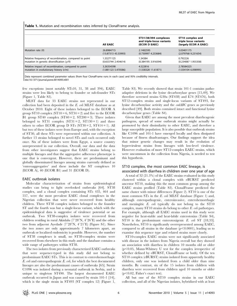
The role of recombination, relative to point mutations, in E. coli
evolution is uncertain, with different studies and methods
producing different rate estimates [13,60,61]. Some studies have
found that in E. coli and other organisms where there is no
consensus on recombination rates, that recombination occurs
more commonly in some lineages of a species than in others
[13,62]. Among the EAEC sequence-typed in this study,
Figure 1. BURST output for strains in the E. coli MLST database with STs containing EAEC strains ringed in pink. The size of the node isproportional to the number of isolates in the STs. Blue nodes represent predicted founder STs and sub-founders are indicated in yellow. All other STsmarked as black dots. STs 12, 40 and the six clonal complexes containing four or more of the EAEC strains from this study are circled in green. Brownarrows point to the two outbreak isolates.doi:10.1371/journal.pone.0014093.g001
MLST of EAEC from Nigeria
PLoS ONE | www.plosone.org 4 November 2010 | Volume 5 | Issue 11 | e14093

ClonalFrame identified strong evidence for recombination at all
principal nodes of the tree. The relative frequency of recombina-
tion, as compared to point mutation, (r/h), computed using
ClonalFrame was 1.333 in the complete EAEC set (Table 1). This
was greater than rates computed for the E. coli species by Perez-
Losada et al (who estimated recombination at essentially zero [60])
but comparable to those proposed by Wirth et al (0.321–2.139
[13]) and Touchon et al (close to one [63]). However, as with
Wirth et al, we found considerable variation in recombination for
different lineages. Most notably, recombination rates were lower
in ST10-complex EAEC and strains sharing up to four alleles
with this group compared with all EAEC (Table 1), a finding that
has previously been reported for the ECOR-defined group A
compared to the species as a whole [13]. The net consequence of
recombination, which is measured by the rate at which a given
nucleotide is substituted by recombination versus point mutation
(r/m), demonstrates that a change is almost twice as likely to be
produced by recombination in the ST31 complex and its triple-
locus variants than in ST10 and triple locus variants (Table 1).
We examined the E. coli MLST database at mlst.net (http://
mlst.ucc.ie/mlst/dbs/Ecoli/) for other strains present in predom-
inant EAEC STs. As at October 2010, E. coli ST394 complex
contained 16 isolates (12 from this study) and all but three
members of this complex were annotated as EAEC or E. coli from
diarrhea. (One isolate is annotated as unknown and two are
urinary tract infection isolates). The ST394 complex may
represent an EAEC-specific complex but no other ST-complexes
containing more than three EAEC strains were EAEC-specific.
The most common clonal complexes in which EAEC strains were
identified, including those with predicted ancestors ST10, ST23,
ST31 and ST38 contained strains belonging to multiple
pathogenic categories as well as likely commensals and, with a
Figure 2. 75% Consensus ClonalFrame tree for MLST data from 150 EAEC strains including strains from Nigerian children withdiarrhea (Red), healthy Nigerian children (Blue), and cases of diarrhea from other parts of the world (Yellow). The tree incorporatesrecombination as well as mutation. The principal E. coli sub-clades corresponding to the four major groups originally defined by MLEE - A, B1, D andB2 - are marked to the right of the tree respectively in brown, purple, grey and orange. Boxed numbers to the right of the tree indicate strainsbelonging to the most common ST complexes identified by eBURST.doi:10.1371/journal.pone.0014093.g002
MLST of EAEC from Nigeria
PLoS ONE | www.plosone.org 5 November 2010 | Volume 5 | Issue 11 | e14093
few exceptions (most notably STs10, 31, 38 and 394), EAEC
strains were less likely to belong to founder or sub-founder STs
(Figure 1, Table S3).
MLST data for 33 EAEC strains not represented in our
collection had been deposited in the E. coli MLST database as at
October 2010. Eight of these isolates belonged to the ECOR A
group ST10 complex (ST10 = 6, ST34 = 2) and five to the ECOR
B1 group ST40 complex (ST40 = 2, ST200 = 3). Three isolates
belonged to ST31 complex (ST31 = 2, ST130 = 1) and three
others to other ECOR group D STs (ST30 = 2, ST414 = 1). All
but two of these isolates were from Europe and, with the exception
of ST30, all these STs were represented within our collection. A
further 13 strains belonged to novel STs and each occurred only
once. Six of these isolates were from Egypt, a source country
unrepresented in our collection. Overall, our data and the data
from other investigators suggest that EAEC strains belong to
multiple lineages and that the aggregative adherence phenotype is
one that is convergent. However, there are predominant and
globally disseminated lineages among strains currently defined as
‘enteroaggregative’ and these include the ST complexes 10
(ECOR A), 40 (ECOR B1) and 31 (ECOR D).
EAEC outbreak isolatesMolecular characterization of strains from epidemiological
studies can bring to light overlooked outbreaks [64]. ST38
complex, and a clonal complex containing STs 435, 464 and
557, were the most prevalent EAEC clonal complexes in the
Nigerian collection that were never recovered from healthy
children. Three ST38 complex isolates belonged to the founder
ST and the fourth was but a single-locus variant, which with the
epidemiological data is suggestive of virulence potential or an
outbreak. Two ST38-complex isolates were recovered from
children residing in rural Akinlalu (7u289N, 4u269E) and the other
two from adjacent Ayedade (7u289N, 4u219E) (Figure S1). Since
the two zones are only approximately 7 kilometers apart, an
outbreak or localized endemicity is possible. However, the number
of ST38 complexes is small, no ST38-complex strains were
recovered from elsewhere in this study and the database contains a
wide range of pathotypes within ST38.
The two isolates from previously characterized EAEC outbreaks
that were sequence-typed in this study did not belong to
predominant EAEC STs. This is in contrast to enterohemorrhagic
E. coli and enteropathogenic E. coli, for which the best documented
lineages are also the predominant causes of outbreaks [65]. Strain
C1096 was isolated during a neonatal outbreak in Serbia, and is
unique to singleton ST490. The largest documented EAEC
outbreak occurred in Japan and was caused by strain 101-1 [14],
which is the single strain in ST493 (ST complex 12) (Figure 1,
Table S3). We recently showed that strain 101-1 contains patho-
adaptive deletions in the lysine decarboxylase genes [15,49]. We
therefore screened strains G30a (ST438) and E74 (ST476), both
ST12-complex strains and single-locus variants of ST493, for
lysine decarboxylase activity and the cadABC genes as previously
described [49]. Both strains contained intact and functional lysine
decarboxylase genes (Table S4).
Given that EAEC are among the most prevalent diarrheagenic
pathogens, spread of some outbreak strains might actually be
promoted by their dissimilarity to other EAEC, and therefore a
large susceptible population. It is also possible that outbreak strains
like C1096 and 101-1 have emerged locally and then dissipated
because of fitness disadvantages. Our findings support the idea
that minor genetic changes may result in the evolution of
hypervirulent strains from lineages with low-level virulence.
However evaluation of more ST12-complex EAEC strains, which
were uncommon in the collection from Nigeria, is needed to test
this hypothesis.
ST10 complex, the most common EAEC lineage, isassociated with diarrhea in children over one year of age
A total of 32 (21.3%) of the EAEC strains evaluated in this study
clustered within a clonal complex with eBURST predicted
ancestor ST10, making this the most common group among the
EAEC strains profiled (Table S3). ClonalFrame predicted the
same cluster with minor differences (Figure 2). ST10 is one of the
most common STs in the E. coli MLST database (Figure 1) and,
although enteropathogenic, enteroinvasive, enterohemorrhagic
and meningitic E. coli typically do not belong to the ST10
complex, many ST10 strains do belong to other E. coli pathotypes.
For example, although all EAEC strains used in this study were
negative for heat-stable and heat-labile enterotoxins (Table S4),
ST10 is the predominant enterotoxigenic E. coli ST [18,33].
Nonetheless, ST10 is significantly overrepresented among EAEC,
compared to all strains in the database (p,0.0001), leading us to
examine this sequence type and related strains more closely.
ST10-complex EAEC strains were not significantly associated
with disease in the isolates from Nigeria overall but they showed
an association with diarrhea in children 10 months old or older
(p,0.05, Mann-Whitney U test for the complex irrespective of
whether defined by eBURST, ClonalFrame or both). Of the ten
ST10 complex (eBURST) strains isolated from apparently healthy
children, only one was isolated from a child older than nine
months. By contrast, six of the 15 isolates from children with
diarrhea were recovered from children aged 10 months or older
(p,0.02, Fisher’s exact test).
All but one of the ST10 complex strains in our EAEC
collection, and all of the Nigerian isolates, hybridized with at least
Table 1. Mutation and recombination rates inferred by ClonalFrame analysis.
All EAEC
ST31/38/394 complexesand triple-locus variants(ECOR D EAEC)
ST10 complex andtriple locus variants(largely ECOR A EAEC)
Mutation rate (h) 26.8984715(13.87313 43.33993)
2.1482595(0.730997 4.233684)
5.03401175(2.079768 9.231654)
Relative frequency of recombination, compared to pointmutation in genetic diversification (r/h)
1.33271125(0.653744 2.454614)
1.34384(0.384195 3.916594)
0.792226(0.234087 1.955358)
Relative impact of recombination, compared to pointmutation in evolutionary history (r/m)
3.26354598(1.881123 5.470586)
4.522816(1.558328 11.61871)
2.78584325(1.034104 6.098995)
Data represent combined parameter values (from four ClonalFrame runs in each case) and 95% credibility intervals.doi:10.1371/journal.pone.0014093.t001
MLST of EAEC from Nigeria
PLoS ONE | www.plosone.org 6 November 2010 | Volume 5 | Issue 11 | e14093
one pAA-derived probe (Table S4). All ST10 and related isolates
were negative for chuA, which is seen in the ECOR B2 and D
lineages. No other chromosomal loci sought in this study were
identified in more than two ST10-complex strains. All ST10
complex isolates from Nigeria were resistant to at least two
antimicrobial classes. ST10 complex-EAEC resistance patterns
were highly variable and there were no predominant resistance
profiles, in contrast to ST complexes 31 and 394 described below
(Table S4). Ten multiply-resistant ST10 complex strains were able
to transfer one or more resistance markers via conjugation [18]
suggesting a high prevalence of mobile elements within this ST
complex.
Early prototypical EAEC strain 17-2 from Chile, which
produced diarrhea in only one of 24 adults in one study and
none in a second study, belongs to ST10 [31,32]. As a result of
poor diarrheagenicity in North American adults, interest in this
strain has waned. However, as Nataro [66] has observed, while
outbreak information and volunteer studies can authoritatively
implicate a putative pathogen in disease they cannot prove that a
given strain is non-pathogenic. Our data suggest that immune
protection to this lineage may be acquired early in life, given the
high prevalence of ST10 in general, so that virulence of 17-2 and
related strains cannot be assessed in adult volunteers. This is
supported by the fact that in one study 13 of 19 volunteers post-
screened in another and 40 of 60 volunteer candidates pre-
screened had antibodies to a 14 KDa 17-2 protein before they
were infected with the strain [32].
In contrast to adult human models, we found that two ST10
complex strains 17-2 (ST10) and 60A (ST34) were among the most
virulent in our Caenorhabditis elegans model for EAEC [49].
Fernandez-Prada et al [67] reported that, like Shigella, ST10
EAEC strain 17-2 caused cytolysis of human monocyte-derived
cell line and apoptosis in the murine cell line JM774 which they
attributed the cytotoxicity to hemolysin. In addition, the ST10
complex EAEC strains show exceptional biofilm forming capacity
compared to other EAEC strains [50,68]. The pathogenic
potential shown in these models, the high prevalence of ST10-
complex strains in this study – from Nigeria and elsewhere -
together with the association of ST10 complex EAEC with disease
among weaned Nigerian children, suggest that pathogenesis
research on these strains should be resumed and that future
studies evaluating the pathogenic role of ST10 complex EAEC
need to be suitably age stratified. ST10 E. coli appear to become
enterotoxigenic by the acquisition of plasmid-borne virulence
factors alone [69]. It is possible that the same is true for
aggregative adherence in this lineage. In a recent report, ST10
complex isolates were shown to commonly carry horizontally
acquired genes encoding extended spectrum b-lactamases [70].
This possible propensity to acquire mobile elements may not be
unrelated to the presence of error-prone repair genes on aggA-
bearing aggregative plasmids, which are harbored by many ST10
EAEC isolates [50].
An EAEC lineage with a conserved antimicrobialresistance island
The most distinct EAEC lineage identified by eBURST and
ClonalFrame in this study corresponds to the MLEE-defined
ECOR D clade (Figure 2). The cluster includes prototypical and
genome-sequenced EAEC strain 042 (ST414) from Peru [29]. As
shown in Figure 3, this cluster is comprised largely of ST31-,
ST38- and ST394 complexes. While all but one ST394 EAEC
were identified in the Nigerian collection, ST31 complex strains
from Peru and Thailand were identified in addition to strains from
Nigeria. From the Nigerian collection, ST394 complex strains
were predominantly recovered from children with diarrhea whilst
ST31 complex strains were predominantly recovered from healthy
children. ST394 (n = 10) was the most common EAEC ST after
ST10 (n = 13).
Strains belonging to ST31, ST38 and ST394 complexes share
between two and five alleles with ECOR D ST69, which includes
uropathogenic E. coli strains belonging to the internationally
disseminated multiply-resistant clonal group A [57]. We have
previously reported that ST31- and ST394-complex EAEC strains
are multiply-resistant EAEC, pAA-positive, chuA-positive, and
almost always bear a flagellin gene encoding the H18 antigen [71].
Seeking to determine whether these strains share other chromo-
somal loci outside the core E. coli genome, we screened them for
hra1, a chromosomal gene encoding an accessory colonization
factor, which we have recently described in ST31-complex EAEC
strain 042 [72]. Seventeen (48.6%) of EAEC strains in the ECOR
D group but only 25 (13.0%) of other EAEC strains possessed this
gene (Table S4).
Most ST31 and ST394 complex EAEC hybridized with probes
for antimicrobial resistance genes cat and tet and class 1 integrase
gene intI1, loci that were occasionally, but not commonly present
in other EAEC (Figure 3, Table S4). In the recently published
genome of EAEC strain 042, the intI1 gene lies within a mosaic
chromosomal island of plasmid origin, which is composed of a
4,988 bp long polar fimbrial operon and Tn2411, a 20,321 bp
Tn21-like element bearing a class 1 integron with an aadA cassette
encoding resistance to streptomycin. Tn2411 also carries chlor-
amphenicol, tetracycline and mercury resistance genes [29]. This
antimicrobial resistance island is inserted between the pstS and
glmS open reading frames on the K-12 backbone (Figure 4).
To determine the prevalence of this island in ECOR D EAEC
strains we first used primers spanning the pstS-glmS junction. These
primers, pstSF and glmSR, failed to generate a predicted 945 bp
amplicon for all the ST31 and ST394 complex, and related,
isolates, but not ST38 complex strains. Primers specific for four
genes on the island, lpfA, cat3, intI1 and insA, were used to test the
content and distribution of the island in EAEC strains by PCR. As
shown in Figure 3, the island is a mosaic and only five strains
possess all four island loci.
Eight ST31 and ST394 complex strains and 13 EAEC strains
from other STs, were positive for only one island marker, lpfA
(Table S4). This suggests that these isolates might carry a smaller
version of the island, which is present in EHEC O157:H7 strains,
and if so that the fimbrial island may have been acquired ahead of
the resistance region in a strain ancestral to ST31 and ST394.
Fifteen EAEC strains that did not have the resistance island or the
lpfA gene inserted at this site but still had a pstS-glmS insertion were
found to carry Tn7, a transposon known to preferentially integrate
at this site. Tn7 carries a class 2 integron with dfrA1, sat and aadA
and genes encoding resistance to trimethoprim and the amino-
glycosides [73]. Seven of the Tn7-positive strains belonged to
ST10 complex and none of them were positive for any of the
ST31/ST394 resistance island-markers.
The mutual exclusivity of these two resistance islands is
interesting and the presence of at least one of them in a substantial
proportion of EAEC strains screened explains several anecdotal
reports associating antimicrobial resistance with EAEC
[18,48,74,75,76,77]. It is possible that antimicrobial use over the
last half-century has provided selective pressure that permitted
expansion of resistant EAEC in a manner similar to what has
occurred in prominent uropathogenic lineages. For example,
‘Clonal Group A’ strains (ST69) contain a 23 Kb resistance island
inserted at leuX, which contains cat, dfrA7, aadA, sulI and blaTEM
conferring resistance to chloramphenicol, trimethoprim, strepto-
MLST of EAEC from Nigeria
PLoS ONE | www.plosone.org 7 November 2010 | Volume 5 | Issue 11 | e14093
mycin, sulphonamides and ampicillin respectively [78]. Resistance
islands could be an important feature ensuring success of
pathogenic E. coli lineages in this era of antimicrobial overuse.
Recent studies have shown that genomic islands integrated in
the chromosomes of Vibrio cholerae, Yersinia, Salmonella, and Shigella
isolates can be mobilized [79]. We tested the mobility of the ST31
resistance island. Solid and liquid mating experiments were
performed using twelve ST31 and ST394 strains as candidate
donors and a nalidixic acid resistant derivative of E. coli K-12
C600 as recipient. Matings were performed at room temperature
or 37uC for 3 hours or overnight but no transconjugants carrying
island genes were isolated. LexA autoproteolysis following DNA
damage has been shown to induce transfer of some genomic
islands but we were unable to induce transfer by UV induction.
We therefore conclude that the island is not mobile under the
conditions tested and at our level of detection (about 1 in 109).
Computing the ratio between the lengths of tree external and
internal branches provides some information about the demo-
graphics of bacterial populations. For the complete EAEC data
set, ClonalFrame-inferred ratios of external-to-internal branches
were significantly lower than the coalescent expectation
(p = 0.00905, 0.0181) (Figure S2). This usually is indicative of a
population that subdivided or split, but allows for occasional
recombination but this interpretation must be made with caution
in E. coli where the lengths of recombined sequence are often short
and can overlap, thereby potentially producing an exaggerated
influence on branch lengths [37,63,80,81]. Significantly lower
actual ratios were seen in most other parts of the tree, to varying
degrees. However, the internal-external branch ratio for the
ECOR D lineage (p = 0.7098), in contrast to the ECOR A lineage,
including ST10 complex (p = 0.04405), is not significantly different
from the coalescent model prediction suggesting clonal expansion
of this lineage (Figure S2).
By definition, a pathotype consists of a sub-specific category that
produces a specific disease syndrome in a susceptible host by virtue
of common virulence factors and mechanisms. A pathogenic clone
is comprised of organisms of the same pathotype with common
ancestry [82]. O:H serotyping, multilocus enzyme electrophoresis
and, most recently, multilocus sequence typing have assisted in
defining pathotypes of extraintestinal E. coli and some diarrheal
Figure 3. Presence of absence of markers of the 042 antimicrobial resistance island and other chromosomal loci in EAEC belongingto phylogenetic group ECOR D mapped onto a 75% consensus tree. Columns from left to right of the matrix indicate strain source (green =Nigeria, red = Thailand, grey = Peru), host status (orange = diarrhea, blue = healthy), columns 3–8 lpf A, cat 3, insA (Tn 1723), hra1, chuA, iucA (grey =positive, white = negative).doi:10.1371/journal.pone.0014093.g003
MLST of EAEC from Nigeria
PLoS ONE | www.plosone.org 8 November 2010 | Volume 5 | Issue 11 | e14093
pathogens like enterohemorrhagic E. coli and enteropathogenic E.
coli. Strains from these pathotypes carry plasmid-borne virulence
genes but are defined by genomic islands acquired step-wise by
pathogenic clones [57,83]. By contrast, principal virulence factors
of enterotoxigenic E. coli are typically plasmid-borne so that the
pathotype is defined by those factors, rather than by any distinct
lineage [69]. However, probably because chromosomal back-
ground is a determinant in the horizontal acquisition of mobile
elements, some lineages are more likely to harbor toxigenic
plasmids than others [11,84]
Unlike the ST38-complex strains, ST31- and ST394-complex
isolates were from children residing at various locations across the
study area and abroad (Figure S1). Altogether, seven of the 26
isolates not from Nigeria belonged to ST31- (6 strains) or ST394 (1
strain) complex and therefore a localized outbreak cannot explain
the predominance of this group. Instead, ECOR D EAEC appear
to comprise a disseminated EAEC pathogenic clone, or at the very
least a pathotype. Like other pathogenic clones, these strains share
chromosomal and plasmid virulence loci and have a demonstrated
clonal origin. ST38 complex strains are part of this lineage but do
not carry the 042 resistance island. However, the presence of the
island in co-clustering singleton STs 362, 501 and 507 strains
strongly suggests that the ST38 complex strains identified in this
study may have acquired and then lost the island (Figure 2).
ConclusionIn this study, we developed a molecular phylogenetic framework
for EAEC, which can be built upon without highly specialized
methodology. The considerable diversity we observed within a
single epidemiological strain set, confirms that EAEC are indeed
very heterogeneous and demonstrates that the EAEC pathotype,
as presently defined, is comprised of E. coli strains whose defining
superior colonization phenotype does not reflect a common
ancestry or even a manageable number of clonal groups. These
findings are supported by a recent comparative study of draft
genomes from EAEC strains 042 and 101-1 that identified only
three ‘EAEC-specific’ genes shared between the two strains, but
absent from other E. coli [15].
The E. coli genome is extraordinarily plastic, allowing for dif-
ferent phenotypes to emerge convergently multiple times [13,63].
In this study, we have identified multiple EAEC subgroups of
interest from children with diarrhea and healthy controls in
Nigeria. The principal lineages were represented in a reference
collection drawn from more diverse locations. Our data provide
convincing evidence that aggregative adherence is a bacterial
outcome of co-evolution of human hosts and has been acquired by
different E. coli lineages through multiple paths. This study
confirms previous reports pointing to extreme heterogeneity in the
fecal E. coli category described as enteroaggregative and strongly
suggests that ‘EAEC’ is likely a conglomerate of convergently
evolved enterovirulent lineages some of which may share similar
but non-identical mobile elements. Among them are the
predominant ECOR group D EAEC pathotype and the ST10-
complex lineage both of which this study suggests deserve further
study. However we also demonstrate that strains arising from less
common lineages may have important pathogenic potential and
that there is probably geographical variation in the epidemiology
of EAEC lineages. Although this study was largely focused on
Figure 4. Gene order and arrangement in the 042 antimicrobial resistance island inserted between pstS and glmS on the E. coligenomic backbone (shaded). GC content is plotted on top of the gene-content schematic and regions used to screen for the different segmentsof the island are indicated by yellow boxes. The island consists of a 59 long-polar fimbrial operon (outlined in red) with lower overall GC content, anda resistance region similar to that of Tn21 (outlined in green). The 39 end of the island is identical to part of Tn1723. The resistance region and the 39end comprise Tn2411.doi:10.1371/journal.pone.0014093.g004
MLST of EAEC from Nigeria
PLoS ONE | www.plosone.org 9 November 2010 | Volume 5 | Issue 11 | e14093

isolates obtained from a single time and place, it recovered strains
that spanned the known range of E. coli phylogeny. Therefore no
single strain can be considered representative of EAEC. Instead,
there is need to identify specific virulence genes and mechanisms
within lineages in order to produce a nuanced assessment of the
pathogenesis and epidemiology of strains presently categorized as
EAEC. Additionally, MLST studies on EAEC from controlled
studies performed in other geographic locations could identify
other pathogenic lineages.
Supporting Information
Table S1 Oligonucleotide primers used in this study.
Found at: doi:10.1371/journal.pone.0014093.s001 (0.06 MB
DOC)
Table S2 MLST allele profiles of EAEC strains that have
previously been phylogenetically categorized by MLEE by
Czeczulin et al (1999).
Found at: doi:10.1371/journal.pone.0014093.s002 (0.12 MB
DOC)
Table S3 BURST results for 150 EAEC isolates.
Found at: doi:10.1371/journal.pone.0014093.s003 (0.04 MB
XLS)
Table S4 Allele profiles, virulence gene and antimicrobial
resistance properties of EAEC isolates.
Found at: doi:10.1371/journal.pone.0014093.s004 (0.15 MB
XLS)
Figure S1 Distribution of the ratios of external branch length to
internal branch length of trees resulting from ClonalFrame
analysis of (A) all EAEC isolates, (B) ST10 and related strains
and (C) ST31 and related strains. External/internal branch length
ration (x-axis) is plotted against tree sample frequency (y-axis). The
external to internal branch length ratio is significantly lower than
that for trees simulated under the coalescent model for the
complete EAEC data set (p,0.01) and ST10 complex (p,0.05).
Found at: doi:10.1371/journal.pone.0014093.s005 (0.23 MB TIF)
Figure S2 Geographic source of ST31, ST38 and ST394 EAEC
isolates. The location of central towns and villages in the zones
from which Nigerian patients and controls were drawn are
indicated by red markers. S, or sub-urban, indicates small towns,
R, rural villages.
Found at: doi:10.1371/journal.pone.0014093.s006 (0.55 MB TIF)
Acknowledgments
We are grateful to Mark Achtman for helpful discussions at the inception of
this project and for valuable comments on the manuscript. We thank Fiona
Cooke, Craig Corton, Gordon Dougan, James Kaper, Adebayo Lami-
kanra, Satheesh Nair, James Nataro, and Nicholas Thompson for helpful
discussions at different stages of this project. We are grateful to Thomas
Whittam and Jonathan Fletcher for strains. Rachel Compton, Claire-
Louise Crichton, Sheraz Said, Justin Dorff, Jessica Glaubman and Amanda
Muir provided valuable technical assistance. This study was assisted by
access to in-process sequence data produced by the Escherichia coli and
Shigella spp. comparative Sequencing Group at the Sanger Institute, which
can be accessed at http://www.sanger.ac.uk/Projects/Escherichia_
Shigella/.
Author Contributions
Conceived and designed the experiments: INO NM JW. Performed the
experiments: INO FWG HRS NM ASL JH. Analyzed the data: INO
FWG HRS ASL JH JW. Contributed reagents/materials/analysis tools:
INO JW. Wrote the paper: INO FWG JW.
References
1. Nataro JP, Kaper JB, Robins-Browne R, Prado V, Vial P, et al. (1987) Patterns
of adherence of diarrheagenic Escherichia coli to HEp-2 cells. Pediatr Infect Dis J
6: 829–831.
2. Huang DB, Mohanty A, DuPont HL, Okhuysen PC, Chiang T (2006) A review
of an emerging enteric pathogen: enteroaggregative Escherichia coli. J Med
Microbiol 55: 1303–1311.
3. Harrington SM, Dudley EG, Nataro JP (2006) Pathogenesis of enteroaggrega-
tive Escherichia coli infection. FEMS Microbiol Lett 254: 12–18.
4. Okeke IN, Nataro JP (2001) Enteroaggregative Escherichia coli. Lancet Infect Dis
1: 304–313.
5. Guerrant RL, Oria R, Bushen OY, Patrick PD, Houpt E, et al. (2005) Global
impact of diarrheal diseases that are sampled by travelers: the rest of the
hippopotamus. Clin Infect Dis 41(Suppl 8): S524–530.
6. Huang DB, Nataro JP, DuPont HL, Kamat PP, Mhatre AD, et al. (2006)
Enteroaggregative Escherichia coli is a cause of acute diarrheal illness: a meta-
analysis. Clin Infect Dis 43: 556–563.
7. Shah N, DuPont HL, Ramsey DJ (2009) Global etiology of travelers’ diarrhea:
systematic review from 1973 to the present. Am J Trop Med Hyg 80: 609–614.
8. Tompkins DS, Hudson MJ, Smith HR, Eglin RP, Wheeler JG, et al. (1999) A
study of infectious intestinal disease in England: microbiological findings in cases
and controls. Commun Dis Public Health 2: 108–113.
9. Jenkins C, Tembo M, Chart H, Cheasty T, Willshaw GA, et al. (2006) Detection
of enteroaggregative Escherichia coli in faecal samples from patients in the
community with diarrhoea. J Med Microbiol 55: 1493–1497.
10. Cohen MB, Nataro JP, Bernstein DI, Hawkins J, Roberts N, et al. (2005)
Prevalence of diarrheagenic Escherichia coli in acute childhood enteritis: a
prospective controlled study. J Pediatr 146: 54–61.
11. Escobar-Paramo P, Clermont O, Blanc-Potard AB, Bui H, Le Bouguenec C,
et al. (2004) A specific genetic background is required for acquisition and
expression of virulence factors in Escherichia coli. Mol Biol Evol 21: 1085–1094.
12. Reid S, Herbelin C, Bumbaugh A, Selander R, Whittam T (2000) Parallel
evolution of virulence in pathogenic Escherichia coli. Nature 406: 64–67.
13. Wirth T, Falush D, Lan R, Colles F, Mensa P, et al. (2006) Sex and virulence in
Escherichia coli: an evolutionary perspective. Mol Microbiol 60: 1136–1151.
14. Itoh Y, Nagano I, Kunishima M, Ezaki T (1997) Laboratory investigation of
enteroaggregative Escherichia coli O untypeable:H10 associated with a massive
outbreak of gastrointestinal illness. J Clin Microbiol 35: 2546–2550.
15. Rasko DA, Rosovitz MJ, Myers GS, Mongodin EF, Fricke WF, et al. (2008) The
pangenome structure of Escherichia coli: comparative genomic analysis of E. coli
commensal and pathogenic isolates. J Bacteriol 190: 6881–6893.
16. Dudley EG, Abe C, Ghigo JM, Latour-Lambert P, Hormazabal JC, et al. (2006)
An IncI1 plasmid contributes to the adherence of the atypical enteroaggregative
Escherichia coli strain C1096 to cultured cells and abiotic surfaces. Infect Immun
74: 2102–2114.
17. Nataro JP (2005) Enteroaggregative Escherichia coli pathogenesis. Curr Opin
Gastroenterol 21: 4–8.
18. Okeke I, Lamikanra A, Czeczulin J, Dubovsky F, Kaper J, et al. (2000)
Heterogeneous virulence of enteroaggregative Escherchia coli strains isolated from
children in Southwest Nigeria. J Infectious Dis 181: 252–260.
19. Czeczulin J, Whittam T, Henderson I, Navarro-Garcia F, Nataro J (1999)
Phylogenetic analysis of virulence genes in enteroaggregative and diffusely-
adherent Escherichia coli. Infect Immun 67: 2692–2699.
20. Jenkins C, van Ijperen C, Dudley EG, Chart H, Willshaw GA, et al. (2005) Use of
a microarray to assess the distribution of plasmid and chromosomal virulence
genes in strains of enteroaggregative Escherichia coli. FEMS Microbiol Lett 253:
119–124.
21. Elias WP, Uber AP, Tomita SK, Trabulsi LR, Gomes TA (2002) Combinations of
putative virulence markers in typical and variant enteroaggregative Escherichia coli
strains from children with and without diarrhoea. Epidemiol Infect 129: 49–55.
22. Mendez-Arancibia E, Vargas M, Soto S, Ruiz J, Kahigwa E, et al. (2008)
Prevalence of different virulence factors and biofilm production in enteroag-
gregative Escherichia coli isolates causing diarrhea in children in Ifakara
(Tanzania). Am J Trop Med Hyg 78: 985–989.
23. Kahali S, Sarkar B, Rajendran K, Khanam J, Yamasaki S, et al. (2004)
Virulence characteristics and molecular epidemiology of enteroaggregative
Escherichia coli isolates from hospitalized diarrheal patients in Kolkata, India.
J Clin Microbiol 42: 4111–4120.
24. Regua-Mangia AH, Gomes TA, Vieira MA, Irino K, Teixeira LM (2009)
Molecular typing and virulence of enteroaggregative Escherichia coli strains
isolated from children with and without diarrhoea in Rio de Janeiro city, Brazil.
J Med Microbiol 58: 414–422.
25. Okeke IN, Scaletsky IC, Soars EH, Macfarlane LR, Torres AG (2004)
Molecular epidemiology of the iron utilization genes of enteroaggregative
Escherichia coli. J Clin Microbiol 42: 36–44.
MLST of EAEC from Nigeria
PLoS ONE | www.plosone.org 10 November 2010 | Volume 5 | Issue 11 | e14093
26. Henderson I, Czeczulin J, Eslava C, Noriega F, Nataro J (1999) Characteriza-tion of pic, a secreted protease of Shigella flexneri and enteroaggregative Escherichia
coli. Infection and Immunity 67: 5587–5596.
27. Dudley EG, Thomson NR, Parkhill J, Morin NP, Nataro JP (2006) Proteomicand microarray characterization of the AggR regulon identifies a pheU
pathogenicity island in enteroaggregative Escherichia coli. Mol Microbiol 61:1267–1282.
28. Sheikh J, Dudley EG, Sui B, Tamboura B, Suleman A, et al. (2006) EilA, a HilA-
like regulator in enteroaggregative Escherichia coli. Mol Microbiol 61: 338–350.
29. Chaudhuri RR, Sebaihia M, Hobman JL, Webber MA, Leyton DL, et al. (2010)
Complete genome sequence and comparative metabolic profiling of theprototypical enteroaggregative Escherichia coli strain 042. PLoS ONE 5: e8801.
30. Jiang ZD, Okhuysen PC, Guo DC, He R, King TM, et al. (2003) Genetic
Susceptibility to enteroaggregative Escherichia coli diarrhea: polymorphism in theinterleukin-8 promotor region. J Infect Dis 188: 506–511.
31. Mathewson JJ, Johnson PC, DuPont HL (1986) Pathogenicity of enteroadherentEscherichia coli in adult volunteers. J Infect Dis 154: 524–527.
32. Nataro JP, Deng Y, Cookson S, Cravioto A, Savarino SJ, et al. (1995)
Heterogeneity of enteroaggregative Escherichia coli virulence demonstrated involunteers. J Infect Dis 171: 465–468.
33. Okeke IN, Lamikanra A, Steinruck H, Kaper JB (2000) Characterization of
Escherichia coli strains from cases of childhood diarrhea in provincial southwesternNigeria. J Clin Microbiol 38: 7–12.
34. Sanger F, Nicklen S, Coulson AR (1977) DNA sequencing with chain-terminating inhibitors. Proc Natl Acad Sci U S A 74: 5463–5467.
35. West A, Clee C, Rogers J (2005) The Sanger Institute high-throughput
sequencing pipeline. In: Kieleczawa J, editor. DNA sequencing: optimizing theprocess and analysis. Sudbury, Mass.: Jones and Bartlett Publishers. xix, 204 p.
36. Feil EJ, Li BC, Aanensen DM, Hanage WP, Spratt BG (2004) eBURST:inferring patterns of evolutionary descent among clusters of related bacterial
genotypes from multilocus sequence typing data. J Bacteriol 186: 1518–1530.
37. Didelot X, Falush D (2007) Inference of bacterial microevolution usingmultilocus sequence data. Genetics 175: 1251–1266.
38. Gelman A, Rubin D (1992) Inference from iterative simulation using multiple
sequences. Stat Sci 7: 457–511.
39. Johnson TJ, Wannemuehler YM, Johnson SJ, Logue CM, White DG, et al.
(2007) Plasmid replicon typing of commensal and pathogenic Escherichia coli
isolates. Appl Environ Microbiol 73: 1976–1983.
40. Tobe T, Hayashi T, Han C, Schoolnik G, Ohtsubo E, et al. (1999) Complete
DNA sequence and structural analysis of the enteropathogenic Escherichia coli
adherence factor plasmid. Infect Immun 67: 5455–5462.
41. Brinkley C, Burland V, Keller R, Rose DJ, Boutin AT, et al. (2006) Nucleotidesequence analysis of the enteropathogenic Escherichia coli adherence factor
plasmid pMAR7. Infect Immun 74: 5408–5413.
42. Parkhill J, Dougan G, James KD, Thomson NR, Pickard D, et al. (2001)Complete genome sequence of a multiple drug resistant Salmonella enterica serovar
Typhi CT18. Nature 413: 848–852.
43. Lu J, Manchak J, Klimke W, Davidson C, Firth N, et al. (2002) Analysis and
characterization of the IncFV plasmid pED208 transfer region. Plasmid 48:
24–37.
44. NCCLS (1990) Performance standards for antimicrobial disk susceptibility tests,
4th Edition; Approved standard. Villanova, PA: National Committee forClinical Laboratory Standards. NCCLS Document M7-A4 NCCLS Document
M7-A4.
45. Simonsen C, Chen E, Levinson A (1983) Identification of the type Itrimethoprim-resistant dihydrofolate reductase specified by the Escherichia coli
R-plasmid R483: comparision with procaryotic and eucaryotic dihydrofolatereductases. J Bacteriol 155: 1001–1008.
46. Sundstrom L, Radstrom P, Swedberg G, Skold O (1988) Site-specific
recombination promotes linkage between trimethoprim- and sulfonamideresistance genes. Sequence characterization of dhfrV and sulI and a recombina-
tion active locus of Tn21. Mol Gen Genet 213: 191–201.
47. Sambrook J, Fritsch EF, Maniatis T (1989) Molecular cloning; a laboratorymanual. New York: Coldspring Harbor Laboratory Press.
48. Okeke IN, Steinruck H, Kanack KJ, Elliott SJ, Sundstrom L, et al. (2002)Antibiotic-resistant cell-detaching Escherichia coli strains from Nigerian children.
J Clin Microbiol 40: 301–305.
49. Hwang J, Mattei L, VanArendonk L, Meneely P, Okeke I (2010) Apathoadaptive deletion in an enteroaggregative Escherichia coli outbreak strain
enhances virulence in a Caenorhabditis elegans model. Infect Immun 78:4068–4076.
50. Joo LM, Macfarlane-Smith LR, Okeke IN (2007) Error-prone DNA repair
system in enteroaggregative Escherichia coli identified by subtractive hybridization.J Bacteriol 189: 3793–3803.
51. Clermont O, Bonacorsi S, Bingen E (2000) Rapid and simple determination ofthe Escherichia coli phylogenetic group. Appl Environ Microbiol 66: 4555–4558.
52. Saadi S, Maas WK, Hill DF, Bergquist PL (1987) Nucleotide sequence analysis
of RepFIC, a basic replicon present in IncFI plasmids P307 and F, and itsrelation to the RepA replicon of IncFII plasmids. J Bacteriol 169: 1836–1846.
53. Baudry B, Savarino SJ, Vial P, Kaper JB, Levine MM (1990) A sensitive andspecific DNA probe to identify enteroaggregative Escherichia coli, a recently
discovered diarrheal pathogen. J Infect Dis 161: 1249–1251.
54. Johnson TJ, Nolan LK (2009) Pathogenomics of the virulence plasmids ofEscherichia coli. Microbiol Mol Biol Rev 73: 750–774.
55. Bean DC, Livermore DM, Hall LM (2009) Plasmids imparting sulfonamide
resistance in Escherichia coli: implications for persistence. Antimicrob Agents
Chemother 53: 1088–1093.
56. Parks AR, Peters JE (2009) Tn7 elements: engendering diversity from
chromosomes to episomes. Plasmid 61: 1–14.
57. Tartof SY, Solberg OD, Manges AR, Riley LW (2005) Analysis of a
uropathogenic Escherichia coli clonal group by multilocus sequence typing.
J Clin Microbiol 43: 5860–5864.
58. Lau SH, Reddy S, Cheesbrough J, Bolton FJ, Willshaw G, et al. (2008) Major
uropathogenic escherichia coli strain isolated in the northwest of England
identified by multilocus sequence typing. J Clin Microbiol 46: 1076–1080.
59. Escobar-Paramo P, Grenet K, Le Menac’h A, Rode L, Salgado E, et al. (2004)
Large-scale population structure of human commensal Escherichia coli isolates.
Appl Environ Microbiol 70: 5698–5700.
60. Perez-Losada M, Browne EB, Madsen A, Wirth T, Viscidi RP, et al. (2006)
Population genetics of microbial pathogens estimated from multilocus sequence
typing (MLST) data. Infect Genet Evol 6: 97–112.
61. Didelot X, Maiden MC (2010) Impact of recombination on bacterial evolution.
Trends Microbiol 18: 315–322.
62. Wirth T, Morelli G, Kusecek B, van Belkum A, van der Schee C, et al. (2007)
The rise and spread of a new pathogen: seroresistant Moraxella catarrhalis.
Genome Res 17: 1647–1656.
63. Touchon M, Hoede C, Tenaillon O, Barbe V, Baeriswyl S, et al. (2009)
Organised genome dynamics in the Escherichia coli species results in highly diverse
adaptive paths. PLoS Genet 5: e1000344.
64. Okeke IN, Ojo O, Lamikanra A, Kaper JB (2003) Etiology of acute diarrhea in
adults in southwestern Nigeria. J Clin Microbiol 41: 4525–4530.
65. Whittam T, McGraw E (1995) Clonal analysis of EPEC serogroups. Revista de
Microbiologia 27: 7–16.
66. Nataro JP (2006) Atypical enteropathogenic Escherichia coli: typical pathogens?
Emerg Infect Dis 12: 696.
67. Fernandez-Prada C, Tall BD, Elliott SE, Hoover DL, Nataro JP, et al. (1998)
Hemolysin-positive enteroaggregative and cell-detaching Escherichia coli strains
cause oncosis of human monocyte-derived macrophages and apoptosis of
murine J774 cells. Infect Immun 66: 3918–3924.
68. Sheikh J, Hicks S, Dall’Agnol M, Phillips AD, Nataro JP (2001) Roles for Fis and
YafK in biofilm formation by enteroaggregative Escherichia coli. Mol Microbiol
41: 983–997.
69. Turner SM, Chaudhuri RR, Jiang ZD, DuPont H, Gyles C, et al. (2006)
Phylogenetic comparisons reveal multiple acquisitions of the toxin genes by
enterotoxigenic Escherichia coli strains of different evolutionary lineages. J Clin
Microbiol 44: 4528–4536.
70. Oteo J, Diestra K, Juan C, Bautista V, Novais A, et al. (2009) Extended-
spectrum beta-lactamase-producing Escherichia coli in Spain belong to a large
variety of multilocus sequence typing types, including ST10 complex/A, ST23
complex/A and ST131/B2. Int J Antimicrob Agents 34: 173–176.
71. Wallace-Gadsden F, Wain J, Johnson JR, Okeke IN (2007) Enteroaggregative
Escherichia coli related to uropathogenic Clonal Group A. Emerg Infect Dis 13:
757–760.
72. Bhargava S, Johnson BB, Hwang J, Harris TA, George AS, et al. (2009) The
heat resistant agglutinin 1 is an accessory enteroaggregative Escherichia coli
colonization factor. J Bacteriol 191: 4934–4942.
73. Peters JE, Craig NL (2001) Tn7: smarter than we thought. Nat Rev Mol Cell
Biol 2: 806–814.
74. Yamamoto T, Echeverria P, Yokota T (1992) Drug resistance and adherence to
human intestines of enteroaggregative Escherichia coli. J Infect Dis 165: 744–749.
75. Gassama A, Aidara-Kane A, Chainier D, Denis F, Ploy MC (2004) Integron-
associated antibiotic resistance in enteroaggregative and enteroinvasive Esche-
richia coli. Microb Drug Resist 10: 27–30.
76. Vila J, Vargas M, Casals C, Urassa H, Mshinda H, et al. (1999) Antimicrobial
resistance of diarrheagenic Escherichia coli isolated from children under the age of
5 years from Ifakara, Tanzania. Antimicrob Agents Chemother 43: 3022–3024.
77. Mendez Arancibia E, Pitart C, Ruiz J, Marco F, Gascon J, et al. (2009)
Evolution of antimicrobial resistance in enteroaggregative Escherichia coli and
enterotoxigenic Escherichia coli causing traveller’s diarrhoea. J Antimicrob
Chemother 64: 343–347.
78. Lescat M, Calteau A, Hoede C, Barbe V, Touchon M, et al. (2009) A module
located at a chromosomal integration hot spot is responsible for the multidrug
resistance of a reference strain from Escherichia coli clonal group A. Antimicrob
Agents Chemother 53: 2283–2288.
79. Luck SN, Turner SA, Rajakumar K, Adler B, Sakellaris H (2004) Excision of the
Shigella resistance locus pathogenicity island in Shigella flexneri is stimulated by a
member of a new subgroup of recombination directionality factors. J Bacteriol
186: 5551–5554.
80. Robinson DA, Falush D, Feil EJ (2010) Bacterial population genetics in
infectious disease. Hoboken NJ, Wiley J, eds.
81. Lang P, Lefebure T, Wang W, Pavinski Bitar P, Meinersmann RJ, et al. (2010)
Expanded multilocus sequence typing and comparative genomic hybridization
of Campylobacter coli isolates from multiple hosts. Appl Environ Microbiol 76:
1913–1925.
82. Donnenberg MS, Whittam TS (2001) Pathogenesis and evolution of virulence in
enteropathogenic and enterohemorrhagic Escherichia coli. J Clin Invest 107:
539–548.
MLST of EAEC from Nigeria
PLoS ONE | www.plosone.org 11 November 2010 | Volume 5 | Issue 11 | e14093
83. Lacher DW, Steinsland H, Blank TE, Donnenberg MS, Whittam TS (2007)
Molecular evolution of typical enteropathogenic Escherichia coli: Clonal analysisby multilocus sequence typing and virulence gene allelic profiling. J Bacteriol
189: 342–350.
84. Steinsland H, Lacher DW, Sommerfelt H, Whittam TS (2010) Ancestral
lineages of human enterotoxigenic Escherichia coli. J Clin Microbiol 48:
2916–2924.
MLST of EAEC from Nigeria
PLoS ONE | www.plosone.org 12 November 2010 | Volume 5 | Issue 11 | e14093


