Embed Size (px)
Citation preview
Brief Communication
Motoneuron-Derived Neurotrophin-3 Is a Survival Factor forPAX2-Expressing Spinal Interneurons
Catherine Bechade, Catherine Mallecourt, Frederic Sedel, Sheela Vyas, and Antoine Triller
Laboratoire de Biologie Cellulaire de la Synapse Normale et Pathologique, Institut National de la Sante et de la RechercheMedicale U497, Ecole Normale Superieure, 75005 Paris, France
Rat spinal cord interneurons undergo programmed cell deathshortly after birth. We investigated here whether cell death ofinterneurons could be regulated by trophic factors produced bymotoneurons, one of their main targets. To test this hypothesis,we studied the effect of the selective destruction of motoneu-rons on the survival of interneurons in organotypic cultures ofembryonic rat spinal cords. Motoneurons were eliminated by ananti-p75NTR-specific immunotoxin (192 IgG-saporin). We thenobserved a decrease of 28% in the number of ventral spinalinterneurons immunoreactive (IR) for the homeoprotein PAX2.This was correlated with an increase in the number of apoptotic
nuclei in the same area. Because neurotrophin-3 (NT-3) is spe-cifically produced by motoneurons and because interneuronsexpress the NT-3 high-affinity receptor trkC, we examinedthe role of NT-3 in the survival of PAX2-IR interneurons. Additionof NT-3 to 192 IgG-saporin-treated explants rescued ventralPAX2-IR interneurons. Depletion of secreted NT-3 by anti-NT-3antibodies induced 66% loss of ventral PAX2-IR interneurons.We conclude that motoneuron-derived NT-3 is a trophic factorfor ventral PAX2-IR interneurons.
Key words: programmed cell death; spinal interneuron; mo-toneuron; 192 IgG-saporin; neurotrophin-3; PAX2
In the spinal cord, developmental cell death has been studiedextensively for motoneurons. In rat, approximately half of mo-toneurons die between embryonic day 15 (E15) and postnatal day(P1) (Oppenheim, 1986). Although interneurons constitute themajority of neurons within the spinal cord, there are few data ontheir developmental cell death. A first study in chick, based on theclassic Nissl stain, found no evidence for developmental cell deathof interneurons (McKay and Oppenheim, 1991). However, in rat,apoptosis-specific methods have shown that spinal interneuronsalso undergo programmed cell death (Lawson et al., 1997). Otherstudies have also reported apoptotic cells throughout the spinalcord in neonatal mice and rat (Oliveira et al., 1997; Grieshammeret al., 1998; White et al., 1998). In rat, the first apoptotic nucleilocated outside the motor column appear after E16. At E20, thedistribution of apoptotic nuclei extends into the intermediate graymatter, and, by P2, most of the apoptotic cells are detected in thedorsal horns (Lawson et al., 1997). The peak of interneuronapoptosis occurs between E20 and P2 and, after that, of motoneu-rons. Because motoneurons represent the principal target ofventral interneurons, we investigated whether the death of thelatter could be regulated by motoneuron-derived trophic factors.
This was tested by analyzing the effect of the selective destruc-tion of motoneurons on the survival of spinal interneurons usingembryonic rat spinal cord explants. In this system, three-dimensional organization and connectivity are conserved, andmotoneurons as well as interneurons undergo apoptosis as they
do in vivo (Sedel et al., 1999). Motoneurons were selectively killedwith a monoclonal antibody (IgG-192), raised against the low-affinity neurotrophin receptor p75NTR, which is coupled to theribosome-inactivating protein saporin (Wiley and Kline, 2000). Inthe developing rat spinal cord, only motoneurons express p75NTR
(Yan and Johnson, 1988) and thus specifically bind this immuno-toxin (192 IgG-saporin).
Using this approach, we show that elimination of motoneuronsresults in the death of ventral spinal interneurons expressing thehomeoprotein PAX2. Neurotrophin-3 (NT-3) is specifically ex-pressed by spinal motoneurons during the period of interneuroncell death (Henderson et al., 1993; Buck et al., 2000), and inter-neurons express trkC, the high-affinity NT-3 receptor (Hender-son et al., 1993). Thus, we hypothesized that NT-3 exerts atrophic effect on PAX2-expressing interneurons. Such a functionis supported by our experiments.
MATERIALS AND METHODSExplant cultures. The rostral part of brachial neural tubes from E13 ratembryos was dissected in PBS–glucose (33 mM). Explants (4 mm inlength) corresponding to the neural tubes were opened dorsally andflattened on Biopore membranes (Millipore, Bedford, MA) as describedpreviously (Sedel et al., 1999). The culture medium contained Neuro-basal medium completed with B27, penicillin–streptomycin (100 U/ml),200 mM L-glutamine, and 5% horse serum (reagents from Invitrogen).Explants were cultured in the absence (control) or presence of thefollowing molecules diluted in culture medium: 192 IgG-saporin (200ng/ml; Advanced Targeting Systems, San Diego, CA), NT-3 (200 ng/ml;Peprotech, London, UK), and rabbit anti-NT-3 (100 �g/ml, AB1780SP;Chemicon, Temecula, CA).
Rat motoneuron and spinal interneuron cultures. Motoneurons werepurified from E14 embryos as described previously (Arce et al., 1999),plated at 2 � 10 3 cells/cm 2 in four-well dishes, and cultured in Neuro-basal–B27 supplemented with 2% horse serum, 0.5 mM L-glutamine, 12.5�M �-mercaptoethanol, ciliary neurotrophic factor (1 ng/ml), and glialcell line-derived neurotrophic factor (100 pg/ml) (Peprotech).
Primary cultures of spinal cord neurons were prepared from E14embryos as described previously (Bechade et al., 1996). Neurons were
Received April 1, 2002; revised July 8, 2002; accepted July 16, 2002.This work was supported by Institut National de la Sante et de la Recherche
Medicale grants from the Association Francaise contre les Myopathies and Institutpour la Recherche sur la Moelle Epiniere.
Correspondence should be addressed to Dr. C. Bechade, Ecole Normale Su-perieure, Laboratoire de Biologie Cellulaire de la Synapse, Institut National de laSante et de la Recherche Medicale U497, 46 Rue d’Ulm, 75005 Paris, France.E-mail: [email protected] © 2002 Society for Neuroscience 0270-6474/02/228779-06$15.00/0
The Journal of Neuroscience, October 15, 2002, 22(20):8779–8784
plated at 10 5 cells/cm 2 in four-well culture plates and maintained inNeurobasal–B27 medium.
Antibodies and immunochemistry. Primary antibodies used were asfollows: rabbit anti-PAX2 (1:200; Zymed, San Francisco, CA), polyclonalgoat anti-choline acetyltransferase (AB144, 1:1000; Chemicon), mono-clonal anti-Islet-1 (1:100, clone 4D5; Developmental Studies HybridomaBank, University of Iowa, Iowa City, IA). Secondary antibodies werecarboxymethyl indocyanine-3 (CY3)-goat anti-mouse IgG (1:200), TexasRed-donkey anti-goat IgG (1:200), and FITC-goat anti-rabbit IgG (1:200) (Jackson ImmunoResearch, West Grove, PA). Explants were fixedby immersion in 4% paraformaldehyde overnight at 4°C, transferred toPBS–30% sucrose for 24 hr at 4°C, and frozen in Tissue-Tek OCT. Onetransverse cryostat section 16-�m-thick from every 10 sections wasmounted on Superfrost plus glass slides, incubated for 15 min in PBSwith 0.1% Triton and 0.2% gelatin (PBGT), and incubated overnight at4°C with the primary antibody diluted in PBGT. Slides were rinsed inPBS–Tween 20 (0.1%) (PBT) and incubated with the secondary antibodydiluted in PBGT for 2 hr at room temperature. After washes in PBS,slides were mounted with Vectashield (Vector Laboratories, Burlingame,CA) supplemented with 4�,6�-diamidino-2-phenylindole (DAPI) (100ng/ml; Roche Diagnostics, Hertforshire, UK). Controls without the pri-mary antibody were negative for all immunostainings.
Double terminal deoxynucleotidyl transferase-mediated biotinylated UTPnick end labeling staining and immunochemistry. Explants were fixed asabove. Cryostat sections were permeabilized in PBS–Triton X-100(0.1%) for 5 min, preincubated 15 min with terminal deoxynucleotidyltransferase buffer (Amersham Biosciences, Little Chalfont, UK), fol-lowed by incubation for 4 hr at 37°C in the terminal deoxynucleotidyltransferase-mediated biotinylated UTP nick end labeling (TUNEL) re-action solution as described previously (Sedel et al., 1999). Then, theywere washed three times in PBT and incubated overnight at 4°C with theanti-PAX2 antibody and streptavidin-FITC (1:200; Amersham Bio-sciences) diluted in PBT. After three washes in PBT, sections wereincubated for 2 hr at room temperature with a donkey anti-rabbit IgGcoupled with CY3 (Jackson ImmunoResearch) diluted 1:500 in PBT.Sections were washed three times in PBS and mounted in Vectashieldcontaining DAPI.
Confocal microscopy and quantification. Sections were analyzed undera Leica (Nussloch, Germany) confocal microscope (objective 40�). Thequantification of PAX2-immunoreactive (IR) interneurons and apoptoticnuclei were performed on digitized images. For each explant, the meannumber of PAX2-IR interneurons and apoptotic nuclei per section werecomputed within the entire ventral horn. In the dorsal horn, the meannumber of PAX2-IR interneurons was determined, in each experimentalcondition, from two squares (88 � 88 �m 2) sampled at random. At leastthree independent experiments, using two to four explants, were done foreach experimental condition. To evaluate the effects of the treatments,the means of PAX2-IR interneurons and apoptotic nuclei were expressedas percentages of control values. The SD for the control experimentsderives from a normalization of each control value by the average of thenumber of PAX2-IR interneurons or apoptotic nuclei obtained in controlexplants. Comparisons between the mean values of control and treatedexplants were performed by statistical analysis using ANOVA, followedby a Scheffe’s F test.
RESULTSExpression of PAX2 in spinal cord explantsDuring development, groups of spinal interneurons can be iden-tified by expression of homeodomain transcription factors (Ma-tise and Joyner, 1997), including PAX2, which is expressed inmultiple spinal interneuron populations in chick and mouse(Nornes et al., 1990; Burrill et al., 1997). We used explants of E13rat brachial neural tubes disconnected from supra-medullary af-ferents and peripheral target tissues as described previously(Sedel et al., 1999). After 6 d in vitro (DIV), PAX2 immunore-activity was detected in many neurons of the ventral and dorsalhorns (Fig. 1A), and the distribution pattern resembled thatobserved in vivo at E19, a stage equivalent to that analyzed inexplants (E13 plus 6 DIV) (Fig. 1B). Double immunolabelingwith choline acetyl transferase (ChAT) antibody, a specific mo-
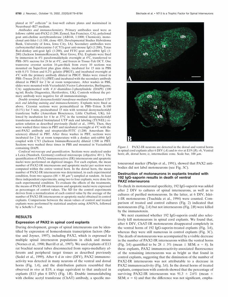
toneuronal marker (Phelps et al., 1991), showed that PAX2 anti-bodies did not label motoneurons (see Fig. 3C).
Destruction of motoneurons in explants treated with192 IgG-saporin results in death of ventralPAX2 interneuronsTo check its motoneuronal specificity, 192 IgG-saporin was addedafter 2 DIV to cultures of spinal interneurons, as well as tocultures of purified motoneurons. In the latter, at 6 DIV, Islet-1-IR motoneurons (Tsuchida et al., 1994) were counted. Com-parison of treated and control cultures (Fig. 2) indicated thatmotoneurons (Fig. 2A) but not interneurons (Fig. 2B) were killedby the immunotoxin.
We next examined whether 192 IgG-saporin could also selec-tively kill motoneurons in spinal cord explants. We found that,after 6 DIV, ChAT-IR motoneurons disappeared completely inthe ventral horns of 192 IgG-saporin-treated explants (Fig. 3A),whereas they were still numerous in control explants (Fig. 3C).The death of motoneurons was accompanied by a visible decreasein the number of PAX2-IR interneurons within the ventral horns(Fig. 3A) quantified to be 28 � 3% (mean � SEM; n � 8). Inthese explants, PAX2 immunoreactivity-associated fluorescenceof the remaining interneurons was as bright as that found incontrol explants, suggesting that the diminution of the number ofPAX2-IR interneurons was not attributable to a decrease inPAX2 immunoreactivity (Fig. 3A). In the dorsal horns of treatedexplants, comparison with controls showed that the percentage ofsurviving PAX2-IR interneurons was 91.3 � 2.6% (mean �SEM; n � 6) and that the difference was not significant, empha-
Figure 1. PAX2-IR neurons are detected in the dorsal and ventral hornsin spinal cord explants after 6 DIV (A) and in vivo at E19 (B). vh, Ventralhorn; dh, dorsal horn; iz, intermediate zone. Scale bar, 100 �m.
8780 J. Neurosci., October 15, 2002, 22(20):8779–8784 Bechade et al. • NT-3 Is a Trophic Factor for Spinal Interneurons

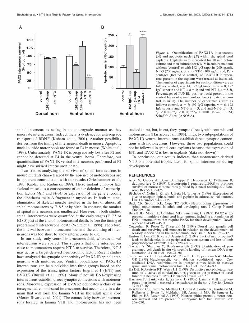
sizing the lack of toxicity of 192 IgG-saporin (Fig. 4A). TUNELstaining (Gavrieli et al., 1992) was used to investigate whether thisloss resulted from apoptotic cell death. As seen from directobservations, the number of apoptotic nuclei was higher in thetreated ventral horns (Fig. 3D) compared with control culturesand displayed a 105 � 43% (mean � SEM; n � 6) increase intreated explants compared with control explants (Fig. 4B). Thisincrease did not correspond to dying motoneurons because, at 6DIV, motoneurons were already dead in 192 IgG-saporin-treatedexplants (Fig. 3A). The increase in the number of apoptotic nucleiwas greater than expected from the decrease in number ofPAX2-IR interneurons. This could result from the fact that somePAX2-negative cells (glial cells or neurons) also underwent apo-ptosis. Altogether, our results established that the selective elim-ination of motoneurons resulted in the death of PAX2-IR inter-neurons. This suggested that motoneurons produced trophicfactors required for the survival of these interneurons.
NT-3 rescues PAX2-IR interneurons in 192IgG-saporin-treated explantsIf ventral PAX2-IR interneurons depend on NT-3 produced bymotoneurons for their survival, addition of exogenous NT-3 to192 IgG-saporin-treated explants should prevent them from dy-ing. Indeed, in spinal cord explants cultured with both 192 IgG-saporin and NT-3, we found that the number of PAX2-IR ventralinterneurons (Fig. 4A) and the number of TUNEL-positive nu-clei (Fig. 4B) were comparable with that of controls (untreatedexplants) after 6 DIV. This indicates that the effect of NT-3 wasnot attributable to an increase in PAX2 immunoreactivity butrather results from a diminution of cell death. Thus, addition ofexogenous NT-3 to 192 IgG-saporin-treated explants resulted inthe rescue of virtually all PAX2-IR interneurons. Motoneuronswere not rescued from death because ChAT immunoreactivity
was still undetectable in the treated explants (data not shown).Moreover, we showed previously that addition of NT-3 to spinalcord explants does not promote motoneuronal survival (Sedel etal., 1999).
Anti-NT-3 treatment results in death of PAX2-IRventral interneuronsNT-3 is expressed by motoneurons in spinal cord explants (Sedelet al., 1999). We therefore treated explants with an antiserum thatspecifically blocks the biological activity of NT-3 (Zhou andRush, 1995). After this treatment, very few PAX2-IR interneu-rons were observed in the ventral horn area (Fig. 3C): the de-crease was 66 � 4% (mean � SEM; n � 5) compared withcontrols (Fig. 4A). This diminution of PAX2-IR ventral interneu-rons was associated with a 140 � 15% (mean � SEM; n � 3)increase in the number of apoptotic nuclei in the same area (Figs.3F, 4B). Again, the increase in the number of apoptotic nuclei washigher than that expected given the decrease in the number ofPAX2 interneurons. This suggests that anti-NT-3 most likelycauses death of cells not expressing PAX2. In the dorsal horn,anti-NT-3 antibody had no effect on the number of PAX2-IRneurons (91 � 2.6% of the control; mean � SEM; n � 8) (Fig.4A). Therefore, the survival effect of NT-3 is specific to ventralPAX2-IR interneurons.
In anti-NT-3-treated explants, labeling for anti-PAX2 and anti-ChAT antibodies resulted in yellow labeling of motoneurons (Fig.3C). The FITC-conjugated anti-rabbit antibody used to reveal theanti-PAX2 antibody also binds to the rabbit anti-NT-3 antibodyassociated with ChAT-IR motoneurons visualized by a CY3secondary antibody. This double labeling shows that the anti-NT-3 antibody is selectively bound to motoneurons. This is inagreement with the finding that NT-3 transcripts are specificallysynthesized by motoneurons in spinal cord explants (Sedel et al.,1999). For additional confirmation, anti-NT-3 explants culturedfor 6 DIV were immunostained with the anti-ChAT antibodyrevealed with a CY3 secondary antibody and a FITC secondaryantibody to visualize the anti-NT-3 binding sites: we observedthat ChAT-IR motoneurons, but not ChAT-negative neurons,were NT-3-IR (Fig. 3B�).
DISCUSSION192 IgG-saporin has been used extensively in vitro and in vivo asa powerful tool to eliminate p75NTR-expressing neurons (Wileyand Kline, 2000). We found that the selective destruction ofmotoneurons by 192 IgG-saporin caused the death of ventralPAX2-IR interneurons. This death does not result from a directkilling effect of 192 IgG-saporin because (1) 192 IgG-saporin hadno effect on the survival of spinal interneurons in culture nor onthat of dorsal spinal interneurons in the explants, and (2) onlyp75NTR-IR neurons (i.e., motoneurons) were killed by 192 IgG-saporin. We checked by immunochemistry that interneurons didnot express p75NTR in treated explants (data not shown). Anindirect toxic effect attributable to the liberation of toxic sub-stances by dying motoneurons is unlikely because, in other in vitrosystems, in cocultures containing basal forebrain and corticalneurons, elimination of basal forebrain neurons with 192 IgG-saporin induced death of a specific population of cortical neuronsbut did not damage other neurons (Ha et al., 1998). Therefore,the survival of PAX2-IR interneurons is most likely regulated bytrophic factors produced by motoneurons.
We show here that the supply of NT-3 in the absence ofmotoneurons rescued PAX2-IR ventral interneurons from death
Figure 2. 192 IgG-saporin kills motoneurons (A) but not interneurons(B) in spinal cord primary cultures. Spinal interneurons and purifiedmotoneurons were cultured for 2 DIV in culture medium and then for 4 din the same medium without (control) or with 192 IgG-saporin. Thenumber of motoneurons per well was 103 � 11 and 2 � 0.11 in control andtreated cultures, respectively. The number of interneurons counted in afield with a 40� objective was 111 � 11 and 104 � 7.5 in control andtreated cultures, respectively. Data are expressed as the percentage ofsurviving neurons compared with that of untreated neurons. Results fromtwo independent experiments were pooled (mean � SEM; n � 4 wells).
Bechade et al. • NT-3 Is a Trophic Factor for Spinal Interneurons J. Neurosci., October 15, 2002, 22(20):8779–8784 8781
and that depletion of endogenous NT-3 caused death ofPAX2-IR ventral interneurons. Altogether, these data are com-patible with the notion that NT-3 produced by motoneurons is asurvival factor for PAX2-IR ventral interneurons. Anti-NT-3treatment leads to a 66% decrease in PAX2-IR ventral interneu-rons, whereas 192 IgG-saporin had a weaker effect resulting in adecrease of only 26%. This difference may be accounted for bytheir mechanisms of action. The effect of anti-NT-3 is immediatebecause it blocks the trophic effect of NT-3 by an antibody-antigen reaction. In contrast, 192-IgG-saporin induces a progres-sive death of motoneurons, as indicated by results of immunola-beling with anti-ChAT antibody (data not shown) and thus acontinuous and progressive decrease in NT-3 availability.
NT-3 knock-out mutant mice or in vivo administration ofblocking antibodies against NT-3 indicated that NT-3 impair-ment induces a severe loss in sensory and sympathetic neuronsbut has no effect on the survival of CNS neurons (Ernfors et al.,
1994; Tessarollo et al., 1994; Oakley et al., 1995). We quantifiedthe number of PAX2-IR ventral interneurons in the spinal cord ofNT3�/�, NT-3�/�, and wild-type mice at P2 and found nostatistical difference between the NT-3 knock-out and the controlmice (data not shown). A straightforward interpretation for thediscrepancy between these quantifications and the trophic effectof NT-3 found in vitro is that in vivo spinal interneurons haveaccess to additional factors with overlapping or redundant func-tions with regard to that of NT-3. These survival factors could beassociated with sources such as supraspinal or dorsal root affer-ents that are absent in the explants after their dissection out ofthe embryos. In rat, the peak of naturally occurring cell deathfor DRG occurs at E17–E19 just before interneuronal death(Coggeshall et al., 1994). BDNF and NT-4/5 are synthesized inDRG during development, and trkB, their specific receptor, isexpressed throughout the spinal cord (Henderson et al., 1993).Therefore, these neurotrophins could act as survival factors for
Figure 3. Effects of 192 IgG-saporin and anti-NT-3 antiserum on the survival of PAX2-IR interneurons (A–C) and on the presence of apoptotic nuclei(D–F) within the ventral horns of spinal cord explants. A–C, Double immunostaining using anti-ChaT (red) and anti-PAX2 ( green) antibodies ontransverse sections of explants cultured for 6 DIV with 192 IgG-saporin (A), with anti-NT-3 antibody (B), or in controls (C). ChaT-IR motoneuronswere absent in 192 IgG-saporin-treated explants (A) but were present in control (C) and anti-NT-3-treated (B) explants. In B, motoneurons are in yellow(see arrows) because the FITC-conjugated antibody used to detect the PAX2 antiserum revealed the presence of the anti-NT-3 bound to ChaT-IR (red)motoneurons. Note the reduction in the number of PAX2-IR interneurons in 192 IgG-saporin (A) and anti-NT-3-treated explants (B). B�, ChaT-IRmotoneurons express NT-3. Anti-NT-3-treated explants were immunostained using an anti-ChaT antibody revealed with a CY3-conjugated antibody(red), and anti-NT-3 binding sites were visualized using an FITC-conjugated antibody ( green). D–F, TUNEL-stained nuclei of transverse sections ofexplants cultured for 6 DIV with 192 IgG-saporin (A), with anti-NT-3 (B), or in controls (C). Note the increased number of apoptotic nuclei comparedwith control in both 192 IgG-saporin- and anti-NT-3-treated explants. Scale bar: A–F, 50 �m; B�, 25 �m.
8782 J. Neurosci., October 15, 2002, 22(20):8779–8784 Bechade et al. • NT-3 Is a Trophic Factor for Spinal Interneurons
spinal interneurons acting in an anterograde manner as theyinnervate interneurons. Indeed, there is evidence for anterogradetransport of BDNF (Kohara et al., 2001). Another possibilityderives from the timing of interneuron death in mouse. Apoptoticnuclei outside motor pools are found at P4 in mouse (White et al.,1998). Unfortunately, PAX2-IR is progressively lost after P2 andcannot be detected at P4 in the ventral horns. Therefore, ourquantification of PAX2-IR ventral interneurons performed at P2might have missed interneuron death.
Two studies analyzing the survival of spinal interneurons inmouse mutants characterized by the absence of motoneurons arein apparent contradiction with our results (Grieshammer et al.,1998; Kablar and Rudnicki, 1999). These mutant embryos lackskeletal muscle as a consequence of either deletion of transcrip-tion factors Myf5 and MyoD or expression of the gene encodingthe diphtheria toxin A fragment in myoblasts. In both mutants,elimination of skeletal muscle resulted in the loss of almost allspinal motoneurons by E18.5 or by birth. In contrast, the numberof spinal interneurons was unaffected. However, in both studies,spinal interneurons were quantified at the early stages (E17.5 orE18.5) just at the end of motoneuronal death, before the timing ofprogrammed interneuronal death (White et al., 1998). Therefore,the interval between motoneuron loss and the counting of inter-neurons was too short to allow interneurons to die.
In our study, only ventral interneurons died, whereas dorsalinterneurons were spared. This suggests that only interneuronsclose to motoneurons require NT-3 to survive. Therefore, NT-3may act as a target-derived neurotrophic factor. Recent studieshave analyzed the synaptic connectivity of PAX2-IR spinal inter-neurons with motoneurons. Ventral populations of PAX2-IRinterneurons can be subdivided into three groups based on theexpression of the transcription factors Engrailed-1 (EN1) andEVX1/2 (Burrill et al., 1997). Many if not all EN1-expressinginterneurons establish direct synaptic connections with motoneu-rons. Moreover, expression of EVX1/2 delineates a class of in-tersegmental commissural interneurons that accumulate in a do-main that will form the lamina VIII in the adult spinal cord(Moran-Rivard et al., 2001). The connectivity between interneu-rons located in lamina VIII and motoneurons has not been
studied in rat, but, in cat, they synapse directly with contralateralmotoneurons (Harrison et al., 1986). Thus, two subpopulations ofPAX2-IR ventral interneurons establish direct synaptic connec-tions with motoneurons. However, these two populations couldnot be followed in spinal cord explants because the expression ofEN1 and EVX1/2 is lost in explants (data not shown).
In conclusion, our results indicate that motoneuron-derivedNT-3 is a potential trophic factor for spinal interneurons duringdevelopment.
REFERENCESArce V, Garces A, Bovis B, Filippi P, Henderson C, Pettmann B,
deLapeyriere O (1999) Cardiotropin-1 requires LIFR� to promotesurvival of mouse motoneurons purified by a novel technique. J Neu-rosci Res 55:119–126.
Bechade C, Colin I, Kirsch J, Betz H, Triller A (1996) Expression ofglycine receptor alpha subunits and gephrin in cultured spinal neurons.Eur J Neurosci 8:429–435.
Buck CR, Seburn KL, Cope TC (2000) Neurotrophin expression byspinal motoneurons in adult and developing rats. J Comp Neurol416:309–318.
Burrill JD, Moran L, Goulding MD, Saueressig H (1997) PAX2 is ex-pressed in multiple spinal cord interneurons, including a population ofEN1� interneurons that require PAX6 for their development. Devel-opment 124:4493–4503.
Coggeshall R, Pover C, Fitzgerald M (1994) Dorsal root ganglia celldeath and surviving cell numbers in relation to the development ofsensory innervation in the rat hindlimb. Dev Brain Res 82:193–212.
Ernfors P, Lee KF, Kucera J, Jaenisch R (1994) Lack of neurotrophin-3leads to deficiencies in the peripheral nervous system and loss of limbproprioceptive afferents. Cell 77:503–512.
Gavrieli Y, Sherman Y, Ben-Sasson SA (1992) Identification of pro-grammed cell death in situ via specific labeling of nuclear DNA frag-mentation. J Cell Biol 119:493–501.
Grieshammer U, Lewandoski M, Prevette D, Oppenheim RW, MartinGR (1998) Muscle-specific cell ablation conditional upon Cre-mediated DNA recombination in transgenic mice leads to massivespinal and cranial motoneuron loss. Dev Biol 197:234–247.
Ha DH, Robertson RT, Weiss JH (1998) Distinctive morphological fea-tures of a subset of cortical neurons grown in the presence of basalforebrain neurons in vitro. J Neurosci 18:4201–4215.
Harrison PJ, Jankowska E, Zytnicki D (1986) Lamina VIII interneu-rones interposed in crossed reflex pathways in the cat. J Physiol (Lond)371:147–166.
Henderson CE, Camu W, Mettling C, Gouin A, Poulsen K, Karihaloo M,Rullamas J, Evans T, McMahon SB, Armanini MP, Berkemeier L,Phillips HS, Rosenthal A (1993) Neurotrophins promote motor neu-ron survival and are present in embryonic limb bud. Nature 363:266–270.
0
25
50
75
100
125
con
tro
l
*
**
PAX
2in
tern
euro
nsu
rviv
al(%
of
con
tro
l)
192-
IgG
-sap
+NT
3
anti
-NT
3
con
tro
l
192-
IgG
-sap
192-
IgG
-sap
+NT
3an
ti-N
T3
** *
A B*
0
50
Ap
oto
tic
nu
clei
(%o
fco
ntr
ol)
100
200
150
150250
con
tro
l
192-
IgG
-sap
192-
IgG
-sap
anti
-NT
3
Dorsal horn Ventral horn Ventral horn
Figure 4. Quantification of PAX2-IR interneurons(A) and apoptotic nuclei ( B) within the spinal cordexplants. Explants were incubated for 10 min beforeculture and then cultured for 6 DIV in culture mediumwithout (control) or with 192 IgG-saporin (200 ng/ml),NT-3 (200 ng/ml), or anti-NT-3 (100 �g/ml). A, Per-centages (treated vs control) of PAX2-IR interneu-rons present in the explants were treated as indicated.The number of experiments for each condition were asfollows: control, n � 14; 192 IgG-saporin, n � 8; 192IgG-saporin and NT-3, n � 3; and anti-NT-3, n � 5. B,Percentages of TUNEL-positive nuclei present in theventral horns of spinal cord explants (treated vs con-trol as in A). The number of experiments were asfollows: control, n � 7; 192 IgG-saporin, n � 6; 192IgG-saporin and NT-3, n � 3; and anti-NT-3, n � 3.*p � 0.05; **p � 0.01; ***p � 0.001. Mean � SEM;Scheffe’s F test (ANOVA).
Bechade et al. • NT-3 Is a Trophic Factor for Spinal Interneurons J. Neurosci., October 15, 2002, 22(20):8779–8784 8783

Kablar B, Rudnicki MA (1999) Development in the absence of skeletalmuscle results in the sequential ablation of motor neurons from thespinal cord to the brain. Dev Biol 208:93–109.
Kohara K, Kitamura A, Morishima M, Tsumoto T (2001) Activity-dependent transfer of brain-derived neurotrophic factor to postsynapticneurons. Science 291:2419–2423.
Lawson S, Davies H, Bennett J, Lowrie M (1997) Evidence that spinalinterneurons undergo programmed cell death postnatally in the rat. EurJ Neurosci 9:794–799.
Matise MP, Joyner AL (1997) Expression patterns of developmentalcontrol genes in normal and Engrailed-1 mutant mouse spinal cordreveal early diversity in developing interneurons. J Neurosci17:7805–7816.
McKay SE, Oppenheim RW (1991) Lack of evidence for cell deathamong avian spinal cord interneurons during normal development andfollowing removal of targets and afferents. J Neurobiol 22:721–733.
Moran-Rivard L, Kagawa T, Saueressig H, Gross MK, Burrill J, GouldingM (2001) Evx1 is a postmitotic determinant of v0 interneuron identityin the spinal cord. Neuron 29:385–399.
Nornes HO, Dressler GR, Knapik EW, Deutsch U, Gruss P (1990)Spatially and temporally restricted expression of Pax2 during murineneurogenesis. Development 109:797–809.
Oakley RA, Garner AS, Large TH, Frank E (1995) Muscle sensoryneurons require neurotrophin-3 from peripheral tissues during theperiod of normal cell death. Development 121:1341–1350.
Oliveira AL, Risling M, Deckner M, Lindholm T, Langone F, Cullheim
S (1997) Neonatal sciatic nerve transection induces TUNEL labelingof neurons in the rat spinal cord and DRG. NeuroReport 8:2837–2840.
Oppenheim RW (1986) The absence of significant postnatal motoneu-ron death in the brachial and lumbar spinal cord of the rat. J CompNeurol 246:281–286.
Phelps PE, Barber RP, Vaughn JE (1991) Embryonic development ofcholine acetyltransferase in thoracic spinal motor neurons: somatic andautonomic neurons may be derived from a common cellular group.J Comp Neurol 307:77–86.
Sedel F, Bechade C, Triller A (1999) Nerve growth factor (NGF) in-duces motoneuron apoptosis in rat embryonic spinal cord in vitro. EurJ Neurosci 11:3904–3912.
Tessarollo L, Vogel KS, Palko ME, Reid SW, Parada LF (1994) Tar-geted mutation in the neurotrophin-3 gene results in loss of musclesensory neurons. Proc Natl Acad Sci USA 91:11844–11848.
Tsuchida T, Ensini M, Morton SB, Baldassare M, Edlund T, Jessell TM,Pfaff SL (1994) Topographic organization of embryonic motor neu-rons defined by expression of LIM homeobox genes. Cell 79:957–970.
White FA, Keller-Peck CR, Knudson CM, Korsmeyer SJ, Snider WD(1998) Widespread elimination of naturally occurring neuronal deathin Bax-deficient mice. J Neurosci 18:1428–1439.
Wiley RG, Kline IR (2000) Neuronal lesioning with axonally trans-ported toxins. J Neurosci Methods 103:73–82.
Yan Q, Johnson E (1988) An immunohistochemical study of the growthfactor receptor in developing rats. J Neurosci 8:3481–3498.
Zhou XF, Rush R (1995) Sympathetic neurons in neonatal rats requireendogenous neurotrophin-3 for survival. J Neurosci 15:6521–6530.
8784 J. Neurosci., October 15, 2002, 22(20):8779–8784 Bechade et al. • NT-3 Is a Trophic Factor for Spinal Interneurons