Embed Size (px)
Citation preview
Molecular Consequences of AlteredNeuronal Cholesterol Biosynthesis
Zeljka Korade,1,2* Anne K. Kenworthy,3 and Karoly Mirnics2,4
1Department of Biochemistry, Vanderbilt University, Nashville, Tennessee2Vanderbilt Kennedy Center for Research on Human Development, Nashville, Tennessee3Departments of Molecular Physiology and Biophysics and Cell and Developmental Biology,Vanderbilt, Nashville, Tennessee4Department of Psychiatry, Vanderbilt University, Nashville, Tennessee
The first dedicated step in de novo cholesterol biosyn-thesis begins with formation of squalene and ends withthe reduction of 7-dehydrocholesterol by 7-dehydro-cholesterol reductase (Dhcr7) into cholesterol, which isan essential structural and signaling molecule. Muta-tions in the Dhcr7 gene lead to Smith-Lemli-Opitzsyndrome (SLOS), which is characterized by develop-mental deformities, incomplete myelination, and mentalretardation. To understand better the molecular conse-quences of Dhcr7 deficiency in neuronal tissue, weanalyzed the effect of cholesterol deficiency on thetranscriptome in Neuro2a cells. Transient down-regula-tion of Dhcr7 by siRNA led to altered expression ofmultiple molecules that play critical roles in intracellularsignaling or vesicular transport or are inserted intomembrane rafts (e.g. Egr1, Snx, and Adam19). A similardown-regulation was also observed in stable Dhrc7-shRNA-transfected cell lines, and the findings wereverified by qPCR. Furthermore, we investigated theDhcr7-deficient and control cells for the expression ofseveral critical genes involved in lipid biosynthesis.Among these, fatty acid synthase, sterol-regulatoryelement binding protein 2, SREBF chaperone, site-1protease, and squalene synthase showed a significantdown-regulation, suggesting that, in a neuronal cellline, Dhcr7 is a potent regulator of lipid biosynthesis.Importantly, the gene expression changes were presentin both lipid-containing and cholesterol-deficient media,suggesting that intrinsic cholesterol biosynthesis is nec-essary for normal neuronal function and cannot be sup-plemented from extrinsic sources. VVC 2008 Wiley-Liss, Inc.
Key words: gene expression; lipid metabolism;cholesterol; neuroblastoma; SREBP
Cholesterol is a critical building block of cellularmembranes (Maxfield and Tabas, 2005). Cholesterol bio-synthesis is a complex cascade of events that involves atleast 20 dedicated enzymes (Lutton, 1991). The rate-lim-iting enzyme in cholesterol biosynthesis is 3-hydroxy-3-methylglutaryl-coenzyme A reductase (Hmgcr; EC2.3.3.10), which catalyzes conversion of Hmg-CoA tomevalonic acid (Luskey and Stevens, 1985). The cascade
ends with reduction of 7-dehydrocholesterol that is cata-lyzed by 7-dehydrocholesterol reductase (Dhcr7; EC1.3.1.21) enzyme (Bae et al., 1999). Liver and smallintestine are the major sites of cholesterol biosynthesisfor the whole body (Strandberg and Tilvis, 1988). How-ever, all of the brain cholesterol is synthesized locally,with the highest rate of synthesis occurring during firstpostnatal weeks in humans and rodents (Jurevics andMorell, 1995; Jurevics et al., 1997). This peak of earlypostnatal biosynthesis coincides with myelination (Aniteiand Pfeiffer, 2006). The expression pattern of transcriptsinvolved in cholesterol biosynthesis and the role and reg-ulation of cholesterol biosynthesis in the nervous systemremain understudied and are mostly unknown to date(Korade and Kenworthy, 2008).
Recent studies showed that changes in cholesterolhomeostasis lead to a wide variety of CNS disorders(Maxfield and Tabas, 2005). For example, deficient cho-lesterol biosynthesis in oligodendrocytes delays myelina-tion (Saher et al., 2005), and defects in cholesterol traf-ficking lead to Niemann-Pick type C disease (Paul et al.,2004). Importantly, partial or complete lack of Dhcr7enzymatic activity causes Smith-Lemli-Opitz syndrome(SLOS), characterized by severe developmental malfor-mations and mental retardation (Smith et al., 1964; Tier-ney et al., 2000). In SLOS, mutations in Dhcr7, the lastenzyme in cholesterol biosynthesis pathway, lead toaccumulation of 7-dehydrocholesterol and reduced cho-lesterol levels (Jira et al., 2003). 7-Dehydrocholesterol
Additional Supporting Information may be found in the online version
of this article.
Contract grant sponsor: NIH; Contract grant number: K02 MH070786
(to K.M.); Contract grant number: R01 MH079299 (to K.M.); Contract
grant number: R01 GM073846 (to A.K.K.); Contract grant sponsor: VU
Kennedy Center for Research on Human Development (to Z.K.).
*Correspondence to: Zeljka Korade, DVM, PhD, Department of Bio-
chemistry, 8124A MRB III, Vanderbilt University School of Medicine,
Nashville, TN 37232. E-mail: [email protected]
Received 15 April 2008; Revised 21 August 2008; Accepted 20 August
2008
Published online 24 October 2008 in Wiley InterScience (www.
interscience.wiley.com). DOI: 10.1002/jnr.21917
Journal of Neuroscience Research 87:866–875 (2009)
' 2008 Wiley-Liss, Inc.

differs from cholesterol in a double bond at the seventhposition in the sterol ring, and this structural differencetranslates into a functional difference: whereas 7-dehy-drocholesterol can be incorporated into the membranesjust like cholesterol, its presence perturbs the proteincontent of lipid rafts (Keller et al., 2004).
Lipid rafts (microdomains) are cholesterol- andsphingolipid-enriched areas within cellular membranesthat concentrate and segregate proteins within the mem-brane bilayer (Pike, 2005). Membrane rafts are presentin both neurons and glial cells, and they contain differentreceptors and neurotransmitter transporters, regulateintramembrane proteolysis of many transmembrane pro-teins, and serve as organizing sites for neurotrophin sig-naling (Korade and Kenworthy, 2008).
In addition to its structural role as a building blockof membranes, cholesterol has important role in tran-scription of genes containing the SRE domain (Wanget al., 1994; Horton et al., 2002). This regulation is welldescribed in liver, where SCAP, through the sterol-sens-ing domain, regulates availability of SREBPs, transcrip-tion factors critical for regulation of lipid genes (Hortonet al., 2002). This mechanism has not been studied inneuronal tissue. Therefore, we hypothesized that alteringcholesterol biosynthesis in cells of neuronal origin willlead to changed transcription of genes belonging to mul-tiple cellular pathways. As a result, the present study wasaimed to 1) develop an in vitro model for assessment ofthe role and consequences of endogenous neuronal cho-lesterol biosynthesis, 2) uncover and validate the molec-ular consequences of the reduced intrinsic cholesterolbiosynthesis, and 3) evaluate the dependence of theuncovered molecular changes on extrinsic vs. intrinsicsources of cholesterol.
MATERIALS AND METHODS
Cell Culture and Reagents
Neuroblastoma cell line Neuro2a was purchased fromthe American Type Culture Collection (Rockville, MD).Neuro2a cells were maintained in alpha modification of mini-mal essential medium (Eagle) with Earle’s salt and supple-mented with L-glutamine, sodium bicarbonate, nonessentialamino acids, sodium pyruvate, 10% fetal bovine serum (FBS;Thermo Scientific HyClone, Logan, UT), and penicillin/streptomycin at 378C and 5% CO2. According to the vendor,the FBS contained cholesterol at concentrations of 32 mg/100ml. This translates into a final cholesterol concentration of 32lg/ml in our culture medium. To evaluate the role of exoge-nous cholesterol on gene expression, cells were also culturedwith medium containing 10% cholesterol-deficient serum(Thermo Scientific HyClone Lipid Reduced FBS). This FBSmedium did not have detectable cholesterol levels. The Neu-ro2a cells were subcultured once per week, and the culturemedium was changed every 2 days. SiRNA oligonucleotideswere purchased from Qiagen (Valencia, CA): AllStarsNegative Control siRNA, Mm_Dhcr7_1 HP siRNA,Mm_Dhcr7_2 HP siRNA, and Mm_Dhcr7_3 HP siRNA.For stable transfections, we obtained plasmids from Open Bio-
systems through the Vanderbilt Microarray Shared Resource.These include OligoID V2LMM_9686 and nonsilencingGIPZ shRNAmir control.
Transient and Stable siRNA Transfections
Neuro2a cells were cultured for 2 days before transfec-tions. Cells were transfected with three different Dhcr7siRNA oligonucleotides (Qiagen) using a Nucleofector instru-ment and Nucleofector Kit V (Amaxa GmbH, Cologne, Ger-many) optimized for use with Neuro2a cells. Briefly, 2 3 106
cells were resuspended in 100 ll transfection buffer, siRNAwas added, and cells were electroporated using program T-24.The cells were grown for 24 hr following transfection, andthe expression of Dhcr7 was monitored by quantitative RT-PCR. To establish stable down-regulation of Dhcr7, aftertransfection of cells with pGIPZ plasmids, cells were grown inthe presence of puromycin.
RNA Preparation and Array Hybridization
Total RNA was isolated from the cells using Trizol(Life Technologies, Rockville, MD). Purification of totalRNA was done using RNeasy Mini Kit (Qiagen), and on-column digestion of DNA was performed during the RNApurification step using RNase-Free DNase Set (Qiagen). Theconcentration of total RNA was measured on a Nanodropinstrument (Thermo Scientific, Wilmington, DE), and the in-tegrity of the RNA was established by electrophoresis usingthe Agilent Bioanalyzer 2100 instrument (Agilent Technolo-gies, Santa Clara, CA). Sample preparation, cRNA synthesis,IVT, hybridization, and array scanning were performed as permanufacturers’ instructions. The fragmented cRNA washybridized to Affymetrix MG-430 mouse oligonucleotidemicroarrays.
Data Analysis
All microarrays had exceptional quality based on presentcalls and 50:30 GAPDH integrity ratios calculated by GCOS.Segmented images were normalized and log2 transformedusing robust multiarray analysis (RMA) in Expression Console(Affymetrix). Clustering and secondary data analysis was per-formed in GenePattern 2.0 (Reich et al., 2006).
In the microarray study, genes were considered differen-tially expressed between Dhcr7-deficient and control samplesif they reported an absolute ALR > 0.383 (>30% change)and a groupwise statistical significance of P < 0.05. All micro-array data will be publicly available at the time of publicationat http://mirnicslab.vanderbilt.edu/mirnicslab/ordering.htm.Because this was an exploratory experiment to identify puta-tive downstream effects, we did not correct for multiple test-ing, and the most critical data were verified by qPCR.
Northern Blotting
Northern hybridization was done using the Northern-Max system (Ambion, Austin, TX) as previously described(Korade et al., 2007). Briefly, 20 lg total RNA was loadedon a formamide gel, electrophoresed at 5 V/cm, transferred toa Bright Star nylon membrane, and cross-linked by UV lightexposure. Hmgcr and Dhcr7 probes were amplified using SP6
Consequences of Dhcr7 Deficiency 867
Journal of Neuroscience Research
and T7 primers from plasmid as previously described (Koradeet al., 2007). Gapdh probe were generated using gene-specificprimers and Neuro2a cDNA template in a standard PCR.The purified PCR product was radioactively labeled usinga32P-dCTP and a Random Primed DNA labeling kit (Roche,Indianapolis, IN). The labeled probe was purified with Probe-Quant G-50 MicroColumns (Amersham Biosciences, Piscat-away, NJ). The denatured probe, 106 cpm/ml, was added tothe hybridization buffer (10 ml/100 cm2 membrane). Thehybridization was done overnight at 428C. The Northernmembrane was washed for 3 hr at 458C and exposed to X-rayfilm.
Quantitative PCR
Total RNA (100 ng) from each sample was reversetranscribed to cDNA using a High Capacity cDNA ArchiveKit (Applied Biosystems, Foster City, CA). Real-time PCRwas performed with an ABI Prism 7300 System (Applied Bio-systems) using 1 ng cDNA per 50 ll reaction volume, 23SYBR green master mix, and gene-specific primers. All sam-ples were run in triplicate. Data from the PCRs was analyzedusing the comparative cycle number determined as threshold(Ct) method (Kurrasch et al., 2004). Differential expressionwas calculated as DDCt against expression of Pgk1 as a nor-malizer. We designed primers (�20 bp) to yield 85–110-bpPCR amplicons in Primer3 software (http://frodo.wi.mit.edu/) for different genes. For each gene, we designed foursets of primers. Each set was tested using no template andthree different concentrations of a specific template. Thistranslates into 16 wells for each primer set (four wells withno-template controls, four wells for each of three differenttemplate concentrations). From this PCR, we calculated theefficiency of PCR primers and R2 value (coefficient of corre-lation). All mouse gene-specific primers used showed a slopebetween –3.10 and –3.58, with R2 > 0.99 (Supp. Info. Fig.2A). Lipid-specific qPCR primers have been described byWang et al. (2002). All qPCR amplicons were checked by gelelectrophoresis, and all of the qPCR reactions gave rise to asingle product of predicted size. Furthermore, the qPCR dis-sociation curve of the amplicons, performed after each qPCRrun (using Dissociation Curve 1.0 software; ABI), also con-firmed specific amplification. The gel electrophoresis data arepresented in Supporting Information Figure 2B.
Analysis of Total Cholesterol Level by Amplex Red
Total cholesterol levels were measured using anAmplex-Red Cholesterol Assay kit (Molecular Probes,Eugene, OR). Dhcr7-deficient and control cells were grownfor 4 days in either regular or cholesterol-deficient culturemedium and then lysed in 0.1 M phosphate lysis buffer. Cho-lesterol standard and samples were prepared for measurementaccording to the manufacturer’s instructions. Fluorescence wasmeasured with a fluorescence microplate reader using excita-tion at 545 nm and fluorescence detection at 590 nm. Thebackground fluorescence, determined for the no-cholesterolcontrol reaction, was subtracted from each value. The totalcholesterol level was determined by comparing experimental
data with the data obtained for cholesterol standards. Totalcholesterol values were normalized to protein values.
UV Spectrometric Analysis of 7DHC Levels
7DHC shows characteristic ultraviolet absorption max-ima (kmax) at 271, 282, and 294 nm (Nes, 1985). Therefore,we used spectrophotometric measurement to detect 7DHC instably transfected Neuro2a cells (Honda et al., 1997). Stablytransfected Neuro2a cells were cultured for 3–5 days in cho-lesterol-deficient medium. After removal of cell culture me-dium, cells were washed twice in 13 PBS and scraped intoPBS. Cell were spun down, PBS was removed, and 200 ll of100% EtOH was added to the cell pellet, with incubation inan ultrasonic bath for 15 min. After addition of 200 ll waterand vortexing for 10 sec, 1 ml n-hexane was added. The con-tent was vortexed for 20 sec and spun down at 400g for1 min. After centrifugation, the clear n-hexane layer was col-lected in a 1-ml quartz cuvet and used for UV measurementon a spectrophotometer. The samples were scanned using n-hexane blank in the reference beam, and the absorption max-ima at 271, 282, and 294 nm were used for detection of7DHC in lipid extracts from cells.
RESULTS
Dhcr7 siRNA Reduces Dhcr7Transcript Expression
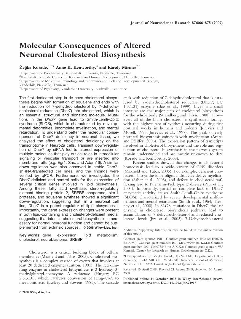
Both neurons and neuronal cell lines have the abil-ity to synthesize cholesterol, yet the level of cholestero-genic enzyme expression varies across distinct cellularphenotypes (Korade et al., 2007). We therefore firstexamined the expression of the cholesterogenic enzymesin our model system, the Neuro2a neuroblastoma cellline. Northern hybridization revealed that Neuro2a cellsstrongly expressed the first and the last enzymes (Dhcr7and Hmgcr, respectively) of the cholesterol biosynthesispathway (Fig. 1). The expression of these transcripts sug-gested that Neuro2a cells are an appealing model tostudy the regulation and role of neuronal cholesterol
Fig. 1. Hmgcr and Dhcr7 are highly expressed in Neuro2a cells.Total RNA was isolated from Neuro2a cells and analyzed by North-ern blotting for the expression of cholesterogenic enzymes. The samemembrane was probed sequentially with Hmgcr-, Dhcr7-, andGapdh-32P-dCTP-labelled cDNA probes. The experiment was per-formed in triplicate. Note that Hmgcr and Dhcr7, the first and thelast enzymes in the cholesterol biosynthesis pathway, are highlyexpressed in Neuro2a neuroblastoma cells.
868 Korade et al.
Journal of Neuroscience Research
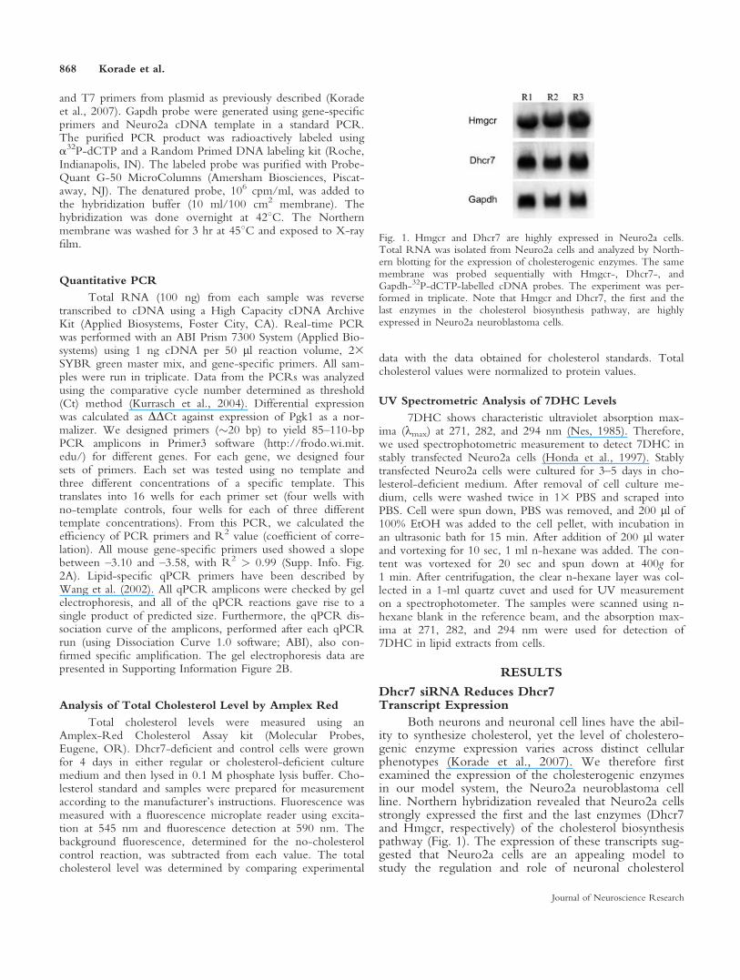
biosynthesis. Thus, following the assessment of theexpression of these two critical enzymes, we establishedan in vitro experimental system based on down-regula-tion of Dhcr7 in these cells. To achieve this, we testedthe efficacy of three different siRNAs designed todown-regulate Dhcr7. Two of the three tested siRNAssuccessfully down-regulated Dhcr7 transcript and choles-terol levels (achieving >70% transcript reduction),whereas one failed to achieve a specific Dhcr7 down-regulation (Fig. 2). Given these results, we decided toperform our DNA microarray experiments using Dhcr7-siRNA No. 3.
Down-regulation of Dhcr7 Transcript Leads toTransient Changes in Cellular Transcriptome
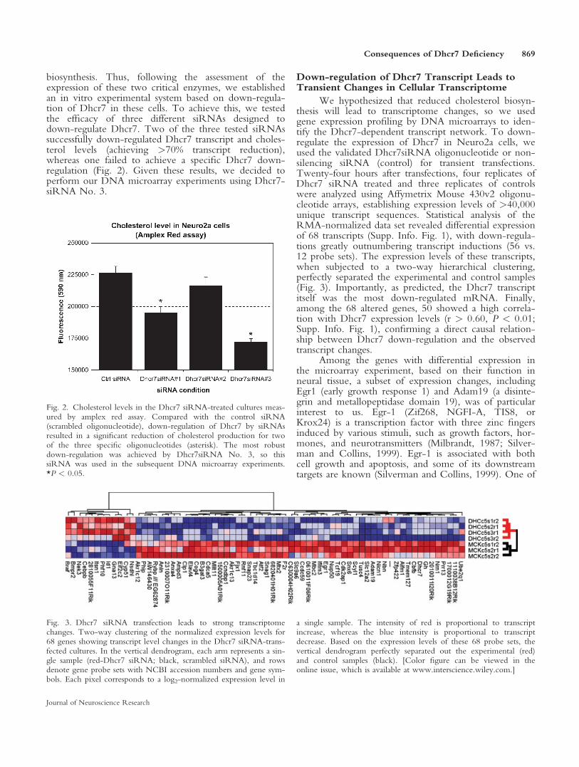
We hypothesized that reduced cholesterol biosyn-thesis will lead to transcriptome changes, so we usedgene expression profiling by DNA microarrays to iden-tify the Dhcr7-dependent transcript network. To down-regulate the expression of Dhcr7 in Neuro2a cells, weused the validated Dhcr7siRNA oligonucleotide or non-silencing siRNA (control) for transient transfections.Twenty-four hours after transfections, four replicates ofDhcr7 siRNA treated and three replicates of controlswere analyzed using Affymetrix Mouse 430v2 oligonu-cleotide arrays, establishing expression levels of >40,000unique transcript sequences. Statistical analysis of theRMA-normalized data set revealed differential expressionof 68 transcripts (Supp. Info. Fig. 1), with down-regula-tions greatly outnumbering transcript inductions (56 vs.12 probe sets). The expression levels of these transcripts,when subjected to a two-way hierarchical clustering,perfectly separated the experimental and control samples(Fig. 3). Importantly, as predicted, the Dhcr7 transcriptitself was the most down-regulated mRNA. Finally,among the 68 altered genes, 50 showed a high correla-tion with Dhcr7 expression levels (r > 0.60, P < 0.01;Supp. Info. Fig. 1), confirming a direct causal relation-ship between Dhcr7 down-regulation and the observedtranscript changes.
Among the genes with differential expression inthe microarray experiment, based on their function inneural tissue, a subset of expression changes, includingEgr1 (early growth response 1) and Adam19 (a disinte-grin and metallopeptidase domain 19), was of particularinterest to us. Egr-1 (Zif268, NGFI-A, TIS8, orKrox24) is a transcription factor with three zinc fingersinduced by various stimuli, such as growth factors, hor-mones, and neurotransmitters (Milbrandt, 1987; Silver-man and Collins, 1999). Egr-1 is associated with bothcell growth and apoptosis, and some of its downstreamtargets are known (Silverman and Collins, 1999). One of
Fig. 3. Dhcr7 siRNA transfection leads to strong transcriptomechanges. Two-way clustering of the normalized expression levels for68 genes showing transcript level changes in the Dhcr7 siRNA-trans-fected cultures. In the vertical dendrogram, each arm represents a sin-gle sample (red-Dhcr7 siRNA; black, scrambled siRNA), and rowsdenote gene probe sets with NCBI accession numbers and gene sym-bols. Each pixel corresponds to a log2-normalized expression level in
a single sample. The intensity of red is proportional to transcriptincrease, whereas the blue intensity is proportional to transcriptdecrease. Based on the expression levels of these 68 probe sets, thevertical dendrogram perfectly separated out the experimental (red)and control samples (black). [Color figure can be viewed in theonline issue, which is available at www.interscience.wiley.com.]
Fig. 2. Cholesterol levels in the Dhcr7 siRNA-treated cultures meas-ured by amplex red assay. Compared with the control siRNA(scrambled oligonucleotide), down-regulation of Dhcr7 by siRNAsresulted in a significant reduction of cholesterol production for twoof the three specific oligonucleotides (asterisk). The most robustdown-regulation was achieved by Dhcr7siRNA No. 3, so thissiRNA was used in the subsequent DNA microarray experiments.*P < 0.05.
Consequences of Dhcr7 Deficiency 869
Journal of Neuroscience Research
the Egr-1 targets is SREBP transcription factor (Fer-nandez-Alvarez et al., 2008). On the other hand,Adam19 (Meltrin b, FKSG34) belongs to a gene familyof proteins with metalloproteinase domain that areinvolved in cell adhesion, cell fusion, migration, mem-brane protein shedding, and proteolysis (Mochizuki andOkada, 2007). Furthermore, ADAMs play a role in celladhesion through interaction with other proteins (synde-cans and fibronectin) with their cysteine-rich domains(Iba et al., 2000). Although not much is known aboutAdam19, the functions of some other family membersare better described (Rocks et al., 2008). For example,Adam17 cleaves several transmembrane proteins, and itsactivity can be regulated by membrane cholesterol levels(Tellier et al., 2006, 2008). Finally, in addition to Egr1and Adam19, the three other genes that were also cho-sen for follow-up included Prr13 (proline-rich 13),Snag1 (sorting nexin associated golgi protein 1), andSnx6 (sorting nexin 6) along with an Est with unknownfunction (2010011I20Rik).
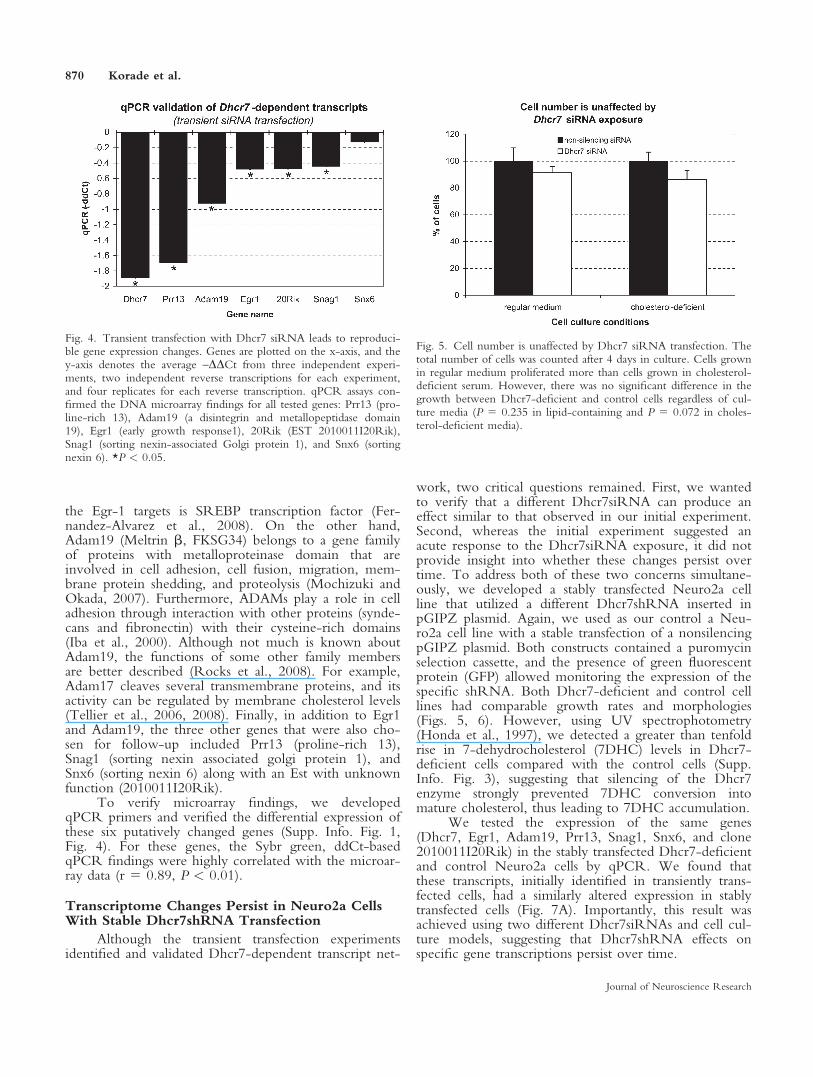
To verify microarray findings, we developedqPCR primers and verified the differential expression ofthese six putatively changed genes (Supp. Info. Fig. 1,Fig. 4). For these genes, the Sybr green, ddCt-basedqPCR findings were highly correlated with the microar-ray data (r 5 0.89, P < 0.01).
Transcriptome Changes Persist in Neuro2a CellsWith Stable Dhcr7shRNA Transfection
Although the transient transfection experimentsidentified and validated Dhcr7-dependent transcript net-
work, two critical questions remained. First, we wantedto verify that a different Dhcr7siRNA can produce aneffect similar to that observed in our initial experiment.Second, whereas the initial experiment suggested anacute response to the Dhcr7siRNA exposure, it did notprovide insight into whether these changes persist overtime. To address both of these two concerns simultane-ously, we developed a stably transfected Neuro2a cellline that utilized a different Dhcr7shRNA inserted inpGIPZ plasmid. Again, we used as our control a Neu-ro2a cell line with a stable transfection of a nonsilencingpGIPZ plasmid. Both constructs contained a puromycinselection cassette, and the presence of green fluorescentprotein (GFP) allowed monitoring the expression of thespecific shRNA. Both Dhcr7-deficient and control celllines had comparable growth rates and morphologies(Figs. 5, 6). However, using UV spectrophotometry(Honda et al., 1997), we detected a greater than tenfoldrise in 7-dehydrocholesterol (7DHC) levels in Dhcr7-deficient cells compared with the control cells (Supp.Info. Fig. 3), suggesting that silencing of the Dhcr7enzyme strongly prevented 7DHC conversion intomature cholesterol, thus leading to 7DHC accumulation.
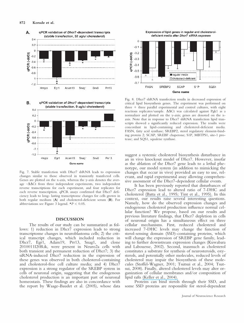
We tested the expression of the same genes(Dhcr7, Egr1, Adam19, Prr13, Snag1, Snx6, and clone2010011I20Rik) in the stably transfected Dhcr7-deficientand control Neuro2a cells by qPCR. We found thatthese transcripts, initially identified in transiently trans-fected cells, had a similarly altered expression in stablytransfected cells (Fig. 7A). Importantly, this result wasachieved using two different Dhcr7siRNAs and cell cul-ture models, suggesting that Dhcr7shRNA effects onspecific gene transcriptions persist over time.
Fig. 4. Transient transfection with Dhcr7 siRNA leads to reproduci-ble gene expression changes. Genes are plotted on the x-axis, and they-axis denotes the average –DDCt from three independent experi-ments, two independent reverse transcriptions for each experiment,and four replicates for each reverse transcription. qPCR assays con-firmed the DNA microarray findings for all tested genes: Prr13 (pro-line-rich 13), Adam19 (a disintegrin and metallopeptidase domain19), Egr1 (early growth response1), 20Rik (EST 2010011I20Rik),Snag1 (sorting nexin-associated Golgi protein 1), and Snx6 (sortingnexin 6). *P < 0.05.
Fig. 5. Cell number is unaffected by Dhcr7 siRNA transfection. Thetotal number of cells was counted after 4 days in culture. Cells grownin regular medium proliferated more than cells grown in cholesterol-deficient serum. However, there was no significant difference in thegrowth between Dhcr7-deficient and control cells regardless of cul-ture media (P 5 0.235 in lipid-containing and P 5 0.072 in choles-terol-deficient media).
870 Korade et al.
Journal of Neuroscience Research
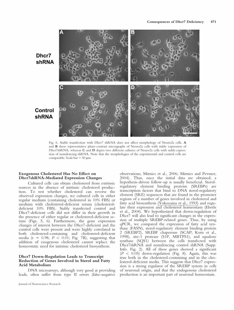
Exogenous Cholesterol Has No Effect onDhcr7shRNA-Mediated Expression Changes
Cultured cells can obtain cholesterol from extrinsicsources in the absence of intrinsic cholesterol produc-tion. To test whether cholesterol can reverse theobserved expression changes, we cultured cells in eitherregular medium (containing cholesterol in 10% FBS) ormedium with cholesterol-deficient serum (cholesterol-deficient 10% FBS). Stably transfected control andDhcr7-deficient cells did not differ in their growth inthe presence of either regular or cholesterol-deficient se-rum (Figs. 5, 6). Furthermore, the gene expressionchanges of interest between the Dhcr7-deficient and thecontrol cells were present and were highly correlated inboth cholesterol-containing and cholesterol-deficientmedia (r 5 0.98, P < 0.01; Fig. 7B), suggesting thataddition of exogenous cholesterol cannot replace thehomeostatic need for intrinsic cholesterol biosynthesis.
Dhcr7 Down-Regulation Leads to TranscriptReduction of Genes Involved in Sterol and FattyAcid Metabolism
DNA microarrays, although very good at providingleads, often suffer from type II errors (false-negative
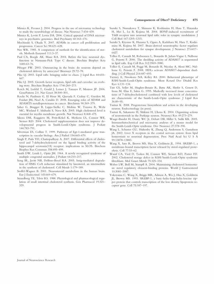
observations; Mirnics et al., 2006; Mirnics and Pevsner,2004). Thus, once the initial data are obtained, ahypothesis-driven follow-up is usually beneficial. Sterol-regulatory element binding proteins (SREBPs) aretranscription factors that bind to DNA sterol-regulatoryelement (SRE) sequences that are found in the promoterregions of a number of genes involved in cholesterol andfatty acid biosynthesis (Yokoyama et al., 1993) and regu-late their expression and cholesterol homeostasis (Eberleet al., 2004). We hypothesized that down-regulation ofDhcr7 will also lead to significant changes in the expres-sion of multiple SREBP-related genes. Thus, by usingqPCR, we compared the expression of fatty acid syn-thase (FASN), sterol-regulatory element binding protein2 (SREBP2), SREBF chaperone (SCAP; Korn et al.,1998), site-1 protease (S1P, MBTPS1), and squalenesynthase (SQS1) between the cells transfected withDhcr7shRNA and nonsilencing control shRNA (Supp.Info. Fig. 2). All of these genes showed a significant(P < 0.05) down-regulation (Fig. 8). Again, this wastrue both in the cholesterol-containing and in the cho-lesterol-deficient media. This suggests that Dhcr7 expres-sion is a strong regulator of the SREBP system in cellsof neuronal origin, and that the endogenous cholesterolproduction is an important part of neuronal homeostasis.
Fig. 6. Stable transfection with Dhcr7 shRNA does not affect morphology of Neuro2a cells. Aand B show representative phase-contrast micrographs of Neuro2a cells with stable expression ofDhcr7shRNA, whereas C and D depict two different cultures of Neuro2a cells with stable expres-sion of nonsilencing shRNA. Note that the morphologies of the experimental and control cells arecomparable. Scale bar5 50 lm.
Consequences of Dhcr7 Deficiency 871
Journal of Neuroscience Research
DISCUSSION
The results of our study can be summarized as fol-lows: 1) reduction in Dhcr7 expression leads to strongtranscriptome changes in neuroblastoma cells; 2) the crit-ical transcript changes, which included reduction inDhcr7, Egr1, Adam19, Prr13, Snag1, and clone2010011I20Rik, were present in Neuro2a cells withboth transient and permanent reduction of Dhcr7; 3) thesiRNA-induced Dhcr7 reduction in the expression ofthese genes was observed in both cholesterol-containingand cholesterol-free cell culture media; and 4) Dhcr7expression is a strong regulator of the SREBP system incells of neuronal origin, suggesting that the endogenouscholesterol production is an important part of neuronalhomeostasis. These findings are also in concordance withthe report by Waage-Baudet et al. (2005), whose data
suggest a systemic cholesterol biosynthesis disturbance inan in vivo knockout model of Dhcr7. However, insofaras the ablation of the Dhcr7 gene leads to a lethal phe-notype, our model system (in addition to mimicking thechanges that occur in vivo) provided an easy to use, rel-evant, and rapid experimental assay allowing comprehen-sive assessment of the Dhcr7-dependent cellular events.
It has been previously reported that disturbances ofDhcr7 expression lead to altered ratio of 7-DHC andcholesterol (Batta et al., 1995; Tint et al., 1995). In thiscontext, our results raise several interesting questions.Namely, how do the observed expression changes andendogenous cholesterol production influence normal cel-lular function? We propose, based on our results andprevious literature findings, that Dhcr7 depletion in cellsof neuronal origin has a simultaneous effect on threecellular mechanisms. First, reduced cholesterol andincreased 7-DHC levels may change the function ofsterol-sensing domain (SSD)-containing proteins, whichwill change the expression of SREBP gene family, lead-ing to further downstream expression changes (Kuwabaraand Labouesse, 2002). Second, inasmuch as cholesterolconstitutes a substrate for synthesis of neurosteroids, oxy-sterols, and potentially other molecules, reduced levels ofcholesterol may impair the biosynthesis of these mole-cules (Stoffel-Wagner, 2001; Tsutsui et al., 2004; Tsut-sui, 2008). Finally, altered cholesterol levels may alter or-ganization of cellular membranes and/or composition oflipid rafts (Keller et al., 2004).
Proteins can bind sterols through their SSD, andsome SSD proteins are responsible for sterol-dependent
Fig. 8. Dhcr7 shRNA transfection results in decreased expression ofcritical lipid biosynthesis genes. The experiment was performed onthree 1 three parallel experimental and control cultures, with eightreactions replicates/sample. DDCt was calculated against Pgk1 as anormalizer and plotted on the y-axis; genes are denoted on the x-axis. Note that in response to Dhcr7 shRNA transfection lipid tran-scripts showed a significantly reduced expression. The results wereconcordant in lipid-containing and cholesterol-deficient media.FASN, fatty acid synthase; SREBP2, sterol regulatory element-bind-ing protein 2; SCAP, SREBF chaperone; S1P, MBTPS1, site-1 pro-tease; and SQS1, squalene synthase.
Fig. 7. Stable transfection with Dhcr7 shRNA leads to expressionchanges similar to those observed in transiently transfected cells.Genes are plotted on the x-axis, whereas the y-axis denotes the aver-age –DDCt from three independent experiments, two independentreverse transcriptions for each experiment, and four replicates foreach reverse transcription. qPCR assays confirmed that Dhcr7 defi-ciency leads to long- lasting transcriptome changes for cells grown inboth regular medium (A) and cholesterol-deficient serum (B). Forabbreviations see Figure 3 legend. *P < 0.05.
872 Korade et al.
Journal of Neuroscience Research

proteolysis in the Golgi and transcriptional regulation ofproteins containing an SRE domain (Kuwabara and Lab-ouesse, 2002; Weber et al., 2004). For example, SCAP,through its SSD, regulates transcription of target genesthrough SREBPs, by either retaining them in the ER orescorting them to the Golgi (Brown and Goldstein,1999; Brown et al., 2002; Horton et al., 2002; Goldsteinet al., 2006). Dhcr7-siRNA transfection introduces animbalance between the amount of ‘‘mature’’ cholesterol(decreased) and 7-DHC (increased). It is known that dif-ferent sterols have slightly different effects on specificproteins, putatively altering their structure and function(Song et al., 2005). For example, the cholesterol inter-mediate lanosterol is more efficient in stimulating theubiquitination and degradation of Hmgcr than choles-terol (Song et al., 2005), and previous studies led to theproposal that 7-DHC may perturb the function of otherSSD-containing proteins in SLOS (Wassif et al., 2002).Interestingly, 7-DHC leads to increased proteolysis ofHmgcr, thus further exacerbating the deficit in SLOS(Fitzky et al., 2001). Finally, fibroblast cultures ofpatients with CHILD syndrome (an inborn error of thecholesterol biosynthesis pathway) report an accumulationof sterol precursors leading to formation of lipid vacuoleswith a lamellar appearance (Hashimoto et al., 1998). Inagreement with these findings, the expression changesobserved in our experiments also strongly support thenotion that 7-DHC accumulation has a critical action onthe function of proteins with SSD. We observedreduced transcripts of both SSD-containing proteins(SCAP) and SRE element-containing genes (FASN,SREBP2, S1P-MBTPS1, and SQS1). Therefore, wehypothesize that altered balance between 7-DHC andcholesterol will change the structure and function ofSSD-containing protein SCAP, which will change theexpression of multiple SREBP gene family members.More specifically, we believe that Srebp2 (one of themost down-regulated genes in our experiment) has acritical role in this process. Srebp2 is a transcription fac-tor, and it controls the transcription of multiple lipidbiosynthesis genes (Goldstein et al., 2006). Interestingly,Srebp2 contains three Egr1 binding sites in its promoterregion (McMullen et al., 2005). Thus, we propose thatEgr1 (another transcription factor with changed expres-sion in the Dhcr7-siRNA treated cells) is likely to be animportant upstream regulator of neuronal cholesterolbiosynthesis.
Dhcr7 siRNA treatment reduces ‘‘mature’’ choles-terol levels. Because cholesterol is a substrate for biosyn-thesis of neurosteroids and oxysterols, reduction of cho-lesterol levels will cause use of 7-DHC for neurosteroidsynthesis. Although literature data suggest that 7-DHCcan serve as a substrate for neurosteroid synthesis, it isnot clear whether the efficacy of the 7-DHC-derivedneurosteroid synthesis is comparable to that generatedfrom cholesterol or whether the activities of the 7-DHC-generated neurosteroids are similar to those of‘‘normal’’ neurosteroids (Marcos et al., 2004). Supportfor this hypothesis is provided by the observation that,
in Niemann-Pick disease, reduced cholesterol levels leadto reduced alopregnanole synthesis (Chen et al., 2007),suggesting that normal cholesterol level likely arerequired for maintenance of normal CNS neurosteroidlevels.
Lipid rafts are a critical place for receptor insertion,neurotrophin signaling, neurotransmitter release, andregulated intramembrane proteolysis of transmembraneproteins (Pike, 2005). Reduced Dhcr7 levels will lead toaccumulation of 7-dehydrocholesterol, which is insertedinto the rafts instead of cholesterol (Keller et al., 2004).However, because the structures of 7-dehydrocholesteroland cholesterol are different, the structure and composi-tion of the lipid rafts will be altered. Thus, altered inser-tion of many receptors could change signaling from thecell surface (Suzuki et al., 2004; Freeman et al., 2007).Although our experimental data do not provide directevidence for such a mechanism, this hypothesis is sup-ported by several critical, literature-based lines of evi-dence: 1) cholesterol content of rafts affects membranethickness, elasticity, and curvature (Allende et al., 2004;Bacia et al., 2005); 2) cholesterol content of rafts affectsion conductance and excitability of membranes, traffick-ing of ionotropic receptors to and from the cell mem-brane, size and number of some postsynaptic receptorclusters, and neurotransmitter signaling through G-pro-tein coupled receptors (Korade and Kenworthy, 2008);3) protein activity may be modulated by the composi-tion of lipid rafts (Pike, 2003); and 4) presence of 7-de-hydrocholesterol in hippocampal membranes impairsligand-binding activity of the serotonin 1A receptor(Singh et al., 2007).
Finally, our findings suggest that the role of endog-enous cholesterol biosynthesis in neuronal cells deservesequal consideration. The human brain contains as muchas 25% of total cholesterol, and the CNS cholesterolpool is regulated independently of the peripheral choles-terol pool (Brown and Goldstein, 1999; Dietschy andTurley, 2001). Although the role, origin, metabolism,and regulation of the CNS cholesterol pool remainunderstudied and mostly unknown to date (Korade andKenworthy, 2008), it has been historically assumed thatcholesterol of nonneuronal origin is the only importantsource of cholesterol for the brain cells (Pfrieger, 2003).Recent evidence that 1) cholesterogenic enzymes arecoexpressed at high level in neurons across various brainstructures, 2) hippocampal and cholinergic neuronscoexpress the highest levels of cholesterogenic enzymes,and 3) growth factors regulate cholesterol biosynthesis ina neuroblastoma cell line suggest that, in at least some ofthe neurons, endogenous neuronal cholesterol synthesisis likely to be an important homeostatic factor (Koradeet al., 2007; Suzuki et al., 2007). Our current findingsstrongly underscore this notion. That the expressionchanges we observed were present in both cholesterol-containing and cholesterol-free cell culture mediastrongly argues that endogenous cholesterol biosynthesisis critical for neuronal homeostasis, and extrinsic supple-mentation of cholesterol cannot reverse the effects of
Consequences of Dhcr7 Deficiency 873
Journal of Neuroscience Research

reduced intrinsic neuronal cholesterol biosynthesis. Thisfinding may also contribute to our understanding of thepathophysiological mechanism underlying some of theCNS-related disturbances in SLOS. SLOS, characterizedby a range of developmental brain abnormalities, is dueto inactivation of the Dchr7 enzyme and a consequentreduction in cholesterol biosynthesis (Jira et al., 2003).Unfortunately, cholesterol supplementation has only alimited therapeutic effect in these patients, and webelieve that this is due to the need of some neurons foran endogenous neuronal cholesterol biosynthesis thatcannot be supplemented from sources outside the cell(Sikora et al., 2004). Similarly, our findings warrant fur-ther follow-up studies on the role of statins (inhibitors ofcholesterol biosynthesis) in neuronal cholesterol biosyn-thesis and their effects on neuronal homeostasis.
ACKNOWLEDGMENTS
We thank Charles Asher Day for help with theUV spectrometry assay.
REFERENCES
Allende D, Vidal A, McIntosh TJ. 2004. Jumping to rafts: gatekeeper
role of bilayer elasticity. Trends Biochem Sci 29:325–330.
Anitei M, Pfeiffer SE. 2006. Myelin biogenesis: sorting out protein traf-
ficking. Curr Biol 16:R418–R421.
Bacia K, Schwille P, Kurzchalia T. 2005. Sterol structure determines the
separation of phases and the curvature of the liquid-ordered phase in
model membranes. Proc Natl Acad Sci U S A 102:3272–3277.
Bae SH, Lee JN, Fitzky BU, Seong J, Paik YK. 1999. Cholesterol bio-
synthesis from lanosterol. Molecular cloning, tissue distribution, expres-
sion, chromosomal localization, and regulation of rat 7-dehydrocholes-
terol reductase, a Smith-Lemli-Opitz syndrome-related protein. J Biol
Chem 274:14624–14631.
Batta AK, Salen G, Tint GS, Shefer S. 1995. Identification of 19-nor-
5,7,9(10)-cholestatrien-3 beta-ol in patients with Smith-Lemli-Opitz
syndrome. J Lipid Res 36:2413–2418.
Brown AJ, Sun L, Feramisco JD, Brown MS, Goldstein JL. 2002. Cho-
lesterol addition to ER membranes alters conformation of SCAP, the
SREBP escort protein that regulates cholesterol metabolism. Mol Cell
10:237–245.
Brown MS, Goldstein JL. 1999. A proteolytic pathway that controls the
cholesterol content of membranes, cells, and blood. Proc Natl Acad Sci
U S A 96:11041–11048.
Chen G, Li HM, Chen YR, Gu XS, Duan S. 2007. Decreased estradiol
release from astrocytes contributes to the neurodegeneration in a mouse
model of Niemann-Pick disease type C. Glia 55:1509–1518.
Dietschy JM, Turley SD. 2001. Cholesterol metabolism in the brain.
Curr Opin Lipidol 12:105–112.
Eberle D, Hegarty B, Bossard P, Ferre P, Foufelle F. 2004. SREBP tran-
scription factors: master regulators of lipid homeostasis. Biochimie
86:839–848.
Fernandez-Alvarez A, Tur G, Lopez-Rodas G, Casado M. 2008. Recip-
rocal regulation of the human sterol regulatory element binding protein
(SREBP)-1a promoter by Sp1 and EGR-1 transcription factors. FEBS
Lett 582:177–184.
Fitzky BU, Moebius FF, Asaoka H, Waage-Baudet H, Xu L, Xu G,
Maeda N, Kluckman K, Hiller S, Yu H, Batta AK, Shefer S, Chen T,
Salen G, Sulik K, Simoni RD, Ness GC, Glossmann H, Patel SB, Tint
GS. 2001. 7-Dehydrocholesterol-dependent proteolysis of HMG-CoA
reductase suppresses sterol biosynthesis in a mouse model of Smith-
Lemli-Opitz/RSH syndrome. J Clin Invest 108:905–915.
Freeman MR, Cinar B, Kim J, Mukhopadhyay NK, Di Vizio D, Adam
RM, Solomon KR. 2007. Transit of hormonal and EGF receptor-de-
pendent signals through cholesterol-rich membranes. Steroids 72:210–
217.
Goldstein JL, DeBose-Boyd RA, Brown MS. 2006. Protein sensors for
membrane sterols. Cell 124:35–46.
Hashimoto K, Prada S, Lopez AP, Hoyos JG, Escobar M. 1998. CHILD
syndrome with linear eruptions, hypopigmented bands, and verruciform
xanthoma. Pediatr Dermatol 15:360–366.
Honda A, Batta AK, Salen G, Tint GS, Chen TS, Shefer S. 1997.
Screening for abnormal cholesterol biosynthesis in the Smith-Lemli-
Opitz syndrome: rapid determination of plasma 7-dehydrocholesterol
by ultraviolet spectrometry. Am J Med Genet 68:288–293.
Horton JD, Goldstein JL, Brown MS. 2002. SREBPs: activators of the
complete program of cholesterol and fatty acid synthesis in the liver.
J Clin Invest 109:1125–1131.
Iba K, Albrechtsen R, Gilpin B, Frohlich C, Loechel F, Zolkiewska A,
Ishiguro K, Kojima T, Liu W, Langford JK, Sanderson RD, Brake-
busch C, Fassler R, Wewer UM. 2000. The cysteine-rich domain of
human ADAM 12 supports cell adhesion through syndecans and trig-
gers signaling events that lead to beta1 integrin-dependent cell spread-
ing. J Cell Biol 149:1143–1156.
Jira PE, Waterham HR, Wanders RJ, Smeitink JA, Sengers RC, Wevers
RA. 2003. Smith-Lemli-Opitz syndrome and the DHCR7 gene. Ann
Hum Genet 67:269–280.
Jurevics H, Morell P. 1995. Cholesterol for synthesis of myelin is made
locally, not imported into brain. J Neurochem 64:895–901.
Jurevics HA, Kidwai FZ, Morell P. 1997. Sources of cholesterol during
development of the rat fetus and fetal organs. J Lipid Res 38:723–733.
Keller RK, Arnold TP, Fliesler SJ. 2004. Formation of 7-dehydrocholes-
terol-containing membrane rafts in vitro and in vivo, with relevance to
the Smith-Lemli-Opitz syndrome. J Lipid Res 45:347–355.
Korade Z, Kenworthy A. 2008. Lipid rafts, cholesterol, and the brain.
Neuropharmacology (in press).
Korade Z, Mi Z, Portugal C, Schor NF. 2007. Expression and p75 neu-
rotrophin receptor dependence of cholesterol synthetic enzymes in
adult mouse brain. Neurobiol Aging 28:1522–1531.
Korn BS, Shimomura I, Bashmakov Y, Hammer RE, Horton JD, Gold-
stein JL, Brown MS. 1998. Blunted feedback suppression of SREBP
processing by dietary cholesterol in transgenic mice expressing sterol-re-
sistant SCAP(D443N). J Clin Invest 102:2050–2060.
Kurrasch DM, Huang J, Wilkie TM, Repa JJ. 2004. Quantitative real-
time polymerase chain reaction measurement of regulators of G-protein
signaling mRNA levels in mouse tissues. Methods Enzymol 389:3–15.
Kuwabara PE, Labouesse M. 2002. The sterol-sensing domain: multiple
families, a unique role? Trends Genet 18:193–201.
Luskey KL, Stevens B. 1985. Human 3-hydroxy-3-methylglutaryl coen-
zyme A reductase. Conserved domains responsible for catalytic activity
and sterol-regulated degradation. J Biol Chem 260:10271–10277.
Lutton C. 1991. Dietary cholesterol, membrane cholesterol and choles-
terol synthesis. Biochimie 73:1327–1334.
Marcos J, Guo LW, Wilson WK, Porter FD, Shackleton C. 2004. The
implications of 7-dehydrosterol-7-reductase deficiency (Smith-Lemli-
Opitz syndrome) to neurosteroid production. Steroids 69:51–60.
Maxfield FR, Tabas I. 2005. Role of cholesterol and lipid organization
in disease. Nature 438:612–621.
McMullen MR, Pritchard MT, Wang Q, Millward CA, Croniger CM,
Nagy LE. 2005. Early growth response-1 transcription factor is essential
for ethanol-induced fatty liver injury in mice. Gastroenterology
128:2066–2076.
Milbrandt J. 1987. A nerve growth factor-induced gene encodes a possi-
ble transcriptional regulatory factor. Science 238:797–799.
874 Korade et al.
Journal of Neuroscience Research
Mirnics K, Pevsner J. 2004. Progress in the use of microarray technology
to study the neurobiology of disease. Nat Neurosci 7:434–439.
Mirnics K, Levitt P, Lewis DA. 2006. Critical appraisal of DNA microar-
rays in psychiatric genomics. Biol Psychiatry 60:163–176.
Mochizuki S, Okada Y. 2007. ADAMs in cancer cell proliferation and
progression. Cancer Sci 98:621–628.
Nes WR. 1985. A comparison of methods for the identification of ster-
ols. Methods Enzymol 111:3–37.
Paul CA, Boegle AK, Maue RA. 2004. Before the loss: neuronal dys-
function in Niemann-Pick Type C disease. Biochim Biophys Acta
1685:63–76.
Pfrieger FW. 2003. Outsourcing in the brain: do neurons depend on
cholesterol delivery by astrocytes? Bioessays 25:72–78.
Pike LJ. 2003. Lipid rafts: bringing order to chaos. J Lipid Res 44:655–
667.
Pike LJ. 2005. Growth factor receptors, lipid rafts and caveolae: an evolv-
ing story. Biochim Biophys Acta 1746:260–273.
Reich M, Liefeld T, Gould J, Lerner J, Tamayo P, Mesirov JP. 2006.
GenePattern 2.0. Nat Genet 38:500–501.
Rocks N, Paulissen G, El Hour M, Quesada F, Crahay C, Gueders M,
Foidart JM, Noel A, Cataldo D. 2008. Emerging roles of ADAM and
ADAMTS metalloproteinases in cancer. Biochimie 90:369–379.
Saher G, Brugger B, Lappe-Siefke C, Mobius W, Tozawa R, Wehr
MC, Wieland F, Ishibashi S, Nave KA. 2005. High cholesterol level is
essential for myelin membrane growth. Nat Neurosci 8:468–475.
Sikora DM, Ruggiero M, Petit-Kekel K, Merkens LS, Connor WE,
Steiner RD. 2004. Cholesterol supplementation does not improve de-
velopmental progress in Smith-Lemli-Opitz syndrome. J Pediatr
144:783–791.
Silverman ES, Collins T. 1999. Pathways of Egr-1-mediated gene tran-
scription in vascular biology. Am J Pathol 154:665–670.
Singh P, Paila YD, Chattopadhyay A. 2007. Differential effects of choles-
terol and 7-dehydrocholesterol on the ligand binding activity of the
hippocampal serotonin(1A) receptor: implications in SLOS. Biochem
Biophys Res Commun 358:495–499.
Smith DW, Lemli L, Opitz JM. 1964. A newly recognized syndrome of
multiple congenital anomalies. J Pediatr 64:210–217.
Song BL, Javitt NB, DeBose-Boyd RA. 2005. Insig-mediated degrada-
tion of HMG CoA reductase stimulated by lanosterol, an intermediate
in the synthesis of cholesterol. Cell Metab 1:179–189.
Stoffel-Wagner B. 2001. Neurosteroid metabolism in the human brain.
Eur J Endocrinol 145:669–679.
Strandberg TE, Tilvis RS. 1988. Physiological and pharmacological regu-
lation of small intestinal cholesterol synthesis. Gen Pharmacol 19:321–
329.
Suzuki S, Numakawa T, Shimazu K, Koshimizu H, Hara T, Hatanaka
H, Mei L, Lu B, Kojima M. 2004. BDNF-induced recruitment of
TrkB receptor into neuronal lipid rafts: roles in synaptic modulation. J
Cell Biol 167:1205–1215.
Suzuki S, Kiyosue K, Hazama S, Ogura A, Kashihara M, Hara T, Koshi-
mizu H, Kojima M. 2007. Brain-derived neurotrophic factor regulates
cholesterol metabolism for synapse development. J Neurosci 27:6417–
6427.
Tellier E, Canault M, Rebsomen L, Bonardo B, Juhan-Vague I, Nalbone
G, Peiretti F. 2006. The shedding activity of ADAM17 is sequestered
in lipid rafts. Exp Cell Res 312:3969–3980.
Tellier E, Canault M, Poggi M, Bonardo B, Nicolay A, Alessi MC, Nal-
bone G, Peiretti F. 2008. HDLs activate ADAM17-dependent shed-
ding. J Cell Physiol 214:687–693.
Tierney E, Nwokoro NA, Kelley RI. 2000. Behavioral phenotype of
RSH/Smith-Lemli-Opitz syndrome. Ment Retard Dev Disabil Res
Rev 6:131–134.
Tint GS, Seller M, Hughes-Benzie R, Batta AK, Shefer S, Genest D,
Irons M, Elias E, Salen G. 1995. Markedly increased tissue concentra-
tions of 7-dehydrocholesterol combined with low levels of cholesterol
are characteristic of the Smith-Lemli-Opitz syndrome. J Lipid Res
36:89–95.
Tsutsui K. 2008. Progesterone biosynthesis and action in the developing
neuron. Endocrinology (in press).
Tsutsui K, Sakamoto H, Shikimi H, Ukena K. 2004. Organizing actions
of neurosteroids in the Purkinje neuron. Neurosci Res 49:273–279.
Waage-Baudet H, Dunty WC Jr, Dehart DB, Hiller S, Sulik KK. 2005.
Immunohistochemical and microarray analyses of a mouse model for
the Smith-Lemli-Opitz syndrome. Dev Neurosci 27:378–396.
Wang L, Schuster GU, Hultenby K, Zhang Q, Andersson S, Gustafsson
JA. 2002. Liver X receptors in the central nervous system: from lipid
homeostasis to neuronal degeneration. Proc Natl Acad Sci U S A
99:13878–13883.
Wang X, Sato R, Brown MS, Hua X, Goldstein JL. 1994. SREBP-1, a
membrane-bound transcription factor released by sterol-regulated prote-
olysis. Cell 77:53–62.
Wassif CA, Vied D, Tsokos M, Connor WE, Steiner RD, Porter FD.
2002. Cholesterol storage defect in RSH/Smith-Lemli-Opitz syndrome
fibroblasts. Mol Genet Metab 75:325–334.
Weber LW, Boll M, Stampfl A. 2004. Maintaining cholesterol homeosta-
sis: sterol regulatory element-binding proteins. World J Gastroenterol
10:3081–3087.
Yokoyama C, Wang X, Briggs MR, Admon A, Wu J, Hua X, Goldstein
JL, Brown MS. 1993. SREBP-1, a basic-helix-loop-helix-leucine zip-
per protein that controls transcription of the low density lipoprotein re-
ceptor gene. Cell 75:187–197.
Consequences of Dhcr7 Deficiency 875
Journal of Neuroscience Research


