Embed Size (px)
Citation preview
Molecular characterization of Mst77F and implication in Drosophila spermatogenesis
Dissertation for the award of the degree
“Doctor rerum naturalium” (Dr. rer. nat.) Division of Mathematics and Natural Sciences
of the Georg-August-Universität Göttingen
submitted by Nils Kost
from Lübben, Germany Göttingen 2012
2
Committee members: Dr. Wolfgang Fischle (1st reviewer), Research group Chromatin Biochemistry Max Planck Institute for Biophysical Chemistry, Göttingen Prof. Dr. Peter Rehling (2nd reviewer), Department of Biochemistry II Georg-August-University Göttingen Prof. Dr. Steven Johnson, Department of Molecular Oncology Georg-August-University Göttingen Date of the oral examination: August 03, 2012

3
I affirm that the presented thesis “Molecular characterization of Mst77F and implication in Drosophila spermatogenesis” has been written independently and with no other sources and aids than quoted. June 30, 2012, Göttingen

4
Acknowledgments
First and foremost, I am grateful to Dr. Wolfgang Fischle, for his trust, constant
support and fruitful discussions on several projects.
I would like to thank my PhD Thesis committee members Prof. Peter Rehling and
Prof. Steven Johnson for their support and guidance throughout this project.
Many thanks go to all members of the Chromatin Biochemistry group for providing a
great working atmosphere. In particular, I would like to thank Claudia Fahlbusch,
Lydia Abdelhalim and Winfried Lendeckel for their help in the lab.
I want to thank Prof. Renate Renkawitz-Pohl, Dr. Christina Rathke and Sophie Kaiser
for their collaboration on the Mst77F project.
Special thanks goes to Dr. Alf Herzig for providing me with reagents and his help on
Drosophila methodology
I thank the GGNB administration for the constant support, organization of lectures,
method courses as well as retreats.
I am grateful to my parents, Karin and Michael Scholta, for their constant hold up.
Lastly, I want to thank my own family, Daniela and Maxi, for their love, support and
cheering me up when it was necessary.

5
6
Table of contents
List of figures .................................................................................................................. 8
List of tables ................................................................................................................... 9
Abbreviations ............................................................................................................... 10
1. Introduction ........................................................................................................... 14
1.1 Spermatogenesis ............................................................................................................. 14 1.1.1 Premeiotic stages and meiosis ................................................................................................................... 14 1.1.2 Structural organization of the DNA after meiosis .............................................................................. 16
1.1.2.1 The nucleosome core particle - the basic unit of chromatin ................................................. 16 1.1.2.2 Histone posttranslational modifications in early differentiating spermatids ............... 17
1.1.3 Postmeiotic spermatid maturation .......................................................................................................... 18 1.1.4 Sperm nuclear proteins involved in chromatin condensation ...................................................... 19 1.1.5 The details of spermatid differentiation ................................................................................................ 20
1.1.5.1 The condensation of the DNA ............................................................................................................. 20 1.1.5.2 Nuclear shaping (Fuller, 1993) .......................................................................................................... 21
1.1.6 Mst77F - a suggested chromosomal architectural protein ............................................................. 22
1.2 Linker histone H1 ............................................................................................................ 23 1.2.1 H1 localisation and structural function .................................................................................................. 23 1.2.2 H1 structure and function - the globular domain ............................................................................... 24 1.2.3 H1 structure and function - the N-terminal domain ......................................................................... 25 1.2.4 H1 structure and function - the C-terminal domain .......................................................................... 25
1.2.4.1 Intrinsically unstructured proteins (IUPs) ................................................................................... 25 1.2.4.2 Functional implication of the intrinsically unstructured histone H1 CTD ...................... 27
1.4 Objectives of the thesis ................................................................................................... 28
2 Material & Methods ............................................................................................... 30 2.1 Laboratory equipment ...................................................................................................................................... 30 2.2 Chemicals / Reagents ......................................................................................................................................... 32 2.3 Bioinformatic tools ............................................................................................................................................. 35 2.4 Preparation of SDS-PAGE Gels and Electrophoresis ............................................................................. 35 2.5 Protein staining within SDS Gels ................................................................................................................... 36 2.6 Determination of Nucleic Acid and Protein Concentrations.............................................................. 36 2.7 Molecular Cloning................................................................................................................................................ 37
2.7.1 Bacterial transformation.......................................................................................................................... 38 2.7.2 PCR based DNA amplification ................................................................................................................ 38 2.7.3 Purification of PCR products .................................................................................................................. 39 2.7.4 Restriction digest of DNA ........................................................................................................................ 39 2.7.5 Dephosphorylation of Plasmid DNA ................................................................................................... 39 2.7.6 Purification of DNAs .................................................................................................................................. 40 2.7.7 Agarose electrophoresis & ethidium bromid visualization of DNA ....................................... 40 2.7.8 Ligation ........................................................................................................................................................... 40
2.8 Mini - Preparation of Plasmid DNA .............................................................................................................. 41 2.9 Site - Directed - Mutagenesis of Plasmid DNA ......................................................................................... 41 2.10 Expression and Purification of Recombinant Proteins ..................................................................... 42
2.10.1 Bacterial Expression ............................................................................................................................... 42
7
2.10.2 HIS-tag protein purification ................................................................................................................. 42 2.10.3 Histone Inclusion Body Purification ................................................................................................. 43 2.10.4 Chromatographic Purification of Histones .................................................................................... 43
2.11 In vitro chromatin reconstitution & analysis ........................................................................................ 44 2.11.1 Preparation of DNA templates for chromatin reconstitution ................................................ 44 2.11.2 Histone octamer reconstitution ......................................................................................................... 45 2.11.3 Size exclusion chromatographie of histone octamers ............................................................... 45 2.11.4 Reconstitution of mono and oligonucleosomes ........................................................................... 45 2.11.5 In vitro chromatin transcription assay ............................................................................................ 46 2.11.6 Micrococcus nuclease digestion assay ............................................................................................. 47
2.12 Biochemical and biophysical binding assays ........................................................................................ 47 2.12.1 Generation of DNA templates .............................................................................................................. 47 2.12.2 12mer duplex DNA Pulldown experiments ................................................................................... 49 2.12.3 Chromatin co-precipitation experiments ....................................................................................... 50 2.12.4 Fluorescence polarisation (FP) ........................................................................................................... 50 2.12.5 Isothermal Titration Calorimetry (ITC) .......................................................................................... 51 2.12.6 Electrophoretic Mobility Shift Assay (EMSA) ............................................................................... 52 2.12.7 Protein cross - linking assay ................................................................................................................ 53
2.13 DNA / Chromatin compaction assays ....................................................................................................... 53 2.13.1 Atomic Force Microscopy ...................................................................................................................... 53 2.13.2 Centrifugation Fractionation Assay .................................................................................................. 55 2.13.3 In vitro DNA cross linking assay ........................................................................................................ 56
2.14 Other techniques ............................................................................................................................................... 57 2.14.1 Circular Dichroism (CD) ........................................................................................................................ 57 2.14.2 Analytical Ultra Centrifugation ........................................................................................................... 57
3. Results ....................................................................................................................... 60 3.1 Mst77F is a protein of bivalent structural organization .................................................................... 60 3.2 The Mst77F-DNA interaction.......................................................................................................................... 63
3.2.1 The Mst77F C-terminal domain is necessary and sufficient for DNA binding .................. 64 3.2.2 Mst77F binding to DNA is based on sequence unspecific ionic interactions ..................... 67
3.3 Thermodynamics of the Mst77F DNA interaction ................................................................................ 70 3.4 The mechanism of the Mst77F-DNA interaction .................................................................................... 71
3.4.1 Mst77F aggregates DNA ........................................................................................................................... 72 3.4.2 Mst77F effects on DNA are mediated by the N-terminus of the protein ............................. 75
3.5 The Mst77F N-terminus functions as multimerization interface upon DNA recognition ..... 79 3.6 Mst77F induces DNA clustering through its multimerization .......................................................... 82 3.7 Mst77F tightly compacts long DNA in vitro.............................................................................................. 85 3.8 Mst77F inhibits transcription in vitro ........................................................................................................ 89 3.9 Mst77F displays similar function in the context of recombinant chromatin ............................. 92
4. Discussion ............................................................................................................... 98 4.1 The implication of intrinsically unstructured, charged domains in DNA binding .................... 98 4.2 Induction of structural elements in intrinsically unstructured domains is functionally
relevant ................................................................................................................................................................. 100 4.3 Structural aspects of Mst77F DNA complexes ...................................................................................... 102 4.4 The biological role of the S149T mutant ................................................................................................. 105 4.5 Mst77F association with chromatin in vivo .......................................................................................... 106 4.6 Outlook ................................................................................................................................................................. 109
Summary ..................................................................................................................... 110
Bibliography ................................................................................................................ 113
8
List of figures Fig. 1.1 Schematic illustration of Drosophila Spermatogenesis ................................................. 15
Fig. 1.2 The structure of the nucleosome core particel: ................................................................ 17
Fig. 1.3 The postmeiotic differentiation of Drosophila spermatids .......................................... 21
Fig. 3.1 Structural parameters of Mst77F ........................................................................................... 63
Fig. 3.2 Mst77F binds DNA with its C-terminal positively charged tail ................................... 65
Fig. 3.3 Mst77F binding to DNA is based on an ionic interaction mechanism ...................... 69
Fig. 3.4 Isothermal Titration Calorimetry with Mst77F and DNA dodecamers ................... 70
Fig. 3.5 Unspecific charge mediated protein – DNA interactions in EMSA experiments .. 74
Fig. 3.6 Mst77F induces aggregation of DNA ..................................................................................... 79
Fig. 3.7 The N-terminus of Mst77F multimerizes ............................................................................ 82
Fig. 3.8 Mst77Fs N-terminus triggers quantitatively DNA aggregation .................................. 84
Fig. 3.9 AFM uncovers structural differences caused by Mst77F from effects triggered
by other charged proteins ......................................................................................................... 88
Fig. 3.10 Mst77F inhibits transcription in vitro ................................................................................ 90
9
List of tables Tabl. 1.1 Functional implications of intrinsically unstructured domains ................................ 26
Tabl. 2.1 Laboratory equipment used for the experiments ................................................... 30
Tabl. 2.2 Chemicals / reagents used for the experiments .................................................... 32
Tabl. 2.3 Molecular weights and extinction coefficients of the proteins analyzed ................. 36
Tabl. 2.4 Bacterial cells that were used throughout this study ............................................. 37
Tabl. 2.5 Formulations of the bacterial growth media used ................................................. 38
Tabl. 2.6 Typical PCR program used for Pfu-Polymerase (native)....................................... 39
Tabl. 2.7 Primers used for the mutagenesis of Mst77F serin149 into threonine ................... 41
Tabl. 2.8 PCR program used for the mutagenesis of pET3a Mst77F S149T ....................... 41
Tabl. 2.9 Oligonucleotides used for thermal annealing ........................................................ 48
Tabl. 2.10 Primer sequences used for the PCR based synthesis of 234 bp DNA used in
DNA cross linking assays .................................................................................. 49
Tabl. 2.11 PCR program used for the PCR based synthesis of 234 bp DNA with Pfu-
Polymerase (native) ........................................................................................... 49
Tabl. 2.11 AFM settings applied for the recorded images ................................................... 55
Tabl. 3.1 Equilibrium dissociation constants measured by fluorescence polarization. .......... 68
10
Abbreviations °C degree centigrade
A absorbance
aa amino acid
AFM atomic force microscopy
amp ampère
APS Ammoniumperoxodisulfat
bp basepaire
BSA bovine serum albumine
C concentration
cDNA complementary DNA
CTD C-terminal domain
Dm Drosophila melanogaster
DNA deoxyribonucleic acid
dNTPs deoxynucleotides
DTT Dithiothreitol
G Gibb's free energy
H change in enthalpy (heat energy)
TS change in entropy
Extinction coefficient
EDTA ethylenediaminetetraacetic acid
EMSA electrophoretic mobility shift assay
et al. et alteres; et alii
fig. figure
FP fluorescence polarisation
FPLC fast protein liquid chromatography
fwd forward
g gram
g gravitation
h hour
H1 histone 1; linker histone
H2A histone 2A
H2B histone 2B
H3 histone 3
11
H4 histone 4
HA hemagglutinin
HMG high mobility group
HP1 heterochromatin protein 1
ITC isothermal titration calorimetry
kb kilobase
kDa kilodalton
LB Laura-Bertani-media
m milli
M molar
min minutes
ml milliliter
mm milimeter
mM millimolar
Mst male specific transcribed
MW molecular weight
ng nanogram
nm nanometer
OD optical density
PBS phosphate buffered saline
PCR polymerase chain reaction
pH potentium hydrogenii
PMSF phenylmethanesulphonylfluoride
pp. paginae
rev reverse
RNA ribonucleic acid
rpm rounds per minute
RT room temperature
s seconds
SMART simple modular architecture research tool
tabl. table
TBE TRIS-Borat-EDTA-buffer
TEMED tetramethylethylendiamin
U Units
v/v volume/volume
WT wild type
12
w/v weight/volume
x times
YT-medium yeast - tripton media
µ micro
µg microgram
µM micromolar
Amino acids and nucleic acids are shortened according to the international one letter -
codes.

13
Introduction
14
1. Introduction Fertilization of female oocytes requires extraordinary specialized male gametes, the
spermatides. In the course of spermatogenesis spermatids undergo a series of
morphological as well as molecular rearrangements that are unique and found in no
other cell type. In this process the DNA becomes structurally reorganized in a
completely distinct manner that avoids the ordinary somatic histone configuration.
The inherited program that underlies this development ensures the faithful
transmission of the genetic material from one generation to another. Though, the
molecular mechanisms that drive differentiation are unclear in many aspects.
1.1 Spermatogenesis
Spermatogenesis, or the development from a stem cell towards a heavily specified,
differentiated male gamete takes place in the testis. This organ can be considered a
one-dimensional spacio-temporal array of all spermatid developmental stages and
has the morphological characteristics of a coiled up, thickened tube.
Spermatogenesis follows a gradual differentiation program with dramatic changes in
morphology, gene expression and cell cycle dynamics. In Drosophila this whole
process is manifested by the transformation of a 15 µm diameter round spermatid
into a 1.8 mm long needle shaped, motile sperm accompanied by a 200-fold
condensation of the genome. About 50% of the genes expressed during this
development are testis specific. On the cellular level spermatogenesis is apparently
well described in Drosophila as well as in mice. However, the molecular mechanisms
that drive these processes and in particular the condensation of the genome, are
very little understood.
1.1.1 Premeiotic stages and meiosis The germinal proliferation center is situated at the apical tip of the testis. About eight
germ-line stem cells, each associated with two cyst progenitor cells, are situated in
close vicinity to a set of twenty somatic cells called “the hub”. Spermatogenesis
commences with the simultaneous division of the germ-line- and the cyst cells
Introduction
15
resulting in a spermatogonium encapsulated by two cyst cells. The cyst cells stop
dividing whereas the spermatogonium undergoes four mitotic divisions followed by
the premeiotic S-phase. At this point the cells interconnected by cytoplasmic bridges
and still associated with the initial two cyst cells form a cyst of sixteen primary
spermatocytes (McKearin, 1997). Cyst maturation is characterized by cell growth,
extensive gene expression and storage of translationally repressed transcripts for
la
Fig. 1.1 Schematic illustration of Drosophila Spermatogenesis
The gonadial stem cells are situated at the apical tip of the testis in close vicinity to somatic hub cells. The gonial stem cells undergo four mitotic divisions and form a cyst of 16 primary spermatocytes that are interconnected by cytoplamic bridges. Prior to meiosis, as the cyst matures, the cells grow and undergo heavy transcription. After two meiotic divisions a cyst of 64 early spermatocytes enters postmeiotic differentiation. The chromatin is restructured and condenses 200-fold. Concomitantly morphological changes towards needle shape in mature sperm cells are initiated. Finally the previously interconnected spermatids individualize. Scheme taken and adapted from http://www.sickkids.ca/research/brilllab/sper_pop2.asp.
Introduction
16
for later stages of spermatogenesis. Importantly, at the end of this stage bulk
transcription is shut down (Gould-Somero, 1974; Olivieri and Olivieri, 1965). The
mature cyst enters the first meiotic divisions resulting in a secondary spermatocyte
cyst of 32 diploid cells still interconnected and encapsulated by the two cyst cells.
After a short interphase the second meiotic division cycle ensues. Eventually, the end
product is a cyst of 64 haploid interconnected spermatids surrounded by the two cyst
cells (Fig. 1.1).
The premeiotic stages are essentially cellular amplification steps. Nevertheless, the
cells are also configured for the postmeiotic differentiation that involves dramatic
morphological as well as molecular rearrangements.
1.1.2 Structural organization of the DNA after meiosis
1.1.2.1 The nucleosome core particle - the basic unit of chromatin
As spermatids exit meiosis they carry a single set of chromosomes. Nevertheless,
the linear array of genomic DNA exceeds the size of a spermatid nucleus by a
multitude. The condensation of the genetic material is achieved through packaging
into nucleic acid protein complexes, referred to as chromatin. This macromolecular
entity is the basis for all DNA related metabolic processes and constantly subject of
regulatory processes.
The simplest unit of chromatin is the nucleosome core particel (Kornberg, 1974).
Each core particel consists of 147 bp of DNA that is wrapped around an octameric
proteinacious core built up from small basic proteins, the (core) histones, in
approximately 1.7 helical turns. Each octamer comprises two copies each of the
highly conserved histones H2A, H2B, H3 and H4.
Apart from the globular core that serves as the interaction surface for the DNA, the
N- and C-terminal unstructured histone “tails” protrude out of the nucleosome. This is
a fundamental feature since mainly the tails are target of posttranslational
modification and interaction partners thereby serving as functionalized units in
chromatin related processes (Grant, 2001; Hacques et al., 1990).
Introduction
17
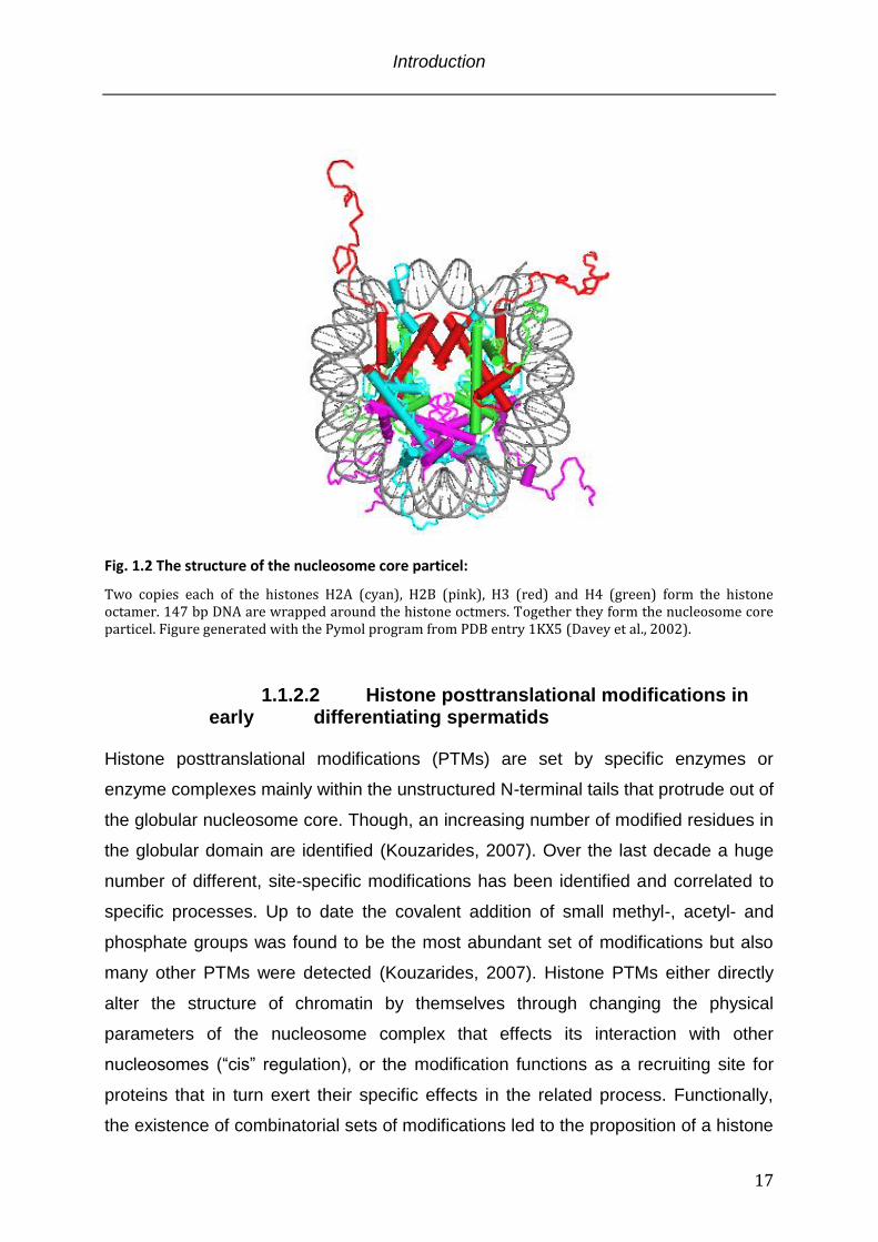
Fig. 1.2 The structure of the nucleosome core particel:
Two copies each of the histones H2A (cyan), H2B (pink), H3 (red) and H4 (green) form the histone octamer. 147 bp DNA are wrapped around the histone octmers. Together they form the nucleosome core particel. Figure generated with the Pymol program from PDB entry 1KX5 (Davey et al., 2002).
1.1.2.2 Histone posttranslational modifications in early differentiating spermatids
Histone posttranslational modifications (PTMs) are set by specific enzymes or
enzyme complexes mainly within the unstructured N-terminal tails that protrude out of
the globular nucleosome core. Though, an increasing number of modified residues in
the globular domain are identified (Kouzarides, 2007). Over the last decade a huge
number of different, site-specific modifications has been identified and correlated to
specific processes. Up to date the covalent addition of small methyl-, acetyl- and
phosphate groups was found to be the most abundant set of modifications but also
many other PTMs were detected (Kouzarides, 2007). Histone PTMs either directly
alter the structure of chromatin by themselves through changing the physical
parameters of the nucleosome complex that effects its interaction with other
nucleosomes (“cis” regulation), or the modification functions as a recruiting site for
proteins that in turn exert their specific effects in the related process. Functionally,
the existence of combinatorial sets of modifications led to the proposition of a histone
Introduction
18
code (Jenuwein and Allis, 2001). In this context a differential read out of one
modification is generated through intra- and intermolecular crosstalk between
modifications.
The early stages of postmeiotic spermatid differentiation are characterized by
extensively modified chromatin. Methylation, phosphorylation, acetylation and
ubiquitinylation were detected. Whereas the importance of methylated histone H3
(K4, K9 and K27) and H4 (R3) or phosphorylated H4 (S1) is unclear, global H4
hyperacetylation (K5 K8, K12, K16) and H2A ubiquitinylation are thought to exert
“cis” effects by opening up the chromatin structure thereby preparing the DNA for
subsequent condensation steps (Braun, 2001; Rathke et al., 2007). Apart from the
proposed physical alteration of the chromatin fiber by acetylation and ubiquitinylation
nothing is known about the contribution of additional modifications and their putative
binding partners in the DNA condensation process.
1.1.3 Postmeiotic spermatid maturation
The terminal stages of sperm development encompass the most intriguing
differentiations steps of eukaryotic cells. These steps are best studied in mice and
Drosophila whereas Drosophila as a spermatogenesis model system only emerged
recently. However, the basic concepts were described being conserved even though
the degree of DNA condensation was found to be different. Generally, the final
differentiation steps that involve DNA condensation and morphological changes
towards needle shaped cells are proposed to fulfill the following three functions: (a)
The strong condensation of the DNA accompanied by the morphological
rearrangements are suggested to create a hydrodynamic favorable shape that
assists sperm motility. (b) The tight condensation of the genome assures protection
from mutagenic damage. (c) The removal of histones resets the genome in respect of
a functional histone modification status. Upon fertilization this allows de novo
deposition of maternal histones.
In Drosophila, postmeiotic spermatid maturation can be divided into different
morphological stages according to the nuclear shape of the cells: round spermatids
directly after meiosis II, young elongating spermatids, early canoe stage spermatids,
late canoe stage spermatids, spermatids during individualization and mature sperm
Introduction
19
cells (Fig. 1.2). The gradual morphological change is accompanied by several
molecular rearrangements that affect amongst others the DNA.
1.1.4 Sperm nuclear proteins involved in chromatin condensation
The switch from the nucleosomal towards a highly condensed DNA organization is
essential to obtain small hydrodynamic sperm heads that are capable to fertilize the
oocyte. The condensation of DNA is thought to be accomplished by two classes of
small very basic, nuclear proteins: transition proteins and protamines. Whereas the
transition proteins constitute short-term histone replacement factors that are not
present in differentiated spermatids, the protamines are the major structural protein in
mature sperm cells (Braun, 2001). However, this model is not valid for all species
and a coherent pattern is missing. In mammals and flies, transition proteins as well
as protamines have been described (Braun, 2001; Sunil Jayaramaiah Raja, 2005).
Fishes and birds lack transition proteins and protamines directly condense the DNA.
Annelids and echinoderms keep the nucleosomal configuration and abandon
spermatid specific genome organizing proteins (Wouters-Tyrou et al., 1998). This
inconsistent picture is further confused by the fact that protamines are dispensable
for fertility and vitality in some organisms whereas in others they are essential
(Rathke et al., 2010). Furthermore, in some species additional proteins are proposed
to contribute to condensation processes. In mice and humans the linker histone H1-
like protein (Hils1) is suggested to participate in chromatin remodeling during mice
and human spermatogenesis.
The functional mechanism of how DNA is condensed by transition proteins and
protamines is unknown. The high content of Cystein residues in protamines was
suggested to cause intermolecular oxidation that in turn leads to condensation. In
vitro, DNA protamine complexes have been reconstituted and analyzed by Atomic
force Microscopy. The imaged structures resembled stacked doughnuts (Allen et al.,
1997; Braun, 2001). However, if these structures are formed in vivo is not known.
Introduction
20
1.1.5 The details of spermatid differentiation
1.1.5.1 The condensation of the DNA As illustrated in Fig. 1.3 the major step in chromatin reorganization takes place in the
canoe stage. First, histone proteins are short term replaced by the transition proteins.
These proteins in turn are substituted by protamines. In this configuration the
Drosophila genome displays a 200-fold stronger condensation state (Fuller, 1993).
The removal of histone proteins is preceded by global hyperacetylation of histone H4
and ubiquitinylation of H2A and is also conserved across species. For histone
acetylation it could be shown that it actually is necessary but not sufficient for
chromatin rearrangements (Awe and Renkawitz-Pohl, 2010). Both modifications are
thought to open up chromatin structure thereby facilitating histone eviction. The exact
mechanism of the eviction process is unclear. However, concomitant with histone
removal numerous DNA breaks are detectable and transition proteins start
associating with the DNA (Rathke et al., 2007). The current model derived from these
data also involves chromatin remodeling complexes that gain access to the DNA
upon ubiquitinylation and acetylation. The DNA breaks are suggested to enhance the
accessibility so that the remodelers can evict the histone proteins that in turn are
replaced by transition proteins as intermediate genome organizers. Apart from the
implications of acetylation and ubiquitinylation also other histone posttranslational
modifications (phosphorylation, methylation) as well as high levels of SUMO and
CTCF proteins are detectable. Their contribution to chromatin restructuring is
unknown.
Postmeiotic spermatid differentiation is an only little understood process. The
functional integration of cellular events like histone modifications, histone eviction,
DNA breaks, transition protein- and protamine deposition is to date hardly possible.
The signaling pathways involved in these processes as well as the structural
organization of DNA intermediate- as well as highly compacted states (in vivo) are
unknown.
Introduction
21
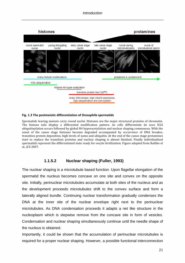
Fig. 1.3 The postmeiotic differentiation of Drosophila spermatids
Spermatids leaving meiosis carry round nuclei. Histones are the major structural proteins of chromatin. The histone tails display a differential modification pattern. As cells differentiate de novo H2A ubiquitinylation occurs followed by global H4 hyperacetylation and nuclear shaping commences. With the onset of the canoe stage histones become degraded accompanied by occurrence of DNA breakes, transition protein deposition, high levels of sumo and ubiquitin. At the end of the canoe stage protamines start to replace the transition proteins and nuclear shaping is almost finished. Finally individualized spermatids represent the differentiated state ready for oocyte fertilization. Figure adapted from Rathke et al., JCS 2007.
1.1.5.2 Nuclear shaping (Fuller, 1993) The nuclear shaping is a microtubule based function. Upon flagellar elongation of the
spermatid the nucleus becomes concave on one site and convex on the opposite
site. Initially, perinuclear microtubules accumulate at both sites of the nucleus and as
the development proceeds microtubules shift to the convex surface and form a
laterally aligned bundle. Continuing nuclear transformation gradually condenses the
DNA at the inner site of the nuclear envelope right next to the perinuclear
microtubules. As DNA condensation proceeds it adapts a net like structure in the
nucleoplasm which is stepwise remove from the concave site in form of vesicles.
Condensation and nuclear shaping simultaneously continue until the needle shape of
the nucleus is obtained.
Importantly, it could be shown that the accumulation of perinuclear microtubules is
required for a proper nuclear shaping. However, a possible functional interconnection
Introduction
22
of nuclear shaping and DNA condensation is elusive.
1.1.6 Mst77F - a suggested chromosomal architectural protein The Mst77F gene was discovered by Russel and Kaiser twenty years ago (Russell
and Kaiser, 1993). The Mst77F protein is a small, very basic protein that is
exclusively expressed in spermatids. Sequence alignments classify the protein as a
distant relative of H1/H5 linker histones, major structural proteins of chromatin.
Accordingly functions in chromatin compaction were proposed. Expression studies
showed that Mst77F mRNA is expressed already at the primary spermatocyte stage,
translationally inhibited and stored until translation in young elongating spermatids
(Fig. 1.2) (Sunil Jayaramaiah Raja, 2005). At this time histone proteins are still the
major genome organizers and Mst77F displays a clear histone colocalization
suggesting an association with chromatin (Rathke et al., 2010). The observations that
Mst77F stays associated with the DNA in differentiated, mature sperm cells and that
Drosophila mutants in a protamine-/- background condense chromatin normally and
are fertile shed light on Mst77F as a protein of redundant protamine function involved
in DNA condensation (Rathke et al., 2010). However, in contrast to protamines the
nuclear Mst77F distribution in mature sperm cells is not homogeneous. Additionally,
protamine-/- flies are more susceptible to genome damage suggesting at least in part
diverging functions of Mst77F and protamines. Additionally, along with the suggested
contribution to DNA condensation a second Mst77F function was proposed. This
function was implicated by the discovery of a mutant Mst77F allele termed ms(3)nc3
that was identified in a non-complementation screen of 2-tubulin mutants. This
Mst77F mutant is characterized by a single amino acid substitution of serine 149
toward threonine. As already described nuclear shaping is a tubulin dependent
process and in the Mst77F ms(3)nc3 background the cells fail to undergo nuclear
elongation and display a roundish phenotype (Rathke et al., 2010). Nevertheless, the
mutant displays a wild-type like spaciotemporal expression pattern. Proper DNA
condensation is observed and Mst77F ms(3)nc3 is also associated with the tightly
compacted DNA in differentiated spermatids. A direct link between Mst77F and
tubulin was suggested by the observation that in young elongating nuclei Mst77F
specifically localizes to the convex site of the nucleus in a parallel alignment with the
Introduction
23
perinuclear microtubule bundle. This colocalization along with nuclear shaping
defects in the background of the Mst77F ms(3)nc3 mutant led to the hypothesis that
Mst77F also is a factor involved in morphological shaping of differentiating
spermatids (Rathke et al., 2010). A stabilizing function on 2-tubulin, the major tubulin
isoform in testis, was suggested. This hypothesis was consolidated by reduced 2-
tubulin levels in Mst77F ms(3)nc3 background (Rathke et al., 2010). Alternatively, a
coordinating function in positioning 2-tubulin and chromatin in close vicinity prior to
nuclear elongation was proposed. However, no evidence for a simultaneous
interaction of Mst77F with chromatin and tubulin could be presented.
In Drosophila as well as in mice our current knowledge of the molecular
mechanisms that drive spermatogenesis is very limited. Especially the details of
histone eviction and the genome condensation by transition proteins and finally
protamines are not described at all. The proposed dual functionality of Mst77F in
DNA condensation and morphogenesis during spermatid maturation impede the
proposition of Mst77F being a classical structural component in this system.
However, Mst77F associates with DNA shortly before condensation processes
commence. Along with protamines Mst77F is a component of differentiated sperm
cells. A strong argument for Mst77F being a DNA condensing protein in Drosophila
spermatogenesis is the fact that protamine-/- flies show normal DNA condensation
and Mst77F to date is the only known protein associated with this DNA condensation
state. The distant Mst77F relationship towards H1/H5 linker histone family proteins
that are implicated in somatic and spermatid specific condensation processes, makes
a related Mst77F function in spermatogenesis conceivable.
1.2 Linker histone H1
1.2.1 H1 localisation and structural function Apart from the canonical core histones and their variants a fifth different kind of
histone, linker histone H1 is associated with the nucleosome. H1 exhibits much less
conservation across species and in contrast to the core histones possesses no
histone-fold domain. It exhibits a short N- and a long C-terminal basic domain
discontinued by a globular domain. The globular domain binds to the nucleosome
Introduction
24
where the DNA enters/leaves the histone octamer. Additional contact is made by the
C-terminal domain that interacts with the linker DNA in a range of approximately 20
bp aside the nucleosome core particle. Because of its “exterior” association with the
nucleosome it shows much higher mobility in comparison to the core histones (Catez
et al., 2004; Phair et al., 2004).
In mammals eleven H1 subtypes have been identified including specialized isoforms
in testis and ovaries (Happel and Doenecke, 2009). Besides the knowledge about
their existence very little is known about functional differences on the molecular level.
In yeast, C.elegans and some other model organsisms they have been proven to be
nonessential, but defects in development and life span were observed (Jedrusik and
Schulze, 2001; Patterton et al., 1998). However, recent work in higher eukaryots
such as mice and Drosophila indicate their necessity for viability (Fan et al., 2005; Lu
et al., 2009b).
The exact mechanism how H1 proteins exert their functions is not resolved.
However, much data suggest its involvement in regulation of chromatin and
chromosome structure. In Tetrahymena thermophila deletion of the somatic H1 gene
resulted in increased nuclear volume, suggesting chromatin condensation defects
(Shen et al., 1995). Likewise, similar effects were observed in embryonic stem cells
derived from H1 knockout mice (Fan et al., 2005). Moreover, H1 was also reported to
be required for metaphase chromosome structure in X.leavis. Depletion led to
aberrant morphology and segregation defects (Maresca et al., 2005).
However, our today’s understanding of H1 related chromatin condensation processes
is limited. Recent work set out to investigate the molecular details which enable H1
proteins to achieve their proposed functions that are mainly attributed to the C-
terminal domain.
1.2.2 H1 structure and function - the globular domain The structure of the globular domains of linker histones H1 and isoform H5 have
been resolved two decades ago by x-ray crystallography and NMR studies (Cerf et
al., 1993) (Cerf et al., 1993). These domains consist of a three-helix fold for which the
term “winged-helix” has been coined. This fold adapts the classical helix-turn-helix
motif. In comparison to the N- and C-terminal domains of H1 the “winged-helix” is
Introduction
25
well conserved across species and necessary to generate chromatosome stops in
nuclease digestion experiments. Additionally, it could be shown that the isolated
globular domain is sufficient for nucleosome binding (Allan et al., 1980). The exact
interaction position of the globular domain on the nucleosome is controversial.
However, generally accepted is its contact with two strands of DNA at the
nucleosome where the DNA enters/exits. This position is implicated to stabilize the
wrapping of the DNA around the histone octamer (Brown et al., 2006; Crane-
Robinson, 1997; Fan and Roberts, 2006; Syed et al., 2010).
1.2.3 H1 structure and function - the N-terminal domain Depending on the isoform the N-terminal domain is 20 to 35 amino acids in length.
Sequence analysis revealed a bipartite amino acid distribution with a high content of
basic amino acids proximal the “winged-helix” globular domain (Bohm and Mitchell,
1985). This part of the N-terminal domain was suggested to contribute to binding
stability of H1 (Allan et al., 1986; Vila et al., 2001). In aqueous solutions the N-
terminal domain shows an unstructured coiled coil conformation that under stabilizing
conditions adapts a -helical conformation. In light of the emerging field of
intrinsically unstructured proteins a folding upon interaction with DNA was proposed
(Vila et al., 2001; Vila et al., 2002).
1.2.4 H1 structure and function - the C-terminal domain The C-terminal domain (CTD) is approximately 100 amino acids in length and
displays sequence variation between isoforms and across species (Ponte et al.,
2003). A striking feature is the high content of basic residues that turn the C-terminal
domain into a highly charged moiety with 30 to 50 evenly distributed net positive
charges (Subirana, 1990). The sequence content of the CTD resembles that of
intrinsically unstructured proteins (Hansen et al., 2006; Lu et al., 2009a).
1.2.4.1 Intrinsically unstructured proteins (IUPs) “Structure determines function” is the classical definition of functional proteins. In the
Introduction
26
recent years more and more proteins were discovered that deviate from this classical
definition. Intrinsic unstructured proteins - that is the hindrance of spontaneous
folding into well-organized structures in the absence of stabilizing interactions -
emerge in the field of protein science. Even though these characteristics were put
forward recently, they were originally discovered more than 25 years ago (Mitchell
and Tjian, 1989; O'Hare and Williams, 1992). In contrast to the prediction of 3D
structures of globular proteins that to date remains a key challenge, the identification
of sequences that adapt unstructured characteristics is straightforward. The
fingerprint of unstructured domains is their low sequence complexity. This often
involves repetitive or periodic elements of a limited set of amino acids and a
compositional
Tabl. 1.1 Functional implications of intrinsically unstructured domains
Listed are the different function of intrinsically unstructured domains and the number of proteins found for each function. A common function of intrinsically unstructured domains is their involvement in protein protein interactions. However, multiple examples were also found for their role in DNA binding events. Posttranslational modifications in intrinsically unstructured domains might reflect regulatory mechanisms that modulate the function. Table modified from Duner et al., Biochemistry 2002.
Introduction
27
compositional bias towards a low content of bulky hydrophobic amino acids and a
high proportion of polar/charged amino acids (Obradovic et al., 2003; Romero et al.,
2001).
The molecular function of IUPs is versatile (Tabl. 1.1). The region of disorder can
manifest in flexible linkers that connect globular domains within one polypeptide
chain. This allows for rotational and conformational flexibility in search for binding
partners and additionally allows proteins that bind the linker to induced interdomain
conformational changes. Another important aspect of intrinsically unstructured
domains is the coupled binding and folding mechanism upon recognition of
interaction partners. The folding occurs locally but also can concern entire domains
(Gunasekaran et al., 2003). Moreover, intrinsically unstructured domains are regions
that are targeted by regulatory mechanisms. Binding of a small molecule or a
posttranslational modification alters the inducible structures and this correlates with
functional diversity (Sandhu and Dash, 2007).
The growing field of intrinsic unstructured proteins illustrates the surprisingly manifold
aspect of protein structure and function that is not appreciated by protein
crystallography approaches. Even though only a fractional amount of intrinsically
unstructured proteins has been discovered, their vital role across cellular functions
already becomes clear.
1.2.4.2 Functional implication of the intrinsically unstructured histone H1 CTD
Recently, spectroscopic experiments could show that in the presence of secondary
structure stabilizing reagents the H1 CTD acquires a substantial -helical
conformation (Verdaguer et al., 1993). Moreover, the addition of DNA under
physiological conditions also induces structures in the CTD (Roque et al., 2005).
Further experiments identified two regions within the long H1 CTD that are primarily
responsible for alteration of the linker DNA structure and chromatin condensation (Lu
and Hansen, 2004). It could be shown that these subdomain adapt -helical
structures upon molecular recognition of DNA (Lu et al., 2009a). Furthermore,
posttranslational modifications in the H1 C-terminal domain are proposed to “switch”
between distinct H1 functions (Happel and Doenecke, 2009). Roque and coworkers
Introduction
28
could show in 2008 that cyclin-dependent kinase-mediated phosphorylation of
specific sites in the C-terminal domain cause a decrease in the proportion of α-helix
and an increase in the β-sheet content (Roque et al., 2008). This effect depended on
the amount of introduced phosphate groups and interestingly the fully phosphorylated
CTD had a higher chromatin aggregation potential.
1.4 Objectives of the thesis DNA condensation and structural reorganization is a conserved mechanism in the
process of spermatogenesis. The exact molecular mechanisms and determinants
underlying this process, such as histone displacement, degradation, incorporation of
transition proteins and protamines together with the role of linker histone like proteins
are poorly understood.
In Drosophila the Mst77F protein is put forward as an additional DNA condensing
protein. The cellular spaciotemporal expression pattern has been described whereas
the molecular function is unknown. In this work I set out to characterize Mst77F on a
molecular level by biochemical and biophysical methods. I wanted to answer the
question if Mst77F is a protein involved in DNA condensation during Drosphila
spermatogenesis and by what mechanism it conducts its function. To obtain this goal
the following question were addressed:
1. Mst77F colocalizes with DNA in vivo but does it directly interact with DNA?
2. Which part of the protein binds DNA?
3. What is the interaction mode? Is it similar to linker histone H1 proteins, the
proposed homologs?
4. Does Mst77F introduce structural changes in DNA?
5. How does Mst77F alter DNA structure?
6. Mst77F colocalizes with DNA when histones are still the structural organizers.
In this respect, is Mst77F sufficient to evict histones from the DNA?
7. How inert is the DNA Mst77F complex? Does Mst77F serve as a general
transcriptional inhibitor?

Introduction
29
Material and Methods
30
2 Material and Methods
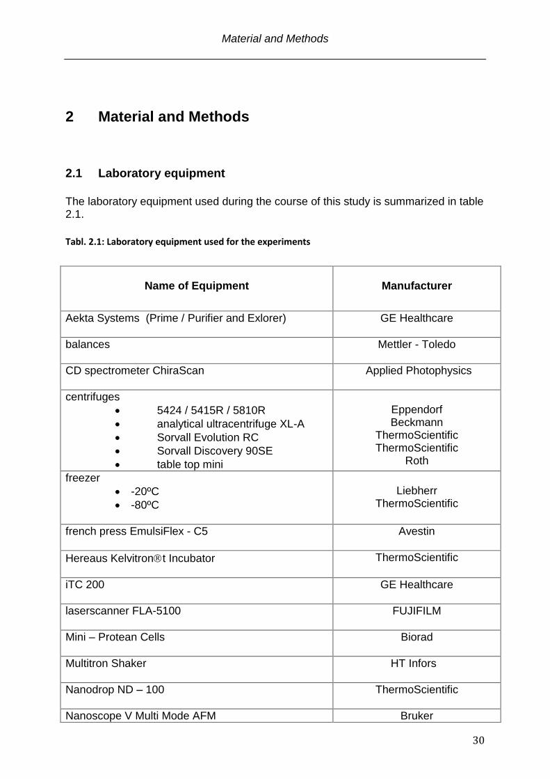
2.1 Laboratory equipment
The laboratory equipment used during the course of this study is summarized in table 2.1.
Tabl. 2.1: Laboratory equipment used for the experiments
Name of Equipment
Manufacturer
Aekta Systems (Prime / Purifier and Exlorer)
GE Healthcare
balances
Mettler - Toledo
CD spectrometer ChiraScan
Applied Photophysics
centrifuges
5424 / 5415R / 5810R
analytical ultracentrifuge XL-A
Sorvall Evolution RC
Sorvall Discovery 90SE
table top mini
Eppendorf Beckmann
ThermoScientific ThermoScientific
Roth
freezer
-20ºC
-80ºC
Liebherr
ThermoScientific
french press EmulsiFlex - C5
Avestin
Hereaus Kelvitront Incubator
ThermoScientific
iTC 200
GE Healthcare
laserscanner FLA-5100
FUJIFILM
Mini – Protean Cells
Biorad
Multitron Shaker
HT Infors
Nanodrop ND – 100
ThermoScientific
Nanoscope V Multi Mode AFM Bruker
Material and Methods
31
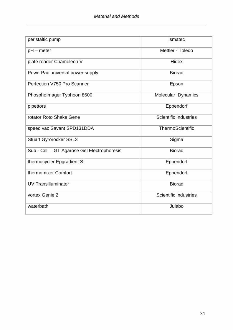
peristaltic pump
Ismatec
pH – meter
Mettler - Toledo
plate reader Chameleon V
Hidex
PowerPac universal power supply
Biorad
Perfection V750 Pro Scanner
Epson
PhosphoImager Typhoon 8600
Molecular Dynamics
pipettors
Eppendorf
rotator Roto Shake Gene
Scientific Industries
speed vac Savant SPD131DDA
ThermoScientific
Stuart Gyrorocker SSL3
Sigma
Sub - Cell – GT Agarose Gel Electrophoresis
Biorad
thermocycler Epgradient S
Eppendorf
thermomixer Comfort
Eppendorf
UV Transilluminator
Biorad
vortex Genie 2
Scientific industries
waterbath
Julabo
Material and Methods
32
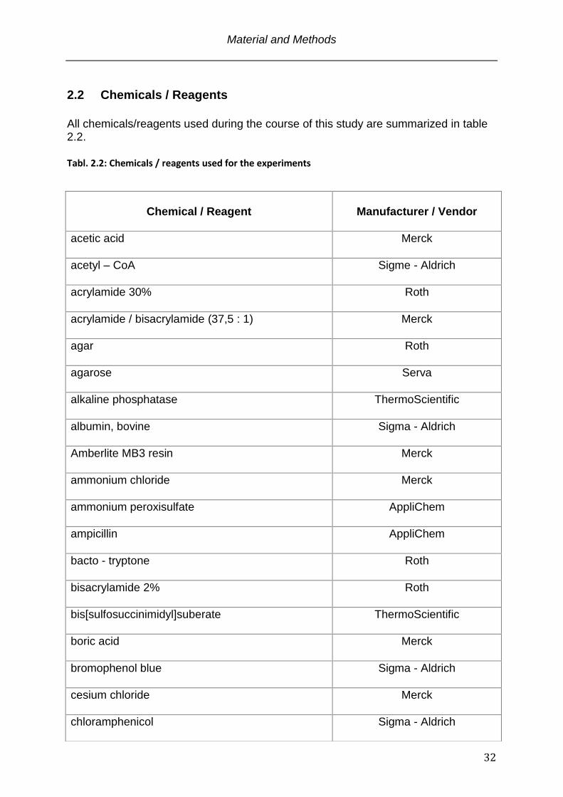
2.2 Chemicals / Reagents
All chemicals/reagents used during the course of this study are summarized in table 2.2.
Tabl. 2.2: Chemicals / reagents used for the experiments
Chemical / Reagent
Manufacturer / Vendor
acetic acid
Merck
acetyl – CoA
Sigme - Aldrich
acrylamide 30%
Roth
acrylamide / bisacrylamide (37,5 : 1)
Merck
agar
Roth
agarose
Serva
alkaline phosphatase
ThermoScientific
albumin, bovine
Sigma - Aldrich
Amberlite MB3 resin
Merck
ammonium chloride
Merck
ammonium peroxisulfate
AppliChem
ampicillin
AppliChem
bacto - tryptone
Roth
bisacrylamide 2%
Roth
bis[sulfosuccinimidyl]suberate
ThermoScientific
boric acid
Merck
bromophenol blue
Sigma - Aldrich
cesium chloride
Merck
chloramphenicol
Sigma - Aldrich
Material and Methods
33
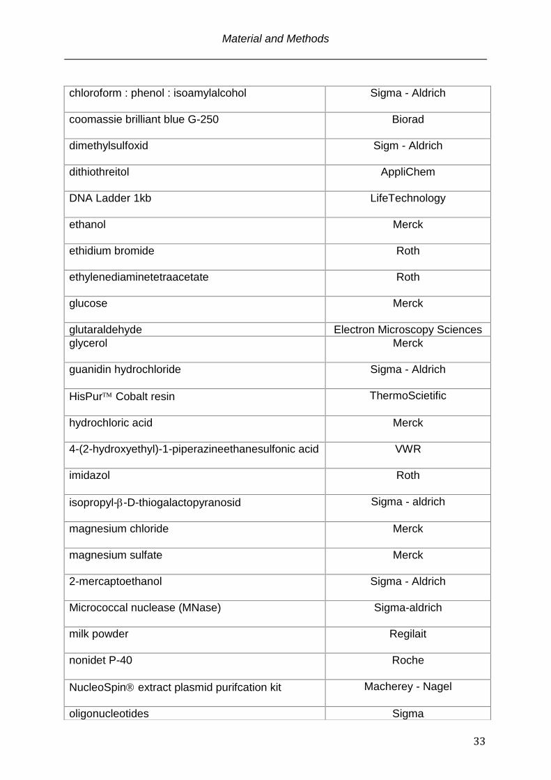
chloroform : phenol : isoamylalcohol
Sigma - Aldrich
coomassie brilliant blue G-250
Biorad
dimethylsulfoxid
Sigm - Aldrich
dithiothreitol
AppliChem
DNA Ladder 1kb
LifeTechnology
ethanol
Merck
ethidium bromide
Roth
ethylenediaminetetraacetate
Roth
glucose
Merck
glutaraldehyde Electron Microscopy Sciences
glycerol
Merck
guanidin hydrochloride
Sigma - Aldrich
HisPur Cobalt resin
ThermoScietific
hydrochloric acid
Merck
4-(2-hydroxyethyl)-1-piperazineethanesulfonic acid
VWR
imidazol
Roth
isopropyl--D-thiogalactopyranosid
Sigma - aldrich
magnesium chloride
Merck
magnesium sulfate
Merck
2-mercaptoethanol
Sigma - Aldrich
Micrococcal nuclease (MNase) Sigma-aldrich
milk powder
Regilait
nonidet P-40
Roche
NucleoSpin extract plasmid purifcation kit
Macherey - Nagel
oligonucleotides Sigma

Material and Methods
34
peptone
Roth
Pfu DNA Polymerase (native)
ThermoScientific
phenylmethylsulfonylfluorid
Serva
polyethylenglycol 6000
Merck
potassium chloride
Merck
protein ladder Seablueplus 2
LifeTechnology
QiaEX II gel extraction kit
Qiagen
radionucleotides
Perkin - Elmar
reaction tubes 1.5 ml low binding properties
Nerbe
restriction endonucleases
New England Biolabs / ThermoScientific
RNase T1
Roche
sodium acetate
Merck
sodium chloride
Merck
sodium dihydrogen phosphate
Merck
sodium dodecyl sulfate
Roth
sodium fluoride
Merck
sodium hydroxide
Merck
sodium monohydrogen phosphate
Merck
streptavidin coated paramagnetic particels
Promega
streptavidin coated 96 well plates
ThermoScientific
T4 DNA Ligase
ThermoScientific
tetramethylethylendiamine
Sigma - Aldrich
triethanolamine
VWR
tris(hydroxymethyl)aminoethane Roth
Material and Methods
35
2.3 Bioinformatic tools Protein structural domain prediction was carried out using the Simple Modular
Architecture Reasearch Tool (SMART) algorithm (http://smart.embl-
heidelberg.de/)(Schultz et al., 1998).
The relative protein disorder was calculated by the Intrinsic Unstructured Protein
relative disorder (IUPred) algorithm (http://iupred.enzim.hu/) on the basis of the
primary structure by estimation of the pairwise energy content between amino acids.
The algorithm predicts the structural properties of a given protein by identifying the
number of favorable interactions all amino acids can form (Dosztanyi et al., 2005a,
b).
2.4 Preparation of SDS-PAGE Gels and Electrophoresis
SDS-PAGE was performed using standard protocols (Gallagher, 2006). The
resolving gel contained 0.4 M TrisHCl pH = 8.8, 0.1% (w/v) SDS, 15% acrylamide-
bisacrylamide (37.5:1), 0.1% APS, 0.04% TEMED and the stacking gel was
composed of 0.68 M TrisHCl pH = 6.8, 0.1% (w/v) SDS, 5% acrylamide-
bisacrylamide (37.5:1), 0.1% APS, 0.1% TEMED. Casting and running of the gels
was performed with the Mini-Protean System. The sample loading buffer contained
62.5 mM TrisHCl pH = 6.8, 8.5% (v/v) glycerol, 2 % (w/v) SDS, 4 mM DTT and 0.05%
bromophenol blue. The protein samples were boiled in loading buffer for 5 min at 95
ºC. The gel running buffer contained 25 mM Tris, 200 mM glycine and 0.1 % (w/v)
SDS. Electrophoresis was performed at a constant current of 25 mA until the
bromophenol blue migrated out of the gel. SeeBlue Plus 2 marker was used as a size
triton X-100
Merck
urea
Merck
well plates 96 format (transparent)
Greiner
well plates 384 format (black)
Corning
yeast extract
Mobio
Material and Methods
36
standard.
2.5 Protein staining within SDS Gels
For protein visualization the SDS-PAGE gels were soaked with coomassie G-250
protein staining solution (Meyer, 1965) at RT for 15 min and constant agitation (0.05
% Coomassie R-250, 10% (v/v) acetic acid, 50% (v/v) methanol). The staining
solution was replaced by destaining solution (10 % (v/v) acetic acid, 7.5 % (v/v)
methanol) until the desired contrast was reached.
2.6 Determination of Nucleic Acid and Protein Concentrations
Nucleic acid concentrations were determined with the NanoDrop Spectrophotometer
(ThermoScientific) using the DNA/RNA program implemented in the NanoDrop
software. In order to determine protein concentrations the OD of the samples was
measured with the UV-Vis program of the NanoDrop software. Extinction coefficients
and theoretical molecular weights of proteins were calculated on the basis of the
primary proteins sequences using the ProtParam online tool (Gasteiger E., 2005).
Protein concentrations were calculated using the Lambert - Beer equation:
C A
* d
with c being the concentration, A the measured absorbence, the molar extinction
coefficient and d the pathlenght of the cuvette. Table 2.3 summarizes the molecular
weights and the extinction coefficients of the proteins analyzed.
Tabl. 2.3: Molecular weights and extinction coefficients of the proteins analyzed
Protein
Extinction coefficient ()
MW (Da)
Mst77F WT 17420 26421
Mst77F S149T 17420 26435
Mst 77F shuffled C 17420 26421
Material and Methods
37
Mst77F 20C 17420 24222
Mst77F 40C 17420 21973
Mst77F 60C 17420 19748
Mst77F 110C 17420 14338
Mst77F 100N 2980 16589
hH1.4 2980 22529
xPr-Set7 27850 42246
xH2A 4050 13960
xH2B 6070 13774
xH3 4040 15273
xH4 5040 11236
2.7 Molecular Cloning
Tabl. 2.4: Bacterial cells that were used throughout this study
Strain
Genotyp
Application
Vendor
DH5α E. coli F- φ80lacZΔM15
Δ(lacZYAargF)U169 deoR
recA1 endA1 hsdR17 (rk-,
mk+) phoA supE44 λ- thi-1
gyrA96 relA1
molecular cloning
DNA production
LifeTechnologies
BL21-Codon
Plus (DE3)-
RIL
E. coli B F– ompT
hsdS(rB– mB–)
dcm+ Tetr gal endA Hte
[argU ileY
leuW Camr
protein expression Agilent
Material and Methods
38
Tabl. 2.5: Formulations of the bacterial growth media used
Type
Formulation
Application
LB 1% w/v bacto-tryptone, 0.5% w/v yeast extract,
1% w/v NaCl
cloning, DNA production
2xYT 1.6% w/v bacto-tryptone,
1% w/v yeast extract,
0.5% w/v NaCl
protein expression
SOC 2% w/v bacto-tryptone, 0.5% w/v yeast extract,
10 mM NaCl
2.5 mM KCl
10 mM MgCl2
10 mM MgSO4
cloning
2.7.1 Bacterial transformation Transformation of plamids (Griffith, 1928) was carried out with chemically competent
E.coli cells throughout this work. The bacterial cells (table 2.4), were prepared
according to Sambrook and Russell (Sambrook, 2001). The cells were thawed on ice
and 10 ng DNA or 3 µL of a ligation reaction of were added. The DNA was allowed to
attach to the bacterial membrane for 30 min on ice and transformation was induced
by heat-shock for 30 s at 42ºC in a water bath. After 2 min incubation on ice, 250 µL
of SOC medium were added. The bacteria were incubated in a thermomixer at 37ºC
for 1 h and shaking at 1000 rpm to induce resistance. The cells were plated on agar-
plates containing the respective antibiotic. Incubation of the plates was carried out
over night at 37ºC.
2.7.2 PCR based DNA amplification
Material and Methods
39
Standard polymerase chain reactions (Mullis et al., 1986) were carried out in a 50 µL
reaction volume with 0.2 µM of forward and reverse primer, 0.2 µM dNTP’s,
1 U Pfu native DNA Polymerase, 1x Pfu buffer and 10 – 100 ng template DNA. PCR
reactions were performed in an Eppendorf MasterCycler Epgradient S using the
protocol in table 2.6.
Tabl. 2.6: Typical PCR program used for Pfu – Polymerase (native)
Step Temperature (C) Time (s) cycles
1 95 180 1
2 95 30
35 3 50-65 30
4 72 120/kb
5 72 120/kb 1
6 10 -
2.7.3 Purification of PCR products The PCR products were purified using the NucleoSpin PCR and gel extraction kit
according to the manufacturers protocol.
2.7.4 Restriction digest of DNA PCR derived DNAs and plasmid DNAs were digested using New England Biolabs
Restriction Endonucleases (Hartl, 2001) according to the manufacturers
recommendations. In brief, 1 ug of Plasmid DNA was digested with 5 Weiss Units
each of the appropriate set of Restriction Endonucleases for 1 h at their respective
temperature. PCR products were digested according to the plasmid digestion
parameters but over night. The restriction reaction was terminated by thermal
inactivation in all possible cases.
2.7.5 Dephosphorylation of Plasmid DNA
Material and Methods
40
Restriction endonuclease treated plasmid DNA was dephosphorylated with Fast
Alkaline Phosphatase in the respective restriction buffer for 15 min at 37C followed
by thermal inactivation at 75C for 5 min.
2.7.6 Purification of DNAs For molecular cloning reactions DNAs subjected to restriction endonucleases were
purified by agarose gel electrophoresis and subsequent extration from the gel with
the QiaEX II gel extraction kit according to the manufacturers recommendations.
2.7.7 Agarose electrophoresis & ethidium bromid visualization of DNA
Typically, 0.5 % to 2 % (w/v) agarose was dissolved by boiling in TBE (90 mM
TrisHCl pH = 8.0, 90 mM Sodiumborate, 2 mM EDTA) and gels were prepared using
a Sub-Cell GT electrophoresis system. Prior to loading onto the gel the DNA samples
were mixed with 10x loading dye (TBE with 30 % (v/v) glycerol and 0.05% (w/v)
bromphenolblue). Electrophoresis was carried out at a constant voltage of 80 V in 1x
TBE buffer. As size standard served the 1kb Plus DNA ladder. The DNA within the
gel was stained with ethidiumbromide solution (0.5 µg / ml ethidium bromide in 1X
TBE - buffer) for 30 min followed by visualization on a gel - doc system in the
Department of Cellular Biochemistry at the Max – Planck Institute for Biophysical
Chemistry, Goettingen, Germany.
For native agarose gel electrophoresis 0.5% to 1.5% agarose gels (0.5% - 1.5% w/v
agarose, 18 mM Tris, 18 mM boric acid, pH 8.0) were run in 0.2X TB buffer (18 mM
Tris, 18 mM boric acid, pH 8.0) at a constant voltage of 80 V for 2 hrs at 4°C.
Staining and visualization was carried out as discribed above.
2.7.8 Ligation Ligation of PCR products with the destination vector was carried out using 30 ng of
plasmid vector and a three fold molar excess of the insert DNA in a 20 µL reaction
volume. 1 Unit of T4 DNA Ligase catalyzed the reaction for 1 h at 22C. The ligation
reaction was transformation into E.coli DH5α according to paragraph 2.7.1.
Material and Methods
41
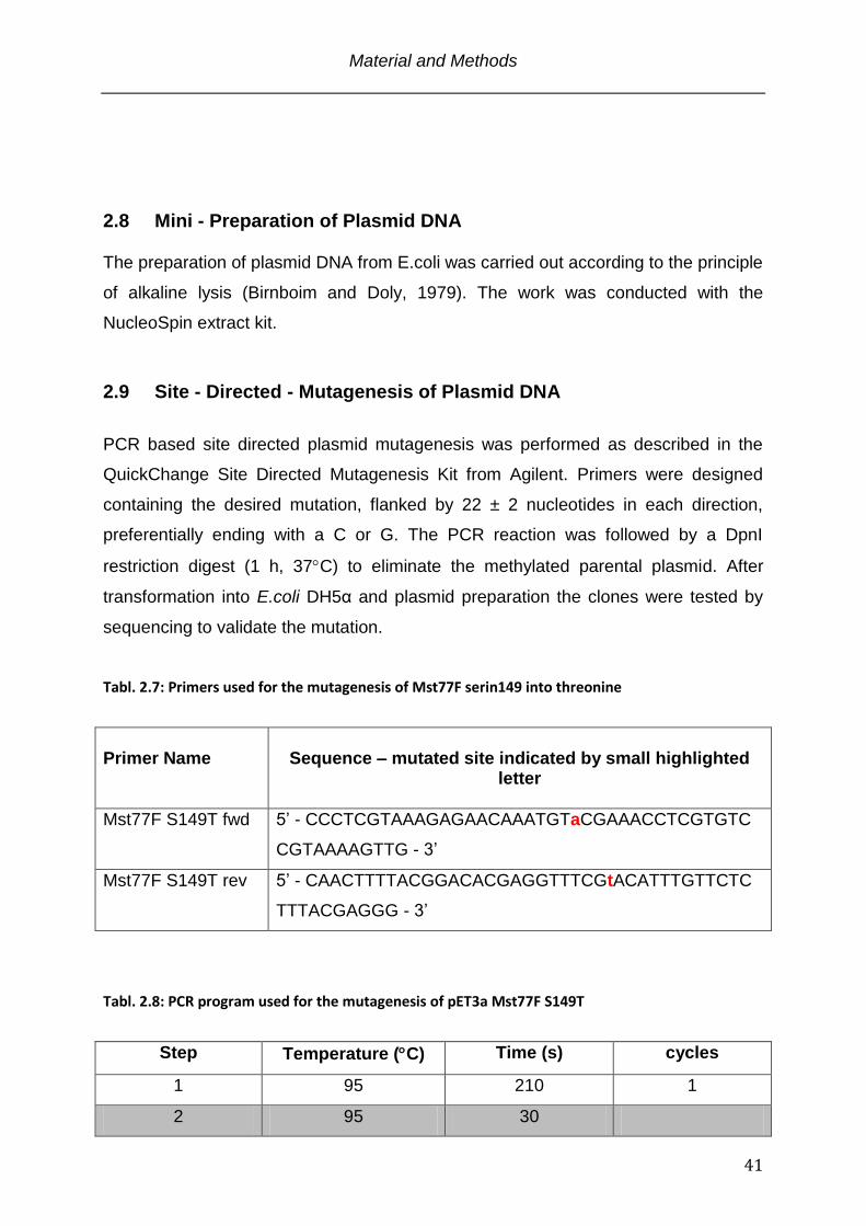
2.8 Mini - Preparation of Plasmid DNA The preparation of plasmid DNA from E.coli was carried out according to the principle
of alkaline lysis (Birnboim and Doly, 1979). The work was conducted with the
NucleoSpin extract kit.
2.9 Site - Directed - Mutagenesis of Plasmid DNA
PCR based site directed plasmid mutagenesis was performed as described in the
QuickChange Site Directed Mutagenesis Kit from Agilent. Primers were designed
containing the desired mutation, flanked by 22 ± 2 nucleotides in each direction,
preferentially ending with a C or G. The PCR reaction was followed by a DpnI
restriction digest (1 h, 37C) to eliminate the methylated parental plasmid. After
transformation into E.coli DH5α and plasmid preparation the clones were tested by
sequencing to validate the mutation.
Tabl. 2.7: Primers used for the mutagenesis of Mst77F serin149 into threonine
Primer Name
Sequence – mutated site indicated by small highlighted
letter
Mst77F S149T fwd 5’ - CCCTCGTAAAGAGAACAAATGTaCGAAACCTCGTGTC
CGTAAAAGTTG - 3’
Mst77F S149T rev 5’ - CAACTTTTACGGACACGAGGTTTCGtACATTTGTTCTC
TTTACGAGGG - 3’
Tabl. 2.8: PCR program used for the mutagenesis of pET3a Mst77F S149T
Step Temperature (C) Time (s) cycles
1 95 210 1
2 95 30
Material and Methods
42
3 65 30 18
4 72 720
5 72 720 1
6 10 -
2.10 Expression and Purification of Recombinant Proteins
2.10.1 Bacterial Expression
A single colony of E.coli BL21 (DE3) RIL cells (table 2.4) transformed with the
plasmid carrying the protein encoding sequence was used to inocculate a 50 mL pre-
expression culture of LB medium containing the respective antibiotic. The pre-culture
was grown over night at 37C and 130 rpm. The next morning the pre-culture was
diluted 1:100 in prewarmed 2xYT medium (table 2.5) and grown to an OD of 0.6 at
37C and 130 rpm. Protein expression was induced by the addition of IPTG to a final
concentration of 0.3 mM. The protein production was allowed to go on for 1.5 h
followed by centrifugation of the E.coli cells in a Sorvall Evolution centrifuge using a
SLC-1500 rotor (10 min, 6000 x g). Cells were subsequently either directly lysed for
protein purification or shock frozen with liquid nitrogen and stored at -80C until the
proteins were to be purified.
2.10.2 HIS-tag protein purification Bacterial pellets from E.coli BL21 (DE3) RIL cultures (see previous paragraph) were
thoroughly resuspended in HIS - lysis/wash buffer (20 mM Hepes pH = 7.4, 1 M
NaCl, 40 mM Imidazole, 1 mM -Mercaptoethanol, 1 mM PMSF) and lysed by three
passages through an EmulsiFlex - C5 cell disrupter (Avestin) at 4°C using a pressure
setting of 100 - 150 bar. Insoluble material was removed by centrifugation for 25 min
at 20000 x g. The supernatant was either loaded onto a 1 mL Ni-NTA column using
the Äkta FPLC system or purified on a 1 mL Cobalt resin by gravity flow. In either
case the column material was washed with at least 100mL of HIS - lysis/wash buffer.
The bound proteins were eluted with HIS – elution buffer (20 mM Hepes (NaOH) pH
= 7.4, 1 M NaCl, 250 mM Imidazole,
1 mM -Mercaptoethanol, 1 mM PMSF) in 15 mL. Directly after elution all proteins
Material and Methods
43
were extensivly dialysed against storage buffer (50 mM Imidazol, 300 mM NaCl,
5 mM -Mercaptoethanol, 10% Glycerol (v/v), pH = 6.4 adjusted with 3 M NaAc pH =
5.2 (final NaAc 40 mM)), concentrated to 20 – 100 µM, aliquoted, snap frozen in
liquid nitrogen and finally stored at -80°C until usage.
2.10.3 Histone Inclusion Body Purification The purification of histone proteins under denaturing conditions was largely
performed as documented (Luger et al., 1999), with some modifications that were
introduced in the Tsukiyama Lab by Marnie E. Gelbart. Plasmids encoding the X.
laevis histone proteins H2A (GeneBank: CAD89676), H2B (GeneBank: CAD89678),
H3 (GeneBank: CAD89679), and H4 (GeneBank: CAD89677) were obtained from
Karolin Luger, Colorado State University, Dept. of Biochemistry and Molecular
Biology. 2 L of E.coli BL21 (DE3) RIL Bacteria were grown as described in paragraph
2.10.1 with a protein expression period of 4 h and lysed in Wash Buffer (50 mM
TrisCl pH = 7.5, 100 mM NaCl, 1 mM EDTA, 2 mM DTT, 1 mM DTT) by three
passages through an EmulsiFlex - C5 cell disrupter at 4°C using a pressure setting of
100 – 150 bar. Inclusion bodies were pelleted by centrifugation for 20 min at a speed
of 20000 x g. The pellet was washed two times with TW - Buffer (Wash Buffer with 1
% (v/v) Triton X-100) and subsequently two times with Wash Buffer. To solubilize the
histones from the inclusion body pellets, 350 µL DMSO was added to the pellet. The
sample was stirred thoroughly with a spatula and incubated for 30 min at RT. Then,
13.3 mL unfolding buffer (7 M guanidine - HCl, 20 mM TrisHCl pH = 7.5, 10 mM
DTT) were added, the sample was mixed and the suspension was rotated for 1 h at
RT. Insoluble material was removed by centrifugation for 15 min at 25000 x g and
4°C. The supernatant was dialysed 3 times against 2 L each of Urea Dialysis Buffer
(7 M Urea, 1 mM EDTA, 10 mM TrisHCl pH = 7.5, 100 mM NaCl, 2 mM DTT, 0.2 mM
PMSF) with one dialysis step over night.
2.10.4 Chromatographic Purification of Histones The denatured histone proteins were purified by ion exchange chromatography on a
XK26/20 Q sepharose and a XK26/20 SP sepharose column tandem setup. The
columns were equilibrated in 90 % Urea Buffer A (7 M Urea, 10 mM TrisHCl pH =
Material and Methods
44
7.5, 1 mM EDTA, 2 mM DTT, 0.2 mMPMSF) and 10 % Urea Buffer B (Urea Buffer A
with 1 M NaCl). The dialyzed protein was cleared from precipitate by centrifugation at
20000 x g for 10 min and 4°C. The sample was loaded at a constant flow rate of 2
mL/min and both columns were washed with 10 column volumes buffer. The Q
sepharose column with the bound DNA and contaminating proteins was removed.
The elution was carried out with a isocratic gradient from 10 – 100 % Urea Buffer B
over 10 column volumes. 5 mL fractions were collected and analyzed by SDS-PAGE,
pooled and dialyzed 3 times against 2 L each ddH2O for 48 h in total. For storage,
the histones were aliquoted, lyophilized and stored at -80°C until usage.
2.11 In vitro chromatin reconstitution & analysis
2.11.1 Preparation of DNA templates for chromatin reconstitution
The pG5ML plasmid used in in vitro transcription assays was obtained from (An,
2004) and prepared in quantities by using a Plasmid Giga Kit according to the
manufacturers recommendations. The super coiled form of the plasmids was isolated
by CsCl gradient centrifugation (Sambrook, 2001).
For the reconstitution of mono- and oligonucleosomal complexes the non-natural
nucleosome positioning sequence “601” was used in order to generate high affinity
binding sites for the histone octamers (Lowary and Widom, 1998). The 12 x 200 x
601 template was generated from a pUC18 plasmid containing a 12 x 200 x 601
insert. This plasmid was kindly provided by Daniela Rhodes, MRC Laboratory of
Molecular Biology, Cambridge, UK. The DNA was prepared in quantities using the
Plasmid Giga Kit (Qiagen) according to the manufacturers recommendations. 10 mg
of the pUC18 12 x 200 x 601 plasmid were applied for the enzymatic digestion with
the restriction endonucleases DdeI, HaeII, BfuCI, and EcoRI (New England Biolabs)
over night. The obtained 2437 bp 12 x 200 x 601 repeat sequence and small
fragments which correspond to the pUC18 backbone were separated by sequential
PEG precipitation as described previously (Lis and Schleif, 1975) with a final PEG
concentration of 8%.
The 147 bp 601 DNA sequence without linker DNA was used for mononucleosome
reconstitutions. It was generated by PCR applying a monomeric 187 bp 601
Material and Methods
45
sequence as DNA template and was kindly provided by Dr. Alexandra Stuetzer, Max
- Planck Institute for Biophysical Chemistry, Goettingen, Germany.
2.11.2 Histone octamer reconstitution The reconstitution of histone octamers was performed according to (Luger et al.,
1999). In brief, the lyophilized histones were dissolved in unfolding buffer (see
paragraph 2.10.3) and mixed in equimolar amounts according to their UV
absorbance spectra (paragraph 2.6 and table 2.3). After dialysis 3 times against 2 L
each of RB high buffer (10 mM TrisHCl pH = 7.5, 1 mM EDTA, 2 M NaCl, 1 mM
DTT), the sample was concentrated to a final volume of 1 mL and subjected to size
exclusion chromatography.
2.11.3 Size exclusion chromatographie of histone octamers
The histone octamers reconstituted by salt dialysis (previous paragraph 2.11.2) were
separated from H2A/H2B dimers on a HR16/60 Superdex 200 column (GE-
Healthcare) in RB high buffer and a constant flow of 1 mL/min. Peak fractions were
collected in 1 mL volumes and analysed by SDS – PAGE. The fractions containing all
four histone proteins in stoichometric ratios were pooled and concentrated. The
histone octamers were either used immediately, stored for short term at 4°C, or were
diluted 1:1 with 100 % glycerol and stored for long term use at -20°C.
2.11.4 Reconstitution of mono and oligonucleosomes In order to assemble regularly spaced nucleosomes on DNA, a method of continuous
dialysis was utilized (Luger et al., 1999). The concentration of the histone octamer
was determined photometrically assuming that an OD276 = 0.45 corresponds to 1
mg/mL of histone octamer. For a test assembly typically 50 μg of DNA were used.
The reconstituted octamers (paragraph 2.11.5) were mixed with the respective DNA
in a 0.6 : 1 to 1.3 : 1 with 0.1 increments of octamer/DNA molar ratio for
mononucleosomal DNA and 0.8 : 1 to 1.2 : 1 molar ratio for 12 x 200 x 601
oligonucleosomal arrays in RB high buffer. The dialysis vessels were placed in 400
mL RB high buffer and the buffer was slowly exchanged against 2 L RB low buffer
Material and Methods
46
(RB high but 10 mM NaCl) over a period of 36 h using a peristaltic pump. The
samples were subsequently dialyzed against TEA 20 storage buffer (10 mM
Triethanolamine-HCl pH = 7.5, 20 mM NaCl, 0.1 mM EDTA), analyzed by native
agarose gel electrophoresis and kept at 4C. Typically, a ratio of 0.9 to 1 resulted in
saturated but not aggregated material. The ratio that was determined in the pre-
assembly was then used to produce larger quantities.
2.11.5 In vitro chromatin transcription assay The chromatin in vitro transcription assay is based on a protocol that was developed
in the lab of Dr. James Manley, Columbia University, New York, USA (Hirose and
Manley, 1998). The method was adapted by Dr. Adrian Schomburg within the Lab of
Chromatin Biochemistry, Max-Planck Institute for Biophysical Chemistry, Goettingen,
Germany in order to apply in vitro assembled pG5ML plasmid chromatin as a
transcriptional template.
To evaluate the effects of Mst77F on the transcription process the pG5ML plasmid
(An, 2004) was either used in it’s “naked“ form (plasmid only) or was assembled into
chromatin. 1 µL/reaction of the “naked“ pG5ML or pG5ML chromatin (0.1 μg/µL) was
diluted in 9 µL/reaction transcription mix (2 mM MgCl2, 2 mM spermidine, 20 mM
creatinphosphate, 3 % (w/v) polyvinylalcohol, 500 μM ATP, 500 μM UTP, 500 μM
GTP, 20 μM CTP, 80 nM TSA, 10 mM sodiumbutyrate, 20 ng Gal4-VP16). In the
case of the “naked“ plasmid 1µL/reaction of Mst77F in BC200 buffer (20 mM TrisHCl
pH = 7.9, 20% glycerol, 0.2 mM EDTA, 200 mM KCl, 1 mM DTT, 0.5 mM PMSF) at
varying concentrations was added and the whole reaction was incubated for 20 min
at 30°C. When pG5ML chromatin was the respective template two sequential
incubation periods became necessary. The first incubation was carried out at 30°C
for 20 min after the addition of 1 µL/reaction p300 HAT (20 ng/µL). This allowed p300
mediated histone acetylation that turned the transcriptional repressed pG5ML
chromatin into a bona fide transcriptional template. Subsequently varying
concentrations of Mst77F in BC200 buffer were added to the reaction. After another
20 min incubation at 30°C the transcription reaction was initiated by addition of 1
µL/reaction 10 μCi α32P-CTP (3000 Ci/mM) and 10 µL/reaction HeLa nuclear extract,
which was obtained from the Department of Cellular Biochemisty, Max Planck
Material and Methods
47
Institute for Biophysical Chemistry, Goettingen, Germany (Dignam et al., 1983). After
incubation at 30°C for 1 h, 2 µL/reaction RNAseT1 (10 U/µL) were added and the
incubation was continued for another 15 min at 30°C. 1/10 volume of a loading
control (pG5MLΔ50, generated by inserting a G into the G-less cassette at position -
50) that was transcribed in a separate reaction were added to each sample. The
transcripts were recovered using the RNeasy Mini Kit according to the manufacturers
recommendations. The purified RNA was dried in a speed - vac and resuspended in
RNA loading dye (95 % formamide, 50 mM TrisHCl pH = 7.5, 0.05% (w/v)
bromphenol blue and xylenecyanol). After boiling for 5 min, the samples were loaded
onto a 5 % urea PAGE gel (50 % w/v urea, 5 % acrylamid-bisacrylamide 30:1 in
TBE) and electrophoresis at 25 W for 30 min. The gel was subsequently dried at
80°C for 2 h and the radioactive signal was visualized by autoradiography on a
storage phosphor screen for 1h. Quantifications of signal intensities were performed
using ImageJ version 1.42q.
2.11.6 Micrococcus nuclease digestion assay Digestion of mononucleosomes was performed according to [154] with variations. 5
µg mononucleosomes were incubated with 0.2 units Micrococcal nuclease (MNase)
in a 1 ml reaction volume in MNase buffer (20 mM TrisHCl pH=7.2, 5 mM MgCl2, 3
mM CaCl2) at 20°C for 1 min, 2 min, 4 min and 8 min. After the corresponding time a
200 µL aliquot of the reactionmixture was collected and mixed with 400 µl of binding
buffer (taken from the Macherey&Nagel PCRpurification kit) to instantaneously stop
the reaction. DNA was purified withthe Macherey&Nagel PCR purification kit
according to manufacturer’s protocol, and samples were analyzed by 5% TBE-
PAGE.
2.12 Biochemical and biophysical binding assays
2.12.1 Generation of DNA templates In order to investigate aspects of the protein DNA interaction several different DNA
templates were used. The short DNAs used for most of the experiments (6mer
duplex, 12mer duplex and 20mer duplex, poly AT duplex, poly GC duplex) were
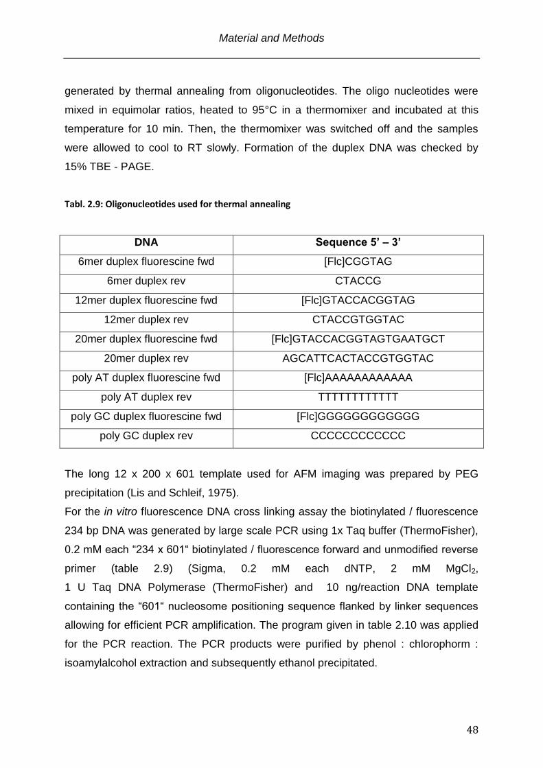
Material and Methods
48
generated by thermal annealing from oligonucleotides. The oligo nucleotides were
mixed in equimolar ratios, heated to 95°C in a thermomixer and incubated at this
temperature for 10 min. Then, the thermomixer was switched off and the samples
were allowed to cool to RT slowly. Formation of the duplex DNA was checked by
15% TBE - PAGE.
Tabl. 2.9: Oligonucleotides used for thermal annealing
DNA Sequence 5’ – 3’
6mer duplex fluorescine fwd [Flc]CGGTAG
6mer duplex rev CTACCG
12mer duplex fluorescine fwd [Flc]GTACCACGGTAG
12mer duplex rev CTACCGTGGTAC
20mer duplex fluorescine fwd [Flc]GTACCACGGTAGTGAATGCT
20mer duplex rev AGCATTCACTACCGTGGTAC
poly AT duplex fluorescine fwd [Flc]AAAAAAAAAAAA
poly AT duplex rev TTTTTTTTTTTT
poly GC duplex fluorescine fwd [Flc]GGGGGGGGGGGG
poly GC duplex rev CCCCCCCCCCCC
The long 12 x 200 x 601 template used for AFM imaging was prepared by PEG
precipitation (Lis and Schleif, 1975).
For the in vitro fluorescence DNA cross linking assay the biotinylated / fluorescence
234 bp DNA was generated by large scale PCR using 1x Taq buffer (ThermoFisher),
0.2 mM each “234 x 601“ biotinylated / fluorescence forward and unmodified reverse
primer (table 2.9) (Sigma, 0.2 mM each dNTP, 2 mM MgCl2,
1 U Taq DNA Polymerase (ThermoFisher) and 10 ng/reaction DNA template
containing the “601“ nucleosome positioning sequence flanked by linker sequences
allowing for efficient PCR amplification. The program given in table 2.10 was applied
for the PCR reaction. The PCR products were purified by phenol : chlorophorm :
isoamylalcohol extraction and subsequently ethanol precipitated.
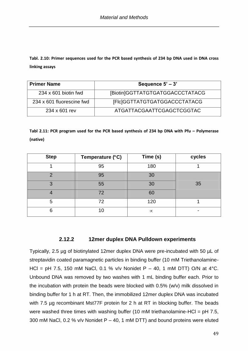
Material and Methods
49
Tabl. 2.10: Primer sequences used for the PCR based synthesis of 234 bp DNA used in DNA cross
linking assays
Primer Name Sequence 5’ – 3’
234 x 601 biotin fwd [Biotin]GGTTATGTGATGGACCCTATACG
234 x 601 fluorescine fwd [Flc]GGTTATGTGATGGACCCTATACG
234 x 601 rev ATGATTACGAATTCGAGCTCGGTAC
Tabl 2.11: PCR program used for the PCR based synthesis of 234 bp DNA with Pfu – Polymerase
(native)
Step Temperature (C) Time (s) cycles
1 95 180 1
2 95 30
35 3 55 30
4 72 60
5 72 120 1
6 10 -
2.12.2 12mer duplex DNA Pulldown experiments Typically, 2.5 µg of biotinylated 12mer duplex DNA were pre-incubated with 50 µL of
streptavidin coated paramagnetic particles in binding buffer (10 mM Triethanolamine-
HCl = pH 7.5, 150 mM NaCl, 0.1 % v/v Nonidet P – 40, 1 mM DTT) O/N at 4°C.
Unbound DNA was removed by two washes with 1 mL binding buffer each. Prior to
the incubation with protein the beads were blocked with 0.5% (w/v) milk dissolved in
binding buffer for 1 h at RT. Then, the immobilized 12mer duplex DNA was incubated
with 7.5 µg recombinant Mst77F protein for 2 h at RT in blocking buffer. The beads
were washed three times with washing buffer (10 mM triethanolamine-HCl = pH 7.5,
300 mM NaCl, 0.2 % v/v Nonidet P – 40, 1 mM DTT) and bound proteins were eluted
Material and Methods
50
by boiling the beads in protein loading buffer. Samples were analyzed by SDS -
PAGE and visualized by Coomassie staining.
2.12.3 Chromatin co-precipitation experiments 10 ng/µL of nucleosomal arrays (OD260 = 0.2) were incubated in a titration series
with recombinant Mst77F protein for 1 hr at 4°C and a total volume of 100 µL in
binding buffer (10 mM Triethanolamine pH = 7.4. 150 mM NaCl, 1mM DTT). 5 mM
MgCl2 was added to the nucleosomal arrays with Mst77F to induce nucleosome
clustering (Schwarz et al., 1996). Oligonucleosomes were pelleted by centrifugation
for 30 min at 16,000 x g and 4°C. Pellets were washed once with 1 mL of binding
buffer containing 5 mM MgCl2 and centrifuged for 15 min at 16,000 x g and 4°C.
Pellets were solubilized in protein loading buffer. Samples were run on a SDS -
PAGE and proteins were visualized by Coomassie staining.
2.12.4 Fluorescence polarisation (FP)
Fluorescence polarisation binding assays were performed according to (Jacobs,
2004) with 5 nM fluorescein-labeled 12mer duplex DNA (table 2.8) and 1 nM to 10
µM of purified Mst77F protein in 10 µL of 10 mM triethanolamine-HCl pH = 7.5 and
150 mM NaCl at 4°C in black microtiter plates. Fluorescence polarization was
measured using a Chameleon multi-label counter equipped with a 485 nm
fluorescence polarization excitation filter and 535 nm fluorescence polarization
emission filters in parallel and perpendicular orientation. Anisotropy (A) values were
calculated using the equation:
P is the degree of fluorescence polarization calculated by the equation:
Iparallel and Iperpendicular are the intensities of the emitted fluorescence light in
parallel and perpendicular orientation. For the analysis of binding curves, non-linear
Material and Methods
51
least-square fitting of the data was carried out using the equation:
where Af and Ab are the measured anisotropy values of free and bound peptides,
cprotein is the known protein concentration and Kd is the dissociation constant. All data
sets were plotted and fitted with the Kaleidagraph software.
2.12.5 Isothermal Titration Calorimetry (ITC) The 12mer duplex DNA used in this assay was produced by thermal annealing
according to paragraph 2.12.1 and table 2.8. The sample was concentrated to 50 µM
using Amicon Ultra centrifugation filter units (Millipore) and dialyzed extensively
against 10 mM TriethanolamineHCl = pH 7.5, 150 mM NaCl, 1mM TCEP.
Recombinant Mst77F was concentrated to 100 µM and dialyzed in parallel against
the exact same buffer as the DNA sample. All ITC measurements were performed on
an iTC200 calorimeter at 20°C in the Department of Physical Chemistry at the Max-
Planck Institute for Biophysical Chemistry, Goettingen, Germany. The
thermodynamic parameters of the protein : DNA interaction were recorded through
the emitted heats of 20 sequential protein injections into the 12mer duplex DNA. At
each injection step 2 µL of 34.4 µM Mst77F protein solution were released into the
Calorimeter cell containing 13.8 µM 12mer duplex DNA. Each injection was spaced
by a 120 s interval of constant stirring at 1000 rpm to allow for baseline stabilisation.
The heats of dilution, obtained by titration of Mst77F into buffer, were subtracted from
raw data before analysis. Raw data were integrated with respect to time, normalized
per mol of injected Mst77F and the apparent heat change (q) was plotted against
the molecular ratio of Mst77F vs. 12mer duplex DNA by the implemented Origin
analysis software. For determination of apparent enthalpy changes(Happ), the molar
association constant (KA), and the stoichiometry (n) of the interaction, nonlinear least-
square fitting of the q values was performed by the Origin software using a one set
of identical binding sites model. The heat change for each injection is described by
the equation:
Material and Methods
52
qi is the molar heat change, V the cell volume, ca,i the total concentration of the
injected component in the cell after the ith injection step, and cb,i the total
concentration of the macromolecule in the cell after the ith injection (Jelesarov and
Bosshard, 1999).
2.12.6 Electrophoretic Mobility Shift Assay (EMSA) The EMSA experiment was carried out on the basis of the original protocoll (Garner
and Revzin, 1981) and adopted in the following way. The 12mer duplex fluorescine
labeled DNA template (random sequence) used in this assay was assembled by
thermal annealing according paragraph 2.12.1 and table 2.8. Mononucleosomes and
12 x 200 x 601 nucleosomal arrays were assembled according to the paragraph
2.11.4. 50 ng / reaction 12 mer duplex DNA, 250 ng / reaction mononucleosomes
and 12 x 200 x 601 nucleosomal arrays, respectivly, were used as binding partners in
a titration series of various Mst77F and control proteins. The protein titration was
ranging from 20 nM to 5.12 µM in 2-fold dilution steps. All reactions were carried out
in EMSA buffer (10 mM Hepes (NaOH) pH = 7.4, 50 mM KCl, 0.25 mg / mL BSA, 5
mM DTT and 5% (v/v) glycerol) (Hayashihara et al., 2010) with the proteins to be
added last. EMSA reactions were incubated at room temperature for 20 min and
subsequently were loaded either onto 15% 0.5X TBE – PAGE gels (12 mer duplex
DNA), 5% 0.5X TBE – PAGE gels (Mononucleosomes) or 1% 0.5X TBE – Agarose
gels (12 x 200 x 601 oligonucleosomal arrays). All gels were run at a constant 100 V
at 4°C in the cold room with variation in the run time (90 min for the PAGE gels and
240 min for the Agarose gels). The EMSA reactions carried out with the 12mer
fluorescine duplex DNA were visualized with the FLA-5100 imaging system in the
Department of Physical Biochemistry at the Max – Planck Institute for Biophysical
Chemistry, Goettingen, Germany. Mononucleosome and Oligonucleosome EMSAs
were stained and visualized according to paragraph 2.7.7. Quantifications of signal
intensities were performed using ImageJ version 1.42q (Rasband, 1997 - 2011) and
plotted with the Kaleidagraph software.
Material and Methods
53
2.12.7 Protein cross - linking assay
Protein multimerization was investigated in the presence of DNA. The protein specific
amine reactive chemical cross – linker BS3 (Bis[sulfosuccinimidyl]suberate) was used
to stabilize the protein – protein interaction and to exclude any unspecific reaction
with the DNA present in the reaction mix (Sinz, 2003). Prior to the cross – linking all
proteins were dialysed into 10 mM triethanolamineHCl pH = 7.4, 150 mM NaCl and 1
mM DTT containing buffer. A final protein concentration of 20 µM in a 10 µL reaction
volume was used for each cross – linking reaction in the above specified buffer. 12 x
200 x 601 DNA was titrated to the proteins. At the lowest ratio a 500-fold molar
excess of protein over DNA was used. The DNA amount was increased 2-fold in
each titration ending up with 30 fold molar excess of protein over DNA. After addition
of the DNA the reaction was incubated at room temperature for 30 min. Then, the
chemical cross linker BS3 was added to the reaction in a 20-fold molar excess (400
µM) over the protein and the reaction was incubated at room temperature for another
90 min. The cross - linking was terminated by the addition of 100 mM final TrisHCl
pH = 7.4 buffer and an incubation period of 15 min at room temperature. The SDS –
PAGE and protein staining was carried out according to paragraphs 2.4 and 2.5
2.13 DNA / Chromatin compaction assays
2.13.1 Atomic Force Microscopy
Sample preparation and AFM imaging was basicly performed according to published
protocols (Leuba and Bustamante, 1999) with modifications towards the imaging of
DNA. All buffers used for sample preparation were filtered through a Anotop 10 - 0.02
µM filter Unit. The 12 x 200 x 601 DNA and the respective proteins were dialysed
against 10 mM triethanolamineHCl pH = 7.4, 150 mM NaCl and 1 mM DTT over
night. The next day 3.71 x 10⁻¹⁵ mol (0.3 ng/µL) of the DNA were incubated with
varying amounts of Mst77F and control proteins (4-fold, 20-fold and 100-fold molar
excess of protein over DNA) in a total volume of 20 µL. The reactions were incubated
at RT for 30 min and subsequently deposited onto the surface (~ 0.5 cm2) of a freshly
cleaved mica. The sample was incubated for 10 min at RT. The surface was drained
Material and Methods
54
with the help of a KIMTECH paper towel and washed with 2 mL of ddH2O. Prior to
imaging the mica was dried for 20 min in a stream of filtered compressed air.
The 12 x 200 x 601 nucleosomal arrays and recombinant Mst77F protein were
dialyzed against 10 mM triethanolamineHCl pH = 7.4, 50 mM NaCl and 1 mM DTT.
3.44 x 10⁻¹³ mol (10 ng / µL) chromatin were preincubated for 1 h at 4°C with 1.38 x
10-12 mol (4-fold molar excess) of Mst77F protein in a total volume of 100 µL. Fresh
glutaraldehyde was added to a final concentration of 0.05% (v/v) and the sample was
incubated over night at 4°C. The next day the glutaraldehyde was removed by
extensive dialysis against 10 mM triethanolamineHCl pH = 7.4 and 50 mM NaCl. 10
µL of the reaction was deposited onto the surface (~ 0.5 cm2) of a freshly cleaved
mica. The sample was incubated for 10 min at RT. The surface was drained with the
help of a paper towel and washed carefully with 2 mL of ddH2O. Prior to imaging the
mica was dried for 20 min in a stream of filtered compressed air.
The Mst77F WT and Mst77F 100N proteins in conjunction with the 12mer duplex
DNA were complexed at concentrations and conditions that were applied for the
differential centrifugation assay (paragraph 2.13.2). 150 ng of 12mer duplex DNA
were incubated with either 40 nM or 640 nM of protein in a final volume of 50 µL.
Prior to deposition the samples were diluted 1 : 10 in the reaction buffer and 20 µL
were pipetted onto the surface of a freshly cleaved mica. As before, the surface was
drained with the help of a KIMTECH paper towel and washed carefully with 2 mL of
ddH2O. Prior to imaging the mica was dried for 20 min in a stream of filtered
compressed air.
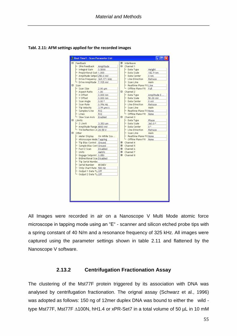
Material and Methods
55
Tabl. 2.11: AFM settings applied for the recorded images
All Images were recorded in air on a Nanoscope V Multi Mode atomic force
microscope in tapping mode using an “E“ - scanner and silicon etched probe tips with
a spring constant of 40 N/m and a resonance frequency of 325 kHz. All images were
captured using the parameter settings shown in table 2.11 and flattened by the
Nanoscope V software.
2.13.2 Centrifugation Fractionation Assay
The clustering of the Mst77F protein triggered by its association with DNA was
analysed by centrifugation fractionation. The orignal assay (Schwarz et al., 1996)
was adopted as follows: 150 ng of 12mer duplex DNA was bound to either the wild -
type Mst77F, Mst77F 100N, hH1.4 or xPR-Set7 in a total volume of 50 µL in 10 mM
Material and Methods
56
triethanolamineHCl pH = 7.4 buffer supplemented with 150 mM NaCl and 1 mM DTT.
The proteins were applied at concentrations of 40 nM to 1.28 µM in concentration
doubling increments. Immediatly after addition of the proteins and thorough mixing 20
µL of each sample were removed and saved as input. All reactions were
subsequently kept on ice for 30 min to allow for equilibration. Then the protein DNA
complexes were pelleted by centrifugation for 30 min at 16000 x g and 4°C. Again,
20 µL of the supernatant were removed and saved as output. To correlate the
amounts of DNA within the respective input and corresponding output the DNA of all
samples was stained with 0.5 µg / mL ethidium bromide (Bonasera et al., 2007). The
fluorescence was measured on a Chameleon multi-label counter equipped with a 360
nm excitation filter and a 612 nm emission filter. For each sample the ratio of input
DNA vs. output DNA was determined and plotted vs. the respective protein
concentration with the Kaleidagraph software.
2.13.3 In vitro DNA cross linking assay The assays were performed according to (Vogel et al., 2011). All incubations were
carried out at room temperature. In brief, black 96 well plates coated with
Streptavidin were pre-washed three times with 200 µL each of 1X PBS. 500 ng of
biotinylated 234 bp duplex DNA were synthesized according to paragraph 2.12.1
(table 2.9 and 2.10) and immobilized on the surface of the 96 well plate in
immobilization buffer (10 mM TrisHCl pH = 7.4, 2 M NaCl, 1 mM EDTA) for 1 h under
constant agitation. The wells were briefly washed 3 times with 200 µL each of 1X
PBS. 500 ng of 5’ fluorescine 234 bp duplex DNA (synthesized according to
paragraph 2.12.1 (table 2.9 and 2.10)) were mixed in a titration series ranging from
160 nM to 5.12 µM with recombinant Mst77F proteins or control proteins in binding
buffer (10 mM triethanolamineHCl pH = 7.4, 150 mM NaCl, 1 mM DTT), and were
immediatly transfered to the wells with the immobilized biotinylated DNA. The
reactions were incubated for 1 h under constant agitation. After measuring the input
fluorescence for all proteins the wells were washed 10 times for 5 min each with 200
µL 1X PBS under constant agitation and the remaining fluorescence was measured.
The fluorescence was recorded using a Chameleon multi - label counter (Hidex)
equipped with a 485 nm excitation filter and a 535 nm emission filter. Relative
Material and Methods
57
fluorescence was calculated from the mean absolute values (output fluorescence /
input fluorescence) of three independant experiments. Control measurements were
carried out using either fluorescence DNA alone without any immobilized biotin -
DNA or fluorescence DNA + the respective protein without biotin - DNA or
fluorescence DNA in the background of immobilized biotin - DNA.
2.14 Other techniques
2.14.1 Circular Dichroism (CD) The protein samples were dialyzed into 10 mM triethanolamineHCl pH = 7.4, 150 mM
sodiumfluoride buffer and concentrated / diluted to a final concentration of 30 µM.
The Circular Dichroism spectra were recorded on a ChiraScan CD spectrometer at
20°C in a 2 mm Quartz Suprasil CD cell in the Department of Neurobiology, Max -
Planck Institute for Biophysical Chemistry, Göttingen, Germany.
2.14.2 Analytical Ultra Centrifugation For protein analysis by the sedimentation velocity method (Schuck, 2000), 0.8
OD260 of recombinant Mst77 was prepared in 400 μL triethanolamineHCl pH = 7.4,
150 mM NaCl and 1 mM -mercaptoethanol. Double-sector charcoal filled epon cells
were filled with 412 μL of buffer in the reference sector and 392 μL protein solution in
the sample sector. After loading the centrifuge with the rotor, the bucket of the
instrument was evacuated to 1 micron vacuum pressure. Prior to starting the run the
sample was equilibrated to 20°C for a 1 h. The sample was centrifuged at a speed of
35.000 rpm and sedimentation of the molecules was monitored over 200 scans.
During the run, scans were continuously acquired. The data analysis was performed
with the SEDFIT software (Schuck, 2000) using a partial specific volume of Mst77
calculated from its’ amino acid composition with the software SEDNTERP.
Generally, after positioning the meniscus and the bottom, a simplex fit for the
meniscus position was performed at a resolution of 50-100. The frictional ratio was
fitted with the simplex algorithm. The initial fits were further refined by the simulated
annealing algorithm until the root mean square deviation converged at a minimum.
Figures of the analytical ultracentrifugation analyses were prepared by exporting the
Material and Methods
58
raw data at a resolution of 200 to the Origin software.

Material and Methods
59
Results
60
3. Results
The Mst77F protein is exclusively expressed in the testis of Drosophila males. During
postmeiotic spermatid maturation it arises in the transition phase from a nucleo-
histone towards a protamine based organization of the genome. In vivo observations
suggest a role in the condensation of the DNA in order to reach a high level of
compaction. Beyond the descriptive work that characterized the gene and its timely
and spacial expression no information about the proteins structure or its exact
function in vivo are available. In this study I set out to characterize Mst77F on the
molecular level and its functional implication in the compaction of DNA by in vitro
techniques.
3.1 Mst77F is a protein of bivalent structural organization Since the discovery of the Drosophila Mst77F gene in 1993 (Russell and Kaiser,
1993) little knowledge about the molecular structure or the functional role of the
Mst77F protein during Drosophila spermatogenesis could be obtained. To develop
hypotheses that help to characterize the protein in experimental assays I applied, as
a starting point several online available protein domain prediction algorithms to the
Mst77F protein sequence. The only tool amongst several that identified putatively
structured domains of Mst77F was the Simple Modular Architecture Research Tool
(SMART) (Schultz et al., 1998)(Fig. 3.1 panal A) that assigned a N-terminal coiled-
coil motif to the region of amino acids 26 to 49 based on the coils2 algorithm (Lupas
et al., 1991). Coiled-coils represent a class of -helical protein- protein interaction
interfaces in a multitude of proteins with different functions (Mason and Arndt, 2004).
The relationship between the amino acid sequence and the finally folded structure
adopting a coiled-coil conformation is well studied (Mason and Arndt, 2004;
Nishikawa and Scheraga, 1976) and a high confidence prediction is therefore
possible.
The second motif which was annotated within the Mst77F sequence identified by
SMART shows two regions of low compositional complexity between amino acids
171 to 191 and 200 to 214 (Wootton JC, 1996) (Fig. 3.1 panal A). Such regions
Results
61
display only little diversity in their amino acid composition and show loosely
clustered, irregularly spaced or periodic patterns of these residues (Coletta et al.,
2010). Structural information is not available, because a) there is no sequence
conservation due to rapid evolution across species (Ekman et al., 2006) and b) those
regions do not readily crystallize, arguing towards their structural undefined nature
(Huntley and Golding, 2002). Nevertheless, low compositional complexity regions are
found in a number of distinct proteins that either adopt folded conformations or
display intrinsically unstructured properties (Tatham and Shewry, 2000; Tompa,
2002). For some proteins the pivotal biological function of low complexity regions
could be demonstrated (Phatnani and Greenleaf, 2006; Wanker et al., 1995).
To confirm the protein motif prediction and to obtain additional structural properties I
conducted a secondary structure analysis using the secondary structure prediction
online tool (http://npsa-pbil.ibcp.fr/cgi-bin/npsa_automat.pl?page=npsa_sspred.html)
(Combet et al., 2000) in conjunction with the determination of the relative protein
disorder by the IUPred tool (see paragraph 2.3) (Dosztanyi et al., 2005a, b).
Secondary structure and relative disorder prediction were coherent in their results
(Fig. 3.1 panel B, data not shown for sec. structure prediction). The N-terminal region
(amino acids 10 to 90) of Mst77F adopts an ordered confirmation due to several
predicted -helices and a few extended strands. This is in good agreement with the
putative coiled-coil motif identified in the SMART blast. Additionally, the possible -
helical organization of Mst77F is extended up to amino acid 90. More then half of the
protein (amino acids 91 to 215) cannot faithfully be correlated with any regularly
folded secondary structure but random coils. This is also reflected in the relative
disorder prediction and by the SMART annotation predicting an overall highly bivalent
protein organization: A putatively structured N-terminus organized by -helices and
an unstructured C-terminus in flexible random coil conformation.
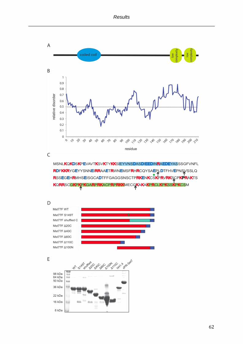
Results
62
Results
63
Fig. 3.1: Structural parameters of Mst77F
(A) Mst77F domain architecture according to SMART prediction: N-terminal coiled-coil motif (blue) as possible protein-protein interaction module and C-terminal low compositional complexity regions (green) with undefined structure and function for Mst77F. (B) Protein relative disorder prediction for Mst77F shows a bivalent organization of the protein. Roughly the first half of the protein except the very N-terminal 10 amino acids are assumed to have folded characteristics whereas the C-terminus is predicted to be largely unstructured. (C) Mst77F primary structure annotations: coiled-coil (blue background), low complexity region (green background), positively charged amino acids (red), negatively charged amino acids (blue), arrows represent the cloned Mst77F truncation mutants from the C-terminus and N-terminus respectively, Mst77F S149T point mutant (green asterisk). (D) Schematic representation of the Mst77F construct expressed in E.coli. 6xHis affinity purification tag (blue box), Mst77F with compositionally intact but shuffled amino acid order in the highly charged C-terminus (cyan box). (E) Affinity purified Mst77F proteins, hH1.4 and xPR-Set7 resolved on 15% Tris-Glycine SDS-PAGE
By taking a closer look at the possibly unstructured C-terminus it was striking that this
part of the protein is very rich in basic amino acids (Fig. 3.1 panel C). 60% of all
positive charges are situated within the last 70 amino acids of Mst77F. This local
positive charge accumulation in the C-terminal domain in conjunction with its
predicted random coil conformation resembles the structural properties of histone H1
family proteins and some members of HMG-box family of chromosomal, architectural
proteins (Dow et al., 2000; Dragan et al., 2003). In these proteins the positively
charged N- or C-terminal tails execute a major contribution to DNA binding via
electrostatic interactions with the negatively charged phosphodiester backbone of the
DNA (Churchill, 1999; Lu et al., 2009a). For histone H1 it could be demonstrated that
the CTD undergoes structural rearrangements upon interaction with DNA.
Despite the physical and structural relationship of the Mst77F C-terminus with
histone H1 proteins and some HMG proteins no conserved H1/H5 globular domain or
a HMG-box motif could be detected in Mst77F.
3.2 The Mst77F-DNA interaction
Previous studies reported Mst77F to be an important factor in postmeiotic spermatid
maturation in Drosophila. Functions in chromatin condensation and nuclear shaping
were proposed (Rathke et al., 2010; Sunil Jayaramaiah Raja, 2005). These findings
are solely based on cytogenetic experiments and no molecular prove for a direct
interaction with DNA has been found. However, the Mst77F sequence analysis
Results
64
revealed a physiochemical similarity of the C-terminal tail towards a class of HMG
proteins. These proteins functionally act as structural components of chromatin
through a direct interaction with DNA in an unspecific, charge-mediated manner
(Churchill, 1999; Dragan et al., 2003). Also, histone H1 type proteins, major structural
components of cellular chromatin exhibit a highly charged C-terminus that is only little
structured in solution (Allan et al., 1980; Dootz et al., 2011; Happel and Doenecke,
2009; Lu et al., 2009a). Additionally, large patches of positive charges have been
suggested to be indicative for protein nucleic acid interfaces (Honig et al., 1989;
Jones et al., 2003; Shazman et al., 2007). On the basis of these data I hypothesized
that Mst77F binds DNA via its extremely basic C-terminal domain. Due to its high
positive charge density and putatively unstructured conformation the interaction
might be unspecific and mainly of ionic nature.
3.2.1 The Mst77F C-terminal domain is necessary and sufficient for DNA binding
To test these hypotheses I expressed along with the wild-type several deletion-
mutant Mst77F proteins (Fig. 3.1 panel D, E). These proteins were subsequently
tested in two conceptually different assays. On the one hand I performed pulldown
experiments with the recombinant Mst77F proteins and immobilized dodecamer
duplex DNA (paragraph 2.12.2). This assay revealed qualitative and kinetic
information about the respective interactions. Alternatively, I conducted quantitative
measurements of the interaction under equilibrium conditions by fluorescence
polarization (FP) using the same dodecameric duplex DNA but a 5’ fluorescine
instead of a biotin-tag (paragraph 2.12.4). In both assays the wild-type protein
displayed strong binding to the respective DNA template (Fig. 3.2 A and B left) and
the interaction was quantified with an equilibrium dissociation constant of 90 + 20 nM
(Tabl. 3.1). Mst77F 20C, Mst77F 40C and Mst77F 60C are characterized by their
stepwise deletions of the C-terminal basic domain thereby reducing the local, high-
density charge in 25% intervals. These proteins should reveal the functional impact
of the tail in the DNA binding reaction. In pulldown assays Mst77F 20C and Mst77F
40C showed a similar recovery rate compared to the wild-type protein even though
the overall charge sum is reduced by 25% and 50%, respectively. Mst77F 60C
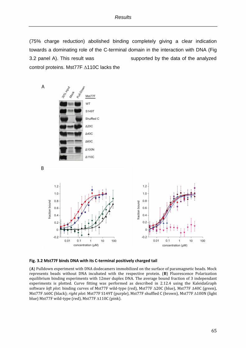
Results
65
(75% charge reduction) abolished binding completely giving a clear indication
towards a dominating role of the C-terminal domain in the interaction with DNA (Fig
3.2 panel A). This result was supported by the data of the analyzed
control proteins. Mst77F 110C lacks the
Fig. 3.2 Mst77F binds DNA with its C-terminal positively charged tail
(A) Pulldown experiment with DNA dodecamers immobilized on the surface of paramagnetic beads. Mock represents beads without DNA incubated with the respective protein. (B) Fluorescence Polarization equilibrium binding experiments with 12mer duplex DNA. The average bound fraction of 3 independant experiments is plotted. Curve fitting was performed as described in 2.12.4 using the KaleidaGraph software left plot: binding curves of Mst77F wild-type (red), Mst77F 20C (blue), Mst77F 40C (green), Mst77F 60C (black); right plot: Mst77F S149T (purple), Mst77F shuffled C (brown), Mst77F 100N (light blue) Mst77F wild-type (red), Mst77F 110C (pink).
Results
66
C-terminal domain and displayed no binding to DNA. Vice versa the Mst77F 100N
mutant that misses the putative -helical N-terminus restored binding to WT level.
Additionally, the shuffled C-terminal domain Mst77F mutant with a randomized order
of the C-terminal domain amino acids showed equal recovery in respect to the WT
protein ruling out a possible structural contribution coming from the C-terminus.
The Mst77F S149T point mutant has been reported to show severe phenotypes in
cytogenetic experiments in Drosophila. Nuclear shaping defects of spermatids and
male sterility is observed but chromatin condensation appears to be normal (Rathke
et al., 2010). In conformity with the in vivo observed chromatin compaction in the
Mst77F S149T background, the S149T mutant interacted with DNA to a similar
extent as the wild-type protein.
Quantitatively, even though the charge decline occurred in steps of 25% in the
Mst77F 20C, Mst77F 40C and Mst77F 60C mutants, the decrease in measured
affinity was not proportional to the percentage of charge reduction (Fig 3.2 panel B
left and Tabl. 3.1). The changes in the equilibrium dissociation constants observed
between Mst77F WT and Mst77F 20C mutant (2.5 fold decrease; WT 90 + 20 nM
vs. 20C 230 + 70 nM) or Mst77F 40C and Mst77F 60C (3.5 fold decrease; 40C
8.42 + 3.13 µM vs. 60C 28.6 + 12.5 µM) are rather mild when compared to the
transition from Mst77F 20C to Mst77F 40C (40 fold decrease; 20C 230 + 70 nM
vs. 40C 8.42 + 3.13 µM). However, this binding effect with its abrupt decrease in
affinity in the transition from 20C to 40C was not reflected by the protein recovery
in the pulldown assay pointing towards kinetic effects under the applied experimental
conditions. The differences observed might manifest by altered association kinetics
that became apparent in one assay but not the other. Strikingly, bioinformatic
analysis assigned a low compositional complexity motif to this region. The recorded
effect might therefore be brought about by a functional sub-domain within the
unstructured region. Such functional sub-domains within intrinsically disordered
domains have been identified (Lu and Hansen, 2004).
Coherent to the pulldown results the equilibrium dissociation constants for Mst77F
100N, Mst77F shuffled C-terminal domain and Mst77F S149T were not significantly
different from the wild-type protein and no binding for the Mst77F 110C protein
could be observed (Fig. 3.2 panel B right and Tabl. 3.1).
Results
67
Taken together, the results from the pulldown experiment and the FP measurements
strongly indicate a high affinity interaction of Mst77F with DNA mediated by the C-
terminal domain of the protein.
3.2.2 Mst77F binding to DNA is based on sequence unspecific ionic interactions
Binding experiments with Mst77F and short DNAs identified the C-terminal domain to
be sufficient for the interaction (paragraph 3.2.1). The high positive charge content
along with a predicted structurally undefined conformation allows for an ionic
interaction type that is sequence unspecific. Binding that virtually depends on
opposing charges should not be influenced by structural properties of the DNA or
sequence. Therefore, I tested the binding of Mst77F to different subtypes of B-DNA.
Poly(dA)*poly(dT) duplexes have been described to form a narrow minor groove with
a wider major groove whereas poly(dG)*poly(dC) DNA structurally displays a wide
minor groove and a narrow major groove (Nelson et al., 1987; Yoon et al., 1988). The
interactions of Mst77F with poly(dA)*poly(dT) as well as poly(dG)*poly(dC)
dodecamers were compared to regular B form DNA (random sequence) of the same
length (same negative charge sum) in fluorescence polarization binding assays (Fig.
3.3 panel A). If major and minor groove are the predominant high affinity binding
parameters in the protein DNA complex changing their dimensions should result in
altered binding affinities. This was not observed. The measured equilibrium
dissociation constants are not significantly different (Tabl. 3.1).
Along with natural structural variations of B-DNA caused by repetitive AT or GC
sequences the canonical Watson & Crick B-form DNA was structurally modified by
pre-incubation with ethidium bromid. Moreover, also single strand DNA that has no
helical structure was measured in a Mst77F binding reaction (Fig. 3.3 panel B). The
observed deviations of the measured equilibrium dissociation constants were
marginal from the Mst77F interaction with regular B-type DNA and not significant
(Tabl. 3.1).
If the conformational status and the sequence of the DNA are irrelevant for high
affinity interactions, the origin must be different. In fact, a salt titration series that
stepwise compensates the opposing charges of DNA and protein resulted in loss of
binding (Fig. 3.3 panel C). The binding strength of an ionic interaction type is based
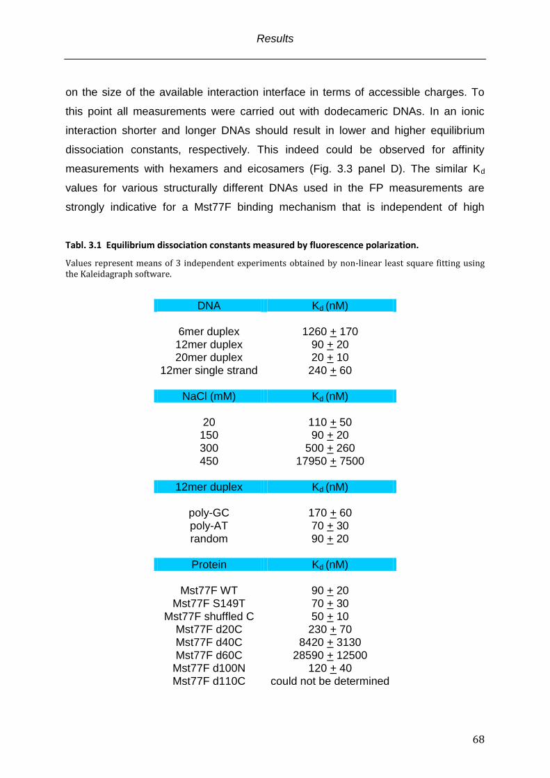
Results
68
on the size of the available interaction interface in terms of accessible charges. To
this point all measurements were carried out with dodecameric DNAs. In an ionic
interaction shorter and longer DNAs should result in lower and higher equilibrium
dissociation constants, respectively. This indeed could be observed for affinity
measurements with hexamers and eicosamers (Fig. 3.3 panel D). The similar Kd
values for various structurally different DNAs used in the FP measurements are
strongly indicative for a Mst77F binding mechanism that is independent of high
affinity
Tabl. 3.1 Equilibrium dissociation constants measured by fluorescence polarization.
Values represent means of 3 independent experiments obtained by non-linear least square fitting using the Kaleidagraph software.
DNA Kd (nM)
6mer duplex 1260 + 170 12mer duplex 90 + 20 20mer duplex 20 + 10
12mer single strand 240 + 60
NaCl (mM) Kd (nM)
20 110 + 50 150 90 + 20 300 500 + 260 450 17950 + 7500
12mer duplex Kd (nM)
poly-GC 170 + 60 poly-AT 70 + 30 random 90 + 20
Protein Kd (nM)
Mst77F WT 90 + 20
Mst77F S149T 70 + 30 Mst77F shuffled C 50 + 10
Mst77F d20C 230 + 70 Mst77F d40C 8420 + 3130 Mst77F d60C 28590 + 12500
Mst77F d100N 120 + 40 Mst77F d110C could not be determined
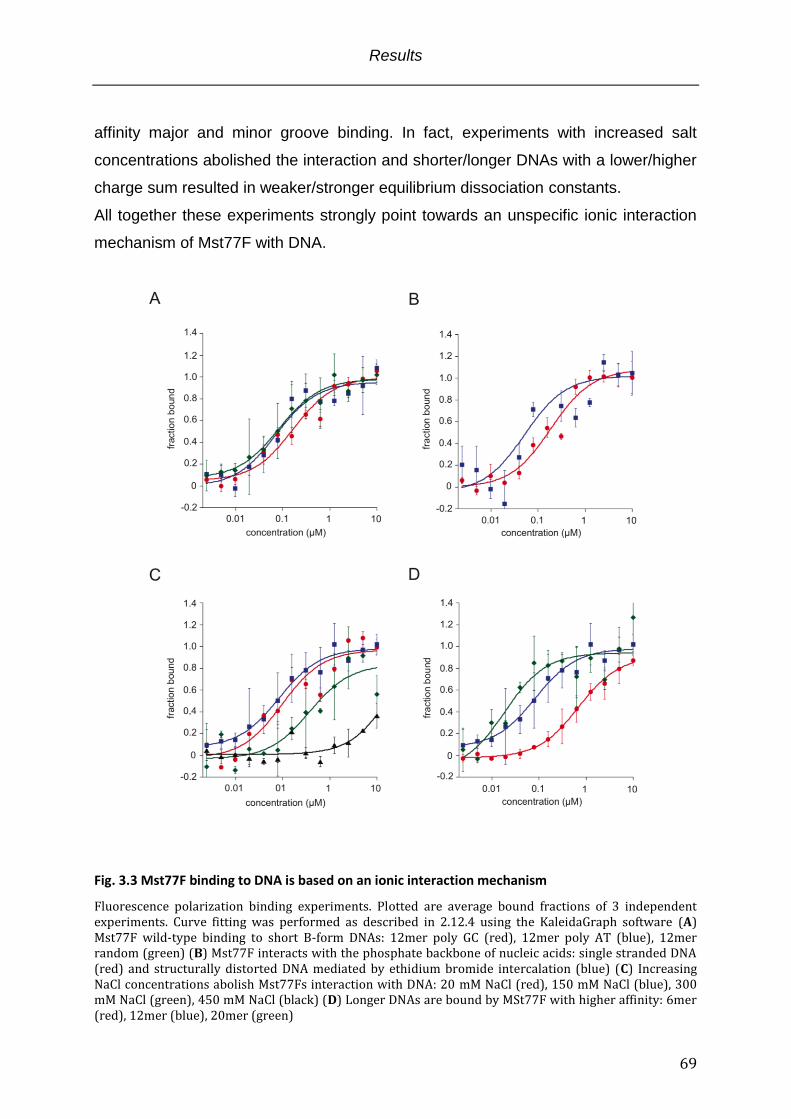
Results
69
affinity major and minor groove binding. In fact, experiments with increased salt
concentrations abolished the interaction and shorter/longer DNAs with a lower/higher
charge sum resulted in weaker/stronger equilibrium dissociation constants.
All together these experiments strongly point towards an unspecific ionic interaction
mechanism of Mst77F with DNA.
Fig. 3.3 Mst77F binding to DNA is based on an ionic interaction mechanism
Fluorescence polarization binding experiments. Plotted are average bound fractions of 3 independent experiments. Curve fitting was performed as described in 2.12.4 using the KaleidaGraph software (A) Mst77F wild-type binding to short B-form DNAs: 12mer poly GC (red), 12mer poly AT (blue), 12mer random (green) (B) Mst77F interacts with the phosphate backbone of nucleic acids: single stranded DNA (red) and structurally distorted DNA mediated by ethidium bromide intercalation (blue) (C) Increasing NaCl concentrations abolish Mst77Fs interaction with DNA: 20 mM NaCl (red), 150 mM NaCl (blue), 300 mM NaCl (green), 450 mM NaCl (black) (D) Longer DNAs are bound by MSt77F with higher affinity: 6mer (red), 12mer (blue), 20mer (green)
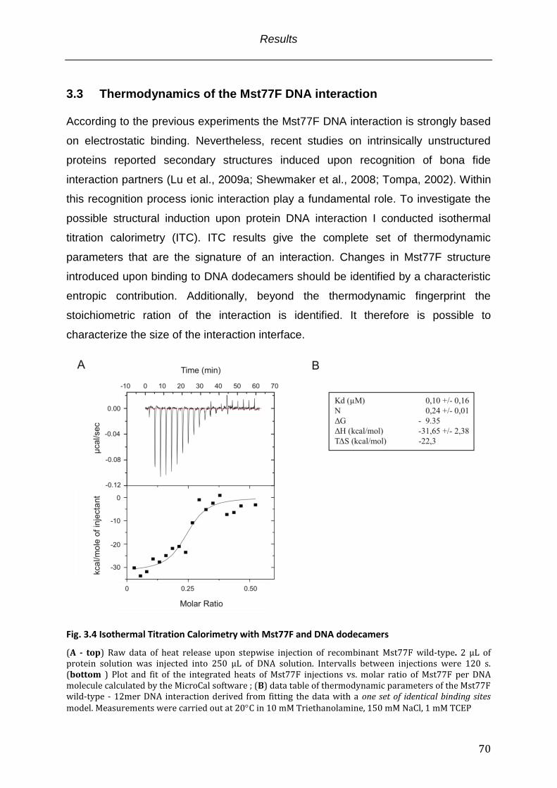
Results
70
3.3 Thermodynamics of the Mst77F DNA interaction According to the previous experiments the Mst77F DNA interaction is strongly based
on electrostatic binding. Nevertheless, recent studies on intrinsically unstructured
proteins reported secondary structures induced upon recognition of bona fide
interaction partners (Lu et al., 2009a; Shewmaker et al., 2008; Tompa, 2002). Within
this recognition process ionic interaction play a fundamental role. To investigate the
possible structural induction upon protein DNA interaction I conducted isothermal
titration calorimetry (ITC). ITC results give the complete set of thermodynamic
parameters that are the signature of an interaction. Changes in Mst77F structure
introduced upon binding to DNA dodecamers should be identified by a characteristic
entropic contribution. Additionally, beyond the thermodynamic fingerprint the
stoichiometric ration of the interaction is identified. It therefore is possible to
characterize the size of the interaction interface.
Fig. 3.4 Isothermal Titration Calorimetry with Mst77F and DNA dodecamers
(A - top) Raw data of heat release upon stepwise injection of recombinant Mst77F wild-type. 2 µL of protein solution was injected into 250 µL of DNA solution. Intervalls between injections were 120 s. (bottom ) Plot and fit of the integrated heats of Mst77F injections vs. molar ratio of Mst77F per DNA molecule calculated by the MicroCal software ; (B) data table of thermodynamic parameters of the Mst77F wild-type - 12mer DNA interaction derived from fitting the data with a one set of identical binding sites model. Measurements were carried out at 20C in 10 mM Triethanolamine, 150 mM NaCl, 1 mM TCEP
Results
71
From the measured thermodynamic parameters of Mst77F binding DNA dodecamers
a stoichiometric ratio of one protein binding four short DNAs could be derived (Fig.
3.4 panel B). This suggests a brought interface across the charges of the C-terminal
domain, even though information about the exact interaction positions is not
available. Noticeable are the high negative enthalpy (H) and entropy (TS) values
indicating the type of interaction. Whereas the strong negative H value reflects the
ionic interaction mode (salt bridges, hydrogen bonding), the high negative TS is
difficult to explain. A strong negative contribution is usually a measure of high
flexibility or hydrophobic interactions whereas positive contributions in entropy reflect
protein folding. To this point there is no indication of structural induction upon binding
to DNA. Due to the proposed random coil conformation it is likely a combination of
high flexibility due to electrostatic repulsion between amino acids of the same charge
and additional hydrophobic interactions between amino acids and DNA bases. In
agreement with the FP measurements the derived equilibrium dissociation constant
determined by ITC is 0.1 µM. This result confirms the previously obtained Kd values
by FP and underpins the validity of the thermodynamic parameters.
3.4 The mechanism of the Mst77F-DNA interaction
Despite the characterization of the Mst77F binding domain and the determination of
the interaction parameters with DNA by FP and ITC the question of functional
relevance of this unspecific interaction emerged. Usually unspecific binding
mechanisms are involved in one-dimensional scanning along the DNA. This lateral
diffusion in search for a bona fide binding site that is eventually recognized with high
affinity is a general feature of sequence-specific DNA binding proteins (e.g.
transcription factors). However, this usually also involves amino acids of additional
protein motifs that insert into major or minor groove and convey the cognate
sequence specificity (Winter et al., 1981). These extensively studied mechanisms
were also confirmed for structural DNA binding proteins that show no sequence
preferences but exhibit their function by motifs that recognize major/minor groove
and thereby introduce structural changes (Klass et al., 2003). The previous binding
experiments with Mst77F did not point towards an interaction mode that involves
Results
72
groove insertion of particular amino acids. However, Mst77F has been implicated in
genomic shaping during spermatid maturation (Rathke et al., 2010). Previous studies
suggested Mst77F being a distant member of the histone H1 family of structural
proteins and sequence alignments show a similarity in the C-terminal domains (CTD)
of both proteins (Russell and Kaiser, 1993). Importantly, the H1 CTD is assumed to
be intrinsically unstructured and its necessity for H1 function in chromatin compaction
could be demonstrated (Lu et al., 2009a). The initial interaction between protein and
DNA/chromatin is therein mediated by electrostatic mechanisms. On this basis I
hypothesized that the Mst77F DNA interaction is similar to histone H1 interactions
with DNA. Moreover, I wished to demarcate effects that are based on simple
adhesion of positively charged patches on their surface of proteins that are not
necessarily involved in a direct interaction with DNA.
3.4.1 Mst77F aggregates DNA To further refine the binding of Mst77F to DNA I studied the gel migration behavior of
dodecameric fluorescine tagged duplex DNA in the context of previously tested
Mst77F proteins in electrophoretic mobility shift assays (EMSA) (Fig. 3.5 panel C and
partially B). Ordinary EMSA experiments usually result in a discrete shifted band that
appears upon binding of a protein. In a 1:1 interaction model a single band is
expected and multiple interaction result in a complex pattern of numerous bands.
However, the shifting of the free DNA upon binding of a protein is concentration
dependant and typically follows the law of mass action. Figure 3.5 panel C and panel
B (partially) show that Mst77F did not shift the free DNA towards a discrete high
molecular weight band. This was surprising since the ITC experiment uncovered a
stoichiometric ratio of four short DNA dodecamers per protein. Instead it generated
smeary shifting patterns starting at Kd concentrations in a narrow window. At
saturating conditions the DNA was removed from the gel reflecting the initial
formation of distinct stoichiometric complexes and finally their aggregation. The
observed dose effect within the tested Mst77F concentrations depended on the
length of the C-terminal tail (Fig. 3.5 panel C). As expected the Mst77F 110C
mutant that misses the charged tail did not “shift” the DNA. Mst77F shuffled C-
terminal domain and Mst77F S149T behave very similar to the wild-type protein (Fig.
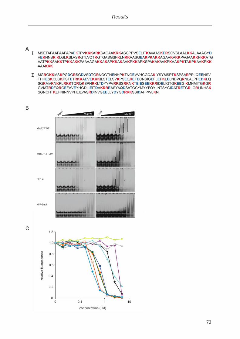
Results
73
Results
74
Fig. 3.5 Unspecific charge mediated protein – DNA interactions in EMSA experiments
(A) Protein sequences of hH1.4 (I) and xPR-Set7 (II); basic amino acids are annotated in red, acidic amino acids are displayed in blue (B) EMSA experiment: Mst77F wild-type, Mst77F 100N, hH1.4 and xPR-Set7 were complexed with DNA dodecamers in a concentration titration range from 0.02 to 5.12 µM in 2 fold increments. Samples were run on 15% TBE-PAGE gels for 90 min @ 80 volts. Input represents DNA dodecamers only. Parallel arranged gel within the same row show different exposures times. Unspecific DNA protein complexes are marked with bars (C) DNA dodecamer band intensities of EMSA experiments were integrated using the ImageJ software and plotted with Kaleidagraph: Mst77F wild-type (orange), Mst77F S149T (blue), Mst77F shuffled C (green), Mst77F 20C (black). Mst77F 40C (purple), Mst77F 60C (yellow), Mst77F 100N (brown), Mst77F 110C (light blue), hH1.4 (), xPR-Set7 (turquoise).
3.5 panel C). Very interesting was the gel migration behavior of DNA Mst77F 100N
complexes. It was only slightly different from the wild-type protein towards a stronger
dose response (Fig. 3.5 panel B). A functional contribution of the suggested -helical
N-terminus in the DNA binding event became not obvious.
The unexpected behavior of Mst77F DNA interaction in the EMSA experiments
raised the question weather the observed effects are explainable by the ionic type of
interaction. On this account I compared Mst77F to positively charged proteins that
either have been described to interact with DNA unspecifically by charge or proteins
that have not been reported to bind DNA at all.
Histone H1, a major structural component of chromatin (Happel and Doenecke,
2009; van Holde and Zlatanova, 1996) is highly basic (pI = 11.4) and has been
shown to strongly bind DNA unspecifically (Dootz et al., 2011). Distinct from Mst77F
it displays an even higher charge density that is not limited to the C-terminal tail but
almost spans the whole sequence (Fig 3.5 panel A I). On the other hand xPR-Set7 is
a known histone modifier with an enzymatic activity that introduces mono-methylation
on histone H4 lysine 20 in nucleosomal assemblies (Nishioka et al., 2002). xPR-Set7
resembles in its basicity Mst77F with a pI of 9.4 (Mst77F pI = 9.7) but shows a
uniform positive charge distribution across the sequence. Importantly, it has not been
demonstrated to physically interact with DNA (Fig 3.5 panel A II). Apart from their
common net positive charge those proteins show little structural similarity (Allan et
al., 1980; Ramakrishnan et al., 1993; Xiao et al., 2005). Surprisingly, the migration
properties of DNA complexed with hH1.4 as well as xPR-Set7 showed the same
smear like distribution within a narrow concentration window. However, the dose
response of hH1.4 became visible at lower concentrations finally resulting in loss of
the DNA from the gel. Within the tested concentrations the loss of DNA was not seen
for xPR-Set7 (Fig 3.5 panel B and C).
Results
75
Taken together, the EMSA assays did not reveal a specific Mst77F on DNA upon
binding. Analogous effects were observed for the hH1.4 and xPR-Set7 DNA
interactions pointing towards a phenomenon that generally holds true for ion guided
interaction, no matter if unspecific or adhesive, and reflects the differential
stoichiometric complexes that are formed. However, the observed formation of high
molecular weight complexes/aggregates might demarcate the charge mediated
unspecific binding effect from simple charge adhesion effects. These complexes
were further investigated in the upcoming paragraphs.
3.4.2 Mst77F effects on DNA are mediated by the N-terminus of the protein
Mst77F and hH1.4 are thought to contribute to the structural organization of DNA and
chromatin, respectively (Rathke et al., 2010{Thoma, 1977 #62)}(Shen et al., 1995).
Their role in the formation of condensed DNA/chromatin states was suggested or has
been shown and differences within these complexes due to functional deviation of
the proteins are possible. On the other hand xPR-Set7 as a histone modifying
enzyme is not expected to have direct structural effects on DNA or chromatin. On this
basis I hypothesized that unspecific charge mediated binding exerts effects that can
be discriminated from simple charge based adhesion. Moreover, the functions of
different unspecific DNA binding proteins and their effects on DNA are also distinct.
To test these hypotheses I performed centrifugation fractionation assays to uncover
molecular weight/density differences in the formed protein DNA complexes that
became not apparent in EMSA experiments. Additionally, I conducted Atomic Force
Microscopy (AFM) studies that convey visual information on the respective size of the
complexes but also provide useful information on molecular shape. To guarantee
consistency throughout the different experiment these assays were carried out with
the DNA dodecamers also used for previous experiments.
The principle of the centrifugation assay is outlined in Fig. 3.6 panel A and has been
adapted from (Carruthers et al., 1998; Nikitina et al., 2007). The sedimentation
behavior of DNA dodecamers in dependence of increasing concentrations of Mst77F
wild-type, Mst77F 100N, hH1.4 and xPR-Set7 was investigated (Fig 3.6 panel B). At
protein concentrations from 0.04 µM to 0.32 µM no significant difference in the
sedimentation of the DNA became visible across proteins. The DNA was floating in
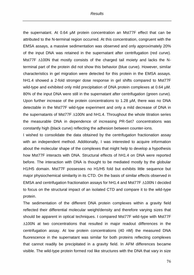
Results
76
the supernatant. At 0.64 µM protein concentration an Mst77F effect that can be
attributed to the N-terminal region occurred. At this concentration, congruent with the
EMSA assays, a massive sedimentation was observed and only approximately 20%
of the input DNA was retained in the supernatant after centrifugation (red curve).
Mst77F 100N that mostly consists of the charged tail moiety and lacks the N-
terminal part of the protein did not show this behavior (blue curve). However, similar
characteristics in gel migration were detected for this protein in the EMSA assays.
hH1.4 showed a 2-fold stronger dose response in gel shifts compared to Mst77F
wild-type and exhibited only mild precipitation of DNA protein complexes at 0.64 µM.
80% of the input DNA were still in the supernatant after centrifugation (green curve).
Upon further increase of the protein concentrations to 1.28 µM, there was no DNA
detectable in the Mst77F wild-type experiment and only a mild decrease of DNA in
the supernatants of Mst77F 100N and hH1.4. Throughout the whole titration series
the measurable DNA in dependence of increasing PR-Set7 concentrations was
constantly high (black curve) reflecting the adhesion between counter-ions.
I wished to consolidate the data obtained by the centrifugation fractionation assay
with an independent method. Additionally, I was interested to acquire information
about the molecular shape of the complexes that might help to develop a hypothesis
how Mst77F interacts with DNA. Structural effects of hH1.4 on DNA were reported
before. The interaction with DNA is thought to be mediated mostly by the globular
H1/H5 domain. Mst77F possesses no H1/H5 fold but exhibits little sequence but
major physiochemical similarity in its CTD. On the basis of similar effects observed in
EMSA and centrifugation fractionation assays for hH1.4 and Mst77F 100N I decided
to focus on the structural impact of an isolated CTD and compare it to the wild-type
protein.
The sedimentation of the different DNA protein complexes within a gravity field
reflected their differential molecular weight/density and therefore varying sizes that
should be apparent in optical techniques. I compared Mst77F wild-type with Mst77F
100N at two concentrations that resulted in major readout differences in the
centrifugation assay. At low protein concentrations (40 nM) the measured DNA
fluorescence in the supernatant was similar for both proteins reflecting complexes
that cannot readily be precipitated in a gravity field. In AFM differences became
visible. The wild-type protein formed rod like structures with the DNA that vary in size
Results
77
from a few nanometers up to several hundred nanometers. Mst77F 100N in
contrast showed small globular complexes with very short DNA (Fig. 3.6 panel C).
Importantly, in DNA binding experiments no differences between both proteins could
be detected neither in pulldown nor FP assays. The structural information on the
formed complexes clearly showed differences pointing towards detached functions of
N- and C-terminus. However, the density of both structures was not sufficient to
precipitate under the applied conditions. Increased protein concentrations (640 nM)
that led in the centrifugation assay to rapid sedimentation of DNA Mst77F wild-type
complexes were in AFM visible as massive clusters of more than five hundred
nanometers in XY dimensions. This was not visible with the 100N mutant.
Analogous to the centrifugation assay that shows no DNA precipitation evoked by the
protein, no structural differences compared to lower concentrations were detectable
(Fig. 3.6 panel C right).
In summary, Mst77F exerts specific effects on DNA that are depending on its N-
terminus in vitro. These effects can be characterized by hydrodynamic assays and
imaging techniques and become visible in form of DNA protein aggregates. These
aggregates are not seen with similar charged proteins or proteins of high positive
charge that already have been reported to structurally alter DNA.
Results
78
Results
79
Fig 3.6 Mst77F induces aggregation of DNA
(A) Schematic representation of the centrifugation fractionation experiment (B) Mst77F forms with DNA high molecular weight complexes that can be pelleted by centrifugation. DNA dodecamers were incubated with Mst77F wild – type, Mst77F 100N, hH1.4 and xPR-Set7 from 0.04 to 1.28 µM protein concentration in 2 fold increments. The ratio of floating DNA before and after centrifugation was plotted with the Kaleidagraph software. Data represents the mean of three independent experiments: Mst77F wild-type (red), Mst77F 100N (blue), hH1.4 (green) and xPR-Set7 (black) (C) Mst77F – DNA complexes pelleted by centrifugation can be visualized by Atomic Force Microscopy. Mst77F wild-type and Mst77F 100N DNA dodecamer complexes were imaged at 40 nM and 640 nM, respectively. Images were recorded in tapping mode. Scale bars represent 500 nm
3.5 The Mst77F N-terminus functions as multimerization interface upon DNA recognition
The differences between Mst77F wild-type and 100N mutant seen in fractionated
centrifugation and AFM experiments leading to a massive DNA aggregation strongly
point towards an additional functional contribution of the N-terminus. This effect
seems not to be related to a binding event since DNA binding assays did not uncover
a N-terminal contribution. However, the bio- informatics sequence analysis predicted
a -helical coiled-coil motif, a well-studied protein-protein interaction interface,
establishing the possibility of Mst77F self-interaction (Mason and Arndt, 2004). These
facts led me to hypothesize that Mst77F multimerizes through its N-terminal domain.
This multimerizaton is necessary to effectuate DNA aggregation.
To test this, I first performed circular dichroism (CD) spectroscopy. I compared the
110C mutant that according to the prediction should have a high -helical content to
the full-length protein that is suggested to contain an overall elevated random coil
structure (Fig. 3.7 panel A). Indeed, the CD spectrum of the 110C mutant
corroborated the predicted helical structure that dominates the N-terminus as seen
for the minima around 220 nm and 205 nm (blue curve). Nevertheless, -sheets also
contributed to the overall N-terminal fold reflected by the plateau around 215 nm.
However, as expected within the overall structure of the full-length protein random
coils constituted an abundant fraction (red curve) (Johnson, 1990). Nonetheless, the
verified helices do not automatically imply a functional multimerization of the protein.
A sedimentation velocity analytical ultracentrifugation experiment that records the
hydrodynamic sedimentation behavior in a gravity field, revealed a small fraction of
the protein that represents a dimer (6%) (Fig. 3.7 panel B). The low ratio of Mst77F
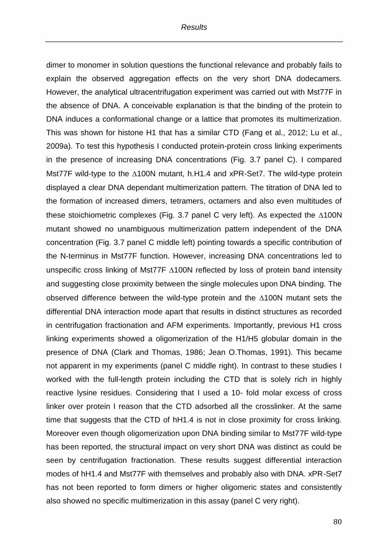
Results
80
dimer to monomer in solution questions the functional relevance and probably fails to
explain the observed aggregation effects on the very short DNA dodecamers.
However, the analytical ultracentrifugation experiment was carried out with Mst77F in
the absence of DNA. A conceivable explanation is that the binding of the protein to
DNA induces a conformational change or a lattice that promotes its multimerization.
This was shown for histone H1 that has a similar CTD (Fang et al., 2012; Lu et al.,
2009a). To test this hypothesis I conducted protein-protein cross linking experiments
in the presence of increasing DNA concentrations (Fig. 3.7 panel C). I compared
Mst77F wild-type to the 100N mutant, h.H1.4 and xPR-Set7. The wild-type protein
displayed a clear DNA dependant multimerization pattern. The titration of DNA led to
the formation of increased dimers, tetramers, octamers and also even multitudes of
these stoichiometric complexes (Fig. 3.7 panel C very left). As expected the 100N
mutant showed no unambiguous multimerization pattern independent of the DNA
concentration (Fig. 3.7 panel C middle left) pointing towards a specific contribution of
the N-terminus in Mst77F function. However, increasing DNA concentrations led to
unspecific cross linking of Mst77F 100N reflected by loss of protein band intensity
and suggesting close proximity between the single molecules upon DNA binding. The
observed difference between the wild-type protein and the 100N mutant sets the
differential DNA interaction mode apart that results in distinct structures as recorded
in centrifugation fractionation and AFM experiments. Importantly, previous H1 cross
linking experiments showed a oligomerization of the H1/H5 globular domain in the
presence of DNA (Clark and Thomas, 1986; Jean O.Thomas, 1991). This became
not apparent in my experiments (panel C middle right). In contrast to these studies I
worked with the full-length protein including the CTD that is solely rich in highly
reactive lysine residues. Considering that I used a 10- fold molar excess of cross
linker over protein I reason that the CTD adsorbed all the crosslinker. At the same
time that suggests that the CTD of hH1.4 is not in close proximity for cross linking.
Moreover even though oligomerization upon DNA binding similar to Mst77F wild-type
has been reported, the structural impact on very short DNA was distinct as could be
seen by centrifugation fractionation. These results suggest differential interaction
modes of hH1.4 and Mst77F with themselves and probably also with DNA. xPR-Set7
has not been reported to form dimers or higher oligomeric states and consistently
also showed no specific multimerization in this assay (panel C very right).
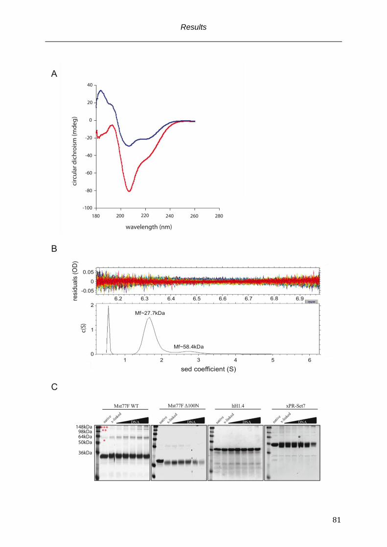
Results
81
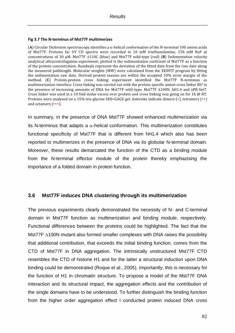
Results
82
Fig 3.7 The N-terminus of Mst77F multimerizes
(A) Circular Dichroism spectroscopy identifies a -helical conformation of the N-terminal 100 amino acids of Mst77F. Proteins far UV CD spectra were recorded in 10 mM triethanolamine, 150 mM NaF at concentrations of 30 µM. Mst77F 110C (blue) and Mst77F wild-type (red) (B) Sedimentation velocity analytical ultracentrifugation experiment: plotted is the sedimentation coeficient of Mst77F as a function of the protein concentration. Residuals represent the deviation of the fitted data from the raw date along the measured pathlenght. Molecular weights (MW) were calculated from the SEDFIT program by fitting the sedimentation raw data. Derived protein masses are within the accepted 10% error margin of the method. (C) Protein-protein cross linking experiment identified the Mst77F N-terminus as multimerization interface. Cross linking was carried out with the protein specific amine cross linker BS3 in the presence of increasing amounts of DNA for Mst77F wild-type, Mst77F 100N, hH1.4 and xPR-Set7. Cross linker was used in a 10 fold molar excess over protein and cross linking was going on for 1h @ RT. Proteins were analysed on a 15% tris-glycine SDS=GAGE gel. Asterisks indicate dimers (), tetramers () and octamers ().
In summary, in the presence of DNA Mst77F showed enhanced multimerization via
its N-terminus that adapts a -helical conformation. This multimerization constitutes
functional specificity of Mst77F that is different from hH1.4 which also has been
reported to multimerizes in the presence of DNA via its globular N-terminal domain.
Moreover, these results demarcated the function of the CTD as a binding module
from the N-terminal effector module of the protein thereby emphazising the
importance of a folded domain in protein function.
3.6 Mst77F induces DNA clustering through its multimerization
The previous experiments clearly demonstrated the necessity of N- and C-terminal
domain in Mst77F function as multimerization and binding module, respectively.
Functional differences between the proteins could be highlighted. The fact that the
Mst77F 100N mutant also formed smaller complexes with DNA raises the possibility
that additional contribution, that exceeds the initial binding function, comes from the
CTD of Mst77F in DNA aggregation. The intrinsically unstructured Mst77F CTD
resembles the CTD of histone H1 and for the latter a structural induction upon DNA
binding could be demonstrated (Roque et al., 2005). Importantly, this is necessary for
the function of H1 in chromatin structure. To propose a model of the Mst77F DNA
interaction and its structural impact, the aggregation effects and the contribution of
the single domains have to be understood. To further distinguish the binding function
from the higher order aggregation effect I conducted protein induced DNA cross
Results
83
linking experiments (Fig. 3.8) (Vogel et al., 2011). The experimental set up is outlined
in Fig. 3.8 panel A and described in the Material and Methods paragraph 2.13.2 in
detail. 234 bp DNA was immobilized in a 96 well plate through biotin-streptavidin
coupling. The same DNA but with a fluorescine instead of biotin-tag was added in
equal quantities. Protein induced DNA cross linking was examined in protein titration
experiments and analyzed by plotting the output vs. input signal ratio after washing.
In good agreement to previous assays the functional difference between the wild type
protein and the 100N mutant became apparent. However, a difference in the dose
response in comparison to the other assays (EMSA, centrifugation fractionation,
AFM) was also obvious. Basal fluorescence was observed for the initial three titration
points (0.16 µM, 0.32 µM, 0.64 µM - less then 10% retained input signal), reflecting
low level immobilization of the tracer DNA. A steep increase in the fluorescence
signal was seen at 1.28 µM for both proteins, with approximately 30% retained
fluorescence. Within a 2-fold concentration margin from 1.28 µM to 2.56 µM the wild-
type protein recovered 90% of the input fluorescence signal. 100% immobilization of
the tracer DNA in the well was accomplished with 5.12 µM Mst77F wild-type protein.
In contrast to the Mst77F wild-type, the recovered fluorescence signal did not
increase with elevated 100N concentrations and stayed steady at approximately
30% of the input signal over the last three concentrations tested. I reasoned that
Mst77F 100N mutant indeed formed stable complexes with independent DNAs but
this capability is limited. The additive effect of the N-terminal domain in the wild type
protein provokes highly efficient aggregation.
To further dissect the functional contribution of the CTD in DNA aggregation, I also
tested the Mst77F shuffled C-terminal domain, 20C and 40C mutants (Fig 3.8
panel B). These mutants showed equivalent recovery in pulldown assays towards the
wild-type protein. Importantly, Mst77F shuffled C-terminal domain has a similar
equilibrium dissociation constant as the wild-type protein but displayed only 2/3 of the
DNA aggregation potential. Additionally, consecutive deletions of the CTD resulted in
50% and 90% reduction of DNA-DNA crosslinking mediated by Mst77F 20C and
40C, respectively. These effects cannot be explained by the equilibrium dissociation
constants of the respective protein, since under the applied conditions 96% of 20C
and 40% of 40C were bound to the DNA. However, an induced structure within the
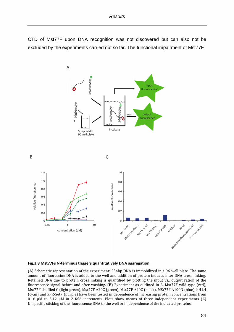
Results
84
CTD of Mst77F upon DNA recognition was not discovered but can also not be
excluded by the experiments carried out so far. The functional impairment of Mst77F
Fig.3.8 Mst77Fs N-terminus triggers quantitatively DNA aggregation
(A) Schematic representation of the experiment: 234bp DNA is immobilized in a 96 well plate. The same amount of fluorescine DNA is added to the well and addition of protein induces inter DNA cross linking. Retained DNA due to protein cross linking is quantified by plotting the input vs,. output ration of the fluorescence signal before and after washing. (B) Experiment as outlined in A. Mst77F wild-type (red), Mst77F shuffled C (light green), Mst77F 20C (green), Mst77F 40C (black), MSt77F 100N (blue), hH1.4 (cyan) and xPR-Set7 (purple) have been tested in dependence of increasing protein concentrations from 0.16 µM to 5.12 µM in 2 fold increments. Plots show means of three independent experiments (C) Unspecific sticking of the fluorescence DNA to the well or in dependence of the indicated proteins.
Results
85
shuffled C-terminal domain, 20C and 40C pointed towards an additional
component within the CTD that is needed for highly efficient DNA aggregation. Even
though hH1.4 and Mst77F 100N displayed similar effects on DNA in other
experiments, hH1.4 had no steady cross linking potential in this assay. The impact of
H1 proteins on DNA structure has been documented extensively and the
predominant model is that H1 forms sandwich structures with DNA bridging DNA
molecules through a layer of histone H1 (Jean O.Thomas, 1991). However, these
studies were solely based on imaging techniques and did not include kinetic aspects
that are an important aspect of this assay. It is conceivable that hH1.4 forms the
mentioned complexes but that they dissociated in the consecutive washing steps.
Also, xPR-Set7 had no cross linking potential and it is likely that previous interactions
with DNA represented simple electrostatic adhesion.
In summary, Mst77F efficiently interconnected individual DNA molecules in vitro. This
effect relied on the integral function of the C-terminal tail binding to DNA and the N-
terminal protein multimerization domain as the 100N mutant showed strongly
reduced cross linking. However, the impaired cross linking potential of Mst77F
shuffled C-terminal domain strongly pointed towards an additional functional
contribution of the CTD that exceeds DNA binding. Importantly, hH1.4 displayed no
cross linking at all, emphasizing different functionality in DNA/chromatin organization.
3.7 Mst77F tightly compacts long DNA in vitro
Within the nucleus of spermatids the carrier of the genetic information is several
orders of magnitude longer than the very short DNAs that have been used for the
previous experiments. During the postmeiotic maturation process of Drosophila
spermatids the DNA adapts a 200-fold higher compaction status compared to
somatic cells. This state requires extraordinary tight arrangement of the DNA.
According to the data I generated in the course of this study which clearly show
Mst77Fs potential to aggregate and interconnect even short DNAs, I proposed a
massive structural impact of the Mst77F wild-type protein on longer DNA. This effect
Results
86
should be discriminative from the 100N multimerization mutant that displays less
potential to link up individual DNA molecules, the Mst77F 60C binding mutant,
hH1.4 and xPR-Set7.
To investigate the structural effects that Mst77F and the other proteins exert on
longer, linear DNA I conducted again AFM with DNA that is 2434 bp in lenght. The
structural properties of Mst77F wild-type DNA complexes were compared to DNA
complexes that are shaped by the Mst77F 100N multimerization mutant and the
Mst77F 60C DNA binding mutant. Histone hH1.4 as a member of the H1 family
proteins that have been reported to structurally impact DNA/chromatin, was used to
discriminate the Mst77F effect on DNA from the effects of well-studied structural
proteins (Dootz et al., 2011; Shen et al., 1995). Previous experiments within this
study showed clear functional differences between Mst77F and hH1.4 and on this
basis I expected also structurally different complexes. Last but not least, xPR-Set7
that was not implicated in direct DNA binding, though displayed certain DNA binding
properties in EMSA assays, was used as a further control. The structural properties
of the DNA were imaged in the presence of 4-fold, 20-fold and 100-fold molar excess
of the indicated proteins over DNA (Fig. 3.9).
At little molar excess of protein over DNA (4:1) the observed effects were limited to
single molecules. Compared to free DNA imaging that showed DNA molecules in
their extended but flexible conformation, a 4-fold molar excess of Mst77F wild-type
structurally induced bending and looping of a fraction of molecules. Mst77F formed
hubs that interconnected parts within one molecule (top row 1:4). Importantly this
effect was intra-molecular and limited to individual molecules. A similar effect was
observed for the 100N mutant that also exerted intra-molecular connections
between parts of the same DNA molecule. DNA complexed with Mst77F 60C was
indistinguishable from DNA “only” structures reflecting its mutant DNA association.
Histone H1 has previously been shown to form fibrillar, branched and network-like
structures (“tramtracks”) with DNA mediated through sandwiching of DNA molecules
by the protein (Lu et al., 2009a; Lucius et al., 2001). At a molar ration 1 : 4 the fibrillar
and branched species were seen with low frequency. The predominant form was
indistinguishable from DNA “only” images. As expected for a non DNA binding
protein xPR-Set7 DNA complexes showed similar results to DNA only at the lowest
molar DNA protein ratio.
Results
87
Increased molar DNA-protein rations of 1:20 uncovered the specific Mst77F effect on
DNA that can be attributed to the N-terminal domain of the protein (Fig. 3.9 ; 1:20
column). Individual DNA molecules clustered and formed a “nucleus” with protruding
DNA “tentacles” that seemed to be free of protein. Within this “nucleus”
substructures were not recognizable suggesting a tightly packed, dense DNA protein
entity. With lower frequency the second observable species was individual DNA
molecules that displayed, by the majority high intra-molecular associations.
Interestingly, the DNA structures built up by the Mst77F 100N mutant were very
much alike the structures formed by hH1.4 suggesting a similar interaction mode
with the proteins packaging DNA molecules in a parallel aligned manner. Both
proteins converted individual DNA molecules into fibrillar, branched complexes
consisting of several DNAs. The recorded differences between Mst77F wild-type,
Mst77F 100N and hH1.4 nicely reflected the specific function that extend the simple
charge mediated binding to DNA. Again, as predicted no significant differences
between DNA incubated with Mst77F 60C, xPR-Set7 and DNA “only” species
became obvious.
Further increase of the molar DNA protein ratios to 1:100 led to an amplification of
the formed structures (Fig. 3.9 ; 1:100 column). The “nucleus” structures elicited by
the Mst77F wild-type protein expanded their dimensions in the Z plane. Individual
DNA molecules could not be detected anymore. A major change from scattered
fibrillar, branched towards tightly arranged, network like structures was seen for the
100N mutant possibly reflecting the impact of an excess of flexible, highly charged
peptide moieties on their counter-ion polymer. Even though hH1.4 has a similar
charge density to the 100N mutant and showed previously similar results, the
formed complexes with long DNA were distinct. At the highest monitored molar ratio
fibrillar, branched conformations were still evident but the major species constituted
spacious, parallel (and maybe lateral) interconnected DNA molecules with a small,
central high-density entity. Fundamentally, this structure was very different from the
structures built up by Mst77F wild-type. As previously shown for the lower molar
ratios there was also no effect of the Mst77F 60C protein on DNA structure at the
1:100 molar ratio. However, a 100-fold molar excess of xPR-Set7 led to branched
interconnection of DNA molecules distinct from structures seen for lower molar ratios
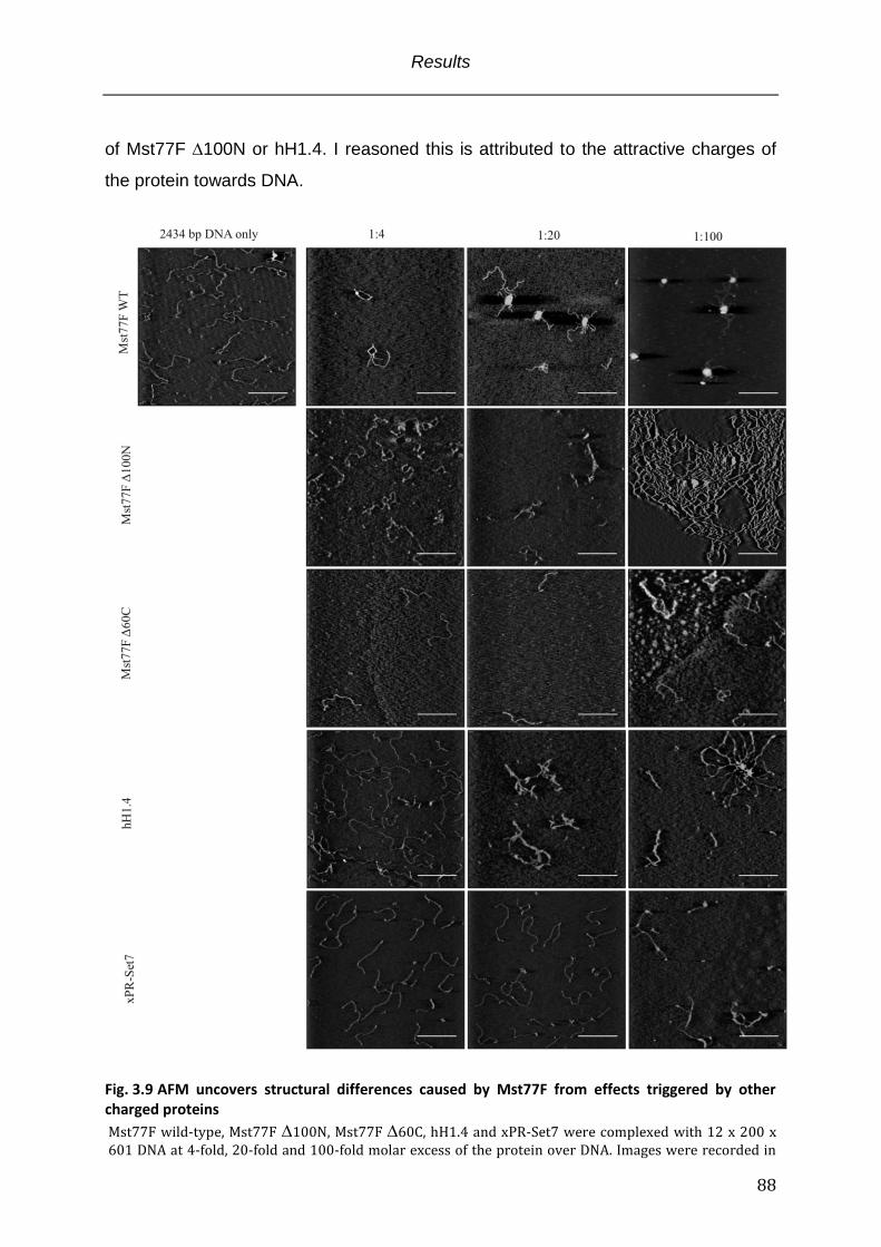
Results
88
of Mst77F 100N or hH1.4. I reasoned this is attributed to the attractive charges of
the protein towards DNA.
Fig. 3.9 AFM uncovers structural differences caused by Mst77F from effects triggered by other charged proteins
Mst77F wild-type, Mst77F 100N, Mst77F 60C, hH1.4 and xPR-Set7 were complexed with 12 x 200 x 601 DNA at 4-fold, 20-fold and 100-fold molar excess of the protein over DNA. Images were recorded in
Results
89
tapping mode. The scale bar represents 500 nm. The height scale of all images is 3 nm but for Mst77F wild-type 1:100: due to illustration the height scale was increased to 20 nm.
Taken together, Mst77F tightly compacted DNA in vitro. In very good agreement with
the centrifugation fractionation and DNA crosslinking assays this effect can be
attributed to the N-terminal region of the protein that constitutes a multimerization
module. Consequently, mutants that were impaired in DNA binding show no
structural effect on DNA. The Mst77F mutant that lacked the N-terminus induced
network like complexes with long DNAs. Importantly, histone H1 proteins that have
been reported to elicit structural effects on DNA showed different structures.
3.8 Mst77F inhibits transcription in vitro
The postmeiotic spermatid maturation requires timely onset of sequential processes
that lead to a fundamental reorganization of the genome. Mst77F has been
suggested to participate in the compaction of DNA in the transition from nucleo-
histone to a protamine organized genome (Rathke et al., 2010). My in vitro data
clearly identified Mst77F as a potent compactor of DNA, corroborating its proposed in
vivo function. However, immunocytology experiments on developing spermatids
clearly showed a heterogenous nuclear distribution of Mst77F questioning its
exclusive role as a structural protein involved in nuclear condensation (Rathke et al.,
2010). Transcriptional processes in Drosophila postmeiotic spermatids are thought to
be inhibited. Though, recent work identified elevated transciption from some loci
(Barreau et al., 2008; Vibranovski et al., 2010; Vibranovski et al., 2009). This raised
the possibility that Mst77F functions as a general repressor of transcriptional
processes.
To test the Mst77F impact on transcriptional processes, I applied an in vitro
transcription system that is based on the plasmid pG5ML (Fig. 3.10 panel A) (An,
2004). This plasmid contains five sequential Gal4 binding sites, which are used to
recruit the Gal4-VP16 transcriptional activator (Sadowski et al., 1988). The Gal4
sequences are followed by a strong viral adML-promotor and a G-less transcriptional
cassette. The transcripts of this cassette selectively can be recovered after RNAse
T1 digestion that only cleaves after G - nucleotides. The experimental scheme is
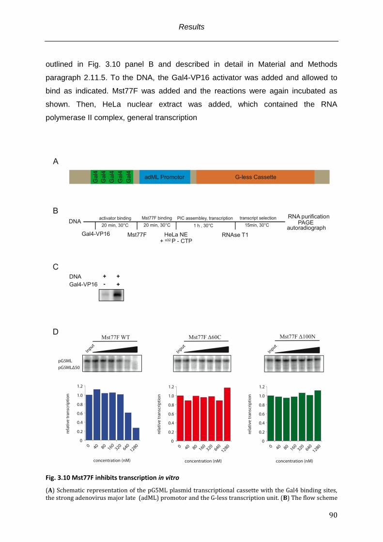
Results
90
outlined in Fig. 3.10 panel B and described in detail in Material and Methods
paragraph 2.11.5. To the DNA, the Gal4-VP16 activator was added and allowed to
bind as indicated. Mst77F was added and the reactions were again incubated as
shown. Then, HeLa nuclear extract was added, which contained the RNA
polymerase II complex, general transcription
Fig. 3.10 Mst77F inhibits transcription in vitro
(A) Schematic representation of the pG5ML plasmid transcriptional cassette with the Gal4 binding sites, the strong adenovirus major late (adML) promotor and the G-less transcription unit. (B) The flow scheme
Results
91
of the transcription experiment with DNA as transcriptional template: the system is entirely composed of recombinant components until addition of the HeLa nuclear extract. (C) Transcription reactions require an activator (Gal4-VP16): transcripts are only abundant in reactions that incl. Gal4-VP16. (D) Transcription experiments with MSt77F wild type (left), Mst77F 60C (middle) and Mst77F 100N (right): Transcription reaction was carried out according to the scheme in (B). Mst77F proteins were titrated from 40 nM up to 1.28 µM final concentrations in 2-fold increments. Purified RNAs were run on 5% urea TBE-PAGE gels. The gel was dried and exposed for 1h to a “storage-phosphor” visualization screen. The transcript band intensities were integrated with ImageJ and normalized towards the loading control pG5ML 50. The signal ratio was plotted with the Kaleidagraph software.
factors etc. that are necessary for transcription. Along with the nuclear extract a
radioactively labeled nucleotide was included and the transcription was allowed to
proceed for 1h. Unspecific transcripts were removed by RNAseT1 treatment. The G-
less cassette transcript was purified and resolved on an urea-PAGE gel and
visualized by autoradiography. In order to account for unequal recovery of
transcripts, a control transcript pG5ML50 that was co-transcribed and not incubated
with Mst77F served as purification control.
To test the functionality of the complex transcription system I compared two reaction
conditions. One transcription reaction was conducted in the presence of the
transcriptional activator Gal4-VP16. The other one missed Gal4-VP16. As expected,
in the presence of a transcriptional activator RNA transcripts could be observed (Fig
3.10 panel C). Therefore, the impact of Mst77F on transcription processes could be
tested.
Clear structural differences in DNA Mst77F complexes could be observed for Mst77F
wild-type, Mst77F 60C and Mst77F 100N. These proteins were tested for their
capability to suppress transcription (Fig 3.10 panel D). In this experiment, Mst77F
wild-type caused a dose dependent transcription repression similar to the dose
responses seen with other experiments with basically no transcripts detectable at
1.28 µM protein concentration. As expected from other experiments, Mst77F 60C
had no impact on the transcription process. No reduction in transcripts was observed.
Surprisingly, Mst77F 100N that formed network like structures with DNA was also
not capable to reduce the transcription within the concentrations tested.
Take together, the structurally different effects of MSt77F wild-type, 60C and 100N
on DNA functionally also translated into effects that effect DNA metabolism, more
precisely transcription. Whereas the wild type protein was a potent repressor both
deletion mutants, effecting either DNA binding or multimerization of the protein failed
to inhibit transcriptional processes.
Results
92
3.9 Mst77F displays similar function in the context of recombinant chromatin
Previous experiments were solely carried out with “naked” DNA as Mst77F
interaction partner because previous immunocytology experiments in Drosophila
showed that Mst77F mainly exists when histone proteins already have been
degraded (Rathke et al., 2010; Sunil Jayaramaiah Raja, 2005). However, there is a
short overlap of both protein signals in vivo that might functionally be important for
the transition from a nucleo-histone based DNA structure towards a highly
condensed protamine organization. To test the hypothesis that Mst77F is an
important factor for the progression of postmeiotic spermatid maturation, I conducted
experiments that monitored its competence to bind to recombinant chromatin. I was
interested to characterize its interaction mode with nucleosomes in comparison to
histone H1, its suggested homolog. Mst77Fs potential to cause histone eviction from
recombinant oligonucleosomal arrays was investigated along with AFM of the
resulting structures. Finally, I looked at the previously reported repressive function on
transcriptional processes in the context of recombinant chromatin.
The recombinant chromatin was assembled according to material and methods
paragraph 2.11.4 from linear 2434 bp of DNA named 12x200x601 or 147 bp
mononucleosome core particle templates. EMSA binding experiments with
12x200x601 oligonucleosomal arrays and the Mst77F wild-type protein showed the
same binding behavior as the EMSA experiments with the DNA dodecamers (Fig.
3.11 panel A left). Clear band shifting was not observable and the dose response
was very similar. At 0.64 µM protein most of the chromatin already disappeared from
the gel with complete clearance at 1.28 µM. Interestingly within a similar experiment
but with nucleosome core particles (147 bp DNA complexed with histone octamers)
that have no linker DNA Mst77F showed the exact same effect. This suggests that
the proposed unspecific electrostatic interaction mode holds also true for Mst77F
interaction with DNA in the context of chromatin (Fig. 3.11 panel A right). Histone H1
binds the nucleosome at the dyad axis where the DNA enters/leaves the histone
octamer and additionally binds approximately 20 bp of the linker DNA between
nucleosomes with its C-terminal domain (Allan et al., 1980). Mst77F was suggested
to be a distant H1 relative on the basis of sequence alignments that indeed showed a
similarity in the CTD of both proteins. My in vitro experiments uncovered fundamental
Results
93
Fig. 3.11 Mst77F effects on chromatin are similar to the observed effects with DNA
(A) EMSA experiments with Mst77F wild-type and 12x200x601 oligonucleosomes (left) or nucleosome core particles (right): the protein was titrated from 0.02 µM to 1.28 µM in 2-fold increments. The samples were run at 80 volts for 90 min on 0.6% TB-Agarose in the case of 12x200x601 chromatin or on a 5% TB-PAGE gel for the nucleosome core particles. Both gels were stained with ethidium bromide for 15 min. (B) MNase digest of 187x601 mononucleosomes alone (left), in the presence of Mst77F (middle) or hH1.4
Results
94
(right). Input means no MNase in the reaction. 200 mU of Mnase were incubated for 1 min, 2 min, 4 min or 8 min with the mononucleosomes or mononucleosome Mst77F and hH1.4 complexes, respectively, as indicated by the time bar. The DNA was phenol:chlorophorm:isoamylalcohol extracted and ethanol precipitated. For fragment size analysis DNA was run on a 5% TBE-PAGE gel for 1h @ 80 volts. (C) Mst77F chromatin coprecipitation: 12x200x601 oligonucleosomes were incubated with increasing concentrations of Mst77F wild-type protein from 0.08 to 1.28 µM in 2-fold increments. Clustering of chromatin complexes was triggered by the addition of 5 mM MgCl2. Complexes were pelleted by centrifugation and analyzed on 15% Tris-Glycine SDS-PAGE. (D) AFM with 12 x 200 x 601 chromatin and Mst77F wild-type: The addition of the protein causes intramolecular compaction at a 4-fold molar excess of Mst77F over DNA. Images were recorded in tapping mode and scale bars represent 100 nm (E) Outline of the order of addition of components during the transcription experiment with chromatin being the transcriptional template. (F) Transcription from chromatin templates requires histone acetylation: Gal4-VP16 was present in all reactions. Autoradiograph of the resolved RNAs purified after the transcription reaction on a 5% Urea TBE-PAGE gel. (G) Mst77F also inhibits transcription from pG5ML chromatin templates. Autoradiograph of purified RNAs after transcription from pG5ML chromatin and increasing Mst77F concentrations. RNAs were resolved on a 5% Urea TBE-PAGE gel and the gel was dried. Transcript band intensities were integrated and normalized towards the pG5ML50 signal with the help of the ImageJ software. Bar plot was generated with the Kaleidagraph software.
functional differences between both proteins. However, I did not work with testis
specific H1 and that might account for the different observations. To investigate the
localization of Mst77F on the nucleosome I performed MNase protection assays with
187x601 mononucleosomes, mononucleosomes complexed with hH1.4 and
mononucleosomes complexed with Mst77F (paragraph 2.11.6) (Fig. 3.11 panel B).
Without protection of the linker DNA by a protein MNase rapidly degraded the
overlapping DNA to nucleosome core particle size of 147 bp within the time course of
8 min. Addition of hH1.4 prior to the MNase resulted, as expected, in a
chromatosome stop with DNA length of approximately 167 bp caused by H1
protection. Preincubation of the mononucleosomes with Mst77F showed a
Mononucleosome only like digestion pattern with only slide protection. This result
strongly suggested that Mst77F does not bind specifically to the nucleosome in a H1
like manner.
Mst77Fs capability to evict histone proteins from the DNA was tested in chromatin
co-precipitation assays (Material and Methods paragraph 2.12.3)(Schwarz et al.,
1996). Recombinant 12x200x601 chromatin arrays were incubated with increasing
amounts of Mst77F. MgCl2 was added to induce internucleosomal clustering. Along
with the clustered oligonucleosomes associated proteins were pelleted by
centrifugation. The Mst77F and histone recovery as a result of increasing Mst77F
concentrations were visualized by SDS-PAGE (Fig. 3.11 panel C). The addition of
increasing amounts of Mst77F showed no reduction in the recovered amounts of
histone proteins compared to the absolute amounts in the input lane and the
Results
95
precipitation of 12x200x601 chromatin alone (chromatin lane). Therefore, under the
applied conditions Mst77F exerted no histone eviction.
Since Mst77F binding to chromatin could be shown by EMSA and co-precipitation
experiments I wondered weather the structural effects seen for its interaction with
DNA also hold true for its interaction with recombinant chromatin. 12x200x601
chromatin alone and with a 4-fold molar excess of Mst77F was imaged by AFM (Fig.
3.11 panel D). At the applied conditions with 50 mM NaCl the chromatin fiber
appeared loosly bend but single nucleosomes were recognizable (left). The addition
of Mst77F triggered intranucleosomal compaction (right). Remarkably, the effect of a
4-fold molar excess of Mst77F appeared stronger compared to its effect on DNA at
the same molar ratio.
Mst77Fs potential to inhibit transcriptional processes from DNA templates in vitro
could be demonstrated (paragraph 3.8). The eviction of histones in the transition
phase from histone to protamine organization during spermatid maturation may lead
to a global leakage of transcription. To counteract this effect Mst77F might function
as a structural repressor that holds up the repressed state until complete
reorganization of the genome. Its inhibitory effect on chromatin transcription was
tested again with the in vitro transcription assay. The experiment was basically
carried out as described before but had to be adapted in order to allow the
transcription from a chromatin template that is inherently silenced. Figure 3.11 panel
E shows the flow of the experiment. Along with the transcriptional activator Gal4-
VP16 the coactivator p300 (histone acetyl transferase) and its substrate acetyl-CoA
were added in the first incubation step. In this set up Gal4-VP16 recruits the added
p300 that in turn acetylates the histones. Additionally Gal4-VP16 also recruits
chromatin remodeling complexes required for ongoing transcription from the nuclear
extract. The inherently silenced state that can be activated by p300 mediated
acetylation of histones is shown in figure 3.11 panel F. That showed that the system
can actually be activated and that observed silencing effects are indeed mediated by
Mst77F. In the subsequent transcription experiment Mst77F displayed a stronger
inhibitory effect compared to the transcription from DNA templates. At 160 nM
Mst77F only 40% of the transcripts observed for the control reaction without Mst77F
are detected. At 640 nM no transcripts are detectable anymore (Figure 3.11 panel
G).
Results
96
Taken together, Mst77F also interacted with recombinant chromatin arrays. This
interaction seemed to be similar to the binding of short DNAs. Additionally,
mononucleosomes without linker DNA were bound equally well emphasizing the ionic
character of the interaction. Unlike H1 Mst77F showed no association preference for
the nucleosomal dyad axis at the entry/exit point of the DNA suggesting a functional
distinction also in the chromatin context. Under the applied conditions Mst77F was
not capable to evict histones from oligonucleosomal complexes suggesting a function
after histone removal. The strong Mst77F mediated compaction of DNA was also
seen for chromatin templates even though the Mst77F effect seemed to be stronger
at equal molar ratios. Finally, Mst77F also inhibited transcription from chromatin
templates but again showed stronger inhibition at lower concentrations. The latter
observations possibly can be accounted to the special physiochemical properties of
the chromatin fiber that enhance the Mst77F function.

Results
97
Discussion
98
4. Discussion A fundamental conserved process of evolution is the sexual reproduction of
organisms. In higher eukaryots this involves the fusion of the male and female
genomes along with their respective gametes in a process described as fertilization.
Male germ cells, the spermatids undergo a series of developmental processes in
order to be capable of fertilizing the female oocyte. This involves mitotic as well as
meiotic divisions and finally results in a superior compaction of the sperm DNA along
with substantial morphological rearrangements, a process referred to as spermatid
maturation.
Although the Mst77F gene has been discovered almost two decades ago and the
spaciotemporal expression pattern of the protein has been investigated, its function
in the process of spermatid maturation is not resolved. The very few existing studies
on the protein suggest an involvement in DNA compaction and nuclear shaping but
by what mechanism is unclear. Due to limited experimental tools in vivo no prove for
these hypothesis could be demonstrated. To circumvent the existing in vivo
limitations I characterized the molecular organization of Mst77F and its interaction
with DNA by in vitro techniques. I identified Mst77F to be a protein of bivalent
structural functionality. The intrinsically unstructured C-terminus binds DNA
unspecifically with high affinity in a charge-mediated manner. This interaction induces
homo-multimerization of Mst77 through its N-terminus and results in tight compaction
of DNA. Importantly, these effects are distinct from histone H1, the proposed
homolog. My results support the suggested function of Mst77F as a structural
protein involved in the condensation of the Drosophila sperm genome during
postmeiotic spermatid maturation.
4.1 The implication of intrinsically unstructured, charged domains in DNA binding
The spatiotemporal expression pattern of Mst77F in Drosophila postmeiotic
spermatid maturation has been investigated (Rathke et al., 2010; Sunil Jayaramaiah
Raja, 2005). The protein becomes expressed prior to tight condensation of the
Discussion
99
genome. Due to its sequence similarities of the C-terminal domain (CTD) towards
histone H1 family proteins, major structural proteins of cellular chromatin, it has been
proposed to be part of the DNA compaction process. This requires a physical
interaction with DNA in the first place. In my experiments I could show that Mst77F
binds DNA in vitro via its highly charged C-terminal domain (Fig. 3.2). Protein
structural analyses that were based on the Mst77F sequence and CD spectroscopy
identified an intrinsically unstructured random coil conformation of the recombinant
Mst77F C-terminus in solution (Fig. 3.1 and 3.7). Binding experiments with a Mst77F
sequence shuffled CTD protein showed wild-type like binding affinity strongly pointing
towards little structural contribution in the binding process. Additional binding
experiments with DNA protected by histone octamers, distorted DNA and single
stranded DNA resulted in wild-type like interactions underpinning the electrostatic
binding determinant (Fig. 3.2, 3.3 and Tabl. 3.1).
On first sight this proposed interaction mode appears unusual. However, intrinsically
unstructured domains often are involved in binding and macromolecular recognition
processes (Dunker and Obradovic, 2001; Hansen et al., 2006). Within such domains
an elevated level of charged residues is characteristic and suggests strong
electrostatic contributions in binding events (Vucetic et al., 2003). In particular, it
could be shown that proteins that alter chromatin folding and compaction frequently
make use of long, charged, intrinsically disordered domains. Recent work
demonstrated that the side specific repressive function of Polycomb Repressive
Complex 1 that compacts chromatin and inhibits remodeling processes, relies on one
of its subunits that interacts with DNA through an intrinsically unstructured, highly
charged domain (Beh et al., 2012; Brown et al., 2011). Also, studies on global
genome architectural proteins show that unspecific protein DNA interactions guided
by electrostatic binding modes are common mechanisms within this group (Dow et
al., 2000; Lu et al., 2009a; Luger et al., 1997).
In the nucleus of almost every eukaryotic cell the organization of DNA is
predominantly achieved through core histone DNA complexes, the nucleosomes.
These complexes are held together by a multitude of electrostatic interactions.
Moreover, beyond the repetitive nucleosomal units the DNA is organized through
additional proteins that physically interact with DNA and further affect the genome
structure. Importantly, some of these auxiliary proteins share common
Discussion
100
physiochemical properties that are also present in Mst77F: (a) they are in part
unstructured and (b) additionally show highly basic domains of low compositional
complexity. Histone H1, amongst others implicated in structural stabilization and
compaction of chromatin, shows high affinity binding to the nucleosomal linker DNA
largely mediated by its long unstructured basic C-terminus (Allan et al., 1980; Fang et
al., 2012). Moreover, the HMG-D protein that is associated with condensed
chromatin during developmental processes, binds unspecifically to DNA, in part via
its highly charged, unstructured C-terminus (Dow et al., 2000; Ner et al., 2001).
The involvement of intrinsically disorder domains with high charge density seems to
be a general mechanism in recognition and organization of DNA on the genome- and
local level, respectively. Recently, unstructured domains were also implicated in
protein-protein interactions and the observed physiochemical deviations across
proteins may therefore reflect functionalized domains that convey dedicated effects.
However, the rapid evolution of unstructured domains results in low sequence
conservation and makes a functional prediction difficult (Coletta et al., 2010; Tompa,
2002).
4.2 Induction of structural elements in intrinsically unstructured domains is functionally relevant
A proposed feature of intrinsically unstructured domains is their transition from
disordered to ordered conformations (Uversky, 2002). This process is coupled to
recognition of their respective binding partners. For histone H1 proteins it could be
shown that the C-terminal domain adapts -helical structures upon binding to DNA.
However, the conformational stabilization does not occur across the long C-terminal
domain but is limited to two short, discontinued sub-domains. The formation of these
local secondary structural elements is essential for protein function even though the
initial protein DNA interaction is considered to be purely ionic (Clark et al., 1988; Lu
et al., 2009a; Lu and Hansen, 2004; Vila et al., 2001).
The suggested function of Mst77F in sperm DNA compaction makes it conceivable
that Mst77F compacts DNA by a similar mechanism as histone H1 influences
chromatin structure. This essentially involves the structural refinement of H1 CTD
sub-domains upon DNA binding through electrostatic interactions (Fang et al., 2012).
Discussion
101
Whereas the electrostatic contribution in the binding reaction could be demonstrated
for Mst77F, an induction of secondary structural elements became not apparent in
my experiments. The thermodynamic parameters of the Mst77F DNA interaction
determined by microcalorimetry experiments could underpin the ionic interaction
mode. But, ITC did not hint the formation of structural elements in the CTD as would
be reflected by a positive entropy of the binding reaction. However, the high negative
entropy value suggests a very flexible conformation of Mst77F, something that is also
seen for histone H1 (Fang et al., 2012). A possible reason why I could not detect
structural formation might be due to the small DNA dodecamers used in this
experiment. The size of the continuous interaction interface might be critical for
secondary structure formation.
However, the non-gradual loss in binding affinity observed for the transition from
Mst77F 20C towards 40C does not correlate with the proportional decrease in the
charge sum and might indeed reflect functional rearrangements upon DNA binding in
the region between 20C and 40C. Interestingly, the Mst77F bioinformatic
sequence analysis revealed two regions of low compositional complexity that can be
correlated with this part of the protein. In comparing Mst77F wild-type proteins with
amino acid shuffled CTD Mst77F no significant change in binding affinity was
observed. However, experiments that addressed intermolecular cross linking of DNA
by these proteins show less efficient interconnection of individual DNA molecules by
the shuffled CTD Mst77. These observations point towards separation of molecular
recognition and function. Importantly, in the histone H1 CTD secondary structure
formation seems not to depend on the amino acid sequence. Rather the overall
composition and the relative intra-molecular position towards other structural or
functional reference points was found to be relevant (Lu et al., 2009a and references
therein). By shuffling the last 70 amino acids of the Ms77F C-terminus the low
compositional complexity regions received a distinct composition of amino acids. For
the histone H1 CTD it could be shown that upon replacement of nonpolar amino
acids in one of its structurally inducible regions, H1 effects on chromatin folding were
partially disrupted. Similarly, the Mst77F shuffled CTD mutant was less efficient in
interconnecting/aggregating individual DNA molecules. This finding might indeed
point towards an induced secondary structure within the last 40 amino acids of the
Mst77F CTD upon DNA binding.
Discussion
102
Unfortunately, only little is known about the protein chemistry that underlies the
connections between amino acid composition, intrinsic disorder, molecular
recognition and folding. To what extend the CTD contributes to Mst77F function,
effects that exceed molecular recognition, could not be clearified in this study. On the
assumption that the Mst77F CTD is a intrinsically unstructured domain I propose a
induced structure upon DNA binding. This structure might be necessary for high
efficient interconnection of DNA and therefore DNA condensation during Drosophila
spermatogenesis. Therefore, further experiments that address structural details
within the Mst77F CTD and how they possibly impact Mst77Fs function are
necessary to underpin these interpretations.
4.3 Structural aspects of Mst77F DNA complexes AFM imaging of Mst77F and H1 DNA complexes identified major differences in their
ability to alter DNA structures. hH1.4 induces so called “tramtrack” structures with
long DNA (Fang et al., 2012; Jean O.Thomas, 1991), structures of parallel aligned
DNA molecules sandwiched by H1 proteins. Mst77F in contrast induced massive
nucleation of individual DNA molecules forming highly condensed structures.
Importantly, this effect relied on the N-terminal domain of Mst77F and the DNA
molecules protruding out of these “nuclei” are void of Mst77F. Bioinformatic analysis
of the Mst77F sequence in conjunction with CD spectroscopy of Mst77F 110C
identified predominant -helical structures in this part of the protein. In particular, a
coiled coil motif could be annotated. Chemical cross linking of the wild-type protein in
presence of DNA revealed a stoichiometric multimerization pattern that was not seen
without DNA or with the 100N mutant. In solution only little Mst77F dimer and no
multimers could be detected suggesting either a weak coiled coil multimerization
stabilized through immobilization of molecules on the DNA via the CTD or an induced
cooperative multimerization mode upon DNA binding. The latter model seems more
likely since AFM with long DNA and Mst77F clearly showed non-distributive binding
of Mst77F along the DNA. Some regions displayed massive DNA condensation
whereas others were free of protein and adapted extended conformations. Therefore,
Discussion
103
the CTD interaction with DNA likely causes high affinity homomultimerization of
Mst77F that is several orders of magnitude stronger than in solution.
Supportive data came from the centrifugation fractionation experiments with Mst77F
and very short DNA. Mst77F and DNA dodecamers formed fast sedimenting high
molecular weight complexes clearly visible in AFM. The formation of lattice like
structures through simple electrostatic interactions can be ruled out since the Mst77F
100N mutant and hH1.4 did not show aggregation. However, for histone H1 a
cooperative binding mode to DNA could be demonstrated and the interaction
interface was mapped to the H1/H5 globular domain (Clark and Thomas, 1986; Jean
O.Thomas, 1991). Importantly, although H1 cooperativity has been reported
aggregation of the short DNA dodecamers was not observed. If both proteins,
Mst77F and H1 induce altered DNA structures through cooperative binding, and both
need their globular domains what are the molecular determinants for the observed
major differences? 1. Cooperative DNA binding between H1 molecules is mediated
by the globular domain and the contribution of the CTD is unclear (Clark and
Thomas, 1986; Jean O.Thomas, 1991). In contrast, binding of Mst77F to DNA takes
place through its CTD and induces cooperativity that likely involves the N-terminus
(Fig. 3.6 and 3.8). The structural requirements for cooperativity are unknown and
might involve structural adaptation of the CTD upon DNA recognition. From my
experiments it became not clear whether the Mst77F CTD undergoes structural
stabilization even though the protein mediated DNA aggregation assay indicates
functional relevant contribution of the Mst77F CTD. This might indeed be due to
induced structures in this region of the protein 2. Additionally, the association with
DNA seems to result in differential kinetic stability of the different protein DNA
complexes. Surprisingly, even though H1 proteins strongly associate with DNA and
also form aggregates, the complex is not stable. In the protein induced DNA cross
linking assay extensive washing removed all fluorescence DNA in the wells were
hH1.4 was tested for its cross-linking potential. This is actually in good agreement
with previous studies that identified an average H1 residence time of 100 s in vivo
(Stasevich et al., 2010). Mst77F 100N DNA complexes displayed kinetic stability
even though the overall recovery was largely reduced compared to the wild-type
DNA complexes. The suggested kinetic stability of the protein DNA complex could
Discussion
104
also be shown by SPR experiments with the wild-type protein and DNA dodecamers
that showed virtually no dissociation (data not shown).
According to the binding, thermodynamic and structural data I propose the following
model how Mst77F interacts with DNA:
In solution the Mst77F CTD is flexible and extended due to electrostatic repulsion
between positively charged amino acids. In this conformation protein protein
interactions via the N-terminus are rare and the protein samples its environment for
interaction partners. Upon recognition of DNA by the CTD, Mst77F undergoes a
conformational change that increases its affinity for self-interactions dramatically. The
CTD on it own can aggregate multiple individual DNA molecules in a one-
dimensional manner. However, spreading of aggregation towards highly condensed
structures is mediated by cooperative protein multimerization (Fig 4.1).
Two scenarios how cooperativity is induced are conceivable. The first scenario
implies a self-inhibitory mechanism. The flexible extended CTD folds back on the N-
terminus and blocks the self-interaction domain. The recognition of DNA that
constitutes a longer continuous interaction interface and is therefore bound with
higher affinity (Fig. 3.3) liberates the N-terminus that is now prone to multimerization.
The second possibility involves a functional structure in the CTD that becomes
induced upon DNA recognition and in turn interacts with the -helical N-terminus of a
different Mst77F molecule. I favor the second model for the following reasons: (1)
The stabilizing charged amino acids within the coiled coil are all anionic and
homomultimers likely become destabilized. This is probably what is seen for Mst77F
in solution where only little dimer can be detected. However, induced folded
structures within the cationic CTD that are capable to interact with the N-terminal
coiled coil will therefore be stabilized. (2) For histone H1 it could be shown that a
change in CTD amino acid composition partially disrupts its function likely through
alterations in the induced structures that convey functionality upon DNA recognition.
For the Mst77F shuffled CTD protein a decrease in protein induced DNA cross-
linking capacity could be demonstrated (Fig. 3.8). The importance of a dedicated
amino acid composition for function supports the idea of inducible secondary
structures in the Mst77F CTD that interact with the N-terminal part of the protein to
form multimers.
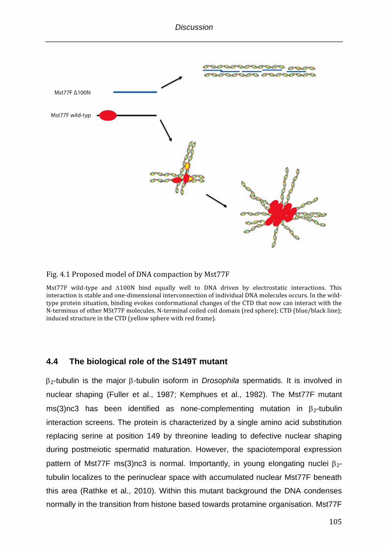
Discussion
105
Fig. 4.1 Proposed model of DNA compaction by Mst77F
Mst77F wild-type and 100N bind equally well to DNA driven by electrostatic interactions. This interaction is stable and one-dimensional interconnection of individual DNA molecules occurs. In the wild-type protein situation, binding evokes conformational changes of the CTD that now can interact with the N-terminus of other MSt77F molecules. N-terminal coiled coil domain (red sphere); CTD (blue/black line); induced structure in the CTD (yellow sphere with red frame).
4.4 The biological role of the S149T mutant 2-tubulin is the major -tubulin isoform in Drosophila spermatids. It is involved in
nuclear shaping (Fuller et al., 1987; Kemphues et al., 1982). The Mst77F mutant
ms(3)nc3 has been identified as none-complementing mutation in 2-tubulin
interaction screens. The protein is characterized by a single amino acid substitution
replacing serine at position 149 by threonine leading to defective nuclear shaping
during postmeiotic spermatid maturation. However, the spaciotemporal expression
pattern of Mst77F ms(3)nc3 is normal. Importantly, in young elongating nuclei 2-
tubulin localizes to the perinuclear space with accumulated nuclear Mst77F beneath
this area (Rathke et al., 2010). Within this mutant background the DNA condenses
normally in the transition from histone based towards protamine organisation. Mst77F
Discussion
106
ms(3)nc3 is associated with the DNA but the spermatids do not undergo elongation.
In good aggrement with this in vivo observation, Mst77F S149T behaved similarly as
the wild-type protein in DNA binding and aggregation assays (data not shown)
confirming the intact protein DNA interaction interface and condensation function in
vitro (Fig 3.2). The CTD of Mst77F is likely intrinsically unstructured and shows
physiochemical similarities towards the histone H1 CTD. Along with the proposed
DNA binding and structural functions, the histone H1 CTD exhibits interface functions
in histone H1 protein-protein interactions (Kim K, 2008; Widlak P, 2005).
Interestingly, 2-tubulin is proposed to be unstable and in spermatids of sea urchins
axonems (microtubule structures of the sperm tail) of the a tubulin stabilizing function
of histone H1 could be shown suggesting a similar function of Mst77F in the process
of nuclear shaping during spermatid development (Multigner et al., 1992). Supportive
data comes from the observation that 2-tubulin is less abundant in later stages of
spermatid maturation. The stabilization likely would involve steady physical
interaction with 2-tubulin and an interaction interface involving serine 149. The
mutation towards threonine would abolish the interaction leading to tubulin
degradation. However, this hypothesis suggests a dual function of Mst77F: (a) DNA
condensation and (b) nuclear shaping by tubulin stabilization. Whether this involves
an orchestrated dual binding of Mst77F to DNA and to 2-tubulin is elusive. Additional
experiments will have to clearify if both proteins interact in the first place.
4.5 Mst77F association with chromatin in vivo Mst77Fs nuclear localization in vivo is detectable at the onset of the young
elongating nuclei stage. At this time histone proteins are abundant and might
interfere with Mst77Fs DNA association. As expected Mst77F also bound
recombinant chromatin in vitro. In my experiments this binding was conceptually
different from histone H1 binding since no association preference for the nucleosome
dyad axis at the entry/exit point of the DNA could be found in MNase protection
assays. However, Mst77F bound nucleosome core particles suggesting that the
association is random and also driven by electrostatic mechanisms. In vivo this might
be different due to chaperone proteins that guide Mst77F deposition similar to what
Discussion
107
could be shown for histone H1 (Finn et al., 2008). Though, the differences between
Mst77F and histone H1 in their molecular organsization translate into distinct
functionality in interactions with DNA in vitro. The diverging interaction mode with
recombinant chromatin might also reflect specialized functions in the context of
chromatin. In mus musculus and homo sapiens the Hils1 protein is proposed to be a
spermatid specific linker histone-like protein. The spaciotemporal expression pattern
of Mst77F and Hils1 is related but not coherent. Hils1 disappears at the stage of
elongating nuclei whereas Mst77F is a component of mature spermatids.
Unfortunately, also only very little is known about Hils1 but in vitro it also binds
mononucleosomes in a H1 like manner and unlike Mst77F produces chromatosome
stops (Yan et al., 2003). On the assumption that related processes are guided by
similar mechanisms it is unlikely that Mst77F and Hils1 represent homologs.
The distinct binding mode from H1 in vitro and the global colocalisation with the core
histones in vivo functionally might reflect a Mst77F role in the histone eviction
processes. Mst77F first localizes to chromatin concomitantly with histone H4 hyper-
acetylation in young elongating nuclei. Hyper-acetylation is proposed to be necessary
but not sufficient for histone eviction (Rathke et al., 2007). In vitro Mst77F was not
capable to evict histone proteins from unmodified recombinant chromatin raising the
possibility that acetylation might be a prerequisite. However, the direct eviction of
histone proteins by Mst77F is rather unlikely since most of the histones are removed
in later stages of spermatid maturation between the early and late canoe stage.
Conceivable is a scenario that involves Mst77F as a recruiting molecule. This
function could involve the N-terminal coiled coil domain as well as the suggested
induced structure in the CTD as interaction domains.
In early canoe stage nuclei the chromatin has been primed for histone eviction by
global ubiquitinylation and acetylation and DNA bound Mst77F could function as a
platform for executing factors. This would involve enzymes that introduce DNA nicks
and chromatin remodelling comlexes which facilitate histone expulsion (Rathke et al.,
2007). For histone H1 it could be shown that it recruits apoptosis specific nucleases
via its CTD and stimulates DNA cleavage in programmed cell death (Widlak P,
2005). A similar function of Mst77F in the process of histone removal by recruiting
DNA nicking enzymes is therefore conceivable and would make Mst77F essential for
the transition from nucleo-histone towards protamine organization.
Discussion
108
Furthermore, it is a generally accepted paradigm that transcription is silenced during
postmeiotic spermatid maturation in Drosophila whereas in mammals transcription
has been reported. However, recent studies challenged that point of view by
providing evidence for solid ongoing transcription. Importantly, this transcription was
found to occur during chromatin remodeling (Barreau et al., 2008; Vibranovski et al.,
2010). The functional implications of these transcripts and their products are largely
unknown. However, a repressive function of Mst77F on transcription could be
demonstrated in vitro. It might be that Mst77F localization “marks” regions of silent
chromatin whereas Mst77F free regions are prone to transcription during remodeling
processes. Whether Mst77F exerts its repressive effect on its own or represents the
platform for interacting proteins that in turn execute their repressive function is
unclear. A genome wide Mst77F localization study and correlation of these data with
mRNA expression profiles could give answers to this question.
The apparently oversimplified in vitro situation where Mst77F strongly condenses
DNA upon recognition is not found in vivo. The initial chromatin association in early
canoe shaped nuclei does not instantly cause condensation of the genome.
According to the model that I proposed in the previous paragraph, Mst77F might be
structurally inactivated - that is the incapability to multimerize. This effect might be
due to an interaction partner (like a chaperone) that binds to the induced structure in
the CTD upon DNA recognition or stays associated also after deposition. Again, for
histone H1 proteins interaction partner dependent functions could be demonstrated
and this might also hold true for Mst77F (Happel and Doenecke, 2009). Upon histone
expulsion the inhibiting factor dissociates, Mst77F becomes ”activated” and replaces
the histones as a genome architectural protein. The reason why Mst77F initially binds
DNA/chromatin in an “inactive” conformation or becomes “inactivated” might be due
to additional functions prior to DNA condensation (see above: nuclear shaping –
interaction with tubulin; transcriptional inhibition etc.). Concomitantly also transition
proteins and little later protamines associate with the nascent DNA structures. The
functional boundaries between Mst77F, transition proteins and protamine are totally
unclear. Additionally, transition proteins as well as protamines are dispensible in
Drosophila leaving Mst77F as the major structural genome organizing protein.
Discussion
109
4.6 Outlook To understand the complex process of spermatogenesis, in particular the
transition from the nucleo-histone towards protamine organisation of the genome,
it will be necessary to integrate the function of orchestrated processes like histone
modifications, histone eviction and degradation, DNA breaks and condensation,
nuclear shaping, variant structural proteins etc.. One piece in this puzzle is
Mst77F that is suggested to take part in condensation processes, though is also
implicated in nuclear shaping mediated by microtubules. The spaciotemporal
expression pattern that shows Mst77F association with the nucleo-histone
genome configuration questions the simple function of a genome organizer
suggested by my in vitro experiments. Similar to histone H1 proteins the Mst77F
function might be versatile and different functions may depend on the respective
interaction partner or posttranslational modification (Happel and Doenecke,
2009). On the basis of my in vitro data and the accessible information from other
studies I consider Mst77F to be different from histone H1 proteins even though
they share physiochemical parameters and intrinsically unstructured domains that
are vital in varying processes. To date no Mst77F homolog in mammals could be
faithfully identified. The fact that the Drosophila genome condenses
approximately 10 fold stronger than its mammalian counterpart raises the
possibility that a homolog does not exist.
Further experiments addressing interaction partners, global genome localization
and Mst77Fs proposed role in nuclear shaping have to be carried out to
understand its exact role in Drosophila spermatogenesis. Eventually a null mutant
will answer the question if Mst77F is really essential for DNA condensation in
vivo.
Summary
110
Summary The mechanisms of spermatogenesis, in particular the process of postmeiotic
spermatid maturation, are little understood. During this developmental stage
massive molecular and morphological rearrangements occur and the major
events are conserved across higher eukaryots. This involves the condensation of
the DNA after histone removal by two major classes of proteins: (a) transition
proteins and (b) protamines. Beyond these proteins nothing is known about
additional factors that directly impact DNA condensation. In Drosophila Mst77F
has been implicated in the DNA condensation process on the basis of
immunocytology experiments and its distant sequence homology towards histone
H1 family proteins and protamines. Even though Mst77F clearly localizes inside
the nucleus and is a component of the condensed DNA complex in differentiated
sperm cells, a direct interaction with DNA, the prerequisite for condensation,
could not be demonstrated.
On the basis of these findings my hypothesis was that Mst77F is a structural
component of spermatid DNA that contributes to the condensation process. To
address this hypothesis I conducted bioinformatic analysis of the Mst77F protein
sequence to identify a putative DNA interaction domain. I found a highly charged,
intrinsically unstructured C-terminal domain. This domain shows similarity towards
the C-terminal domain of linker histone H1 that could be shown to bind
DNA/chromatin. Qualitative and quantitativ binding experiments with recombinant
Mst77F mutants and short DNAs corroborated the in silico predicted DNA
interaction interface. The Mst77F DNA interaction was further characterized in
detail by equilibrium binding experiments that identified an ionic interaction mode
and provided the thermodynamic parameters. Besides the binding to DNA the
proposed Mst77F function in DNA condensation processes was investigated.
Therefore, Mst77F complexes with short DNAs were analysed by different
biochemical assay systems and compared to a proposed homolog, histone
hH1.4. I found Mst77F specific DNA aggregation properties that rely on the N-
terminal domain of the protein. These observed differences were subsequently
Summary
111
further characterized by Atomic Force Microscopy with long DNAs. Based on the
recorded structural differences protein multimerization experiments were
conducted and the following model of the Mst77F-DNA interaction was proposed:
The Mst77F C-terminal domain constitutes a flexible, extended domain in
solution. In this conformation protein-protein interactions via the N-terminus are
rare. Upon recognition of DNA by the C-terminal domain, Mst77F undergoes a
conformational change that increases its affinity for self-interactions dramatically.
The C-terminal domain on its own can aggregate multiple individual DNA
molecules in a one-dimensional manner. However, spreading of aggregation
towards highly condensed structures is mediated by cooperative protein
multimerization.
Additionally, the functional consequences of the formed Mst77F DNA
complexes were tested in an in vitro transcription experiment. Lastly, since
Mst77F arises when the genome is still in histone-based configuration my aim
was to investigate binding-, structural- and functional effects also in the context of
recombinant chromatin.
The present work presents the first comprehensive in vitro study on a DNA
architecture protein presumably involved in DNA condensation during postmeiotic
spermatid maturation in Drosophila. The insights obtained by this study could help
to improve the understanding of spermatogenesis, in particular observed
differences between mice and flies in DNA condensation.

Summary
112
References
113
Bibliography
Combet, C., Blanchet, C., Geourjon, C., and Deleage, G. (2000). NPS@: network protein sequence analysis. Trends in biochemical sciences 25, 147-150.
Crane-Robinson, C. (1997). Where is the globular domain of linker histone located on the nucleosome? Trends in biochemical sciences 22, 75-77.
Davey, C.A., Sargent, D.F., Luger, K., Maeder, A.W., and Richmond, T.J. (2002). Solvent mediated interactions in the structure of the nucleosome core particle at 1.9 a resolution. Journal of molecular biology 319, 1097-1113.
Dignam, J.D., Lebovitz, R.M., and Roeder, R.G. (1983). Accurate transcription initiation by RNA polymerase II in a soluble extract from isolated mammalian nuclei. Nucleic acids research 11, 1475-1489.
Dootz, R., Toma, A.C., and Pfohl, T. (2011). Structural and dynamic properties of linker histone H1 binding to DNA. Biomicrofluidics 5, 24104.
Dosztanyi, Z., Csizmok, V., Tompa, P., and Simon, I. (2005a). IUPred: web server for the prediction of intrinsically unstructured regions of proteins based on estimated energy content. Bioinformatics 21, 3433-3434.
Dosztanyi, Z., Csizmok, V., Tompa, P., and Simon, I. (2005b). The pairwise energy content estimated from amino acid composition discriminates between folded and intrinsically unstructured proteins. Journal of molecular biology 347, 827-839.
Dow, L.K., Jones, D.N., Wolfe, S.A., Verdine, G.L., and Churchill, M.E. (2000). Structural studies of the high mobility group globular domain and basic tail of HMG-D bound to disulfide cross-linked DNA. Biochemistry 39, 9725-9736.
Dragan, A.I., Klass, J., Read, C., Churchill, M.E., Crane-Robinson, C., and Privalov, P.L. (2003). DNA binding of a non-sequence-specific HMG-D protein is entropy driven with a substantial non-electrostatic contribution. Journal of molecular biology 331, 795-813.
Dunker, A.K., and Obradovic, Z. (2001). The protein trinity--linking function and disorder. Nature biotechnology 19, 805-806.
Ekman, D., Light, S., Bjorklund, A.K., and Elofsson, A. (2006). What properties characterize the hub proteins of the protein-protein interaction network of Saccharomyces cerevisiae? Genome biology 7, R45.
Fan, L., and Roberts, V.A. (2006). Complex of linker histone H5 with the nucleosome and its implications for chromatin packing. Proceedings of the National Academy of Sciences of the United States of America 103, 8384-8389.
References
114
Fan, Y., Nikitina, T., Zhao, J., Fleury, T.J., Bhattacharyya, R., Bouhassira, E.E., Stein, A., Woodcock, C.L., and Skoultchi, A.I. (2005). Histone H1 depletion in mammals alters global chromatin structure but causes specific changes in gene regulation. Cell 123, 1199-1212.
Fang, H., Clark, D.J., and Hayes, J.J. (2012). DNA and nucleosomes direct distinct folding of a linker histone H1 C-terminal domain. Nucleic acids research 40, 1475-1484.
Finn, R.M., Browne, K., Hodgson, K.C., and Ausio, J. (2008). sNASP, a histone H1-specific eukaryotic chaperone dimer that facilitates chromatin assembly. Biophysical journal 95, 1314-1325.
Fuller, M.T. (1993). The development of Drosophila (Cold Spring Harbor, NY: Cold Spring Harbor Press ).
Fuller, M.T., Caulton, J.H., Hutchens, J.A., Kaufman, T.C., and Raff, E.C. (1987). Genetic analysis of microtubule structure: a beta-tubulin mutation causes the formation of aberrant microtubules in vivo and in vitro. The Journal of cell biology 104, 385-394.
Gallagher, S.R. (2006). One-dimensional SDS gel electrophoresis of proteins. Current protocols in molecular biology / edited by Frederick M Ausubel [et al] Chapter 10, Unit 10 12A.
Garner, M.M., and Revzin, A. (1981). A gel electrophoresis method for quantifying the binding of proteins to specific DNA regions: application to components of the Escherichia coli lactose operon regulatory system. Nucleic acids research 9, 3047-3060.
Gasteiger E., H.C., Gattiker A., Duvaud S., Wilkins M.R., Appel R.D., Bairoch A. (2005). Protein Identification and Analysis Tools on the ExPASy Server (Humana Press).
Gould-Somero, M.a.H., L. (1974). The timing of RNA synthesis for spermiogenesis in organ cultures of Drosophila melanogaster testes. Wilhelm Roux Arch, 133–148.
Grant, P.A. (2001). A tale of histone modifications. Genome biology 2, REVIEWS0003.
Griffith, F. (1928). The Significance of Pneumococcal Types. The Journal of hygiene 27, 113-159.
Gunasekaran, K., Tsai, C.J., Kumar, S., Zanuy, D., and Nussinov, R. (2003). Extended disordered proteins: targeting function with less scaffold. Trends in biochemical sciences 28, 81-85.
Hacques, M.F., Muller, S., De Murcia, G., Van Regenmortel, M.H., and Marion, C. (1990). Accessibility and structural role of histone domains in chromatin. biophysical and immunochemical studies of progressive digestion with immobilized proteases. Journal of biomolecular structure & dynamics 8, 619-641.
References
115
Hansen, J.C., Lu, X., Ross, E.D., and Woody, R.W. (2006). Intrinsic protein disorder, amino acid composition, and histone terminal domains. The Journal of biological chemistry 281, 1853-1856.
Happel, N., and Doenecke, D. (2009). Histone H1 and its isoforms: contribution to chromatin structure and function. Gene 431, 1-12.
Hartl, D.L., Jones, Elizabeth W. (2001). Genetics: Analysis of Genes and Genomes, Vol fifth Edition.
Hayashihara, K., Uchiyama, S., Shimamoto, S., Kobayashi, S., Tomschik, M., Wakamatsu, H., No, D., Sugahara, H., Hori, N., Noda, M., et al. (2010). The middle region of an HP1-binding protein, HP1-BP74, associates with linker DNA at the entry/exit site of nucleosomal DNA. The Journal of biological chemistry 285, 6498-6507.
Hirose, Y., and Manley, J.L. (1998). RNA polymerase II is an essential mRNA polyadenylation factor. Nature 395, 93-96.
Honig, B., Sharp, K., and Gilson, M. (1989). Electrostatic interactions in proteins. Progress in clinical and biological research 289, 65-74.
Huntley, M.A., and Golding, G.B. (2002). Simple sequences are rare in the Protein Data Bank. Proteins 48, 134-140.
Jacobs, S.A., W. Fischle, and S. Khorasanizadeh (2004). Assays for the determination of structure and dynamics of the interaction of the chromodomain with histone peptides, Vol 376.
Jean O.Thomas, C.R.a.J.T.F. (1991). Cooperative binding of the globular domains of histones H1 and H5 to DNA. Nucleic acids research 20, 187-194.
Jedrusik, M.A., and Schulze, E. (2001). A single histone H1 isoform (H1.1) is essential for chromatin silencing and germline development in Caenorhabditis elegans. Development 128, 1069-1080.
Jelesarov, I., and Bosshard, H.R. (1999). Isothermal titration calorimetry and differential scanning calorimetry as complementary tools to investigate the energetics of biomolecular recognition. Journal of molecular recognition : JMR 12, 3-18.
Jenuwein, T., and Allis, C.D. (2001). Translating the histone code. Science 293, 1074-1080.
Johnson, W.C., Jr. (1990). Protein secondary structure and circular dichroism: a practical guide. Proteins 7, 205-214.
Jones, S., Shanahan, H.P., Berman, H.M., and Thornton, J.M. (2003). Using electrostatic potentials to predict DNA-binding sites on DNA-binding proteins. Nucleic acids research 31, 7189-7198.
References
116
Kemphues, K.J., Kaufman, T.C., Raff, R.A., and Raff, E.C. (1982). The testis-specific beta-tubulin subunit in Drosophila melanogaster has multiple functions in spermatogenesis. Cell 31, 655-670.
Kim K, C.J., Heo K, Kim H, Levens D, Kohno K, Johnson EM, Brock HW, An W. (2008). Isolation and characterization of a novel H1.2 complex that acts as a repressor of p53-mediated transcription. The Journal of biological chemistry.
Klass, J., Murphy, F.V.t., Fouts, S., Serenil, M., Changela, A., Siple, J., and Churchill, M.E. (2003). The role of intercalating residues in chromosomal high-mobility-group protein DNA binding, bending and specificity. Nucleic acids research 31, 2852-2864.
Kornberg, R.D. (1974). Chromatin structure: a repeating unit of histones and DNA. Science 184, 868-871.
Kouzarides, T. (2007). SnapShot: Histone-modifying enzymes. Cell 131, 822.
Leuba, S.H., and Bustamante, C. (1999). Analysis of chromatin by scanning force microscopy. Methods Mol Biol 119, 143-160.
Lis, J.T., and Schleif, R. (1975). Size fractionation of double-stranded DNA by precipitation with polyethylene glycol. Nucleic acids research 2, 383-389.
Lowary, P.T., and Widom, J. (1998). New DNA sequence rules for high affinity binding to histone octamer and sequence-directed nucleosome positioning. Journal of molecular biology 276, 19-42.
Lu, X., Hamkalo, B., Parseghian, M.H., and Hansen, J.C. (2009a). Chromatin condensing functions of the linker histone C-terminal domain are mediated by specific amino acid composition and intrinsic protein disorder. Biochemistry 48, 164-172.
Lu, X., and Hansen, J.C. (2004). Identification of specific functional subdomains within the linker histone H10 C-terminal domain. The Journal of biological chemistry 279, 8701-8707.
Lu, X., Wontakal, S.N., Emelyanov, A.V., Morcillo, P., Konev, A.Y., Fyodorov, D.V., and Skoultchi, A.I. (2009b). Linker histone H1 is essential for Drosophila development, the establishment of pericentric heterochromatin, and a normal polytene chromosome structure. Genes & development 23, 452-465.
Lucius, H., Haberland, A., Zaitsev, S., Dalluge, R., Schneider, M., and Bottger, M. (2001). Structure of transfection-active histone H1/DNA complexes. Molecular biology reports 28, 157-165.
Luger, K., Mader, A.W., Richmond, R.K., Sargent, D.F., and Richmond, T.J. (1997). Crystal structure of the nucleosome core particle at 2.8 A resolution. Nature 389, 251-260.
Luger, K., Rechsteiner, T.J., and Richmond, T.J. (1999). Expression and purification of recombinant histones and nucleosome reconstitution. Methods Mol Biol 119, 1-16.
References
117
Lupas, A., Van Dyke, M., and Stock, J. (1991). Predicting coiled coils from protein sequences. Science 252, 1162-1164.
Maresca, T.J., Freedman, B.S., and Heald, R. (2005). Histone H1 is essential for mitotic chromosome architecture and segregation in Xenopus laevis egg extracts. The Journal of cell biology 169, 859-869.
Mason, J.M., and Arndt, K.M. (2004). Coiled coil domains: stability, specificity, and biological implications. Chembiochem : a European journal of chemical biology 5, 170-176.
McKearin, D. (1997). The Drosophila fusome, organelle biogenesis and germ cell differentiation: if you build it. BioEssays : news and reviews in molecular, cellular and developmental biology 19, 147-152.
Meyer, T.S.L., B. L. (1965). Use of Coomassie brilliant blue R250 for the electrophoresis of microgram quantities of parotid saliva proteins on acrylamide-gel strips. Biochimica et Biophysica Acta 107 (1): 144–145.
Mitchell, P.J., and Tjian, R. (1989). Transcriptional regulation in mammalian cells by sequence-specific DNA binding proteins. Science 245, 371-378.
Mullis, K., Faloona, F., Scharf, S., Saiki, R., Horn, G., and Erlich, H. (1986). Specific enzymatic amplification of DNA in vitro: the polymerase chain reaction. Cold Spring Harbor symposia on quantitative biology 51 Pt 1, 263-273.
Multigner, L., Gagnon, J., Van Dorsselaer, A., and Job, D. (1992). Stabilization of sea urchin flagellar microtubules by histone H1. Nature 360, 33-39.
Nelson, H.C., Finch, J.T., Luisi, B.F., and Klug, A. (1987). The structure of an oligo(dA).oligo(dT) tract and its biological implications. Nature 330, 221-226.
Ner, S.S., Blank, T., Perez-Paralle, M.L., Grigliatti, T.A., Becker, P.B., and Travers, A.A. (2001). HMG-D and histone H1 interplay during chromatin assembly and early embryogenesis. The Journal of biological chemistry 276, 37569-37576.
Nikitina, T., Shi, X., Ghosh, R.P., Horowitz-Scherer, R.A., Hansen, J.C., and Woodcock, C.L. (2007). Multiple modes of interaction between the methylated DNA binding protein MeCP2 and chromatin. Molecular and cellular biology 27, 864-877.
Nishikawa, K., and Scheraga, H.A. (1976). Geometrical criteria for formation of coiled-coil structures of polypeptide chains. Macromolecules 9, 395-407.
Nishioka, K., Rice, J.C., Sarma, K., Erdjument-Bromage, H., Werner, J., Wang, Y., Chuikov, S., Valenzuela, P., Tempst, P., Steward, R., et al. (2002). PR-Set7 is a nucleosome-specific methyltransferase that modifies lysine 20 of histone H4 and is associated with silent chromatin. Molecular cell 9, 1201-1213.
O'Hare, P., and Williams, G. (1992). Structural studies of the acidic transactivation domain of the Vmw65 protein of herpes simplex virus using 1H NMR. Biochemistry 31, 4150-4156.
References
118
Obradovic, Z., Peng, K., Vucetic, S., Radivojac, P., Brown, C.J., and Dunker, A.K. (2003). Predicting intrinsic disorder from amino acid sequence. Proteins 53 Suppl 6, 566-572.
Olivieri, G., and Olivieri, A. (1965). Autoradiographic study of nucleic acid synthesis during spermatogenesis in Drosophila melanogaster. Mutation research 2, 366-380.
Patterton, H.G., Landel, C.C., Landsman, D., Peterson, C.L., and Simpson, R.T. (1998). The biochemical and phenotypic characterization of Hho1p, the putative linker histone H1 of Saccharomyces cerevisiae. The Journal of biological chemistry 273, 7268-7276.
Phair, R.D., Scaffidi, P., Elbi, C., Vecerova, J., Dey, A., Ozato, K., Brown, D.T., Hager, G., Bustin, M., and Misteli, T. (2004). Global nature of dynamic protein-chromatin interactions in vivo: three-dimensional genome scanning and dynamic interaction networks of chromatin proteins. Molecular and cellular biology 24, 6393-6402.
Phatnani, H.P., and Greenleaf, A.L. (2006). Phosphorylation and functions of the RNA polymerase II CTD. Genes & development 20, 2922-2936.
Ponte, I., Vila, R., and Suau, P. (2003). Sequence complexity of histone H1 subtypes. Molecular biology and evolution 20, 371-380.
Ramakrishnan, V., Finch, J.T., Graziano, V., Lee, P.L., and Sweet, R.M. (1993). Crystal structure of globular domain of histone H5 and its implications for nucleosome binding. Nature 362, 219-223.
Rasband, W.S. (1997 - 2011). ImageJ.
Rathke, C., Baarends, W.M., Jayaramaiah-Raja, S., Bartkuhn, M., Renkawitz, R., and Renkawitz-Pohl, R. (2007). Transition from a nucleosome-based to a protamine-based chromatin configuration during spermiogenesis in Drosophila. Journal of cell science 120, 1689-1700.
Rathke, C., Barckmann, B., Burkhard, S., Jayaramaiah-Raja, S., Roote, J., and Renkawitz-Pohl, R. (2010). Distinct functions of Mst77F and protamines in nuclear shaping and chromatin condensation during Drosophila spermiogenesis. European journal of cell biology 89, 326-338.
Romero, P., Obradovic, Z., Li, X., Garner, E.C., Brown, C.J., and Dunker, A.K. (2001). Sequence complexity of disordered protein. Proteins 42, 38-48.
Roque, A., Iloro, I., Ponte, I., Arrondo, J.L., and Suau, P. (2005). DNA-induced secondary structure of the carboxyl-terminal domain of histone H1. The Journal of biological chemistry 280, 32141-32147.
Roque, A., Ponte, I., Arrondo, J.L., and Suau, P. (2008). Phosphorylation of the carboxy-terminal domain of histone H1: effects on secondary structure and DNA condensation. Nucleic acids research 36, 4719-4726.
References
119
Russell, S.R., and Kaiser, K. (1993). Drosophila melanogaster male germ line-specific transcripts with autosomal and Y-linked genes. Genetics 134, 293-308.
Sadowski, I., Ma, J., Triezenberg, S., and Ptashne, M. (1988). GAL4-VP16 is an unusually potent transcriptional activator. Nature 335, 563-564.
Sambrook, J., and Russell, D.W. (2001). Molecular cloning: A Laboratory Manual 3rd Edition (Cold Spring Harbor, N.Y., Cold Spring Harbor Laboratory Press).
Sandhu, K.S., and Dash, D. (2007). Dynamic alpha-helices: conformations that do not conform. Proteins 68, 109-122.
Schultz, J., Milpetz, F., Bork, P., and Ponting, C.P. (1998). SMART, a simple modular architecture research tool: identification of signaling domains. Proceedings of the National Academy of Sciences of the United States of America 95, 5857-5864.
Schwarz, P.M., Felthauser, A., Fletcher, T.M., and Hansen, J.C. (1996). Reversible oligonucleosome self-association: dependence on divalent cations and core histone tail domains. Biochemistry 35, 4009-4015.
Shazman, S., Celniker, G., Haber, O., Glaser, F., and Mandel-Gutfreund, Y. (2007). Patch Finder Plus (PFplus): a web server for extracting and displaying positive electrostatic patches on protein surfaces. Nucleic acids research 35, W526-530.
Shen, X., Yu, L., Weir, J.W., and Gorovsky, M.A. (1995). Linker histones are not essential and affect chromatin condensation in vivo. Cell 82, 47-56.
Shewmaker, F., Ross, E.D., Tycko, R., and Wickner, R.B. (2008). Amyloids of shuffled prion domains that form prions have a parallel in-register beta-sheet structure. Biochemistry 47, 4000-4007.
Sinz, A. (2003). Chemical cross-linking and mass spectrometry for mapping three-dimensional structures of proteins and protein complexes. Journal of mass spectrometry : JMS 38, 1225-1237.
Stasevich, T.J., Mueller, F., Brown, D.T., and McNally, J.G. (2010). Dissecting the binding mechanism of the linker histone in live cells: an integrated FRAP analysis. The EMBO journal 29, 1225-1234.
Subirana, J.A. (1990). Analysis of the charge distribution in the C-terminal region of histone H1 as related to its interaction with DNA. Biopolymers 29, 1351-1357.
Sunil Jayaramaiah Raja, R.R.-P. (2005). Replacement by Drosophila melanogaster Protamines and Mst77F of Histones during Chromatin Condensation in Late Spermatids and Role of Sesame in the Removal of These Proteins from the Male Pronucleus†. MCB 25, 6165-6177.
Syed, S.H., Goutte-Gattat, D., Becker, N., Meyer, S., Shukla, M.S., Hayes, J.J., Everaers, R., Angelov, D., Bednar, J., and Dimitrov, S. (2010). Single-base resolution mapping of H1-nucleosome interactions and 3D organization of the nucleosome.
References
120
Proceedings of the National Academy of Sciences of the United States of America 107, 9620-9625.
Tatham, A.S., and Shewry, P.R. (2000). Elastomeric proteins: biological roles, structures and mechanisms. Trends in biochemical sciences 25, 567-571.
Tompa, P. (2002). Intrinsically unstructured proteins. Trends in biochemical sciences 27, 527-533.
Uversky, V.N. (2002). Natively unfolded proteins: a point where biology waits for physics. Protein science : a publication of the Protein Society 11, 739-756.
van Holde, K., and Zlatanova, J. (1996). What determines the folding of the chromatin fiber? Proceedings of the National Academy of Sciences of the United States of America 93, 10548-10555.
Verdaguer, N., Perello, M., Palau, J., and Subirana, J.A. (1993). Helical structure of basic proteins from spermatozoa. Comparison with model peptides. European journal of biochemistry / FEBS 214, 879-887.
Vibranovski, M.D., Chalopin, D.S., Lopes, H.F., Long, M., and Karr, T.L. (2010). Direct evidence for postmeiotic transcription during Drosophila melanogaster spermatogenesis. Genetics 186, 431-433.
Vibranovski, M.D., Lopes, H.F., Karr, T.L., and Long, M. (2009). Stage-specific expression profiling of Drosophila spermatogenesis suggests that meiotic sex chromosome inactivation drives genomic relocation of testis-expressed genes. PLoS genetics 5, e1000731.
Vila, R., Ponte, I., Collado, M., Arrondo, J.L., Jimenez, M.A., Rico, M., and Suau, P. (2001). DNA-induced alpha-helical structure in the NH2-terminal domain of histone H1. The Journal of biological chemistry 276, 46429-46435.
Vila, R., Ponte, I., Jimenez, M.A., Rico, M., and Suau, P. (2002). An inducible helix-Gly-Gly-helix motif in the N-terminal domain of histone H1e: a CD and NMR study. Protein science : a publication of the Protein Society 11, 214-220.
Vogel, B., Loschberger, A., Sauer, M., and Hock, R. (2011). Cross-linking of DNA through HMGA1 suggests a DNA scaffold. Nucleic acids research 39, 7124-7133.
Vucetic, S., Brown, C.J., Dunker, A.K., and Obradovic, Z. (2003). Flavors of protein disorder. Proteins 52, 573-584.
Wanker, E.E., Sun, Y., Savitz, A.J., and Meyer, D.I. (1995). Functional characterization of the 180-kD ribosome receptor in vivo. The Journal of cell biology 130, 29-39.
Widlak P, K.M., Parseghian MH, Lu X, Hansen JC, Garrard WT. (2005). The histone H1 C-terminal domain binds to the apoptotic nuclease, DNA fragmentation factor (DFF40/CAD) and stimulates DNA cleavage. Biochemistry.
References
121
Winter, R.B., Berg, O.G., and von Hippel, P.H. (1981). Diffusion-driven mechanisms of protein translocation on nucleic acids. 3. The Escherichia coli lac repressor--operator interaction: kinetic measurements and conclusions. Biochemistry 20, 6961-6977.
Wootton JC, F.S. (1996). Analysis of compositionally biased regions in sequence databases, Vol 266.
Wouters-Tyrou, D., Martinage, A., Chevaillier, P., and Sautiere, P. (1998). Nuclear basic proteins in spermiogenesis. Biochimie 80, 117-128.
Xiao, B., Jing, C., Kelly, G., Walker, P.A., Muskett, F.W., Frenkiel, T.A., Martin, S.R., Sarma, K., Reinberg, D., Gamblin, S.J., et al. (2005). Specificity and mechanism of the histone methyltransferase Pr-Set7. Genes & development 19, 1444-1454.
Yan, W., Ma, L., Burns, K.H., and Matzuk, M.M. (2003). HILS1 is a spermatid-specific linker histone H1-like protein implicated in chromatin remodeling during mammalian spermiogenesis. Proceedings of the National Academy of Sciences of the United States of America 100, 10546-10551.
Yoon, C., Prive, G.G., Goodsell, D.S., and Dickerson, R.E. (1988). Structure of an alternating-B DNA helix and its relationship to A-tract DNA. Proceedings of the National Academy of Sciences of the United States of America 85, 6332-6336.

122


