Embed Size (px)
Citation preview

Free Radical Biology & Medicine 52 (2012) 635–645
Contents lists available at SciVerse ScienceDirect
Free Radical Biology & Medicine
j ourna l homepage: www.e lsev ie r .com/ locate / f reeradb iomed
Original Contribution
Modulatory effects of low-dose hydrogen peroxide on the function of humanplasmacytoid dendritic cells
Kitti Pazmandi a, Zoltan Magyarics a, Istvan Boldogh b, Aniko Csillag a, Eva Rajnavolgyi a, Attila Bacsi a,⁎a Department of Immunology, Faculty of Medicine, Medical and Health Science Center, University of Debrecen, Debrecen H-4012, Hungaryb Department of Microbiology and Immunology, University of Texas Medical Branch at Galveston, Galveston, TX 77555, USA
Abbreviations: 7-AAD, 7-aminoactinomycin-D; DCzyme-linked immunospot; FCS, fetal calf serum; H2DCFDescein diacetate; ICOS-L, inducible costimulator ligmonoclonal antibody; NAC, N-acetylcysteine, pDC, plasmperipheral blood mononuclear cell; PDL-1, programmedoxygen species; Th, helper T cell; TLR, Toll-like receptor⁎ Corresponding author. Fax: +36 52 417 159.
E-mail address: [email protected] (A. Bacsi).
0891-5849/$ – see front matter © 2011 Elsevier Inc. Alldoi:10.1016/j.freeradbiomed.2011.11.022
a b s t r a c t
a r t i c l e i n f oArticle history:Received 22 July 2011Revised 11 November 2011Accepted 14 November 2011Available online 8 December 2011
Keywords:Plasmacytoid dendritic cellsOxidative stressInflammationImmune regulationFree radicals
Under normal conditions, plasmacytoid dendritic cells (pDCs) are located in peripheral lymphoid organs orcirculate in the blood, from where they can migrate to sites of infection or inflammation. In inflamed tissues,pDCs can be exposed to elevated levels of reactive oxygen species produced by inflammatory cells and wepresume that oxidative stress could affect the cellular responses of pDCs to microenvironmental stimuli. Toexplore this possibility, human pDCs isolated from peripheral blood of healthy donors were treated withH2O2 and R837 (a Toll-like receptor 7 ligand), separately and in combination. Our results demonstrate thattreatment with a low concentration (0.01 μM) of H2O2 resulted in only slight changes in the expression ofCD40, CD80, CD86, and CD83; however, low-dose H2O2 markedly decreased the expression of HLA-DQ onpDCs. Exposure to H2O2 did not trigger the release of IL-6, TNF-α, IL-8, or IFN-α from pDCs. Although additionof H2O2 did not modify the capacity of pDCs to activate allogeneic IL-17- or IFN-γ-producing T cells, it signif-icantly increased the ability of pDCs to stimulate IL-4-secreting T cells. Exposure of pDCs to H2O2 beforecocultivation with naïve autologous T cells significantly lowered IL-10 production by T cells, but did not affectIL-17 release. It was also observed that H2O2-exposed pDCs provided stronger stimuli for Th2 than for Th1differentiation upon autologous activation, compared to untreated pDCs, possibly because of elevated surfaceexpression of OX40-L. Most importantly, when pDCs were stimulated with R837 in the presence of H2O2, de-creased phenotypic activation, decreased chemokine and cytokine release, and impaired allo- and autostimu-latory functions of pDCs were detected, indicating that pDCs exposed to oxidative stress in vivo may have ananti-inflammatory or tolerogenic role in regulating adaptive immune responses.
© 2011 Elsevier Inc. All rights reserved.
Plasmacytoid dendritic cells (pDCs) are a unique and rare cell pop-ulation of the immune system. Immature pDCs exhibit a sphericalshape and are referred to as plasmacytoid predendritic cells. Theyare specialized for the direct recognition of viral and microbial infec-tions by their selectively expressed endosomal nucleic acid-sensingToll-like receptors (TLRs) such as TLR7 and TLR9 [1]. After detectionof pathogen-derived nucleic acids, plasmacytoid predendritic cells se-crete large amounts of type I interferons (IFNs) and other inflamma-tory cytokines involved in innate immune responses [2]. Uponactivation, the shape of plasmacytoid predendritic cells changes todendritic cell morphology. In this state, the expression of MHC classII and costimulatory molecules is up-regulated and pDCs are able toprime naïve CD4+ T lymphocytes [3]. Depending on the type of
, dendritic cell; ELISPOT, en-A, 2′,7′-dihydrodichlorofluor-and; IFN, interferon; mAb,acytoid dendritic cell; PBMC,death ligand 1; ROS, reactive
.
rights reserved.
activation and maturation signals, pDCs have the ability to facilitate Thelper (Th) 1, Th2, and regulatory T cell development [4–6]. Despitethe fact that pDCs and conventional dendritic cells (DCs) may arisefrom common precursors through overlapping developmental path-ways [7,8], pDCs displaymany features of lymphocytes and in their phe-notypic and functional features they are explicitly distinct fromconventional DCs. Plasmacytoid DCs lack expression of the myeloidmarkers such as CD11c, CD13, CD33, and mannose receptors; however,they express several lymphoid markers (CD2, CD5, and CD7), as well astranscripts for pre-T cell receptor α and immunoglobulin λ-like 14.2[9,10]. Furthermore, expression of granzyme B and Spi-B, a lymphoid-restricted transcription factor, has also been reported in pDCs but notin conventional DCs [11]. Based on their tissue localization andmigrato-ry patterns, pDCs also appear different from conventional DCs but sim-ilar to lymphocytes [12]. After development in the bone marrow, pDCsare released into the blood circulation, and through high endothelial ve-nules they can migrate from the bloodstream into secondary lymphoidorgans and peripheral tissues [13]. Although it is difficult to detect pDCsunder steady-state conditions in most peripheral tissues, their numberincreases dramatically in many tissues during inflammatory responses[14–16].

636 K. Pazmandi et al. / Free Radical Biology & Medicine 52 (2012) 635–645
Several lines of evidence indicate that inflammation is associatedwith oxidative stress. The mechanisms described for this phenomenoninclude generation of reactive oxygen species (ROS) by inflammatorycells recruited to the infected and/or damaged tissues and ROS inducedby exposure to environmental factors such as ozone, cigarette smoke orpollen grains [17,18]. Elevated levels of ROS can cause cellular injury invarious ways; membrane lipids can be attacked leading to the forma-tion of peroxide derivatives, protein side chains can be modified, andeven peptide backbones can be broken, and finally, DNA can be dam-aged resulting in strand breaks or nucleotide modifications. In contrastto these harmful events, low concentrations of ROS are being recog-nized as essential participants of several signal transduction pathways[19] and ROS have been demonstrated to induce phenotypic and func-tional maturation of human monocyte-derived DCs [20,21].
It has been recently demonstrated that in addition to detection ofinfection and induction of adaptive immunity, pDCs have an impor-tant function in sensing tissue damage and initiating tissue repair[22]. During infiltration into inflamed tissues and recognition ofnucleic acids released by injured cells, pDCs can be exposed to elevat-ed levels of ROS. We hypothesized that these stimuli could affect thecellular responses of pDCs to signals from the surrounding microenvi-ronment. Thus, in this study we have investigated how oxidativestress conditions can change the phenotype of pDCs and modulatetheir cytokine production and T-cell-polarizing capacity.
Materials and methods
Cell purification
Human pDCsPeripheral blood mononuclear cells (PBMCs) were isolated by
Ficoll–Paque (GE Healthcare, Uppsala, Sweden) density gradient centri-fugation of heparinized leukocyte-enriched buffy coats of healthy do-nors drawn at the Regional Blood Center of the Hungarian NationalBlood Transfusion Service (Debrecen, Hungary) in accordance withthe written approval of the Director of the National Blood TransfusionService and the Regional and Institutional Ethics Committee of theUniversity of Debrecen, Medical and Health Science Center (Debrecen,Hungary). Plasmacytoid DCs were purified from PBMCs by negative se-lection using immunomagnetic cell separation kit (Miltenyi Biotec,Bergish Gladbach, Germany), according to the manufacturer's instruc-tions. After separation on a VarioMACS magnet, the purity ofBDCA2+BDCA4+CD123+ pDCs was >98%, as confirmed by flowcytometry.
Human monocytesCD14+ monocytes were obtained from PBMCs by positive selec-
tion using immunomagnetic cell separation with anti-CD14 microbe-ads (Miltenyi Biotec), according to the manufacturer's instructions.After separation, 96 to 99% of the cells were CD14+ monocytes asmeasured by flow cytometry.
Human CD3+ pan-T cellsAnti-CD3 microbeads (Miltenyi Biotec) were used for the positive
selection of CD3+ T cells from PBMCs, according to the manufac-turer's instructions. Freshly isolated CD3+ T cells had a purity of atleast 98% based on flow cytometric analysis.
Human naïve CD4+ T cellsNaïve CD4+CD45RA+CD45RO− T cellswere isolated fromPBMCsby
magnetic depletion of CD4+CD45RO+ memory T cells and non-CD4+ Tcells with the Naïve CD4+ T Cells Isolation Kit II (Miltenyi Biotec),according to the manufacturer's instructions. The homogeneity of un-touched naïve T cells was >97%, as measured by flow cytometry.
Generation of conventional DCs from human CD14+ monocytes
Freshly isolated monocytes were cultured in 24-well tissue cultureplates at a density of 2×10⁶ cells/ml in RPMI 1640 medium (Sigma–Aldrich, St. Louis, MO, USA) supplemented with 2 mM L-glutamine(Sigma–Aldrich), 100 U/ml penicillin, 100 ng/ml streptomycin, 10%heat-inactivated fetal calf serum (FCS) (Invitrogen, Carlsbad, CA,USA), and 80 ng/ml granulocyte–macrophage colony-stimulating fac-tor (Gentaur Molecular Products, Brussels, Belgium) and 100 ng/mlIL-4 (Peprotech EC, London, UK). On day 2, the same amounts ofgranulocyte–macrophage colony-stimulating factor and IL-4 wereadded to the cell cultures. More than 90% of the cells showed imma-ture DC phenotype (DC-SIGN/CD209+, CD14low) and the percentageof CD1a+ DCs varied among individuals (75–90%) on day 5 whenthey were used for experiments.
Assessment of cell viability
Isolated untouched pDCs and conventional DCs were seeded at adensity of 5×10⁵ cells/ml in RPMI 1640 medium containing 2 mML-glutamine, 100 U/ml penicillin, 100 ng/ml streptomycin, and 10%heat-inactivated FCS (culture medium for pDCs was also supplemen-ted with 50 ng/ml recombinant human IL-3; Peprotech) and treatedwith increasing concentrations (ranging from 0.01 to 10 μM) ofH2O2 (Sigma–Aldrich) for 24 h. In control experiments, cells werepretreated with an antioxidant (30 mM N-acetylcysteine, NAC;Sigma–Aldrich) for 1 h and then cotreated with H2O2. Cell viabilitywas determined by 7-aminoactinomycin-D (7-AAD; 10 μg/ml;Sigma–Aldrich) staining for 15 min immediately before flow cyto-metric analysis. Fluorescence intensities were measured by a FACSCa-libur flow cytometer (BD Biosciences Immunocytometry Systems,Franklin Lakes, NJ, USA) and analysis of data was performed by FlowJosoftware (TreeStar, Ashland, OR, USA).
Measurement of intracellular ROS levels
Freshly isolated untouched pDCs and conventional DCs were load-ed with 50 μM 2′,7′-dihydrodichlorofluorescein diacetate (H2DCFDA;Invitrogen) at 37°C for 20 min. After excess fluorescent dye was re-moved, the cells were exposed to increasing concentrations of H2O2
for 2 h. Changes in DCF fluorescence intensity were detected on theFL1 (530±15 nm) channel using a BD FACSCalibur flow cytometer.Data were analyzed by FlowJo software.
Consumption of H2O2 in cell culture medium of pDCs and conventional DCs
Freshly isolated untouched pDCs and conventional DCs were seed-ed at a density of 5×10⁵ cells/ml in RPMI 1640 medium containing10% heat-inactivated FCS, allowed to equilibrate for 30 min, andthen treated with 0.01 μM H2O2. Samples were withdrawn every5 min to assay H2O2 content. To measure H2O2 levels, 50 μl of cell-free supernatant was mixed with 50 μl H2DCFDA (50 μM) and fluores-cence intensity was assessed by a Synergy HT reader (Bio-Tek Instru-ments, Winooski, VT, USA) using a 485/20 excitation filter and a 528/20 emission filter. The level of H2O2 at time 0 was determined by tak-ing a sample immediately after addition of H2O2 to the cells. For con-trol experiments, H2O2 was added to cell-free medium andconsumption of H2O2 was assayed as described above. The assaywas linear over the concentration range from 20 nM to 156.25 pM(r2>0.99) with a minimum detectable limit of quantitation of 312.5pM H2O2.
Stimulation of the cells
Freshly isolated pDCs were seeded at 1×10⁵ cells/well in 96-wellflat-bottom plates in RPMI 1640 medium supplemented with 2 mM

637K. Pazmandi et al. / Free Radical Biology & Medicine 52 (2012) 635–645
L-glutamine, 100 U/ml penicillin, 100 ng/ml streptomycin, 10% heat-inactivated FCS, and 50 ng/ml recombinant human IL-3. For stimula-tion, pDCs were treated with H2O2 at a final concentration of 0.01 μMand a TLR7 ligand (imiquimod, R837, 2.5 μg/ml; Invivogen, SanDiego, CA, USA), separately and in combination. After treatments,cells were incubated at 37 °C in a humidified atmosphere containing5% CO2 for 24 h and then supernatants were collected and stored at−70°C until cytokine measurements.
Phenotypic analysis of pDCs by flow cytometry
Cell surface protein expression was analyzed by staining the cellswith FITC-labeled human monoclonal antibodies (mABs) against CD40(BD Pharmingen, San Diego, CA, USA) and CD80 (BioLegend, Uithoorn,The Netherlands); PE-labeled human mABs against CD86 (R&D Sys-tems, Minneapolis, MN, USA), HLA-DQ (BioLegend), inducible costimu-lator ligand (ICOS-L; eBioscience, Vienna, Austria), OX40-L, andprogrammed death ligand 1 (PDL-1); and PE-Cy5-labeled anti-CD83mAb (BD Pharmingen). Isotype-matched control antibodies wereobtained from BD Pharmingen. Fluorescence intensities weremeasuredby FACSCalibur flow cytometer and analysis of data was performed byFlowJo software.
Measurement of cytokines produced by pDCs
ELISA kits (BD OptEIA; BD Biosciences, San Diego, CA, USA) wereused to quantify IL-8 chemokine, as well as IL-6 and TNF-α cytokines.A human IFN-α ELISA kit was purchased from PBL InterferonSource(Piscataway, NJ, USA). Assays were performed according to the man-ufacturer's instructions. Absorbance measurements were performedwith a Synergy HT reader at 450 nm.
Detection of cytokine secretion by CD3+ pan-T cells by means ofenzyme-linked immunospot (ELISPOT)
ELISPOT was performed using human IL-17, IFN-γ, and IL-4 ELI-SPOT kits (eBioscience) according to the manufacturer's instructions.CD3+ pan-T cells were cocultured with allogeneic pDCs pretreatedwith various agents as indicated above, at a ratio of 1:10 (pDC/Tcell) in 48-well tissue culture plates. After 4 days, the cells werewashed twice with phosphate-buffered saline (PBS; PAA LaboratoriesGmbH, Pasching, Austria) and plated in serum-free test medium (Cel-lular Technology Ltd. (C.T.L.), Bonn, Germany) at 1×10⁵ cells/100 μl/well in polyvinylidene difluoride-coated 96-well MultiScreen FilterPlates (Millipore, Schwalbach, Germany) for 24 or 48 h. Plates werepreviously prepared by adding capture antibody and by blockingwith RPMI 1640 medium supplemented with 10% heat-inactivatedFCS. After cultivation, the cells were removed from the plates and de-tection antibody was added for 2 h. The plates were incubated for45 min in the presence of avidin–horseradish peroxidase conjugateand finally, BD ELISPOT AEC (3-amino-9-ethylcarbazole) substrate(BD Biosciences) was added and color change was allowed to developfor 5–30 min at room temperature, followed by rinsing with distilledwater. The plates were dried completely, and spots were read on anImmunoScan analyzer using ImmunoSpot 4.0 software (C.T.L.).
Analysis of T cell polarization by intracellular cytokine staining and ELISA
Plasmacytoid DCs treated with H2O2 and TLR7 ligand, separatelyand in combination, for 24 h were washed once with PBS and thencocultured with autologous CD4+CD45RA+ naïve T cells (1×10⁶
cells/well; pDC:T cell ratio, 1:10) in 48-well tissue culture plates inthe presence of 2.5 µg/ml purified anti-human CD3 mAb (cloneHIT3a; BD Pharmingen). Six days after coculture, the cells were resti-mulated for 7 h with 2.5 µg/ml purified anti-human CD3 mAb,100 ng/ml phorbol 12-myristate 13-acetate, and 1 μg/ml ionomycin
(Sigma–Aldrich), in the presence of GolgiStop (BD Biosciences) pro-tein transport inhibitor for the final 5 h. After restimulation, thecells were fixed and permeabilized with eBioscience Fixation/Per-meabilization buffer, according to the manufacturer's instructions.The T cells were further incubated with FITC- and PE-conjugatedmouse anti-IFN-γ/IL-4 mABs (clones 25723.11, 3010.211; BD Biosci-ences) and respective isotype controls from the same sources. Flowcytometry was performed using a FACSCalibur flow cytometer anddata were analyzed by FlowJo software. From the supernatants ofthe cocultures, the levels of IL-10 (BD OptEIA; BD Biosciences) andIL-17 (eBioscience) cytokines secreted by T lymphocytes were mea-sured by ELISA.
Statistical analysis
Data from the various treatment groups were analyzed by Stu-dent's paired t test or ANOVA, followed by Bonferroni's post hoc testfor least-significant differences. Data analysis was performed withSPSS version 12.0 for Windows (SPSS, Inc., Chicago, IL, USA). Differ-ences were considered statistically significant at Pb0.05.
Results
Sensitivity of pDCs to oxidative stress
To investigate the sensitivity of pDCs and conventional DCs to oxida-tive stress, freshly isolated pDCs and monocyte-derived DCs were trea-ted with increasing concentrations of H2O2 for 24 h and their viabilitywas assessed by 7-AAD staining. Exposure to 0.1, 1, and 10 μM H2O2
resulted in a 48, 60, and 88% reduction in the viability of the pDCs, re-spectively, whereas the viability of conventional DCs significantly de-creased only at a concentration of 10 μM (Fig. 1A). The LD50 value forH2O2 was 0.7 μM in pDCs, compared with 130 μM in conventionalDCs. Pretreatment with an antioxidant (NAC) before the addition ofH2O2 almost completely protected both pDCs and conventional DCsfromH2O2-induced cell death, indicating that reductions in cell viabilityweremediated by ROS and not by other factors (Fig. 1A). It isworth not-ing that at the highest H2O2 concentration (10 μM), despite the pres-ence of NAC, an 87% decrease in pDC viability was detected (Fig. 1A).
To study the effects of a low concentration of H2O2 on intracellularROS levels, both pDCs and conventional DCs were loaded with redox-sensitive H2DCFDA and exposed to 0.01 μM H2O2. Flow cytometricanalysis revealed that even this low concentration of H2O2 was ableto induce a 3.7±2.3-fold increase in median DCF fluorescence inpDCs compared to untreated control cells (Fig. 1B, top). In contrast,the same concentration of H2O2 did not trigger any changes in DCFfluorescence in conventional DCs and exposure of these cells to a100-times higher H2O2 concentration (1 μM) was needed to elicit anotable increase in intracellular DCF signals (Fig. 1B, bottom).
Next, depletion of 0.01 μM H2O2 by cell-free medium and culturemedium of conventional DCs and pDCs was analyzed by means of fluo-rimetry. In the cell-freemedium, 31.4±3.1% of H2O2 was eliminated by35min after H2O2 addition (Fig. 1C). In the culture medium of conven-tional DCs, rapid depletion of H2O2 was observed. By 20 min after H2O2
addition to the cell culture, H2O2 levels fell below the detection limit(Fig. 1C). However, in the medium of pDCs, H2O2 was degraded at alower rate; its concentration decreased nearly to the detection limitby 35 min after its administration. These observations indicate that incomparison with conventional DCs, pDCs are more sensitive to celldeath and oxidative stress induced by exogenous H2O2.
Phenotypic characterization of H2O2-treated pDCs
Based on the results described above, in all further experiments ofour study 0.01 μM H2O2 was applied for treatments; this caused lessthan a 5% decline in the viability of pDCs. To assess the phenotypic

Fig. 1. Sensitivity of pDCs and conventional DCs to oxidative stress. (A) Effect of exposure to H2O2 on the viability of pDCs and conventional DCs. Cells were treated with increasing concen-trations of H2O2 for 24 h. In control experiments, cellswere pretreatedwith an antioxidant,N-acetylcysteine, for 1 h and then cotreatedwithH2O2. After treatments, cellswere stainedwith 7-AAD and the proportion of 7-AAD-negative (living) cells was assessed by flow cytometry. Percentages of viable cells are displayed. Data are presented as means±SE of three individual ex-periments. **Pb0.01, ***Pb0.001 vs untreated control cells, ###Pb0.001 vs cells pretreatedwithN-acetylcysteine. (B) Exposure to low-dose H2O2 (0.01 μM) increases intracellular ROS levelsin pDCs, but not in conventional DCs. Cells were loaded with redox-sensitive H2DCFDA and treated with H2O2 for 2 h. Changes in intracellular DCF fluorescence were determined by flowcytometry. Results are representative of three independent experiments. (C) Consumption of H2O2 in cell culture medium of pDCs and conventional DCs. Cells were treated with 0.01 μMH2O2 and samples were withdrawn from the supernatant every 5 min to assay H2O2 content. Tomeasure H2O2 levels, cell-free supernatant samples weremixedwith H2DCFDA and fluores-cence intensity was assessed by fluorimetry. Data are presented as means±SE of three individual experiments. NAC, N-acetylcysteine; cDC, conventional DCs.
638 K. Pazmandi et al. / Free Radical Biology & Medicine 52 (2012) 635–645
changes in pDCs induced by exposure to a low concentration of exo-genous H2O2, the expression of costimulatory molecules (CD40, CD80,and CD86); CD83, a specific maturation marker; and the antigen-presenting molecule HLA-DQ was analyzed by means of flow cytom-etry. Treatment of pDCs with H2O2 resulted in minor changes in theexpression of CD40, CD80, CD86, and CD83; however, it markedly de-creased the expression of HLA-DQ (Fig. 2). In parallel experiments,pDCs were treated with R837, a synthetic TLR7 ligand, alone and incombination with H2O2. Stimulation of pDCs with R837 alone trig-gered remarkable increases in the expression of all cell-surfacemarkers analyzed in our experiment (Fig. 2). When R837 was addedtogether with H2O2, the expression of costimulatory moleculesCD83 and HLA-DQ was close to that induced by H2O2 alone (Fig. 2).To exclude the possibility that oxidation of R837 by H2O2, leading toaltered binding of R837 to TLR7, stands behind this observed phe-nomenon, various experimental settings were tested. In the next se-ries of experiments, cells were treated with R837 and H2O2 spaced30 min apart; this was performed with R837 exposure both precedingand after H2O2 treatment. We found that the levels of cell-surfacemolecules were identical to those measured after simultaneous ad-ministration of these activators (data not shown), suggesting thatlow-concentration H2O2 treatment does not trigger phenotypic matu-ration of pDCs; moreover, low-dose H2O2 down-regulates the matu-ration and activation program induced by TLR7-mediated signals,whereas it does not influence the TLR7–ligand interaction.
Chemokine and cytokine production of pDCs in response to H2O2
treatment
To assess the potential effects of low-dose H2O2 treatment on che-mokine and cytokine release by pDCs, levels of IL-8, IL-6, TNF-α, andIFN-α were measured in the cell culture supernatant by ELISA.Twenty-four hours after administration of H2O2, the levels of IL-8,IL-6, and TNF-α produced by treated cells were similar to levels gen-erated by untreated cells (Fig. 3). Neither pDCs exposed to H2O2 noruntreated cells released IFN-α into the culture supernatant (Fig. 3).However, treatment of pDCs with R837 induced significant elevationsin the levels of IL-8 and proinflammatory cytokines and triggered theproduction of IFN-α (Fig. 3). Compared to treatment with R837 alone,simultaneous administration of R837 and H2O2 notably decreased IL-6 production, significantly lowered both IL-8 and TNF-α levels, andcompletely eliminated the release of IFN-α (Fig. 3). These observa-tions indicate that exposure to low-dose H2O2 does not induceincreased chemokine or cytokine release from pDCs; however, low-dose H2O2 markedly reduces the production of these mediators trig-gered by TLR7 activation.
Effects of H2O2 treatment on the allostimulatory capacity of pDCs
Next, to investigate their allostimulatory capacity, freshly isolatedpDCs were exposed to H2O2 and R837, separately and in combination,
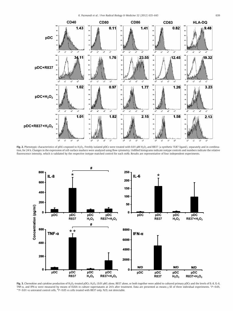
Fig. 2. Phenotypic characteristics of pDCs exposed to H2O2. Freshly isolated pDCs were treated with 0.01 μM H2O2 and R837 (a synthetic TLR7 ligand), separately and in combina-tion, for 24 h. Changes in the expression of cell-surface markers were analyzed using flow cytometry. Unfilled histograms indicate isotype controls and numbers indicate the relativefluorescence intensity, which is validated by the respective isotype-matched control for each mAb. Results are representative of four independent experiments.
Fig. 3. Chemokine and cytokine production of H2O2-treated pDCs. H2O2 (0.01 μM) alone, R837 alone, or both together were added to cultured primary pDCs and the levels of IL-8, IL-6,TNF-α, and IFN-α were measured by means of ELISA in culture supernatants at 24 h after treatment. Data are presented as means±SE of three individual experiments. *Pb0.05,**Pb0.01 vs untreated control cells, #Pb0.05 vs cells treated with R837 only. N/D, not detectable.
639K. Pazmandi et al. / Free Radical Biology & Medicine 52 (2012) 635–645

640 K. Pazmandi et al. / Free Radical Biology & Medicine 52 (2012) 635–645
for 24 h and cocultured with allogeneic CD3+ pan-T cells. Activationof CD3+ pan-T cells was assessed by IL-17, IFN-γ, and IL-4 ELISPOTassays. Addition of H2O2 to pDCs did not change their capacity to ac-tivate allogeneic IL-17- or IFN-γ-producing T cells (Figs. 4A–D).However, coculture of allogeneic CD3+ pan-T cells with pDCs trea-ted with R837 alone significantly increased the frequency of bothIL-17- and IFN-γ-producing T cells (Figs. 4A–D). Combined treat-ment with H2O2 and R837 caused only a slight decrease in the fre-quency of IL-17-secreting T cells (Figs. 4A and B); however,compared to only R837 treatment, combined treatment significantlyimpaired the ability of pDCs to induce IFN-γ secretion by pan-T cells(Figs. 4C and D). On the other hand, exposure of pDCs to H2O2 signif-icantly increased their ability to stimulate allogeneic IL-4-secreting Tcells (Figs. 4E and F). Treatment with only R837 also significantly en-hanced the capacity of pDCs to induce activation of allogeneic IL-4-producing T cells (Figs. 4E and F). Compared to R837 treatmentalone, simultaneous application of H2O2 and R837 did not alter the
Fig. 4. Effects of H2O2 treatment on the ability of pDCs to activate allogeneic T cells. Purified pDthen cocultured with allogeneic CD3+ pan-T cells. After 4 days of cocultivation, activation of CDvidual (A) IL-17-, (C) IFN-γ-, and (E) IL-4-producing T cells in representative assays from threedividual experiments. **Pb0.01, ***Pb0.001 vs T cells cocultured with untreated pDCs. ##Pb0.
ability of pDCs to stimulate allogeneic T cells to produce IL-4(Figs. 4E and F).
T-cell-polarizing capacity of pDCs exposed to H2O2
To assess the impact of H2O2 treatment on T cell polarization, pDCswere stimulated with H2O2 and R837, separately and in combination,for 24 h and thenwashed and cocultured for up to 6 dayswith naïve au-tologous CD4+CD45RA+ T cells. To investigate whether coculture withstimulated pDCs drives the differentiation of naïve autologous T cells to-ward IL-10-producing T lymphocytes, the amount of IL-10 in the culturesupernatants was measured. Exposure of pDCs to H2O2 before coculti-vationwith naïve autologous T cells significantly lowered IL-10 produc-tion by T cells compared to those coculturedwith untreated pDCs, pDCstreated with only R837, and pDCs stimulated with H2O2 and R837 incombination (Fig. 5A). To identify molecular mechanisms behind thisphenomenon, the expression of ICOS-L and PDL-1 on pDCs was
Cs were treated with 0.01 μMH2O2 and R837, separately and in combination, for 24 h and3+ pan-T cells was assessed by IL-17, IFN-γ, and IL-4 ELISPOT assays. Spots indicate indi-independent experiments. (B, D, and F) Results are presented as means±SE of three in-
01 vs T cells cocultured with R837-exposed pDCs. N/D, not detectable.
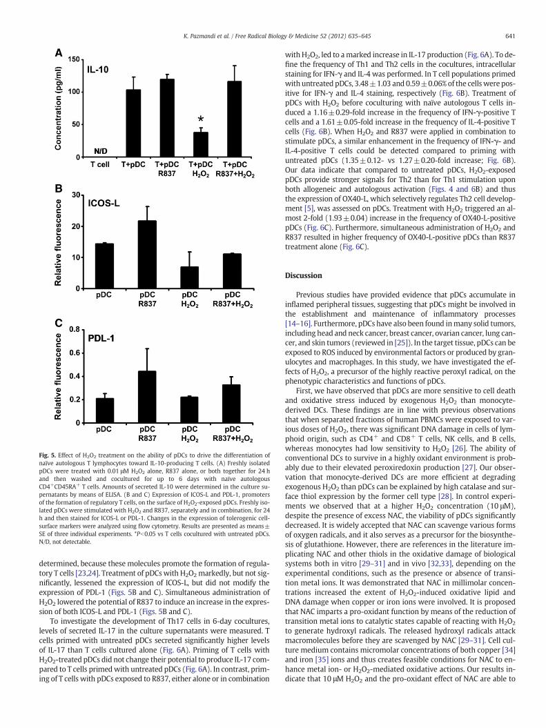
Fig. 5. Effect of H2O2 treatment on the ability of pDCs to drive the differentiation ofnaïve autologous T lymphocytes toward IL-10-producing T cells. (A) Freshly isolatedpDCs were treated with 0.01 μM H2O2 alone, R837 alone, or both together for 24 hand then washed and cocultured for up to 6 days with naïve autologousCD4+CD45RA+ T cells. Amounts of secreted IL-10 were determined in the culture su-pernatants by means of ELISA. (B and C) Expression of ICOS-L and PDL-1, promotersof the formation of regulatory T cells, on the surface of H2O2-exposed pDCs. Freshly iso-lated pDCs were stimulated with H2O2 and R837, separately and in combination, for 24h and then stained for ICOS-L or PDL-1. Changes in the expression of tolerogenic cell-surface markers were analyzed using flow cytometry. Results are presented as means±SE of three individual experiments. *Pb0.05 vs T cells cocultured with untreated pDCs.N/D, not detectable.
641K. Pazmandi et al. / Free Radical Biology & Medicine 52 (2012) 635–645
determined, because these molecules promote the formation of regula-tory T cells [23,24]. Treatment of pDCswith H2O2markedly, but not sig-nificantly, lessened the expression of ICOS-L, but did not modify theexpression of PDL-1 (Figs. 5B and C). Simultaneous administration ofH2O2 lowered the potential of R837 to induce an increase in the expres-sion of both ICOS-L and PDL-1 (Figs. 5B and C).
To investigate the development of Th17 cells in 6-day cocultures,levels of secreted IL-17 in the culture supernatants were measured. Tcells primed with untreated pDCs secreted significantly higher levelsof IL-17 than T cells cultured alone (Fig. 6A). Priming of T cells withH2O2-treated pDCs did not change their potential to produce IL-17 com-pared to T cells primedwith untreated pDCs (Fig. 6A). In contrast, prim-ing of T cells with pDCs exposed to R837, either alone or in combination
withH2O2, led to amarked increase in IL-17 production (Fig. 6A). To de-fine the frequency of Th1 and Th2 cells in the cocultures, intracellularstaining for IFN-γ and IL-4 was performed. In T cell populations primedwith untreated pDCs, 3.48±1.03 and 0.59±0.06% of the cellswere pos-itive for IFN-γ and IL-4 staining, respectively (Fig. 6B). Treatment ofpDCs with H2O2 before coculturing with naïve autologous T cells in-duced a 1.16±0.29-fold increase in the frequency of IFN-γ-positive Tcells and a 1.61±0.05-fold increase in the frequency of IL-4-positive Tcells (Fig. 6B). When H2O2 and R837 were applied in combination tostimulate pDCs, a similar enhancement in the frequency of IFN-γ- andIL-4-positive T cells could be detected compared to priming withuntreated pDCs (1.35±0.12- vs 1.27±0.20-fold increase; Fig. 6B).Our data indicate that compared to untreated pDCs, H2O2-exposedpDCs provide stronger signals for Th2 than for Th1 stimulation uponboth allogeneic and autologous activation (Figs. 4 and 6B) and thusthe expression of OX40-L, which selectively regulates Th2 cell develop-ment [5], was assessed on pDCs. Treatment with H2O2 triggered an al-most 2-fold (1.93±0.04) increase in the frequency of OX40-L-positivepDCs (Fig. 6C). Furthermore, simultaneous administration of H2O2 andR837 resulted in higher frequency of OX40-L-positive pDCs than R837treatment alone (Fig. 6C).
Discussion
Previous studies have provided evidence that pDCs accumulate ininflamed peripheral tissues, suggesting that pDCs might be involved inthe establishment and maintenance of inflammatory processes[14–16]. Furthermore, pDCs have also been found inmany solid tumors,including head and neck cancer, breast cancer, ovarian cancer, lung can-cer, and skin tumors (reviewed in [25]). In the target tissue, pDCs can beexposed to ROS induced by environmental factors or produced by gran-ulocytes and macrophages. In this study, we have investigated the ef-fects of H2O2, a precursor of the highly reactive peroxyl radical, on thephenotypic characteristics and functions of pDCs.
First, we have observed that pDCs are more sensitive to cell deathand oxidative stress induced by exogenous H2O2 than monocyte-derived DCs. These findings are in line with previous observationsthat when separated fractions of human PBMCs were exposed to var-ious doses of H2O2, there was significant DNA damage in cells of lym-phoid origin, such as CD4+ and CD8+ T cells, NK cells, and B cells,whereas monocytes had low sensitivity to H2O2 [26]. The ability ofconventional DCs to survive in a highly oxidant environment is prob-ably due to their elevated peroxiredoxin production [27]. Our obser-vation that monocyte-derived DCs are more efficient at degradingexogenous H2O2 than pDCs can be explained by high catalase and sur-face thiol expression by the former cell type [28]. In control experi-ments we observed that at a higher H2O2 concentration (10 μM),despite the presence of excess NAC, the viability of pDCs significantlydecreased. It is widely accepted that NAC can scavenge various formsof oxygen radicals, and it also serves as a precursor for the biosynthe-sis of glutathione. However, there are references in the literature im-plicating NAC and other thiols in the oxidative damage of biologicalsystems both in vitro [29–31] and in vivo [32,33], depending on theexperimental conditions, such as the presence or absence of transi-tion metal ions. It was demonstrated that NAC in millimolar concen-trations increased the extent of H2O2-induced oxidative lipid andDNA damage when copper or iron ions were involved. It is proposedthat NAC imparts a pro-oxidant function by means of the reduction oftransition metal ions to catalytic states capable of reacting with H2O2
to generate hydroxyl radicals. The released hydroxyl radicals attackmacromolecules before they are scavenged by NAC [29–31]. Cell cul-ture medium contains micromolar concentrations of both copper [34]and iron [35] ions and thus creates feasible conditions for NAC to en-hance metal ion- or H2O2-mediated oxidative actions. Our results in-dicate that 10 μM H2O2 and the pro-oxidant effect of NAC are able to

Fig. 6. Th-polarizing ability of pDCs exposed to H2O2. Freshly isolated pDCs were treated with 0.01 μMH2O2 and R837, separately and in combination for 24 h, and then washed andcocultured for up to 6 days with naïve autologous CD4+CD45RA+ T cells. (A) Th17-polarizing ability of H2O2-treated pDCs. To assess the development of Th17 cells in the cocul-tures, levels of secreted IL-17 were determined in the culture supernatants by ELISA. (B) Th1- and Th2-polarizing ability of H2O2-exposed pDCs. After 6 days of cocultivation, T cellswere restimulated with anti-CD3 mAb, phorbol 12-myristate 13-acetate, and ionomycin, and intracellular IFN-γ and IL-4 staining was performed. The percentages of IFN-γ- andIL4-positive cells were analyzed by means of flow cytometry. The dot-plot diagrams represent the results from one of four individual experiments. (C) Expression of OX40-L, a reg-ulator of Th2 cell development, on the surface of pDCs treated with H2O2. Freshly isolated pDCs were stimulated with H2O2 only, R837 only, or both together for 24 h and thenstained for OX40-L. Changes in the frequency of OX40-L-positive cells were analyzed using flow cytometry. Results are presented as means±SE of three individual experiments.**Pb0.01, ***Pb0.001 vs T cells cocultured with untreated pDCs.
642 K. Pazmandi et al. / Free Radical Biology & Medicine 52 (2012) 635–645
overwhelm the antioxidant defense mechanisms of pDCs but notthose of conventional DCs.
Furthermore, H2O2 seems to be an activating signal for conven-tional DCs, as it induces an up-regulation of several DC surfacemarkers involved in the activation of naïve T cells, including HLA-DQ and HLA-DR and the costimulatory molecules CD40 and CD86[20]. Moreover, it has also been observed that H2O2-treatedmonocyte-derived DCs acquire an enhanced ability to promote autol-ogous T cell proliferation compared to untreated DCs [20]. In contrast,pDCs displaying several lymphoid features responded to H2O2 treat-ment in radically different ways. Exposure of pDCs to H2O2 did notlead to significantly increased expression of either antigen-presenting or costimulatory molecules. Additionally, H2O2 treatmentblocked the activation of pDCs induced by a TLR7 ligand. It is worthnoting that in addition to the different phenotypic and functionalcharacteristics of the two major DC subsets, another factor may alsobe involved in the two distinct observed phenomena, namely, con-ventional DCs were treated with a high (300 μM) concentration ofH2O2 [20], whereas pDCs were exposed to 0.01 μM H2O2 in ourexperiments.
Investigating chemokine and cytokine release by H2O2-treatedpDCs, we have found that exposure to low-dose H2O2 did not increasethe production of IL-8, IL-6, TNF-α, or IFN-α. In addition, treatmentwith H2O2 notably inhibited the production of these mediators trig-gered by a TLR7-mediated stimulus. It is known that the expression ofIL-8, IL-6, and TNF-α is regulated by NF-κB [36]. It has been shownthat cytokine production of activated/memory T cells is reduced afterexposure to micromolar levels of H2O2 and the impaired cytokine ex-pression induced by H2O2 correlates with a block in NF-κB activation
[37]. Based on these observations, we propose that blockage and/orthe disturbed balance of the NF-κB- and IRF-mediated activation path-ways might be the underlying mechanism for the impaired IL-8, IL-6,and TNF-α production in pDCs treated with a combination of TLR7 li-gand andH2O2. In amurinemodel, it has been found that aged pDCs ex-hibit an impaired ability to produce IFN-α, because of a decreased up-regulation of IRF7, a key adaptor molecule in the type I IFN signalingpathway during TLR9 activation [38]. Other adaptor molecules up-stream of IRF7 were not altered by aging under experimental circum-stances. It has also been noted that aged pDCs have increasedintracellular levels of ROS both at rest and during TLR9 activation com-pared to young pDCs. Importantly, reduction of age-induced oxidativestress in pDCs led to augmented IFN-α production upon TLR9 activation[38]. As TLR7 and TLR9 appear to trigger very similar signaling pathwayswith similar functional outcomes, we hypothesize that not only in mu-rine but also in human pDCs the impaired up-regulation of IRF7 is thepivotal event in blocking TLR7/9-induced IFN-α production under oxi-dative stress conditions. However, further studies are needed to provethis hypothesis.
Although there is evidence for distinct and complementary rolesof conventional DCs and pDCs in regulating T-cell-mediated immuni-ty, both DC subsets show wide functional plasticity in determining Tcell responses. Conventional DCs at an immature stage have the abil-ity to prime naïve T cells toward IL-10-producing T lymphocytes,whereas pDCs at a mature stage seem to have an intrinsic capacityto perform this priming activity [24]. Maturing pDCs but not conven-tional DCs express high levels of ICOS-L, a key molecule in driving thegeneration of IL-10-producing T cells [24]. Our finding that treatmentof pDCs with H2O2 markedly lessens the expression of ICOS-L, leading

Fig. 7. An overview of the modulatory effects of low-dose H2O2 on the functions ofpDCs. (A) Exposure of resting pDCs to 0.01 μM H2O2 results in a slight increase in theexpression of costimulatory molecules (CD80 and CD86); CD83, a specific maturationmarker; and OX40-L, a selective regulator of Th2 cell development; however, it mark-edly decreases the expression of the antigen-presenting molecule HLA-DQ. Low-doseH2O2 does not trigger production of type I IFNs or modify the basal proinflammatorycytokine and chemokine release by pDCs. Furthermore, H2O2-exposed pDCs withoutTLR-mediated activation provide stronger signals for Th2 than for Th1 stimulation.(B) Human pDCs selectively express both TLR7 and TLR9 localized in the endoplasmicreticulum. TLR7 and TLR9 recognize RNA and DNA fragments of viruses and bacteria,respectively, and initiate a signal transduction pathway through a key adaptor mole-cule, MyD88. MyD88-mediated signals activate IRF7 and NF-κB, leading to secretionof type I IFNs, proinflammatory cytokines, and chemokines. Ligation of TLR receptorsalso leads to high expression levels of costimulatory molecules, as well as CD83 andHLA-DQ. In this activation state pDCs are able to induce a strong Th1 response upon in-teraction with naïve T cells. (C) In contrast, when pDCs are stimulated with TLR ligandsin the presence of low-dose H2O2, decreased cytokine and chemokine release as well aslowered expression of cell-surface molecules can be detected, indicating that pDCs ex-posed to ROS in vivo may have an anti-inflammatory or a tolerogenic phenotype.
643K. Pazmandi et al. / Free Radical Biology & Medicine 52 (2012) 635–645
to impaired capacity of pDCs to induce IL-10-producing T cells, fur-ther confirms the inhibitory effect of H2O2 on pDCs. A recent studyhas demonstrated that pDCs are able to promote Th17 differentiationunder certain circumstances [39]. We have also observed that prim-ing of naïve T cells with TLR7-stimulated pDCs triggers a marked in-crease in IL-17 production and that the presence of H2O2 duringTLR7 ligation does not influence this phenomenon. Interestingly, al-though exposure of pDCs to H2O2 alone significantly decreases theirability to promote differentiation of IL-10-producing T cells, it doesnot affect the Th17-priming capacity of pDCs. Regarding Th1/Th2 po-larization, H2O2-exposed pDCs provide stronger signals for Th2 thanfor Th1 stimulation during both allogeneic and autologous activation.Elevated expression of OX40-L on pDCs may be responsible for thisphenomenon, as we observed that treatment with H2O2 almost dou-bled the frequency of OX40-L-positive pDCs and it has been previous-ly demonstrated that pDCs prime Th2 cells through OX40-L-dependent mechanisms [5]. Importantly, when pDCs were stimulatedwith R837 in the presence of H2O2, decreased phenotypic activation,lower chemokine and cytokine release, as well as impaired allo- andautostimulatory capacities of pDCs could be detected, suggestingthat during in vivo circumstances pDCs exposed to oxidative stresshave an anti-inflammatory or a tolerogenic role in regulating adaptiveimmune responses (Fig. 7).
Human pDCs represent only 0.1–0.6% of PBMCs [40], and thereforelimited numbers of these cells can be isolated from blood of healthyvolunteers for in vitro experiments. This leads to a significant techni-cal restriction of pDC studies. Indeed, much less information on thefunctions of pDCs in human immune responses is available comparedwith the more abundant conventional DCs. Our in vitro finding thatpDCs are highly sensitive to oxidative stress raises the question howpDCs can be long-lived in vivo if they cannot withstand elevatedlevels of ROS, which occur during infection and inflammation. Themain difficulty in interpreting our results is the lack of a proper meth-od to measure the exact amount of reactive radicals the pDCs have toface in situ [41]. Another issue that should be considered is that al-though infection and inflammation are characterized by increasedproduction of ROS, levels of enzymatic and nonenzymatic antioxi-dants are also elevated in affected tissues to balance the harmful ef-fects of oxidation products [42–44]. Furthermore, inflammationenhances vascular permeability, allowing plasma antioxidant sub-stances to move into inflamed tissues [45–48]. Thus, in inflamed tis-sues pDCs are exposed to a net effect of both pro-oxidant andantioxidant factors. The fact that pDCs can be identified in inflamedtissues proves that these cells can survive oxidative stress in vivo.However, our findings indicate that pDCs may have lower intracellu-lar antioxidant capacity than conventional DCs. Additionally, it seemsthat pDCs release (or express on their surface) lower amounts of an-tioxidant factors than conventional DCs do. Although the molecularmechanisms behind our findings need further investigations, our invitro observations may explain why pDCs respond to oxidative stressin vivo in radically different ways compared to conventional DCs.
Indeed, it has been reported that in a murine model of allergic air-way inflammation, which is associated with oxidative stress [18],pDCs perform anti-inflammatory activities irrespective of their matura-tion state [23]. In solid tumors, in which tumor-derived macrophages[49] and granulocytes [50] secrete H2O2, pDCs are present in a nonacti-vated state and are associatedwith the development of an immunosup-pressive microenvironment [25]. Furthermore, oxidative stress isimplicated as a pathogenic factor in a number of viral infections[51–53]. A lack of dietary antioxidants leading to nutritionally inducedoxidative stress, or exposure to air pollutants, including diesel exhaustand cigarette smoke, resulting in oxidative stress in the airways, isthought to be associated with an increase in severity from and suscep-tibility to viral infections [54,55]. The iron-catalyzed Haber–Weiss reac-tion generates hydroxyl radicals, which are by far the most reactiveoxygen radicals, thus iron-overload-mediated oxidative stress may
also contribute to viral pathogenicity [53,56]. Although oxidative stressmay have profound effects on several antiviral mechanisms, includingeffector functions of activated Th1 and Th2 lymphocytes [57], and alsoon pathogens [54], our findings raise the possibility that impaired pDCfunctions are responsible, at least in part, for exacerbation of symptoms

644 K. Pazmandi et al. / Free Radical Biology & Medicine 52 (2012) 635–645
in viral infections associated with oxidative stress. In accordance withour observations, a recent study has demonstrated that cigarettesmoke, which is known to induce oxidative stress, suppresses TLR7-mediated responses to virus infection in pDCs [58].
In conclusion, here we provide evidence that human pDCs re-spond to oxidative stress in a manner similar to that of cells of lym-phoid origin. The inhibitory effects of H2O2 on TLR-inducedphenotypic activation, cytokine production, and the T-cell-polarizingcapacity of pDCs may be involved in the development and mainte-nance of their anti-inflammatory or tolerogenic properties observedin several pathologic conditions.
Acknowledgments
This work was supported by the Hungarian Scientific Research Fund(K-73347 to A.B. andNK-72937 to E.R.), theU.S. National Institute of En-vironmental Health Science (RO1-ES018948 to I.B.), the U.S. NationalInstitute of Allergic and Infectious Diseases (AI062885-01 to I.B.), andthe TAMOP 4.2.1/B-09/1/KONV-2010-0007 project (to A.B. and E.R.).The project is cofinanced by the European Union and the European So-cial Fund.
References
[1] Kadowaki, N.; Ho, S.; Antonenko, S.; Malefyt, R. W.; Kastelein, R. A.; Bazan, F.; Liu,Y. J. Subsets of human dendritic cell precursors express different toll-like recep-tors and respond to different microbial antigens. J. Exp. Med. 194:863–869; 2001.
[2] Colonna, M.; Trinchieri, G.; Liu, Y. J. Plasmacytoid dendritic cells in immunity. Nat.Immunol. 5:1219–1226; 2004.
[3] Kadowaki, N.; Antonenko, S.; Lau, J. Y.; Liu, Y. J. Natural interferon alpha/beta-producingcells link innate and adaptive immunity. J. Exp. Med. 192:219–226; 2000.
[4] Cella, M.; Facchetti, F.; Lanzavecchia, A.; Colonna, M. Plasmacytoid dendritic cellsactivated by influenza virus and CD40L drive a potent Th1 polarization. Nat.Immunol. 1:305–310; 2000.
[5] Ito, T.; Amakawa, R.; Inaba, M.; Hori, T.; Ota, M.; Nakamura, K.; Takebayashi, M.;Miyaji, M.; Yoshimura, T.; Inaba, K.; Fukuhara, S. Plasmacytoid dendritic cells reg-ulate Th cell responses through OX40 ligand and type I IFNs. J. Immunol. 172:4253–4259; 2004.
[6] Moseman, E. A.; Liang, X.; Dawson, A. J.; Panoskaltsis-Mortari, A.; Krieg, A. M.; Liu,Y. J.; Blazar, B. R.; Chen, W. Human plasmacytoid dendritic cells activated by CpGoligodeoxynucleotides induce the generation of CD4+CD25+ regulatory T cells. J.Immunol. 173:4433–4442; 2004.
[7] Naik, S. H.; Sathe, P.; Park, H. Y.; Metcalf, D.; Proietto, A. I.; Dakic, A.; Carotta, S.;O'Keeffe, M.; Bahlo, M.; Papenfuss, A.; Kwak, J. Y.; Wu, L.; Shortman, K. Develop-ment of plasmacytoid and conventional dendritic cell subtypes from single pre-cursor cells derived in vitro and in vivo. Nat. Immunol. 8:1217–1226; 2007.
[8] Onai, N.; Obata-Onai, A.; Schmid, M. A.; Ohteki, T.; Jarrossay, D.; Manz, M. G. Iden-tification of clonogenic common Flt3+M−CSFR+ plasmacytoid and conventionaldendritic cell progenitors in mouse bone marrow. Nat. Immunol. 8:1207–1216;2007.
[9] Spits, H.; Couwenberg, F.; Bakker, A. Q.; Weijer, K.; Uittenbogaart, C. H. Id2 andId3 inhibit development of CD34(+) stem cells into predendritic cell (pre-DC)2but not into pre-DC1: evidence for a lymphoid origin of pre-DC2. J. Exp. Med.192:1775–1784; 2000.
[10] Bendriss-Vermare, N.; Barthelemy, C.; Durand, I.; Bruand, C.; Dezutter-Dambuyant,C.; Moulian, N.; Berrih-Aknin, S.; Caux, C.; Trinchieri, G.; Briere, F. Human thymuscontains IFN-alpha-producing CD11c(−), myeloid CD11c(+), and mature interdig-itating dendritic cells. J. Clin. Invest. 107:835–844; 2001.
[11] Rissoan, M. C.; Duhen, T.; Bridon, J. M.; Bendriss-Vermare, N.; Peronne, C.; deSaint Vis, B.; Briere, F.; Bates, E. E. Subtractive hybridization reveals the expres-sion of immunoglobulin-like transcript 7, Eph-B1, granzyme B, and 3 novel tran-scripts in human plasmacytoid dendritic cells. Blood 100:3295–3303; 2002.
[12] Randolph, G. J.; Ochando, J.; Partida-Sanchez, S. Migration of dendritic cell subsetsand their precursors. Annu. Rev. Immunol. 26:293–316; 2008.
[13] Yoneyama, H.; Matsuno, K.; Zhang, Y.; Nishiwaki, T.; Kitabatake, M.; Ueha, S.;Narumi, S.; Morikawa, S.; Ezaki, T.; Lu, B.; Gerard, C.; Ishikawa, S.; Matsushima,K. Evidence for recruitment of plasmacytoid dendritic cell precursors to inflamedlymph nodes through high endothelial venules. Int. Immunol. 16:915–928; 2004.
[14] Jahnsen, F. L.; Lund-Johansen, F.; Dunne, J. F.; Farkas, L.; Haye, R.; Brandtzaeg, P.Experimentally induced recruitment of plasmacytoid (CD123high) dendriticcells in human nasal allergy. J. Immunol. 165:4062–4068; 2000.
[15] Farkas, L.; Beiske, K.; Lund-Johansen, F.; Brandtzaeg, P.; Jahnsen, F. L. Plasmacy-toid dendritic cells (natural interferon-alpha/beta-producing cells) accumulatein cutaneous lupus erythematosus lesions. Am. J. Pathol. 159:237–243; 2001.
[16] Gerlini, G.; Mariotti, G.; Bianchi, B.; Pimpinelli, N. Massive recruitment of type Iinterferon producing plasmacytoid dendritic cells in varicella skin lesions. J. In-vest. Dermatol. 126:507–509; 2006.
[17] Riedl, M. A.; Nel, A. E. Importance of oxidative stress in the pathogenesis andtreatment of asthma. Curr. Opin. Allergy Clin. Immunol. 8:49–56; 2008.
[18] Boldogh, I.; Bacsi, A.; Choudhury, B. K.; Dharajiya, N.; Alam, R.; Hazra, T. K.; Mitra,S.; Goldblum, R. M.; Sur, S. ROS generated by pollen NADPH oxidase provide a sig-nal that augments antigen-induced allergic airway inflammation. J. Clin. Invest.115:2169–2179; 2005.
[19] Reth, M. Hydrogen peroxide as second messenger in lymphocyte activation. Nat.Immunol. 3:1129–1134; 2002.
[20] Rutault, K.; Alderman, C.; Chain, B. M.; Katz, D. R. Reactive oxygen species activatehuman peripheral blood dendritic cells. Free Radic. Biol. Med. 26:232–238; 1999.
[21] Csillag, A.; Boldogh, I.; Pazmandi, K.; Magyarics, Z.; Gogolak, P.; Sur, S.; Rajnavolgyi, E.;Bacsi, A. Pollen-induced oxidative stress influences both innate and adaptive immuneresponses via altering dendritic cell functions. J. Immunol. 184:2377–2385; 2010.
[22] Gregorio, J.; Meller, S.; Conrad, C.; Di Nardo, A.; Homey, B.; Lauerma, A.; Arai, N.; Gallo,R. L.; Digiovanni, J.; Gilliet, M. Plasmacytoid dendritic cells sense skin injury and pro-mote wound healing through type I interferons. J. Exp. Med. 207:2921–2930; 2010.
[23] Kool, M.; van Nimwegen, M.; Willart, M. A.; Muskens, F.; Boon, L.; Smit, J. J.; Coyle,A.; Clausen, B. E.; Hoogsteden, H. C.; Lambrecht, B. N.; Hammad, H. An anti-inflammatory role for plasmacytoid dendritic cells in allergic airway inflamma-tion. J. Immunol. 183:1074–1082; 2009.
[24] Ito, T.; Yang, M.; Wang, Y. H.; Lande, R.; Gregorio, J.; Perng, O. A.; Qin, X. F.; Liu, Y.J.; Gilliet, M. Plasmacytoid dendritic cells prime IL-10-producing T regulatory cellsby inducible costimulator ligand. J. Exp. Med. 204:105–115; 2007.
[25] Lande, R.; Gilliet, M. Plasmacytoid dendritic cells: key players in the initiation andregulation of immune responses. Ann. N. Y. Acad. Sci. 1183:89–103; 2010.
[26] Weng, H.; Lu, Y.;Weng, Z.;Morimoto, K. Differential DNAdamage induced byH2O2 andbleomycin in subpopulations of humanwhite blood cells.Mutat. Res. 652:46–53; 2008.
[27] Rivollier, A.; Perrin-Cocon, L.; Luche, S.; Diemer, H.; Strub, J. M.; Hanau, D.; vanDorsselaer, A.; Lotteau, V.; Rabourdin-Combe, C.; Rabilloud, T.; Servet-Delprat,C. High expression of antioxidant proteins in dendritic cells: possible implicationsin atherosclerosis. Mol. Cell. Proteomics 5:726–736; 2006.
[28] Thoren, F. B.; Betten, A.; Romero, A. I.; Hellstrand, K. Antioxidative properties ofmyeloid dendritic cells: protection of T cells and NK cells from oxygen radical-induced inactivation and apoptosis. J. Immunol. 179:21–25; 2007.
[29] Oikawa, S.; Yamada, K.; Yamashita, N.; Tada-Oikawa, S.; Kawanishi, S. N-acetylcysteine,a cancer chemopreventive agent, causes oxidative damage to cellular and isolatedDNA. Carcinogenesis 20:1485–1490; 1999.
[30] Sagrista, M. L.; Garcia, A. E.; Africa De Madariaga, M.; Mora, M. Antioxidant andpro-oxidant effect of the thiolic compounds N-acetyl-L-cysteine and glutathioneagainst free radical-induced lipid peroxidation. Free Radic. Res. 36:329–340; 2002.
[31] Su, M.; Yang, Y.; Yang, G. Quantitative measurement of hydroxyl radical inducedDNA double-strand breaks and the effect of N-acetyl-L-cysteine. FEBS Lett. 580:4136–4142; 2006.
[32] Wang, A. L.; Wang, J. P.; Wang, H.; Chen, Y. H.; Zhao, L.; Wang, L. S.; Wei, W.; Xu,D. X. A dual effect of N-acetylcysteine on acute ethanol-induced liver damage inmice. Hepatol. Res. 34:199–206; 2006.
[33] Childs, A.; Jacobs, C.; Kaminski, T.; Halliwell, B.; Leeuwenburgh, C. Supplementa-tion with vitamin C and N-acetyl-cysteine increases oxidative stress in humansafter an acute muscle injury induced by eccentric exercise. Free Radic. Biol. Med.31:745–753; 2001.
[34] Freedman, J. H.; Weiner, R. J.; Peisach, J. Resistance to copper toxicity of culturedhepatoma cells: characterization of resistant cell lines. J. Biol. Chem. 261:11840–11848; 1986.
[35] Kakuta, K.; Orino, K.; Yamamoto, S.; Watanabe, K. High levels of ferritin and itsiron in fetal bovine serum. Comp. Biochem. Physiol. A Physiol. 118:165–169; 1997.
[36] Pahl, H. L. Activators and target genes of Rel/NF-kappaB transcription factors. On-cogene 18:6853–6866; 1999.
[37] Malmberg, K. J.; Arulampalam, V.; Ichihara, F.; Petersson, M.; Seki, K.; Andersson,T.; Lenkei, R.; Masucci, G.; Pettersson, S.; Kiessling, R. Inhibition of activated/memory (CD45RO(+)) T cells by oxidative stress associated with block of NF-kappaB activation. J. Immunol. 167:2595–2601; 2001.
[38] Stout-Delgado, H.W.; Yang, X.;Walker,W. E.; Tesar, B. M.; Goldstein, D. R. Aging im-pairs IFN regulatory factor 7 up-regulation in plasmacytoid dendritic cells duringTLR9 activation. J. Immunol. 181:6747–6756; 2008.
[39] Yu, C. F.; Peng, W. M.; Oldenburg, J.; Hoch, J.; Bieber, T.; Limmer, A.; Hartmann, G.;Barchet, W.; Eis-Hubinger, A. M.; Novak, N. Human plasmacytoid dendritic cellssupport Th17 cell effector function in response to TLR7 ligation. J. Immunol. 184:1159–1167; 2010.
[40] Magyarics, Z.; Csillag, A.; Pazmandi, K.; Rajnavolgyi, E.; Bacsi, A. Identification ofplasmacytoid pre-dendritic cells by one-color flow cytometry for phenotypescreening. Cytometry A 73:254–258; 2008.
[41] Halliwell, B.; Whiteman, M. Measuring reactive species and oxidative damage invivo and in cell culture: how should you do it and what do the results mean?Br. J. Pharmacol. 142:231–255; 2004.
[42] Tkaczyk, J.; Vizek, M. Oxidative stress in the lung tissue—sources of reactive oxy-gen species and antioxidant defence. Prague Med. Rep. 108:105–114; 2007.
[43] Kohen, R.; Gati, I. Skin low molecular weight antioxidants and their role in agingand in oxidative stress. Toxicology 148:149–157; 2000.
[44] Kiroglu, A. F.; Noyan, T.; Oger, M.; Kara, T. Oxidants and antioxidants in tonsillarand adenoidal tissue in chronic adenotonsillitis and adenotonsillar hypertrophyin children. Int. J. Pediatr. Otorhinolaryngol. 70:35–38; 2006.
[45] Cross, C. E.; van der Vliet, A.; O'Neill, C. A.; Louie, S.; Halliwell, B. Oxidants, antiox-idants, and respiratory tract lining fluids. Environ. Health Perspect. 102 (Suppl.10):185–191; 1994.
[46] Polidori, M. C.; Stahl, W.; Eichler, O.; Niestroj, I.; Sies, H. Profiles of antioxidants inhuman plasma. Free Radic. Biol. Med. 30:456–462; 2001.

645K. Pazmandi et al. / Free Radical Biology & Medicine 52 (2012) 635–645
[47] Roche, M.; Rondeau, P.; Singh, N. R.; Tarnus, E.; Bourdon, E. The antioxidant prop-erties of serum albumin. FEBS Lett. 582:1783–1787; 2008.
[48] Nieto, F. J.; Iribarren, C.; Gross, M. D.; Comstock, G.W.; Cutler, R. G. Uric acid and serumantioxidant capacity: a reaction to atherosclerosis? Atherosclerosis 148:131–139; 2000.
[49] Kono, K.; Salazar-Onfray, F.; Petersson, M.; Hansson, J.; Masucci, G.; Wasserman,K.; Nakazawa, T.; Anderson, P.; Kiessling, R. Hydrogen peroxide secreted bytumor-derived macrophages down-modulates signal-transducing zeta moleculesand inhibits tumor-specific T cell- and natural killer cell-mediated cytotoxicity.Eur. J. Immunol. 26:1308–1313; 1996.
[50] Schmielau, J.; Finn, O. J. Activated granulocytes and granulocyte-derived hydro-gen peroxide are the underlying mechanism of suppression of T-cell function inadvanced cancer patients. Cancer Res. 61:4756–4760; 2001.
[51] Schwarz, K. B. Oxidative stress during viral infection: a review. Free Radic. Biol.Med. 21:641–649; 1996.
[52] Israel, N.; Gougerot-Pocidalo, M. A. Oxidative stress in human immunodeficiencyvirus infection. Cell. Mol. Life Sci. 53:864–870; 1997.
[53] Choi, J.; Ou, J. H. Mechanisms of liver injury. III. Oxidative stress in the pathogenesisof hepatitis C virus. Am. J. Physiol. Gastrointest. Liver Physiol. 290:G847–G851; 2006.
[54] Beck, M. A.; Handy, J.; Levander, O. A. The role of oxidative stress in viral infec-tions. Ann. N. Y. Acad. Sci. 917:906–912; 2000.
[55] Gowdy, K. M.; Krantz, Q. T.; King, C.; Boykin, E.; Jaspers, I.; Linak, W. P.; Gilmour,M. I. Role of oxidative stress on diesel-enhanced influenza infection in mice. Part.Fibre Toxicol. 7:34; 2010.
[56] Savarino, A.; Pescarmona, G. P.; Boelaert, J. R. Iron metabolism and HIV infection:reciprocal interactions with potentially harmful consequences? Cell Biochem.Funct. 17:279–287; 1999.
[57] Frossi, B.; De Carli, M.; Piemonte, M.; Pucillo, C. Oxidative microenvironment ex-erts an opposite regulatory effect on cytokine production by Th1 and Th2 cells.Mol. Immunol. 45:58–64; 2008.
[58] Castro, S. M.; Chakraborty, K.; Guerrero-Plata, A. Cigarette smoke suppresses TLR-7 stimulation in response to virus infection in plasmacytoid dendritic cells. Toxi-col. In Vitro 25:1106–1113; 2010.


