Embed Size (px)
Citation preview
Mineral-Microbe Interactions Probed in Force, Energy, and Distance Nanospace
Steven K. Lower
Dissertation submitted to the Faculty of the
Virginia Polytechnic Institute and State University
in partial fulfillment of the requirements for the degree of
Doctor of Philosophy
in
Geological Sciences and Biochemistry
Michael F. Hochella, Jr., Chair
Duane Berry
Malcolm Potts
J. Donald Rimstidt
Christopher Tadanier
February 22, 2001
Blacksburg, Virginia
Keywords: AFM, bacteria, force, mineral, nanoscale, nanotechnology
Copyright 2001, Steven K. Lower

ii
Mineral-Microbe Interactions Probed in Force, Energy, and Distance Nanospace
Steven K. Lower
(ABSTRACT)
Biological force microscopy (BFM) was developed to quantitatively measure pico- to nano-Newton forces (10-9 to 10-12 N) as a function of the nanoscale distance (nanometers) between living bacteria and mineral surfaces, in aqueous solution. Native cells were linked to a force-sensing probe, which was used in a force microscope to measure attractive and repulsive forces as a mineral surface approached, made contact with, and subsequently withdrew from a bacterium on the probe. The resulting data were used to interpret the interactive dynamics operative between bacteria and mineral surfaces under environmentally relevant conditions.
BFM was used to study bacterial adhesion to mineral surfaces. In the case of Escherichia coli interactions with goethite, graphite, and muscovite, attractive and repulsive forces were detected at ranges up to 400 nanometers, the magnitude and sign depending on the ionic strength of the intervening solution and the mineral surface charge and hydrophobicity. Adhesion forces, up to several nanoNewtons in magnitude and exhibiting various fibrillation dynamics, were also measured and reflect the complex interactions of structural and chemical functionalities on the bacteria and mineral surfaces. In the study of Burkholderia cepecia interactions with mica, it was found that the physiological condition of the cell affected the observed adhesion forces. Cells grown under oligotrophic conditions exhibited an increased affinity for the mineral surface as opposed to cells grown under eutropic conditions. BFM was also used to characterize the transfer of electrons from biomolecules on Shewanella oneidensis to Fe(III) in the structure of goethite. Force measurements with picoNewton resolution were made in aqueous solution under aerobic and anaerobic conditions. Energy values (in attoJoules) derived from these measurements show that the affinity between S. oneidensis and goethite rapidly increases by two to five times under anaerobic conditions where electron transfer from bacterium to mineral is expected. Specific signatures in the force curves, analyzed with the worm-like chain model of protein unfolding, suggest that the bacterium recognizes the mineral surface such that a 150 kDa putative, iron reductase is quickly mobilized within the outer membrane of S. oneidensis and specifically interacts with the goethite surface to facilitate the electron transfer process.

iii
Acknowledgements
Thanks first to Mike for giving me the freedom to explore my scientific interests, the knowledge to successfully pursue those interests, and the ability to share my interests in scientific venues. I look forward to working with Mike during the rest of my scientific career. I have gained not only an exceptionally great colleague but also a truly great friend. Second, I’d like to acknowledge my brother, Brian, whose knowledge of biochemistry and laboratory know-how is unprecedented. I could not have completed my PhD without him. I thank my committee members - Don, Malcolm, Duane, and Chris - for their interdisciplinary instruction and education. Special thanks to Don for his insight into geochemistry and the use of his laboratory. I am also thankful for being surrounded by the many talented peers that did or currently shape our research group, namely Catherine, Andy, Tracy, Jeanne, Treavor, Barry, Eric, Erin, Kevin, Jodi, Chris, and Rob. Thanks to Cahit for being such an admirable department head; Jill Banfield for insight into geomicrobiology; William Ducker for discussions on forces; Al Yousten, Eugene Gregory, Laura Link, Ann Stevens, Brenda Winkel, and Terry Beveridge for instruction in biochemistry and microbiology; Mark Lemon for the computer assistance; Susan Eriksson for providing samples; Kristy DeCourcy for assistance with laser scanning microscopy and molecular biology. All the ladies in the main office kept me in good hands with administrative and academic needs. Special thanks to Linda for allowing me to spend money, Mary for helping with travel expenses, and Connie for helping with everything from teaching duties to advice on being a good husband. I am also thankful for the time I have spent with Barbara and family, Beth and family, Paul Ribbe, Katy, Christine, Keiko and family, Matt Eick, Maddy, Trish, Colin, Kevin, and Christina. On a more personal note, I wish to give thanks to those who give my life meaning. My wife and best friend, Joanna, who has put up with me for over 11 years! This dissertation cannot hold all the things she means to me. She is the inspiration for everything I do. All other things will come and go, but I will have her forever. I love you Joanna! I thank my children Tyler (My friend, my main man, the one and only. B’ball games and canoeing trips were a blast), Alexis (Always my little girl. Manicures and face paintings were messy and fun), and Kirsten (Always my baby. I won’t forget our dollhouse and tea parties). I can’t image life without any of you! I pray that wherever life takes you, I will always be “your daddy”. I love you Tyler, Alexis, and Kirsten!!! I thank my mother and father for family vacations at the beach. We had fun! I thank my father-in-law, Dave Diehl, for being such a wonderful grandfather (and father to Joanna and an okay father-in-law :), for financial help and for personal encouragement. Thanks to my grandfather-in-law, Wendell Diehl, for instruction and guidance on my road to a PhD. Finally, my dissertation is dedicated to my mother-in-law, Brenda Diehl. I literally, could not have become a geologist without her help. More importantly, I could not have had such a wonderful wife and children without her! I miss you Brenda.
iv
Table of Contents
Abstract ........................................................................................................................................... ii
Acknowledgements........................................................................................................................ iii
Table of Figures ............................................................................................................................. vi
Table of Tables ............................................................................................................................. vii
Chapter 1 - Introduction.................................................................................................................. 1
References ................................................................................................................................... 2
Chapter 2 - Measuring interfacial and adhesion forces between bacteria and mineral surfaces
with biological force microscopy.................................................................................................... 3
Abstract........................................................................................................................................ 3
Introduction ................................................................................................................................. 3
Material and Methods.................................................................................................................. 4
Mineral and Bacteria Specimens.............................................................................................. 4
Design and Characterization of Biologically-Active-Force-Probes......................................... 5
Biological Force Microscopy Measurements........................................................................... 7
Results ......................................................................................................................................... 9
Discussion.................................................................................................................................. 11
Categories of Interfacial and Adhesion Forces ...................................................................... 11
Mineralogical and Microbiological Aspects of Forces ......................................................... 12
Conclusion ................................................................................................................................. 14
Acknowledgments ..................................................................................................................... 15
References ................................................................................................................................. 15
Chapter 3 - Dynamics of the mineral-microbe interface: Use of biological force microscopy in
biogeochemistry and geomicrobiology......................................................................................... 22
Abstract...................................................................................................................................... 22
Introduction ............................................................................................................................... 22
Materials and Methods .............................................................................................................. 25
Mineral and Bacteria .............................................................................................................. 25
Force Sensor Preparation and Characterization ..................................................................... 25
Force Measurements .............................................................................................................. 26
v
Results and Discussion .............................................................................................................. 29
Characterization of Biologically-Active-Force-Probes.......................................................... 29
Forces of interaction between bacteria and mineral surfaces in-situ ..................................... 33
Potential Applications of Biological Force Microscopy to Geomicrobiology Studies.......... 35
Acknowledgments ..................................................................................................................... 37
References ................................................................................................................................. 37
Chapter 4 - Bacterial recognition of mineral surfaces: Nanoscale interactions between
Shewanella and α-FeOOH............................................................................................................ 44
Abstract...................................................................................................................................... 44
Introduction ............................................................................................................................... 44
Methods and Materials .............................................................................................................. 45
Results and Discussion .............................................................................................................. 46
References and Acknowledgments............................................................................................ 51
Curriculum Vitae .......................................................................................................................... 54

vi
Table of Figures
Figure 2.1 Biological force microscopy and biologically-active-force-probe................................ 6
Figure 2.2 Force measurements between muscovite and Escherichia coli .................................. 10
Figure 3.1 Biological force microscopy........................................................................................ 24
Figure 3.2 Force curves................................................................................................................. 28
Figure 3.3 Optical lever sensitivity.............................................................................................. 30
Figure 3.4 Biologically-active-force-probes................................................................................. 31
Figure 3.5 Force measurements between mica and Burkholderica spp........................................ 34
Figure 4.1 Force measurements between goethite and Shewanella oneidensis............................ 47

vii
Table of Tables
Table 4.1 Force and Energy Measurements.................................................................................. 48

1
Chapter 1 - Introduction
The fundamental forces between a bacterium and mineral surface are central to
understanding the intricacies of interfacial phenomena such as bacterial attachment to or
detachment from minerals, biofilm formation and structure, bacterial affinity for or recognition
of specific mineral surfaces, and dispersal of microorganisms in the environment. In nature, a
myriad of physical and chemical interactions occur at bacteria-mineral interfaces due to the
mosaic of macromolecular structures on bacteria, and the diversity of mineral surface
functionality and crystallography. Mineral-microbe interactions are governed by the cumulative
effects of fundamental, nano- (10-9 N) to pico-scale (10-12 N) forces such as van der Waals,
electrostatics, solvation interactions, and steric interactions. However, acquiring even an
elementary appreciation of these forces presents a daunting challenge, primarily due to the
minute scale at which these interfaces must be probed, and the difficulty in developing a
technique that preserves the natural intricacies of bacteria surfaces. This dissertation describes
the development and use of a new technique, biological force microscopy (BFM), which is
capable of quantitatively measuring interfacial and adhesion forces between living bacterial cells
and mineral surfaces (or other living cells), in situ.
Chapter two was published in Geochimica et Cosmochimica Acta, 2000, 64: 3133-3139.
Access to this manuscript may be obtained at http://www.elsevier.com/. In this chapter, BFM is
used to measure attractive and repulsive forces in the nanoNewton range between Escherichia
coli and muscovite, goethite, and graphite surfaces at separations of <1 µm in aqueous solutions
(pH 6, 25 °C) of varying ionic strength. The resulting data are used to interpret the
intermolecular forces operative between living bacteria and mineral surfaces in natural
environments.
Chapter three was published in Geomicrobiology Journal, 2001, 18: 63-76. Access to
this manuscript may be obtained at http://www.tandf.co.uk/. This chapter presents a detailed
description of the use of BFM to accurately measure nanoscale force and distance relationships
between a bacterium and mineral surface. The fabrication of biologically-active-force-probes
(i.e., force-sensing probes activated with living cells) is also discussed in detail. BFM is used to
2
measure approach and retraction forces between mica and Burkholderia spp. grown under high
or low nutrient conditions. The resulting data are used to discuss the role that physiology plays
in adhesion strategies.
Chapter four is in review in Science, 2001. If accepted, access to this manuscript may be
obtained at http://www.sciencemag.org/. In this chapter, BFM is used to probe electron transfer
processes between a dissimilatory-metal-reducing-bacterium and iron oxyhydroxides. Force
measurements with picoNewton resolution were made with living cells of Shewanella oneidensis
(also called S. putrefaciens), in aqueous solution under aerobic and anaerobic conditions as a
function of the nanoscale (in nanometers) distance between the cell and mineral surface. Energy
values (in attoJoules = 10-18 J) derived from these measurements show that S. oneidensis
recognizes goethite such that the affinity between the bacterium and mineral rapidly increases by
two to five times under anaerobic conditions where electron transfer from bacterium to mineral is
expected. Specific signatures in the force curves, analyzed with a theoretical model describing
conformational changes in proteins, suggest that a 150 kDa putative, iron reductase is quickly
mobilized within the outer membrane of S. oneidensis and specifically interacts with the goethite
surface to facilitate the electron transfer process.
References
Lower, S.K., Tadanier, C.J., and Hochella, M.F., Jr. (2000) Measuring interfacial and adhesion
forces between bacteria and mineral surfaces with biological force microscopy. Geochimica et
Cosmochimica Acta, 64: 3133-3139. Copyright 2000 Elsevier Science Ltd.
Lower, S.K., Tadanier, C.J., and Hochella, M.F., Jr. (2001) Dynamics of the mineral-microbe
interface: Use of biological force microscopy in biogeochemistry and geomicrobiology.
Geomicrobiology Journal, 18: 63-76. Copyright 2001 Taylor & Francis.
Lower, S.K., Beveridge, T.J., and Hochella, M. F., Jr. (2001) Bacterial recognition of mineral
surfaces: Nanoscale interactions between Shewanella and α-FeOOH. Science, in review.

3
Chapter 2 - Measuring interfacial and adhesion forces between bacteria and mineral
surfaces with biological force microscopy
Published in Geochimica et Cosmochimica Acta, 2000, 64:3133-3139
Copyright 2000 Elsevier Science Ltd
Abstract
Interfacial and adhesion forces between living, unmodified bacterial cells (Escherichia coli) and
mineral surfaces (muscovite, goethite, and graphite) have been directly measured in aqueous
solution using a force microscope. Native cells are linked to a force-sensing probe that is used to
characterize interactions as a mineral surface approaches, makes contact with, and withdraws
from bacteria on the probe. Attractive and repulsive interfacial forces were detected at ranges up
to 400 nanometers separation, the magnitude and sign depending on the ionic strength of the
intervening solution and the mineral surface charge and hydrophobicity. Adhesion forces, up to
several nanoNewtons in magnitude and exhibiting various fibrillation dynamics, were also
measured and reflect the complex interactions of structural and chemical functionalities on the
bacteria and mineral surfaces.
Introduction
The fundamental forces between a bacterium and mineral surface are central to
understanding the intricacies of interfacial phenomena such as bacterial attachment to or
detachment from minerals (van Loosdrecht et al., 1989; Fletcher, 1996; Yee et al., 2000), mineral
dissolution and crystal growth (Myers and Nealson, 1988; Hiebert and Bennett, 1992; Schultze-
Lam et al., 1992; Roden and Zachara, 1996; Fortin et al., 1997), biofilm formation and structure
(Lawrence et al., 1991; Davies et al., 1998), bacterial affinity for or recognition of specific
mineral surfaces (Ohmura et al., 1993; Fleminger and Shabtai, 1995; Bhosle et al., 1998; Dziurla
et al., 1998; Edwards et al., 1998), and dispersal of genetically engineered microorganisms in the
environment (Gannon et al., 1991; Mills and Powelson, 1996; Trevors and van Elsas, 1997). A
myriad of physical and chemical interactions occur at bacteria-mineral interfaces in nature, due

4
to (i) the mosaic of spatially discrete macromolecular cell envelope structures on bacteria, (ii) the
dynamic nature of these structures imposed by various environmental conditions, and (iii) the
diversity of mineral surface functionality and crystallography. These interactions are governed
by the cumulative effects of interfacial forces when bacteria and minerals are separated by some
finite distance, and by adhesion forces when in intimate contact (Israelachvili and McGuiggan,
1988; Israelachvili, 1992; Kendall, 1994; Butt et al., 1995; Fletcher, 1996; Gay and Leibler,
1999). However, acquiring even an elementary appreciation of these forces presents a daunting
challenge, primarily due to the minute scale at which these interfaces must be probed, and the
difficulty in developing a technique that preserves the natural intricacies of the bacteria surface.
Here we introduce and describe a new technique, biological force microscopy (BFM),
which is capable of quantitatively measuring interfacial and adhesion forces between native
bacterial cells and mineral surfaces, in situ. BFM was inspired by research that uses atomic force
microscopy to study inter- and intra-molecular interactions between organic and inorganic
surfaces with resolutions as small as a few picoNewtons (Ducker et al., 1991; Tsao et al., 1993;
Florin et al., 1994; Frisbie et al., 1994; Lee et al., 1994; Moy et al., 1994; Boland and Ratner,
1995; Dammer et al., 1995; Hinterdorfer et al., 1996; Rief et al., 1997a; Rief et al., 1997b; Wong
et al., 1998; Grandbois et al., 1999). In this study we use BFM to measure attractive and
repulsive forces in the nN range between Escherichia coli and muscovite, goethite, and graphite
surfaces at separations of <1 µm in aqueous solutions (pH 6, 25 °C) of varying ionic strength
(low I = 10-5 M, high I = 10-1 M). The resulting data are used to interpret the interactive
dynamics operative between living bacteria and mineral surfaces.
Material and Methods
Mineral and Bacteria Specimens
Mineral specimens were selected to span a range of surface charges and hydrophobicities.
Their points-of-zero-charge (pzc) and contact angles (θ) are (Stumm and Morgan, 1996;
Adamson and Gast, 1997): pzc = 2-3, θ = near 0° for muscovite; pzc = 8-9, θ = near 0° for
goethite; and pzc = 7-8, θ = 85-90° for graphite. Muscovite and graphite were cleaved
immediately before using them, whereas goethite crystals were cleaned by agitation in ultrapure

5
water (Milli-Pore), acetone, and ethanol to remove adventitious carbon (Stipp and Hochella,
1991).
E. coli K-12 was selected because of the wealth of structural, physiological, and
molecular information on this organism and also because of increasing concerns related to the
release of this bacterium into the environment. Wastewater treatment facilities and wastewater
reclamation projects, for example, are interested in the interaction of this bacterium with sorbtive
mineral phases (Crook et al., 1998). Furthermore, as E. coli can develop natural competence
(i.e., genetic transformation in natural environments) (Baur et al., 1996), improper disposal of
genetically altered strains may pose a significant threat to human health and natural ecosystems
(Av-Gay, 1999).
Electroporation was used to transform bacterial cells with a plasmid (pGLO, Bio-Rad)
encoding a green fluorescent protein (Dower et al., 1988; Miller et al., 1988). Electro-competent
cells were transformed with a Gene-Pulser (Bio-Rad) at 25 µF, 12.5 kV cm-1, 4 msec. Induction
of the plasmid was accomplished by culturing the cells in 0.02 M L-arabinose (C5H10O5).
Transformed cells were grown to exponential phase, plated onto agar, and allowed to grow
overnight. Several colonies were scraped from the plate, washed in 10-5 M sodium chloride, and
used in BFM experiments (see below). These transformed bacteria produced an intracellular
fluorophore, which allowed us to characterize the distribution of cells on the force sensors
without using dyes that would bind to outer surface macromolecules potentially altering cell
surface properties (see below).
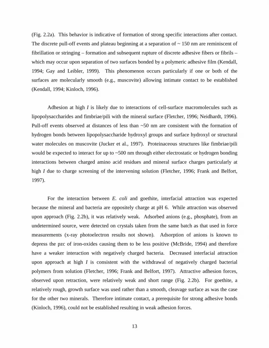
Design and Characterization of Biologically-Active-Force-Probes
Force-sensing probes, termed biologically-active-force-probes (BAFPs), were fabricated
by linking a minute bacteria-coated-bead to a silicon nitride cantilever in a manner that
conserved the orientation, structural integrity, and conformation of macromolecules on the
bacterial surface (Fig. 2.1). Glass beads (~5 µm radius; Duke Scientific or Polysciences) were

F
b
li
su
li
b
w
(b
lo
B
th
Si
6
igure 2.1. (a) Biological force microscopy components highlighting the linkage between
acterial cells and the force-sensing cantilever. Illustrated is one of many possible poly-lysine
nkages between negatively charged silanol groups on the bead and negatively charged cell-
rface functional groups on biomolecules, such as phospholipids (shown here) or
popolysaccharides. A piezoelectric scanner was used to translate a mineral to and from a
iologically-active-force-probe (BAFP). Forces between bacteria on the probe and the mineral
ere detected by reflecting a laser off the top of the cantilever and into a photodiode detector.
) Scanning laser confocal micrograph of a BAFP (perspective is from the mineral surface
oking towards the BAFP). Bacteria are fluorescing due to excitation of an intracellular protein.
iological activity of these cells was demonstrated by culturing a colony on an agar plate using
e probe as the inoculum. Scale bar 10 µm.
b
piezoscanner
BAFP
laserObead
O O
CH2
PO4
H2C CH
O O
C C
CHn CHn
cell
a
O
NH3
H
C
(CH2) 4
(CH2) 4
N CC
OH
N
H
H
C
poly-lysine
mineral
NH3

7
cleaned with hydrofluoric acid (1 % solution for 1.5 minutes) or sodium hydroxide (50 %
solution for 60 minutes), rinsed with ultrapure water (Milli-Pore), and functionalized with amino
groups by incubation in a 2% solution of 135 kDa poly-D-lysine (Sigma) for ~4 hours. This
procedure creates a 1-3 nm thick, positively charged monolayer on glass beads (Pagac et al.,
1998a; Pagac et al., 1998b). Amine-functionalized beads were dispersed in a suspension of
washed cells and spun at 8,000 x g for 5 minutes at 4 °C. A single bead supporting a monolayer
of cells was attached to a cantilever using a small amount of epoxy resin, which has previously
been found to be inert in aqueous solutions (Pincet et al., 1995; Yoon et al., 1997). This
attachment procedure was conducted in solution with the aid of a microscope (Nikon, 200X
magnification) and a micromanipulator to translate the cantilever.
By using whole bacteria expressing macromolecules in their natural state rather than
individual biomolecules (e.g., exopolysaccharides, proteins) purified from bacterial surfaces, we
avoided situations in which the linkage procedure modified the conformation of biomolecules
such that they were no longer in a natural state (Stotzky, 1986; Ellen and Burne, 1996; Turner et
al., 1996; Ingersoll and Bright, 1997; Turner et al., 1997). Linkage of cells to the cantilever is
extremely stable. The strength of bonds between poly-lysine and either glass or bacteria are on
the order of 1000 kJ molecule-1 (Voet and Voet, 1995; West et al., 1997), which is at least one to
four orders of magnitude greater than interfacial and adhesion forces expected to occur between
bacteria and mineral surfaces (Israelachvili, 1992).
A BAFP was placed in the fluid-cell used in force measurements and imaged with a
scanning laser confocal microscope LSM-510 Axiovert 100M (Zeiss) using a 100X, 1.4 N.A.
objective lens. The confocal ability of the microscope, and cellular expression of the
intracellular fluorophore encoded by the inserted plasmid, allowed noninvasive characterization
of the three-dimensional nature of BAFPs.
Biological Force Microscopy Measurements
BFM measurements were performed in sodium chloride solutions using a NanoScope IIIa
Multimode SPM (Digital Instruments). The deflection of a BAFP was monitored as an oriented
mineral grain (mounted on a piezoelectric scanner) was indexed towards, made contact with, and

8
retracted from bacteria on the probe (Fig. 2.1). The mineral was translated at rates of <3 µm
sec-1 which is within the range of velocities of motile bacteria (Marshall, 1976). Interfacial
forces were measured as the mineral approached the bacteria on the probe; whereas adhesion
forces were measured upon contact and subsequent retraction of the mineral from the bacteria.
Mineral samples were driven to the same contact force to normalize the effect that loading can
have on measured forces during retraction (Weisenhorn et al., 1992). To ensure reproducibility,
measurements were taken as solution in the fluid-cell was cycled between low I and high I four
to five times per mineral.
Force-distance curves were constructed from photodiode-voltage versus piezo
displacement data (i.e., “force curves”) (Ducker et al., 1991; Ducker et al., 1992). Diode
response (in volts) was converted to cantilever deflection (in meters) using the
diode/displacement conversion factor defined by the region of constant compliance (i.e., slope on
the force curve where diode response becomes a linear function of piezo displacement). Hooke’s
Law, F = ksp d, where d is cantilever deflection and ksp is the cantilever spring constant (0.17 N
m-1; determined according to Cleveland et al., 1993), was then used to obtain the force of
interaction (in Newtons). The distance-axis origin was defined as the point of intimate contact
(i.e., beginning of the region of constant compliance). This conversion method is appropriate
when the cantilever is the most compliant component of the system (Hutter and Bechhoefer,
1993). A region of constant compliance with 1:1 correspondence between probe deflection and
piezo displacement was observed in all approach-retraction cycles in this study. Recent elasticity
measurements of various bacterial surface macromolecules suggest that this situation is valid for
most bacteria (Xu et al., 1996; Yao et al., 1999). However, for cells that are more compliant than
the cantilever or have fragile appendages, it will be necessary to use a method that does not
require bacteria-mineral contact (D'Costa and Hoh, 1995; Sader et al., 1999).
The results presented herein illustrate the interaction between a mineral surface and an
aggregate of cells rather than single bacterium. The surface properties of an aggregate of cells
may differ from those of a single cell. It should be noted, however, that force measurements
made with force microscopes are presumed to involve the interaction of only a few tens to
hundreds of square nanometers (Butt et al., 1995). These dimensions are closer to the size of a

9
single bacterium than the entire aggregate of cells on the cantilever. Single bacterium have been
attached to cantilevers (results not shown), but this linkage is extremely difficult. Recent
advances in optical tweezer (Svoboda and Block 1994) and nanotweezer (Kim and Lieber 1999)
technologies could significantly enhance the linkage of single cells to a cantilever.
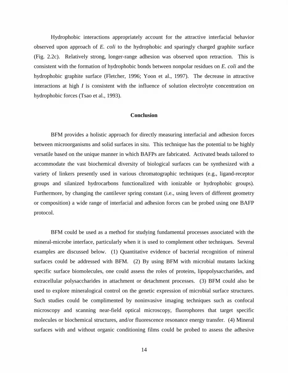
Results
Net repulsive interfacial forces were observed for muscovite approach curves (Fig. 2.2a).
Conversely, E. coli-goethite and -graphite systems exhibited more attractive interfacial forces on
approach (Fig. 2.2b, 2.2c). For these latter systems, jump-to-contact events were observed at
distances of <20 nm because the force gradient between bacteria and mineral surface exceeded
the cantilever spring constant. Ionic strength effects were noted for all mineral-bacteria systems.
In general, the magnitude of interfacial forces and distances over which they were operative
diminished at high I.
Adhesion forces of varying magnitude were observed for each bacteria-mineral system
upon retraction and were sensitive to solution I. At high I the muscovite system showed
adhesion and the retraction curve displayed a large number of discrete “pull-off” events to
separations of ∼ 400 nm (Fig. 2.2a). At low I the muscovite system did not exhibit attractive
adhesion forces. Goethite and graphite systems were markedly different as they exhibited
decreased adhesion force at high I and displayed jump-from-contact events because the
cantilever spring constant exceeded the force gradient between bacteria and mineral surface (Fig.
2.2b, 2.2c).
Results from the control experiment between muscovite and a naked poly-lysine coated
bead (no bacteria) (Fig. 2.2d) were very different from those between muscovite and an E. coli
coated bead (Fig. 2.2a). As expected, the positively charged poly-lysine exhibited strong
attraction towards the (001) surface of muscovite which is negatively charged at pH 6.
Hysteresis between the approach and retraction curves was absent for the interaction between
muscovite and poly-lysine. These observations indicate that the poly-lysine linker did not affect
forces measured between the bacteria and mineral.

10
Figure 2.2. Force-distance curves for Escherichia coli K-12 and (a; top left) muscovite (001)
surface, (b; top right) goethite (010) surface, and (c; bottom left) graphite (001) surface at pH 6,
25ºC and varying ionic strengths (I). (d; bottom right) Control experiment for muscovite (001)
surface and naked poly-lysine coated bead. Jump-to- or jump-from-contact events result in linear
segments with a slope corresponding to the cantilever spring constant (0.17 N m-1). Curve
convention: (triangles) I = 10-5 M, approach; (squares) I = 10-5 M, retraction; (diamonds) I = 10-1
M, approach; (circles) I = 10-1 M, retraction. Force sign convention: (+) repulsive; (−) attractive.
Note differences in axes scales.
-5
-4
-3
-2
-1
0
1
0 10 20 30 40Distance (nm)
Fo
rce
(nN
)
-14-12-10-8-6-4-202
0 25 50 75 100 125 150Distance (nm)
Fo
rce
(nN
)
-40
-30
-20
-10
0
10
0 40 80 120 160 200 240Distance (nm)
Fo
rce
(nN
)
-20
-15
-10
-5
0
5
10
0 100 200 300 400 500Distance (nm)
Fo
rce
(nN
)

11
The bacteria-mineral data presented above were performed by applying the same BAFP
to muscovite, goethite, graphite, and then once again to muscovite. Only minor variation was
observed between the initial and replicate muscovite experiments indicating that bacterial
surfaces were not significantly altered by repeated contact with mineral surfaces. Results shown
in Fig. 2.2 are representative of 300-400 force-distance curves collected using three separate E.
coli BAFPs. While results were highly reproducible (e.g., errors in measurement of muscovite-
bacteria approach curves are ~± 0.2 nN in force and ~± 2.0 nm in distance), some variation was
observed particularly in retraction curves. The dynamic nature of the macromolecular cell
envelope mosaic (Beveridge, 1999) and specific cell orientation on a BAFP are likely to account
for the variation.
Discussion
Categories of Interfacial and Adhesion Forces
All intermolecular forces ultimately depend on the distribution of electrons surrounding
interacting particles or surfaces (Israelachvili, 1992). Unfortunately, theoretical and
experimental studies of the electronic structures of complex biological and mineralogical
surfaces are still in their infancy (e.g., Becker et al., 1996; Beveridge, 1999; Rosso et al., 1999).
Therefore, the fundamental interactions responsible for interfacial and adhesion forces have been
operationally classified into a number of force categories which differ in magnitude, sign, and
operative range.
Van der Waals forces and hydrophobic interactions are generally considered attractive
and predominate from contact to tens of nm separation, whereas electrostatic forces may be
attractive or repulsive and tend to predominate at greater separations (Israelachvili and
McGuiggan, 1988; Israelachvili, 1992; Butt et al., 1995; Fletcher, 1996; Yoon et al., 1997). The
magnitude and operative distance of electrostatic forces decrease with increasing I, commonly
termed the electrostatic double-layer effect. Polymer interactions or bridging forces, involving
long-chain organic molecules, represent the combined effects of several forces including
hydrophobics, electrostatics and van der Waals. These interactions may be attractive or
repulsive depending on ionization of functional groups and typically extend outwards to

12
hundreds of nm (van Loosdrecht et al., 1990; Israelachvili, 1992; Biggs, 1995; Jucker et al.,
1998). Forces attributable to ionic-polymers decrease with increasing I, whereas neutral-
polymers are unaffected by different I (Frank and Belfort, 1997).
At intermediate distances (0.5 to 5 nm), repulsive forces related to solvation/hydration
and steric/entropic effects become operative and may be influenced by I dependent conformation
changes in polymeric surface molecules or solution charge screening (Israelachvili and
McGuiggan, 1988; Israelachvili, 1992; Butt et al., 1995). At extremely small separations (< 1
nm), attractive, specific interactions between surfaces such as hydrogen bonding, cation
bridging, and receptor-ligand interactions may occur (Israelachvili and McGuiggan, 1988;
Israelachvili, 1992; Butt et al., 1995; Fletcher, 1996; Kinloch, 1996).
Mineralogical and Microbiological Aspects of Forces
Deconvolution of the net force between two surfaces into contributions made by
individual interaction mechanisms would require detailed spectroscopic, microscopic, and
surface structural information. Additionally, it is beyond the scope of this paper to compare the
measured interfacial and adhesion forces to those predicted by models such as DLVO (Derjaguin
and Landau, 1941; Verwey and Overbeek, 1948) and JKR (Johnson et al., 1971), respectively.
None-the-less, a general interpretation consistent with the characteristics of the force types
outlined above can be made from the force-distance curves shown in Fig. 2.2.
For the interaction between E. coli and muscovite, both of which are negatively charged at pH 6,
repulsive interfacial forces observed for the approach curves are consistent with electrostatic
forces (Fig. 2.2a). The magnitude of repulsion and distance over which the force was operative
diminished at high ionic strength (I) as expected due to compression of the electrostatic double
layers around the bacteria and muscovite (Israelachvili, 1992) and the flattened conformation of
negatively charged polymers on the bacteria surface (Frank and Belfort, 1997; Jucker et al.,
1998).
An adhesion force was observed for the E. coli-muscovite system at high I as the
retraction curve displayed a large number of discrete “pull-off” events to separations of ∼ 400 nm
13
(Fig. 2.2a). This behavior is indicative of formation of strong specific interactions after contact.
The discrete pull-off events and plateau beginning at a separation of ~ 150 nm are reminiscent of
fibrillation or stringing – formation and subsequent rupture of discrete adhesive fibers or fibrils –
which may occur upon separation of two surfaces bonded by a polymeric adhesive film (Kendall,
1994; Gay and Leibler, 1999). This phenomenon occurs particularly if one or both of the
surfaces are molecularly smooth (e.g., muscovite) allowing intimate contact to be established
(Kendall, 1994; Kinloch, 1996).
Adhesion at high I is likely due to interactions of cell-surface macromolecules such as
lipopolysaccharides and fimbriae/pili with the mineral surface (Fletcher, 1996; Neidhardt, 1996).
Pull-off events observed at distances of less than ~50 nm are consistent with the formation of
hydrogen bonds between lipopolysaccharide hydroxyl groups and surface hydroxyl or structural
water molecules on muscovite (Jucker et al., 1997). Proteinaceous structures like fimbriae/pili
would be expected to interact for up to ~500 nm through either electrostatic or hydrogen bonding
interactions between charged amino acid residues and mineral surface charges particularly at
high I due to charge screening of the intervening solution (Fletcher, 1996; Frank and Belfort,
1997).
For the interaction between E. coli and goethite, interfacial attraction was expected
because the mineral and bacteria are oppositely charge at pH 6. While attraction was observed
upon approach (Fig. 2.2b), it was relatively weak. Adsorbed anions (e.g., phosphate), from an
undetermined source, were detected on crystals taken from the same batch as that used in force
measurements (x-ray photoelectron results not shown). Adsorption of anions is known to
depress the pzc of iron-oxides causing them to be less positive (McBride, 1994) and therefore
have a weaker interaction with negatively charged bacteria. Decreased interfacial attraction
upon approach at high I is consistent with the withdrawal of negatively charged bacterial
polymers from solution (Fletcher, 1996; Frank and Belfort, 1997). Attractive adhesion forces,
observed upon retraction, were relatively weak and short range (Fig. 2.2b). For goethite, a
relatively rough, growth surface was used rather than a smooth, cleavage surface as was the case
for the other two minerals. Therefore intimate contact, a prerequisite for strong adhesive bonds
(Kinloch, 1996), could not be established resulting in weak adhesion forces.
14
Hydrophobic interactions appropriately account for the attractive interfacial behavior
observed upon approach of E. coli to the hydrophobic and sparingly charged graphite surface
(Fig. 2.2c). Relatively strong, longer-range adhesion was observed upon retraction. This is
consistent with the formation of hydrophobic bonds between nonpolar residues on E. coli and the
hydrophobic graphite surface (Fletcher, 1996; Yoon et al., 1997). The decrease in attractive
interactions at high I is consistent with the influence of solution electrolyte concentration on
hydrophobic forces (Tsao et al., 1993).
Conclusion
BFM provides a holistic approach for directly measuring interfacial and adhesion forces
between microorganisms and solid surfaces in situ. This technique has the potential to be highly
versatile based on the unique manner in which BAFPs are fabricated. Activated beads tailored to
accommodate the vast biochemical diversity of biological surfaces can be synthesized with a
variety of linkers presently used in various chromatographic techniques (e.g., ligand-receptor
groups and silanized hydrocarbons functionalized with ionizable or hydrophobic groups).
Furthermore, by changing the cantilever spring constant (i.e., using levers of different geometry
or composition) a wide range of interfacial and adhesion forces can be probed using one BAFP
protocol.
BFM could be used as a method for studying fundamental processes associated with the
mineral-microbe interface, particularly when it is used to complement other techniques. Several
examples are discussed below. (1) Quantitative evidence of bacterial recognition of mineral
surfaces could be addressed with BFM. (2) By using BFM with microbial mutants lacking
specific surface biomolecules, one could assess the roles of proteins, lipopolysaccharides, and
extracellular polysaccharides in attachment or detachment processes. (3) BFM could also be
used to explore mineralogical control on the genetic expression of microbial surface structures.
Such studies could be complimented by noninvasive imaging techniques such as confocal
microscopy and scanning near-field optical microscopy, fluorophores that target specific
molecules or biochemical structures, and/or fluorescence resonance energy transfer. (4) Mineral
surfaces with and without organic conditioning films could be probed to assess the adhesive

15
properties of humic or fulvic acids. (5) BFM could be used to study inter- and intra-species
signaling of microorganisms within a biofilm or other community by probing bacteria-bacteria
interactions, host-pathogen recognition, and/or the effects aqueous biochemical signals have on
quorum sensing phenomena. (6) BFM could provide insight into enzyme activity or
conformation changes in cell surface proteins involved in oxidation/reduction reactions at
mineral surfaces. In so doing, BFM may contribute to a better understanding of electron transfer
at the microbe-mineral interface. (7) Forces measured with BFM could be compared to force
models (e.g., DLVO and JKR) to gain a fundamental understanding of attachment and
detachment phenomena. Techniques like biological force microscopy have the potential to open
a new door in mineral-microbe research, one in which researchers are able to gain a fundamental
insight into the nanoscale world that exists at the interface between microorganisms and minerals
in nature.
Acknowledgments
This work benefited from discussions with W. Ducker, A. Razatos, J. Wightman, and members
of the Virginia Tech mineral-microbe group including D. Berry, M. Eick, T. Kendall, J. Little,
and M. Potts. The presentation of this study was enhanced by the constructive comments of J.
Fein and three anonymous reviewers. K. DeCourcy assisted with electroporation and scanning
laser microscopy. S.K.L. greatly appreciates the support of J. Tak. Mineral samples were
provided by the Virginia Tech Geological Sciences Museum. Funding was provided by the
American Chemical Society grant ACS-PRF 34326-AC2 and the Department of Energy grant
DE-FG02-99ER 15002.
References
Adamson A. W. and Gast A. P. (1997) Physical Chemistry of Surfaces. John Wiley & Sons, New
York.
Av-Gay Y. (1999) Uncontrolled release of harmful microorganisms. Science 284, 1621.
16
Baur B., Hanselmann K., Schlimme W., and Jenni B. (1996) Genetic transformation in
freshwater: Escherichia coli is able to develop natural competence. Appl. Envir.
Microbiol. 62, 3673-3678.
Becker U., Hochella, Jr., M. F., and Apra E. (1996) The electronic structure of hematite {001}
surfaces: Applications to the interpretation of STM images and heterogeneous surface
reactions. Amer. Mineral. 81, 1301-1314.
Beveridge T. J. (1999) Structures of Gram-negative cell walls and their derived membrane
vesicles. J. Bacteriol. 181, 4725-4733.
Bhosle N., Suci P. A., Baty A. M., Weiner R. M., and Geesey G. G. (1998) Influence of divalent
cations and pH on adsorption of a bacterial polysaccharide adhesin. J. Colloid Interface
Sci. 205, 89-96.
Biggs S. (1995) Steric and bridging forces between surfaces bearing adsorbed polymer: An
atomic force microscopy study. Langmuir 11, 156-162.
Boland T. and Ratner B. D. (1995) Direct measurement of hydrogen bonding in DNA nucleotide
bases by atomic force microscopy. Proc. Nat. Acad. Sci. USA 92, 5297-5301.
Butt H. J., Jaschke M., and Ducker W. (1995) Measuring surface forces in aqueous electrolyte
solution with the atomic force microscope. Bioelectrochem. Bioenergetics 38, 191-201.
Cleveland J. P., Manne S., Bocek D., and Hansma P. K. (1993) A nondestructive method for
determining the spring constant of cantilevers for scanning force microscopy. Rev. Sci.
Instrum. 64, 403-405.
Crook J., Engelbrecht R. S., Benjamin M. M., Bull R. J., Fowler B. A., Griffin H. E., Hass C. N.,
Moe C. L., Rose J. B., and Trussel R. R. (1998) Issues in Potable Reuse: The Viability of
Augmenting Drinking Water Supplies with Reclaimed Water. National Academy Press,
Washington DC.
Dammer U., Popescu O., Wagner P., Anselmetti D., Guntherodt H.-J., and Misevic G. N. (1995)
Binding strength between cell adhesion proteoglycans measured by atomic force
microscopy. Science 267, 1173-1175.
Davies D. G., Parsek M. R., Pearson J. P., Iglewski B. H., Costerton J. W., and Greenberg E. P.
(1998) The involvement of cell-to-cell signals in the development of a bacterial biofilm.
Science 280, 295-298.
17
D'Costa N. P. and Hoh J. H. (1995) Calibration of optical lever sensitivity for atomic force
microscopy. Rev. Sci. Instrum. 66, 5096-5097.
Derjaguin B. V. and Landau L. (1941) Theory of the stability of strongly charged lyophobic sols
and the adhesion of strongly charged particles in solutions of electrolytes. Acta
Physiochim. URSS 14, 633-662.
Dower W. J., Miller J. F., and Ragsdale C. W. (1988) High efficiency transformation of E. coli
by high voltage electroporation. Nucleic Acids Res. 16, 6127-6145.
Ducker W. A., Senden T. J., and Pashley R. M. (1991) Direct measurement of colloidal forces
using an atomic force microscope. Nature 353, 239-241.
Ducker W. A., Senden T. J., and Pashley R. M. (1992) Measurements of forces in liquids using a
force microscope. Langmuir 8, 1831-1836.
Dziurla M., -A., Achouak W., Lam B., -T., Heulin T., and Berthelin J. (1998) Enzyme-linked
immunofiltration assay to estimate attachment of Thiobacilli to pyrite. Appl. Envir.
Microbiol. 64, 2937-2942.
Edwards K. J., Schrenk M. O., Hamers R., and Banfield J. F. (1998) Microbial oxidation of
pyrite: Experiments using microorganisms from an extreme acidic environment. Amer.
Mineral. 83, 1444-1453.
Ellen R. P. and Burne R. A. (1996) Conceptual advances in research on the adhesion of bacteria
to oral surfaces. In Bacterial Adhesion: Molecular and Ecological Diversity. (ed. M.
Fletcher). American Society for Microbiology, Washington DC. pp. 201-247.
Fleminger G. and Shabtai Y. (1995) Direct and rapid analysis of the adhesion of bacteria to solid
surfaces: Interaction of fluorescently labeled Rhodococcus strain GIN-1 (NCIMB 40340)
cells with titanium-rich particles. Appl. Envir. Microbiol. 61, 4357-4361.
Fletcher M. (1996) Bacterial Adhesion: Molecular and Ecological Diversity. Wiley-Liss, New
York.
Florin E.-L., Moy V. T., and Gaub H. E. (1994) Adhesion forces between individual ligand-
receptor pairs. Science 264, 415-417.
Fortin D., Ferris F. G., and Beveridge T. J. (1997) Surface-mediated mineral development by
bacteria. Rev. Mineral. 35, 161-180.
Frank B. P. and Belfort G. (1997) Intermolecular forces between extracellular polysaccharides
measured using the atomic force microscope. Langmuir 13, 6234-6240.

18
Frisbie C. D., Rozsnyai L. F., Noy A., Wrighton M. S., and Lieber C. M. (1994) Functional
group imaging by chemical force microscopy. Science 265, 2071-2074.
Gannon J. T., Manilal V. B., and Alexander M. (1991) Relationship between cell surface
properties and transport of bacteria through soil. Appl. Envir. Microbiol. 57, 190-193.
Gay C. and Leibler L. (1999) On stickiness. Phys. Today 52, 48-52.
Grandbois M., Beyer M., Rief M., Clausen-Schaumann H., and Gaub H. E. (1999) How strong is
a covalent bond? Science 283, 1727-1730.
Hiebert F. K. and Bennett P. C. (1992) Microbial control of silicate weathering in organic-rich
ground water. Science 258, 278-281.
Hinterdorfer P., Baumgartner W., Gruber H. J., Schilcher K., and Schindler H. (1996) Detection
and localization of individual antibody-antigen recognition events by atomic force
microscopy. Proc. Nat. Acad. Sci. USA 93, 3477-3481.
Hutter J. L. and Bechhoefer J. (1993) Calibration of atomic-force microscope tips. Rev. Sci.
Instrum. 64, 1868-1873.
Ingersoll C. M. and Bright F. V. (1997) Using fluorescence to probe biosensor interfacial
dynamics. Anal. Chem. 69, 403A-408A.
Israelachvili J. (1992) Intermolecular and Surface Forces. Academic Press, London.
Israelachvili J. N. and McGuiggan P. M. (1988) Forces between surfaces in liquids. Science 241,
795-800.
Johnson K. L., Kendall K., and Roberts A. D. (1971) Surface energy and the contact of elastic
solids. Proc. Royal Soc. London 324A, 301-313.
Jucker B. A., Harms H., Hug S. J., and Zehnder A. J. B. (1997) Adsorption of bacterial surface
polysaccharides on mineral oxides is mediated by hydrogen bonds. Colloids Surf. B 9,
331-343.
Jucker B. A., Zehnder A. J. B., and Harms H. (1998) Quantification of polymer interactions in
bacterial adhesion. Environ. Sci. Technol. 32, 2909-2915.
Kendall K. (1994) Adhesion: Molecules and mechanics. Science 263, 1720-1725.
Kim P. and Lieber C. M. (1999) Nanotube nanotweezers. Science 286, 2148-2150.
Kinloch A. J. (1996) Adhesives in engineering. Proc. Instit. Mech. Engineers, 1-32. London.
Lawrence J. R., Korber D. R., Hoyle B. D., Costerton J. W., and Caldwell D. E. (1991) Optical
sectioning of microbial biofilms. J. Bacteriol. 173, 6558-6567.
19
Lee G. U., Chrisey L. A., and Colton R. J. (1994) Direct measurement of the forces between
complementary strands of DNA. Science 266, 771-773.
Marshall K. C. (1976) Interfaces in Microbial Ecology. Harvard University Press, Cambridge,
Massachusetts.
McBride M. B. (1994) Environmental Chemistry of Soils. Oxford University Press, New York.
Miller J. F., Dower W. J., and Tompkins L. S. (1988) High-voltage electroporation of bacteria:
genetic transformation of Campylobacter jejuni with plasmid DNA. Proc. Nat. Acad. Sci.
USA 85, 856-860.
Mills A. L. and Powelson D. K. (1996) Bacterial interactions with surfaces in soils. In Bacterial
Adhesion: Molecular and Ecological Diversity. (ed. M. Fletcher). American Society for
Microbiology, Washington DC. pp. 25-57.
Moy V. T., Florin E.-L., and Gaub H. E. (1994) Intermolecular forces and energies between
ligands and receptors. Science 266, 257-259.
Myers C. and Nealson K. H. (1988) Bacterial manganese reduction and growth with manganese
oxide as the sole electron acceptor. Science 240, 1319-1321.
Neidhardt F. C. (1996) Escherichia coli and Salmonella: Cellular and Molecular Biology.
American Society for Microbiology, Washington DC.
Ohmura N., Kitamura K., and Saiki H. (1993) Selective adhesion of Thiobacillus ferrooxidans to
pyrite. Appl. Envir. Microbiol. 59, 4044-4050.
Pagac E. S., Prieve D. C., and Tilton R. D. (1998a) Kinetics and mechanisms of cationic
surfactant adsorption and coadsorption with cationic polyelectrolytes at the silica-water
interface. Langmuir 14, 2333-2342.
Pagac E. S., Tilton R. D., and Prieve D. C. (1998b) Depletion attraction caused by unadsorbed
polyelectrolytes. Langmuir 14, 5106-5112.
Pincet F., Perez E., and Wolfe J. (1995) Does glue contaminate the surface forces apparatus?
Langmuir 11, 373-374.
Rief M., Oesterhelt F., Heymann B., and Gaub H. E. (1997a) Single molecule force spectroscopy
on polysaccharides by atomic force microscopy. Science 275, 1295-1297.
Rief M., Gautel M., Oesterhelt F., Fernandez J. M., and Gaub H. E. (1997b) Reversible
unfolding of individual titin immunoglobulin domains by AFM. Science 276, 1109-1112.

20
Roden E. E. and Zachara J. M. (1996) Microbial reduction of crystalline iron(III) oxides:
Influence of oxide surface area and potential for cell growth. Environ. Sci. Technol. 30,
1618-1628.
Rosso K. M., Becker U., and Hochella M. F., Jr. (1999) Atomically resolved electronic structure
of pyrite {100} surfaces: An experimental and theoretical investigation with implications
for reactivity. Amer. Mineal. 84, 1535-1548.
Sader J. E., Chon J. W. M., and Mulvaney P. (1999) Calibration of rectangular atomic force
microscope cantilevers. Rev. Sci. Instrum. 70, 3967-3969.
Schultze-Lam S., Harauz G., and Beveridge T. J. (1992) Participation of a cyanobacterial S-layer
in fine-grain mineral formation. J. Bacteriol. 174, 7971-7981.
Stipp S. L. and Hochella, Jr., M. F. (1991) Structure and bonding environments at the calcite
surface as observed with X-ray photoelectron spectroscopy (XPS) and low energy
electron diffraction (LEED). Geochim. Cosmochim. Acta 55, 1723-1736.
Stotzky G. (1986) Influence of soil mineral colloids on metabolic processes, growth, adhesion,
and ecology of microbes and viruses. In Interactions of Soil Minerals with Natural
Organics and Microbes, Vol. 17 (eds. P. M. Huang and M. Schnitzer). Soil Science
Society of America, Madison, Wisconsin. pp. 305-428.
Stumm W. and Morgan J. J. (1996) Aquatic Chemistry. John Wiley & Sons, New York.
Svoboda K. and Block S. M. (1994) Biological applications of optical forces. Annu. Rev.
Biophys. Biomol. Struct. 23, 247-285.
Trevors J. T. and van Elsas J. D. (1997) Quantification of gene transfer in soil and the
rhizosphere. In Manual of Environmental Microbiology. (ed. C. J. Hurst). American
Society for Microbiology, Washington DC. pp. 500-508.
Tsao Y.-H., Evans D. F., and Wennerstrom H. (1993) Long-range attractive force between
hydrophobic surfaces observed by atomic force microscopy. Science 262, 547-550.
Turner D. C., Peek B. M., Wertz T. E., Archibald D. D., Geer R. E., and Gaber B. P. (1996)
Enzymatic modification of a chemisorbed lipid monolayer. Langmuir 12, 4411-4416.
Turner D. C., Testoff M. A., Conrad D. W., and Gaber B. P. (1997) Enzymatic hydrolysis of a
chemisorbed peptide film using beads activated with covalently bound chymotrypsin.
Langmuir 13, 4855-4860.

21
van Loosdrecht M. C. M., Lyklema J., Norde W., and Zehnder A. J. B. (1989) Bacterial
adhesion: A physicochemical approach. Microbial Ecology 17, 1-15.
van Loosdrecht M. C. M., Norde W., Lyklema J., and Zehnder A. J. B. (1990) Hydrophobic and
electrostatic parameters in bacterial adhesion. Aquatic Sci. 52, 103-114.
Verwey E. J. W. and Overbeek J. Th. G. (1948) Theory of the Stabilityof Lyophobic Colloids.
Elsevier, Amsterdam.
Voet D. and Voet J. (1995) Biochemistry. John Wiley & Sons, New York.
Weisenhorn A. L., Maivald P., Butt H.-J., and Hansma P. K. (1992) Measuring adhesion,
attraction, and repulsion between surfaces in liquids with an atomic-force microscope.
Phys. Review B 45, 11226-11232.
West J. K., Latour R., Jr., and Hench L. L. (1997) Molecular modeling study of adsorption of
poly-L-lysine onto silica glass. J. Biomed. Materials Res. 37, 585-591.
Wong S. S., Joselevich E., Woolley A. T., Cheung C. L., and Lieber C. M. (1998) Covalently
functionalized nanotubes as nanometre-sized probes in chemistry and biology. Nature
394, 52-55.
Xu W., Mulhern P. J., Blackford B. L., Jericho M. H., Firtel M., and Beveridge T. (1996)
Modeling and measuring the elastic properties of an Archaeal surface, the sheath of
Methanospirillum hungatei, and the implications for methane production. J. Bacteriol.
178, 3106-3112.
Yao X., Jericho M., Pink D., and Beveridge T. (1999) Thickness and elasticity of Gram-negative
murein sacculi measured by atomic force microscopy. J. Bacteriol. 181, 6865-6875.
Yee N., Fein J. B., and Daughney C. J. (2000) Experimental study of the pH, ionic strength, and
reversibility behavior of bacteria-mineral adsorption. Geochim. Cosmochim. Acta 64,
609-617.
Yoon R. H., Flinn D. H., and Rabinovich Y. I. (1997) Hydrophobic interactions between
dissimilar surfaces. J. Colloid Interface Sci. 185, 363-370.
22
Chapter 3 - Dynamics of the mineral-microbe interface: Use of biological force microscopy
in biogeochemistry and geomicrobiology
Published in Geomicrobiology Journal, 2001, 18: 63-76.
Copyright 2000 Taylor & Francis
Abstract
At the most fundamental level, inter- and intra-molecular forces delineate the interface
between a microorganism and a mineral surface. A new technique, termed biological force
microscopy (BFM), is described which can be used to directly probe the dynamics of the
mineral-microbe interface. BFM quantifies attractive and repulsive forces in the nanoNewton
range between living microbial cells and mineral surfaces in aqueous solution. Native bacterial
cells are linked to a force-sensor that is used in a force microscope to measure bacteria-mineral
interactions as a function of the distance between the mineral surface and the cells on the sensor.
The magnitudes and ranges of the measured forces reflect the chemical and structural intricacies
of the mineral-microbe interface. BFM is presented with potential applications to studies
assessing the role microbes or biomolecules play in geochemical and mineralogical processes.
Introduction
Mineral-microbe interactions have been occurring for at least 3 billion years on and
within the earth. Thousands of mineral species with enormous variability in surface chemistry
and structure may interact with any of millions of bacterial species that display diverse surface
mosaics and physiologies. Minerals and microorganisms are intimately linked such that one
often cannot exist without the other in nature. Microbial processes play roles in the cycling of
elements and sorption of metals (Ehrlich 1990; Fein et al. 1997; Langley and Beveridge 1999;
Marshall 1996; Stotzky 1986), the dissolution of minerals (Banfield and Hamers 1997; Barker et
al. 1998; Edwards et al. 1998a; Edwards et al. 1998b; Forsythe et al. 1998; Grantham et al. 1997;
Hersman et al. 1995; Hersman et al. 1996; Krumbein et al. 1991; Lovley and Phillips 1986;
Lovley and Phillips 1987; Myers and Nealson 1988; Robert and Berthelin 1986; Roden and

23
Zachara 1996; Schrenk et al. 1998; Stone 1997; Welch and Ullman 1993; Welch and
Vandevivere 1994), and mineral crystallization (Beveridge and Doyle 1989; Fortin and
Beveridge 1997; Fortin et al. 1997; Pentecost and Bauld 1988; Schultze-Lam et al. 1996;
Schultze-Lam et al. 1992; Warren and Ferris 1998). Conversely, mineralogical processes
influence the distribution, activity, and diversity of microbes (Barker et al. 1997; Bennett et al.
1996; Fletcher 1996a; Fredrickson et al. 1995; Marshall 1996; Rogers et al. 1998; Schrenk et al.
1998; Stotzky 1986), the expression of genes (Arredondo et al. 1994; Dziurla et al. 1998;
Fletcher 1996a; Gehrke et al. 1998), community structure and development (Brown et al. 1997;
Kennedy and Gewin 1997; Lawrence et al. 1991; Thorseth et al. 1995; Wolfaardt et al. 1994),
and transfer of genetic material (Holben 1997; Trevors and van Elsas 1997).
The thread linking these unimaginably complex interactions is the fact that mineral-
microbe processes are dependent upon the intimate juxtaposition of a living and nonliving entity,
that is, the interface between a microbial cell and a mineral surface. A fundamental appreciation
of this interface is dependent upon our understanding and characterization of the symphony of
inter- and intra-molecular forces between microbes and mineral surfaces in nature. Despite the
vast amount of work on microbial affects on mineralogical processes and vice versa, the
interface remains largely unexplored due primarily to the fact that it is difficult to directly probe
this minute and dynamic space.
Atomic force microscopy – and variations thereof – is an elegant tool for measuring inter-
and intramolecular forces between organic and inorganic surfaces (Figure 3.1) (for review see
Butt et al. 1995; Cappella and Dietler 1999; Lower and Maurice 2001). Recently, we created
biological force microscopy (BFM) as a method to directly probe interfacial and adhesion forces
between bacteria and mineral surfaces in aqueous solution (Lower et al. 2000). Living cells are
linked to a sensor that is used to quantitatively measure attractive and repulsive forces in the
nanoNewton range between bacteria and mineral surfaces at distances between zero (i.e.,
contact) and 1 µm. Measured forces reflected the complex interactions of structural and
chemical functionalities on the bacteria and mineral surfaces. Herein, we demonstrate and

Figure 3.1. Sc
mineral and forc
measurements a
attractive or repu
mounted on a p
sensor. Deflecti
a split segment
sensor thereby c
adhesive forces
24
hematic diagram showing the key components of a force microscope. The
e sensor are within a fluid cell (not shown) containing aqueous solution. Force
re made by recording the deflection of a sensor (i.e., cantilever) in response to
lsive forces between itself and the sample (a mineral in this case). The sample,
iezoelectric scanner, indexes towards, makes contact with, and retracts from the
on of the sensor is detected by reflecting a laser off the top of the sensor and into
photodiode. In this study, bacteria (shown as a sphere) have been linked to the
reating a biologically-active-force-probe (BAFP) used to measure interfacial and
between bacteria and mineral surfaces, in-situ.
piezoelectricscanner
mineralsensor
photodiode
laser

25
discuss this technique and suggest potential applications of BFM to studies assessing the role that
microbes or biomolecules play in geochemical and mineralogical processes.
Materials and Methods
Mineral and Bacteria
Freshly cleaved muscovite (KAl2(AlSi3O10)(OH)2) and a Gram-negative soil bacteria
were used for all experiments. The bacteria strain was obtained from a chemostat inoculated
with an iron-oxide rich soil from Pandapas Pond, Jefferson National Forest, Virginia (Tadanier et
al. 2000). Through comparison of 500 bp of 16S rRNA sequence, this bacterium has been
aligned at the species level (0.67% difference) with the pseudomonad Burkholderia cepacia
(MIDI Labs, Newark, DE). Burkholderia sp. were cultured on agar plates under oligotrophic
(glucose concentration = 0.6 mM) or eutrophic conditions (glucose concentration = 2.8 mM) and
used in BFM experiments as described below (see Tadanier et al. 2000 for complete description
of growth media).
Force Sensor Preparation and Characterization
Biologically-active-force-probes (BAFPs) can be created by linking bacteria either
directly to a silicon or silicon nitride force sensor (i.e., cantilever), or indirectly by linking a
monolayer of bacteria to a small glass bead that is then fixed to the end of a cantilever. Direct-
linkage of bacteria to the cantilever was accomplished by first placing a cantilever in a 1-5%
solution of the polycationic molecule, poly-D-lysine (135 to 150 kDa; pH near neutral). Bacteria
were linked to the poly-lysine functionalized cantilever by lowering it into a colony of live
bacteria with the aid of a microscope (Nikon, 200x magnification) and a micromanipulator to
translate the cantilever. For the indirect-linkage method, small glass beads (Polysciences or
Duke Scientific, radii 3-7 µm, cleaned with a solution of 1% hydrofluoric acid or 10% sodium
hydroxide) were activated with a 1-5% solution of poly-lysine. The activated beads were placed
in a suspension of Burkholderia sp. and centrifuged at 8000 x g for 5 minutes. A single bacteria-
coated-bead was attached to a cantilever using a small quantity of epoxy resin, which has

26
previously been found to be inert in aqueous solutions (Pincet et al. 1991; Yoon et al. 1997).
This attachment procedure was conducted in solution using a micromanipulator.
Prior to BFM measurements, scanning laser confocal microscopy (Zeiss LSM 510) was
used to characterize the three-dimensional nature of a BAFP. A probe was placed in the fluid
cell used for force measurements and imaged with a 100x, 1.4 N.A. objective. This imaging
procedure was facilitated by transforming bacteria with a plasmid (pGLO, Bio-Rad or pSMC2,
provided by G. A. O’Toole, Dartmouth University) that encoded an intracellular green
fluorescent protein. The bacteria fluoresced when excited by light at 458 nm or 488 nm.
Fluorescence emitted by the epoxy resin revealed that it was confined to the region between the
cantilever and glass bead (i.e., resin was not in a position that would alter the interaction between
the bacteria and mineral during BFM experiments).
Cantilever spring constants (N m-1), essential for measuring force magnitudes with a
force microscope (see below), can vary substantially from the nominal value listed by the
manufacturer (Cleveland et al. 1993; Senden and Ducker 1994). Spring constants were
determined by attaching known masses to the end of a cantilever and recording the change in
cantilever resonant frequency (Cleveland et al. 1993). A linear relationship was observed
between added mass and resonant frequency with the slope being the spring constant (0.17 N m-
1). These measurements were accomplished using ten cantilevers from the same wafer as that
used to create BAFPs. The variability of spring constants from cantilever to cantilever within the
same wafer has been determined to be very small (Senden and Ducker 1994). This was
confirmed by the linear fit of our data and the reproducibility of the resonant frequency of
unloaded cantilevers (13.7 kHz ±0.2).
Force Measurements
BFM measurements were performed using a NanoScope IIIa Multimode SPM (Digital
Instruments) in aqueous solution (pH 6, ionic strength 10-5 M, 25 °C). A piezoelectric scanner
was used to translate the mineral towards and away from bacteria on a BAFP at rates of 0.1 to 3
µm sec-1. For comparison, this is within the range of velocities of motile bacteria (Marshall
1976). Interfacial forces were measured as the mineral approached the bacteria on the probe;

27
whereas adhesion forces were measured upon contact and subsequent retraction of the mineral
from the bacteria. Mineral samples were driven to the same contact force to normalize the effect
that loading can have on adhesion forces during retraction (Weisenhorn et al. 1992).
Force measurement data are collected as cantilever deflection (diode voltage) and
corresponding piezo displacement, typically termed a force curve (Figure 3.2A). This data must
be manipulated to produce the familiar force-distance curve, which describes interfacial and
adhesion forces (Figure 3.2B). Distance (i.e., separation between bacteria and mineral) is
calculated by correcting the recorded piezo position (i.e., displacement) by the measured
deflection of the cantilever. For example, if the mineral attached to the piezo scanner moves 10
nm towards bacteria attached to the cantilever, and the bacteria are repelled 2 nm due to
repulsive forces, then the actual mineral-bacteria distance (or separation) changes by only 8 nm.
The distance axis origin is chosen as the point on the force curve where sensor deflection
becomes a linear function of piezo displacement (the sensor is in contact with the sample).
Force (F) is determined using Hooke’s Law, F = ksp d, where d is cantilever deflection
(meters) and ksp (N m-1) is the cantilever spring constant. In order to use Hooke’s Law,
cantilever deflection measured by the photodiode in volts must be converted to meters. A
diode/displacement conversion factor (also called “optical lever sensitivity”) is defined from the
slope of the force curve region where the cantilever is in contact with the sample on the piezo
(Figure 3.2Aiii, called the region of constant compliance). The reciprocal of this conversion
factor (in nm V-1) can be used to convert measured cantilever deflection in volts to meters. A
zero force reference value is determined as the force curve region where sensor deflection is
independent of piezo displacement (Figure 3.2Ai, the sensor and sample are not interacting
because they are far apart).
It is important to note that using the constant compliance region of the force curve to
convert photodiode response into force will overestimate the force of interaction if the bacteria
are more compliant than the cantilever. Recent measurements of the elasticity of bacterial
surface macromolecules suggest that bacteria are less compliant (i.e., stiffer) than cantilevers
having spring constants smaller than 10 N m-1 (Xu et al. 1996; Yao et al. 1999).

Figure 3.2. Schematic diag
sensor and sample are far
sample approaches the sens
sensor to deflect upwards (
contact with the sample
compliance). The sample i
to their original positions
milliseconds). Hysteresis,
text for discussion on con
distance, respectively. Int
measured upon retraction; r
A
28
rams of a force curve (A) and force-distance curve (B). When the
apart (i) they exhibit no interaction (region of zero force). As the
or, intermolecular forces between the bacteria and mineral cause the
ii) due to repulsive forces shown here. Eventually the probe makes
(iii) and their movement becomes coupled (region of constant
s then retracted from the probe (iv) until the sensor and sample return
thereby completing one cycle (an entire cycle requires nano- to
shown here, may occur upon retraction due to adhesion forces. See
verting cantilever deflection and piezo displacement into force and
erfacial forces are measured on approach and adhesion forces are
epulsive forces are positive and attractive forces are negative.
def
lect
ion
(V
)
piezo displacement (nm)
iii
iii
ivfo
rce
(nN
)
distance (nm)
B
attractive
repulsive
adhesive forces
interfacial forces+
_

29
In instances where bacteria (or biomolecules) linked to the cantilever are more compliant
than the cantilever or for cells with fragile appendages, other methods must be used to accurately
convert the measured deflection of the cantilever (in volts) into a force of interaction (in
Newtons) (D’Costa and Hoh 1995; Sader et al. 1999). For example, because the optical lever
sensitivity is strongly dependent upon the shape of the laser spot on the photodiode detector, the
“photodiode shift voltage” can be used to convert volts of cantilever deflection into meters of
deflection (D’Costa and Hoh 1995). Photodiode shift voltage is measured as the difference in
output voltage when the photodiode detector is shifted approximately 318 µm (one full turn of
the positioning screw) on either side of the zero setting. Figure 3.3 illustrates the strong
correlation between photodiode shift voltages and optical lever sensitivities measured by
directing the laser to different positions on the cantilever. Once this correlation is established for
a given instrument, piezoelectric scanner, fluid cell, and cantilever (e.g., 200 µm long, V-shaped,
silicon nitride cantilevers), the optical lever sensitivity can be accurately determined without
pressing the cantilever against any other surface. This method ensures that forces can be
determined regardless of the compliance of the cantilever relative to any microorganism attached
to it, and also ensures the preservation of fragile macromolecules on microorganisms attached to
the cantilever.
Results and Discussion
Characterization of Biologically-Active-Force-Probes
BAFPs were created by either attaching a single bacterium to a force sensor (Figure
3.4A) or by attaching a single bacteria-coated-bead (Figure 3.4B) to a force sensor. Fluorescent
dyes (ViaGram, Molecular Probes) and plating techniques revealed that cell membranes on these
bacteria are intact and the cells are viable. Both the direct and indirect method of attaching
bacteria to a cantilever preserves the orientation and structural integrity of macromolecules on
the cell surface. This is important because the native conformation of macromolecules on a
bacteria surface is critical to their function, activity, and role in nature. By using whole bacteria
expressing macromolecules in their natural state rather than individual biomolecules (e.g.,
exopolysaccharides, proteins) purified from bacterial surfaces, we avoid situations in which the
linkage procedure modifies the conformation of biomolecules such that they are no longer in a

Figure 3.3. Variati
a J-type piezoelect
measurements were
solutions ranging in
regression line is 64
lever sensitivity can
voltage. Furthermo
compliant compone
30
on of photodiode shift voltage as a function of optical lever sensitivity using
ric scanner and eight silicon nitride cantilevers (200 µm in length). All
performed on muscovite and hematite in a fluid cell containing aqueous
ionic strength from zero (i.e., Milli-Q water) to 10-2 M. The slope of the
2 nm and the intercept is 1.3 V (R2 = 0.95). With this correlation, the optical
be determined in a matter of seconds by simply using the photodiode shift
re, this method is valid regardless of whether the cantilever is the most
nt of the system.
02468
1012141618
0 0.005 0.01 0.015 0.02 0.025
optical lever sensitivity (V/nm)
ph
oto
dio
de
shif
t vo
ltag
e (V
)

31
Figure 3.4. (A) Scanning laser micrograph of a BAFP created by directly linking a single cell of
Burkholderia sp. to a force-sensing cantilever. (B) Confocal micrograph focused on the mid-
plane of bacteria-coated beads revealing a layer of cells. Focusing up and down on the beads
reveal that many are evenly coated with cells. Beads showing the most uniform coverage are
attached to cantilevers, thereby creating BAFPs. These cells have been transformed with a
plasmid encoding a green fluorescent protein and are emitting “natural” fluorescence. This
fluorescent protein, being intracellular, together with the confocal abilities of the laser scanning
microscope allow noninvasive characterization of the orientation and distribution of cells on the
force sensor, in-situ, without affecting the surface chemistry of the microbes. Plating
experiments in which BAFPs were used as the inoculum, revealed that the bacteria on the
sensors are viable and presumably active during force measurements. Scale bars = 10 µm.
BB
A

32
natural state (Ellen and Burne 1996; Ingersoll and Bright 1997; Stotzky 1986; Turner et al. 1996;
Turner et al. 1997).
As both Gram-negative and Gram-positive bacteria are typically negatively charged at
most pH, poly-lysine with its high pKa is a more-or-less universal linker molecule. However,
due to the large diversity in bacterial surfaces, situations will likely arise in which other linker
molecules are necessary. The versatility of our linkage protocol allows for modification on a
case-by-case basis. For example, we have also fabricated BAFPs using
aminopropyltriethoxysilane and polyethyleneimine with species of Shewanella and
Pseudomonas. Additionally, methods used to attach proteins or antibodies to small glass spheres
or sensors (e.g., Caruso et al. 1998; Florin et al. 1994; Frey and Corn 1996; Rezania et al. 1999;
Turner et al. 1997) could be applied to our protocol. For example, lysozyme, which has been
genetically altered such that it possesses an active binding site but an inactive catalytic site (Voet
and Voet 1995), is an attractive choice because of its high affinity for bacterial cell walls.
Antibodies specific for cell surface receptors may also be effective linkers.
A critical component to consider when designing BAFPs is the relative difference
between those forces binding the cells to the sensor, and interfacial and adhesive forces that will
be probed by the cells on the sensor. Forces holding bacteria to the sensor must be greater than
the forces that are actually probed, otherwise the bacteria will be stripped from the cantilever
during force measurements. Electrostatic attachment of bacteria to sensors via poly-lysine is
very strong. Approximations using Coulomb’s law and molecular dynamic calculations show
that the energy associated with amine-silanol (i.e., the poly-lysine glass bead linkage or poly-
lysine cantilever linkage) and amine-carboxylic pairs (i.e., the poly-lysine bacteria linkage)
(Pagac et al. 1998a; Pagac et al. 1998b; Voet and Voet 1995; West et al. 1997) are at least one to
four orders of magnitude stronger than potential intermolecular forces between a bacteria and
mineral (Israelachvili 1992). Organosilane linkages like that of aminopropyltriethoxysilane form
covalent bonds with silanol groups (Plueddemann 1991) and should therefore be even stronger
than poly-lysine linkages.
33
Comparison of the direct and indirect linkage protocols suggests that the latter is more
desirable. Direct linkage of a single cell to a cantilever (Figure 3.4A) is very difficult.
Techniques such as optical tweezers (Svoboda and Block 1994) or nanotweezers (Kim and
Lieber 1999) could significantly enhance the linkage of a bacterium to a cantilever, but such
techniques are not at present easily applicable to such procedures. Another practical reason for
selecting the indirect protocol is the need to use cantilevers with appropriate spring constants in
order to probe the entire range of potential forces between microbes and minerals. Cantilevers of
different composition (e.g., glass, silicon, silicon nitride, and gold coated cantilevers) are often
used to vary the spring constant. Gold coated and uncoated silicon nitride cantilevers, for
example, have very different surface properties and silicon nitride itself can vary from Si3N4 to
Si15N4 (Weisenhorn et al. 1992). These compositional differences would require use of more
than one linker molecule to directly attach one particular strain of bacteria to different
cantilevers. Conversely, indirect linkage allows the use of one protocol to attach bacteria to
glass (or latex) beads of uniform composition, which can then be attached to cantilevers of
different compositions.
Forces of interaction between bacteria and mineral surfaces in-situ
The interactions between muscovite and poly-lysine (Figure 3.5A) and muscovite and the
soil Burkholderia sp. (Figures 3.5B and 3.5C) were studied with BFM at pH 6, ionic strength 10-
5 M, 25 ˚C. As expected, the positively charged poly-lysine exhibited a strong attraction towards
the (001) surface of muscovite which is negatively charged (Figure 3.5A). Hysteresis was not
observed for the interaction between muscovite and poly-lysine. In contrast, the Burkholderia
sp. exhibited a repulsive interaction with muscovite beginning at ∼ 80 nm (Figure 5B, approach).
However, once contact was made this repulsive force was overcome and the bacteria exhibited
an attractive adhesion towards the mineral (Figure 5B, retraction).
The repulsive interfacial forces between the Burkholderia sp. and muscovite illustrated in
Figure 3.5B (approach) are consistent with the negative surface charges of both the bacteria and
mineral at circum-neutral pH. The distance at which repulsion occurs is slightly smaller than
that expected based solely on electrostatic interactions (Debye length thickness is ~100 nm at
this ionic strength). Other forces associated with hydrophobic, steric, and entropic effects likely

34
Figure 3.5. Biological force microcopy (BFM) force-distance curves between the (001) surface
of muscovite, and a poly-lysine functionalized cantilever (A; top left), versus Burkholderia sp.
grown in a eutropic environment (B; top right), versus Burkholderia sp. grown in an oligotropic
environment (C; bottom). Solution conditions were pH 6, ionic strength 10-5 M, 25 °C. The
force of interaction is plotted as a function of the distance between the two surfaces (i.e.,
separation between poly-lysine or bacteria on the sensor and the mineral surface). Note
difference in x-axis scale. Curves begin on the right and proceed towards the left as the mineral
is brought towards the sensor (solid circles, approach curve). The sensor makes contact with the
mineral surface and subsequently withdraws from the muscovite (open squares, retraction curve)
to complete one cycle.
-35-30-25-20-15-10-505
0 50 100 150 200 250
distance (nm)
forc
e (n
N)
approachretraction
-35-30-25-20-15-10-505
0 100 200 300 400 500
distance (nm)
forc
e (n
N)
approachretraction
-35-30-25-20-15-10-505
0 100 200 300 400 500
distance (nm)
forc
e (n
N)
approachretraction
35
play a key role in bacteria-mineral interactions due to the relatively long polymers on bacterial
surfaces (Israelachvili and McGuiggan 1988; Israelachvili 1992).
The adhesive behavior between Burkholderia sp. and muscovite is drawn out for
hundreds of nanometers (Figure 3.5B, retraction). This is likely due to stretching and fibrillation
of biomolecules (e.g., lipopolysaccharides or flagella) as a result of attractive forces such as
hydrogen bonds between polymers on the bacteria and surface hydroxyls or structured water
molecules on muscovite (Jucker et al. 1997).
When the same Burkholderia sp. was grown in nutrient poor media it exhibited very
strong affinity for muscovite (Figure 3.5C). Interfacial repulsion was not detected (Figure 3.5C,
approach) and attractive adhesion forces were very large and long range (Figure 3.5C,
retraction). This phenomena is consistent with the observation that many bacterial species alter
their surface properties under oligotrophic versus eutropic environments (e.g., Bengtsson 1991).
In fact, many bacteria show greater affinity for mineral surfaces under oligotrophic rather than
eutropic conditions (Fletcher 1996b; Marshall 1996). This cycling between attached and
planktonic states is believed to be a strategy bacteria use to proliferate in nature where many
environments have eutropic/oligotropic sequences.
Potential Applications of Biological Force Microscopy to Geomicrobiology Studies
BFM could provide a unique perspective into several aspects of geomicrobiology (e.g.,
mineral dissolution, crystal growth, and biofilm formation), particularly if used to complement
other techniques. Listed below are several examples presented in a question and answer format.
1. Do bacteria “recognize” particular minerals or crystallographic orientations? Probe
different minerals or different faces on the same mineral with one BAFP.
2. Do bacteria show an increased affinity for particular minerals under oligotrophic or
anaerobic conditions? Change solution conditions within fluid cell while collecting BFM
measurements.
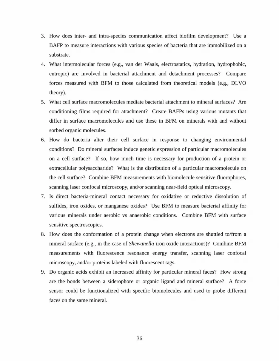
36
3. How does inter- and intra-species communication affect biofilm development? Use a
BAFP to measure interactions with various species of bacteria that are immobilized on a
substrate.
4. What intermolecular forces (e.g., van der Waals, electrostatics, hydration, hydrophobic,
entropic) are involved in bacterial attachment and detachment processes? Compare
forces measured with BFM to those calculated from theoretical models (e.g., DLVO
theory).
5. What cell surface macromolecules mediate bacterial attachment to mineral surfaces? Are
conditioning films required for attachment? Create BAFPs using various mutants that
differ in surface macromolecules and use these in BFM on minerals with and without
sorbed organic molecules.
6. How do bacteria alter their cell surface in response to changing environmental
conditions? Do mineral surfaces induce genetic expression of particular macromolecules
on a cell surface? If so, how much time is necessary for production of a protein or
extracellular polysaccharide? What is the distribution of a particular macromolecule on
the cell surface? Combine BFM measurements with biomolecule sensitive fluorophores,
scanning laser confocal microscopy, and/or scanning near-field optical microscopy.
7. Is direct bacteria-mineral contact necessary for oxidative or reductive dissolution of
sulfides, iron oxides, or manganese oxides? Use BFM to measure bacterial affinity for
various minerals under aerobic vs anaerobic conditions. Combine BFM with surface
sensitive spectroscopies.
8. How does the conformation of a protein change when electrons are shuttled to/from a
mineral surface (e.g., in the case of Shewanella-iron oxide interactions)? Combine BFM
measurements with fluorescence resonance energy transfer, scanning laser confocal
microscopy, and/or proteins labeled with fluorescent tags.
9. Do organic acids exhibit an increased affinity for particular mineral faces? How strong
are the bonds between a siderophore or organic ligand and mineral surface? A force
sensor could be functionalized with specific biomolecules and used to probe different
faces on the same mineral.
37
Tools such as BFM offer biochemists, biophysicists, geochemists, environmental engineers,
microbiologists, and mineralogists a unique insight into the fundamental intricacies of the
ubiquitous interfaces between microbes and minerals.
Acknowledgments
The presentation of this research benefited from the comments of two anonymous
reviewers. S.K.L. appreciates the support of J. Tak. Funding was provided by American
Chemical Society grant ACS-PRF 34326-AC2 and Department of Energy grant DE-FG02-99ER
15002.
References
Arredondo R, Garcia A, Jerez CA. 1994. Partial removal of lipopolysaccharide from
Thiobacillus ferrooxidans affects its adhesion to solids. Appl Envir Microbiol 60: 2846-
2851.
Banfield JF, Hamers RJ. 1997. Processes at minerals and surfaces with relevance to
microorganisms and prebiotic synthesis. Rev Mineral 35: 81-122.
Barker WW, Welch SA, Banfield JF. 1997. Biogeochemical weathering of silicate minerals. Rev
Mineral 35: 391-428.
Barker WW, Welch SA, Chu S, Banfield JF. 1998. Experimental observations of the effects of
bacteria on aluminosilicate weathering. Amer Mineral 83: 1551-1563.
Bennett PC, Hiebert FK, Choi WJ. 1996. Microbial colonization and weathering of silicates in
petroleum-contaminated groundwater. Chem Geol 132: 45-53.
Beveridge TJ, Doyle RJ. 1989. Metal Ions and Bacteria. John Wiley & Sons, New York.
Bengtsson G. 1991. Bacterial exopolymer and PHB production in fluctuating ground-water
habitats. FEMS Microbiol Ecol 86: 15-24.
Brown DA, Sherriff BL, Sawicki JA. 1997. Microbial transformation of magnetite to hematite.
Geochim Cosmochim Acta 61: 3341-3348.
Butt HJ, Jaschke M, Ducker W. 1995. Measuring surface forces in aqueous electrolyte solution
with the atomic force microscope. Bioelectrochem Bioenergetics 38: 191-201.

38
Cappella B, Dietler G. 1999. Force-distance curves by atomic force microscopy. Surf Sci
Reports 34: 1-104.
Caruso F, Caruso RA, Mohwald H. 1998. Nanoengineering of inorganic and hybrid hollow
spheres by colloidal templating. Science 282: 1111-1114.
Cleveland JP, Manne S, Bocek D, Hansma PK. 1993. A nondestructive method for determining
the spring constant of cantilevers for scanning force microscopy. Rev Sci Instrum 64: 403-
405.
D'Costa NP, Hoh JH. 1995. Calibration of optical lever sensitivity for atomic force microscopy.
Rev Sci Instrum 66: 5096-5097.
Dziurla M, -A., Achouak W, Lam B, -T., Heulin T, Berthelin J. 1998. Enzyme-linked
immunofiltration assay to estimate attachment of Thiobacilli to pyrite. Appl Envir
Microbiol 64: 2937-2942.
Edwards KJ, Goebel BM, Rodgers TM, Schrenk MO, Gihring TM, Cardona MM, Hu B,
McGuire MM, Hamers RJ, Pace NR, Banfield JF. 1998a. Geomicrobiology of pyrite
(FeS2) dissolution: A case study at Iron Mountain, California. Geomicrobiol 16: 1-25.
Edwards KJ, Schrenk MO, Hamers R, Banfield JF. 1998b. Microbial oxidation of pyrite:
Experiments using microorganisms from an extreme acidic environment. Amer Mineral 83:
1444-1453.
Ehrlich HL. 1990. Geomicrobiology. New York: Marcel Dekker.
Ellen RP, Burne RA. 1996. Conceptual advances in research on the adhesion of bacteria to oral
surfaces. In M Fletcher, editor. Bacterial Adhesion: Molecular and Ecological Diversity,
Washington DC: American Society for Microbiology, p 201-247.
Fein JB, Daughney CJ, Yee N, Davis T. 1997. A chemical equilibrium model for metal
adsorption onto bacterial surfaces. Geochim Cosmochim Acta 61: 3319-3328.
Fletcher M. 1996a. Bacterial Adhesion: Molecular and Ecological Diversity. New York: Wiley-
Liss.
Fletcher M. 1996b. Bacterial attachment in aquatic environments: A diversity of surfaces and
adhesion strategies. In M Fletcher, editor. Bacterial Adhesion: Molecular and Ecological
Diversity, Washington DC: American Society for Microbiology, p 1-24.
Florin E-L, Moy VT, Gaub HE. 1994. Adhesion forces between individual ligand-receptor pairs.
Science 264: 415-417.

39
Forsythe JH, Maurice PA, Hersman LE. 1998. Attachment of a Pseudomonas sp. to Fe(III)-
(hydr)oxide surfaces. Geomicrobiol 15: 293-308.
Fortin D, Beveridge TJ. 1997. Role of the bacterium, Thiobacillus, in the formation of silicates in
acidic mine tailings. Chem Geol 141: 235-250.
Fortin D, Ferris FG, Beveridge TJ. 1997. Surface-mediated mineral development by bacteria.
Rev Mineral 35: 161-180.
Fredrickson JK, McKinley JP, Nierzwicki-Bauer SA, White DC, Ringelberg DB, Rawson SA,
Shu-Mei L, Brockman FJ, Bjornstad BN. 1995. Microbial community structure and
biogeochemistry of Miocene subsurface sediments: Implications for long-term microbial
survival. Molecular Ecol 4: 619-626.
Frey BL, Corn RM. 1996. Covalent attachment and derivatization of poly(L-lysine) monolayers
on gold surfaces as characterized by polarization-modulation FT-IR spectroscopy. Anal
Chem 68: 3187-3193.
Gehrke T, Telegdi J, Dominique T, Sand W. 1998. Importance of extracellular polymeric
substances from Thiobacillus ferrooxidans for bioleaching. Appl Environ Microbiol 64:
2743-2747.
Grantham MC, Dove PM, DiChristina TJ. 1997. Microbially catalyzed dissolution of iron and
aluminum oxyhydroxide mineral surface coatings. Geochim Cosmochim Acta 61: 4467-
4477.
Hersman L, Lloyd T, Sposito G. 1995. Siderophore-promoted dissolution of hematite. Geochim
Cosmochim Acta 59: 3327-3330.
Hersman L, Maurice PA, Sposito G. 1996. Iron acquisition from hydrous Fe(III)-oxides by an
aerobic Pseudomonas sp. Chem Geol 132: 25-31.
Holben WE. 1997. Isolation and purification of bacterial community DNA from environmental
samples. In CJ Hurst, editor. Manual of Environmental Microbiology, Washington DC:
American Society for Microbiology, p 431-436.
Ingersoll CM, Bright FV. 1997. Using fluorescence to probe biosensor interfacial dynamics.
Anal Chem 69: 403A-408A.
Israelachvili J. 1992. Intermolecular and Surface Forces. London: Academic Press.
Israelachvili JN, McGuiggan PM. 1988. Forces between surfaces in liquids. Science 241: 795-
800.
40
Jucker BA, Harms H, Hug SJ, Zehnder AJB. 1997. Adsorption of bacterial surface
polysaccharides on mineral oxides is mediated by hydrogen bonds. Colloids Surf B 9: 331-
343.
Kennedy AC, Gewin VL. 1997. Soil microbial diversity: Present and future considerations. Soil
Sci 162: 607-617.
Kim P, Lieber CM. 1999. Nanotube nanotweezers. Science 286: 2148-2150.
Krumbein WE, Urzi CE, Gehrmann C. 1991. Biocorresion and biodeteriorationof antique and
medieval glass. Geomicrobiol 9: 139-160.
Langley S, Beveridge TJ. 1999. Effect of O-side-chain-lipopolysaccharide chemistry on metal
binding. Appl Envir Microbiol 65: 489-498.
Lawrence JR, Korber DR, Hoyle BD, Costerton JW, Caldwell DE. 1991. Optical sectioning of
microbial biofilms. J Bacteriol 173: 6558-6567.
Lovley DR, Phillips EJP. 1986. Organic matter mineralization with reduction of ferric iron in
anaerobic sediments. Appl Envir Microbiol 51: 683-689.
Lovley DR, Phillips EJP. 1987. Competitive mechanisms for inhibition of sulfate reduction and
methane production in the zone of ferric iron reduction in sediments. Appl Envir Microbiol
53: 2636-2641.
Lower SK, Tadanier CJ, Hochella MF Jr. 2000. Measuring interfacial and adhesion forces
between bacteria and mineral surfaces with biological force microscopy. Geochim
Cosmochim Acta 64: 3133-3139.
Lower SK, Maurice PM. 2001. From minerals and crystal lattices to microorganisms and nucleic
acids: The use atomic force microscopy in geomicrobiology. Earth Sci Rev. in press.
Marshall KC. 1976. Interfaces in Microbial Ecology. Cambridge, Massachusetts: Harvard
University Press.
Marshall KC. 1996. Adhesion as a strategy for access to nutrients. In M Fletcher, editor.
Bacterial Adhesion: Molecular and Ecological Diversity, Washington DC: American
Society for Microbiology, p 59-87.
Myers C, Nealson KH. 1988. Bacterial manganese reduction and growth with manganese oxide
as the sole electron acceptor. Science 240: 1319-1321.

41
Pagac ES, Prieve DC, Tilton RD. 1998a. Kinetics and mechanisms of cationic surfactant
adsorption and coadsorption with cationic polyelectrolytes at the silica-water interface.
Langmuir 14: 2333-2342.
Pagac ES, Tilton RD, Prieve DC. 1998b. Depletion attraction caused by unadsorbed
polyelectrolytes. Langmuir 14: 5106-5112.
Pentecost A, Bauld J. 1988. Nucleation of calcite on the sheaths of cyanobacteria using a simple
diffusion cell. Geomicrobiol 6: 129-135.
Pincet F, Perez E, Wolfe J. 1995. Does glue contaminate the surface forces apparatus? Langmuir
11: 373-374.
Plueddemann EP. 1991. Silane Coupling Agents. New York: Plenum Press.
Rezania A, Johnson R, Lefkow A, Healy KE. 1999. Bioactivation of metal oxide surfaces. 1.
Surface characterization and cell response. Langmuir 15: 6931-6939.
Robert M, Berthelin J. 1986. Role of biological and biochemical factors in soil mineral
weathering. In PM Huang, M Schnitzer, editors. Interactions of Soil Minerals with Natural
Organics and Microbes, Madison: Soil Science of America, p 453-495.
Roden EE, Zachara JM. 1996. Microbial reduction of crystalline iron(III) oxides: Influence of
oxide surface area and potential for cell growth. Envir Sci Technol 30: 1618-1628.
Rogers JR, Bennett PC, Choi WJ. 1998. Feldspars as a source of nutrients for microorganisms.
Amer Mineral 83: 1532-1540.
Sader JE, Chon JWM, Mulvaney P. 1999. Calibration of rectangular atomic force microscope
cantilevers. Rev Sci Instrum 70: 3967-3969.
Schrenk MO, Edwards KJ, Goodman RM, Hamers RJ, Banfield JF. 1998. Distribution of
Thiobacillus ferrooxidans and Leptospirillum ferrooxidans: Implications for generation of
acid mine drainage. Science 279: 1519-1522.
Schultze-Lam S, Fortin D, Davis BS, Beveridge TJ. 1996. Mineralization of bacterial surfaces.
Chem Geol 132: 171-181.
Schultze-Lam S, Harauz G, Beveridge TJ. 1992. Participation of a cyanobacterial S-layer in fine-
grain mineral formation. J Bacteriol 174: 7971-7981.
Senden TJ, Ducker WA. 1994. Experimental determination of spring constants in atomic force
microscopy. Langmuir 10: 1003-1004.

42
Stone AT. 1997. Reactions of extracellular organic ligands with dissolved metal ions and mineral
surface. Rev Mineral 35: 309-341.
Stotzky G. 1986. Influence of soil mineral colloids on metabolic processes, growth, adhesion,
and ecology of microbes and viruses. In PM Huang, M Schnitzer, editors. Interactions of
Soil Minerals with Natural Organics and Microbes, Madison: Soil Science Society of
America, p 305-428.
Svoboda K, Block SM. 1994. Biological applications of optical forces. Annu Rev Biophys
Biomolecular Structure. 23: 247-285.
Tadanier CJ, Little JC, Berry DF, Hochella MF Jr. 2000. Energetics of microbial nutrient
acquisition from mineral surfaces. In R Hollmann, SA Wood, editors. Geochemical Society
Special Publication – David A. Crerar Honorary Volume, Washington DC: Geochemical
Society of America.
Thorseth IH, Torsvik T, Furnes H, Muehlenbachs K. 1995. Microbes play an important role in
the alteration of oceanic crust. Chem Geol 126: 137-146.
Trevors JT, van Elsas JD. 1997. Quantification of gene transfer in soil and the rhizosphere. In CJ
Hurst, editor. Manual of Environmental Microbiology, Washington DC: American Society
for Microbiology, p 500-508.
Turner DC, Peek BM, Wertz TE, Archibald DD, Geer RE, Gaber BP. 1996. Enzymatic
modification of a chemisorbed lipid monolayer. Langmuir 12: 4411-4416.
Turner DC, Testoff MA, Conrad DW, Gaber BP. 1997. Enzymatic hydrolysis of a chemisorbed
peptide film using beads activated with covalently bound chymotrypsin. Langmuir 13:
4855-4860.
Voet D, Voet J. 1995. Biochemistry. New York: John Wiley & Sons.
Warren LA, Ferris FG. 1998. Continuum between sorption and precipitation of Fe(III) on
microbial surfaces. Envir Sci Technol 32: 2331-2337.
Weisenhorn AL, Maivald P, Butt H-J, Hansma PK. 1992. Measuring adhesion, attraction, and
repulsion between surfaces in liquids with an atomic-force microscope. Phys Rev B 45:
11226-11232.
Welch SA, Ullman WJ. 1993. The effect of organic acids on plagioclase dissolution rates and
stoichiometry. Geochim Cosmochim Acta 57: 2725-2736.

43
Welch SA, Vandevivere P. 1994. Effect of microbial and other naturally occurring polymers on
mineral dissolution. Geomicrobiol 12: 227-238.
West JK, Latour R, Jr., Hench LL. 1997. Molecular modeling study of adsorption of poly-L-
lysine onto silica glass. J Biomed Materials Res 37: 585-591.
Wolfaardt GM, Lawrence JR, Roberts RD, Caldwell SJ, Caldwell DE. 1994. Multicellular
organization in a degradative biofilm community. Appl Envir Microbiol 60: 434-446.
Xu W, Mulhern PJ, Blackford BL, Jericho MH, Firtel M, Beveridge T. 1996. Modeling and
measuring the elastic properties of an Archaeal surface, the sheath of Methanospirillum
hungatei, and the implications for methane production. J Bacteriol 178: 3106-3112.
Yao X, Jericho M, Pink D, Beveridge T. 1999. Thickness and elasticity of Gram-negative murein
sacculi measured by atomic force microscopy. J Bacteriol 181: 6865-6875.
Yoon RH, Flinn DH, Rabinovich YI. 1997. Hydrophobic interactions between dissimilar
surfaces. J Colloid Interface Sci 185: 363-370.

44
Chapter 4 - Bacterial recognition of mineral surfaces: Nanoscale interactions between
Shewanella and αααα-FeOOH
In review in
Science
Abstract
Biological force microscopy has been used to quantitatively measure the minute forces that
characterize interactions between Shewanella oneidensis, a dissimilatory metal reducing
bacterium, and the mineral goethite, α-FeOOH, both commonly found in Earth near-surface
environments. Force measurements with sub-nanoNewton resolution were made with living
cells, in aqueous solution under aerobic and anaerobic conditions as a function of the nanoscale
distance between the cell and mineral surface. Energy values derived from these measurements
show that the affinity between S. oneidensis and goethite rapidly increases by two to five times
under anaerobic conditions where electron transfer from bacterium to mineral is expected.
Specific signatures in the force curves suggest that a 150 kDa putative, iron reductase is quickly
mobilized within the outer membrane of S. oneidensis and specifically interacts with the goethite
surface to facilitate the electron transfer process.
Introduction
The Fe(II)-Fe(III) redox cycle represents a major energy flux at the earth’s surface. A
critical component of this system is the reduction of iron-containing minerals by biological
processes (1, 2). Shewanella - a Gram-negative, dissimilatory metal reducing bacterium found in
soils, sediments, surface waters, and groundwaters - is able to conserve energy for growth by
using oxygen or ferric iron as a terminal electron acceptor (3, 4). In many natural environments
oxygen is in limited supply but Fe(III) is present as a major element in the crystal structure of
minerals. Hence in many cases, dissimilatory metal reducing bacteria oxidize various carbon
substrates by reductively dissolving Fe(III)-containing minerals, the most ubiquitous in the near-
surface environments being iron oxyhydroxides (e.g., ferrihydrite, goethite, and hematite) (5-12).
45
This impacts a wide array of processes including the biogeochemical cycle of iron and
phosphorus, oxidation of natural and anthropogenic carbon sources, biocorrosion, and the release
of heavy metals associated with iron oxyhydroxides.
The dissimilatory reduction of iron-containing minerals presents a rather unique situation
because unlike oxygen, Fe(III) in a solid form cannot diffuse across the cell wall to the plasma
membrane which, in most bacteria, houses the proteins involved in electron transfer, proton
translocation, and subsequent generation of ATP. Microorganisms like Shewanella seem to be
able to generate two energized membranes using a unique system of proteins that shuttle
electrons from an energy source in the cytoplasm, across the plasma membrane and periplasmic
space, to the outer membrane (13, 14). Once in the outer membrane, iron reductases appear to
transfer electrons directly to ferric iron in the crystal structure of minerals causing a weakening
of the iron-oxygen bond and reductive dissolution of the mineral (6, 15-18). A great deal of
research has focused independently on either the microbiological or mineralogical aspects of the
interaction between Shewanella and iron oxyhydroxides. However, virtually no studies have
been conducted on the interface between the bacterium and iron containing minerals. It is this
interface, delineated by chemical and structural features on the surface of the organism (e.g.,
concentration and localization of cytochromes and reductases, physical structure of the outer
membrane) and the mineral surface (e.g., density and coordination of iron-oxide moieties,
surface microtopography, crystallographic orientation), which likely has a major impact on the
kinetics and extent of bacterial reduction of Fe(III)-minerals.
Methods and Materials
In this study, we used biological force microscopy (19, 20) to probe the interface between a
living cell of Shewanella oneidensis, formerly classified as S. putrefaciens MR1 (21), and the
(010) surfaces of goethite (α-FeOOH) and diaspore (α-AlOOH; isostructural with goethite;
surface properties such as charge and hydrophobicity are very similar to goethite) at the
nanoscale level in anaerobic and aerobic solutions. S. oneidensis were grown under anaerobic
conditions using lactate as the carbon and energy source and Fe(III)-citrate as the electron
acceptor (22). To preserve the natural, complex biomolecular network on the bacteria surface,

46
fully functional cells (0.5 x 2 µm) were linked in their native state to a small bead (5 µm radius)
situated on the end of a silicon nitride cantilever thereby creating a biologically-active-force-
probe (22). A commercial force microscope (Digital Instruments, Santa Barbara, California) was
used to measure the deflection of a biologically-active-force-probe by reflecting a laser off the
top of the cantilever and into a photodiode-detector while an oriented mineral crystal, mounted
on a piezoelectric scanner, was translated towards (i.e., approach data), made contact with, and
was subsequently retracted from (i.e., retraction data) a bacterium on the probe. Photodiode
response (in volts) was converted to cantilever deflection (in meters) using a conversion factor
(in meters volt-1) determined from the photodiode shift voltage (23). Cantilever deflection was
multiplied by the cantilever spring constant (ksp = 0.07 N m-1, determined as described previously
(24)), to calculate the force of interaction (in Newtons). Displacement of the mineral on the
piezoelectric scanner was converted to separation between the bacteria and mineral by correcting
for cantilever deflection (25) and selecting the point of contact using jump-to-contact and jump-
from-contact events for approach and retraction curves, respectively. For retraction curves it is
more appropriate to refer to extension rather than separation because polymers on the cell surface
form a bridge with the mineral.
Results and Discussion
In a typical experiment with goethite, approach and retraction forces were measured after
varying the contact time between S. oneidensis and the mineral in anaerobic solutions
supplemented with lactate as an energy source (Fig. 4.1a). An aerobic solution supplemented
with lactate was then injected into the fluid-cell of the force microscope and allowed to incubate
for 30-45 min, after which forces were again measured as a function of the contact time between
the bacteria and mineral (Fig. 4.1b). To characterize the complex physical and chemical
intricacies involved in these reactions, the work or energy of each curve was determined by
integrating force with respect to distance (Table 4.1). In general the retraction curves may
provide greater insight into the transfer of electrons from biomolecules on S. oneidensis to iron
atoms in goethite because direct cell-mineral contact is a prerequisite for this reaction (6, 17, 26,
27). However,

47
Figure 4.1. Representative force-distance curves between Shewanella oneidensis and goethite
(α-FeOOH) or diaspore (α-AlOOH) at 25 °C, pH 6, 10 mM sodium chloride, and 15 mM lactate
in solution. For each approach-retraction cycle in this figure, the upper data set corresponds to
approach values, whereas the lower data set corresponds to retraction values. Positive values are
repulsive and negative values are attractive. (a) Goethite-S. oneidensis interactions in which the
cell and mineral remained in contact for 0-5 min (green circles) or 30-45 min (blue squares)
under anaerobic conditions. Black curves correspond to the worm-like chain model prediction
for the 83 kDa and 150 kDa outer membrane, putative iron reductases. (b) Goethite-S.
oneidensis interactions in which the cell and mineral remained in contact for 0-5 min under
aerobic conditions. There was no significant difference between the 0-5 min and 30-45 min
contact times under aerobic conditions. (c) Diaspore-S. oneidensis interactions under anaerobic
conditions. There was no significant difference between the aerobic and anaerobic experiments
between diaspore and S. oneidensis.
-1
-0.8
-0.6
-0.4
-0.2
0
0.2
0 100 200 300 400 500 600
distance (nm)
forc
e (n
N)
a
83 kDa 150 kDa
-1
-0.8
-0.6
-0.4
-0.2
0
0.2
0 40 80 120 160 200
distance (nm)
forc
e (n
N)
b
-1
-0.8
-0.6
-0.4
-0.2
0
0.2
0 40 80 120 160 200
distance (nm)
forc
e (n
N)
c
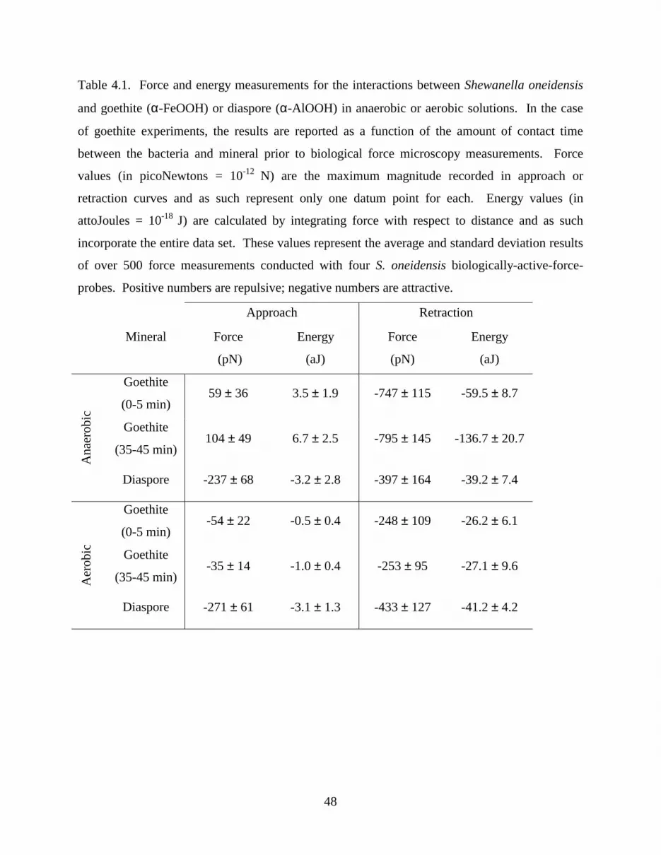
48
Table 4.1. Force and energy measurements for the interactions between Shewanella oneidensis
and goethite (α-FeOOH) or diaspore (α-AlOOH) in anaerobic or aerobic solutions. In the case
of goethite experiments, the results are reported as a function of the amount of contact time
between the bacteria and mineral prior to biological force microscopy measurements. Force
values (in picoNewtons = 10-12 N) are the maximum magnitude recorded in approach or
retraction curves and as such represent only one datum point for each. Energy values (in
attoJoules = 10-18 J) are calculated by integrating force with respect to distance and as such
incorporate the entire data set. These values represent the average and standard deviation results
of over 500 force measurements conducted with four S. oneidensis biologically-active-force-
probes. Positive numbers are repulsive; negative numbers are attractive.
Approach Retraction
Mineral Force
(pN)
Energy
(aJ)
Force
(pN)
Energy
(aJ)
Goethite
(0-5 min) 59 ± 36 3.5 ± 1.9 -747 ± 115 -59.5 ± 8.7
Goethite
(35-45 min) 104 ± 49 6.7 ± 2.5 -795 ± 145 -136.7 ± 20.7
Ana
erob
ic
Diaspore -237 ± 68 -3.2 ± 2.8 -397 ± 164 -39.2 ± 7.4
Goethite
(0-5 min) -54 ± 22 -0.5 ± 0.4 -248 ± 109 -26.2 ± 6.1
Goethite
(35-45 min) -35 ± 14 -1.0 ± 0.4 -253 ± 95 -27.1 ± 9.6
Aer
obic
Diaspore -271 ± 61 -3.1 ± 1.3 -433 ± 127 -41.2 ± 4.2

49
the approach curves give an indication of the intermolecular forces that will effect whether S.
oneidensis is able to bridge the solution gap between itself and the mineral.
For the approach curves, repulsion between S. oneidensis and goethite under anaerobic
conditions was greater after the bacteria and mineral remained in contact for 30-45 min (Fig.
4.1a; Table 4.1). Infusion of an oxygenated solution caused this repulsive interaction to decrease
to the point where it became slightly attractive (Fig. 4.1b; Table 4.1). The point-of-zero-charge
of Shewanella is 5-6 (determined under aerobic conditions) (28) and 7-8 for goethite (29). Based
on electrostatic considerations alone, a slight attraction would be expected at a pH of ∼ 6.
However, there is a high degree of electrochemical heterogeneity on the surface of Shewanella
and its surface charge is dependent upon the concentration and localization of at least five
different types of functional groups which are arrayed over the cell surface (28). Under aerobic
conditions, amine-groups are embedded within the outer membrane and underlying
peptidoglycan layer such that they do not significantly contribute to the surface charge (28).
Anaerobic conditions induce the expression of a significant quantity of proteins in the outer
membrane of S. oneidensis (15-17). The amine-groups on these proteins would likely lead to an
increased positive charge on the cell surface such that repulsion would be expected between S.
oneidensis and goethite in anaerobic solutions, as observed in approach curves.
Retraction curves represent in many cases a complex dynamic of adhesion involving, among
other things, bond breaking, intermolecular forces, and extension of outer membrane proteins
that form a bridge between a bacterium and the mineral surface. Under aerobic conditions, S.
oneidensis exhibited a stronger overall adhesion to diaspore relative to goethite (Fig. 4.1b and
4.1c, Table 4.1). Under anaerobic conditions S. oneidensis exhibited a greatly increased affinity
for goethite, as indicated by the retraction curves, whereas the attractive energy between diaspore
and S. oneidensis was indifferent to oxygen concentrations in solution (Fig. 4.1a and 4.1c, Table
4.1). Furthermore, biologically-active-force-probes fabricated with non-viable cells exhibited an
attraction towards goethite but this affinity did not change with oxygen concentration in solution.
Taken together, this indicates that the adhesion between S. oneidensis and goethite is due, at least
in part, to processes associated with dissimilatory iron reduction. The retraction energies
between S. oneidensis and goethite were 2-5 times stronger under anaerobic as opposed to

50
aerobic conditions (Table 4.1). Under anaerobic conditions, this attraction increased with the
amount of contact time for goethite (Table 4.1) but not for diaspore. This provides direct
evidence suggesting that the bacteria are able to quickly recognize the mineral surface such that
they produce and/or localize biomolecules at the interface with goethite. This type of
recognition between a microorganism and mineral surface has been previously suggested but
never quantified (30, 31). Clearly, this could be one of the functions of the fluidity of the outer
membrane allowing the bacterium to react rapidly to the mineral surface and to congregate
reactive membrane macromolecules at sites of contact. Dynamic modeling of outer membrane
lipopolysaccharides suggests molecular motion over microsecond timeframes (32), and it is
possible that cytochromes and reductases could also have rapid motional attributes.
Anaerobic retraction curves between goethite and S. oneidensis exhibit a strong force of
interaction within the first few hundred nanometers followed by a jagged plateau (Fig. 4.1a). In
many cases, the retraction curves terminate in this saw-tooth-like discontinuity (Fig. 4.1a), which
we suggest is a signature of the unfolding of outer membrane proteins that form a bridge between
a living bacterium and the mineral surface. The saw-tooth like patterns in the retraction curves
were analyzed to gain more definitive evidence for the presence of outer membrane proteins that
may be involved in the direct transfer of electrons from the cell to the mineral. A total of four
putative iron-reductases ranging in size from 53 to 150 kDa have been isolated from the outer
membrane of S. oneidensis (17, 18). According to the worm-like chain model (33) which can be
used to calculate the force (F) needed to stretch a polypeptide chain in a solvent to a length x,
F(x) = (kT/b) [0.25 (1 – x/L)-2 – 0.25 + x/L] , where k is the Boltzmann’s constant, T is the
temperature, b is the persistence length (i.e., length of the stiff segment of the chain) which is
0.38 nm for the distance between two Cα atoms in a polypeptide (34-36), and L is the contour
length (i.e., length of the completely stretched chain) calculated for each putative iron reductase
assuming an average molecular weight of 110 Da per amino acid residue (18, 37) and an average
molecular length of 0.4 nm per amino acid residue (35).
Of the four putative iron reductases, the 83 kDa protein was emphasized in previous reports
(17, 18); however recent studies involving mutants incapable of producing this protein provide
evidence that it is not an iron reductase (C. Myers, personal communication). Predictions based
51
on the worm-like chain model, when compared to our anaerobic retraction curves (Fig. 4.1a), are
consistent with this. Of the remaining three putative iron reductases, the worm-like chain model
prediction for the 150 kDa protein fit 82% of the retraction curves for S. oneidensis and goethite
under anaerobic conditions and longer contact time (Fig. 4.1a). Evidence of this protein was not
found in the retraction curves of the aerobic-goethite nor the diaspore experiments. This strongly
suggests that the 150 kDa protein in the outer membrane of S. oneidensis specifically interacts
with the surface of goethite fulfilling some aspect of the electron transfer reaction.
We have shown that under anaerobic conditions S. oneidensis responds to the surface of
goethite by rapidly developing stronger adhesion forces at the interface. We interpret these data
to indicate that following recognition of goethite as a terminal electron acceptor, S. oneidensis
quickly produces and/or mobilizes proteins (i.e., the 150 kDa putative reductase and perhaps
others) that specifically interact with the mineral surface. This is an example of using
nanomechanical measurements/properties to help isolate likely biochemical and mineralogical
mechanisms that are operative under environmentally relevant conditions. Further, besides an
improved fundamental understanding of bacterial interactions with solid surfaces, the ability to
quantitatively probe and characterize these specific interactions in dimensional, force and energy
nanospace could lead to advances in nanotechnology. For example, one may now be able to
systematically tailor the exquisite, natural specificity between biomolecules, as produced by
living microorganisms, and material surfaces for the purposes of chemical and/or structural
nanoscale modification of the cell, the material surface, or both.
References and Acknowledgments
1. D. R. Lovley, Microbiol. Rev. 55, 259 (1991).
2. K. H. Nealson, D. Saffarini, Ann. Rev. Microbiol. 48, 311 (1994).
3. R. G. Arnold, M. R. Hoffmann, T. J. DiChristina, F. W. Picardal, Appl. Environ. Microbiol.
56, 2811 (1990).
4. C. R. Myers, K. H. Nealson, J. Bacteriol. 172, 6232 (1990).
5. D. R. Lovley, E. J. P. Phillips, Appl. Environ. Microbiol. 52, 751 (1986).
6. R. G. Arnold, T. J. DiChristina, M. R. Hoffmann, Biotech. Bioeng. 32, 1081 (1988).

52
7. D. R. Lovley, E. J. P. Phillips, Appl. Environ. Microbiol. 54, 1472 (1988).
8. E. E. Roden, J. M. Zachara, Environ. Sci. Tech. 30, 1618 (1996).
9. F. Caccavo, Jr., P. C. Schamberger, K. Keiding, P. H. Nielsen, Appl. Environ. Microbiol. 62,
3837 (1997).
10. J. K. Fredrickson, et al., Geochim. Cosmochim. Acta 62, 3239 (1998).
11. J. M. Zachara, et al., Amer. Mineral. 83, 1426 (1998).
12. M. M. Urrutia, E. E. Roden, J. M. Zachara, Environ. Sci. Tech. 33, 4022 (1999).
13. C. R. Myers, J. M. Myers, J. Bacteriol. 179, 1143 (1997).
14. J. M. Myers, C. R. Myers, J. Bacteriol. 182, 67 (2000).
15. C. R. Myers, J. M. Myers, J. Bacteriol. 174, 3429 (1992).
16. C. R. Myers, J. M. Myers, FEMS Microbiol. Let. 108, 15 (1993).
17. C. R. Myers, J. M. Myers, Biochim. Biophys. Acta 1326, 307 (1997).
18. J. M. Myers, C. R. Myers, Biochim. Biophys. Acta 1373, 237 (1998).
19. S. K. Lower, C. J. Tadanier, M. F. Hochella, Jr., Geochim. Cosmochim. Acta 64, 3133
(2000).
20. S. K. Lower, C. J. Tadanier, M. F. Hochella, Jr., Geomicrobiol. J. 18 (2001).
21. K. Venkateswaran, et al., Internat. J. System. Bacteriol. 49, 705 (1999).
22. S. oneidensis (ATCC 700550) was transformed with plasmid pSMC2 which codes for a
constitutive, intracellular green fluorescent protein (38). Transformed cells were grown in an
anaerobic chamber (Coy Laboratory Products, Ann Arbor, Michigan) to mid-logarithmic phase
at room temperature as previously described (4) in defined medium supplemented with 15 mM
lactate and 2 mM ferric citrate (4). Cells were rinsed three times in an anerobic solution of 10
mM sodium chloride. Washed cells were linked to glass beads functionalized with 5-10% 3-
aminopropyltriethoxysilane. A single bacteria coated bead was then attached to a cantilever with
epoxy (19). Scanning laser confocal microscopy was used to verify bacterial coverage of the
probe by imaging the green fluorescent signal emitted from cells. Confocal measurements as
described previously (19) suggest that a single bacterium made contact with the mineral during
force measurements. Viability of cells on the probe was assessed by inoculating an agar plate
with a biologically-active-force-probe subsequent to force measurements.

53
23. The region of constant compliance was not used to determine deflection because it would
have resulted in a significant overestimate of the measured forces due to the fact that the cells
were more compliant than the cantilever (20, 39).
24. J. P. Cleveland, S. Manne, D. Bocek, P. K. Hansma, Rev. Sci. Instrum. 64, 403 (1993).
25. W. A. Ducker, T. J. Senden, R. M. Pashley, Lang. 8, 1831 (1992).
26. T. J. DiChristina, E. F. Delong, J. Bacteriol. 176, 1468 (1994).
27. A. S. Beliaev, D. A. Saffarini, J. Bacteriol. 180, 6292 (1998).
28. I. Sokolov, D. S. Smith, G. S. Henderson, Y. A. Gorby, F. G. Ferris, Environ. Sci. Tech. 35,
341 (2001).
29. W. Stumm, J. J. Morgan, Aquatic Chemistry: Chemical Equilibria and Rates in Natural
Waters (John Wiley & Sons, New York, ed. 3, 1996), pp 534-540.
30. S. Brown, Proc. Natl. Acad. Sci. U.S.A. 89, 8651 (1992).
31. N. Ohmura, K. Kitamura, H. Saiki, Appl. Environ. Microbiol. 59, 4044 (1993).
32. T. J. Beveridge, J. Bacteriol. 181, 4725 (1999).
33. P. J. Flory, Statistical Mechanics of Chain Molecules (Hanser Publishers, New York, 1989),
pp. 401-403.
34. M. Rief, M. Gautel, F. Oesterhelt, J. M. Fernandez, H. E. Gaub, Science 276, 1109 (1997).
35. H. Mueller, H.-J. Butt, E. Bamberg, Biophys. J. 76, 1072 (1999).
36. D. J. Muller, W. Baumeister, A. Engel, Proc. Natl. Acad. Sci. U.S.A. 96, 13170 (1999).
37. J.-J. Karlsson, A. Kadziola, A. Rasmussen, T. E. Rostrup, J. Ulstrup, in Protein Folds: A
Distance-Based Approach, H. Bohr, S. Brunak, Eds. (CRC Press, Boca Raton, 1996), pp. 56-67.
38. G. V. Bloemberg, G. A. O'Toole, B. J. J. Lugtenberg, R. Kolter, Appl. Environ. Microbiol.
63, 4543 (1997).
39. N. P. D'Costa, J. H. Hoh, Rev. Sci. Instrum. 66, 5096 (1995).
40. J. Banfield provided constructive comments. We thank G. O’Toole for providing plasmid
pSMC2. S.K.L. acknowledges the support of J Tak. Mineral samples provided by the Virginia
Tech Geological Sciences Museum. Financial support from the U.S. Department of Energy and
the National Science Foundation.

54
Curriculum Vitae
STEVEN K. LOWER Department of Geological Sciences
Virginia Polytechnic Institute and State University (Virginia Tech) Blacksburg, VA 24061-0420
Tel: 540-231-6521 (ext. 8575), Fax: 540-231-3386 e-mail: [email protected]
Education
Ph.D. GEOLOGICAL SCIENCES, BIOCHEMISTRY, AND MICROBIOLOGY (8/97-12/00) Departments of Geological Sciences and Biochemistry, Virginia Tech, Blacksburg, VA.
Course emphasis: biochemistry, genetics, geochemistry, microbial ecology, microbial physiology, microbiology, mineralogy, molecular biology, surface chemistry. Techniques utilized: atomic force microscopy (AFM), chromatography, chemostats, cloning with plasmid and phage vectors, confocal laser scanning microscopy (CLSM), electrophoresis, fluorescent in-situ hybridization (FISH), gene expression, polymerase chain reaction (PCR), scanning electron microscopy (SEM), Southern blot analysis, temperature gradient gel electrophoresis (TGGE), Western blot analysis. Doctoral research: Development of biological force microscopy. Fundamental nanoscale interactions between a bacterium and mineral surface (e.g., Shewanella-iron oxides; Thiobacillus-sulfides; Pseudomonas-silicates). Macroscale interactions between a community of microorganisms and minerals (e.g., soil microbial community and iron oxides supporting sorbed nutrients and/or metals). Advisor: Dr. Michael F. Hochella, Jr.
M.S. GEOCHEMISTRY (1/95-5/97) Department of Geology, Kent State University, Kent, OH.
Course emphasis: clay mineralogy, environmental organic chemistry, hydrogeochemistry, mineral-water interface chemistry, optical petrography, soil chemistry, statistics, time series analyses, x-ray crystallography. Techniques utilized: atomic absorption spectrophotometer (AA/graphite furnace), atomic force microscopy (AFM), high-performance liquid chromatography (HPLC), inductively coupled plasma spectrometer (ICP), scanning electron microscopy (SEM), transmission electron microscopy (TEM), total organic carbon analyzer; x-ray diffraction (XRD). Thesis research: Mechanisms of lead sorption to calcium-phosphate minerals and the use of apatite to remediate heavy metal contaminated environments. Additional studies include: sorption of dissolved organic matter onto aluminum/iron oxides; dissolution of aluminum/iron oxides by organic acids; pollution associated with urban stormwater runoff. Advisor: Dr. Patricia A. Maurice
B.S. GEOLOGY AND BIOLOGY (9/90-12/94) Departments of Geology and Biology, Kent State University, Kent, OH.
55
Course emphasis: anatomy, aquatic biology, biology, botany, chemistry, climatology, ecology, field camps (biology and geology), forestry, geochemistry, geographic information systems, geomorphology, hydrogeology, mathematics, mineralogy, petrology, physics, physiology, stratigraphy, structural geology, zoology. Independent studies: Characterization of soil samples with x-ray diffraction; development of a constructed wetland for wastewater treatment; preservation of a natural bog. Employment
GRADUATE RESEARCH ASSISTANT (6/99-present) Department of Geological Sciences, Virginia Tech, Blacksburg, VA. Researcher. Responsibilities include primary doctoral research, scanning probe microscopy and laser scanning microscopy maintenance, PC hardware and software administration. GRADUATE TEACHING ASSISTANT (8/97-5/99) Department of Geological Sciences, Virginia Tech, Blacksburg, VA. Head TA for Elements of Geology class. Responsibilities include design and organization of laboratory and field exercises and examinations. GRADUATE RESEARCH ASSISTANT (1/95-8/97) Department of Geology, Kent State University, Kent, OH. Researcher. Responsibilities include primary thesis research and operation/maintenance of x-ray diffraction unit, scanning probe microscopes, and atomic absorption spectrophotometer (graphite furnace). Assess effectiveness of detention basins to attenuate chemical pollutants in urban stormwater runoff. Responsibilities include operation of automated sampling equipment, chemical analysis of water samples for heavy metals and nutrients, and statistical analyses of data. GRADUATE TEACHING ASSISTANT (8/96-12/96) Department of Geology, Kent State University, Kent, OH. Head TA for Mineralogy class. Responsibilities include design and organization of laboratory exercises and examinations. FIELD SCIENTIST (4/91-8/94) Environmental Associates, Akron, OH. Natural Resource specialist. Responsibilities include development of municipal stromwater pollution prevention plans to mitigate environmental degradation of surface waters. FIELD SCIENTIST (4/91-8/94) Buckeye Bison, North Canton, OH. Wildlife manager overseeing a herd of bison. Responsibilities include maintenance of food source, relocation of bison to prevent overgrazing, implementation of immunization program, production of topographic maps showing distribution of various soil and plant types. Society Memberships
GEOCHEMICAL SOCIETY
MINERALOGICAL SOCIETY OF AMERICA
56
AMERICAN SOCIETY FOR MICROBIOLOGY Honors and Awards
C. G. TILLMAN AWARD FOR EXCELLENCE IN TEACHING, Dept. of Geological Sciences, Virginia Tech.
WATER RESOURCE RESEARCH INSTITUTE FELLOWSHIP, Depts. of Biology, Chemistry, Geology, and Geography, Kent State University.
B. J. WALDRON FIELD CAMP AWARD, Dept. of Geology, Kent State University.
OHIO BOYS STATE DELEGATE, Northwest High School. Publications
Lower, S. K., Tadanier, C. J., and Hochella, M. F., Jr. (2000) Measuring interfacial and adhesion forces between bacteria and mineral surfaces with biological force microscopy. Geochimica et Cosmochimica Acta, 64: 3133-3139.
Lower, S. K., Tadanier, C. J., and Hochella, M. F., Jr. (2000) Dynamics of the mineral-microbe interface: Use of biological force microscopy in biogeochemistry and geomicrobiology. Geomicrobiology Journal, 18: 63-76.
Lower, S. K., Maurice, P. A., and Traina, S. J. (1998) Simultaneous dissolution of hydroxylapatite and precipitation of hydroxypyromorphite: Direct evidence of homogeneous nucleation. Geochimica et Cosmochimica Acta, 62: 1773-1780.
Lower, S. K., Maurice, P. A., Traina, S. J., and Carlson, E. H. (1998) Aqueous Pb sorption by hydroxylapatite: Applications of atomic force microscopy to dissolution, nucleation, and growth studies. American Mineralogist 83: 147-158.
Maurice, P. A. and Lower, S. K. (1998) Using atomic force microscopy to study soil mineral reactions. Advances in Agronomy, 62: 1-43.
Maurice, P. A., Namjesnik-Dejanovic, K., Lower, S. K., Pullin, M., Chin, Y. –P., and Aiken, G. R. (1998) Sorption and fractionation of natural organic matter on kaolinite and goethite. In: Water-Rock Interaction (Eds., G. B. Arehart and J.R. Hulston). Balkema Publishers, Rotterdam, Netherlands. Abstracts
Lower, S. K. (2001 – Invited Presentation) “Bacterial recognition of mineral surfaces: Through the eyes of a single bacterium.” Goldschmidt Conference in Roanoke, VA.
Lower, S. K. and Hochella, M. F., Jr. (2000 – Invited Presentation) “Mineral-microbe interactions quantitatively measured in energy and distance nanospace.” American Geophysical Union Meeting in San Francisco, CA.
Lower, S. K. and Hochella, M. F., Jr. (2000) “Probing the dynamic nanoscale world at the interface between microorganisms and minerals.” Goldschmidt Conference in Oxford, UK.
Lower, S. K., Tadanier, C. F., Berry, D. F., Potts, M., and Hochella, M. F., Jr. (1999) “The bacteria-mineral interface: Probing nanoscale forces with biological force microscopy.”

57
Geological Society of America Meeting in Denver, CO (GSA Abstracts with Programs, v. 31, no. 7).
Lower, S. K., Maurice, P. A., Traina, S. J., and Carlson, E. H. (1997) “Pb sorption to hydroxylapatite: A microscopic and spectroscopic study.” American Chemical Society Meeting in San Francisco, CA (Abstracts of Papers ACS, v. 213, GEOC 187).
Carlson, E. H. and Lower, S. K. (1997) “Comparative clay mineralogy of Ordovician Deicke and Millbrig K-bentonites in central Kentucky and central Tennessee.” Geological Society of America Meeting in Salt Lake City, UT (GSA Abstracts with Programs, v. 29, no. 6).


