Embed Size (px)
Citation preview
1
2
3
4
567
8
91011121314151617181920212223
46
47
48
49
50
51
52
53
54
55
56
57
58
59
60
Bone xxx (2010) xxx–xxx
BON-09113; No. of pages: 7; 4C: 4
Contents lists available at ScienceDirect
Bone
j ourna l homepage: www.e lsev ie r.com/ locate /bone
Midkine-deficiency increases the anabolic response of cortical bone tomechanical loading
Astrid Liedert a,⁎, Laura Mattausch a, Viktoria Röntgen a, Robert Blakytny a, Daniel Vogele a, Marcus Pahl a,Ronny Bindl a, Claudia Neunaber b, Thorsten Schinke b, Sheila Harroch c, Michael Amling b, Anita Ignatius a
a Institute of Orthopedic Research and Biomechanics, University of Ulm, Ulm, Germanyb Center for Biomechanics and Skeletal Biology, Department of Trauma, Hand, and Reconstructive Surgery, University Medical Center Hamburg Eppendorf, Hamburg, Germanyc Institut Pasteur, Department of Neuroscience, Paris, France
⁎ Corresponding author. Institute of OrthopedicUniversity of Ulm, Helmholtzstr. 14, 89081 Ulm, Germa
E-mail address: [email protected] (A. Lieder
8756-3282/$ – see front matter © 2010 Published by Edoi:10.1016/j.bone.2010.12.019
Please cite this article as: Liedert A, et al, M(2010), doi:10.1016/j.bone.2010.12.019
a b s t r a c t
a r t i c l e i n f o24
25
26
27
28
29
30
31
32
33
34
Article history:Received 23 September 2010Revised 23 November 2010Accepted 15 December 2010Available online xxxx
Edited by Dr. R. Baron
Keywords:Mechanical loadingMidkineWnt signaling
35
36
37
38
39
40
41
42
The adaptive response of bone to load is dependent on molecular factors, including growth factor signaling,which is involved in the regulation of proliferation, differentiation and function of osteoblasts and osteoclasts.Based on a recent study, which has shown that the deficiency of growth factor midkine (Mdk) in mice at 12and 18 months of age resulted in increased trabecular bone formation, we hypothesized that mechanically-induced bone remodeling may, at least in part, be dependent on Mdk expression. To investigate this, weloaded the ulnae of Mdk-deficient mice and appropriate wild-type mice at the age of 12 months using the invivo ulna loading model. Histomorphometric quantification of the periosteal bone demonstrated an increasedmineralizing surface, mineral apposition rate, and bone formation rate in ulnae of Mdk-deficient micecompared to wild-type mice in response to loading. Because Mdk has been shown to bind to a complex ofreceptor-type protein tyrosine phosphatase zeta (Ptprz) and low density lipoprotein receptor-relatedprotein-6 (Lrp-6) together with the α4β1- and α6β1-integrins, we performed in vitro studies usingosteoblastic cells, transiently over-expressing Mdk, Wnt-3a, and Ptprz to evaluate whether Mdk has a role inregulating bone formation by modulating Wnt signaling. We observed a negative effect of Mdk on Wntsignaling, the extent of which appeared to be dependent on Ptprz expression. Moreover, we performed in vitroloading studies with osteoblasts treated with recombinant Mdk and observed a negative effect on theexpression of Wnt target genes, which play a critical role in osteoblast proliferation. In summary, our datademonstrate that Mdk-deficiency in mice has an anabolic effect on mechanically induced cortical boneformation. This could be due to an improved osteoblast function based on an enhancement of β-catenin-dependent Wnt signaling by both Mdk-deficiency and mechanical loading.
43
Research and Biomechanics,ny. Fax: +49 731 500 55302.t).
lsevier Inc.
idkine-deficiency increases the anabolic resp
© 2010 Published by Elsevier Inc.
4445
61
62
63
64
65
66
67
68
69
70
71
72
73
74
Introduction
The adaptive response of bone to mechanical load plays a criticalrole in the development and maintenance of the skeleton. The abilityof its resident cells to respond to load is influenced by a network ofmolecular factors, including growth factor signaling, which has beenshown to influence the proliferation, differentiation, and function ofosteoblastic and osteoclastic cells [1,2].
One unique family of growth and differentiation factors, whichwasoriginally identified during neural embryogenesis, consists of twohighly homologous proteins, midkine (Mdk) and pleiotrophin (Ptn)[3]. Mdk and Ptn promote the growth, survival and migration of manycell types in vitro. Their expression appears to be highly developmen-tally regulated, because it peaks during murine embryonic organ
75
76
77
78
development and is restricted in adult tissues. Both molecules havebeen implicated to have a role in bone development. Thus, they havebeen shown to be expressed during bone formation and fracturehealing [4,5]. Mouse models with a deficiency in Mdk and Ptnexpression have been generated, which showed no gross anatomicalabnormilities, but changes in the postnatal development of thehippocampus, as well as in the working memory and behavior werefound [6,7]. Recently, both mouse models have been used to elucidatethe physiological role ofMdk and Ptn in bone in vivo. Ptn deficientmicehave been characterized by low bone formation and osteopenia [8]. Incontrast, the lack of Mdk expression in mice resulted in a phenotypewith an increased trabecular bone formation rate, suggesting a role forMdk as a negative modulator of osteoblast function [9].
Several studies have been performed to identify the membranereceptors of both molecules and the mechanisms of their action. TheMdk receptor is thought to be a complex of the transmembranereceptor-type protein tyrosine phosphatase zeta (Ptprz), the lowdensity lipoprotein receptor-related protein-6 (Lrp-6) and the α4β1-
onse of cortical bone to mechanical loading, Bone

79
80
81
82
83
84
85
86
87
88
89
90
91
92
93
94
95
96
97
98
99
100
101
102
103
104
105
106
107
108
109
110
111
112
113
114
115
116
117
118
119
120
121
122
123
124
125
126
127
128
129
130
131
132
133
134
135
136
137
138
139
140
141
142
143
144
145
146
147
148
149
150
151
152
153
154
155
156
157
158
159
160
161
162
163
164
165
166
167
168
169
170
171
172
173
174
175
176
177
178
179
180
181
182
183
184
185
186
187
188
189
190
191
192
193
194
195
196
197
198
199
2 A. Liedert et al. / Bone xxx (2010) xxx–xxx
and α6β1-integrins [10,11]. Lrp-6 is a member of the LDL receptorfamily and is involved as a co-receptor in canonical Wnt signaling,which has an important role in the regulation of bone remodeling. Akey molecule of the canonical Wnt signaling pathway is β-catenin,which can be stabilized by simultaneous binding of specific Wntmolecules to the frizzled (Fzd)-family of cell surface receptors andtheir co-receptors Lrp-5 or Lrp-6. As a consequence of this, β-catenincan migrate into the nucleus where it binds to both the transcriptionfactors lymphoid enhancer binding factor-1 (Lef-1) and T-cell factor(Tcf), thereby modulating Wnt/β-catenin target gene expression. Inaddition, β-catenin has an essential function in cadherin-mediatedcell-cell adhesion by linking cadherins through α-catenin to the actincytoskeleton [12]. There is evidence that tyrosine phosphorylation ofβ-catenin disrupts the association of β-catenin to α-catenin, whichresults in both the loss of cadherin complexes from the cell surfaceand the stimulating of β-catenin signaling [13]. Thus, tyrosinephosphorylation of β-catenin not only regulates the structural andfunctional integrity of cadherin-catenin complexes, but also TCF-mediated gene transcription [14]. Tyrosine phosphorylation iscontrolled by the opposing effects of protein tyrosine kinases (Ptks)and protein tyrosine phosphatases (Ptps), which play a critical role inthe regulation of the development and maintenance of the skeletaltissue [15].
Because Ptprz has been shown to be a potential Mdk receptor,which is involved in tyrosine dephosphorylation of β-catenin [11,16],there may be a link between the binding of Mdk to Ptprz and Wnt/β-catenin signaling, thereby having a regulatory function in boneremodeling. Target genes of Wnt/β-catenin signaling are involved inproliferation and differentiation of osteoblasts. c-Fos and c-Myc, aswell as alkaline phosphatase (Alpl) are three such genes, whoseexpression are critical for cell proliferation and bone matrixmineralization, respectively [17–19].
Based on the remarkable finding, that Mdk has a negativeinfluence on osteoblast function, we hypothesized that the in vivoanabolic response of bone to mechanical loading might also bedependent, at least in part, on Mdk expression. To test this, we usedthe in vivo ulna loading model, which has been shown to stimulatenew bone formation on the endosteal and periosteal surfaces of thecortical bone in mice [20]. Moreover, we performed in vitro studieswith osteoblastic cells, transiently over-expressing Mdk, Wnt-3a, andPtprz, and in vitro loading with osteoblasts treated with recombinantMdk, to clarify the mechanism of how Mdk affects bone formation.
Materials and methods
Mice
The Mdk-deficient mouse model (C57Bl/6 genetic backround) hasalready been described [6]. Genotyping was performed by PCR ongenomic DNA extracted from mouse tails of offsprings fromheterozygous matings. For screening of the wild type allele thesense primer 5′-TAA CCC AGG TTT TAC CCC TA-3′ and antisenseprimer 5′-GTT GCA GGG CAC CTT GCA ATG GAC-3′ and for the mutantallele the sense primer 5′-TAA CCC AGG TTT TAC CCC TA-3′ andantisense primer 5′-GAG AAC CTG CGT GCA ATC CAT C-3′ were used.Mice were fed a standard rodent diet and housed under a regularlight/dark cycle. All experimental procedures were performedaccording to the national and international regulations for the careand the use of laboratory animals and were approved by the NationalEthics Committee (Germany, Regierungspräsidium Tübingen, Reg.Nr.851).
Ex vivo calibration study and mechanical loading in vivo
The ulna loading regimen was calibrated ex vivo using 5 ulnae of51- to 52-week-old Mdk-deficient female mice and 6 ulnae of 51- to
Please cite this article as: Liedert A, et al, Midkine-deficiency increases(2010), doi:10.1016/j.bone.2010.12.019
52-week-old wild type female mice. Strain gauges (Vishay,Germany) were applied directly to the medial surface of the ulnaeof dead mice. Then the forelimbs were positioned in the loadingapparatus established according to the study of Lee et al. [20]. Theloads required to produce peak surface strains of 2000 μstrainrecorded from the lateral ulna midshaft during locomotion weredetermined.
Generated strains did not differ between the two genotypes ofmice, indicating that the same force level was required to be appliedto the ulnae of these mice. Therefore, each loading period comprised120 cycles of a 2 Hz trapezoidal waveform with an axial compressivepeak load of – 2.5 N, corresponding to peak strains of 1825 to 1920μstrain. This loading regimen was used for the right ulnae of nine51- to 52-week-old female Mdk-deficient mice and thirteen 51- to52-week-old female wild type mice for 1 min on 5 consecutive dayseach week for 2 weeks. The left ulnae of these mice were used asnon-loaded paired controls. Mice were anaesthesized during loadingby inhalation with isofluorane (5–6%). For histomorphometricanalysis each mouse was injected intraperitoneally with calceingreen (0.03 g/kg, Sigma) on day 3 and with alizarin red (0.05 g/kg,Sigma) on day 12. On day 16 the mice were sacrificed by carbondioxide inhalation.
Histomorphometry and bone microstructure measurements
The right and left ulnae of each mouse were cleaned of soft tissue,fixed, dehydrated in ethanol, and embedded in methylacrylate. Cross-sections of the undecalcified ulnae perpendicularly to the long axiswere prepared with 300 μm thickness in the mid-diaphyseal region(PS-Grünewald-Exakt). Sections were ground to a final thickness of30 μm.
OsteoMeasure software program (Osteometrix) measurementswere made on 6 consecutive sections of the mid-diaphyseal region ofthe loaded and nonloaded ulna under a fluorescence microscope(Axiophot, Zeiss, Germany). Single (sLS/BS) and double (dLS/BS)labeled bone surface, mineralizing surface (MS), inter-label thickness(Ir.L.Th), and mineral apposition rate (MAR=Ir.L.Th./Ir.L.t) weremeasured and calculated. The inter-label time (Ir.L.t) was 9 days. Boneformation rate with bone surface as the referent (BFR/BS) wascalculated as (MAR×MS/BS×d; μm3/μm2/day).
Three-dimensional (3D) bone reconstructions were obtainedusing a μCT imaging system (Fan Beam μ-Scope, Stratec, Germany)at a spatial resolution of 30 μm. A 1 mm-thick region along the mid-diaphysis was analysed for total tissue cross-sectional area (Tt.Ar.)and cortical area (Ct.Ar.). Marrow area (Ma.Ar.) was obtained fromthe difference between total tissue cross sectional area and corticalarea (Tt.Ar.-Ct.Ar.), and the maximum and minimum second momentof inertia (Imax and Imin) were calculated. Mean cortical thickness(Ct.Th.) was determined from the left ulna by measuring the corticalwidth at four different locations and taking the average value. Allmeasured and derived values were expressed according to thestandard nomenclature [21].
Osteoblast isolation and culture
Cell culture experimentswere performedwith primary osteoblastsisolated from murine calvaria from 3 days old wild type and Ptprz-deficient mice by a standard sequential digestion with 0.1% collage-nase type Ia (Sigma-Aldrich) and 0.2% dispase (Invitrogen). Cells werecultured in alpha-minimum essential medium (α-MEM, Biochrom,Berlin, Germany) supplemented with heat-inactivated 10% fetal calfserum (FCS, PAA Laboratories, Cölbe, Germany), 1% glutamine(Biochrom) in 8.5% CO2 at 37 °C and saturation humidity (expansionmedium). Culture medium was replaced twice a week.
the anabolic response of cortical bone to mechanical loading, Bone
200
201
202
203
204
205
206
207
208
209
210
211
212
213
214
215
216
217
218
219
220
221
222
223
224
225
226
227
228
229
230
231
232
233
234
235
236
237
238
239
240
241
242
243
244
245
246
247
248
249
250
251
252
253
254
255
256
257
258
259
260
261
262
263
264
265
266
267
3A. Liedert et al. / Bone xxx (2010) xxx–xxx
Mechanical loading in vitro
Mechanical loading of osteoblasts was performed at day 21 ofcultivation with L-ascorbate 2-phosphate (50 μg/ml) and disodiumglycerol 2-phosphate (10 mM) by homogenous cyclic stretching asdescribed previously [22]. We used a strain (sinusoidal) at 2% and afrequency of 1 Hz for 3600 cycles (equivalent to 1 h). Controldishes were prepared in parallel using the identical procedure butno load was applied. Recombinant midkine (Dianova, Hamburg,Germany) at a concentration of 100 ng/ml was added to serum-starved osteoblast cultures 24 h and a second time immediatelybefore loading was applied. Control dishes were prepared inparallel using the identical protocol, but without addition ofmidkine protein.
268
269
270
271
272
273
274
275
276
277
278
279
280
281
282
283
284
285
286
287
288
289
Transfection experiments
MC3T3-E1 cells were seeded at an initial density of 100,000 cellsper well of a 6-well plate. Transfection was performed after acultivation time of 16 h using Transfectin (BioRad) according to themanufacturer's instruction with 1 μg of TopFlash (Biomol) DNA and1 μg pCMV5-LacZ control vector DNA in 250 μl serum-free α-MEM.Cotransfection was carried out each with 1 μg of pCMV-Mdk(German Resource Center, Berlin), pRK5-Ptprz and pLNCWnt3aHAplasmid DNA, respectively. The pLNCWnt3aHA plasmid was kindlyprovided by Dr. J. Kitajewski (New York, USA). The pRK5-Ptprz andthe pCMV5-LacZ control plasmids have been described previously[23]. After 48 h cells were washed with PBS and scraped in 100 μl1x reporter lysis buffer. Luciferase activity in the cell lysates wasdetermined by using a luminometer (Monolight 3010, BectonDickenson) and the luciferase assay system (P.J.K. GmbH, Germany),according to the manufacturer's instructions. Luciferase activity ofeach sample was normalized to that of β-galactosidase and theinduction of luciferase activity (x-fold) of each approach wascalculated as a ratio of the activity of the control plasmid withoutWnt3a expression.
290
291
292
293
294
295
296
297
298
299
300
301
302
303
304
305
Western blot analysis
Aliquots of 10 μg cellular lysate protein were resolved by SDS-PAGE (10% resolving gel) and subsequently transferred to a HybondECL membrane (Amersham) or nitrocellulose membrane (BioRad).Membranes were incubated with phospho-β-catenin (Ser33/37/Thr41; Cell Signaling), c-Fos (Cell Signaling), c-Myc (Millipore) andAlpl (R&D Systems) antibodies overnight at 4 °C, respectively. For theECL reaction, membranes were incubated with horseradish peroxi-dase-conjugated goat anti-rabbit secondary antibodies (Perbio Sci-ence), developed in SuperSignal West Pico ChemiluminescentSubstrate (Perbio Science) and the respective proteins were visual-ized using the Fusion Molecular Imaging System (Vilber Lourmat,Germany). To demonstrate equivalency of protein loading, a specificβ-actin antibody (Cell Signaling) was used.
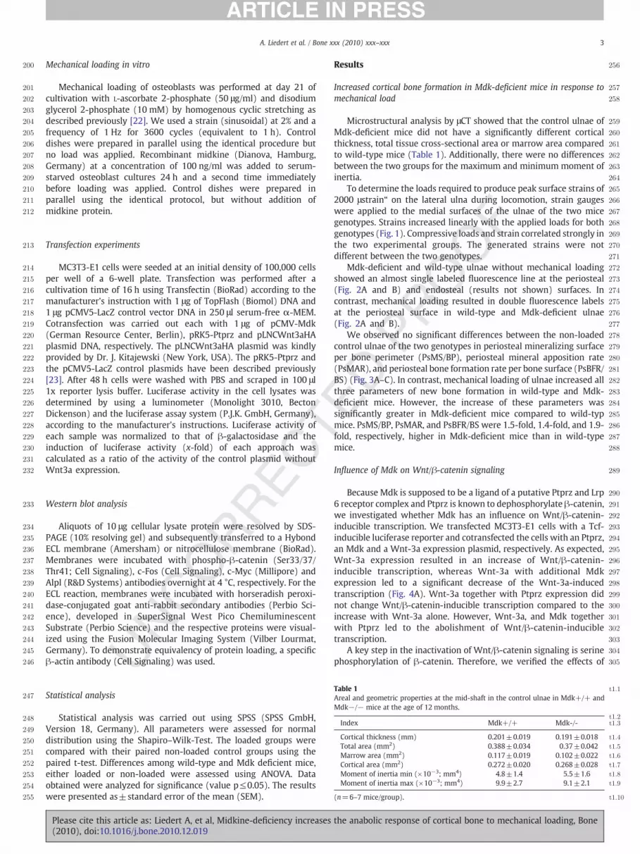
Table 1 t1:1
Areal and geometric properties at the mid-shaft in the control ulnae in Mdk+/+ andMdk−/− mice at the age of 12 months.
t1:2t1:3Index Mdk+/+ Mdk-/-
t1:4Cortical thickness (mm) 0.201±0.019 0.191±0.018t1:5Total area (mm2) 0.388±0.034 0.37±0.042t1:6Marrow area (mm2) 0.117±0.019 0.102±0.022t1:7Cortical area (mm2) 0.272±0.020 0.268±0.028t1:8Moment of inertia min (×10−3; mm4) 4.8±1.4 5.5±1.6t1:9Moment of inertia max (×10−3; mm4) 9.9±2.7 9.1±2.1
(n=6–7 mice/group). t1:10
Statistical analysis
Statistical analysis was carried out using SPSS (SPSS GmbH,Version 18, Germany). All parameters were assessed for normaldistribution using the Shapiro–Wilk-Test. The loaded groups werecompared with their paired non-loaded control groups using thepaired t-test. Differences among wild-type and Mdk deficient mice,either loaded or non-loaded were assessed using ANOVA. Dataobtained were analyzed for significance (value p≤0.05). The resultswere presented as±standard error of the mean (SEM).
Please cite this article as: Liedert A, et al, Midkine-deficiency increases(2010), doi:10.1016/j.bone.2010.12.019
Results
Increased cortical bone formation in Mdk-deficient mice in response tomechanical load
Microstructural analysis by μCT showed that the control ulnae ofMdk-deficient mice did not have a significantly different corticalthickness, total tissue cross-sectional area or marrow area comparedto wild-type mice (Table 1). Additionally, there were no differencesbetween the two groups for the maximum and minimum moment ofinertia.
To determine the loads required to produce peak surface strains of2000 μstrain“ on the lateral ulna during locomotion, strain gaugeswere applied to the medial surfaces of the ulnae of the two micegenotypes. Strains increased linearly with the applied loads for bothgenotypes (Fig. 1). Compressive loads and strain correlated strongly inthe two experimental groups. The generated strains were notdifferent between the two genotypes.
Mdk-deficient and wild-type ulnae without mechanical loadingshowed an almost single labeled fluorescence line at the periosteal(Fig. 2A and B) and endosteal (results not shown) surfaces. Incontrast, mechanical loading resulted in double fluorescence labelsat the periosteal surface in wild-type and Mdk-deficient ulnae(Fig. 2A and B).
We observed no significant differences between the non-loadedcontrol ulnae of the two genotypes in periosteal mineralizing surfaceper bone perimeter (PsMS/BP), periosteal mineral apposition rate(PsMAR), and periosteal bone formation rate per bone surface (PsBFR/BS) (Fig. 3A–C). In contrast, mechanical loading of ulnae increased allthree parameters of new bone formation in wild-type and Mdk-deficient mice. However, the increase of these parameters wassignificantly greater in Mdk-deficient mice compared to wild-typmice. PsMS/BP, PsMAR, and PsBFR/BS were 1.5-fold, 1.4-fold, and 1.9-fold, respectively, higher in Mdk-deficient mice than in wild-typemice.
Influence of Mdk on Wnt/β-catenin signaling
Because Mdk is supposed to be a ligand of a putative Ptprz and Lrp6 receptor complex and Ptprz is known to dephosphorylate β-catenin,we investigated whether Mdk has an influence on Wnt/β-catenin-inducible transcription. We transfected MC3T3-E1 cells with a Tcf-inducible luciferase reporter and cotransfected the cells with an Ptprz,an Mdk and a Wnt-3a expression plasmid, respectively. As expected,Wnt-3a expression resulted in an increase of Wnt/β-catenin-inducible transcription, whereas Wnt-3a with additional Mdkexpression led to a significant decrease of the Wnt-3a-inducedtranscription (Fig. 4A). Wnt-3a together with Ptprz expression didnot change Wnt/β-catenin-inducible transcription compared to theincrease with Wnt-3a alone. However, Wnt-3a, and Mdk togetherwith Ptprz led to the abolishment of Wnt/β-catenin-inducibletranscription.
A key step in the inactivation of Wnt/β-catenin signaling is serinephosphorylation of β-catenin. Therefore, we verified the effects of
the anabolic response of cortical bone to mechanical loading, Bone

306
307
308
309
310
311
312
313
314
315
316
317
318
319
320
321
322
323
324
325
326
Fig. 1. Load–strain relationship in ulnae from Mdk-deficient and wild-type mice. Straingauge measurements were performed ex vivo on the medial surfaces of the ulnae ofboth genotypes of mice. Values are presented as mean±SD. (n=5–6 mice/group;p≤0.05).
4 A. Liedert et al. / Bone xxx (2010) xxx–xxx
Mdk on Wnt/β-catenin signaling by analyzing phosphorylated β-catenin in wild-type and Ptprz-deficient osteoblasts after treatmentwith recombinant Mdk andWnt-3a, respectively. As shown in Fig. 4B,Mdk treatment resulted in an increase of phospho-β-catenincompared to cells without Mdk. Thus, Mdk led to an inactivation ofWnt/β-catenin signaling, which was diminished in cells treated withMdk together withWnt-3a. As expected, Wnt-3a alone resulted in thelowest phospho-β-catenin level in comparison to the three otherapproaches. However, in Ptprz-deficient cells Mdk treatment did notincrease phospho-β-catenin. Therefore, the effects of Mdk onphospho-β-catenin in wild-type osteoblasts appeared to be depen-dent on Ptprz expression.
To verify the effects observed with the Tcf-inducible luciferasereporter vector and Mdk treatment on phospho-β-catenin, weanalyzed Wnt/β-catenin target gene expression in osteoblasts afterthe addition of recombinantMdk in cultures at day 21 after cultivationunder supplementation of ascorbate-2-phosphate and glycerol-2-phosphate. We observed a significant downregulation of Alplexpression on day 21 of cultivation in differentiation medium afterMdk treatment without mechanical loading (25%, p≤0.05). Toconfirm this result at the protein level we performed Western Blot
327
328
329
330
331
332
333
334
335
336
337
Fig. 2. Increased loading-induced new periosteal cortical bone formation in Mdk-deficient mice. (A) Representative fluorescence micrographs showing details of calceingreen- and alizarin red-labeled mid-diaphyseal cross-sections of the unloaded (C, left)and loaded (L, right) ulnae of wild-typ mice. (B) Representative fluorescencemicrographs details of calcein green- and alizarin red-labeled mid-diaphyseal cross-sections of the unloaded (C, left) and loaded (L, right) ulnae of Mdk-deficient mice.Double calcein-alizarin-labels are present on the periosteal surface of the loadedcortical bone. A superimposed bar on the figure indicates the measurement distance.(For interpretation of the references to colour in this figure legend, the reader isreferred to the web version of this article.)
Fig. 3. Histomorphometric analysis of loading-induced new periosteal cortical boneformation in Mdk-deficient mice. The parameters were determined in non-loaded andloaded ulnae of wild-type and Mdk-deficient mice by histomorphometric analysis afterinjection of calcein green and alizarin red. (A) Periosteal mineralizing surface per boneperimeter (PsMS/BPm) in control and loaded ulnae of both genotypes of mice(***p≤0.0005; *p≤0.05). (B) Periosteal mineral apposition rate (PsMAR) in control andloaded ulnae of both genotypes of mice (*p≤0.05; ***p≤0.0005). (C) Periosteal boneformation rate per bone surface (PsBFR/BS) in control and loaded ulnae of bothgenotypes of mice (*p≤0.05; **p≤0.005). All values are presented as mean±SD(n=6–13 mice/group).
Please cite this article as: Liedert A, et al, Midkine-deficiency increases(2010), doi:10.1016/j.bone.2010.12.019
analysis and observed down-regulation of Alpl expression after Mdkaddition (Fig. 4C).
Furthermore, we studied the effect of Mdk without and withmechanical loading on Wnt/β-catenin target protein expression. Asexpected, mechanical loading led to the up-regulation of both c-Fos(„“1.6-fold) and c-Myc expression (1.3-fold), whereas withoutmechanical loading treatment with recombinant Mdk resulted indownregulation of expression (c-Fos, 0.3-fold; c-Myc, 0.5-fold; Fig. 5).Up-regulation of c-Fos and c-Myc expression by mechanical loadingwas almost abolished in Mdk treated osteoblast cultures (c-Fos, 0.6-fold; c-Myc, 0.7-fold).
the anabolic response of cortical bone to mechanical loading, Bone

338
339
340
341
342
343
344
345
346
347
348
349
350
351
352
353
354
355
356
357
358
359
360
361
362
363
364
Fig. 4. Effect of Mdk on Wnt/β-catenin signaling. (A) MC3T3-E1 cells were transfected with a Tcf-inducible luciferase reporter, a β-galactosidase expression plasmid andcotransfected with an Ptprz, an Mdk and a Wnt-3a expression plasmid, respectively. Values are presented as mean±SD (n=6; ***p≤0.0005). (B) Representative immunoblotshowing serine-phosphorylated β-catenin protein levels in wild-type and Ptprz-deficient osteoblasts after treatment with recombinant Mdk and Wnt-3a. The level of β-actinwas used as control. (C) Representative immunoblot using antibodies against the Wnt target protein alkaline phosphatase (Alpl) in osteoblasts treated with recombinant Mdk(100 ng/ml). β-actin was used as control.
5A. Liedert et al. / Bone xxx (2010) xxx–xxx
Discussion
In this study we analyzed the role of the heparin-binding growthfactor Mdk inmechanically-induced bone remodeling. We showed forthe first time that Mdk deficiency increases the sensitivity of corticalbone formation to mechanical loading. Moreover, we demonstratedwith in vitro studies using osteoblastic cells that Mdk has a negativeinfluence onWnt/β-catenin signaling, this pathway playing a key rolein the regulation of bone remodeling.
Our main objective in this study was to determine whether theanabolic response of bone to mechanical loading might be influenced,at least in part, by Mdk expression. Histomorphometric analysis ofnon-loaded wild-type and Mdk-deficient ulnae did not reveal anysignificant differences in PsMS/BPm, PsMAR, and PsBFR/BS between
Fig. 5. Effect of mechanical loading and Mdk treatment on Wnt/β-catenin target protein ewithout mechanical loading (C, control), by mechanical loading (L, loaded) and/or with recowere used for immunodetection. β-actin was used as control.
Please cite this article as: Liedert A, et al, Midkine-deficiency increases(2010), doi:10.1016/j.bone.2010.12.019
the two genotypes. All these three parameters of new bone formationwere increased in both mice genotypes after mechanical loading.However, in Mdk-deficient mice these increases were significantlygreater, indicating that Mdk-deficiency enhances the anabolicresponse of cortical bone to mechanical load in mice. An elevatedresponse to mechanical loading could be caused by an enhancedmechanotransduction, including the process of reception and trans-formation of the mechanical stimulus into the cellular response.Furthermore, an improved osteoblast function could result in anenhancement of the response to mechanical loading. A previous studyhas shown that Mdk-deficiency in mice at 12 and 18 months of ageresulted in increased trabecular bone volume [9]. However, corticalbone mass in femura was unaffected by the Mdk-deficiency in agedmice.
xpression. Representative immunoblot using cellular extracts from osteoblasts treatedmbinant Mdk (100 ng/ml). Antibodies against the transcription factors c-Fos and c-Myc
the anabolic response of cortical bone to mechanical loading, Bone
365
366
367
368
369
370
371
372
373
374
375
376
377
378
379
380
381
382
383
384
385
386
387
388
389
390
391
392
393
394
395
396
397
398
399
400
401
402
403
404
405
406
407
408
409
410
411
412
413
414
415
416
417
418
419
420
421
422
423
424
425
426
427
428
429
430
431
432
433
434
435
436
437
438
439
440
441
442
443
444
445
446
447
448
449
450
451
452
453
454
455
456
457
458
459
460
461
462
463
464
465
466
467
468
469
470
471
472
473
474
475
476
477
478
479
480
481
482
483
484
485
486
487
488
489
490
491
492
493
494
495
496
6 A. Liedert et al. / Bone xxx (2010) xxx–xxx
We performed μCT scanning at the midshaft of the ulnae of wild-type and Mdk-deficient mice, and analyzed areal and geometricproperties. We did not observe any significant differences in theseparameters between the two genotypes, thus supporting the earlierresult that cortical bone mass was unaffected in aged Mdk-deficientmice. This finding was corroborated by the results from ex vivocalibration studies with strain gauges, which were applied to derminethe force-strain curves for wild-type and Mdk-deficient mice at theage of 12 months. We could demonstrate that equal forces wererequired to generate the same surface strains on the lateral midshaftof the ulnae. As we did not observe any significant differences inregard to the analyzed parameters for the control ulnae of bothgenotypes, we could for instance rule out that a thinner bone andaccordingly higher strains were responsible for the enhanced anabolicresponse to mechanical loading in Mdk-deficient mice.
Mdk and Ptn, which form a unique family of heparin-bindinggrowth and differentiation factors, have been shown to bind to Ptprz[24,25]. Ptprz, also known as receptor tyrosine phosphatase β,possesses an extracellular chondroitin sulfate proteoglycan domainand two intracellular PTP domains [26]. These structural featuresprovide the direct coupling of an extracellular adhesive interaction tointracellular tyrosine-phosphorylation-dependent signaling path-ways regulating cell proliferation, differentiation and cytoskeletalintegrity in response to external stimuli [27]. Our group showed up-regulation of Mdk and Ptprz expression during the course ofosteoblast differentiation [23]. Because Ptprz has been shown tohave a role in the inhibition of osteoblast proliferation [23,28], up-regulation of Ptprz during the course of differentiation appears to bephysiologically relevant. Thus, when cell proliferation is no longerrequired, Ptprz-mediated dephosphorylation might inactivate tyro-sine kinase activity induced by the binding of the growth factor to itsreceptor anaplastic lymphoma kinase (Alk). Mdk has been shown toincrease proliferation and differentiation of various cell typs,including osteoblastic and chondrogenic cells [4]. Mdk is implicatedin stimulating tumorigenesis by autocrine and/or paracrine growthloops and binding to Alk [29]. Previously, it has been demonstratedthat treatment with functional antibodies against midkine suppressesgrowth of osteosarcoma cells [30].
In addition to Alk and Ptprz, syndecans, Lrp-6, and integrins havealso been proposed as Mdk membrane receptors [11,24,31–33].Because Ptprz and LRP-6 together with α4β1- and α6β1-integrinshave been identified as a signaling receptor complex for Mdk, andPtprz has been shown to be involved in tyrosine dephosphorylation ofβ-catenin [11,16], we hypothesized that regulation of β-cateninsignaling could be mediated by an interaction of Mdk with Ptprz.Therefore, we performed in vitro studies with MC3T3-E1 cellstransiently expressing Ptprz, Mdk, and Wnt-3a, respectively, andinvestigated their effect on Wnt/β-catenin-inducible transcription.Interestingly, we observed that Mdk alone decreased, while Mdk andPtprz even abolishedWnt3a-induced transcription. Thus, we assumedthat Mdkmight have a negative effect on osteoblast function, which isenhanced by an interaction with Ptprz. Because serine phosphoryla-tion of β-catenin is a key step in the inactivation of Wnt/β-cateninsignaling, we analyzed serine phosphorylation of β-catenin inosteoblasts after treatment with recombinant Mdk. We showed thatMdk had an inactivating effect on Wnt/β-catenin signaling and thatthe extent of this effect was dependent on Ptprz expression. Becausewe and Schinke et al. [23] did not observe any influence on β-catenin-dependentWnt signaling by Ptprz expression alone, it is reasonable toassume that the effect of Ptprz on Wnt/β-catenin signaling dependson additional Mdk expression. We examined Alpl expression inosteoblasts, which plays a critical role in skeletal mineralization [34].We demonstrated that Mdk treatment led to a decrease in Alplexpression. This result is consistent with the findings of Neunaber etal., who in contrast observed a significant increase in alkalinephosphatase activity as well as in matrix mineralization in differen-
Please cite this article as: Liedert A, et al, Midkine-deficiency increases(2010), doi:10.1016/j.bone.2010.12.019
tiatedMdk-deficient osteoblasts derived from bonemarrow precursorcells isolated from 30-week-old animals indicating a cell-autonomousmechanism underlying the increased trabecular bone phenotype ofMdk-deficient mice [9].
Moreover, we studied the influence of recombinant Mdk on Wnt/β-catenin target gene expression in non-loaded and loaded osteo-blasts after cultivation for 21 days under supplementation ofascorbate-2-phosphate and glycerol-2-phosphate. The loading designused in this study has been shown to increase osteoblast proliferation,whereas activities related to differentiation are decreased [22].Therefore, we studied the expression of c-Fos and c-Myc, twotranscription factors, which are involved in osteoblast proliferationand are known to be regulated by Wnt signaling [17,18]. It has beendemonstrated that down-regulation of Wnt/β-catenin signaling is aprerequisite for late stage osteoblast differentiation [35,36]. In fact,Mdk treatment of osteoblast cultures without mechanical loading ledto a decrease of both c-Fos and c-Myc expression. Mdk treatmenttogether with mechanical loading almost abolished the increase of c-Fos and c-Myc expression induced by mechanical loading alone.Clearly, Mdk had a negative effect on Wnt/β-catenin target geneexpression in both non-loaded and loaded osteoblasts. Therefore, it isreasonable to suggest that in our study an improved osteoblastfunction in Mdk-deficient mice might be responsible for the increasedanabolic response to mechanical loading.
We currently do not know the reason for the unaffected corticalbone formation that we observed in the control ulnae of Mdk-deficient mice compared to wild-type mice, which is in line with theunaffected cortical bone formation in femura of aged Mdk-deficientmice [9]. We assume that the age of mice influences the outcome ofMdk-deficiency on cortical bone formation. Neunaber et al. demon-strated that Mdk-deficient tibia of mice compared to their wild-typelittermates have an increased cortical porosity caused by localactivation of bone resorption, which was detectable only at the ageof 18 months [9]. This is consistent with the cortical bone of the ulnaeof midkine-deficient mice, which in our study did not show corticalporosity at the age of 12 months (data not shown). As trabecular boneformation has been shown to be age-dependent in Mdk-deficientmice [9] the cortical phenotype of these mice may similarly be age-dependent. Moreover, Mdk-deficient mice have been shown to becharacterized by an increased anxiety, as demonstrated by theelevated pluze-maze test [6]. Because we also observed a morereclusive behavior of Mdk-deficient mice compared to wild-type miceover the entire experimental period, we cannot rule out that areduced locomotive behavior in Mdk-deficient mice at least in partcontributed the non-affected cortical bone formation phenotype ofthe non-loaded control ulnae. Previously, it was demonstrated thatboth male and female mice doubly deficient in Mdk and Ptn showedreduced locomotive activity compared to wild-type mice [37]. Weapplied load in addition to the physiological load, which the micereceived during their normal activity throughout the experimentalperiod. Thus, it is likely that a specific threshold of load above the levelof physiological loading under normal conditions is necessary toinduce the increase of anabolic response in Mdk-deficient mice.Furthermore, in vivo studies have already shown that Wnt signalingcontributes to the anabolic response of bone to mechanical load[38,39]. We showed in the present study in vitro that Mdk can have anegative effect on Wnt signaling. Therefore, we suggest thatmechanically increased Wnt signaling might be enhanced in micewithout Mdk expression.
There is evidence that Ptn and Mdk are able to bind to Ptprz andinactivate Ptprz, thereby enhancing tyrosine phosphorylation of β-catenin [16,24]. Tyrosine-phoshorylation dependent release of β-catenin from the cadherin complex might not only regulate theintegrity of cell-junctions, but also be an alternative mechanism formodulating β-catenin signaling [14]. When released from cadherin inthe absence of Wnt signaling, uncomplexed β-catenin is rapidly
the anabolic response of cortical bone to mechanical loading, Bone

497
498
499
500
501
502
503
504
505
506
507
508
509
510
511
512
513
514
515
516
517
518
519
520
521
522523524525526527528529530531532533534535536537538539540541542543544545546547548549550551552553554555556557558559560561
562563564565566567568569570571572573574575576577578579580581582583584585586587588589590591592593594595596597598599600601602603604605606607608609610611612613614615616617618619620621622623624625626627628629630631632633634635636637638639
640
7A. Liedert et al. / Bone xxx (2010) xxx–xxx
degraded by proteosomes, thereby inhibiting β-catenin target geneexpression. It has been shown that another receptor protein-tyrosinephosphatase, PCP-2, is able to inhibit β-catenin signaling and toincrease E-cadherin-dependent cell adhesion in the colon cancer cellline SW480 [40]. Thus, upregulation of Mdk and Ptprz expression inosteoblast cultures over the time course of differentiation might be anecessary step to limit osteoblast proliferation in favor of differenti-ation by increasing cadherin-dependent cell adhesion and thusinhibiting β-catenin signaling. Presumably, Mdk binding to Ptprzmight be one mechanism for regulating the balance between β-catenin signaling and adhesive β-catenin. An immunofluorescencestudy of β-catenin in osteoblasts without and after Mdk additionshould be perfomed in future to investigate whether Mdk is able tomobilize β-catenin from the cadherin complex at the cell surface.
In summary, the results presented in this study show that Mdk-deficiency has an anabolic effect on mechanically induced corticalbone remodeling, which could be due to an improved osteoblastfunction caused by the further enhancement of loading-induced β-catenin-dependent Wnt-signaling through Mdk-deficiency.
Acknowledgments
The expert technical assistance of Iris Baum, Elisa Viering, andNadine Todt is gratefully acknowledged. This work was supported bythe German Research Foundation (DFG, grant AM103/10-1 and IG18/3-1).
References
[1] Rubin J, Rubin C, Jacobs CR. Molecular pathwaysmediating mechanical signaling inbone. Gene 2006;367:1–16.
[2] Robling AG, Castillo AB, Turner CH. Biomechanical and molecular regulation ofbone remodeling. Annu Rev Biomed Eng 2006;8:455–98.
[3] Muramatsu T. Midkine and pleiotrophin: two related proteins involved indevelopment, survival, inflammation and tumorigenesis. J Biochem (Tokyo)2002;132:359–71.
[4] Ohta S, Muramatsu H, Senda T, Zou K, Iwata H, Muramatsu T. Midkine is expressedduring repair of bone fracture and promotes chondrogenesis. J Bone Miner Res1999;14:1132–44.
[5] Petersen W, Wildemann B, Pufe T, Raschke M, Schmidmaier G. The angiogenicpeptide pleiotrophin (PTN/HB-GAM) is expressed in fracture healing: animmunohistochemical study in rats. Arch Orthop Trauma Surg 2004;124:603–7.
[6] Nakamura E, Kadomatsu K, Yuasa S, Muramatsu H, Mamiya T, Nabeshima T, et al.Disruption of the midkine gene (Mdk) resulted in altered expression of a calciumbinding protein in the hippocampus of infant mice and their abnormal behaviour.Genes Cells 1998;3:811–22.
[7] Amet LE, Lauri SE, Hienola A, Croll SD, Lu Y, Levorse JM, et al. Enhancedhippocampal long-term potentiation in mice lacking heparin-binding growth-associated molecule. Mol Cell Neurosci 2001;17:1014–24.
[8] Imai S, Heino TJ, Hienola A, Kurata K, Buki K, Matsusue Y, et al. Osteocyte-derivedHB-GAM (pleiotrophin) is associated with bone formation and mechanicalloading. Bone 2009;44:785–94.
[9] Neunaber C, Catala-Lehnen P, Beil FT, Marshall RP, Kanbach V, Baranowsky A,et al. Increased trabecular bone formation in mice lacking the growth factormidkine. J Bone Miner Res 2010;25:1724–35.
[10] Sakaguchi N, Muramatsu H, Ichihara-Tanaka K, Maeda N, Noda M, Yamamoto T,et al. Receptor-type protein tyrosine phosphatase zeta as a component of thesignaling receptor complex for midkine-dependent survival of embryonicneurons. Neurosci Res 2003;45:219–24.
[11] Muramatsu H, Zou P, Suzuki H, Oda Y, Chen GY, Sakaguchi N, et al. alpha4beta1-and alpha6beta1-integrins are functional receptors for midkine, a heparin-bindinggrowth factor. J Cell Sci 2004;117:5405–15.
[12] Mbalaviele G, Shin CS, Civitelli R. Cell–cell adhesion and signaling throughcadherins: connecting bone cells in their microenvironment. J Bone Miner Res2006;21:1821–7.
[13] Piedra J, Miravet S, Castano J, Palmer HG, Heisterkamp N, Garcia de Herreros A,et al. p120 Catenin-associated Fer and Fyn tyrosine kinases regulate beta-cateninTyr-142 phosphorylation and beta-catenin-alpha-catenin Interaction. Mol CellBiol 2003;23:2287–97.
Please cite this article as: Liedert A, et al, Midkine-deficiency increases(2010), doi:10.1016/j.bone.2010.12.019
[14] Nelson WJ, Nusse R. Convergence of Wnt, beta-catenin, and cadherin pathways.Science 2004;303:1483–7.
[15] Schiller KR, Mauro LJ. Tyrosine phosphatases as regulators of skeletal developmentand metabolism. J Cell Biochem 2005;96:262–77.
[16] Meng K, Rodriguez-Pena A, Dimitrov T, Chen W, Yamin M, Noda M, et al.Pleiotrophin signals increased tyrosine phosphorylation of beta beta-cateninthrough inactivation of the intrinsic catalytic activity of the receptor-type proteintyrosine phosphatase beta/zeta. Proc Natl Acad Sci USA 2000;97:2603–8.
[17] Staal FJ, Weerkamp F, Baert MR, van den Burg CM, van Noort M, de Haas EF, et al.Wnt target genes identified by DNA microarrays in immature CD34+ thymocytesregulate proliferation and cell adhesion. J Immunol 2004;172:1099–108.
[18] KatohM.WNT signaling pathway and stem cell signaling network. Clin Cancer Res2007;13:4042–5.
[19] Rodan GA, Noda M. Gene expression in osteoblastic cells. Crit Rev Eukaryot GeneExpr 1991;1:85–98.
[20] Lee KC, Maxwell A, Lanyon LE. Validation of a technique for studying functionaladaptation of the mouse ulna in response to mechanical loading. Bone 2002;31:407–12.
[21] Parfitt AM, Drezner MK, Glorieux FH, Kanis JA, Malluche H, Meunier PJ, et al. Bonehistomorphometry: standardization of nomenclature, symbols, and units. Reportof the ASBMR Histomorphometry Nomenclature Committee. J Bone Miner Res1987;2:595–610.
[22] Kaspar D, Seidl W, Neidlinger-Wilke C, Ignatius A, Claes L. Dynamic cell stretchingincreases human osteoblast proliferation and CICP synthesis but decreasesosteocalcin synthesis and alkaline phosphatase activity. J Biomech 2000;33:45–51.
[23] Schinke T, Gebauer M, Schilling AF, Lamprianou S, Priemel M, Mueldner C, et al.The protein tyrosine phosphatase Rptpzeta is expressed in differentiatedosteoblasts and affects bone formation in mice. Bone 2008;42:524–34.
[24] Maeda N, Ichihara-Tanaka K, Kimura T, Kadomatsu K, Muramatsu T, Noda M. Areceptor-like protein-tyrosine phosphatase PTPzeta/RPTPbeta binds a heparin-binding growth factor midkine. Involvement of arginine 78 of midkine in the highaffinity binding to PTPzeta. J Biol Chem 1999;274:12474–9.
[25] Maeda N, Nishiwaki T, Shintani T, Hamanaka H, Noda M. 6B4 proteoglycan/phosphacan, an extracellular variant of receptor-like protein-tyrosine phosphatasezeta/RPTPbeta, binds pleiotrophin/heparin-binding growth-associated molecule(HB-GAM). J Biol Chem 1996;271:21446–52.
[26] Levy JB, Canoll PD, Silvennoinen O, Barnea G, Morse B, Honegger AM, et al. Thecloning of a receptor-type protein tyrosine phosphatase expressed in the centralnervous system. J Biol Chem 1993;268:10573–81.
[27] Stone RL, Dixon JE. Protein-tyrosine phosphatases. J Biol Chem 1994;269:31323–6.[28] Lau KH, Baylink DJ. Phosphotyrosyl protein phosphatases: potential regulators of
cell proliferation and differentiation. Crit Rev Oncog 1993;4:451–71.[29] Li R, Morris SW. Development of anaplastic lymphoma kinase (ALK) small-
molecule inhibitors for cancer therapy. Med Res Rev 2008;28:372–412.[30] Maehara H, Kaname T, Yanagi K, Hanzawa H, Owan I, Kinjou T, et al. Midkine as a
novel target for antibody therapy in osteosarcoma. Biochem Biophys Res Commun2007;358:757–62.
[31] Stoica GE, Kuo A, Powers C, Bowden ET, Sale EB, Riegel AT, et al. Midkine binds toanaplastic lymphoma kinase (ALK) and acts as a growth factor for different celltypes. J Biol Chem 2002;277:35990–8.
[32] Nakanishi T, Kadomatsu K, Okamoto T, Ichihara-Tanaka K, Kojima T, Saito H, et al.Expression of syndecan-1 and -3 during embryogenesis of the central nervoussystem in relation to binding with midkine. J Biochem 1997;121:197–205.
[33] Muramatsu H, Zou K, Sakaguchi N, Ikematsu S, Sakuma S, Muramatsu T. LDLreceptor-related protein as a component of the midkine receptor. BiochemBiophys Res Commun 2000;270:936–41.
[34] Whyte MP. Physiological role of alkaline phosphatase explored in hypopho-sphatasia. Ann NY Acad Sci 2010;1192:190–200.
[35] van der Horst G, van der Werf SM, Farih-Sips H, van Bezooijen RL, Lowik CW,Karperien M. Downregulation of Wnt signaling by increased expression ofDickkopf-1 and -2 is a prerequisite for late-stage osteoblast differentiation ofKS483 cells. J Bone Miner Res 2005;20:1867–77.
[36] Kahler RA, Galindo M, Lian J, Stein GS, van Wijnen AJ, Westendorf JJ.Lymphocyte enhancer-binding factor 1 (Lef1) inhibits terminal differentiationof osteoblasts. J Cell Biochem 2006;97:969–83.
[37] Muramatsu H, Zou P, Kurosawa N, Ichihara-Tanaka K, Maruyama K, Inoh K, et al.Female infertility in mice deficient in midkine and pleiotrophin, which form adistinct family of growth factors. Genes Cells 2006;11:1405–17.
[38] Robinson JA, Chatterjee-Kishore M, Yaworsky PJ, Cullen DM, Zhao W, Li C, et al.Wnt/beta-catenin signaling is a normal physiological response to mechanicalloading in bone. J Biol Chem 2006;281:31720–8.
[39] Armstrong VJ, Muzylak M, Sunters A, Zaman G, Saxon LK, Price JS, et al. Wnt/beta-catenin signaling is a component of osteoblastic bone cell early responses to load-bearing and requires estrogen receptor alpha. J Biol Chem 2007;282:20715–27.
[40] Yan HX, Yang W, Zhang R, Chen L, Tang L, Zhai B, et al. Protein-tyrosinephosphatase PCP-2 inhibits beta-catenin signaling and increases E-cadherin-dependent cell adhesion. J Biol Chem 2006;281:15423–33.
the anabolic response of cortical bone to mechanical loading, Bone


