Embed Size (px)
Citation preview
MHC Class I/Peptide Transfer between Dendritic CellsOvercomes Poor Cross-Presentation by Monocyte-DerivedAPCs That Engulf Dying Cells1
Chunfeng Qu,2,3 Van Anh Nguyen, Miriam Merad, and Gwendalyn J. Randolph2
In vivo data suggest that monocytes participate critically in cross-presentation, but other data suggest that lymph node residentdendritic cells (DCs) mainly cross-present. Here, we utilized a three-dimensional model of a blood vessel wall that endogenouslysupports DC development from human monocytes, and we incorporated dying autologous cells in the subendothelial matrix of themodel. Flu-infected dying cells promoted monocytes to become mature DCs and cross-present cell-associated Ags for the activationof CTLs. Similar responses were induced by loading the dying cells with the TLR7/8 ligand ssRNA, whereas dying cells loaded withTLR3 ligand were less efficient. Monocyte-derived DCs that developed in this model cross-presented Ag to T cells efficientlyregardless of whether they engulfed detectable amounts of labeled dying cells. Unexpectedly, the monocyte-derived cells thatdirectly engulfed dying cells in vitro were not the major APCs stimulating CD8� lymphocytes. Instead, bystander DCs acquiredmore robust capacity to cross-prime through receipt of MHC class I/peptide from the phagocytic, monocyte-derived cells. In mice,lymph node-homing monocyte-derived DCs processed Ags from engulfed cells and then transferred MHC class I/peptide com-plexes to confer cross-priming capacity to MHC class I-deficient lymph node resident CD8�� DCs. Thus, natural or syntheticTLR7/8 agonists contained within dying cells promote the conversion of monocytes to DCs with capacity for cross-presentation andfor “cross-dressing” other DCs. These data reveal a way in which migratory monocyte-derived DCs and other DCs, like lymphnode resident DCs, both mediate cross-presentation. The Journal of Immunology, 2009, 182: 3650–3659.
M any viral Ags are best detected and combated withinthe immune system through cross-priming, a mecha-nism by which professional APCs, most notably den-
dritic cells (DCs),4 acquire exogenous Ags, likely often from dyingparenchymal cells, for presentation through the MHC class I(MHC I) pathway to CD8� T cells (1, 2). Monocytes readily mo-bilize into peripheral tissues and give rise to lymph node-homingDCs under inflammatory conditions (3), and monocytes newly re-cruited to tissues conditioned by various adjuvants participate crit-ically in cross-priming (4). It has been suggested from studies inmice that monocyte-derived DCs directly primed CD8� T cells
because monocytes that selectively lacked MHC I are unable tosupport cross-priming (4). This observation seems inconsistentwith other findings that a population of DCs in mouse lymph nodesthat expresses CD8�, and hence called CD8�� DCs, has beenshown to manifest superior capacity for cross-presentation (5), andthat these DCs selectively recognize and engulf dying cells (6).Because CD8�� DCs are lymph node resident cells that do notsurvey peripheral tissues, they may in many instances collaboratewith other DCs that mobilize from the periphery to gain access toAg through transfer between DCs (7–10). However, whether andhow monocyte-derived DCs may cooperate with these DCs, ortruly cross-present Ag on their own as suggested (4), have not beenfully determined. Monocyte-derived DCs cultured in GM-CSF andIL-4 for several days to generate immature DCs can cross-presentinfluenza A Ags derived from dying cells (11). However, imma-ture DCs derived from GM-CSF/IL-4-treated monocytes are moreakin to tissue resident DCs, and they may not model the responseof monocytes newly arriving at a site of viral infection. Thus,much remains to be explored regarding how newly recruitedmonocytes may participate in cross-presentation of viral Ags orwhether newly recruited human monocytes do so at all.
To investigate the potential role of newly recruited monocytes inacquiring, processing, and presenting Ag in the periphery to sup-port cross-priming, we took advantage of an in vitro model of asimple connective tissue that consists of an endothelium grown ona matrix of type I collagen. In this model, freshly isolated humanmonocytes readily transmigrate across the endothelium to differ-entiate into macrophages that remain within the subendothelialmatrix and DCs that retraverse, or reverse transmigrate, across theendothelial layer (12). Within the collagen, we grew cell lines thatwere autologous to the transmigrating monocytes; these cell lineswere induced to undergo apoptosis and in some cases were in-fected with influenza A virus (flu) as a model virus or were loadedwith synthetic TLR ligands that mimic viral stimuli to evaluate
Department of Gene and Cell Medicine and Institute for Immunology, Icahn MedicalInstitute, Mount Sinai School of Medicine, New York, NY 10029
Received for publication May 12, 2008. Accepted for publication January 12, 2009.
The costs of publication of this article were defrayed in part by the payment of pagecharges. This article must therefore be hereby marked advertisement in accordancewith 18 U.S.C. Section 1734 solely to indicate this fact.1 This work was supported by the Cancer Research Institute, National Institutes ofHealth Grant AI49653, and Defense Advanced Research Planning Agency contractW81XWH-04-C-0139. The prime contractor of the latter contract is the companyVaxDesign. G.J.R. and Mount Sinai School of Medicine are subcontractors of thisaward. C.Q. later was supported by Grant JK2006A01 from the Chinese Academy ofMedical Sciences.2 Address correspondence and reprint requests to Dr. Chunfeng Qu, State Key Lab-oratory of Molecular Oncology, Cancer Institute, Chinese Academy of Medical Sci-ences, Beijing 100021, China. E-mail address: [email protected] or Dr.Gwendalyn J. Randolph, Department of Gene and Cell Medicine, Mount Sinai Schoolof Medicine, 1425 Madison Avenue, Box 1496, New York, NY 10029. E-mail ad-dress: [email protected] Current address: State Key Laboratory of Molecular Oncology, Cancer Institute,Chinese Academy of Medical Sciences, Beijing 100021.4 Abbreviations used in this paper: DC, dendritic cells; MHC I, MHC class I; LCL,lymphoblastoid cell line; MP, influenza A virus matrix protein; MP-LCL, lympho-blastoid cell line expressing influenza A virus matrix protein; MOI, multiplicity ofinfection; HA, hemagglutinin; �2m, �2-microglobulin.
Copyright © 2009 by The American Association of Immunologists, Inc. 0022-1767/09/$2.00
The Journal of Immunology
www.jimmunol.org/cgi/doi/10.4049/jimmunol.0801532

how these signals delivered to monocytes through dying cellsmight influence cross-presentation. We show that, in particular,TLR7/8 ligands (ssRNA) present within dying cells strongly pro-mote monocytes to become cross-priming APCs. However, mono-cyte-derived DCs that engulfed Ag-bearing dying cells were sur-prisingly poor stimulators of CTLs themselves, but they supportedcross-priming by acting as efficient suppliers of Ag to other DCs.Subsequent in vivo studies in mice then indicated that migratorymonocyte-derived DCs supplied intact MHC I/peptide to lymphnode DCs, allowing MHC I-deficient CD8�� DCs to cross-primeCD8� T cells.
Materials and MethodsLymphoblastoid cell lines (LCLs)
EBV-transformed B cells, or LCLs, were generated as described (13). Asuspension of LCLs in RPMI 1640 containing 20% FBS was mixed withbovine type I collagen (Inamed), a 10� stock of M199 (Lonza), 0.05 NNaOH, and HEPES buffer at relative volumes of 2.5:8:1:2.5, respectively.To monitor phagocytosis, LCLs were labeled with 5 �mol/L CFSE (Mo-lecular Probes) for 15 min at 37°C. The mixture of collagen and cells at avolume of 50 �l was dispensed into microtiter wells and allowed to po-lymerize. The following day, the LCL/collagen constructs were irradiatedat 600 rad. Then HUVECs were applied atop the collagen (30 � 104
HUVECs per well), a density that allowed them to rapidly achieveconfluence.
Plasmids pCAGGS-M (encoding influenza MP) and pCAGGS-GFPwere kindly given by Dr. Garcia-Sastre (Mount Sinai School of Medicine).To produce LCL lines that stably expressed MP, plasmids were linearizedwith Drd I and mixed at a 2:1 ratio of pCAGGS-M to pCAGGS-GFP andused to transfect LCLs. One LCL line was derived from an HLA-A201�
donor, and another was from an HLA-A201� donor. HLA-A201 was typedas previously described (14). The cultured cells were sorted to select thosethat were GFP�. MP expression was confirmed by immunoblot usingmouse MP Ab (Serotec).
Virus infection and poly(I:C) or ssRNA loading of LCLs
In some experiments, PBMCs were directly infected with the influenza APR/8 virus before addition to endothelium, using a multiplicity of infection(MOI) of 0.5, as described (15). LCL were infected (MOI � 25) with livePR/8 in serum-free medium for 1 h. Free virus was removed by centrifu-gation and diluted in 10% human serum. The infected cells were culturedin this medium for 1 day, then mixed with collagen and plated in microtiterwells. The amount of poly(I:C) or ssRNA loaded into cells was estimatedas described previously (16), using fluorescence detection rather thanradiolabeling.
Flow cytometry and immunofluorescence
Abs used for flow cytometric staining included mAbs to CD3, CD4, CD8,CD14, CD16, CD86, and HLA-DR (BD Pharmingen); CCR7 (R&D Sys-tems); anti-hemagglutinin (HA) (gift from Dr. Thomas Moran, Mount Si-nai School of Medicine); anti-granzyme B (eBioscience); and mAb UPC10as isotype-matched control (Sigma-Aldrich). Detection reagents includedFITC-conjugated or PE-conjugated rabbit anti-mouse Ig (Dako) andstreptavidin-allophycocyanin (Caltag Laboratories). Nucleoprotein wasstained with FITC-labeled anti-nucleoprotein mouse mAb (United StatesBiological) (15). To confirm and quantify apoptosis, the LCLs incorporatedin collagen were retrieved from the collagen with collagenase D andstained with biotin-conjugated anti-annexin V (Sigma-Aldrich), followedby detection with streptavidin-allophycocyanin (Caltag Laboratories). Pro-pidium iodide was added before data acquisition.
Transendothelial migration and reverse transmigration assays
Adult peripheral blood was collected according to guidelines approved bythe Institutional Review Board of Mount Sinai School of Medicine andsubjected to density gradient centrifugation on Ficoll. Transmigration as-says were conducted based on previously described methods (12). In brief,collagen with or without LCLs was polymerized as described above andHUVECs were applied and cultured in M199 containing 20% FBS untilconfluent (2–3 days) before addition of PBMCs. For experiments, wholePBMCs or monocytes purified by use of the “untouched monocytes” iso-lation kit from Miltenyi Biotec were applied to the endothelium, incubatedfor 1.5–2 h, then washed thoroughly in medium to remove nontransmi-grated cells from above the endothelium, and continued in culture until
specified. After this wash, some wells received 5 �g/ml ssRNA-DR/LyoVec (InvivoGen), 10 �g/ml poly(I:C) (Amersham), or both in combi-nation. Reverse-transmigrated cells were collected from the above endo-thelium by gently pipetting in the presence of 1 mM EGTA (12).
Transfer of Ag and coculture of DCs
Monocytes were purified using CD14 microbeads (Miltenyi Biotec) andcultured in GM-CSF (50 ng/ml) and IL-4 (34 ng/ml) (PeproTech) for 6days. They were labeled with 2 �M CFSE and matured in the presence of10 ng/ml TNF-�, 10 ng/ml IL-1�, 1000 IU IL-6 (PeproTech), and 0.5�g/ml PGE2 (Sigma-Aldrich) for 2 days as reported (17).
HLA-A201� monocyte-derived reverse-transmigrated and subendothe-lial cells were generated as described above. Subendothelial cells werepositively selected by using CD14 microbeads after digestion of the col-lagen. Both reverse-transmigrated and subendothelial cells were labeledwith HLA-DR-allophycocyanin and mixed with the CFSE-labeled matureDCs from the same or another donor (HLA-A201�) at the ratio of 1:1overnight (18). Then the CFSE� mature DCs and allophycocyanin-posi-tive, CFSE-negative reverse-transmigrated or subendothelial cells weresorted into separate tubes, to be tested as APCs for induction of CTLs.These sorted cells were cocultured with autologous T cells at the ratio of1 APC to 50 T cells for 6 days, and ELISPOT for IFN-�-producing CTLswas conducted as described below. To assess transfer of MHC I/peptide byFACS, matured DCs and reverse-transmigrated cells were cocultured over-night and stained with mouse anti-HLA-A201 (clone BB7.2 from Abcam).Mouse IgG2b (R&D Systems) was used as an isotype control, and PE-conjugated goat anti-mouse IgG (Dako) was used as the secondary Ab.
IFN-� ELISPOT assay
Whole PBMCs or “untouched monocytes” from HLA-A201� donors werecocultured with LCLs in the endothelial/collagen model. Bulk T cells wereisolated by negative selection with anti-HLA II magnetic beads (DynalBiotech). CD8� naive T cells were further sorted to purify cells that wereHLA-DR�CD8�CD45RA�CD27� (19, 20). The reverse-transmigratedcells were used as candidate APCs to coculture with autologous T cells, theMP-specific T cell line, or CD8�CD45RA�CD27� naive T cells in thepresence of 20 U/ml recombinant human IL-2. MP-restricted CD8� T cellswere prepared by culturing HLA-A201� PBMCs in the presence of influ-enza matrix peptide, GILGFVFTL. The proliferated cells were restimu-lated with irradiated autologous LCLs pulsed with the above peptide fortwo cycles and cloned by coculture with T2 cells pulsed with the samepeptide.
After 7 days of T cell/Ag-presenting cell coculture (20:1), naive T cellswere restimulated using the same donor’s candidate APCs from the samecondition for another 5 days. The proliferated cells were collected for as-sessment of CTL activity by ELISPOT.
ELISPOT assays for IFN-� (reagents from Mabtech) release from singleAg-specific CD8� T cells were performed as described (15, 21, 22).Serum-free T2 cells (American Type Culture Collection CRL-1992, aTAP�/�HLA-A201�class II� cell line) were pulsed for 1 h with 1 �Minfluenza matrix peptide, GILGFVFTL. Primed CD8�CD45RA�CD27�
naive T cells were mixed with peptide-pulsed T2 cells at the ratio of 1:1.IFN-� spots were developed using the HRP-3-amino-9-ethylcarbazole sys-tem after being cultured for 20 h. Where specified, background reactivitywas determined using T2 cells pulsed with the HLA-A201-restrictedepitope SLYNTVATL from HIV Gag protein.
Studies on mouse monocytes and DCs
Female Ly5.2 (CD45.1�) C57BL/6 mice and �2-microglobulin (�2m)knockout mice were purchased from the National Cancer Institute and TheJackson Laboratory, respectively, and used at �8 wk of age in accordancewith protocols approved by the Institutional Animal Care and Use Com-mittee at the Mount Sinai School of Medicine. Recruited monocytes wereobtained from CD45.1� donors 22 h after i.p. administration of 4% thio-glycolate, and lymphocytes were depleted with anti-CD3/anti-CD19 Mini-MACS (Miltenyi Biotec). These cells were subsequently cultured in the pres-ence of intact chicken OVA (grade V, Sigma-Aldrich) and the SINFEKLOVA257–264 peptide (AnaSpec) in the presence of a GM-CSF-contain-ing supernatant to preserve DC potential in culture (23). Alternatively,OVA-expressing melanoma cells B16-F10 (kindly given by Dr. Shu-HsiaChen, Mount Sinai School of Medicine) were loaded with ssRNA by ele-tropheresis as described above. These cells were i.p injected into 4% thio-glycolate-treated mice for the final 5 h of the 22-h thioglycolate treatment.Retrieved monocytes were cultured as described above but without addi-tion of OVA or OVA peptide. Subsequently, 1 � 106 of these monocyte-derived cells were injected into the scapular skin (23) of �2m-deficientmice. The draining brachial and axillary lymph nodes or distal mesenteric
3651The Journal of Immunology

lymph nodes were harvested 36 h later, and then digested in collagenase D(Roche). Lymph node cells were pooled from five recipients per treatment,respectively, and stained for CD11c, CD45.1, CD45.2, and CD8� usingFluor-conjugated mAbs from BD Biosciences. CD8��CD11c�CD45.2�,CD8��CD11c�CD45.2�, or CD8��CD11c�CD45.2� DCs were sortedby flow cytometry and then cultured in the presence of CFSE-labeled OT-IT cells that recognize SIINFEKL in the context of presentation by H-2Kb
at a DC-to-T cell ratio of 2:1 (with or without OVA peptide), a modifiedversion of previous protocols (24). Dilution of CFSE by live OT-I T cellswas monitored by flow cytometry.
Statistics
Student’s t test was used to analyze the differences between specifiedgroups.
ResultsEstablishment of the model
We established a three-dimensional culture model to investigate ascenario in which cells that die in peripheral tissues are cleared byphagocytes. In this model, LCLs derived from HLA-A201� andHLA-A201� donors were established and seeded within type Icollagen matrix in microtiter wells. Then HUVECs were appliedon the top of the collagen (Fig. 1A). Some of the LCLs growingwithin the collagen underwent spontaneous apoptosis, with 20–25% of LCLs annexin V� 72–96 h after they were seeded (Fig. 1,B and C). Apoptosis in most of the LCLs could be induced bylow-dose gamma-irradiation (Fig. 1C, right panel) that led to death�2 days later, and this method was employed in subsequent ex-periments. When LCLs were infected with influenza A virus be-fore their incorporation in the collagen, viral HA was detected
(Fig. 1D). This model, therefore, possessed features suitable toinvestigate how newly recruited monocytes respond to autologousdying cells that are actively infected or not with virus.
Monocytes efficiently engulf dying cells, but few monocyte-derivedmigratory DCs carry apoptotic cell fragments
We confirmed our previous observations that monocytes were thevast majority of cells that traversed endothelium in this model (12),even in the presence of dying cells. Less than 1% of the transmi-grated cells were comprised of myeloid human blood DCs, plas-macytoid DCs, or �� T cells that might serve as alternative sourcesof DCs (data not shown). Additionally, the presence of LCLs in thecollagen, whether infected or not with influenza A virus, had noeffect on the number of cells that reverse transmigrated (data notshown); approximately half of initially entering monocytes laterreverse transmigrate (12). To assess how efficiently monocytes thatcrossed the endothelium engulfed apoptotic cells in subendothelialcollagen, irradiated LCLs infected or not with flu were labeledwith CFSE and incorporated in the HUVEC/collagen cultures.Only a small fraction (6–20%) of monocyte-derived cells carryingCFSE� LCL fragments retraversed the endothelium (12) (Fig. 2A).In comparison, monocyte-derived cells that remained in the colla-gen to become macrophages (12) much more efficiently engulfedLCLs (Fig. 2A). Since the fraction of reverse-transmigrated cellsthat carried CFSE� material was low, we conducted additionalexperiments to ensure that proteolytic degradation did not cause usto underestimate the fraction of monocyte-derived cells that
FIGURE 1. A model containing autologous cells in subendothelial connective tissue. A, Low-power micrograph showing a cross-section ofHUVECs grown on type I collagen with LCLs growing within the matrix. B, Higher power image shows the fragmenting nucleus of one LCLundergoing spontaneous death (arrow), surrounded by many living LCLs. C, Staining for propidium iodide and annexin V illustrates the degree ofspontaneous apoptosis within LCLs and the extent to which it can be augmented by low dose gamma-irradiation. D, HA staining to monitor LCLs16 h after infection with influenza A (P/R8) relative to staining with isotype-matched control mAb. Staining was done on nonpermeabilized (left)and permeabilized cells (right).
3652 ROLE OF MONOCYTE-DERIVED APCs IN CROSS-PRESENTATION

acquired CFSE� cellular debris (data not shown). The low level atwhich reverse-transmigrated cells carried material acquired fromLCLs contrasts markedly with the nearly uniform labeling of re-verse-transmigrated cells by uptake of latex beads implanted insubendothelial collagen (12). Thus, only a minority of reverse-transmigrated monocyte-derived DCs phagocytized material fromdying cells, and most of the apoptotic debris was consumed bymacrophages.
ssRNA and natural viral signals contained within dying cellssupport cross-presentation by monocyte-derived DCs
Reverse-transmigrated monocyte-derived cells from culturescontaining virally infected, apoptotic LCLs robustly up-regu-lated surface HLA-DR and CD86 (Fig. 2B). In many donors,up-regulation of such molecules on these reverse-transmigratedcells exceeded the magnitude at which they were increased
FIGURE 2. Characterization of apoptotic cell uptake and its effects on phenotype and Ag presentation of monocyte-derived cells. A, LCLs wereCFSE labeled, irradiated, and incorporated into collagen beneath HUVEC monolayers. The capture and presence of CFSE� LCL-derived material(bold lines) by reverse-transmigrated (R/T) and subendothelial (S/E) monocyte-derived cells, respectively, were analyzed. Shaded profiles indicatethe fluorescence associated with reverse-transmigrated (top) or subendothelial cells (bottom) from cultures wherein LCLs were incorporated in thecollagen without CFSE labeling. In the subendothelial population, monocyte-derived cells were distinguished from LCLs by CD14 counterstaining.B, Cell surface expression of HLA-DR and CD86 on reverse-transmigrated monocyte-derived cells after some of these cells phagocytized apoptotic,autologous LCLs (LCLs, no Flu) or influenza A virus-infected LCLs (LCL/Flu) incorporated within collagen, or after they were directly infectedwith 0.5 MOI live influenza A virus PR/8 (Direct Flu) or were only mock infected (No LCLs, no Flu) before addition to endothelium. C, ELISPOTto quantify the induction of IFN-� during a CTL assay by the HLA-A201-restricted MP58 – 66 peptide (GILGFVFTL)-specific CD8� T cell lineprimed by HLA-A201� reverse-transmigrated cells derived from HUVEC/collagen cultures containing LCLs without influenza MP Ag (backgroundreactivity control, open bar) or cultures in which HLA-A201� LCLs expressed MP with or without flu infection (black bars), or from reverse-transmigrated cells derived from monocytes directly infected with flu (gray bars). Spontaneous activity in the absence of APC activation is shownby the stippled bar. Data shown are from one experiment, representative of four conducted. D, HLA-A201� monocyte-derived reverse-transmigratedcells derived from the conditions described in C were cocultured with autologous CD45RA� memory T cells for 7 days. IFN-�-producing CD8�
CTLs were assayed by ELISPOT using T2 cells (TAP�/�, HLA-A201�) pulsed with influenza A virus MP58 – 66 peptide as target cells. The ratioof T cells to target cells was 1:1. Data shown are representative of five independent experiments. E, Flow cytometric cell sorting of reverse-transmigrated and subendothelial monocyte-derived cells derived from HUVEC/collagen cultures containing flu-infected LCLs was done to comparetheir relative efficacy in stimulating CTL responses by cross-presentation. The open bar shows background reactivity observed with reverse-transmigrated cellswhen T2 cells were pulsed with control HIV Gag peptide rather than with MP peptide. Measurement by ELISA of TNF-� (F) and IFN-� (G) concentrations inconditioned medium collected from cultures 48 h after monocytes were added to the endothelium. Mo � flu indicates monocytes directly infected with influenzaA; LCL � flu, LCLs infected with flu and loaded in collagen. Where specified, media was collected without addition of PBMCs as a source of monocytes.Otherwise, monocytes were present in the cultures for 48 h at the time of supernatant collection. In some such cultures, LPS was added after monocytetransmigration across the endothelium, as described (12). Bar sets to the right of each graph show cultures wherein TLR agonists ssRNA, poly(I:C), or both wereadded to the culture medium (open bars) or LCLs were loaded with the ligands by electroporation (filled bars). Concentrations of these cytokines were measuredin three independent experiments; one representative experiment is shown.
3653The Journal of Immunology

when the input monocytes were directly infected with flu beforetheir addition to endothelium and collection as reverse-trans-migrated cells (Fig. 2B). In contrast, reverse-transmigratedmonocyte-derived cells from cultures containing irradiatedLCLs in the collagen that were not infected with flu possessedlow surface HLA-DR and CD86 (Fig. 2B).
Whether these changes have functional relevance was nextprobed by examining the induction of specific CTL reactivity. Weestablished an HLA-A201� LCL line that expressed influenza Avirus matrix protein (MP) to eliminate the possibility that LCLsmay present Ag to T cells directly. These cells were named MP-LCL. MP-LCL cells that were not infected with flu virus or thesame parental cells infected with flu were incorporated into thecollagen matrices beneath HUVEC monolayers. HLA-A201�
monocyte-derived reverse-transmigrated cells or subendothelialcells were then prepared and collected as test APCs. Reverse-trans-migrated cells from cultures containing MP-LCL rather weaklypresented MP to MP58–66 peptide-specific T cells (Fig. 2C) or tofreshly isolated autologous memory T cells from the same donor(Fig. 2D). The capacity of reverse-transmigrated cells to promoteCTL reactivity increased to at least 3-fold above background re-activity after they phagocytized flu-infected LCLs (Fig. 2, C andD, LCL/Flu) and approached the magnitude of the T cell responseseen when monocytes were directly infected with flu before thegeneration of reverse-transmigrated cells (Fig. 2, C and D, DirectFlu). Reverse-transmigrated cells were an order of magnitude morepotent at provoking CTL reactivity than subendothelial monocyte-derived cells (Fig. 2E).
The effect of flu-infected LCLs on monocytes was not due toshedding of free virus by the LCLs, since viral genomes could notbe detected in supernatants of the LCL cultures, and expression ofnucleoprotein, detectable in newly infected cells, was also not ob-served (data not shown). Furthermore, the profiles of TNF (Fig.2F) and IFN-� (Fig. 2G) production were strikingly distinct be-tween cultures wherein monocytes were directly infected with flu
compared with those obtaining flu Ags through interaction withLCLs. Finally, the high level of serum in our cultures would serveas a major impediment to the propogation of live influenza infec-tion from cell to cell. Instead, uptake of dying cells by recruitedmonocytes permitted them to access pathogen-associated stimu-lants as well as Ag present within the apoptotic cell.
These data indicate that flu-infected apoptotic cells generate sig-nals that can be passed on and perceived by monocyte-derivedcells in a manner that provokes changes in their phenotype. TLR
FIGURE 3. Effect of loading LCL with model TLR li-gands. A, Expression of HLA-DR on monocyte-derived re-verse-transmigrated cells from cultures wherein subendothe-lial collagen contained electroporated LCLs without TLRligand loading (gray profiles); LCLs electroporetically loadedwith poly(I:C), ssRNA, or both ligands (bolded line, openprofiles, labeled “cell-bound”) amounting to �125 ng of eachTLR ligand per well; or cultures in which soluble (“free”)poly(I:C), ssRNA, or both were added to the medium (5�g/ml ssRNA or 10 �g/ml poly(I:C). B and C, IFN-�ELISPOT to detect CTL induction in autologous HLA-A201�CD45RA� naive T cells after two rounds of stimula-tion (B) and IFN-� ELISPOT to detect CTL induction inHLA-A201�CD45RA� memory T cells (C) stimulated for 7days by reverse-transmigrated DCs from cultures lackingLCLs (plain) or cultures containing LCLs without MP (LCL)or MP-positive LCLs (MP-LCL) that were not infected withflu or loaded with TLR ligands (LCL); or were infected withflu (LCL/Flu); or were loaded with poly(I:C) (PIC), ssRNA,or both (cell-bound) at the concentrations described in A. Allof the conditions that used LCLs transduced to express MPare bracketed. Reactivity in cultures containing LCL withoutMP (LCL) or flu infection is indicative of nonspecific back-ground. Alternatively, soluble TLR ligands (Free) were addedto the culture medium. IFN-�-producing CD8� T cells weredetermined by ELISPOT using T2 cells (TAP�/�, HLA-A201�) pulsed with influenza A virus MP58–66 peptide astarget cells. Data shown are from one experiment representa-tive of the outcome of three independent experimentsconducted.
FIGURE 4. Analysis of the relationship between LCL engulfment andCTL induction by monocyte-derived reverse-transmigrated DCs. A, Puri-fied monocytes were added to HUVEC/collagen cultures containing CFSE-labeled flu-infected LCLs, and reverse-transmigrated cells were collectedand sorted using flow cytometry into CFSE� (indicating engulfment ofLCLs) and CFSE� reverse-transmigrated cells. Then these populationswere cocultured with HLA-A201� autologous T cells for 7 days. IFN-�-producing CD8� T cells were determined by ELISPOT using T2 targetcells (TAP�/�, HLA-A201�) pulsed with influenza A virus MP58–66 pep-tide or HIV Gag peptide (to establish background reactivity). Data shownare from one experiment that is representative of two conducted. B, Intra-cellular granzyme B was stained in the proliferated T cells (gated on CD8�
T cells) stimulated by CFSE� (gray profile) or CFSE� reverse-transmi-grated cells (bold line). Open profile indicates PE-conjugated isotype con-trol staining. The bracketed area demarcates the portion of the histogramthat positively detected granzyme�CD8� T cells.
3654 ROLE OF MONOCYTE-DERIVED APCs IN CROSS-PRESENTATION

ligands may particularly be critical elements acquired through theuptake of flu-infected LCLs that led to these changes and cross-presentation. Monocytes express TLR8 constitutively, expressTLR7 weakly or not at all, and lack TLR3 and TLR9 (25). TLR3is up-regulated by monocytes cultured in GM-CSF and IL-4 whilethey remain highly positive for TLR8 (25). Likewise, in our model,expression of TLR3 was detectable by 16 h after incoming mono-cytes interacted with flu-infected LCLs (data not shown).
To determine whether viral TLR ligands could account for the ef-fects of flu infection on stimulating cross-presentation, we loadedLCLs by electroporation (16) with the TLR3 ligand poly(I:C), theTLR7/8 ligand ssRNA, or the two combined. These LCLs were thenincorporated in the collagen underlying HUVEC monolayers. PBMCswere added, and the fate of the transmigrating monocytes was tracedfor 48 h. HLA-DR expression was increased in monocyte-derivedreverse-transmigrated cells when they took up LCLs loaded withpoly(I:C) cell-bound but loading LCLs with ssRNA was even morepotent (Fig. 3A). Poly(I:C) added in soluble form to the culture me-
dium at �50� more than the estimated dose contained within theLCLs conversely down-regulated HLA-DR, whereas addition ofhigh-dose ssRNA to the culture up-regulated HLA-DR, although lessso than the LCL-associated ssRNA (Fig. 3A). LCL-associated Agswere efficiently cross-presented by HLA-A201� monocyte-de-rived reverse-transmigrated cells to autologous naive or mem-ory T cells, respectively (Fig. 3. B and C). LCLs loaded withpoly(I:C) very poorly induced primary, naive CD8� T cells tobecome CTLs, but ssRNA was as effective at doing so as theflu-infected LCLs (Fig. 3, B and C). No additive effects wereseen when poly(I:C) and ssRNA were dually loaded, and, in-deed, for memory T cells, there was a partial inhibitory effect ofpoly(I:C) loading (Fig. 3C). Similar results were obtained whenwe tested purified monocytes or started with the bulk PBMCfraction that enriches mainly for monocytes after transmigration(12). Overall, these data indicate that TLR7/8 signals presentwithin dying cells are a superior stimulus for the conversion ofsome monocytes to cross-presenting cells capable of priming
FIGURE 5. Transfer of the capacity for CTL induction between DCs. A, IFN-� ELISPOT assessing the activation of CD8� MP-specific peripheral blood Tcells by a population of mature CFSE� GM-CSF/IL-4 monocyte-derived DCs after they were cocultured overnight with the same donor’s reverse-transmigratedcells derived from endothelial/LCL/collagen cultures and then reisolated by cell sorting before mixing with T cells. The x-axis shows the type of LCLs that wereoriginally present in the tissue for engulfment by monocyte-derived reverse-transmigrated cells used as the source of “donor DCs” for the overnight cultures.LCL � Flu indicates reverse-transmigrated cells made from HUVEC/collagen cultures containing flu-infected LCLs in the subendothelial matrix. TW indicatesthat the reverse-transmigrated DCs were separated from the other population of mature DCs by a transwell membrane. IFN-�-producing CTLs were assayed byELISPOT using T2 target cells (TAP�/�, HLA-A201�) pulsed with influenza A virus MP58–66 peptide (filled bars) or HIV Gag peptide (open bars, to establishbackground reactivity). B, Same experiments were designed as A, except here reverse-transmigrated (filled bars) and subendothelial (open bars) cells werecompared as the source of “donor DCs” for the overnight cultures. C, Design of two different sources of DC coculture. D, FACS staining of HLA-A201 moleculeson two different sources of DCs before coculture and after coculture overnight (bold line). Data are representative of the outcomes in two independent experiments.E, Similar experiment as designed in A but here the sorted, assayed DCs were derived from HLA-A201� donors. The x-axis shows the type of LCLs that wereoriginally present in the collagen for engulfment by monocyte-derived HLA-A201� reverse-transmigrated (RT) cells used as the source of “donor DCs” duringovernight culture with HLA-A201� recipient DCs. “No coculture” specifies conditions when RT cells were not used as Ag donors, but instead HLA-A201� DCswere used alone (with or without MP peptide) to establish background reactivity. Data are representative of two experiments conducted.
3655The Journal of Immunology
naive CD8� T cells. For this reason, along with the fact thatreverse-transmigrated cells were previously shown to becomeDCs (12), we use the term “reverse-transmigrated DCs” to referto these cells.
Transfer of Ag between monocyte-derived DCs plays animportant role in cross-presentation
A puzzling aspect of the data is that very few reverse-transmigratedmonocyte-derived DCs can be demonstrated to have engulfed apo-ptotic cells, even though most reverse-transmigrated cells movedto a more mature APC phenotype (Fig. 2). To determine whetherthe small number of reverse-transmigrated DCs that engulfeddying cells were the only DCs robustly presenting Ag, we sortedreverse-transmigrated DCs into groups that were CFSE� orCFSE� after inclusion of CFSE� LCLs in the collagen (as in Fig.2). Surprisingly, CFSE� monocyte-derived DCs cross-presentedthe cell-associated MP Ag (Fig. 4A) from LCLs markedly betterthan did their CFSE� counterparts, opposite to what we antici-pated. The fraction of CD8� CTLs that expressed granzyme at theend of the stimulation period was also elevated in groups whereinthe APCs were CFSE� rather than CFSE� reverse-transmigratedcells (35% vs 16% granzyme� T cells, respectively) (Fig. 4B).This finding suggests that uptake of a detectable quantity of dying
(CFSE�) cells fails to support, and may even reduce, robust cross-presentation. The data also raise the question as to how CFSE�
reverse-transmigrated cells gained access to Ag.Thus, we tested the possibility that Ag might be donated by
some cells from the culture system to other DCs. To examine thisin a way where we could be certain that the recipient DCs did nothave access to flu-infected LCLs directly, we collected reverse-transmigrated monocyte-derived DCs from cultures wherein MP-LCL were grown in the collagen under various conditions and thencocultured them with the same donor’s DCs derived from GM-CSF/IL-4-treated monocytes that had matured so their endocyticuptake pathways were down-regulated. Even without the capacityfor endocytosis, mature DCs that never interacted with MP-ex-pressing LCLs could induce CTL reactivity. This ability was ac-quired from reverse-transmigrated cells derived from cultureswherein LCLs were infected with flu or loaded with ssRNA (Fig.5A). This “Ag donation” was more efficient when reverse-trans-migrated cells rather than subendothelial monocyte-derived mac-rophages were used as “Ag donors” (Fig. 5B), even though thelatter phagocytosed most of the Ag (Fig. 2A).
To assess whether recipient DCs might obtain intact MHCI/peptide complexes from “donor” reverse-transmigrated DCs, wecocultured Ag-experienced HLA-A201� reverse-transmigrated
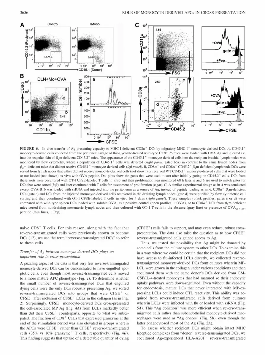
FIGURE 6. In vivo transfer of Ag-presenting capacity to MHC I-deficient CD8�� DCs by migratory MHC I� monocyte-derived DCs. A, CD45.1�
monocyte-derived cells collected from the peritoneal lavage of thioglycolate-treated wild-type C57BL/6 mice were loaded with OVA Ag and injected i.c.into the scapular skin of �2m-deficient CD45.2� mice. The appearance of the CD45.1� monocyte-derived cells into the recipient brachial lymph nodes wasmonitored by flow cytometry, where a population of CD45.1� cells was detected (right panel, gated box) in contrast to the same lymph nodes from�2m-deficient mice that did not receive CD45.1� monocyte-derived cells (left panel). B, CD8�� and CD8�� CD45.2� �2m-deficient lymph node DCs weresorted from lymph nodes that either did not receive monocyte-derived cells (not shown) or received WT CD45.1� monocyte-derived cells that were loadedor not loaded (not shown) ex vivo with OVA peptide. Dot plots show the gates that were used to sort after initially gating on CD45.2� cells. DCs fromthese sorts were cocultured with OT-I CFSE-labeled T cells in vitro and then proliferation was monitored 68 h later. a and b are used to match gates forDCs that were sorted (left) and later cocultured with T cells for assessment of proliferation (right). C, A similar experimental design as in A was conductedexcept OVA-B16 was loaded with ssRNA and injected into the peritoneum as a source of Ag, instead of peptide loading as in A. CD8�� �2m-deficientDCs (gate c) and DCs from the injected monocyte-derived cells recovered in the draining lymph nodes (gate d) were purified by flow cytometric cellsorting and then cocultured with OT-I CFSE-labeled T cells in vitro for 4 days (right panel). These samples (black profiles, gates c or d) werecompared with wild-type spleen DCs loaded with soluble OVA, as a positive control (open profiles, �OVA), or to CD8�� DCs from �2m-deficientmice sorted from nondraining mesenteric lymph nodes and then cultured with OT-1 T cells in the absence (gray line) or presence of OVA257–264
peptide (thin lines, �Pep).
3656 ROLE OF MONOCYTE-DERIVED APCs IN CROSS-PRESENTATION

cells with HLA-A201� mature DCs derived from monocytestreated with GM-CSF and IL-4 (Fig. 5C). Use of anti-HLA-A201-specific mAb revealed that transfer of MHC I/peptide complexindeed occurred (Fig. 5D). Moreover, HLA-A201� DCs that be-came HLA-A201� through coculture with reverse-transmigratedHLA-A201� cells acquired the capacity to stimulate HLA-A2-restricted MP58–66-specific T cells to release IFN-� (Fig. 5E), andthis was evident even with the relatively higher background in thisexperimental design likely due to alloreactivity between HLA-A2� DCs and the T cell line. DCs that were cocultured with re-verse-transmigrated cells from cultures containing LCLs withoutintracellular TLRs were far less effective at inducing CTL reac-tivity (Fig. 5, A, B, and E). Moreover, DCs that were separatedfrom reverse-transmigrated cells by a transwell were unable toacquire robust MP-specific CTLs (Fig. 5A), suggesting a need forclose proximity between the two APC populations in the transferof Ag-presentation capacity. Taken together, we conclude thatappropriately stimulated monocyte-derived DCs, much more thanmonocyte-derived macrophages, can transfer intact MHC I/peptidecomplexes to other DCs in close proximity. Appropriate stimula-tion for monocyte-derived DCs includes uptake of dying cellsbearing the TLR ligand ssRNA.
Finally, we wondered if monocyte-derived DCs might have thecapacity to transfer MHC/peptide to other DCs in vivo. In brief,newly recruited monocytes were obtained from the peritoneal cav-ity of CD45.1� C57BL/6 mice and exposed to OVA/OVA peptidein vitro and then transferred to CD45.2� MHC I-deficient mice(�2m knockout mice) by injection intracutaneously. Alternatively,OVA-expressing B16-F10 cells were loaded with ssRNA in vitroand then injected i.p into mice 17 h after thioglycolate injectionand 5 h before mice were terminated for collection of recruitedmonocyte-derived cells. As described previously (23), after trans-fer of these cells i.c. into recipient mice, some of the transferredmonocyte-derived cells developed into DCs with restricted homingto the draining lymph node, detected as CD45.1� cells in thelymph node of CD45.2� recipients (Fig. 6A). We harvested thedraining and nondraining lymph nodes from the �2m knockoutrecipients 36 h after injection of monocyte-derived cells into theskin, prepared single cell suspensions, and sorted the recipientCD8�� and CD8�� CD45.2� lymph node DCs (Fig. 6, B and C)or CD45.2�CD11c� donor monocyte-derived DCs (Fig. 6C).These sorted cells were cultured in vitro with OVA-restrictedCD8� OT-I T cells, essentially as described (24), to determinewhether recipient lymph node DCs acquired the ability to presentMHC I-restricted Ag to CD8� T cells from MHC I� monocyte-derived DCs. Indeed, CD8�� DCs, but not CD8�� DCs, of re-cipient MHC I-deficient origin presented OVA to OVA-restrictedOT-I T cells, but only when this population was derived from askin-draining lymph node downstream of the injection of OVA-bearing monocyte-derived cells (Fig. 6B). The extent of CD8� Tcell proliferation (a single cell division) was reduced when weused monocyte-derived DCs that took up OVA-expressing tumorcells (Fig. 6C) compared with the OVA peptide-loaded monocyte-derived DCs (Fig. 6B). Monocyte-derived DCs were able to cross-present OVA from the B16 cell line on their own when recoveredfrom the lymph node, and they induced a somewhat stronger pro-liferative response than did the recipient CD8�� �2m-deficientDCs. If monocyte-derived cells were transferred but not OVA-loaded, if OVA-expressing tumor cells were transferred withoutengulfment by monocytes, or if nondraining lymph node were ex-amined, sorted CD8�� or CD8�� DCs did not promote OT-I Tcell proliferation (data not shown). These data suggest that migra-tory monocyte-derived DCs can promote cross-presentation in twoways: by directly cross-priming CD8� lymph node T cells after
acquiring dead cells in tissues or by cross-dressing residentCD8�� DCs.
DiscussionLymph node resident DCs are pivotal for cross-presentation inmany instances (5, 6), and it has been argued that DCs migratingto lymph nodes from the periphery mainly serve to donate Ag tolymph node resident DCs (CD8�� DCs in mice) (7, 8). Newlyrecruited monocytes may in many instances contribute substan-tially to the population of DCs that acquires Ag in the context ofinflammation (3, 4, 26–29) and therefore may serve as a source ofmigratory DCs that deliver Ag to CD8�� DCs. Indeed, monocyteshave recently been implicated as crucial participants in the induc-tion of CTL responses through cross-presentation in vivo (4), butit was proposed that they directly presented Ag to CD8� T cells,rather than transfer Ag to CD8�� DCs, because the monocyte-derived DCs had to express MHC I to promote cross-priming (4).The findings reported herein offer an additional mechanism tothe previous interpretation, as we show that monocyte-derivedDCs also donate intact, functional MHC I/peptide complexes toother DCs, including CD8�� DCs in vivo, thereby using amechanism previously termed “cross-dressing” (30 –32) to sup-port cross-priming.
We draw these conclusions after taking in vitro and in vivoapproaches to delineate in detail the steps involved in how newlyrecruited monocytes, in both humans and mice, contribute to cross-presentation. Our work in vitro utilized a previously developedthree-dimensional model (12) to examine cross-presentation byhuman monocytes. As much as possible, the model is self-drivenby endogenous signals from intercellular interactions to allow theenvironment of the culture system to serve as the primary influenceover the differentiation and behavior of DCs that develop within it.Our culture system incorporated dying cells that expressed influ-enza MP, which has well-defined CTL epitopes, even when noneof the cells were infected with flu. In this model, most of themonocyte-derived cells that engulfed dying cells after traversingthe endothelium remained in the subendothelial matrix as macro-phages (12). A small, but detectable fraction of monocyte-derivedcells took up cellular material acquired from dying subendothelialcells and joined the reverse transmigratory population of DCs.CTL reactivity to Ags expressed by the dying cells was augmentedif they had been naturally infected with flu, fitting with evidence inthe mouse that the presence of TLR ligands within dying cells canpromote DC maturation and cross-presentation (16).
In mouse CD8�� DCs, TLR3 engagement by viral componentswithin dying cells strongly supported cross-priming (16). In ourmodel, human monocyte-derived cells also up-regulated TLR3,but loading dying cells with the TLR3 agonist poly(I:C) was not aseffective at facilitating monocyte maturation to cross-priming DCsthan were flu-infected dying cells. In contrast, dying cells thatwere previously loaded with ssRNA before uptake by mono-cyte-derived cells induced a CTL response to influenza MP thatwas as strong as the CTL response mounted against dying cellsinfected naturally with flu. The failure of TLR3 agonists toaugment the CTL-inducing activity in our model as well aspreviously is likely explained by the difference in sources ofDCs and species studied, as well as by the fact that we focusedon how monocytes responded to TLR signals within dying cellsimmediately after recruitment into a model tissue, rather thanusing already differentiated immature DCs.
The strong response to ssRNA-loaded dying cells is most likelymediated by TLR8, rather than by TLR7. Both TLRs mediate rec-ognition of ssRNA (25), but only TLR8 is expressed by monocyte-derived cells. Use of purified monocytes in our model system was
3657The Journal of Immunology

effective for mediating cross-presentation, indicating that TLR-7�
plasmacytoid DCs were not required. Stimulation of human mono-cytes with TLR8 agonists induces IL-12 and blocks IL-10 produc-tion (33), and stimulation through TLR8 is especially efficacious insupporting the overall Ag-presenting capacity of monocytes fromhuman newborns (34). Furthermore, vaccinations in nonhumanprimate models have identified TLR7/8 agonists as superior adju-vants when the agonists are complexed with the Ag of interest (35,36), in agreement with our findings that a TLR7/8-targeting stim-ulus is a superior agonist for monocyte conversion to cross-pre-senting DCs and that soluble TLR agonists were markedly lesseffective than those present within the dying cells at supporting DCmaturation and CTL reactivity. Our data underscore the possibilitythat the efficacy of these vaccines may relate to how stimulatorythey are for differentiating monocyte-derived cells. Thus, vaccinesthat deliver Ags in the form of cells, such as altered cell lines thatserve as antitumor vaccines (37), may particularly benefit from astep in which the cells used during vaccination are loaded with aTLR8 stimulus like ssRNA, as we did here.
Previous studies demonstrated that DC uptake of cells bearingviral-derived signals promotes cross-presentation, but these studiesdid not exclude the possibility that the presentation itself was beingconducted by DCs in the culture other than the DCs that reallyengulfed the dying cells. In one of these studies, only 20–25% ofthe DCs that cross-presented dying cells had engulfed them (11),whereas 70% of mouse CD8�� DCs had engulfed dying cells inanother study (16); each body of work leaves open the possibilitythat “bystander” DCs in the culture that had not engulfed dyingcells played a key role in presentation. In our system, only aminority of monocyte-derived cells that reverse transmigrated(�20%) could be demonstrated to have engulfed detectable frag-ments of labeled dying cells. When we determined whether thisminority of cells accounted for the bulk of cross-priming, we weresurprised to find that the DCs that had engulfed detectable amountsof dying cells cross-presented notably more poorly than did thosethat did not bear evidence of having acquired cellular materialfrom LCLs. Whereas it is possible that the most phagocytic APCsare inherently the least capable of robust cross-presentation, it isalso possible that the uptake of certain phagocytic material itselfinduces suppressed capacity for cross-presentation.
One way to get around this problem is for such DCs to share ortransfer Ag. Coculture experiments between two populations ofDCs illustrated that presentation capacity, and intact HLA-201 it-self, could be transferred between and from reverse-transmigratedDCs to other, already matured DCs and vice versa. Experiments invivo indicated that monocyte-derived DCs that emigrate to lymphnodes can share intact MHC I/peptide (or at least intact �2m andpeptide) with other DCs, especially the CD8�� lymph node DCs,although in our experimental design their ability to transfer pep-tide/MHC I complexes was lower when the monocyte-derivedcells processed sources of Ag from dying cells as compared withpeptide loading. The common element between our in vitro modeland the in vivo study are the migratory monocyte-derived cellsbearing Ag, and we do not suggest that our in vitro model containshuman equivalents of CD8�� DCs. Instead, in vitro, it appears thatAg sharing takes place between monocyte-derived DCs in closeproximity that had and had not engulfed dying cells, respectively.However, in vivo, the highly organized environment of the lymphnode wherein a number of DC subsets reside may alter the types ofDCs that interact or come into close proximity. Alternatively,through competition, certain interactions may be favored that can-not be modeled in vitro. Furthermore, many DC types may havereceived MHC/peptide by transfer, but CD8�� DCs may be re-
stricted in presenting it, as they also have an advantage in cross-presentation in general.
The overall mode of Ag exchange observed herein has beenpreviously described between DCs or other cell types and is some-times called “cross-dressing” (30–32, 38–41). Much remains to bedetermined mechanistically about the process: phagocytosis ofmembrane has been implicated (39), or alternatively exosomesmay mediate transfer in an �L integrin-dependent manner (41).Our findings differ from the conclusions of Smyth et al., in that we(but not they) found that CD8�� DCs were efficient in presentingAg obtained by cross-dressing (32). Since there may be multiplemechanisms that give rise to cross-dressing, it is possible that therelative efficiency of presentation through cross-dressing may dif-fer depending on context. Here, we argue that cross-dressing oflymph node DCs (such as CD8�� DCs) can serve as a way inwhich populations of DCs that survey inflamed tissues and subse-quently emigrate to lymph nodes (namely monocyte-derived DCs)functionally support cross-priming. Our in vitro data, wherein onlya small fraction of flu-presenting DCs were bearing evidence ofhaving engulfed dying cells that served as a source of Ag, suggestthat one purpose of cross-dressing may be to overcome the rela-tively poorer cross-presentation by monocyte-derived cells that di-rectly engulf dying cells and to spread Ag presentation to a muchlarger number of DCs that would otherwise not directly come intocontact with a given source of Ag. However, in our experiments invivo in which tumor cells expressing OVA were used as a sourceof Ag, we did not observe that the CD8�� DCs presented trans-ferred Ag better than the initial monocyte-derived DCs themselves,if we assume that the monocyte-derived DCs recovered from thelymph node are the same monocyte-derived cells that engulfed theOVA-bearing tumor cells. Thus, the purpose of Ag transfer maynot always be to overcome poor cross-presentation by the initiallyengulfing cells. Instead, the ability of migratory monocyte-derivedDCs to promote cross-presentation in two ways may protect the hostagainst the possibility that a microorganism would evolve a means toshut down the direct pathway of cross-presentation but leave the path-way involving MHC/peptide transfer intact, or vice versa.
In summary, this study allows us to put together a sequence ofevents related to cross-presentation that occurs soon after mono-cytes leave the bloodstream. A few of these steps have been sep-arately proposed before in various mouse or human models, buthere we monitored the process from start to finish to place thesesteps in order and to evaluate their experimental validity and rel-evance to humans in reference to how newly recruited monocytesas a source of DCs during infection and inflammation contribute tocross-priming. As a first step in the sequence, some monocyte-derived cells recently recruited to tissues engulf dying cells in theenvironment. Most of these phagocytic cells differentiate to mac-rophages, but some develop stronger Ag presentation capacity andthe migratory abilities attributed to DCs and leave the tissue. Theup-regulation of Ag presentation capacity and costimulatory mol-ecules by these cells is a function of the contents of the ancillarycells (here, LCLs) that they acquire in the subendothelial tissue. Inparticular, ssRNA, likely acting through TLR8, is a potent cell-associated cue to produce mature DCs from newly recruited mono-cytes that induce CTL responses. For human monocyte-derivedcells in vitro, uptake of dying cells bearing Ag had a negativeimpact on the ability to cross-prime, and the best inducers of CTLswere the DCs that cannot be linked directly to having engulfed thedying cells but instead acquired the Ag, including intact MHC/peptide, from the phagocytic DCs through cross-dressing. Thus,the DC that actually presents the Ag to T cells most efficiently isdistinct from the DC that engulfed the cell that supplied it. Transferof intact MHC-peptide between DCs in vivo can explain why
3658 ROLE OF MONOCYTE-DERIVED APCs IN CROSS-PRESENTATION

monocytes need to express MHC I to support cross-presentation(4) beyond a role for direct cross-presentation themselves.
AcknowledgmentsWe thank Drs. Adolfo Garcia-Sastre, Tom Moran, and Shu-Hsia Chen(Mount Sinai School of Medicine) for providing plasmids, Abs, and tumorcell lines, and we are grateful to Dr. Davor Frleta (Baylor Institute ofImmunology, Dallas, TX) for helpful discussions.
DisclosuresG.J.R. works collaboratively with VaxDesign, the sponsor of this research.She has received stock options from VaxDesign. G.J.R. and Mount SinaiSchool of Medicine have applied for a patent with VaxDesign for tech-nology in which vascular and connective tissue is reconstructed from hu-man cells for the purposes of vaccine testing and selection. If thistechnology were licensed to a commercial entity, then G.J.R. andM.M. would benefit financially.
References1. Bevan, M. J. 2006. Cross-priming. Nat. Immunol. 7: 363–365.2. Heath, W. R., and F. R. Carbone. 2001. Cross-presentation in viral immunity and
self-tolerance. Nat. Rev. Immunol. 1: 126–134.3. Shortman, K., and S. H. Naik. 2007. Steady-state and inflammatory dendritic-cell
development. Nat. Rev. Immunol. 7: 19–30.4. Le Borgne, M., N. Etchart, A. Goubier, S. A. Lira, J. C. Sirard, N. van Rooijen,
C. Caux, S. Ait-Yahia, A. Vicari, D. Kaiserlian, and B. Dubois. 2006. Dendriticcells rapidly recruited into epithelial tissues via CCR6/CCL20 are responsible forCD8� T cell crosspriming in vivo. Immunity 24: 191–201.
5. den Haan, J. M., S. M. Lehar, and M. J. Bevan. 2000. CD8� but not CD8�
dendritic cells cross-prime cytotoxic T cells in vivo. J. Exp. Med. 192:1685–1696.
6. Iyoda, T., S. Shimoyama, K. Liu, Y. Omatsu, Y. Akiyama, Y. Maeda,K. Takahara, R. M. Steinman, and K. Inaba. 2002. The CD8� dendritic cellsubset selectively endocytoses dying cells in culture and in vivo. J. Exp. Med.195: 1289–1302.
7. Allan, R. S., J. Waithman, S. Bedoui, C. M. Jones, J. A. Villadangos, Y. Zhan,A. M. Lew, K. Shortman, W. R. Heath, and F. R. Carbone. 2006. Migratorydendritic cells transfer antigen to a lymph node-resident dendritic cell populationfor efficient CTL priming. Immunity 25: 153–162.
8. Belz, G. T., C. M. Smith, D. Eichner, K. Shortman, G. Karupiah, F. R. Carbone,and W. R. Heath. 2004. Cutting edge: conventional CD8�� dendritic cells aregenerally involved in priming CTL immunity to viruses. J. Immunol. 172:1996–2000.
9. He, Y., J. Zhang, C. Donahue, and L. D. Falo, Jr. 2006. Skin-derived dendriticcells induce potent CD8� T cell immunity in recombinant lentivector-mediatedgenetic immunization. Immunity 24: 643–656.
10. Randolph, G. J. 2006. Migratory dendritic cells: sometimes simply ferries? Im-munity 25: 15–18.
11. Albert, M. L., B. Sauter, and N. Bhardwaj. 1998. Dendritic cells acquire antigenfrom apoptotic cells and induce class I-restricted CTLs. Nature 392: 86–89.
12. Randolph, G. J., S. Beaulieu, S. Lebecque, R. M. Steinman, and W. A. Muller.1998. Differentiation of monocytes into dendritic cells in a model of transendo-thelial trafficking. Science 282: 480–483.
13. Bartido, S. M., and K. Zier. 2004. T-cell responses to multiple antigens presentedby RNA-transfected APCs: a possible immunomonitoring tool. Cancer Immunol.Immunother. 53: 100–109.
14. Krausa, P., M. Brywka, 3rd, D. Savage, K. M. Hui, M. Bunce, J. L. Ngai,D. L. Teo, Y. W. Ong, D. Barouch, C. E. Allsop, et al. 1995. Genetic polymor-phism within HLA-A*02: significant allelic variation revealed in different pop-ulations. Tissue Antigens 45: 223–231.
15. Qu, C., T. M. Moran, and G. J. Randolph. 2003. Autocrine type I IFN and contactwith endothelium promote the presentation of influenza A virus by monocyte-derived APC. J. Immunol. 170: 1010–1018.
16. Schulz, O., S. S. Diebold, M. Chen, T. I. Naslund, M. A. Nolte, L. Alexopoulou,Y. T. Azuma, R. A. Flavell, P. Liljestrom, and C. Reis e Sousa. 2005. Toll-likereceptor 3 promotes cross-priming to virus-infected cells. Nature 433: 887–892.
17. Jonuleit, H., U. Kuhn, G. Muller, K. Steinbrink, L. Paragnik, E. Schmitt, J. Knop,and A. H. Enk. 1997. Pro-inflammatory cytokines and prostaglandins inducematuration of potent immunostimulatory dendritic cells under fetal calf serum-free conditions. Eur. J. Immunol. 27: 3135–3142.
18. Mendoza-Naranjo, A., P. J. Saez, C. C. Johansson, M. Ramirez, D. Mandakovic,C. Pereda, M. N. Lopez, R. Kiessling, J. C. Saez, and F. Salazar-Onfray. 2007.
Functional gap junctions facilitate melanoma antigen transfer and cross-presen-tation between human dendritic cells. J. Immunol. 178: 6949–6957.
19. De Rosa, S. C., and M. Roederer. 2001. Eleven-color flow cytometry: a powerfultool for elucidation of the complex immune system. Clin. Lab. Med. 21:697–712, vii.
20. Hamann, D., P. A. Baars, M. H. Rep, B. Hooibrink, S. R. Kerkhof-Garde,M. R. Klein, and R. A. van Lier. 1997. Phenotypic and functional separation ofmemory and effector human CD8� T cells. J. Exp. Med. 186: 1407–1418.
21. Sauter, B., M. L. Albert, L. Francisco, M. Larsson, S. Somersan, andN. Bhardwaj. 2000. Consequences of cell death: exposure to necrotic tumor cells,but not primary tissue cells or apoptotic cells, induces the maturation of immu-nostimulatory dendritic cells. J. Exp. Med. 191: 423–434.
22. Zuber, B., V. Levitsky, G. Jonsson, S. Paulie, A. Samarina, S. Grundstrom,S. Metkar, H. Norell, G. G. Callender, C. Froelich, and N. Ahlborg. 2005. De-tection of human perforin by ELISpot and ELISA: ex vivo identification of virus-specific cells. J. Immunol. Methods 302: 13–25.
23. Rotta, G., E. W. Edwards, S. Sangaletti, C. Bennett, S. Ronzoni, M. P. Colombo,R. M. Steinman, G. J. Randolph, and M. Rescigno. 2003. Lipopolysaccharide orwhole bacteria block the conversion of inflammatory monocytes into dendriticcells in vivo. J. Exp. Med. 198: 1253–1263.
24. Allan, R. S., C. M. Smith, G. T. Belz, A. L. van Lint, L. M. Wakim, W. R. Heath,and F. R. Carbone. 2003. Epidermal viral immunity induced by CD8�� dendriticcells but not by Langerhans cells. Science 301: 1925–1928.
25. Iwasaki, A., and R. Medzhitov. 2004. Toll-like receptor control of the adaptiveimmune responses. Nat Immunol. 5: 987–995.
26. Randolph, G. J., K. Inaba, D. F. Robbiani, R. M. Steinman, and W. A. Muller.1999. Differentiation of phagocytic monocytes into lymph node dendritic cells invivo. Immunity 11: 753–761.
27. Ginhoux, F., F. Tacke, V. Angeli, M. Bogunovic, M. Loubeau, X. M. Dai,E. R. Stanley, G. J. Randolph, and M. Merad. 2006. Langerhans cells arise frommonocytes in vivo. Nat. Immunol. 7: 265–273.
28. Villadangos, J. A. 2007. Hold on, the monocytes are coming! Immunity 26:390–392.
29. Leon, B., M. Lopez-Bravo, and C. Ardavin. 2007. Monocyte-derived dendriticcells formed at the infection site control the induction of protective T helper 1responses against Leishmania. Immunity 26: 519–531.
30. Dolan, B. P., K. D. Gibbs, Jr., and S. Ostrand-Rosenberg. 2006. Dendritic cellscross-dressed with peptide MHC class I complexes prime CD8� T cells. J. Im-munol. 177: 6018–6024.
31. Dolan, B. P., K. D. Gibbs, Jr., and S. Ostrand-Rosenberg. 2006. Tumor-specificCD4� T cells are activated by “cross-dressed” dendritic cells presenting peptide-MHC class II complexes acquired from cell-based cancer vaccines. J. Immunol.176: 1447–1455.
32. Smyth, L. A., N. Harker, W. Turnbull, H. El-Doueik, L. Klavinskis, D. Kioussis,G. Lombardi, and R. Lechler. 2008. The relative efficiency of acquisition ofMHC:peptide complexes and cross-presentation depends on dendritic cell type.J. Immunol. 181: 3212–3220.
33. Bekeredjian-Ding, I., S. I. Roth, S. Gilles, T. Giese, A. Ablasser, V. Hornung,S. Endres, and G. Hartmann. 2006. T cell-independent, TLR-induced IL-12p70production in primary human monocytes. J. Immunol. 176: 7438–7446.
34. Levy, O., E. E. Suter, R. L. Miller, and M. R. Wessels. 2006. Unique efficacy ofToll-like receptor 8 agonists in activating human neonatal antigen-presentingcells. Blood 108: 1284–1290.
35. Wille-Reece, U., B. J. Flynn, K. Lore, R. A. Koup, R. M. Kedl, J. J. Mattapallil,W. R. Weiss, M. Roederer, and R. A. Seder. 2005. HIV Gag protein conjugatedto a Toll-like receptor 7/8 agonist improves the magnitude and quality of Th1 andCD8� T cell responses in nonhuman primates. Proc. Natl. Acad. Sci. USA 102:15190–15194.
36. Wille-Reece, U., B. J. Flynn, K. Lore, R. A. Koup, A. P. Miles, A. Saul,R. M. Kedl, J. J. Mattapallil, W. R. Weiss, M. Roederer, and R. A. Seder. 2006.Toll-like receptor agonists influence the magnitude and quality of memory T cellresponses after prime-boost immunization in nonhuman primates. J. Exp. Med.203: 1249–1258.
37. Dranoff, G. 2003. GM-CSF-secreting melanoma vaccines. Oncogene 22:3188–3192.
38. Herrera, O. B., D. Golshayan, R. Tibbott, F. Salcido Ochoa, M. J. James,F. M. Marelli-Berg, and R. I. Lechler. 2004. A novel pathway of alloantigenpresentation by dendritic cells. J. Immunol. 173: 4828–4837.
39. Xu, H., K. K. Dhanireddy, and A. D. Kirk. 2006. Human monocytes as inter-mediaries between allogeneic endothelial cells and allospecific T cells: a role fordirect scavenger receptor-mediated endothelial membrane uptake in the initiationof alloimmunity. J. Immunol. 176: 750–761.
40. de Heusch, M., D. Blocket, D. Egrise, B. Hauquier, M. Vermeersch, S. Goldman,and M. Moser. 2007. Bidirectional MHC molecule exchange between migratoryand resident dendritic cells. J. Leukocyte Biol. 82: 861–868.
41. Segura, E., C. Guerin, N. Hogg, S. Amigorena, and C. Thery. 2007. CD8� den-dritic cells use LFA-1 to capture MHC-peptide complexes from exosomes invivo. J. Immunol. 179: 1489–1496.
3659The Journal of Immunology


![3-(Benzo[d][1,3]dioxol-5-ylamino)-N-(4-fluorophenyl)thiophene-2- carboxamide overcomes cancer chemoresistance via inhibition of angiogenesis and P-glycoprotein efflux pump activity](https://img.dokumen.tips/doc/110x75/6325ba2685efe380f306d9d7/3-benzod13dioxol-5-ylamino-n-4-fluorophenylthiophene-2-carboxamide-overcomes.jpg)


