Embed Size (px)
Citation preview
REVIEW Open Access
Messing up disorder: how do missense mutationsin the tumor suppressor protein APC lead tocancer?David P Minde1, Zeinab Anvarian2, Stefan GD Rüdiger1* and Madelon M Maurice2*
Summary: Mutations in the adenomatous polyposis coli (APC) tumor suppressor gene strongly predispose todevelopment of gastro-intestinal tumors. Central to the tumorigenic events in APC mutant cells is the uncontrolledstabilization and transcriptional activation of the protein b-catenin. Many questions remain as to how APC controls b-catenin degradation. Remarkably, the large C-terminal region of APC, which spans over 2000 amino acids and includescritical regions in downregulating b-catenin, is predicted to be natively unfolded. Here we discuss how this uncommonlylarge disordered region may help to coordinate the multiple cellular functions of APC. Recently, a significant number ofgermline and somatic missense mutations in the central region of APC were linked to tumorigenesis in the colon as wellas extra-intestinal tissues. We classify and localize all currently known missense mutations in the APC structure. Themolecular basis by which these mutations interfere with the function of APC remains unresolved. We propose severalmechanisms by which cancer-related missense mutations in the large disordered domain of APC may interfere withtumor suppressor activity. Insight in the underlying molecular events will be invaluable in the development of novelstrategies to counter dysregulated Wnt signaling by APC mutations in cancer.
IntroductionAdenomatous polyposis coli (APC) is a key tumor sup-pressor gene that acts as a gatekeeper of intestinal epithe-lial homeostasis by restraining cytoplasmic cellular levelsof b-catenin, the central activator of transcription in theWnt signaling pathway. At the molecular level, APC co-scaffolds a multiprotein destruction complex, composedof the tumor suppressor Axin and the serine-threoninekinases GSK3b and CK1, which promotes the phosphory-lation and subsequent ubiquitin-mediated degradation ofb-catenin [1]. A Wnt-induced signal at the cell surfaceimpedes the function of the APC-Axin complex, leadingto the stabilization and nuclear import of b-catenin, fol-lowed by the formation of nuclear b-catenin/TCF com-plexes that activate target gene transcription [2,3].Besides regulating proliferation and differentiationthrough Wnt/b-catenin signaling, APC controls multiple
b-catenin-independent fundamental cellular processes.These include cell adhesion and migration, organizationof the cytoskeleton, spindle formation and chromosomesegregation [4,5]. The crucial role of APC in fundamentaldevelopmental cellular processes is illustrated by theembryonic lethality of homozygous Apc-knock-out muta-tions [6-8]. In this review, we focus on how the remark-able lack of structure in the large central domain of APCmay facilitate its tumor suppressor function in the Wnt/b-catenin cascade. Furthermore, by classification andlocalization of known cancer-related APC missensemutations, we uncover different mutational spectra ofgermline and somatic missense mutations along the APCprotein sequence, suggesting variation in functional rele-vance and mechanisms. We discuss how these missensemutations in the large unstructured region of APC maypredispose to cancer.
The large central domain of APC contains multipledomains that control Wnt signalingAPC is a 312 kDa protein composed of 2843 amino acidresidues. It carries multiple designated segments with
* Correspondence: [email protected]; [email protected] Protein Chemistry, Bijvoet Center for Biomolecular Research, UtrechtUniversity, Padualaan 8, 3584CH Utrecht, The Netherlands2Dept. of Cell Biology, University Medical Center Utrecht (UMCU), RmG02.525, Heidelberglaan 100, 3584CX Utrecht, The NetherlandsFull list of author information is available at the end of the article
Minde et al. Molecular Cancer 2011, 10:101http://www.molecular-cancer.com/content/10/1/101
© 2011 Minde et al; licensee BioMed Central Ltd. This is an Open Access article distributed under the terms of the Creative CommonsAttribution License (http://creativecommons.org/licenses/by/2.0), which permits unrestricted use, distribution, and reproduction inany medium, provided the original work is properly cited.
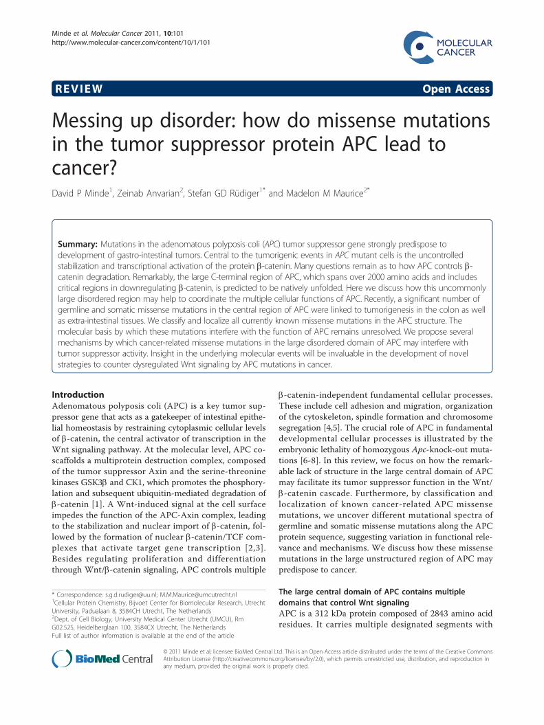
which it coordinates its multiple cellular functions (Fig-ure 1A). The large central region of APC, spanning resi-dues L1021-D2059, has been implicated in thedownregulation of b-catenin [9,10]. It contains four 15aarepeat and seven 20aa repeat segments involved in b-catenin binding [11-13]. The 15aa repeat region alsobinds the transcriptional co-repressor CtBP1 and CtBP2,which prevents nuclear b-catenin activity and facilitatesAPC oligomerization and [14-16]. Interspersed with the20aa b-catenin binding repeats three short recognition
motifs, composed of the highly conserved LxECIxSAMPsequence (called SAMP motif), constitute binding sitesfor Axin [17-19]. The remarkably large number of b-catenin binding sites in the APC protein has instigatedan area of intense research to search for the mechanisticrole of the APC b-catenin binding repeats in thedestruction complex.Each 15aa repeat of APC binds to the structural
groove formed by the armadillo repeats 5-10 on the sur-face of b-catenin in a phosphorylation-independent
Figure 1 The human APC protein carries a large predicted disordered domain which is frequently hit by missense mutations incancer. (A) Schematic representation of the APC scaffold protein and its protein interaction domains. Known interactors are APC (green), CRM1(orange), PP2A (brown), b-catenin, CtBP (15aa repeats, blue), b-catenin (20 aa repeats, cyan), sequence B (yellow), Axin (SAMP-repeats, purple),Microtubules (dark green) and EB1 (light green). (B) Summary of disorder predictions performed for full-length human APC using differentalgorithms from publicly available servers [31-35]. Sequence segments with disorder probability above 50% are represented as black bars. (C)Distribution of missense mutations in the APC protein reported in various tumors, categorized as somatic (red), germline (black) or unknownorigin (grey). Details on the location and nature of amino acid substitutions can be found in additional file 1, Table S1. (D) Summary of disorderpredictions performed for b-catenin (black bars), done as in (B), and b-catenins’s helical secondary structure elements as determined bycrystallography (gray bars) [36].
Minde et al. Molecular Cancer 2011, 10:101http://www.molecular-cancer.com/content/10/1/101
Page 2 of 9
manner [13]. The 20aa repeats require phosphorylationof a consensus SXXSSLSXLS motif to convert into tightbinding sites for b-catenin [20,21]. In addition, twonegatively charged residues in the N-terminal flankingregions of the core 20aa repeats make significant contactwith b-catenin by forming 2 salt bridges with K435 andK312 of b-catenin [20,21]. Once bound, one single phos-phorylated 20aa repeat plus N-terminal flankingsequence of APC occupies almost the entire groove thatspans from armadillo repeats 1 to 12 on the b-cateninsurface. The bound conformation of one extended 20aarepeat is nearly identical to that of other functionalbinding partners of b-catenin, TCF [22] and E-cadherin[23], indicating that these proteins cannot bind b-cate-nin simultaneously.Unlike their equal organization in binding motifs, indi-
vidual 15aa and 20aa repeats in APC vary considerablyin their binding affinities for b-catenin, with the tightestbinding site being the phosphorylated third 20aa repeat(Kd 1.5 nM) [24]. Remarkably, the highly conserved sec-ond 20aa repeat completely lacks binding affinity for b-catenin, even in the phosphorylated state, likely due tothe absence of some of the conserved residues in the N-terminal region flanking the core 20aa sequence. Phos-phorylated APC competes with Axin for binding to b-catenin, whereas unphosphorylated does not [21,25].Based on these findings, various models have been pro-posed on the mechanism by which phosphorylation ofthe 20aa repeats in APC may regulate b-catenin recruit-ment and turnover. In a first model, both the 15aarepeats of APC and the b-catenin binding domain inAxin bind b-catenin side by side to induce b-cateninphosphorylation by GSK3b. As soon as the third 20aarepeat of APC is phosphorylated it will bind phospho-b-catenin thereby releasing Axin from the complex[21,24]. This would facilitate the discharge and degrada-tion of phospho-b-catenin and allow a new phosphoryla-tion cycle to occur. In a second model, b-catenin firstbinds phosphorylated APC with high affinity. Subse-quent dephosphorylation of APC is then required toweaken the interaction between APC and b-catenin,allowing transfer of b-catenin to Axin and phosphoryla-tion by Axin-associated GSK3b [1]. This model isopposed by recent findings that demonstrate a crucialrole of APC in protecting phosho-b-catenin fromdephosphorylation by PP2A [26]. As a consequence,APC would stay tethered to phospho-b-catenin anddirectly deliver it to the E3 ligase b-TrCP for ubiquitina-tion. In a third model, phosphorylation of APC accom-modates the fluctuation in b-catenin levels in the cell inconditions of presence versus absence of a Wnt signal.During active Wnt signaling, abundant levels of b-cate-nin will be dealt with by rapid and transient interactionsbetween b-catenin and nonphosphorylated APC. In the
absence of a Wnt signal, low levels of b-catenin will betightly bound and slowly released by phosphorylatedAPC [21,27].Each of the above models were challenged by a recent
study in which the roles of the 20aa and 15aa repeatswere addressed systematically through the functionalanalysis of a large number of APC variants in humancells and flies [28]. Importantly, separate roles of APCin the cytoplasmic retention and destruction of b-cate-nin were uncovered, involving selective APC regions.The affinities of individual b-catenin binding repeats inAPC were uncovered to be of lesser importance in thedestruction of b-catenin than anticipated in previousmodels. Instead, the b-catenin binding repeats act inconcert to mediate retention of the b-catenin protein inthe cytoplasm, thus preventing its activity in thenucleus. Strikingly, the second 20aa repeat which lacksaffinity for b-catenin, as well as the conserved sequenceB/CID, located in between the second and third 20aarepeat (Figure 1A) [28,29], perform critical roles in theAPC-mediated destruction of b-catenin. How theseregions control APC activity and whether this involvesbinding of co-factors remains to be solved.Further experiments are needed to demonstrate if the
repeat regions act simultaneously or sequentially anddetermine how these events are regulated by APC phos-phorylation as well as by the second 20aa repeat andsequence B in the process of b-catenin destruction.
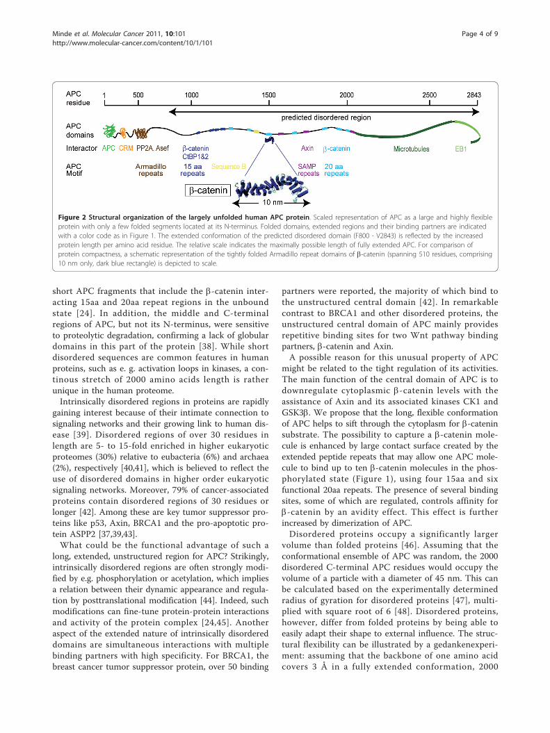
The central domain of APC is intrinsically disorderedStrikingly, APC lacks sequence conservation outside thesmall repetitive b-catenin- and axin-binding regions andsequence B. This led us to investigate the structuralproperties of APC in more detail using nine establishedalgorithms to predict secondary structure and disorder.Each of the algorithms consistently indicate the presenceof an exceptionally large intrinsically disordered regionfrom F800 to V2843 in APC (Figure 1B) [30-35], yield-ing a stretch of 2000 residues with an extended, flexibleconformation (Figure 2). The reliability of those algo-rithms is well established as illustrated using b-cateninand Axin as example proteins, for which we can com-pare bioinformatics results with the experimentally veri-fied structure (Figure 1D) [36,37]. The disorderprediction algorithms for b-catenin confirm unfolded N-and C-terminal segments flanking the folded, helicalcore of the protein. Indeed, for these regions crystallo-graphic studies failed to detect regular structure [36].For the Wnt pathway tumor suppressor Axin, weexperimentally confirmed the intrinsically disorderednature of the functionally important central region, thusconfirming the predictions derived of various algorithms[37]. The prediction data for APC are supported by Far-UV CD spectra and NMR studies on various purified
Minde et al. Molecular Cancer 2011, 10:101http://www.molecular-cancer.com/content/10/1/101
Page 3 of 9
short APC fragments that include the b-catenin inter-acting 15aa and 20aa repeat regions in the unboundstate [24]. In addition, the middle and C-terminalregions of APC, but not its N-terminus, were sensitiveto proteolytic degradation, confirming a lack of globulardomains in this part of the protein [38]. While shortdisordered sequences are common features in humanproteins, such as e. g. activation loops in kinases, a con-tinous stretch of 2000 amino acids length is ratherunique in the human proteome.Intrinsically disordered regions in proteins are rapidly
gaining interest because of their intimate connection tosignaling networks and their growing link to human dis-ease [39]. Disordered regions of over 30 residues inlength are 5- to 15-fold enriched in higher eukaryoticproteomes (30%) relative to eubacteria (6%) and archaea(2%), respectively [40,41], which is believed to reflect theuse of disordered domains in higher order eukaryoticsignaling networks. Moreover, 79% of cancer-associatedproteins contain disordered regions of 30 residues orlonger [42]. Among these are key tumor suppressor pro-teins like p53, Axin, BRCA1 and the pro-apoptotic pro-tein ASPP2 [37,39,43].What could be the functional advantage of such a
long, extended, unstructured region for APC? Strikingly,intrinsically disordered regions are often strongly modi-fied by e.g. phosphorylation or acetylation, which impliesa relation between their dynamic appearance and regula-tion by posttranslational modification [44]. Indeed, suchmodifications can fine-tune protein-protein interactionsand activity of the protein complex [24,45]. Anotheraspect of the extended nature of intrinsically disordereddomains are simultaneous interactions with multiplebinding partners with high specificity. For BRCA1, thebreast cancer tumor suppressor protein, over 50 binding
partners were reported, the majority of which bind tothe unstructured central domain [42]. In remarkablecontrast to BRCA1 and other disordered proteins, theunstructured central domain of APC mainly providesrepetitive binding sites for two Wnt pathway bindingpartners, b-catenin and Axin.A possible reason for this unusual property of APC
might be related to the tight regulation of its activities.The main function of the central domain of APC is todownregulate cytoplasmic b-catenin levels with theassistance of Axin and its associated kinases CK1 andGSK3b. We propose that the long, flexible conformationof APC helps to sift through the cytoplasm for b-cateninsubstrate. The possibility to capture a b-catenin mole-cule is enhanced by large contact surface created by theextended peptide repeats that may allow one APC mole-cule to bind up to ten b-catenin molecules in the phos-phorylated state (Figure 1), using four 15aa and sixfunctional 20aa repeats. The presence of several bindingsites, some of which are regulated, controls affinity forb-catenin by an avidity effect. This effect is furtherincreased by dimerization of APC.Disordered proteins occupy a significantly larger
volume than folded proteins [46]. Assuming that theconformational ensemble of APC was random, the 2000disordered C-terminal APC residues would occupy thevolume of a particle with a diameter of 45 nm. This canbe calculated based on the experimentally determinedradius of gyration for disordered proteins [47], multi-plied with square root of 6 [48]. Disordered proteins,however, differ from folded proteins by being able toeasily adapt their shape to external influence. The struc-tural flexibility can be illustrated by a gedankenexperi-ment: assuming that the backbone of one amino acidcovers 3 Å in a fully extended conformation, 2000
Figure 2 Structural organization of the largely unfolded human APC protein. Scaled representation of APC as a large and highly flexibleprotein with only a few folded segments located at its N-terminus. Folded domains, extended regions and their binding partners are indicatedwith a color code as in Figure 1. The extended conformation of the predicted disordered domain (F800 - V2843) is reflected by the increasedprotein length per amino acid residue. The relative scale indicates the maximally possible length of fully extended APC. For comparison ofprotein compactness, a schematic representation of the tightly folded Armadillo repeat domains of b-catenin (spanning 510 residues, comprising10 nm only, dark blue rectangle) is depicted to scale.
Minde et al. Molecular Cancer 2011, 10:101http://www.molecular-cancer.com/content/10/1/101
Page 4 of 9
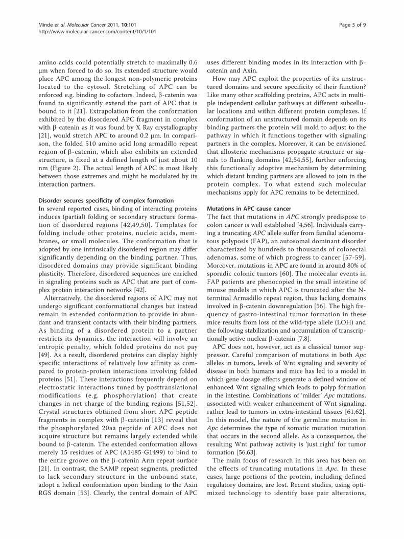
amino acids could potentially stretch to maximally 0.6μm when forced to do so. Its extended structure wouldplace APC among the longest non-polymeric proteinslocated to the cytosol. Stretching of APC can beenforced e.g. binding to cofactors. Indeed, b-catenin wasfound to significantly extend the part of APC that isbound to it [21]. Extrapolation from the conformationexhibited by the disordered APC fragment in complexwith b-catenin as it was found by X-Ray crystallography[21], would stretch APC to around 0.2 μm. In compari-son, the folded 510 amino acid long armadillo repeatregion of b-catenin, which also exhibits an extendedstructure, is fixed at a defined length of just about 10nm (Figure 2). The actual length of APC is most likelybetween those extremes and might be modulated by itsinteraction partners.
Disorder secures specificity of complex formationIn several reported cases, binding of interacting proteinsinduces (partial) folding or secondary structure forma-tion of disordered regions [42,49,50]. Templates forfolding include other proteins, nucleic acids, mem-branes, or small molecules. The conformation that isadopted by one intrinsically disordered region may differsignificantly depending on the binding partner. Thus,disordered domains may provide significant bindingplasticity. Therefore, disordered sequences are enrichedin signaling proteins such as APC that are part of com-plex protein interaction networks [42].Alternatively, the disordered regions of APC may not
undergo significant conformational changes but insteadremain in extended conformation to provide in abun-dant and transient contacts with their binding partners.As binding of a disordered protein to a partnerrestricts its dynamics, the interaction will involve anentropic penalty, which folded proteins do not pay[49]. As a result, disordered proteins can display highlyspecific interactions of relatively low affinity as com-pared to protein-protein interactions involving foldedproteins [51]. These interactions frequently depend onelectrostatic interactions tuned by posttranslationalmodifications (e.g. phosphorylation) that createchanges in net charge of the binding regions [51,52].Crystal structures obtained from short APC peptidefragments in complex with b-catenin [13] reveal thatthe phosphorylated 20aa peptide of APC does notacquire structure but remains largely extended whilebound to b-catenin. The extended conformation allowsmerely 15 residues of APC (A1485-G1499) to bind tothe entire groove on the b-catenin Arm repeat surface[21]. In contrast, the SAMP repeat segments, predictedto lack secondary structure in the unbound state,adopt a helical conformation upon binding to the AxinRGS domain [53]. Clearly, the central domain of APC
uses different binding modes in its interaction with b-catenin and Axin.How may APC exploit the properties of its unstruc-
tured domains and secure specificity of their function?Like many other scaffolding proteins, APC acts in multi-ple independent cellular pathways at different subcellu-lar locations and within different protein complexes. Ifconformation of an unstructured domain depends on itsbinding partners the protein will mold to adjust to thepathway in which it functions together with signalingpartners in the complex. Moreover, it can be envisionedthat allosteric mechanisms propagate structure or sig-nals to flanking domains [42,54,55], further enforcingthis functionally adoptive mechanism by determiningwhich distant binding partners are allowed to join in theprotein complex. To what extend such molecularmechanisms apply for APC remains to be determined.
Mutations in APC cause cancerThe fact that mutations in APC strongly predispose tocolon cancer is well established [4,56]. Individuals carry-ing a truncating APC allele suffer from familial adenoma-tous polyposis (FAP), an autosomal dominant disordercharacterized by hundreds to thousands of colorectaladenomas, some of which progress to cancer [57-59].Moreover, mutations in APC are found in around 80% ofsporadic colonic tumors [60]. The molecular events inFAP patients are phenocopied in the small intestine ofmouse models in which APC is truncated after the N-terminal Armadillo repeat region, thus lacking domainsinvolved in b-catenin downregulation [56]. The high fre-quency of gastro-intestinal tumor formation in thesemice results from loss of the wild-type allele (LOH) andthe following stabilization and accumulation of transcrip-tionally active nuclear b-catenin [7,8].APC does not, however, act as a classical tumor sup-
pressor. Careful comparison of mutations in both Apcalleles in tumors, levels of Wnt signaling and severity ofdisease in both humans and mice has led to a model inwhich gene dosage effects generate a defined window ofenhanced Wnt signaling which leads to polyp formationin the intestine. Combinations of ‘milder’ Apc mutations,associated with weaker enhancement of Wnt signaling,rather lead to tumors in extra-intestinal tissues [61,62].In this model, the nature of the germline mutation inApc determines the type of somatic mutation mutationthat occurs in the second allele. As a consequence, theresulting Wnt pathway activity is ‘just right’ for tumorformation [56,63].The main focus of research in this area has been on
the effects of truncating mutations in Apc. In thesecases, large portions of the protein, including definedregulatory domains, are lost. Recent studies, using opti-mized technology to identify base pair alterations,
Minde et al. Molecular Cancer 2011, 10:101http://www.molecular-cancer.com/content/10/1/101
Page 5 of 9
indicate that in a significant number of cases, however,germline as well as sporadic single amino acid substitu-tions (missense mutations) in Apc predispose to devel-opment of colorectal adenomas [64]. Notably, asignificant number of APC missense mutations werereported in tumors originating from various tissues(listed in additional file 1, Table S1, including referencestherein). Moreover, missense mutations in APC werelinked to worse disease outcome in invasive urothelialcarcinomas [65], suggesting functional relevance ofpoint mutated APC protein in the development ofextra-intestinal tumors. Most of these mutations remainfunctionally uncharacterized although for some missensemutant APC proteins Wnt signaling activating proper-ties were demonstrated [66]. The molecular basis bywhich these mutations interfere with the function ofAPC remains unresolved.
Molecular consequences of missense mutations in thedisordered domain of APCLong, unstructured regions are likely more apt to resistthe effects of point mutations than a folded protein. In
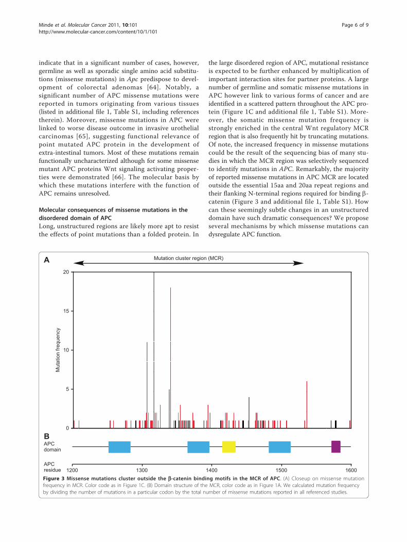
the large disordered region of APC, mutational resistanceis expected to be further enhanced by multiplication ofimportant interaction sites for partner proteins. A largenumber of germline and somatic missense mutations inAPC however link to various forms of cancer and areidentified in a scattered pattern throughout the APC pro-tein (Figure 1C and additional file 1, Table S1). More-over, the somatic missense mutation frequency isstrongly enriched in the central Wnt regulatory MCRregion that is also frequently hit by truncating mutations.Of note, the increased frequency in missense mutationscould be the result of the sequencing bias of many stu-dies in which the MCR region was selectively sequencedto identify mutations in APC. Remarkably, the majorityof reported missense mutations in APC MCR are locatedoutside the essential 15aa and 20aa repeat regions andtheir flanking N-terminal regions required for binding b-catenin (Figure 3 and additional file 1, Table S1). Howcan these seemingly subtle changes in an unstructureddomain have such dramatic consequences? We proposeseveral mechanisms by which missense mutations candysregulate APC function.
A
B
Mutationfrequency
APCresidue 1200 1300 1400 1500 1600
APCdomain
0
10
15
5
20
Mutation cluster region (MCR)
Figure 3 Missense mutations cluster outside the b-catenin binding motifs in the MCR of APC. (A) Closeup on missense mutationfrequency in MCR. Color code as in Figure 1C. (B) Domain structure of the MCR, color code as in Figure 1A. We calculated mutation frequencyby dividing the number of mutations in a particular codon by the total number of missense mutations reported in all referenced studies.
Minde et al. Molecular Cancer 2011, 10:101http://www.molecular-cancer.com/content/10/1/101
Page 6 of 9
(i) Altered protein interaction surface. Many of thereported missense mutations change side chain chargeswhich might have direct implications for the formationof protein-protein interaction interfaces (additional file1, Table S1) [66]. The single known binding partner ofthe MCR region is b-catenin but, surprisingly, most ofthe cancer-related mutations are outside the b-cateninbinding repeats. Possibly, the mutated residues belongto non-redundant, so far unknown protein-protein inter-action sites, although the lower conservation of theseregions would render this option less likely.(ii) Changes in secondary structure formation. Mis-
sense mutations may alter the ability of APC to adoptstructure when bound to its interaction partners in thedestruction complex. This would be of importance forlocal helix formation of APC SAMP-repeat regionsupon binding Axin. Indeed, missense mutations thatlocate to these regions and introduce residues thatreduce helix stability (Gly, Val) were reported as germ-line mutations in cases of adenomatous polyposis coli(Figure 3 and additional file 1, Table S1) [66-68].(iii) Posttranslational modifications. Point mutations
may interfere with posttranslational modifications ofAPC. For instance, mutations in APC may alter recogni-tion sites for responsible kinases such as CK1 andGSK3b. These kinases do not directly dock onto APCbut rather are positioned towards their substrate resi-dues by binding to Axin in the complex [69]. Mutationswithin close proximity of target Ser residues in the 20aarepeats may interfere with phosphorylation of thesemotifs. Alternatively, mutations may hamper the pro-tecting function of APC towards PP2A-mediated depho-sphorylation of phospho-b-catenin and/or the deliveryof b-catenin to b-TrCP [26]. Interference with this roleof APC may lead to rapid dephosphorylation and stabili-zation of b-catenin. It is currently unknown whatregions of APC are involved in these consecutive stepsin b-catenin degradation and whether or not thisrequires extended or folded APC configuration. Thisinformation will be essential to determine whether mis-sense mutations may interfere with this function ofAPC.(iv) Dynamics of conformational equilibrium. Natively
unfolded sequences may adopt a specific three dimen-sional conformation upon binding of a partner protein[70]. This may include a specific spatial arrangement ofthe repeat regions in APC. Point mutations outsidethose regions could prevent the required formation of aspecific three-dimensional structure and, thereby, inhibitAPC’s usual mode of action. Hypothetically, mutationsmay also influence long-range intramolecular signaling.Examples of allosteric regulation of protein signaling arerapidly emerging [71]. Binding to or modification of oneend of an protein elicits a signal that is communicated
through the protein to trigger a response at a remotesite, although it would be less obvious how such signaltransmission may work in an unfolded segment.If and how these mechanisms may play a role in the
tumor suppressor activity of APC remains to be deter-mined. It is obvious that the classical paradigmsobtained for folded proteins fail to explain the pheno-type of APC cancer mutations. We consider it likelythat the effect is related to the dynamic nature of thedisordered regions of APC. In that respect the recentdiscovery of the conserved sequence B as a critical func-tional APC unit is of interest [28,29]. Obviously,sequence B activity may involve binding partners thatremain to be discovered. Alternatively, alterations insequence B may simply disturb the dynamic interplay ofthe b-catenin binding repeats or modulate the competi-tion with Axin for b-catenin binding. The importance ofdisordered regions is a newly emerging field, and itsunusually large disordered stretch make APC a keyparadigm to understand the role of unfolded regions ingeneral.
Conclusions and perspectivesCurrent mechanistic models of APC tumor suppressorfunction leave many questions as to how APC coordi-nates b-catenin degradation. Through its variousdomains, APC is able to interact with many differentproteins. Multiple repeat regions for interaction withboth b-catenin and Axin are implicated in its tumorsuppressor activity. Structural information about howthese proteins are positioned within the b-catenindestruction complex is lacking. Remarkably, the largecentral domain of APC, spanning over 2000 amino acidsand carrying the repeat regions involved in b-catenindownregulation, is predicted to be entirely unstructured.This feature is rather unique in the human genome asonly a few other proteins in the human proteome carrysimilarly sized unfolded domains. Combined structuraland functional analysis of the unstructured domains ofAPC will be needed to reveal if structure is acquiredupon binding to partner proteins. Answers as to howAPC missense mutations contribute to tumorigenesisremains to be uncovered by studying how selectedtumor-associated mutations interfere with essentialtumor suppression mechanisms of APC.
Additional material
Additional file 1: Table S1. Germline and somatic missensemutations in APC reported in human cancer. List of APC missensemutations reported in human tumors. Mutated amino acid residue,affected codon, tumor type, germline or somatic nature of the mutationsand corresponding references are indicated. Nucleotide numberingreflects cDNA numbering with +1 corresponding to the A of the ATGtranslation initiation codon in the reference sequence (Genbank
Minde et al. Molecular Cancer 2011, 10:101http://www.molecular-cancer.com/content/10/1/101
Page 7 of 9
NM_000038.4). The translation initiation codon is codon 1 (GenbankNP_000029). ND = Not described. *In these studies, healthy tissue wasused as control.
AcknowledgementsWe thank Maria Noutsou and Daniele Tauriello for critical reading of themanuscript and Tobias Madl and Andrei Petoukhov for discussions. S.G.D.Rwas supported by a High Potential grant by Utrecht University, a Marie-CurieExcellence Grant by the EU and a VIDI career development grant by theNetherlands Organization for Scientific Research - Chemical Sciences, NWO-CW. M.M.M. was supported by a Dutch Cancer Society grant (UU 2006-3508),a High Potential grant by Utrecht University and a European ResearchCouncil (ERC) Starting grant.
Author details1Cellular Protein Chemistry, Bijvoet Center for Biomolecular Research, UtrechtUniversity, Padualaan 8, 3584CH Utrecht, The Netherlands. 2Dept. of CellBiology, University Medical Center Utrecht (UMCU), Rm G02.525,Heidelberglaan 100, 3584CX Utrecht, The Netherlands.
Authors’ contributionsMM drafted and wrote the manuscript. SR contributed to writing of themanuscript and the generation of essential concepts. Both MM and SRsupervised the project. MM, SR and DM generated the figures andcontributed to the development of the concepts. DM and MM generatedTable S1. ZA and DM collected and summarized essential literature andrevised the manuscript critically for important and intellectual content. Allauthors read and approved the final manuscript.
Competing interestsThe authors declare that they have no competing interests.
Received: 17 March 2011 Accepted: 22 August 2011Published: 22 August 2011
References1. McCartney BM, Nathke IS: Cell regulation by the Apc protein Apc as
master regulator of epithelia. Curr Opin Cell Biol 2008, 20:186-193.2. Logan CY, Nusse R: The Wnt signaling pathway in development and
disease. Annu Rev Cell Dev Biol 2004, 20:781-810.3. Clevers H: Wnt/beta-catenin signaling in development and disease. Cell
2006, 127:469-480.4. Aoki K, Taketo MM: Adenomatous polyposis coli (APC): a multi-functional
tumor suppressor gene. J Cell Sci 2007, 120:3327-3335.5. Okada K, Bartolini F, Deaconescu AM, Moseley JB, Dogic Z, Grigorieff N,
Gundersen GG, Goode BL: Adenomatous polyposis coli protein nucleatesactin assembly and synergizes with the formin mDia1. J Cell Biol 2010,189:1087-1096.
6. Ishikawa TO, Tamai Y, Li Q, Oshima M, Taketo MM: Requirement for tumorsuppressor Apc in the morphogenesis of anterior and ventral mouseembryo. Dev Biol 2003, 253:230-246.
7. Moser AR, Shoemaker AR, Connelly CS, Clipson L, Gould KA, Luongo C,Dove WF, Siggers PH, Gardner RL: Homozygosity for the Min allele of Apcresults in disruption of mouse development prior to gastrulation. DevDyn 1995, 203:422-433.
8. Oshima M, Oshima H, Kitagawa K, Kobayashi M, Itakura C, Taketo M: Loss ofApc heterozygosity and abnormal tissue building in nascent intestinalpolyps in mice carrying a truncated Apc gene. Proc Natl Acad Sci USA1995, 92:4482-4486.
9. Rubinfeld B, Albert I, Porfiri E, Munemitsu S, Polakis P: Loss of beta-cateninregulation by the APC tumor suppressor protein correlates with loss ofstructure due to common somatic mutations of the gene. Cancer Res1997, 57:4624-4630.
10. Munemitsu S, Albert I, Souza B, Rubinfeld B, Polakis P: Regulation ofintracellular beta-catenin levels by the adenomatous polyposis coli(APC) tumor-suppressor protein. Proc Natl Acad Sci USA 1995,92:3046-3050.
11. Rubinfeld B, Souza B, Albert I, Muller O, Chamberlain SH, Masiarz FR,Munemitsu S, Polakis P: Association of the APC gene product with beta-catenin. Science 1993, 262:1731-1734.
12. Su LK, Vogelstein B, Kinzler KW: Association of the APC tumor suppressorprotein with catenins. Science 1993, 262:1734-1737.
13. Eklof Spink K, Fridman SG, Weis WI: Molecular mechanisms of beta-catenin recognition by adenomatous polyposis coli revealed by thestructure of an APC-beta-catenin complex. EMBO J 2001, 20:6203-6212.
14. Hamada F, Bienz M: The APC tumor suppressor binds to C-terminalbinding protein to divert nuclear beta-catenin from TCF. Dev Cell 2004,7:677-685.
15. Sierra J, Yoshida T, Joazeiro CA, Jones KA: The APC tumor suppressorcounteracts beta-catenin activation and H3K4 methylation at Wnt targetgenes. Genes Dev 2006, 20:586-600.
16. Schneikert J, Brauburger K, Behrens J: APC mutations in colorectaltumours from FAP patients are selected for CtBP-mediatedoligomerization of truncated APC. Hum Mol Genet 2011.
17. Behrens J, Jerchow BA, Wurtele M, Grimm J, Asbrand C, Wirtz R, Kuhl M,Wedlich D, Birchmeier W: Functional interaction of an axin homolog,conductin, with beta-catenin, APC, and GSK3 beta. Science 1998,280:596-599.
18. Kishida S, Yamamoto H, Ikeda S, Kishida M, Sakamoto I, Koyama S,Kikuchi A: Axin, a negative regulator of the wnt signaling pathway,directly interacts with adenomatous polyposis coli and regulates thestabilization of beta-catenin. J Biol Chem 1998, 273:10823-10826.
19. Nakamura T, Hamada F, Ishidate T, Anai K, Kawahara K, Toyoshima K,Akiyama T: Axin, an inhibitor of the Wnt signalling pathway, interactswith beta-catenin, GSK-3 beta and APC and reduces the beta-cateninlevel. Genes Cells 1998, 3:395-403.
20. Xing Y, Clements WK, Le Trong I, Hinds TR, Stenkamp R, Kimelman D, Xu W:Crystal structure of a beta-catenin/APC complex reveals a critical role forAPC phosphorylation in APC function. Mol Cell 2004, 15:523-533.
21. Ha NC, Tonozuka T, Stamos JL, Choi HJ, Weis WI: Mechanism ofphosphorylation-dependent binding of APC to beta-catenin and its rolein beta-catenin degradation. Mol Cell 2004, 15:511-521.
22. Graham TA, Weaver C, Mao F, Kimelman D, Xu W: Crystal structure of abeta-catenin/Tcf complex. Cell 2000, 103:885-896.
23. Huber AH, Weis WI: The structure of the beta-catenin/E-cadherincomplex and the molecular basis of diverse ligand recognition by beta-catenin. Cell 2001, 105:391-402.
24. Liu J, Xing Y, Hinds TR, Zheng J, Xu W: The Third 20 Amino Acid Repeat Isthe Tightest Binding Site of APC for β-Catenin. Journal of MolecularBiology 2006, 360:133-144.
25. Xing Y, Clements WK, Kimelman D, Xu W: Crystal structure of a β-catenin/Axin complex suggests a mechanism for the β-catenin destructioncomplex. Genes & Development 2003, 17:2753-2764.
26. Su Y, Fu C, Ishikawa S, Stella A, Kojima M, Shitoh K, Schreiber EM, Day BW,Liu B: APC is essential for targeting phosphorylated beta-catenin to theSCFbeta-TrCP ubiquitin ligase. Mol Cell 2008, 32:652-661.
27. Seo E, Jho E-h: Axin-independent phosphorylation of APC controls[beta]-catenin signaling via cytoplasmic retention of [beta]-catenin.Biochemical and Biophysical Research Communications 2007, 357:81-86.
28. Roberts DM, Pronobis MI, Poulton JS, Waldmann JD, Stephenson EM,Hanna S, Peifer M: Deconstructing the sscatenin destruction complex:mechanistic roles for the tumor suppressor APC in regulating Wntsignaling. Mol Biol Cell 2011, 22:1845-1863.
29. Kohler EM, Chandra SH, Behrens J, Schneikert J: Beta-catenin degradationmediated by the CID domain of APC provides a model for the selectionof APC mutations in colorectal, desmoid and duodenal tumours. HumMol Genet 2009, 18:213-226.
30. Rost B, Yachdav G, Liu J: The PredictProtein server. Nucleic Acids Res 2004,32:W321-326.
31. Obradovic Z, Peng K, Vucetic S, Radivojac P, Dunker AK: Exploitingheterogeneous sequence properties improves prediction of proteindisorder. Proteins 2005, 61(Suppl 7):176-182.
32. Coeytaux K, Poupon A: Prediction of unfolded segments in a proteinsequence based on amino acid composition. Bioinformatics 2005,21:1891-1900.
33. Linding R, Russell RB, Neduva V, Gibson TJ: GlobPlot: Exploring proteinsequences for globularity and disorder. Nucleic Acids Res 2003,31:3701-3708.
Minde et al. Molecular Cancer 2011, 10:101http://www.molecular-cancer.com/content/10/1/101
Page 8 of 9
34. Prilusky J, Felder CE, Zeev-Ben-Mordehai T, Rydberg EH, Man O,Beckmann JS, Silman I, Sussman JL: FoldIndex: a simple tool to predictwhether a given protein sequence is intrinsically unfolded. Bioinformatics2005, 21:3435-3438.
35. Yang ZR, Thomson R, McNeil P, Esnouf RM: RONN: the bio-basis functionneural network technique applied to the detection of nativelydisordered regions in proteins. Bioinformatics 2005, 21:3369-3376.
36. Xing Y, Takemaru K, Liu J, Berndt JD, Zheng JJ, Moon RT, Xu W: Crystalstructure of a full-length beta-catenin. Structure 2008, 16:478-487.
37. Noutsou M, Duarte AM, Anvarian Z, Didenko T, Minde DP, Kuper I, deRidder I, Oikonomou C, Friedler A, Boelens R, Rüdiger SG, Maurice MM:Critical Scaffolding Regions of the Tumor Suppressor Axin1 Are NativelyUnfolded. J Mol Biol 2010.
38. Li Z, Nathke IS: Tumor-associated NH2-terminal fragments are the moststable part of the adenomatous polyposis coli protein and can beregulated by interactions with COOH-terminal domains. Cancer Res 2005,65:5195-5204.
39. Uversky VN, Oldfield CJ, Dunker AK: Intrinsically disordered proteins inhuman diseases: introducing the D2 concept. Annu Rev Biophys 2008,37:215-246.
40. Ward JJ, Sodhi JS, McGuffin LJ, Buxton BF, Jones DT: Prediction andFunctional Analysis of Native Disorder in Proteins from the ThreeKingdoms of Life. Journal of Molecular Biology 2004, 337:635-645.
41. Oldfield CJ, Cheng Y, Cortese MS, Brown CJ, Uversky VN, Dunker AK:Comparing and combining predictors of mostly disordered proteins.Biochemistry 2005, 44:1989-2000.
42. Cortese MS, Uversky VN, Keith Dunker A: Intrinsic disorder in scaffoldproteins: Getting more from less. Progress in Biophysics and MolecularBiology 2008, 98:85-106.
43. Rotem S, Katz C, Benyamini H, Lebendiker M, Veprintsev D, Rudiger S,Danieli T, Friedler A: The structure and interactions of the proline-richdomain of ASPP2. J Biol Chem 2008, 283:18990-18999.
44. Dunker AK, Oldfield CJ, Meng J, Romero P, Yang JY, Chen JW, Vacic V,Obradovic Z, Uversky VN: The unfoldomics decade: an update onintrinsically disordered proteins. BMC Genomics 2008, 9(Suppl 2):S1.
45. Dahlberg CL, Nguyen EZ, Goodlett D, Kimelman D: Interactions betweenCasein kinase Iepsilon (CKIepsilon) and two substrates from disparatesignaling pathways reveal mechanisms for substrate-kinase specificity.PLoS ONE 2009, 4:e4766.
46. Uversky VN, Fink AL: The chicken-egg scenario of protein foldingrevisited. FEBS Lett 2002, 515:79-83.
47. Kohn JE, Millett IS, Jacob J, Zagrovic B, Dillon TM, Cingel N, Dothager RS,Seifert S, Thiyagarajan P, Sosnick TR, Hasan MZ, Pande VS, Ruczinski I,Doniach S, Plaxco KW: Random-coil behavior and the dimensions ofchemically unfolded proteins. Proc Natl Acad Sci USA 2004,101:12491-12496.
48. Cantor CR, Schimmel PR, (Eds.): The Behavior of BiologicalMacromolecules. 1980.
49. Dyson HJ, Wright PE: Intrinsically unstructured proteins and theirfunctions. Nat Rev Mol Cell Biol 2005, 6:197-208.
50. Marsh JA, Dancheck B, Ragusa MJ, Allaire M, Forman-Kay JD, Peti W:Structural diversity in free and bound states of intrinsically disorderedprotein phosphatase 1 regulators. Structure 2010, 18:1094-1103.
51. Mittag T, Kay LE, Forman-Kay JD: Protein dynamics and conformationaldisorder in molecular recognition. J Mol Recognit 2010, 23:105-116.
52. Serber Z, Ferrell JE Jr: Tuning bulk electrostatics to regulate proteinfunction. Cell 2007, 128:441-444.
53. Spink KE, Polakis P, Weis WI: Structural basis of the Axin-adenomatouspolyposis coli interaction. EMBO Journal 2000, 19:2270-2279.
54. Keramisanou D, Biris N, Gelis I, Sianidis G, Karamanou S, Economou A,Kalodimos CG: Disorder-order folding transitions underlie catalysis in thehelicase motor of SecA. Nat Struct Mol Biol 2006, 13:594-602.
55. Bruschweiler S, Schanda P, Kloiber K, Brutscher B, Kontaxis G, Konrat R,Tollinger M: Direct Observation of the Dynamic Process UnderlyingAllosteric Signal Transmission. J Am Chem Soc 2009, 131:3063-3068.
56. Segditsas S, Tomlinson I: Colorectal cancer and genetic alterations in theWnt pathway. Oncogene 2006, 25:7531-7537.
57. Groden J: Identification and characterization of the familial adenomatouspolyposis coli gene. Cell 1991, 66:589-600.
58. Kinzler KW, Nilbert MC, Su LK, Vogelstein B, Bryan TM, Levy DB, Smith KJ,Preisinger AC, Hedge P, McKechnie D, Finniear R, Markham A, Grotten J,
Boguski MS, Altschul SF, Horii A, Ando H, Miyoshi Y, Miki Y, Nishisho I,Nakamura Y: Identification of FAP locus genes from chromosome 5q21.1991, 253:661-665.
59. Fearnhead NS, Winney B, Bodmer WF: Rare Variant Hypothesis forMultifactorial Inheritance: Susceptibility to Colorectal Adenomas as aModel. Cell Cycle 2005, 4:521-525.
60. Kinzler KW, Vogelstein B: Lessons from hereditary colorectal cancer. Cell1996, 87:159-170.
61. Lamlum H, Ilyas M, Rowan A, Clark S, Johnson V, Bell J, Frayling I,Efstathiou J, Pack K, Payne S, Roylance R, Gorman P, Sheer D, Neale K,Phillips R, Talbot I, Bodmer W, Tomlinson I: The type of somatic mutationat APC in familial adenomatous polyposis is determined by the site ofthe germline mutation: a new facet to Knudson’s ‘two-hit’ hypothesis.Nat Med 1999, 5:1071-1075.
62. Fodde R, Smits R: Disease model: familial adenomatous polyposis. Trendsin Molecular Medicine 2001, 7:369-373.
63. Gaspar C, Fodde R: APC dosage effects in tumorigenesis and stem celldifferentiation. Int J Dev Biol 2004, 48:377-386.
64. Bougatef K, Ouerhani S, Moussa A, Kourda N, Coulet F, Colas C, Lahely YB,Najjar T, Ben Jilani S, Benammar-Elgaaied A, Soubrier F, Marrakchi R:Prevalence of mutations in APC, CTNNB1, and BRAF in Tunisian patientswith sporadic colorectal cancer. Cancer Genet Cytogenet 2008, 187:12-18.
65. Kastritis E, Murray S, Kyriakou F, Horti M, Tamvakis N, Kavantzas N,Patsouris ES, Noni A, Legaki S, Dimopoulos MA, Bamias A: Somaticmutations of adenomatous polyposis coli gene and nuclear b-cateninaccumulation have prognostic significance in invasive urothelialcarcinomas: evidence for Wnt pathway implication. Int J Cancer 2009,124:103-108.
66. Azzopardi D, Dallosso AR, Eliason K, Hendrickson BC, Jones N, Rawstorne E,Colley J, Moskvina V, Frye C, Sampson JR, Wenstrup R, Scholl T, Cheadle JP:Multiple Rare Nonsynonymous Variants in the Adenomatous PolyposisColi Gene Predispose to Colorectal Adenomas. 2008, 68:358-363.
67. Nimura Y, Furuwatari C, Fujimori M, Fujimori Y, Nakata S, Ito K, Hama Y,Shingu K, Adachi W, Ogiso Y, Furihata K, Katsuyama T, Amano J: Germlinemutations of the APC gene in two Japanese adenomatous polyposispatients. Jpn J Hum Genet 1997, 42:433-439.
68. Munoz V, Serrano L: Helix design, prediction and stability. Curr OpinBiotechnol 1995, 6:382-386.
69. Ferrarese A, Marin O, Bustos VH, Venerando A, Antonelli M, Allende JE,Pinna LA: Chemical dissection of the APC Repeat 3 multistepphosphorylation by the concerted action of protein kinases CK1 andGSK3. Biochemistry 2007, 46:11902-11910.
70. Sugase K, Dyson HJ, Wright PE: Mechanism of coupled folding andbinding of an intrinsically disordered protein. Nature 2007, 447:1021-1025.
71. Tsai CJ, Del Sol A, Nussinov R: Protein allostery, signal transmission anddynamics: a classification scheme of allosteric mechanisms. Mol Biosyst2009, 5:207-216.
doi:10.1186/1476-4598-10-101Cite this article as: Minde et al.: Messing up disorder: how do missensemutations in the tumor suppressor protein APC lead to cancer?Molecular Cancer 2011 10:101.
Submit your next manuscript to BioMed Centraland take full advantage of:
• Convenient online submission
• Thorough peer review
• No space constraints or color figure charges
• Immediate publication on acceptance
• Inclusion in PubMed, CAS, Scopus and Google Scholar
• Research which is freely available for redistribution
Submit your manuscript at www.biomedcentral.com/submit
Minde et al. Molecular Cancer 2011, 10:101http://www.molecular-cancer.com/content/10/1/101
Page 9 of 9


