Embed Size (px)
Citation preview
Domain-Specific Appendix:
Mechanical Ventilation
Acute Respiratory Failure Domain
REMAP-CAP: Randomized, Embedded, Multifactorial Adaptive Platform trial for
Community-Acquired Pneumonia
Mechanical Ventilation Strategy Domain-Specific Appendix Version 1.0 dated 07 July 2020
Not for
IRB su
bmiss
ion
REMAP-CAP Mechanical Ventilation Domain-Specific Appendix Version 1.0 dated 07 July 2020
CONFIDENTIAL Page 2 of 64
Summary
In this domain of the REMAP-CAP trial, participants meeting the platform entry criteria who are
intubated and are receiving invasive mechanical ventilation will be randomized to receive one of two
mechanical ventilation strategies:
• Protocolized mechanical ventilation strategy
• Clinician-preferred mechanical ventilation strategy
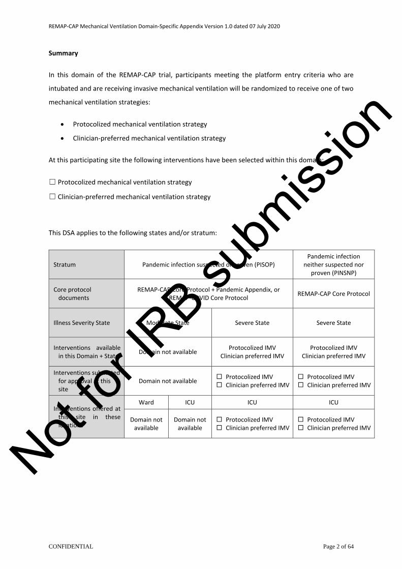
At this participating site the following interventions have been selected within this domain:
☐ Protocolized mechanical ventilation strategy
☐ Clinician-preferred mechanical ventilation strategy
This DSA applies to the following states and/or stratum:
Stratum Pandemic infection suspected or proven (PISOP) Pandemic infection
neither suspected nor proven (PINSNP)
Core protocol documents
REMAP-CAP Core Protocol + Pandemic Appendix, or REMAP-COVID Core Protocol
REMAP-CAP Core Protocol
Illness Severity State Moderate State Severe State Severe State
Interventions available in this Domain + State
Domain not available Protocolized IMV
Clinician preferred IMV Protocolized IMV
Clinician preferred IMV
Interventions submitted for approval at this site
Domain not available Protocolized IMV Clinician preferred IMV
Protocolized IMV Clinician preferred IMV
Interventions offered at this site in these locations
Ward ICU ICU ICU
Domain not available
Domain not available
Protocolized IMV Clinician preferred IMV
Protocolized IMV Clinician preferred IMV
Not for
IRB su
bmiss
ion

REMAP-CAP Mechanical Ventilation Domain-Specific Appendix Version 1.0 dated 07 July 2020
CONFIDENTIAL Page 3 of 64
REMAP-CAP: Mechanical Ventilation Domain Strategy Summary Interventions • Protocolized mechanical ventilation strategy
• Clinician-preferred mechanical ventilation strategy
Unit-of-analysis, Strata, and States
This domain is analyzed in a ventilation statistical model which is separate to both the pandemic and the interpandemic statistical model. Within the ventilation statistical model there are two units-of-analysis corresponding to the Pandemic Infection Suspected or Proven (PISOP) stratum and the Pandemic Infection Neither Suspected nor Proven (PNSNP) stratum. An additional ordinal strata of PaO2:FiO2 ratio at time of randomization will be applied in analysis. Borrowing is permitted between states and stratum. Response adaptive randomization will not be applied during this preliminary phase.
Evaluable Treatment-by-Treatment Interactions
No interactions will be evaluated with any other domain.
Nesting None
Timing of Reveal Randomization with Immediate Reveal and Initiation
Inclusions
Inclusion criteria are those specified in the relevant core protocol documents. Domain-specific inclusion criteria are:
• Receiving invasive mechanical ventilation
• The most recent PaO2:FiO2 ratio obtained within the preceding 6 hours is
less than 200 mmHg
• Treating clinician expects the patient to still require invasive mechanical
ventilation tomorrow
• Treating clinician regards Protocolized Mechanical Ventilation Strategy or
their preferred mode as (equally) appropriate for this patient.
Domain-Specific Exclusions
Patients will be excluded from this domain if they have any of the following:
• More than 48 hours has elapsed since commencement of invasive
mechanical ventilation.
• The treating clinician believes that participation in this domain is not in the
best interests of the patient.
Intervention-Specific Exclusions
Nil, not applicable.
Outcome Measures Primary REMAP endpoint: refer to REMAP-CAP Core Protocol +/- PAtC and REMAP-COVID Core Protocol Secondary REMAP endpoints refer to REMAP-CAP Core Protocol +/- PAtC and REMAP-COVID Core Protocol. Secondary domain-specific endpoints (during hospitalization, censored 90 days from the date of enrollment):
• Serious adverse events (SAE) as defined in core protocol documents
• Administration of rescue therapies
• Occurrence of barotrauma
Not for
IRB su
bmiss
ion
REMAP-CAP Mechanical Ventilation Domain-Specific Appendix Version 1.0 dated 07 July 2020
CONFIDENTIAL Page 4 of 64
CONTENTS TABLE OF TABLES ........................................................................................................................... 7
ABBREVIATIONS ................................................................................................................. 8
PROTOCOL APPENDIX STRUCTURE .................................................................................... 10
MECHANICAL VENTILATION DOMAIN-SPECIFIC APPENDIX VERSION .................................. 11
Version history ...................................................................................................................... 11
MECHANICAL VENTILATION DOMAIN-GOVERNANCE ......................................................... 11
Domain members .................................................................................................................. 11
Contact Details ...................................................................................................................... 12
MECHANICAL VENTILATION DOMAIN-SPECIFIC WORKING GROUP AUTHORIZATION ......... 12
BACKGROUND AND RATIONALE ........................................................................................ 13
Domain definition ................................................................................................................. 13
Introduction .......................................................................................................................... 13
Current recommendations and variability among recommendations ................................. 15
6.3.1. Current guideline recommendations for mechanical ventilation in patients with
CAP 15
6.3.2. Compliance with guidelines and heterogeneity of ventilatory practice .................. 16
6.3.3. Compliance with guidelines and outcome ............................................................... 17
6.3.4. Within unit vs between unit variation ...................................................................... 18
An analytic approach to determining optimal ventilation .................................................... 18
6.4.1. Scope of the problem ............................................................................................... 18
6.4.2. Protocolized guideline-recommended versus wild-type as first questions ............. 21
6.4.3. Isolation of testable strategies within heterogeneity of current practice ............... 22
6.4.4. Analytic approaches to heterogeneity of current practice ...................................... 22
6.4.5. Principles of analysis ................................................................................................. 24
Summary of background ....................................................................................................... 24
DOMAIN OBJECTIVES ........................................................................................................ 24
TRIAL DESIGN .................................................................................................................... 26
Trial design overview ............................................................................................................ 26
Population ............................................................................................................................. 26
Eligibility criteria .................................................................................................................... 27
8.3.1. Domain inclusion criteria .......................................................................................... 27
8.3.2. Domain exclusion criteria ......................................................................................... 27
Interventions ......................................................................................................................... 27
Not for
IRB su
bmiss
ion

REMAP-CAP Mechanical Ventilation Domain-Specific Appendix Version 1.0 dated 07 July 2020
CONFIDENTIAL Page 5 of 64
8.4.1. Mechanical Ventilation Interventions ...................................................................... 27
8.4.2. Physiological targets set prior to reveal of allocation status and common to both
interventions 27
8.4.3. Clinician-preferred mechanical ventilation strategy ................................................ 28
8.4.4. Protocolized mechanical ventilation strategy .......................................................... 28
8.4.5. Duration of administration of domain intervention. ................................................ 30
Concomitant care .................................................................................................................. 31
Endpoints .............................................................................................................................. 31
8.6.1. Primary endpoint ...................................................................................................... 31
8.6.2. Secondary endpoints ................................................................................................ 31
8.6.3. Process end-points ................................................................................................... 31
TRIAL CONDUCT ................................................................................................................ 31
Domain-specific data collection ............................................................................................ 31
9.1.1. Clinical data collection .............................................................................................. 31
Criteria for discontinuation ................................................................................................... 32
Blinding ................................................................................................................................. 32
9.3.1. Blinding ..................................................................................................................... 32
9.3.2. Unblinding................................................................................................................. 32
STATISTICAL CONSIDERATIONS.......................................................................................... 32
Domain-specific stopping rules ............................................................................................. 32
Categorization of clinician-preferred ventilator strategies ................................................... 33
10.2.1. Unit-level self-categorization .................................................................................... 33
10.2.2. Unit-level observed categorization ........................................................................... 33
10.2.3. Patient-level categorization ...................................................................................... 33
10.2.4. Methods of determination of categorization ........................................................... 33
Statistical analysis ................................................................................................................. 33
10.3.1. Principles of statistical analysis................................................................................. 33
10.3.2. Strata ........................................................................................................................ 34
10.3.3. Primary analysis ........................................................................................................ 34
10.3.4. Descriptive analysis of ventilatory patterns ............................................................. 35
10.3.5. Secondary analyses ................................................................................................... 35
10.3.6. Identification of variables that may modify treatment effect .................................. 36
10.3.7. Evaluation of separation between protocolized and clinician-preferred ventilatory
strategies 37
Not for
IRB su
bmiss
ion
REMAP-CAP Mechanical Ventilation Domain-Specific Appendix Version 1.0 dated 07 July 2020
CONFIDENTIAL Page 6 of 64
10.3.8. Sample size and statistical power ............................................................................. 37
Timing of revealing of randomization status ........................................................................ 37
Interactions with interventions in other domains ................................................................ 38
Nesting of interventions ....................................................................................................... 38
ETHICAL CONSIDERATIONS ................................................................................................ 38
Data Safety and Monitoring Board ....................................................................................... 38
Potential domain-specific adverse events ............................................................................ 38
Domain-specific consent issues ............................................................................................ 38
GOVERNANCE ISSUES ........................................................................................................ 39
Funding of domain ................................................................................................................ 39
Funding of domain interventions and outcome measures ................................................... 39
Domain-specific declarations of interest .............................................................................. 39
REFERENCES ...................................................................................................................... 40
APPENDIX 1. ................................................................................................................................. 47
Lung pathology in CAP ....................................................................................................... 47
Physiological principles and options available in setting ventilatory strategy ..................... 48
Introduction .......................................................................................................................... 48
Lung physiology during invasive mechanical ventilation ...................................................... 49
Pressure or volume during inspiration .................................................................................. 49
Characteristics of inspiration and ventilator induced lung injury ......................................... 50
Expiration and PEEP .............................................................................................................. 50
Mandatory and triggered breaths ........................................................................................ 51
Airway Pressure Release Ventilation (APRV) ........................................................................ 51
Recruitment maneuvers ....................................................................................................... 52
Prone positioning .................................................................................................................. 52
Extracorporeal respiratory support ...................................................................................... 52
Spontaneous ventilation and weaning of ventilation ........................................................... 53
Interaction between ventilatory strategy and use of sedative agents and paralysis ........... 53
Hypoxemic Rescue therapies ................................................................................................ 54
APPENDIX 2: Evidence for strategies for different components of ventilatory support .................. 55
Introduction ...................................................................................................................... 55
Characteristics of mandatory inspiratory breaths ................................................................ 55
1.1.1. Are tidal volumes below 6 ml /Kg beneficial? .......................................................... 56
Not for
IRB su
bmiss
ion
REMAP-CAP Mechanical Ventilation Domain-Specific Appendix Version 1.0 dated 07 July 2020
CONFIDENTIAL Page 7 of 64
1.1.2. Positive End Expiratory Pressure (PEEP) ................................................................... 57
1.1.3. Interaction between PEEP and size of inspiration .................................................... 59
1.1.4. Open Lung Strategies incorporating Recruitment maneuvers and elevated PEEP .. 59
1.1.5. Prone positioning ...................................................................................................... 60
1.1.6. Neuro Muscular Blocking Drugs (Paralysis) .............................................................. 60
1.1.7. APRV ......................................................................................................................... 61
1.1.8. ECMO ........................................................................................................................ 62
1.1.9. ECCO2R ...................................................................................................................... 62
1.1.10. Hypoxemic Rescue therapies .................................................................................... 63
1.1.11. Spontaneous ventilation and weaning from ventilation .......................................... 63
1.1.12. Challenges associated with ventilation trials ........................................................... 63
TABLE OF TABLES
Table 1. Sources of variation in practice and potential patient factors that influence ventilator
settings. ................................................................................................................................................. 20
Not for
IRB su
bmiss
ion
REMAP-CAP Mechanical Ventilation Domain-Specific Appendix Version 1.0 dated 07 July 2020
CONFIDENTIAL Page 8 of 64
ABBREVIATIONS
AC Assist Control
APRV Airway Pressure Release Ventilation
ARDS Acute Respiratory Distress Syndrome
ARDSNet Acute Respiratory Distress Syndrome Clinical Trial Network
ATS American Thoracic Society
CAP Community Acquired Pneumonia
COPD Chronic Obstructive Pulmonary Disease
DSA Domain-Specific Appendix
DSWG Domain-Specific Working Group
DSMB Data Safety and Monitoring Board
ECCO2R Extra-Corporeal Carbon Dioxide Removal
ECMO Extra-Corporeal Membrane Oxygenation
ESICM European Society of Intensive Care Medicine
EPVent2 Effect of titrating Positive end-expiratory pressure (PEEP) with an
esophageal pressure-guided strategy vs. an empirical high PEEP-FiO2
strategy on death and days free from mechanical ventilation among patients
with acute respiratory distress syndrome.
FiO2 Fraction of Inspired Oxygen
ICU Intensive Care Unit
ISIG International Statistics Interest Group
ITSC International Trial Steering Committee
NMB Neuromuscular Blockade
LUNG-SAFE Large observational study to UNderstand the Global impact of Severe Acute
respiratory FailurE
MODS Multiple Organ Dysfunction Syndrome
PaO2:FiO2 Ratio Ratio of Partial Pressure of Oxygen in Arterial Blood and Fraction of Inspired
Oxygen Concentration
PBW Predicted Body Weight
PEEP Positive End-Expiratory Pressure
RAR Response Adaptive Randomization
RCT Randomized Controlled Trial
REMAP Randomized, Embedded, Multifactorial Adaptive Platform trial
Not for
IRB su
bmiss
ion
REMAP-CAP Mechanical Ventilation Domain-Specific Appendix Version 1.0 dated 07 July 2020
CONFIDENTIAL Page 9 of 64
REMAP-CAP Randomized, Embedded, Multifactorial, Adaptive Platform trial for
Community-Acquired Pneumonia
RSA Region-Specific Appendix
SAE Serious Adverse Event
SCCM Society of Critical Care Medicine
SIMV Synchronised Intermittent Mandatory Ventilation
Tv Tidal Volume
VFD Ventilator Free Days
VILI Ventilator-Induced Lung Injury
Not for
IRB su
bmiss
ion
REMAP-CAP Mechanical Ventilation Domain-Specific Appendix Version 1.0 dated 07 July 2020
CONFIDENTIAL Page 10 of 64
PROTOCOL APPENDIX STRUCTURE
The structure of this protocol is different to that used for conventional trials because this trial is highly
adaptive and the description of these adaptations is better understood and specified using a ‘modular’
protocol design. While, all adaptations are pre-specified, the structure of the protocol is designed to
allow the trial to evolve over time, for example by the introduction of new domains or interventions
or both (see glossary, Section 1.2 Core Protocol for definitions of these terms) and commencement of
the trial in new geographical regions.
The protocol has multiple modules, in brief, comprising a Core Protocol (overview and design features
of the study), a Statistical Analysis Appendix (details of the current statistical analysis plan and models)
and Simulations Appendix (details of the current simulations of the REMAP), multiple Domain-Specific
Appendices (DSA) (detailing all interventions currently being studied in each domain), and multiple
Regions-Specific Appendices (RSA) (detailing regional management and governance).
The Core Protocol contains all information that is generic to the trial, irrespective of the regional
location in which the trial is conducted and the domains or interventions that are being tested. The
Core Protocol may be amended but it is anticipated that such amendments will be infrequent.
The Core Protocol does not contain information about the intervention(s), within each domain,
because one of the trial adaptations is that domains and interventions will change over time.
Information about interventions, within each domain, is covered in a DSA. These Appendices are
anticipated to change over time, with removal and addition of options within an existing domain, at
one level, and removal and addition of entire domains, at another level. Each modification to a DSA
will be subject of a separate ethics application for approval.
The Core Protocol does not contain detailed information about the statistical analysis or simulations,
because the analysis model will change overtime in accordance with the domain and intervention trial
adaptations but this information is contained in the Statistical Analysis and Simulations Appendices.
These Appendices are anticipated to change over time, as trial adaptations occur. Each modification
will be subject to approval from the International Trial Steering Committee (ITSC) in conjunction with
advice from the International Statistics Interest Group (ISIG) and the Data Safety and Monitoring Board
(DSMB).
The Core Protocol also does not contain information that is specific to a particular region in which the
trial is conducted, as the locations that participate in the trial are also anticipated to increase over
time. Information that is specific to each region that conducts the trial is contained within a RSA. This
Not for
IRB su
bmiss
ion
REMAP-CAP Mechanical Ventilation Domain-Specific Appendix Version 1.0 dated 07 July 2020
CONFIDENTIAL Page 11 of 64
includes information related to local management, governance, and ethical and regulatory aspects. It
is planned that, within each region, only that region’s RSA, and any subsequent modifications, will be
submitted for ethical review in that region.
The current version of the relevant Core Protocol (either REMAP-CAP Core Protocol +/- Pandemic
Appendix or REMAP-COVID Core Protocol), DSAs, RSAs, and the Statistical Analysis Appendix is listed
in the Protocol Summary and on the study website (www.remapcap.org).
MECHANICAL VENTILATION DOMAIN-SPECIFIC APPENDIX VERSION
The version of the Mechanical Ventilation Strategy Domain-Specific Appendix is in this document’s
header and on the cover page.
Version history
Version 1.0: Approved by the Mechanical Ventilation Domain-Specific Working Group (DSWG) on
7th July 2020
MECHANICAL VENTILATION DOMAIN-GOVERNANCE
Domain members
Chair:
Prof. Alistair Nichol
Members:
Prof. Derek Angus
Ms. Wilma van Bentum-Puijk
Dr. Lewis Campbell
Dr. Lennie Derde
Prof. Niall Ferguson
A/Prof. Timothy Girard
A/Prof. Ewan Goligher
Prof. Anthony Gordon
Mr. Cameron Green
Prof. Carol Hodgson
Not for
IRB su
bmiss
ion
REMAP-CAP Mechanical Ventilation Domain-Specific Appendix Version 1.0 dated 07 July 2020
CONFIDENTIAL Page 12 of 64
Prof. Peter Kruger
Prof. John Laffey
Dr. Edward Litton
Prof. John Marshall
Dr. Colin McArthur
Prof. Danny Mc Auley
Prof. Shay McGuiness
Prof. John Laffey
Dr. Neil Orford
Prof. Kathy Rowan
Dr. Ary Neto
Prof. Steve Webb
Contact Details
Chair: Professor Alistair Nichol
Australian and New Zealand Intensive Care Research Centre
School of Public Health, Monash University
553 St Kilda Road, Melbourne, 3004
Phone +61 03 99030513
Fax +61 03 99030513
Email [email protected]
MECHANICAL VENTILATION DOMAIN-SPECIFIC WORKING GROUP
AUTHORIZATION
The Mechanical Ventilation Domain-Specific Working Group (DSWG) have read the appendix and
authorize it as the official Mechanical Ventilation Domain-Specific Appendix for the study entitled
REMAP-CAP. Signed on behalf of the committee, Not for
IRB su
bmiss
ion
REMAP-CAP Mechanical Ventilation Domain-Specific Appendix Version 1.0 dated 07 July 2020
CONFIDENTIAL Page 13 of 64
Chair
Date
07 July 2020
Alistair Nichol
BACKGROUND AND RATIONALE
Domain definition
This is a domain within the REMAP-CAP platform to evaluate the effectiveness of alternative
mechanical ventilation strategies in patients with severe Community-Acquired Pneumonia (CAP),
including patients with suspected or proven COVID-19 infection, who are intubated and receiving
mechanical ventilation.
This version of the ventilation DSA describes a preliminary phase of the domain. The duration of the
preliminary phase is pre-specified. Information available during the preliminary phase will be used to
design subsequent phases of the domain (which will be managed as a subsequent protocol
amendment). This version of the ventilation DSA applies only to the preliminary phase but, so as to
provide context, it does include identification of the goals, but not necessarily the methods that will
be used, for subsequent phases.
Introduction
Severe CAP commonly results in respiratory failure, the need for intubation and the development of
acute respiratory distress syndrome (ARDS) (Bellani et al., 2016). Pneumonia secondary to COVID-19
is associated with a high incidence of acute respiratory failure requiring intubation and mechanical
ventilation (Richardson et al., 2020). Acute respiratory failure secondary to pneumonia manifests by
impairment of transfer of oxygen from the atmosphere into the arterial blood of the patient or
impairment of clearance of carbon dioxide from venous blood into the atmosphere or both (Bellani et
al., 2016). In some patients the application of exogenous oxygen therapy is sufficient to maintain
physiological homeostasis. However, in patients in whom the severity of respiratory failure is a threat
to life, typical management is to institute invasive mechanical ventilation. This involves partial or
complete substitution of the patient’s own efforts to move gas in and out of the lungs by the
application of invasive mechanical ventilation delivered via an endotracheal tube that connects the
patient’s trachea with a mechanical ventilator. The morbidity and mortality of CAP and COVID-19
pneumonia patients who require mechanical ventilation is high (Richardson et al., 2020, Bhatraju et
al., 2020).
Not for
IRB su
bmiss
ion
REMAP-CAP Mechanical Ventilation Domain-Specific Appendix Version 1.0 dated 07 July 2020
CONFIDENTIAL Page 14 of 64
The interventions specified in this domain relate to the settings that are chosen by the treating
clinician on the ventilator. It is universally accepted that the institution of invasive mechanical
ventilation in a patient who would otherwise die from respiratory failure or ventilatory arrest is life-
saving. However, there is little empiric evidence available to guide clinicians as to the optimal settings
of the ventilator and whether the settings should be the same for all patients or differ for patients
with definable characteristics. Furthermore, the lung physiology associated with pneumonia
secondary to COVID-19 appears to have unique features and it is unclear if the optimal ventilatory
strategy is different than for other forms of pneumonia.
We have outlined in detail the pathological changes in the lungs that occur secondary to CAP and the
physiological components that are specified by the treating clinician when providing invasive
mechanical ventilation (Appendix 1). Appendix 1, describes the significant heterogenicity in the
severity and pattern of injury in CAP (irrespective of the causative organism) and that these patients
are a high risk of VILI. In addition, we have summarized in detail the empiric evidence for the
relationship between different options for invasive mechanical ventilation and patient outcomes. It is
clear from Appendix 2, that there is significant heterogenicity in the severity and pattern of injury in
severe CAP, including CAP caused by COVID-19, and that these patients are a high risk of ventilator-
induced lung injury (VILI) and death. However, while many ventilation strategies currently exist, it is
unlikely that one overarching strategy, will be appropriate for all mechanically ventilated patients with
CAP. An immediate issue for the management of patients is that a great deal of the evidence derived
from RCTs regarding mechanical ventilation in CAP, is derived solely from patients who progress to
develop ARDS. Some, but not all patients in RCTs that enroll patients with ARDS have CAP and there
is limited evidence from RCTs in patients with CAP who do not have ARDS. Current guidelines for CAP
extrapolate from patients with ARDS to patients without ARDS. Guidelines for mechanical ventilation
in patients with pneumonia secondary to COVID-19 have been released, but these are based solely on
extrapolation from trials that enrolled patients with other causes of respiratory failure (Poston et al.,
2020, Alhazzani et al., 2020a, Alhazzani et al., 2020b). There is also widespread non-adherence to
current mechanical ventilation guidelines (Bellani et al., 2016). What is unclear is if this lack of
adherence is due to a considered appraisal of the low level of evidence for many current
recommendations, the patient severity illness or heterogenicity of disease. Not for
IRB su
bmiss
ion
REMAP-CAP Mechanical Ventilation Domain-Specific Appendix Version 1.0 dated 07 July 2020
CONFIDENTIAL Page 15 of 64
Current recommendations and variability among recommendations
6.3.1. Current guideline recommendations for mechanical ventilation in patients with CAP
The American Thoracic Society (ATS), European Society of Intensive Care Medicine (ESICM), and
Society of Critical Care Medicine (SCCM) have published consensus guidelines on mechanical
ventilation in patients with ARDS that are directly relevant to those patients with CAP who have ARDS
(Fan et al., 2017). The International Surviving Sepsis Campaign has also published consensus guidelines
on mechanical ventilation in patients with sepsis (Rhodes et al., 2017), a proportion of whom will have
CAP, including those who have developed ARDS. Both sets of guidelines make similar
recommendations with regard to patients requiring mechanical ventilation.
The guidelines for ARDS (Fan et al., 2017) recommend patients receive mechanical ventilation with
strategies that limit tidal volumes (4 to 8 ml/kg predicted body weight, PBW) and inspiratory pressures
(plateau pressure below 30 cm H2O). The initial tidal volume should be set at 6 ml/kg PBW and can be
increased up to 8 ml/kg PBW if the patient is double triggering or if inspiratory pressure decreases
below the set level of positive end-expiratory pressure (PEEP). This is a strong recommendation with
there being moderate confidence in the quality of evidence (Fan et al., 2017). These guidelines identify
substantial uncertainty regarding the question of how long this strategy should be used, before there
is transition to spontaneous ventilation. It is noted that there may be benefit and harm from
facilitating spontaneous ventilation as soon as possible, but that it is not always possible to achieve
strict control of tidal volumes and inspiratory pressures in patients with ARDS while breathing
spontaneously (Fan et al., 2017). There is evidence, derived from experimental animal studies, that
abrogating early spontaneous ventilation may reduce ventilator-induced lung injury (Yoshida et al.,
2012, Yoshida et al., 2013). The guidelines also note that there is need for randomized controlled trials
(RCTs) to evaluate whether tidal volumes lower than 6 ml/kg PBW improve outcome. The guidelines
also assess an observational study, derived from individual patient data from multiple RCTs, that
reported that driving pressure (plateau pressure minus PEEP) was a better predictor of survival in
patients with ARDS than either tidal volume or plateau pressure (Amato et al., 2015). However, as yet,
there are not pilot RCTs that demonstrate feasibility of a strategy that targets driving pressure.
The Surviving Sepsis Campaign Guidelines (Rhodes et al., 2017) recommend a target tidal volume of 6
ml/kg PBW in patients with pneumonia who also have ARDS and regard this as a strong
recommendation supported by high quality evidence. In patients without ARDS, low tidal volumes (4
to 6 ml/kg PBW) are also recommended for patients who do not have ARDS, although this is a weak
recommendation supported only by low quality evidence (Rhodes et al., 2017). No recommendation
Not for
IRB su
bmiss
ion
REMAP-CAP Mechanical Ventilation Domain-Specific Appendix Version 1.0 dated 07 July 2020
CONFIDENTIAL Page 16 of 64
regarding duration of low tidal volume ventilation and transition to spontaneous ventilation is
provided.
Both the ATS/ESICM/SCCM and Surviving Sepsis Campaign Guidelines recommend higher rather than
lower PEEP in patients with ARDS with this recommendation rated as conditional supported by
moderate evidence and weak supported by moderate quality evidence, respectively (Fan et al., 2017).
This recommendation is based an individual patient meta-analysis of three RCTs that reported lower
mortality (adjusted RR, 0.90; 95% CI, 0.81-1.00) (Briel et al., 2010). Higher and lower PEEP are not
defined in either guideline, although the ATS/ESICM/SCCM guideline notes that operationalization of
this recommendation is difficult because different methods were used to achieve higher PEEP in the
pooled RCTs. The Surviving Sepsis Campaign Guidelines (Rhodes et al., 2017) provide three options for
setting PEEP at a higher level comprising bedside measurement of compliance to obtain the best
compliance or lowest driving pressure, to titrate PEEP upwards on a tidal volume of 6 ml/kg PBW until
plateau pressure is 28 cmH2O, or use of the PEEP:FiO2 table used in the RCTs of lower versus higher
PEEP. The ATS/ESICM/SCCM Guidelines note that changes in PEEP will influence inspiratory plateau
pressure but provides no advice about strategy when increases in PEEP result in an increase in plateau
pressure to 30 cm H2O or greater. Since the publication of these Guidelines the EPVent2 trial, which
titrated PEEP according to transpulmonary pressure using a pressure monitor placed in the esophagus
reported no improvement in outcome (Beitler et al., 2019). The Surviving Sepsis Campaign Guidelines
do not provide a recommendation for patients with CAP who do not have ARDS regarding PEEP.
Both the ESICM and SCCM have produced guidelines to guide clinicians caring for critically ill patients
with COVID-19 acute respiratory failure (Alhazzani et al., 2020a, Alhazzani et al., 2020b), but these are
based on extrapolation from other forms of respiratory failure with only current evidence limited to
case-series. Clinical trials to determine optimal ventilation strategy in patients with COVID-19
pneumonia have been initiated to try and address these clear evidence gaps
(https://clinicaltrials.gov/ct2/show/NCT04306393).
6.3.2. Compliance with guidelines and heterogeneity of ventilatory practice
The above guidelines have been widely disseminated and many recommendations have become
routine parts of clinical care globally (Levy et al., 2018). The adoption of many of the recommendations
have become Key Performance Indicators and used as surrogates of quality in some healthcare
intuitions. However, the heterogeneity of patients who require mechanical ventilation and the
complex patient- ventilator interactions make the assessment of compliance with ventilation aspects
of the guidelines problematic. While there is acknowledged uncertainty with regard to some
Not for
IRB su
bmiss
ion
REMAP-CAP Mechanical Ventilation Domain-Specific Appendix Version 1.0 dated 07 July 2020
CONFIDENTIAL Page 17 of 64
recommendations (i.e. Tv, plateau pressures and PEEP) in non-ARDs patients or those with mild ARDS,
the guidelines are clearer on these recommendations for hypoxemic patients with moderate and
severe ARDS. However, these recommendations, even for patients with moderate and severe ARDS,
has not been translated into clinical practice. LUNGSAFE, was a large international observational study
of mechanical ventilation practices in over 454 intensive care units (ICUs), included invasively
mechanically ventilated patients with mild (n=722), moderate (n=1110) and severe (n=564) ARDS. The
mean tidal volume was 7.76, 7.60 and 7.46 for mild, moderate and severe ARDS respectively. Many
patients even with the most severe form of ARDS did not receive ventilation with tidal volume (Tv) <
6 ml /Kg / PBW. A significant number of patients had PEEP levels below the recommended high PEEP
arm of ARDSnet, including patients with severe ARDS.
Furthermore, Esteban et al (Esteban et al., 2002, Esteban et al., 2013) conducted a number of
observational studies in patients with ARDS receiving mechanical ventilation. While this data was a
decade or more before the LUNGSAFE data, it again showed the use of higher than recommended Tv
and lower than recommended PEEP. Furthermore, the compliance to trial ARDSnet protocols is poor
also.
The reasons for non-adherence with guidelines have not been studied systematically. Our anecdotal
understanding is that clinicians generally believe that outcomes for patients are better when the
clinician can use modes of ventilation that do not set tidal volume or when the clinician can tailor the
ventilator settings to the individual patient or both. It is also possible that the guidelines are not
adhered to because the development of ARDS is not recognized or clinicians do not set plans to
monitor and adjust tidal volume and PEEP. What is well established is that non-adherence to the
guidelines is wide-spread.
6.3.3. Compliance with guidelines and outcome
Observational studies report better outcome with adherence to guidelines. In a cohort of 485 patients
with hypoxemic respiratory failure secondary to ARDS, long-term mortality at 2-years was improved
in patients compliant with lung protective ventilation during their ICU stay (Needham et al., 2012).
However, observational studies may not be able to adjust for confounding, for example confounding
caused by other aspects of treatment that may be better in units that can comply with guidelines.
There are no RCTs that compare patients allocated to receive guideline recommended ventilation
compared with a clinician-preferred ventilation strategy. Not for
IRB su
bmiss
ion
REMAP-CAP Mechanical Ventilation Domain-Specific Appendix Version 1.0 dated 07 July 2020
CONFIDENTIAL Page 18 of 64
6.3.4. Within unit vs between unit variation
It is common practice in ICU trials in the critically ill to stratify randomization by site. This recognizes
the many variations in clinical practice that occurs. While the bedside clinician on the day is the final
arbiter of treatment of the patients, this approach recognizes that commonly such practices are more
homogenous on a within unit level (within unit variation) than compared to the practice in another
unit (between unit variation). This stratification approach hopes to balance out such differences as
both groups of patients randomized in a trial would receive non-randomized aspects of care, on
average, equally. In this study, we hope to use this within unit homogeneity towards their clinician -
preferred method of mechanical ventilation (i.e. airway pressure release ventilation, APRV, titration
of PEEP after lung recruitability assessment etc.) to group units with similar common preferred-
mechanical ventilation practices. Furthermore, we will aim to determine a priori the ventilatory
strategies that are common within an individual ICU.
An analytic approach to determining optimal ventilation
6.4.1. Scope of the problem
The empiric investigation of optimal ventilation is complicated by there being enormous variation in
routine clinical practice. This variation occurs in relation to the ventilator setting, for which there are
multiple axes with variation in each axis. A further four factors add to complexity. Firstly, there is
heterogeneity of patient factors, which are potentially effect modifiers for ventilatory settings,
including the possibility of divergent treatment effect (i.e. some settings are beneficial for some
patients and harmful for other patients). Secondly, optimal ventilator settings may be dynamic, in that
optimal settings vary at different times, in conjunction with evolution of underlying lung disease and
other patient factors. Thirdly, some of the axes of ventilator settings may interact with each other.
Fourth, the emergence of the novel COVID-19 acute respiratory failure may induce a nonclassical or
heterogenous for of lung injury, which may respond differently to interventions that non-seasonal
CAP.
Table 1 outlines some of the sources of variation in practice and potential patient factors that could
modify treatment effect from different ventilator settings.
Table 1. Sources of variation in practice and potential patient factors that influence ventilator settings.
Ventilator Settings
Ventilator Setting Characteristic
Options and variable characteristic
Variable Characteristic
Mandatory breaths- inspiratory Determinant
Pressure or Volume Dichotomous and mutually exclusive
Not for
IRB su
bmiss
ion
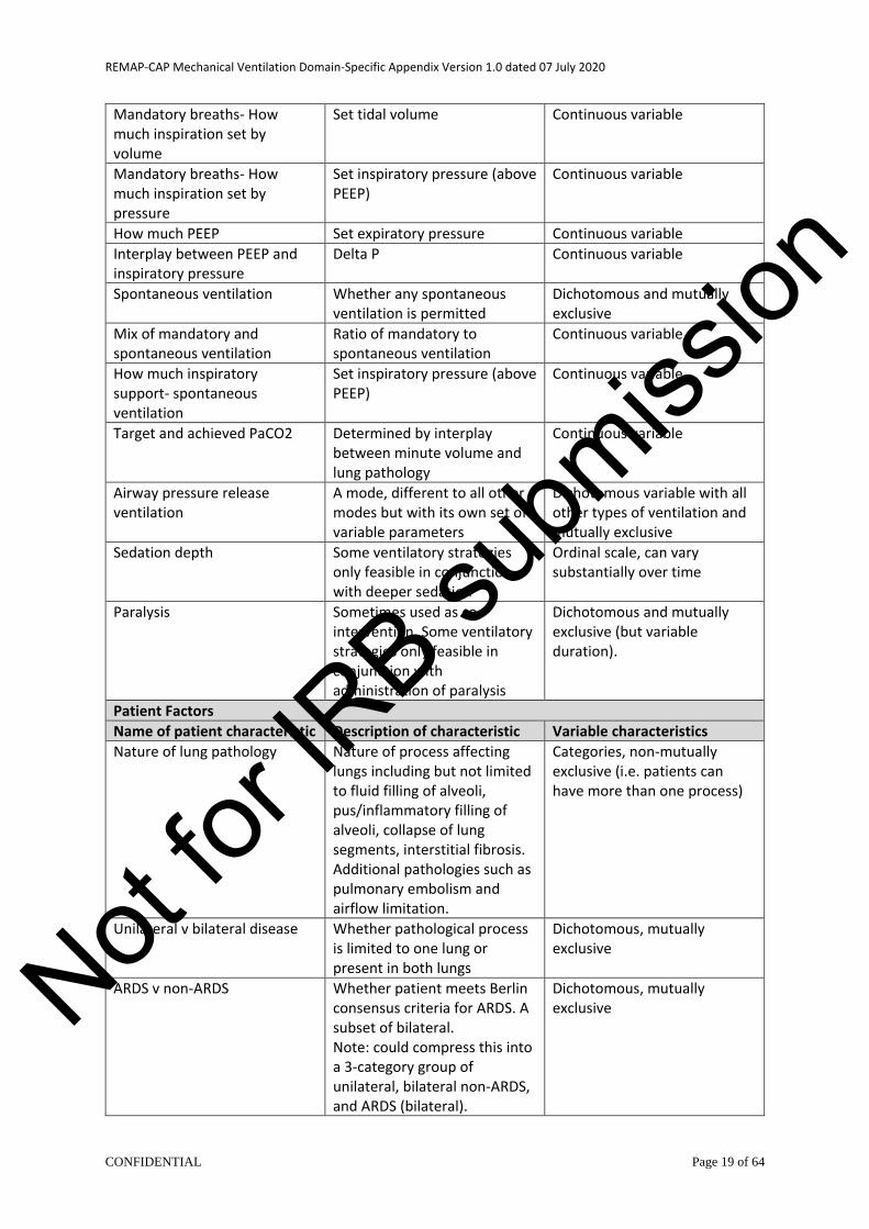
REMAP-CAP Mechanical Ventilation Domain-Specific Appendix Version 1.0 dated 07 July 2020
CONFIDENTIAL Page 19 of 64
Mandatory breaths- How much inspiration set by volume
Set tidal volume Continuous variable
Mandatory breaths- How much inspiration set by pressure
Set inspiratory pressure (above PEEP)
Continuous variable
How much PEEP Set expiratory pressure Continuous variable
Interplay between PEEP and inspiratory pressure
Delta P Continuous variable
Spontaneous ventilation Whether any spontaneous ventilation is permitted
Dichotomous and mutually exclusive
Mix of mandatory and spontaneous ventilation
Ratio of mandatory to spontaneous ventilation
Continuous variable
How much inspiratory support- spontaneous ventilation
Set inspiratory pressure (above PEEP)
Continuous variable
Target and achieved PaCO2 Determined by interplay between minute volume and lung pathology
Continuous variable
Airway pressure release ventilation
A mode, different to all other modes but with its own set of variable parameters
Dichotomous variable with all other types of ventilation and mutually exclusive
Sedation depth Some ventilatory strategies only feasible in conjunction with deeper sedation
Ordinal scale, can vary substantially over time
Paralysis Sometimes used as co-intervention. Some ventilatory strategies only feasible in conjunction with administration of paralysis
Dichotomous and mutually exclusive (but variable duration).
Patient Factors
Name of patient characteristic Description of characteristic Variable characteristics
Nature of lung pathology Nature of process affecting lungs including but not limited to fluid filling of alveoli, pus/inflammatory filling of alveoli, collapse of lung segments, interstitial fibrosis. Additional pathologies such as pulmonary embolism and airflow limitation.
Categories, non-mutually exclusive (i.e. patients can have more than one process)
Unilateral v bilateral disease Whether pathological process is limited to one lung or present in both lungs
Dichotomous, mutually exclusive
ARDS v non-ARDS Whether patient meets Berlin consensus criteria for ARDS. A subset of bilateral. Note: could compress this into a 3-category group of unilateral, bilateral non-ARDS, and ARDS (bilateral).
Dichotomous, mutually exclusive Not
for IR
B subm
ission
REMAP-CAP Mechanical Ventilation Domain-Specific Appendix Version 1.0 dated 07 July 2020
CONFIDENTIAL Page 20 of 64
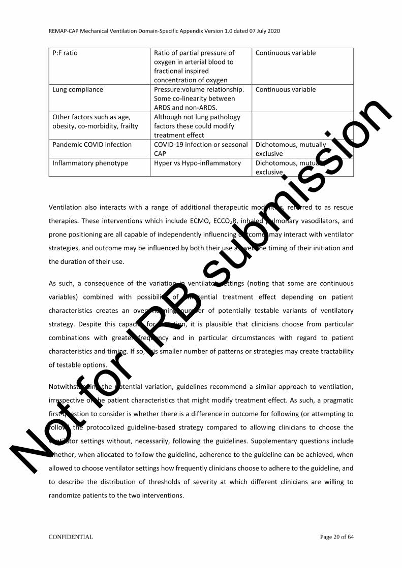
P:F ratio Ratio of partial pressure of oxygen in arterial blood to fractional inspired concentration of oxygen
Continuous variable
Lung compliance Pressure:volume relationship. Some co-linearity between ARDS and non-ARDS.
Continuous variable
Other factors such as age, obesity, co-morbidity, frailty
Although not lung pathology factors these could modify treatment effect
Pandemic COVID infection COVID-19 infection or seasonal CAP
Dichotomous, mutually exclusive
Inflammatory phenotype Hyper vs Hypo-inflammatory Dichotomous, mutually exclusive
Ventilation also interacts with a range of additional therapeutic modalities, referred to as rescue
therapies. These interventions which include ECMO, ECCO2R, inhaled pulmonary vasodilators, and
prone positioning are all capable of independently influencing outcome, may interact with ventilator
strategies, and outcome may be influenced by both their use as well the timing of their initiation and
the duration of their use.
As such, a consequence of the variation in ventilator settings (noting that some are continuous
variables) combined with possibility of differential treatment effect depending on patient
characteristics creates an overwhelming number of potentially testable variants of ventilatory
strategy. Despite this capacity for variation, it is plausible that clinicians choose from particular
combinations with greater frequency and in particular circumstances with regard to patient
characteristics and timing. If so, this smaller number of patterns or strategies may create tractability
of testable options.
Notwithstanding the potential variation, guidelines recommend a similar approach to ventilation,
irrespective of the patient characteristics that might modify treatment effect. As such, a pragmatic
first question to consider is whether there is a difference in outcome for following (or attempting to
follow) the protocolized guideline-based strategy compared to allowing clinicians to choose the
ventilator settings without, necessarily, following the guidelines. Supplementary questions include
whether, when allocated to follow the guideline, adherence to the guideline can be achieved, when
allowed to choose ventilator settings how frequently clinicians choose to adhere to the guideline, and
to describe the distribution of thresholds of severity at which different clinicians are willing to
randomize patients to the two interventions.
Not for
IRB su
bmiss
ion
REMAP-CAP Mechanical Ventilation Domain-Specific Appendix Version 1.0 dated 07 July 2020
CONFIDENTIAL Page 21 of 64
6.4.2. Protocolized guideline-recommended versus wild-type as first questions
It is proposed that the first question that will be assessed within this domain is to compare
protocolized ventilation strategy, derived from the recommendations set by international guidelines,
with the ventilatory strategy that the treating clinician would choose for the patient, including allowing
the clinician to follow guidelines if that is their preference for patients, including patients with COVID-
19. This allows the evaluation of several questions that are necessary for further adaptation of the
domain. The first of these questions is a preliminary consideration of whether there is any difference
in outcome between the two strategies. This is important because of widespread poor adherence to
guideline-recommended strategy. Secondly, it allows assessment of adherence to guideline therapy
in patients with severe CAP. We will aim to understand the barriers to adherence to various aspects
of the guideline-based therapy. If adherence cannot be achieved consistently, and notwithstanding
the recommendation of guidelines, an intervention that cannot be implemented after all reasonable
barriers have be addressed may not be suitable for ongoing evaluation within the platform. Thirdly,
by evaluation of the ventilatory strategies chosen by the clinician it establishes the frequency with
which clinicians choose to follow the strategy recommended by guidelines and, when they choose not
to follow guidelines, establishes the spectrum of ‘wild-type’ strategies that are in use. The latter
information is useful for identifying strategies that are used commonly and might be capable of being
defined as a deliverable intervention in subsequent phases of the ventilation domain. Fourth,
randomization to these two options allows some assessment of the patient characteristics (i.e. the
severity of illness threshold, as measured by a PaO2/FiO2, that each unit choses as the point of
equipoise to randomize) that influence choice of ventilatory strategy and how these patient
characteristics might act as effect modifiers for different treatment strategies.
Some possible outcomes from the preliminary phase include:
• A detailed description of current mechanical ventilation strategies used.
• Evaluation of separation in ventilatory parameters between protocolized and clinician-
preferred strategies and identification of identifiably distinct patterns of ventilation within the
clinician-preferred strategy.
• A preliminary evaluation of any difference in treatment effect between protocolized and
clinician-preferred strategies
• A preliminary evaluation of any difference in treatment effect among identified strata
• If there is no separation between protocolized and clinician-preferred strategies, that
protocolized should be the control group against which further specified variants are
evaluated
Not for
IRB su
bmiss
ion
REMAP-CAP Mechanical Ventilation Domain-Specific Appendix Version 1.0 dated 07 July 2020
CONFIDENTIAL Page 22 of 64
• If clinician-preferred is superior to protocolized then further adaptation should involve testing
identifiable variants with the spectrum of clinician-preferred options
• If there is separation between protocolized and clinician-preferred but there is not clear
superiority of either strategy that it is reasonable to proceed with protocolized as a common
control group and proceed with definable variants of clinical practice.
• If no difference (with limited statistical power after 400 patients) then further adaptation
should proceed with protocolized as one intervention and one or more specified variants of
clinician-preferred strategies as alternative interventions
• Evaluation of the distribution of threshold of severity at which randomization is acceptable to
treating clinicians may be useful for adapting entry criteria or providing ordinal strata to which
participating ICUs are willing to agree to ongoing randomization in patients
6.4.3. Isolation of testable strategies within heterogeneity of current practice
A critical component of the preliminary phase is the identification of definable ventilator strategies
from within patients randomized to the clinician-preferred intervention. Several strategies may be
considered. One approach is the use of qualitative methods to ask participating clinicians to describe
their approach to ventilation for patients with different characteristics and the thresholds of
physiological abnormality at which changes in strategy occur. Within some ICUs there is limited
variation in strategy, i.e. there is a ‘unit-level’ approach to ventilation. Where an ICU believes that it
follows a particular strategy this could be identified and described. Another approach would be to
describe clinical thinking at the time ventilatory strategy is set and adjusted in patients who receive
clinician-preferred ventilation. Lastly, data-driven approaches, such as machine learning, may be
applied to the dataset of patients who receive clinician-preferred ventilation.
6.4.4. Analytic approaches to heterogeneity of current practice
The preliminary phase will enroll patients who meet the platform entry criteria of REMAP-CAP and are
receiving invasive mechanical ventilation in an ICU. The study will collect information related to lung
physiology, ventilation strategy and outcome.
Several different analytic approaches might be considered. Where possible, undertaking several
multiple analytic approaches may be useful as consistency of results, across multiple analytic
approaches, would increase confidence regarding the validity of conclusions. These possible
approaches are outlined in each section. All approaches would be designed to demonstrate, if present,
modification of treatment effect according to the presence of definable patient characteristics such
Not for
IRB su
bmiss
ion
REMAP-CAP Mechanical Ventilation Domain-Specific Appendix Version 1.0 dated 07 July 2020
CONFIDENTIAL Page 23 of 64
as presence of absence of ARDS, lung compliance, unilateral versus bilateral involvement, and P:F
ratio.
6.4.4.1. Pre-specified unit-specific patterns of ventilatory strategy
Each participating ICU will be asked if they believe that there is a ‘unit-level’ approach to ventilatory
strategy for patients that differs according to status with respect to the pandemic infection suspected
or proven strata. This could include asking how the ICU believes it ventilates in a range of scenarios in
patients:
• severe ARDS (i.e. bilateral infiltrates with poor lung compliance)
• less-severe ARDS (i.e. bilateral infiltrates with poor lung compliance and bilateral infiltrates
without poor compliance)
• without ARDS (i.e. unilateral infiltrates but at range of different P:F ratios)
Where possible, strategies that are similar across multiple ICUs will be identified and be analyzed as a
defined strategy. Where there is a belief that the ICU follows a particular strategy, participating ICUs,
nested within a country, will be evaluated within a Bayesian Hierarchical Model as an explanatory
variable. This includes permitting one or more participating ICUs to adopt their choice of a specific
protocol for ventilation in patients with suspected or proven pandemic infection which will be
identified as a defined strategy within the overall ‘clinician-preferred’ ventilation strategy.
6.4.4.2. Observed unit-specific patterns of ventilatory strategy
Where possible, identifiable ventilatory strategies (i.e. those identified in 6.7.3) that are nested within
an ICU, will be evaluated within a model that stratifies by status with respect to suspected or proven
pandemic infection, where appropriate within a Bayesian Hierarchical Model that takes into account
unit and country level effects.
6.4.4.3. Observed individual patient patterns of ventilatory strategy
Where possible, identifiable ventilatory strategies (i.e. those identified in 6.7.3) will be evaluated in a
model, without recourse to nesting of the strategy within an ICU.
6.4.4.4. Individual patient observational analysis
Observational analyses may be conducted that report association between ventilator parameters
and outcome, taking into account, where possible, potential effect modifiers (i.e. with or without
suspected or proven COVID acute respiratory failure) as well as confounding (noting that variables
that contribute to confounding are potentially dynamic).
Not for
IRB su
bmiss
ion
REMAP-CAP Mechanical Ventilation Domain-Specific Appendix Version 1.0 dated 07 July 2020
CONFIDENTIAL Page 24 of 64
6.4.5. Principles of analysis
In the preliminary phase of this domain a fixed sample size will be chosen, after which, analysis will be
conducted with presentation and publication of the results. Subsequent adaptations would then be
planned, on the basis of publicly available results, and, where appropriate, data collected from
patients enrolled in the preliminary phase may be used in analysis or used to generate priors in
ongoing models. Where appropriate, Bayesian Hierarchical Models may be used to capture the
treatment effect of different modifiable components of ventilatory strategy.
This domain complies with the requirements of the core protocol documents. Although no simulations
will be conducted, this is appropriate because the only element of the design that is pre-specified is
the preliminary nature of this phase and the fixed sample size. As such, and to achieve the objectives
of the preliminary phase, it is not necessary to undertake simulations to understand the risks of type
I and type II error.
Summary of background
Severe non-COVID CAP and COVID acute respiratory failure which requires mechanical ventilation is
associated with significant morbidity and mortality. CAP commonly requires invasive mechanical
ventilation. This ventilation has the ability to further damage the injured lung. While international
guidelines offer some principles for care of these patients, many of these recommendations rely either
on weak evidence or on extrapolation of data from patients with ARDS which may not be justified for
all patients with non-COVID CAP or acute respiratory failure due to COVID. There is substantial
uncertainty regarding optimal ventilation strategy. Possibly because of this uncertainty, clinicians
adhere poorly to these recommendations.
The preliminary phase of the ventilation domain will randomize patients to either clinician-preferred
or protocolized therapy with the information obtained from this phase being utilized to design
subsequent phases of the ventilation domain including setting entry criteria, determining
interventions, and setting strata that may be associated with differential treatment effect.
DOMAIN OBJECTIVES
The long-term objective of this domain is to determine the most effective mechanical ventilation
strategies for the treatment of patients with severe CAP and COVID-19 pneumonia who are receiving
invasive mechanical ventilation.
The objectives of the initial phase of the domain are, from a fixed sample size, to:
Not for
IRB su
bmiss
ion
REMAP-CAP Mechanical Ventilation Domain-Specific Appendix Version 1.0 dated 07 July 2020
CONFIDENTIAL Page 25 of 64
1. Collect physiological and ventilatory data on invasively ventilated patients enrolled into the
REMAP-CAP trial.
2. Evaluate separation between protocolized and clinician-preferred mechanical ventilation.
3. Evaluate adherence with protocolized mechanical ventilation in patients with and without
suspected and proven COVID-19 acute respiratory failure.
4. Compare outcome between patients allocated to receive protocolized mechanical ventilation
with patients treated with clinician-preference mechanical ventilation including evaluation of
potential differential treatment effect across strata defined by status with respect to whether
pandemic infection is suspected or proven and across ordinal stratum of PaO2:FiO2 ratio
5. Identify patterns of clinician-preferred mechanical ventilation that can be analyzed as an
‘intervention group’ within the initial phase or potentially deliverable as intervention in
subsequent phases or both
6. Compare outcome in patients with different ‘intervention groups’ within clinician-preferred
mechanical ventilation using protocolized mechanical ventilation as the common comparator
group across evaluable strata
7. Identify variables that may influence treatment effect for different ventilatory strategies that
may be used as strata variables in subsequent phases
8. Utilize information and analysis to revise the protocol design and pre-specified adaptations of
the mechanical ventilation domain in subsequent phases
We hypothesize that the probability of all-cause mortality at 90 days after enrollment will differ based
on the mechanical ventilation strategies treatment received. This preliminary phase will likely be
underpowered to demonstrate superiority. However, it is necessary for the planning of the
subsequent phase. The current mechanical ventilation strategies that will be available to be tested
are:
• Protocolized mechanical ventilation
• Clinician-preferred mechanical ventilation
There are multiple other hypotheses, but these are all regarded as preliminary and hypothesis
generating. These hypotheses are that:
• Within the clinician-preferred ventilation strategy, that 90-day mortality differs depending on
definable patterns of ventilatory strategy Not
for IR
B subm
ission
REMAP-CAP Mechanical Ventilation Domain-Specific Appendix Version 1.0 dated 07 July 2020
CONFIDENTIAL Page 26 of 64
• There is interaction between allocation status and outcome depending on strata (PaO2:FiO2
ratio and pandemic infection strata status), lung compliance, and unilateral v bilateral lung
disease (i.e. infiltrates) at baseline
The long-term objective of the domain is to identify optimal ventilatory strategy and how this varies
between patients with different definable baseline characteristics and how this varies over time and
progression of lung disease. The goals of the preliminary phase are:
• Define testable and deliverable ventilatory strategies that can be introduced as interventions
in subsequent phases of the domain
• Identify baseline stratification variables that can be utilized to allow evaluation of potential
differential treatment effect and be used for implementation of response adaptive
randomization (RAR)
• Adapt the domain, taking these factors into account
TRIAL DESIGN
Trial design overview
This domain will be conducted as part of a REMAP trial for severe CAP (see relevant core documents).
Treatment allocation will not be adaptive during the preliminary phase. This domain is designed so
that it could be supplemented by enrolment of patients with respiratory failure due to causes other
than CAP, but this is not a feature of the current design.
Although this domain is conducted as part of REMAP-CAP, it will use a statistical model that is separate
to the other domains of REMAP-CAP. The strata that apply to this domain are pandemic infection
status and PaO2:FiO2 ratio but, with respect to PaO2:FiO2 the strata that are applied in this domain are
a sub-set of states that are defined in the relevant core protocol documents, but they are applied as
strata, at the time of randomization or reveal of allocation assignment.
Population
The REMAP enrolls patients with severe CAP admitted to ICU and patients with acute illness due to
suspected or proven COVID-19 admitted to ICU. Not for
IRB su
bmiss
ion

REMAP-CAP Mechanical Ventilation Domain-Specific Appendix Version 1.0 dated 07 July 2020
CONFIDENTIAL Page 27 of 64
Eligibility criteria
Patients are eligible for this domain if they meet all of the platform-level inclusion and none of the
platform-level exclusion criteria as specified in either the REMAP-CAP Core Protocol +/- Pandemic
Appendix or the REMAP-COVID Core Protocol. Patients otherwise eligible for REMAP-CAP may have
conditions that exclude them from the Mechanical Ventilation Domain.
8.3.1. Domain inclusion criteria
Patients will be included in this domain if the have the following:
• Receiving invasive mechanical ventilation
• The most recent PaO2:FiO2 ratio obtained within the preceding 6 hours is less than 200 mmHg
• Treating clinician expects the patient to still require invasive mechanical ventilation tomorrow.
• Treating clinician regards Protocolized Mechanical Ventilation Strategy or their preferred
mode as (equally) appropriate for this patient.
8.3.2. Domain exclusion criteria
Patients will be excluded from this domain if they have any of the following:
• More than 48 hours has elapsed since commencement of invasive mechanical ventilation
• The treating clinician believes that participation in this domain is not in the best interests
of the patient.
Interventions
8.4.1. Mechanical Ventilation Interventions
Patients will be randomly assigned to receive one of the following open-label study interventions.
• Protocolized mechanical ventilation
• Clinician-preference mechanical ventilation
8.4.2. Physiological targets set prior to reveal of allocation status and common to both
interventions
In the event of an oxygen saturation target domain of REMAP-CAP, this target would be allocated by
randomization within this domain for patients who receive an allocation status in the oxygen
saturation target domain. In the absence of an oxygen saturation target domain, at the time of
establishing eligibility, the treating clinician will specify a current target range for the oxygen
Not for
IRB su
bmiss
ion

REMAP-CAP Mechanical Ventilation Domain-Specific Appendix Version 1.0 dated 07 July 2020
CONFIDENTIAL Page 28 of 64
saturation. This may be provided as a component of the allocation status for the patient but is
determined by the treating clinician and can be adjusted at any time by the treating clinician.
At the time of establishing eligibility, the treating clinician will specify a target range for the arterial
PaCO2. This may be provided as a component of the allocation status for the patient but is determined
by the treating clinician and can be adjusted at any time by the treating clinician.
8.4.3. Clinician-preferred mechanical ventilation strategy
The clinician will choose the mode of ventilation and all ventilatory parameters that they would use
normally for this patient. Acceptable modes of ventilation include but are not limited to Pressure
Control, synchronised intermittent mandatory ventilation (SIMV), Assist Control (AC), and APRV. Any
and all ventilatory parameters, including mode of ventilation, can be adjusted at any time. If the
treating clinician would usually apply low tidal volume ventilation this is acceptable.
Sedative medications should be administered, as determined by the treating clinician, with or without
administration of a neuromuscular blocker, to provide patient safety and comfort as well as to allow
delivery of the specified ventilator settings.
Each participating ICU will be asked to outline the mechanical ventilation strategy that is used most
commonly, and this may be made available to guide patient care but is regarded only as advisory and
deviation from this guide is permitted at any time.
8.4.4. Protocolized mechanical ventilation strategy
At the time of establishing eligibility, if feasible, the patient’s height will be entered into the on-line
eligibility system which will be used to calculate PBW and to specify a starting tidal volume that will
be set on the ventilator that corresponds to a tidal volume of 6.0 ml/kg PBW. The treating clinician
will set the mode of ventilation as either SIMV, Assist Control, or pressure regulated volume control,
setting the tidal volume to that specified by the on-line eligibility system or calculated by the site. If
the plateau pressure is more than 30 cmH2O, the tidal volume should be reduced in 5 to 10%
increments, but to not less than 65% of the initial specified tidal volume (corresponding to 4.0 ml/kg
PBW). At any time, if the tidal volume is less than that specified following calculation from the patient’s
height and the plateau pressure is 29 cm or less, the tidal volume should be increased in 5% increments
to achieve a tidal volume as close to, but not exceeding, the specified target tidal volume while
maintain plateau pressure of not more than 30 cm H2O. Not
for IR
B subm
ission
REMAP-CAP Mechanical Ventilation Domain-Specific Appendix Version 1.0 dated 07 July 2020
CONFIDENTIAL Page 29 of 64
The respiratory rate is set by the treating clinician with the goal of achieving the targeted PaCO2 and
adjusted, as required, to achieve the PaCO2 target. If the PaCO2 target cannot be achieved the
respiratory rate should be adjusted upwards preserving, at all times (unless pH of arterial blood is less
than 7.15 see below), the tidal volume (and plateau pressure target). The maximum rate is 35 breaths
per minute.
Sedative medications should be administered, as determined by the treating clinician, with or without
administration of a neuromuscular blocker, to provide patient safety and comfort as well as to allow
delivery of the specified ventilator settings.
If arterial pH is less than 7.15, the tidal volume can be increased in 5 to 10% increments until arterial
pH is more than 7.15, noting that, if necessary, to maintain a pH of not less than 7.15, it is permitted
for the plateau pressure to exceed 30 cm H2O. In the presence of severe acidosis, bicarbonate may be
administered, at the discretion of the treating clinician. If the acidosis has a substantial metabolic
component, continuous renal replacement therapy may be instituted, at the discretion of the treating
clinician.
Administration of a tidal volume of more than 6.0 ml/kg PBW or a plateau pressure of 30 cm H20 or
more is a protocol deviation unless the pH is less than 7.15.
The FiO2 and PEEP should be set on the ventilator to the minimum allowable combination of FiO2 and
PEEP that consistently achieves a PaO2 / SaO2 within the target range and adjusted further, as required
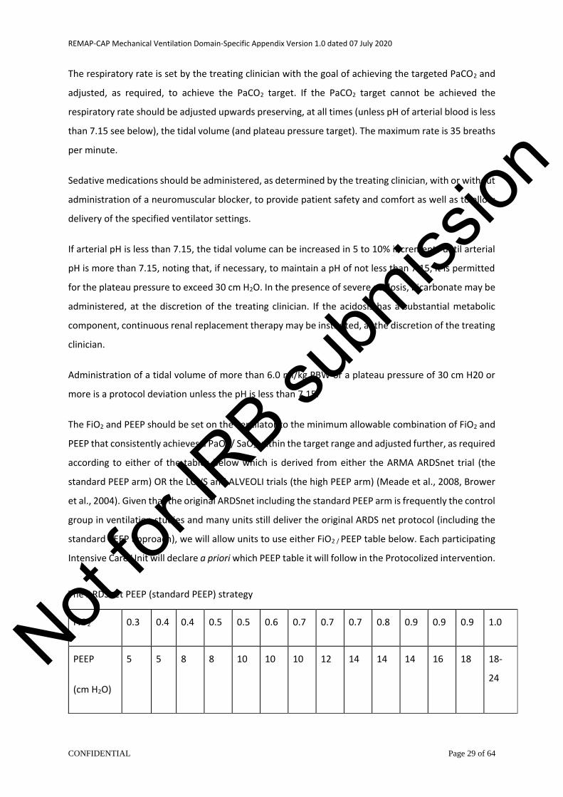
according to either of the tables below which is derived from either the ARMA ARDSnet trial (the
standard PEEP arm) OR the LOVS and ALVEOLI trials (the high PEEP arm) (Meade et al., 2008, Brower
et al., 2004). Given that the original ARDSnet including the standard PEEP arm is frequently the control
group in ventilation studies and many units still deliver the original ARDS net protocol (including the
standard PEEP approach), we will allow units to use either FiO2 / PEEP table below. Each participating
Intensive Care Unit will declare a priori which PEEP table it will follow in the Protocolized intervention.
The ARDSnet PEEP (standard PEEP) strategy
FiO2 0.3 0.4 0.4 0.5 0.5 0.6 0.7 0.7 0.7 0.8 0.9 0.9 0.9 1.0
PEEP
(cm H2O)
5 5
8 8 10 10 10 12 14 14 14 16 18 18-
24 Not
for IR
B subm
ission
REMAP-CAP Mechanical Ventilation Domain-Specific Appendix Version 1.0 dated 07 July 2020
CONFIDENTIAL Page 30 of 64
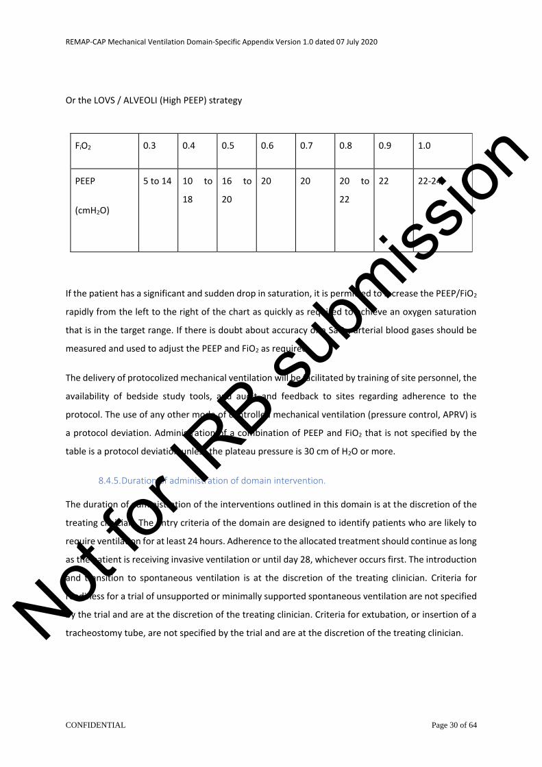
Or the LOVS / ALVEOLI (High PEEP) strategy
If the patient has a significant and sudden drop in saturation, it is permitted to increase the PEEP/FiO2
rapidly from the left to the right of the chart as quickly as required to achieve an oxygen saturation
that is in the target range. If there is doubt about accuracy of a SaO2, arterial blood gases should be
measured and used to adjust the PEEP and FiO2 as required.
The delivery of protocolized mechanical ventilation will be facilitated by training of site personnel, the
availability of bedside study tools, and audit and feedback to sites regarding adherence to the
protocol. The use of any other mode of controlled mechanical ventilation (pressure control, APRV) is
a protocol deviation. Administration of a combination of PEEP and FiO2 that is not specified by the
table is a protocol deviation unless the plateau pressure is 30 cm of H2O or more.
8.4.5. Duration of administration of domain intervention.
The duration of administration of the interventions outlined in this domain is at the discretion of the
treating clinician. The entry criteria of the domain are designed to identify patients who are likely to
require ventilation for at least 24 hours. Adherence to the allocated treatment should continue as long
as the patient is receiving invasive ventilation or until day 28, whichever occurs first. The introduction
and transition to spontaneous ventilation is at the discretion of the treating clinician. Criteria for
readiness for a trial of unsupported or minimally supported spontaneous ventilation are not specified
by the trial and are at the discretion of the treating clinician. Criteria for extubation, or insertion of a
tracheostomy tube, are not specified by the trial and are at the discretion of the treating clinician.
FiO2 0.3 0.4 0.5 0.6 0.7 0.8 0.9 1.0
PEEP
(cmH2O)
5 to 14 10 to
18
16 to
20
20 20 20 to
22
22 22-24
Not for
IRB su
bmiss
ion
REMAP-CAP Mechanical Ventilation Domain-Specific Appendix Version 1.0 dated 07 July 2020
CONFIDENTIAL Page 31 of 64
Concomitant care
All other interventions will be as directed by the treating clinician, including choice of sedative agents,
depth of sedation, use of neuromuscular blocking agents, and use of rescue therapies for severe
hypoxemia including prone positioning, inhaled pulmonary vasodilators, ECMO, and ECCO2R, although
the use of these therapies will be recorded.
Endpoints
8.6.1. Primary endpoint
The primary endpoint for this domain is all-cause mortality at 90 days.
8.6.2. Secondary endpoints
All secondary endpoints as specified in the REMAP-CAP Core Protocol +/- Pandemic Appendix or REMAP-COVID Core Protocol.
The domain-specific secondary outcome measures (occurring during the index hospitalization,
censored 90 days after enrollment) are:
• Administration of rescue therapies
• Occurrence of barotrauma
• SAE as defined in the core protocol documents and qualified in this DSA
8.6.3. Process end-points
Ventilator settings, targets for oxygen saturation and arterial PaCO2, and corresponding results of
arterial blood gases will be recorded. Qualitative and quantitative assessment of factor influencing
threshold for equipoise for randomization, choice of preferred strategy, and barriers to adherence.
TRIAL CONDUCT
Domain-specific data collection
9.1.1. Clinical data collection
Additional domain-specific data will be collected.
• Baseline ventilatory, physiological parameters and arterial blood gas parameters
• Daily ventilatory and physiological parameters and arterial blood gas parameters
Not for
IRB su
bmiss
ion
REMAP-CAP Mechanical Ventilation Domain-Specific Appendix Version 1.0 dated 07 July 2020
CONFIDENTIAL Page 32 of 64
• Daily targets for physiological parameters, if specified
• Occurrence and duration of prone ventilation.
• Administration of rescue therapies
• Occurrence of barotrauma
Criteria for discontinuation
Refer to relevant core protocol documents for criteria for discontinuation of participation in the
REMAP-CAP trial.
Blinding
9.3.1. Blinding
The interventions in the ventilation domain are not suitable for blinding as the appropriate care of the
patient requires the clinician to have knowledge of the ventilator settings.
9.3.2. Unblinding
Not relevant.
STATISTICAL CONSIDERATIONS
Domain-specific stopping rules
The domain can report a Platform Conclusion, as specified in relevant Core Protocol, at any interim
analysis. The possible Platform Conclusions are superiority (with corresponding inferiority) of either
intervention. The Platform Conclusion of equivalence will not be evaluated during the preliminary
phase.
In the absence of a Platform Conclusion, the domain will be analyzed after 400 patients have been
enrolled, although randomization using this version of the protocol will continue beyond 400 patients
while results from the initial phase are being analyzed which will be used to guide subsequent
adaptations to the domain. Not for
IRB su
bmiss
ion

REMAP-CAP Mechanical Ventilation Domain-Specific Appendix Version 1.0 dated 07 July 2020
CONFIDENTIAL Page 33 of 64
Categorization of clinician-preferred ventilator strategies
10.2.1. Unit-level self-categorization
As part of the site initiation process, the opinion of clinicians at that site will be obtained regarding
their usual approaches to ventilation including whether there is a belief that guideline-recommended
ventilation is utilized and, if so, for what category of patients. If guideline-recommended ventilation is
not utilized the common patterns of ventilatory strategy will be identified. Patterns of ventilatory
strategy that are common between sites will be identified and sites will be categorized according to
shared patterns of ventilatory strategy.
10.2.2. Unit-level observed categorization
Data obtained from non-randomized and randomized patients will be analyzed to identify common
patterns of ventilatory strategy in CAP and patients with proven or suspected COVID-19. Patterns of
ventilatory strategy that are common between sites will be identified and sites will be categorized
according to shared patterns of ventilatory strategy.
10.2.3. Patient-level categorization
Data obtained from individual non-randomized and randomized patients will be analyzed to identify
common patterns of ventilatory strategy. Patterns of ventilatory strategy that are common among all
patients, irrespective of participating site, will be identified and patients will be categorized according
to shared patterns of ventilatory strategy.
10.2.4. Methods of determination of categorization
Conventional statistical methods will be applied to identify categories such as classification and
regression trees (CART) and logistic regression. If available to the investigators, additional methods
such as network analysis may be used.
Statistical analysis
10.3.1. Principles of statistical analysis
The statistical analysis of the ventilation domain will follow the principles outlined in the relevant core
protocol documents with some modifications, as outlined in this DSA.
Treatment effects will be evaluated in this domain using a statistical model that is separate to the
model used for other domains of REMAP-CAP. The model will be specified and described in an
Not for
IRB su
bmiss
ion
REMAP-CAP Mechanical Ventilation Domain-Specific Appendix Version 1.0 dated 07 July 2020
CONFIDENTIAL Page 34 of 64
operational document that may be updated periodically. The model will estimate the probability of
superiority, after adjustment for baseline covariates, dependent on allocation status to clinician
preferred or guideline recommended ventilation strategy. Equivalence will not be evaluated during
the initial phase. The statistical model will include stratification by the pandemic infection stratum
with borrowing permitted between stratum. Randomization occurs at the time of reveal, i.e. not
necessarily at the time of first assignment in any domain. This allows for the application of strata that
apply at the time of randomization. The additional strata will comprise ordinal categories of PaO2:FiO2
ratio with borrowing permitted across ordinal stratum categories. The use of Statistical Triggers and
Platform Conclusions will be as specified in the core protocol documents. The sample size for the initial
phase will be a maximum of 400 patients who have been randomized.
The same or similar model, if appropriate in conjunction with a Bayesian Hierarchical Model, will be
used to evaluate the effect of identified ventilatory strategies. In these analyses, patients within the
clinician-preferred strategy will be analyzed according to groups that identify specific ventilatory
strategies with analysis being applied at both site level (site self-reported strategy and site observed
strategy) and patient level. If appropriate, a BHM may be used that takes into account the
relationships between patient- and site-level categorization of ventilatory strategies and evaluates the
impact of ventilatory parameters, as well as progression of the underlying disease and alternative
strata. Sensitivity analyses using data from non-randomized may also be conducted.
10.3.2. Strata
The strata structure of this domain comprises the 2 x 3 table defined by two categories of the
pandemic infection strata combined with 3 ordinal categories defined by the PaO2:FiO2 ratio at time
of randomization (noting that in this domain, randomization occurs at the time of reveal which is
different to the other domains of REMAP-CAP). The ordinal strata are:
• PaO2:FiO2 ratio less than 100
• PaO2:FiO2 ratio of 100 or more but less than 150
• PaO2:FiO2 ratio of 150 or more but less than 200
These categories are a sub-set of state, as defined in core protocol documents.
10.3.3. Primary analysis
The primary analysis will evaluate the main effect of assignment to either clinician-preferred or
protocolized ventilatory strategy. Each stratum is analyzed separately but the model captures
commonalities across stratum, recognizing the ordinal structure of the strata. Additionally, the
Not for
IRB su
bmiss
ion

REMAP-CAP Mechanical Ventilation Domain-Specific Appendix Version 1.0 dated 07 July 2020
CONFIDENTIAL Page 35 of 64
statistical model allows evidence relating to the effectiveness of a ventilatory strategy in one stratum
to contribute (via ‘borrowing’) to the estimation of the posterior probability in other stratum, but this
only occurs to the extent that treatment effect is similar in adjacent stratum. The model will adjust for
demographic and severity factors but will not provide a separate estimate of treatment effect for
these covariates.
The primary analysis set will comprise patients who receive an allocation status, i.e. intention to treat.
Additional analyses may be undertaken on a per protocol basis (i.e. taking into account adherence
with guidelines) and utilizing data from non-randomized patients.
10.3.4. Descriptive analysis of ventilatory patterns
Conventional parametric or non-parametric, as appropriate, statistical methods will be used to
describe observed ventilator settings including mode of ventilation, tidal volume, airway pressures,
PEEP, saturation target and its achievement, PaCO2 target and its achievement.
10.3.5. Secondary analyses
A series of secondary analyses will be conducted. Higher emphasis will be given to results that are
consistent across multiple different analyses.
10.3.5.1. Evaluation of adherence with protocolized ventilatory strategy
Adherence with protocolized ventilatory strategy will be evaluated by the duration for which the
strategy is implemented as a proportion of the total time the patient receives a mandatory ventilation
mode, censored at day 28; and by the proportion of tidal volume and PEEP measurements that are
compliant with the treatment protocol during the period of time for which the strategy is
implemented. Factors associated with adherence will be evaluated using logistic regression utilizing
variables include, but are not limited to age, co-morbidity (including chronic obstructive pulmonary
disease, COPD), pattern of lung involvement (unilateral CAP, bilateral CAP without moderate / severe
ARDS, or moderate / severe ARDS), lung compliance, PaO2:FiO2 ratio stratum, pandemic infection
stratum, and participating site. The analysis set will comprise patients assigned to protocolized
ventilatory strategy.
10.3.5.2. Identification of patterns of clinician-preferred mechanical ventilation that can be
analyzed as an intervention group
Each participating ICU will be asked, using self-report, to identify commonly applied ventilatory
strategies. This will comprise information related to mode of ventilation; targets with respect to one
or more of tidal volume, plateau pressure, oxygen saturation, and PaCO2; use of APRV, and timing of
Not for
IRB su
bmiss
ion
REMAP-CAP Mechanical Ventilation Domain-Specific Appendix Version 1.0 dated 07 July 2020
CONFIDENTIAL Page 36 of 64
switching to spontaneous ventilation. Qualitative thematic analysis of this information will be used to
identify candidate ventilatory strategies which may be mutually exclusive or overlapping.
Several methods will be used to identify common patterns of ventilatory strategy. These include the
extent to which observed ventilatory patterns correspond to the candidate ventilatory strategies
identified by analysis of self-reported strategies, CART or logistic regression-based methods, and
network analysis which identifies commonalities across all recorded parameters and organizes these
into nodes of similarity.
The analysis set will be patients randomized to clinician-preferred strategy.
Identified ventilatory strategies will also be evaluated by investigators to determine the extent that
such strategies could be specified for delivery using a protocol.
10.3.5.3. Evaluation of treatment effect associated with identified patterns of ventilatory
strategy
A Bayesian statistical model will evaluate the treatment effect of different identified patterns of
ventilatory strategy, from among patients assigned to clinician-preferred ventilatory strategy, using
protocolized ventilation as the common comparator. The model will be similar or the same as that
uses for the primary analysis. If appropriate, a hierarchical structure may be utilized that groups
together elements of identified strategy that are similar for example APRV and among non-APRV
patients pressure control versus volume control (and whether tidal volume or inspiratory pressure is
targeted), and the use of PEEP. Analyses will also be conducted in which the unit of analysis is
categories of participating ICU according to self-reported ventilatory strategy as well as observed
ventilatory strategy. The analysis will be conducted using the pandemic infection strata and ordinal
PaO2:FiO2 strata, and take into account the same baseline co-variates.
The analysis set will be all randomized patients.
10.3.6. Identification of variables that may modify treatment effect
An important component of the initial phase is the identification of strata variables for subsequent
phases. Although PaO2:FiO2 ratio has been selected as the strata variable for the initial stage it is
possible that other variables, alone, or in combination with PaO2:FiO2 ratio, may have greater potential
to identify patient characteristics associated with differential treatment effect. The potential for
alternative variables to be used for stratification will be evaluated using a Bayesian statistical model
that evaluates differential treatment effect according to baseline variables including age, co-morbidity
Not for
IRB su
bmiss
ion

REMAP-CAP Mechanical Ventilation Domain-Specific Appendix Version 1.0 dated 07 July 2020
CONFIDENTIAL Page 37 of 64
(including COPD), pattern of lung involvement (unilateral CAP, bilateral CAP without moderate /
severe ARDS or moderate / severe ARDS), and lung compliance on treatment effect for the assigned
ventilatory strategies, as well as identified ventilatory patterns among patients allocated to receive
clinician-preferred ventilation.
10.3.7. Evaluation of separation between protocolized and clinician-preferred ventilatory
strategies
Conventional statistical methods will be used to evaluate separation with regard to ventilatory
parameters including but not limited to mode of ventilation, tidal volume, inspiratory pressures, PEEP,
and initiation of spontaneous ventilation. Logistic regression methods will be used to identify factors
associated with separation. Variables that will be evaluated include, but are not limited to, age, co-
morbidity (including COPD), pattern of lung involvement (unilateral CAP, bilateral CAP without ARDS,
ARDS), lung compliance, stratum, and participating site.
10.3.8. Sample size and statistical power
This initial phase is conducted as part of an ongoing platform, as outlined in the core protocol
documents. There is little rationale for pre-specification of the size of expected treatment effects and,
as such, it is difficult to specify the statistical power that is available for the primary analysis. It is
anticipated that only a large treatment effect would be capable of detection with a sample size of 400
patients. The statistical power available for evaluation of identified variants of clinician-preferred
ventilation is commensurately smaller.
The primary objective of the initial phase is to redesign the domain utilizing the generated data. If data
from 400 patients is insufficient for this purpose, the initial phase can be extended. The sample size
of 400 patients was chosen based on expected rates of recruitment and a compromise between the
need for data to design subsequent phases and the time imperative to move as quickly as possible
from the initial to subsequent phases. In this regard, the initial phase can be regarded as a pilot study.
Timing of revealing of randomization status
The timing of the revealing of allocation status and administration of interventions is as specified to
be Randomization with Immediate Reveal and Initiation. Not for
IRB su
bmiss
ion
REMAP-CAP Mechanical Ventilation Domain-Specific Appendix Version 1.0 dated 07 July 2020
CONFIDENTIAL Page 38 of 64
Interactions with interventions in other domains
No interaction with other domains is capable of being evaluated because of the use of a separate
statistical model for the ventilation domain.
Nesting of interventions
Nesting is not applicable to this domain.
ETHICAL CONSIDERATIONS
Data Safety and Monitoring Board
The DSMB should be aware that the superiority, inferiority, or equivalence of different interventions
with respect to the primary endpoint is possible and will interpret adaptive analyses accordingly and
aware of the objectives of this initial phase of the ventilation domain.
Potential domain-specific adverse events
Potential domain-specific harms related to the ventilation domain include:
• Barotrauma (i.e. pneumothorax, subcutaneous emphysema, etc.)
Other serious adverse events (SAEs) should be reported only where, in the opinion of the site
investigator, the event might reasonably have occurred as a consequence of a study intervention or
study participation (see relevant core protocol documents).
Domain-specific consent issues
All patients eligible for this domain will not be competent to consent because of administration of
sedative agents to facilitate invasive mechanical ventilation. All ventilatory strategies are within the
spectrum of standard care at participating ICUs. The treatment options are either what is
recommended by international guidelines or the treatment the patient would have otherwise
received. Randomization is precluded if the treating clinician believes that participation is not in the
best interests of the participant. On this basis, where available, it is proposed that enrollment will
occur without prior agreement, with agreement being obtained from a patient representative or the
patient or both as soon as possible.
Not for
IRB su
bmiss
ion
REMAP-CAP Mechanical Ventilation Domain-Specific Appendix Version 1.0 dated 07 July 2020
CONFIDENTIAL Page 39 of 64
During a pandemic, visiting by relatives of affected patients may not be possible. In such situations,
alternative methods for confirming consent including electronic and telephone communication, as
permitted by an appropriate ethical review body, may be acceptable methods of confirming
agreement to participate in this (and other) domains of the platform.
Clinicians are directed not to enroll an individual patient if the treating clinician believes that
participation in this domain is not in the best interests of the patient.
GOVERNANCE ISSUES
Funding of domain
Funding sources for the REMAP-CAP trial are specified in the core protocol documents. This domain
has not received any additional domain-specific funding but such funding may be obtained during the
lifetime of the domain.
Funding of domain interventions and outcome measures
All patients will require mechanical ventilation. There are no additional costs for setting of
ventilatory parameters that are available on all ventilators at participating sites.
Domain-specific declarations of interest
All investigators involved in REMAP-CAP maintain a registry of interests on the REMAP-CAP website.
These are updated periodically and publicly accessible on the study website.
Not for
IRB su
bmiss
ion
REMAP-CAP Mechanical Ventilation Domain-Specific Appendix Version 1.0 dated 07 July 2020
CONFIDENTIAL Page 40 of 64
REFERENCES
ABROUG, F., OUANES-BESBES, L., ELATROUS, S. & BROCHARD, L. 2008. The effect of prone positioning in acute respiratory distress syndrome or acute lung injury: a meta-analysis. Areas of uncertainty and recommendations for research. Intensive Care Med, 34, 1002-11.
ACUTE RESPIRATORY DISTRESS SYNDROME, N., BROWER, R. G., MATTHAY, M. A., MORRIS, A., SCHOENFELD, D., THOMPSON, B. T. & WHEELER, A. 2000. Ventilation with lower tidal volumes as compared with traditional tidal volumes for acute lung injury and the acute respiratory distress syndrome. N Engl J Med, 342, 1301-8.
ALHAZZANI, W., MOLLER, M. H., ARABI, Y. M., LOEB, M., GONG, M. N., FAN, E., OCZKOWSKI, S., LEVY, M. M., DERDE, L., DZIERBA, A., DU, B., ABOODI, M., WUNSCH, H., CECCONI, M., KOH, Y., CHERTOW, D. S., MAITLAND, K., ALSHAMSI, F., BELLEY-COTE, E., GRECO, M., LAUNDY, M., MORGAN, J. S., KESECIOGLU, J., MCGEER, A., MERMEL, L., MAMMEN, M. J., ALEXANDER, P. E., ARRINGTON, A., CENTOFANTI, J. E., CITERIO, G., BAW, B., MEMISH, Z. A., HAMMOND, N., HAYDEN, F. G., EVANS, L. & RHODES, A. 2020a. Surviving Sepsis Campaign: Guidelines on the Management of Critically Ill Adults with Coronavirus Disease 2019 (COVID-19). Crit Care Med, 48, e440-e469.
ALHAZZANI, W., MOLLER, M. H., ARABI, Y. M., LOEB, M., GONG, M. N., FAN, E., OCZKOWSKI, S., LEVY, M. M., DERDE, L., DZIERBA, A., DU, B., ABOODI, M., WUNSCH, H., CECCONI, M., KOH, Y., CHERTOW, D. S., MAITLAND, K., ALSHAMSI, F., BELLEY-COTE, E., GRECO, M., LAUNDY, M., MORGAN, J. S., KESECIOGLU, J., MCGEER, A., MERMEL, L., MAMMEN, M. J., ALEXANDER, P. E., ARRINGTON, A., CENTOFANTI, J. E., CITERIO, G., BAW, B., MEMISH, Z. A., HAMMOND, N., HAYDEN, F. G., EVANS, L. & RHODES, A. 2020b. Surviving Sepsis Campaign: guidelines on the management of critically ill adults with Coronavirus Disease 2019 (COVID-19). Intensive Care Med, 46, 854-887.
AMATO, M. B., BARBAS, C. S., MEDEIROS, D. M., MAGALDI, R. B., SCHETTINO, G. P., LORENZI-FILHO, G., KAIRALLA, R. A., DEHEINZELIN, D., MUNOZ, C., OLIVEIRA, R., TAKAGAKI, T. Y. & CARVALHO, C. R. 1998. Effect of a protective-ventilation strategy on mortality in the acute respiratory distress syndrome. N Engl J Med, 338, 347-54.
AMATO, M. B., MEADE, M. O., SLUTSKY, A. S., BROCHARD, L., COSTA, E. L., SCHOENFELD, D. A., STEWART, T. E., BRIEL, M., TALMOR, D., MERCAT, A., RICHARD, J. C., CARVALHO, C. R. & BROWER, R. G. 2015. Driving pressure and survival in the acute respiratory distress syndrome. N Engl J Med, 372, 747-55.
ANDREWS, P. L., SHIBER, J. R., JARUGA-KILLEEN, E., ROY, S., SADOWITZ, B., O'TOOLE, R. V., GATTO, L. A., NIEMAN, G. F., SCALEA, T. & HABASHI, N. M. 2013. Early application of airway pressure release ventilation may reduce mortality in high-risk trauma patients: a systematic review of observational trauma ARDS literature. J Trauma Acute Care Surg, 75, 635-41.
BAO, C., LIU, X., ZHANG, H., LI, Y. & LIU, J. 2020. Coronavirus Disease 2019 (COVID-19) CT Findings: A Systematic Review and Meta-analysis. J Am Coll Radiol.
BEITLER, J. R., SARGE, T., BANNER-GOODSPEED, V. M., GONG, M. N., COOK, D., NOVACK, V., LORING, S. H., TALMOR, D. & GROUP, E. P.-S. 2019. Effect of Titrating Positive End-Expiratory Pressure (PEEP) With an Esophageal Pressure-Guided Strategy vs an Empirical High PEEP-Fio2 Strategy on Death and Days Free From Mechanical Ventilation Among Patients With Acute Respiratory Distress Syndrome: A Randomized Clinical Trial. JAMA, 321, 846-857.
Not for
IRB su
bmiss
ion
REMAP-CAP Mechanical Ventilation Domain-Specific Appendix Version 1.0 dated 07 July 2020
CONFIDENTIAL Page 41 of 64
BELLANI, G., LAFFEY, J. G., PHAM, T., FAN, E., BROCHARD, L., ESTEBAN, A., GATTINONI, L., VAN HAREN, F., LARSSON, A., MCAULEY, D. F., RANIERI, M., RUBENFELD, G., THOMPSON, B. T., WRIGGE, H., SLUTSKY, A. S., PESENTI, A., INVESTIGATORS, L. S. & GROUP, E. T. 2016. Epidemiology, Patterns of Care, and Mortality for Patients With Acute Respiratory Distress Syndrome in Intensive Care Units in 50 Countries. JAMA, 315, 788-800.
BHATRAJU, P. K., GHASSEMIEH, B. J., NICHOLS, M., KIM, R., JEROME, K. R., NALLA, A. K., GRENINGER, A. L., PIPAVATH, S., WURFEL, M. M., EVANS, L., KRITEK, P. A., WEST, T. E., LUKS, A., GERBINO, A., DALE, C. R., GOLDMAN, J. D., O'MAHONY, S. & MIKACENIC, C. 2020. Covid-19 in Critically Ill Patients in the Seattle Region - Case Series. N Engl J Med, 382, 2012-2022.
BRIEL, M., MEADE, M., MERCAT, A., BROWER, R. G., TALMOR, D., WALTER, S. D., SLUTSKY, A. S., PULLENAYEGUM, E., ZHOU, Q., COOK, D., BROCHARD, L., RICHARD, J. C., LAMONTAGNE, F., BHATNAGAR, N., STEWART, T. E. & GUYATT, G. 2010. Higher vs lower positive end-expiratory pressure in patients with acute lung injury and acute respiratory distress syndrome: systematic review and meta-analysis. JAMA, 303, 865-73.
BROCHARD, L. 2010. New goals for positive end-expiratory pressure in acute respiratory distress syndrome: a paradigm shift or the end of an area of uncertainty? Am J Respir Crit Care Med, 181, 528-30.
BROWER, R. G., LANKEN, P. N., MACINTYRE, N., MATTHAY, M. A., MORRIS, A., ANCUKIEWICZ, M., SCHOENFELD, D., THOMPSON, B. T., NATIONAL HEART, L. & BLOOD INSTITUTE, A. C. T. N. 2004. Higher versus lower positive end-expiratory pressures in patients with the acute respiratory distress syndrome. N Engl J Med, 351, 327-36.
CARSETTI, A., DAMIANI, E., DOMIZI, R., SCORCELLA, C., PANTANETTI, S., FALCETTA, S., DONATI, A. & ADRARIO, E. 2019. Airway pressure release ventilation during acute hypoxemic respiratory failure: a systematic review and meta-analysis of randomized controlled trials. Ann Intensive Care, 9, 44.
COMBES, A., HAJAGE, D., CAPELLIER, G., DEMOULE, A., LAVOUE, S., GUERVILLY, C., DA SILVA, D., ZAFRANI, L., TIROT, P., VEBER, B., MAURY, E., LEVY, B., COHEN, Y., RICHARD, C., KALFON, P., BOUADMA, L., MEHDAOUI, H., BEDUNEAU, G., LEBRETON, G., BROCHARD, L., FERGUSON, N. D., FAN, E., SLUTSKY, A. S., BRODIE, D., MERCAT, A., EOLIA TRIAL GROUP, R. & ECMONET 2018. Extracorporeal Membrane Oxygenation for Severe Acute Respiratory Distress Syndrome. N Engl J Med, 378, 1965-1975.
DEANS, K. J., MINNECI, P. C., DANNER, R. L., EICHACKER, P. Q. & NATANSON, C. 2010. Practice misalignments in randomized controlled trials: Identification, impact, and potential solutions. Anesth Analg, 111, 444-50.
ESTEBAN, A., ANZUETO, A., FRUTOS, F., ALIA, I., BROCHARD, L., STEWART, T. E., BENITO, S., EPSTEIN, S. K., APEZTEGUIA, C., NIGHTINGALE, P., ARROLIGA, A. C., TOBIN, M. J. & MECHANICAL VENTILATION INTERNATIONAL STUDY, G. 2002. Characteristics and outcomes in adult patients receiving mechanical ventilation: a 28-day international study. JAMA, 287, 345-55.
ESTEBAN, A., FRUTOS-VIVAR, F., MURIEL, A., FERGUSON, N. D., PENUELAS, O., ABRAIRA, V., RAYMONDOS, K., RIOS, F., NIN, N., APEZTEGUIA, C., VIOLI, D. A., THILLE, A. W., BROCHARD, L., GONZALEZ, M., VILLAGOMEZ, A. J., HURTADO, J., DAVIES, A. R., DU, B., MAGGIORE, S. M., PELOSI, P., SOTO, L., TOMICIC, V., D'EMPAIRE, G., MATAMIS, D., ABROUG, F., MORENO, R. P., SOARES, M. A., ARABI, Y., SANDI, F., JIBAJA, M., AMIN, P., KOH, Y., KUIPER, M. A., BULOW, H. H., ZEGGWAGH, A. A. & ANZUETO, A. 2013. Evolution of mortality over time in patients receiving mechanical ventilation. Am J Respir Crit Care Med, 188, 220-30.
FAN, E., DEL SORBO, L., GOLIGHER, E. C., HODGSON, C. L., MUNSHI, L., WALKEY, A. J., ADHIKARI, N. K. J., AMATO, M. B. P., BRANSON, R., BROWER, R. G., FERGUSON, N. D., GAJIC, O., GATTINONI,
Not for
IRB su
bmiss
ion
REMAP-CAP Mechanical Ventilation Domain-Specific Appendix Version 1.0 dated 07 July 2020
CONFIDENTIAL Page 42 of 64
L., HESS, D., MANCEBO, J., MEADE, M. O., MCAULEY, D. F., PESENTI, A., RANIERI, V. M., RUBENFELD, G. D., RUBIN, E., SECKEL, M., SLUTSKY, A. S., TALMOR, D., THOMPSON, B. T., WUNSCH, H., ULERYK, E., BROZEK, J., BROCHARD, L. J., AMERICAN THORACIC SOCIETY, E. S. O. I. C. M. & SOCIETY OF CRITICAL CARE, M. 2017. An Official American Thoracic Society/European Society of Intensive Care Medicine/Society of Critical Care Medicine Clinical Practice Guideline: Mechanical Ventilation in Adult Patients with Acute Respiratory Distress Syndrome. Am J Respir Crit Care Med, 195, 1253-1263.
FORCE, A. D. T., RANIERI, V. M., RUBENFELD, G. D., THOMPSON, B. T., FERGUSON, N. D., CALDWELL, E., FAN, E., CAMPOROTA, L. & SLUTSKY, A. S. 2012. Acute respiratory distress syndrome: the Berlin Definition. JAMA, 307, 2526-33.
GARNER, W., DOWNS, J. B., STOCK, M. C. & RASANEN, J. 1988. Airway pressure release ventilation (APRV). A human trial. Chest, 94, 779-81.
GATTINONI, L., CAIRONI, P., CRESSONI, M., CHIUMELLO, D., RANIERI, V. M., QUINTEL, M., RUSSO, S., PATRONITI, N., CORNEJO, R. & BUGEDO, G. 2006. Lung recruitment in patients with the acute respiratory distress syndrome. N Engl J Med, 354, 1775-86.
GOLIGHER, E. C., HODGSON, C. L., ADHIKARI, N. K. J., MEADE, M. O., WUNSCH, H., ULERYK, E., GAJIC, O., AMATO, M. P. B., FERGUSON, N. D., RUBENFELD, G. D. & FAN, E. 2017. Lung Recruitment Maneuvers for Adult Patients with Acute Respiratory Distress Syndrome. A Systematic Review and Meta-Analysis. Ann Am Thorac Soc, 14, S304-S311.
GONZALEZ, M., ARROLIGA, A. C., FRUTOS-VIVAR, F., RAYMONDOS, K., ESTEBAN, A., PUTENSEN, C., APEZTEGUIA, C., HURTADO, J., DESMERY, P., TOMICIC, V., ELIZALDE, J., ABROUG, F., ARABI, Y., MORENO, R., ANZUETO, A. & FERGUSON, N. D. 2010. Airway pressure release ventilation versus assist-control ventilation: a comparative propensity score and international cohort study. Intensive Care Med, 36, 817-27.
GOODMAN, P. C. 2000. Radiographic findings in patients with acute respiratory distress syndrome. Clin Chest Med, 21, 419-33, vii.
GUERIN, C., REIGNIER, J., RICHARD, J. C., BEURET, P., GACOUIN, A., BOULAIN, T., MERCIER, E., BADET, M., MERCAT, A., BAUDIN, O., CLAVEL, M., CHATELLIER, D., JABER, S., ROSSELLI, S., MANCEBO, J., SIRODOT, M., HILBERT, G., BENGLER, C., RICHECOEUR, J., GAINNIER, M., BAYLE, F., BOURDIN, G., LERAY, V., GIRARD, R., BABOI, L., AYZAC, L. & GROUP, P. S. 2013. Prone positioning in severe acute respiratory distress syndrome. N Engl J Med, 368, 2159-68.
HAGER, D. N., KRISHNAN, J. A., HAYDEN, D. L., BROWER, R. G. & NETWORK, A. C. T. 2005. Tidal volume reduction in patients with acute lung injury when plateau pressures are not high. Am J Respir Crit Care Med, 172, 1241-5.
HIRSHBERG, E. L., LANSPA, M. J., PETERSON, J., CARPENTER, L., WILSON, E. L., BROWN, S. M., DEAN, N. C., ORME, J. & GRISSOM, C. K. 2018. Randomized Feasibility Trial of a Low Tidal Volume-Airway Pressure Release Ventilation Protocol Compared With Traditional Airway Pressure Release Ventilation and Volume Control Ventilation Protocols. Crit Care Med, 46, 1943-1952.
HODGSON, C., CARTEAUX, G., TUXEN, D. V., DAVIES, A. R., PELLEGRINO, V., CAPELLIER, G., COOPER, D. J. & NICHOL, A. 2013. Hypoxaemic rescue therapies in acute respiratory distress syndrome: Why, when, what and which one? Injury, 44, 1700-9.
HODGSON, C. L., COOPER, D. J., ARABI, Y., KING, V., BERSTEN, A., BIHARI, S., BRICKELL, K., DAVIES, A., FAHEY, C., FRASER, J., MCGUINNESS, S., MURRAY, L., PARKE, R., PAUL, E., TUXEN, D., VALLANCE, S., YOUNG, M. & NICHOL, A. 2019. Maximal Recruitment Open Lung Ventilation in Acute Respiratory Distress Syndrome (PHARLAP). A Phase II, Multicenter Randomized Controlled Clinical Trial. Am J Respir Crit Care Med, 200, 1363-1372.
Not for
IRB su
bmiss
ion
REMAP-CAP Mechanical Ventilation Domain-Specific Appendix Version 1.0 dated 07 July 2020
CONFIDENTIAL Page 43 of 64
KACMAREK, R. M., VILLAR, J., SULEMANJI, D., MONTIEL, R., FERRANDO, C., BLANCO, J., KOH, Y., SOLER, J. A., MARTINEZ, D., HERNANDEZ, M., TUCCI, M., BORGES, J. B., LUBILLO, S., SANTOS, A., ARAUJO, J. B., AMATO, M. B., SUAREZ-SIPMANN, F. & OPEN LUNG APPROACH, N. 2016. Open Lung Approach for the Acute Respiratory Distress Syndrome: A Pilot, Randomized Controlled Trial. Crit Care Med, 44, 32-42.
KAMO, T., AOKI, Y., FUKUDA, T., KURAHASHI, K., YASUDA, H., SANUI, M., NANGO, E., ABE, T., LEFOR, A. K. & HASHIMOTO, S. 2018. Optimal duration of prone positioning in patients with acute respiratory distress syndrome: a protocol for a systematic review and meta-regression analysis. BMJ Open, 8, e021408.
LAMPING, D. L., SCHROTER, S., MARQUIS, P., MARREL, A., DUPRAT-LOMON, I. & SAGNIER, P. P. 2002. The community-acquired pneumonia symptom questionnaire: a new, patient-based outcome measure to evaluate symptoms in patients with community-acquired pneumonia. Chest, 122, 920-9.
LEI, P., MAO, J., HUANG, Z., LIU, G., WANG, P. & SONG, W. 2020. Key Considerations for Radiologists When Diagnosing the Novel Coronavirus Disease (COVID-19). Korean J Radiol, 21, 625-626.
LEVY, M. M., EVANS, L. E. & RHODES, A. 2018. The Surviving Sepsis Campaign Bundle: 2018 Update. Crit Care Med, 46, 997-1000.
LI, X., ZENG, W., LI, X., CHEN, H., SHI, L., LI, X., XIANG, H., CAO, Y., CHEN, H., LIU, C. & WANG, J. 2020. CT imaging changes of corona virus disease 2019(COVID-19): a multi-center study in Southwest China. J Transl Med, 18, 154.
LIM, J. & LITTON, E. 2019. Airway Pressure Release Ventilation in Adult Patients With Acute Hypoxemic Respiratory Failure: A Systematic Review and Meta-Analysis. Crit Care Med, 47, 1794-1799.
LIM, J., LITTON, E., ROBINSON, H. & DAS GUPTA, M. 2016. Characteristics and outcomes of patients treated with airway pressure release ventilation for acute respiratory distress syndrome: A retrospective observational study. J Crit Care, 34, 154-9.
MARIK, P. E., DELGADO, E. M., BARAM, M., GRADWELL, G., ROMEO, S. & DUTILL, B. 2009. Effect of airway pressure release ventilation (APRV) with pressure support (PS) on indices of oxygenation and ventilation in patients with severe ARDS: a cohort study. Crit Care & Shock, 12, 43 - 48.
MARRIE, T. J. 1998. Community-acquired pneumonia: epidemiology, etiology, treatment. Infect Dis Clin North Am, 12, 723-40.
MEADE, M. O., COOK, D. J., GUYATT, G. H., SLUTSKY, A. S., ARABI, Y. M., COOPER, D. J., DAVIES, A. R., HAND, L. E., ZHOU, Q., THABANE, L., AUSTIN, P., LAPINSKY, S., BAXTER, A., RUSSELL, J., SKROBIK, Y., RONCO, J. J., STEWART, T. E. & LUNG OPEN VENTILATION STUDY, I. 2008. Ventilation strategy using low tidal volumes, recruitment maneuvers, and high positive end-expiratory pressure for acute lung injury and acute respiratory distress syndrome: a randomized controlled trial. JAMA, 299, 637-45.
MENTZELOPOULOS, S. D., ROUSSOS, C. & ZAKYNTHINOS, S. G. 2005. Prone position reduces lung stress and strain in severe acute respiratory distress syndrome. Eur Respir J, 25, 534-44.
MERCAT, A., RICHARD, J. C., VIELLE, B., JABER, S., OSMAN, D., DIEHL, J. L., LEFRANT, J. Y., PRAT, G., RICHECOEUR, J., NIESZKOWSKA, A., GERVAIS, C., BAUDOT, J., BOUADMA, L., BROCHARD, L. & EXPIRATORY PRESSURE STUDY, G. 2008. Positive end-expiratory pressure setting in adults with acute lung injury and acute respiratory distress syndrome: a randomized controlled trial. JAMA, 299, 646-55.
Not for
IRB su
bmiss
ion
REMAP-CAP Mechanical Ventilation Domain-Specific Appendix Version 1.0 dated 07 July 2020
CONFIDENTIAL Page 44 of 64
MESSEROLE, E., PEINE, P., WITTKOPP, S., MARINI, J. J. & ALBERT, R. K. 2002. The pragmatics of prone positioning. Am J Respir Crit Care Med, 165, 1359-63.
MINNECI, P. C., EICHACKER, P. Q., DANNER, R. L., BANKS, S. M., NATANSON, C. & DEANS, K. J. 2008. The importance of usual care control groups for safety monitoring and validity during critical care research. Intensive Care Med, 34, 942-7.
NATIONAL HEART, L., BLOOD INSTITUTE, P. C. T. N., MOSS, M., HUANG, D. T., BROWER, R. G., FERGUSON, N. D., GINDE, A. A., GONG, M. N., GRISSOM, C. K., GUNDEL, S., HAYDEN, D., HITE, R. D., HOU, P. C., HOUGH, C. L., IWASHYNA, T. J., KHAN, A., LIU, K. D., TALMOR, D., THOMPSON, B. T., ULYSSE, C. A., YEALY, D. M. & ANGUS, D. C. 2019. Early Neuromuscular Blockade in the Acute Respiratory Distress Syndrome. N Engl J Med, 380, 1997-2008.
NEEDHAM, D. M., COLANTUONI, E., MENDEZ-TELLEZ, P. A., DINGLAS, V. D., SEVRANSKY, J. E., DENNISON HIMMELFARB, C. R., DESAI, S. V., SHANHOLTZ, C., BROWER, R. G. & PRONOVOST, P. J. 2012. Lung protective mechanical ventilation and two year survival in patients with acute lung injury: prospective cohort study. BMJ, 344, e2124.
PAPAZIAN, L., FOREL, J. M., GACOUIN, A., PENOT-RAGON, C., PERRIN, G., LOUNDOU, A., JABER, S., ARNAL, J. M., PEREZ, D., SEGHBOYAN, J. M., CONSTANTIN, J. M., COURANT, P., LEFRANT, J. Y., GUERIN, C., PRAT, G., MORANGE, S., ROCH, A. & INVESTIGATORS, A. S. 2010. Neuromuscular blockers in early acute respiratory distress syndrome. N Engl J Med, 363, 1107-16.
PEEK, G. J., CLEMENS, F., ELBOURNE, D., FIRMIN, R., HARDY, P., HIBBERT, C., KILLER, H., MUGFORD, M., THALANANY, M., TIRUVOIPATI, R., TRUESDALE, A. & WILSON, A. 2006. CESAR: conventional ventilatory support vs extracorporeal membrane oxygenation for severe adult respiratory failure. BMC Health Serv Res, 6, 163.
PETRUCCI, N. & IACOVELLI, W. 2004. Ventilation with lower tidal volumes versus traditional tidal volumes in adults for acute lung injury and acute respiratory distress syndrome. Cochrane Database Syst Rev, CD003844.
POSTON, J. T., PATEL, B. K. & DAVIS, A. M. 2020. Management of Critically Ill Adults With COVID-19. JAMA.
RHODES, A., EVANS, L. E., ALHAZZANI, W., LEVY, M. M., ANTONELLI, M., FERRER, R., KUMAR, A., SEVRANSKY, J. E., SPRUNG, C. L., NUNNALLY, M. E., ROCHWERG, B., RUBENFELD, G. D., ANGUS, D. C., ANNANE, D., BEALE, R. J., BELLINGHAN, G. J., BERNARD, G. R., CHICHE, J. D., COOPERSMITH, C., DE BACKER, D. P., FRENCH, C. J., FUJISHIMA, S., GERLACH, H., HIDALGO, J. L., HOLLENBERG, S. M., JONES, A. E., KARNAD, D. R., KLEINPELL, R. M., KOH, Y., LISBOA, T. C., MACHADO, F. R., MARINI, J. J., MARSHALL, J. C., MAZUSKI, J. E., MCINTYRE, L. A., MCLEAN, A. S., MEHTA, S., MORENO, R. P., MYBURGH, J., NAVALESI, P., NISHIDA, O., OSBORN, T. M., PERNER, A., PLUNKETT, C. M., RANIERI, M., SCHORR, C. A., SECKEL, M. A., SEYMOUR, C. W., SHIEH, L., SHUKRI, K. A., SIMPSON, S. Q., SINGER, M., THOMPSON, B. T., TOWNSEND, S. R., VAN DER POLL, T., VINCENT, J. L., WIERSINGA, W. J., ZIMMERMAN, J. L. & DELLINGER, R. P. 2017. Surviving Sepsis Campaign: International Guidelines for Management of Sepsis and Septic Shock: 2016. Intensive Care Med, 43, 304-377.
RICHARDSON, S., HIRSCH, J. S., NARASIMHAN, M., CRAWFORD, J. M., MCGINN, T., DAVIDSON, K. W., AND THE NORTHWELL, C.-R. C., BARNABY, D. P., BECKER, L. B., CHELICO, J. D., COHEN, S. L., COOKINGHAM, J., COPPA, K., DIEFENBACH, M. A., DOMINELLO, A. J., DUER-HEFELE, J., FALZON, L., GITLIN, J., HAJIZADEH, N., HARVIN, T. G., HIRSCHWERK, D. A., KIM, E. J., KOZEL, Z. M., MARRAST, L. M., MOGAVERO, J. N., OSORIO, G. A., QIU, M. & ZANOS, T. P. 2020. Presenting Characteristics, Comorbidities, and Outcomes Among 5700 Patients Hospitalized With COVID-19 in the New York City Area. JAMA.
Not for
IRB su
bmiss
ion
REMAP-CAP Mechanical Ventilation Domain-Specific Appendix Version 1.0 dated 07 July 2020
CONFIDENTIAL Page 45 of 64
ROCCO, P. R., DOS SANTOS, C. & PELOSI, P. 2012. Pathophysiology of ventilator-associated lung injury. Curr Opin Anaesthesiol, 25, 123-30.
ROUBY, J. J., LU, Q. & GOLDSTEIN, I. 2002. Selecting the right level of positive end-expiratory pressure in patients with acute respiratory distress syndrome. Am J Respir Crit Care Med, 165, 1182-6.
STEWART, T. E., MEADE, M. O., COOK, D. J., GRANTON, J. T., HODDER, R. V., LAPINSKY, S. E., MAZER, C. D., MCLEAN, R. F., ROGOVEIN, T. S., SCHOUTEN, B. D., TODD, T. R. & SLUTSKY, A. S. 1998. Evaluation of a ventilation strategy to prevent barotrauma in patients at high risk for acute respiratory distress syndrome. Pressure- and Volume-Limited Ventilation Strategy Group. N Engl J Med, 338, 355-61.
SUD, S., FRIEDRICH, J. O., ADHIKARI, N. K., TACCONE, P., MANCEBO, J., POLLI, F., LATINI, R., PESENTI, A., CURLEY, M. A., FERNANDEZ, R., CHAN, M. C., BEURET, P., VOGGENREITER, G., SUD, M., TOGNONI, G., GATTINONI, L. & GUERIN, C. 2014. Effect of prone positioning during mechanical ventilation on mortality among patients with acute respiratory distress syndrome: a systematic review and meta-analysis. CMAJ, 186, E381-90.
SUD, S., FRIEDRICH, J. O., TACCONE, P., POLLI, F., ADHIKARI, N. K., LATINI, R., PESENTI, A., GUERIN, C., MANCEBO, J., CURLEY, M. A., FERNANDEZ, R., CHAN, M. C., BEURET, P., VOGGENREITER, G., SUD, M., TOGNONI, G. & GATTINONI, L. 2010. Prone ventilation reduces mortality in patients with acute respiratory failure and severe hypoxemia: systematic review and meta-analysis. Intensive Care Med, 36, 585-99.
TERRAGNI, P. P., ROSBOCH, G., TEALDI, A., CORNO, E., MENALDO, E., DAVINI, O., GANDINI, G., HERRMANN, P., MASCIA, L., QUINTEL, M., SLUTSKY, A. S., GATTINONI, L. & RANIERI, V. M. 2007. Tidal hyperinflation during low tidal volume ventilation in acute respiratory distress syndrome. Am J Respir Crit Care Med, 175, 160-6.
TUXEN, D. & HODGSON, C. 2010. Lung recruitment: who, when and how? Crit Care Resusc, 12, 139-42.
VILLAR, J., KACMAREK, R. M., PEREZ-MENDEZ, L. & AGUIRRE-JAIME, A. 2006. A high positive end-expiratory pressure, low tidal volume ventilatory strategy improves outcome in persistent acute respiratory distress syndrome: a randomized, controlled trial. Crit Care Med, 34, 1311-8.
WALKEY, A. J., GOLIGHER, E. C., DEL SORBO, L., HODGSON, C. L., ADHIKARI, N. K. J., WUNSCH, H., MEADE, M. O., ULERYK, E., HESS, D., TALMOR, D. S., THOMPSON, B. T., BROWER, R. G. & FAN, E. 2017. Low Tidal Volume versus Non-Volume-Limited Strategies for Patients with Acute Respiratory Distress Syndrome. A Systematic Review and Meta-Analysis. Ann Am Thorac Soc, 14, S271-S279.
WEBB, S. A., YOUNG, P. J. & BELLOMO, R. 2012. The "sweet spot" for physiological targets in critically ill patients. Crit Care Resusc, 14, 253-5.
WRITING GROUP FOR THE ALVEOLAR RECRUITMENT FOR ACUTE RESPIRATORY DISTRESS SYNDROME TRIAL, I., CAVALCANTI, A. B., SUZUMURA, E. A., LARANJEIRA, L. N., PAISANI, D. M., DAMIANI, L. P., GUIMARAES, H. P., ROMANO, E. R., REGENGA, M. M., TANIGUCHI, L. N. T., TEIXEIRA, C., PINHEIRO DE OLIVEIRA, R., MACHADO, F. R., DIAZ-QUIJANO, F. A., FILHO, M. S. A., MAIA, I. S., CASER, E. B., FILHO, W. O., BORGES, M. C., MARTINS, P. A., MATSUI, M., OSPINA-TASCON, G. A., GIANCURSI, T. S., GIRALDO-RAMIREZ, N. D., VIEIRA, S. R. R., ASSEF, M., HASAN, M. S., SZCZEKLIK, W., RIOS, F., AMATO, M. B. P., BERWANGER, O. & RIBEIRO DE CARVALHO, C. R. 2017. Effect of Lung Recruitment and Titrated Positive End-Expiratory Pressure (PEEP) vs Low PEEP on Mortality in Patients With Acute Respiratory Distress Syndrome: A Randomized Clinical Trial. JAMA, 318, 1335-1345.
Not for
IRB su
bmiss
ion
REMAP-CAP Mechanical Ventilation Domain-Specific Appendix Version 1.0 dated 07 July 2020
CONFIDENTIAL Page 46 of 64
WRITING GROUP FOR THE, P. I., SIMONIS, F. D., SERPA NETO, A., BINNEKADE, J. M., BRABER, A., BRUIN, K. C. M., DETERMANN, R. M., GOEKOOP, G. J., HEIDT, J., HORN, J., INNEMEE, G., DE JONGE, E., JUFFERMANS, N. P., SPRONK, P. E., STEUTEN, L. M., TUINMAN, P. R., DE WILDE, R. B. P., VRIENDS, M., GAMA DE ABREU, M., PELOSI, P. & SCHULTZ, M. J. 2018. Effect of a Low vs Intermediate Tidal Volume Strategy on Ventilator-Free Days in Intensive Care Unit Patients Without ARDS: A Randomized Clinical Trial. JAMA, 320, 1872-1880.
YOSHIDA, T., UCHIYAMA, A., MATSUURA, N., MASHIMO, T. & FUJINO, Y. 2012. Spontaneous breathing during lung-protective ventilation in an experimental acute lung injury model: high transpulmonary pressure associated with strong spontaneous breathing effort may worsen lung injury. Crit Care Med, 40, 1578-85.
YOSHIDA, T., UCHIYAMA, A., MATSUURA, N., MASHIMO, T. & FUJINO, Y. 2013. The comparison of spontaneous breathing and muscle paralysis in two different severities of experimental lung injury. Crit Care Med, 41, 536-45.
Not for
IRB su
bmiss
ion

REMAP-CAP Mechanical Ventilation Domain-Specific Appendix Version 1.0 dated 07 July 2020
CONFIDENTIAL Page 47 of 64
APPENDIX 1.
LUNG PATHOLOGY IN CAP
CAP can be caused by infection with a variety of different microorganisms, including those with
pandemic potential (i.e. COVID-19) but irrespective of the specific microbiological cause it is marked
by major changes in the alveoli of the lungs where gas exchange occurs (Marrie, 1998). There is
infiltration of inflammatory cells and loss of capillary permeability, leading to leakage of fluid, both of
which result in the filling of the alveoli so that gas is unable to enter or leave the alveoli, thus impairing
transfer of oxygen and carbon dioxide.
Two broad patterns of CAP pathology are recognized- lobar pneumonia and bronchopneumonia.
Lobar pneumonia occurs when one or more lobes of the lung are affected, and can be bilateral,
whereas bronchopneumonia is more widespread, often bilateral, and does not follow a clear lobar
pattern. CAP caused by viruses, such as COVID-19 and influenza, tends to be diffuse
(bronchopneumonia pattern) whereas bacterial infection can result in either pattern (Lei et al., 2020).
The Acute Respiratory Distress Syndrome (ARDS) is an inflammatory lung disease characterized by
acute onset, bilateral lung opacities on radiological imaging, and respiratory failure with impaired
transfer of oxygen that is not caused by heart failure or volume overload (Force et al., 2012). There
are many different causes of ARDS, but CAP is leading cause. Most mechanical ventilation trials have
enrolled patients with ARDS, a substantial proportion of whom will have had CAP as the cause of their
ARDS. As such, many of the strategies utilized in patients with CAP are derived from trials of patients
with ARDS, even if the patient does not have ARDS. The relevance of this is uncertain, but the situation
of greatest likelihood of relevance is in patients with unilateral pneumonia in whom the function of
the unaffected lung is normal or only mildly abnormal. Interestingly, in COVID-19 acute respiratory
failure, while the CXR findings are consistent with ARDS, CT findings are not the classical ARDS
appearance, suggesting that the extrapolation of ARDS ventilation strategies for all COVID-19 patients
might not be appropriate (Lei et al., 2020, Li et al., 2020, Bao et al., 2020, Goodman, 2000).
In addition to CAP and ARDS, per se, mechanical ventilation is known to be capable of causing lung
damage - so called Ventilator Associated Lung Injury (VALI) (Rocco et al., 2012). This damage can be
secondary to excessive volumes or pressures applied to the lung or by repetitive opening and closing
of collapsed alveoli. Volutrauma refers to alveolar damage secondary to excessive tidal volume
breaths delivered by the mechanical ventilation. Barotrauma refers to alveolar damage secondary to
excessive pressures delivered by mechanical ventilation. Atelectatrauma is alveolar damage
Not for
IRB su
bmiss
ion
REMAP-CAP Mechanical Ventilation Domain-Specific Appendix Version 1.0 dated 07 July 2020
CONFIDENTIAL Page 48 of 64
secondary to collapsed alveoli cyclically opening and closing with each mechanical ventilation breath.
Mechanical ventilation strategies have aimed to minimize these injurious effects on the already
damaged lung. This is particularly important as the pathway to death in some patients with CAP or
ARDS or both is Multi-Organ Dysfunction Syndrome (MODS) and not from severe hypoxemia. There is
evidence that inflammation in the lungs, biotrauma, produces inflammatory mediators that contribute
to failure of other organ systems.
The natural history of CAP in survivors, as well as ARDS and lung damage from mechanical ventilation,
is for improvement to occur over time. With respect to CAP, antimicrobial treatment, in combination
with the patient’s own immune response, results in clearance of infection and resolution of
inflammation. A subset of patients with ARDS can develop a condition known as fibroproliferative
ARDS in which lung fibrosis develops which can resolve slowly although it can also result in permanent
lung damage. Nevertheless, most patients with CAP who survive will recover to have normal or near-
normal lung function (Lamping et al., 2002).
PHYSIOLOGICAL PRINCIPLES AND OPTIONS AVAILABLE IN SETTING
VENTILATORY STRATEGY
Introduction
Invasive mechanical ventilation delivers breaths into the lungs during inspiration. Expiration is a
passive process, resulting from the elastic recoil of the lungs and chest wall. There are three features
that characterize invasive mechanical ventilation- how the characteristics of the inspiratory breath are
determined, the application of pressure during expiration which is referred to as positive end-
expiratory pressure (PEEP), and whether breaths are mandatory or triggered. Mandatory breaths are
fully initiated by the ventilator whereas triggered breaths support a respiratory effort initiated by the
patient that is sensed by the ventilator. At any given time, a ventilator can deliver a ventilation strategy
that comprises only mandatory breaths or only triggered breaths or a combination of mandatory and
triggered breaths.
In the following sections, each of these components of ventilation is described. Further sections
describe methods that are applied by the ventilator to minimize VILI, other methods used with the
intention of improving lung function, the process of weaning and separation from invasive mechanical
ventilation, and an explanation for the role of administration of sedative agents and neuromuscular
blocking agents. This section outlines principles that relate predominantly to physiology and does not
Not for
IRB su
bmiss
ion

REMAP-CAP Mechanical Ventilation Domain-Specific Appendix Version 1.0 dated 07 July 2020
CONFIDENTIAL Page 49 of 64
include a description of evidence from trials or observational studies regarding the impact of these
factors on outcome.
Lung physiology during invasive mechanical ventilation
In its simplest form, mechanical ventilation provides breaths to the patient, these are best described
as tidal volume (the size of the breath delivered (mis)) and the rate that these breaths are delivered
(respiratory rate-breaths per minute). The respiratory rate times the tidal volume gives the minute
ventilation for that patient (mis/min). When a tidal volume breath is delivered to a patient this
provides alveolar ventilation. The pressure needed to deliver this tidal volume breath is determined
by the resistance and compliance of the respiratory system. Static respiratory compliance is commonly
measured by clinicians, as a measure of lung disease. Over the last 40 years it has become clear that
the application of a positive pressure at the end of a delivered breath (PEEP, positive end expiratory
pressure) prevents the collapse of alveoli during mechanical ventilation.
Pressure or volume during inspiration
For mandatory breaths, there are two options available to the treating clinician that determine the
size of mandatory breaths, either application of specified pressure for the duration of inspiration
(which is also specified by the treating clinician) or delivery of a specified volume that occurs over a
duration of inspiration (which is also specified). For the purposes of this document, the former is
referred to as Pressure Control ventilation and the latter as Volume Control ventilation. With Pressure
Control ventilation, the positive pressure that is applied during inspiration is set by the treating
clinician but the volume of gas that is delivered each breath is dependent on the compliance
characteristics of the lung and chest wall and, because the compliance characteristics in diseased lungs
are dynamic, so the delivered tidal volume can also change. With Volume Control ventilation, the tidal
volume is set by the treating clinician but the airway pressure that occurs can vary, as a consequence
of changes in the compliance characteristics. Irrespective of whether a clinician chooses to use
Pressure Control or Volume Control ventilation, the clinician must choose settings and be aware of
the potential consequences of changes in compliance characteristics. Decisions regarding the
prescribed pressure or volume will influence minute volume and there is a relationship between
minute volume, and hence the selected pressure or volume, and clearance of carbon dioxide and the
level of carbon dioxide in arterial blood (PaCO2). Not for
IRB su
bmiss
ion

REMAP-CAP Mechanical Ventilation Domain-Specific Appendix Version 1.0 dated 07 July 2020
CONFIDENTIAL Page 50 of 64
Characteristics of inspiration and ventilator induced lung injury
It is believed that there is a relationship between the size of tidal volume or the applied pressure or
both that results in damage to the lung (Rocco et al., 2012). During mechanical ventilation, inspiration
is the process whereby the mechanical ventilator drives gas over an amount of time into the patient’s
lungs. Choices that the treating clinician makes influences the volume, the time, the maximal pressure,
the rate and the pattern of this delivery. These are active processes that require energy.
It is known that higher tidal volumes and higher pressures delivered by the mechanical ventilator can
damage both healthy and already injured lungs (Rocco et al., 2012). This damage can be microscopic,
volutrauma and barotrauma; macroscopic, such a pneumothorax, pneumomediastinum; or functional
due to the expanding lungs compressing other intrathoracic organs, such as the great vessels and the
heart but the constant feature across all of these forms of damage is that there is a relationship
between the size of breath and injury (Acute Respiratory Distress Syndrome et al., 2000, Walkey et al.,
2017).
Expiration and PEEP
As indicated, expiration is a passive process, relying on the elastic recoil of the lungs and chest wall.
During conventional invasive mechanical ventilation, the duration of expiration is typically longer than
the duration of inspiration. In some ventilatory strategies the duration of expiration is set on the
ventilator by the treating clinician while in others the pattern of the patient’s own breathing influences
the duration of expiration.
PEEP is the pressure maintained by the mechanical ventilator after expiration and during invasive
mechanical ventilation and it is usual practice to apply an amount of PEEP. During invasive mechanical
ventilation PEEP serves to promote resolution of areas of collapse and open alveoli that are filled with
edema fluid. There is a relationship between mean airway pressure and effectiveness of oxygen
transfer, and, so, progressive increases in the amount of PEEP is used commonly as a strategy in an
attempt to improve transfer of oxygen in patients with progressive impairment of oxygen transfer.
The usual range of PEEP lies between 5 cm H20 (water) and 25 cm H20. Higher PEEP however can
contribute to the risks of barotrauma and may impair right heart function, reducing cardiac output
and blood pressure.
In addition to fractional inspired oxygen concentration (FiO2), PEEP, in combination with mean airway
pressure, are determinants of transfer of oxygen from inhaled gas into arterial blood. Higher levels of
PEEP are used to improve gas exchange in damaged lungs. However, it is also recognized that PEEP
Not for
IRB su
bmiss
ion
REMAP-CAP Mechanical Ventilation Domain-Specific Appendix Version 1.0 dated 07 July 2020
CONFIDENTIAL Page 51 of 64
may also attenuate VILI. This protective effect of PEEP may be due to splinting open of previously
collapsed alveoli and preventing the repetitive opening and closing of these alveoli (reducing
atelectotrauma) or by opening previously closed alveoli that can now participate in ventilation and
help accommodate a tidal breath (reducing volutrauma and barotrauma).
Mandatory and triggered breaths
With invasive mechanical ventilation, there are two ways in which any given breath can be initiated.
The term mandatory breath applies when the clinician specifies that a certain number of breaths must
be delivered by the ventilator per minute. The term triggered breath applies when the ventilator is
able to sense that the patient has made an inspiratory effort and then support that breath with
application of additional positive pressure (termed Pressure Support) Invasive mechanical ventilation
can be fully mandatory (no triggered breaths), fully triggered (no mandatory breaths), or a
combination of the two.
Airway Pressure Release Ventilation (APRV)
APRV is a mode of ventilation that is qualitatively different from conventional Pressure Control or
Volume Control ventilation (Carsetti et al., 2019). APRV is a pressure-limited, time-cycled mode of
ventilation that allows unrestricted spontaneous breathing throughout the respiratory cycle. The
APRV respiratory cycle comprises a prolonged period of continuous positive airway pressure termed
PHigh that is usually between 80-95% of the cycle time. This is followed by a short time-cycled release
phase, termed TLow, (usually 0.2-0.8 seconds), to a lower pressure termed Plow. Plow is usually set at
zero cmH2O. The TLow release phase is explicitly set to terminate prior to flow dissipation in order to
induce a degree of gas trapping thereby maintaining a degree of positive end-expiratory pressure and
volume.
A number of potential mechanisms that may result in advantage of APRV compared to other
conventional modes of ventilation have been proposed. These include increasing the surface area
available for gas exchange by maximizing time-dependent alveolar opening during the sustained PHigh
phase, enhanced recruitment of dependent lung regions as a result of negative pleural pressure
generated by spontaneous breaths, improving CO2 removal through the use of an expiratory ‘release’
phase, reduction in the pressure or energy required to inflate alveolar units by avoiding the airway
impedance and elastic forces associated with overcoming lower baseline resting lung volumes, and
improved cough and secretion clearance through a reduction in depth of sedation and avoidance of
Not for
IRB su
bmiss
ion
REMAP-CAP Mechanical Ventilation Domain-Specific Appendix Version 1.0 dated 07 July 2020
CONFIDENTIAL Page 52 of 64
neuromuscular blockade as a consequence of the goal of facilitating unrestricted spontaneous
breathing throughout the respiratory cycle.
Recruitment maneuvers
Recruitment maneuvers involve the temporary application of high pressures to the airways in an
attempt to open previously collapsed alveoli (Gattinoni et al., 2006). The pressures used with ‘safe’
conventional mechanical ventilation (<30-35cm H20) are frequently insufficient to open the majority
of the collapsed alveoli in ARDS / severe CAP. These maneuvers can be static (i.e. application of
Continuous Positive Airway Pressure (CPAP) to 40cm H20 for 40 seconds) or dynamic (i.e. stepwise
recruitment where PEEP is increased incrementally in 10 cm H20 steps from 20 cmH20 to 40 cmH20
while ventilation is ongoing). There have been concerns that the higher airway pressures used, even
transiently, may increase the risk of barotrauma and may impair right heart function (Goligher et al.,
2017).
It has been recently recognized that not all patients with ARDS respond (i.e. improved oxygenation or
lung compliance) to a recruitment maneuver. Some patients labelled ‘responders’ have dramatic
increases in recruited lung and may accrue benefits due to reduced volutrauma, barotrauma and
atelectotrauma with ongoing mechanical ventilation. However, others, labeled non-responders (i.e.
no improved oxygenation nor lung compliance), have none of these benefits and may be at higher risk
of injury form these higher pressures. Currently there is no clear consensus of how to clearly define
who might or might not be a responder and how frequently that should be assessed.
Prone positioning
Most patients who are receiving invasive mechanical ventilation in an ICU will be positioned supine
with the degree of recumbency being varied, although patients may spend time nursed on their sides
(so as to reduce skin pressure which can lead to pressure necrosis of skin). In ARDS, the dependent
parts of the lung are much more severely affected. One strategy to improve recruitment of alveoli is
to position the patient prone, often for many hours, so as to promote effective ventilation of alveoli
in the, previously, dependent parts of the lung.
Extracorporeal respiratory support
Extra-Corporeal Life Support (ECLS) refers to the use of machines to circulate patient blood to support
cardiac or ventilatory functions. These circuits require cannula to access and return the blood; a pump
to circulate the blood and an oxygenator to add oxygen and remove carbon dioxide. There are two
Not for
IRB su
bmiss
ion
REMAP-CAP Mechanical Ventilation Domain-Specific Appendix Version 1.0 dated 07 July 2020
CONFIDENTIAL Page 53 of 64
principal forms of ECLS used to support respiratory function- these are Veno-Venous Extra Corporeal
Membrane Oxygenation (VV-ECMO; adds oxygen and removes carbon dioxide) and Veno-Venous-
Extra Corporeal Carbon Dioxide Removal (VV-ECCO2R; only removes carbon dioxide).
ECMO can either partially or completely replace the gas exchange requirements of the patient’s lungs
and the mechanical ventilator. It is invasive and high blood flow rates (4-5L/min) are required to
achieve oxygenation and carbon dioxide removal. Larger cannulae are required to achieve the blood
flow rates required.
ECCO2R can be used to reduce the intensity of mechanical ventilation. As mechanical ventilation is
only required to achieve oxygenation and no carbon dioxide removal much smaller tidal and minute
volumes can be used. Furthermore, the blood flow rates required to remove carbon dioxide are much
less than to achieve oxygenation (0.5-1.0L/min) and smaller cannulae are required.
Spontaneous ventilation and weaning of ventilation
As lung damage in CAP or CAP with ARDS resolves, it becomes possible for the patient to breathe for
themselves. The process by which the patient is transitioned from full mandatory ventilation to fully
breathing for themselves, independent of mechanical ventilation, is termed ventilatory weaning. A
number of different strategies are used, including trials of spontaneous breathing (either with or
without the ventilator providing support for triggered breaths) or gradual reduction of the number of
mandatory breaths per minute that are provided by the ventilator during which time the number of
Pressure Support breaths that the patient triggers increases commensurately, followed by progressive
reduction in the amount of pressure that is provided to support each Pressure Support breath.
Ultimately the patient will be extubated and spontaneously breathing or, if a tracheostomy is inserted,
will be breathing spontaneously through that device. However, there have been some concerns that
the use of pressure support, rather than tightly controlling the tidal volume can potentially lead to
lung injury due to large tidal volumes delivered.
Interaction between ventilatory strategy and use of sedative agents
and paralysis
Invasive mechanical ventilation requires the placement of a tube into the patient’s trachea, typically
with an endotracheal tube that enters through the oral cavity and traverses the pharynx and larynx to
reach the trachea. Endotracheal tubes are quite uncomfortable, and it is standard practice to
administer sedative agents to provide sufficient comfort of the patient and to achieve safety as there
Not for
IRB su
bmiss
ion
REMAP-CAP Mechanical Ventilation Domain-Specific Appendix Version 1.0 dated 07 July 2020
CONFIDENTIAL Page 54 of 64
would otherwise be a risk that the patient would remove the endotracheal tube, while dependent on
invasive mechanical ventilation. Over time, the amount of sedative medication that is necessary to
achieve comfort and safety often decreases. Fully mandatory ventilation often requires higher levels
of sedation than mixed mandatory and triggered ventilation or fully triggered ventilation. In some
patients, but always in the presence of high levels of sedation, agents that cause muscle paralysis are
administered to facilitate fully mandatory ventilation.
As such, different strategies of ventilation may require different levels of sedation or the use of agents
that cause paralysis. These differences provide an alternative pathway by which different ventilatory
strategies can affect outcome i.e. benefit or harm, with different ventilatory strategies, may arise
because of benefit or harm from the sedative or paralysis strategy.
Hypoxemic Rescue therapies
Refractory hypoxemia during mechanical ventilation is generally based on defined and/or
unanimously agreed thresholds that would mandate the use of rescue therapies (Hodgson et al.,
2013). Although the outcome might vary with different rescue procedures, most of them will improve
oxygenation. Commonly used rescue therapies for refractory hypoxemia include prone positioning,
recruitment maneuvers, extracorporeal membrane oxygenation etc.
Not for
IRB su
bmiss
ion

REMAP-CAP Mechanical Ventilation Domain-Specific Appendix Version 1.0 dated 07 July 2020
CONFIDENTIAL Page 55 of 64
APPENDIX 2: EVIDENCE FOR STRATEGIES FOR DIFFERENT COMPONENTS OF
VENTILATORY SUPPORT
INTRODUCTION
Despite the incidence of the use of invasive mechanical ventilation for respiratory failure, which is in
the order of several million patients per year globally, the total sample recruited into clinical trials of
mechanical ventilation strategies over many decades is not more than twenty to thirty thousand
patients. As outlined above, one of the complexities for trials of alternative strategies for invasive
mechanical ventilation is the array of different components of mechanical ventilation that can be
varied in an individual patient including, but not limited to the balance between mandatory and
triggered breaths. For mandatory breaths whether Pressure or Volume Control is used, the size of
breaths in inspiration, the amount of PEEP, what levels of blood carbon dioxide or oxygen are targeted
or regarded as acceptable, the use of prone positioning, the use of extracorporeal support, the use of
recruitment maneuvers, and the independent effect of sedation strategy and use of paralytic agents.
Furthermore, many of the potential impacts of these components are interdependent, i.e. there is
interaction between components. It is also possible that there the treatment effect of some of these
components is different, including divergent, for different categories of patients, for example
unilateral versus bilateral pneumonia and for different levels of severity of ARDS.
The following sections outline the evidence, derived from randomized trials or inferred from
observational studies, regarding the impact of different components of invasive mechanical
ventilation on patient outcomes. Most trials of alternative ventilation strategies that have included
patients with CAP have enrolled patients with ARDS and there is limited evidence derived from
patients with CAP but without ARDS.
Characteristics of mandatory inspiratory breaths
A pivotal RCT compared a strategy of administering Volume Control ventilation to both groups but
targeting a low tidal volume of 6 ml/kg PBW compared with a high tidal volume of 12 ml/kg PBW. In
the low tidal volume group, a maximum end-inspiratory pressure Pplat of less than or equal to 30 cm
H20 was also targeted. Mortality at hospital discharge, censored after 60 days, was reduced for
patients receiving the low tidal volume strategy (31% compared with 40%, P = 0.007) (Acute
Respiratory Distress Syndrome et al., 2000). The results of this trial are supported by several other
Not for
IRB su
bmiss
ion
REMAP-CAP Mechanical Ventilation Domain-Specific Appendix Version 1.0 dated 07 July 2020
CONFIDENTIAL Page 56 of 64
trials that compared similar differences in tidal volume (Amato et al., 1998, Stewart et al., 1998,
Petrucci and Iacovelli, 2004).
Tidal volume is a continuous variable and these trials compared only two tidal volumes and no empiric
implications can be drawn about other tidal volumes (Webb et al., 2012). The possibility that tidal
volumes below, or above, 6 ml/kg PBW may be equally beneficial or even superior cannot be excluded
on the basis of the available evidence from RCTs.
1.1.1. Are tidal volumes below 6 ml /Kg beneficial?
Amato et al demonstrated that the use of lower tidal volume (6 ml/kg PBW) and plateau airway
pressures to minimize alveolar strain reduced mortality compared to higher tidal volume (12 ml/kg)
(Amato et al., 1998), a finding confirmed in the multi-center ARDSnet study (2000). Laboratory work
(Hager et al., 2005) and recent clinical evidence, demonstrate an association between plateau
pressure (even < 30 cmH2O) and mortality in ARDS. This suggests that even the current “non-injurious”
tidal volumes and airway pressures which are commonly accepted may be too high and augment lung
injury. Plateau airway pressures of about 25–28 cm H2O appear to be safer (Terragni et al., 2007, Hager
et al., 2005). Reducing tidal volume, plateau pressure and driving pressure therefore aims to further
minimize alveolar strain.
1.1.1.1. Might tidal volumes above 6 ml /Kg be equally protective compared to 6 ml / Kg
There has been persistent criticisms of the selection of 6 and 12 mls/kg that were used in the ARDSnet
trial (Acute Respiratory Distress Syndrome et al., 2000). Post hoc analysis of the ARDSnet study
suggested that different groups may exist with divergent effects of tidal volume reduction. Deans et
al suggested that while patients with reduced pulmonary compliance have improved outcomes with
reduced tidal volume, the patients with higher pulmonary compliance appeared to have higher
mortality with low tidal volume ventilation (Deans et al., 2010).
Some have advocated that tidal volumes above 6 mls /Kg, but still less than 12 ml/kg may be equally
protective. Interestingly, in mechanically ventilated patients who did not meet the criteria for ARDS,
trials have tested strategies targeting 6mls/Kg versus 10mls /Kg and did not report differences in
duration of ventilation, although such trials may have been underpowered to detect clinically
significant differences (Writing Group for the et al., 2018).
Additional, related, concerns have been raised regarding the interpretation of low versus high tidal
volume trials. The trials randomized patients to two distinct strategies without including a ‘wild-type’
comparison group which would have allowed the treating clinician to specify the tidal volume,
Not for
IRB su
bmiss
ion
REMAP-CAP Mechanical Ventilation Domain-Specific Appendix Version 1.0 dated 07 July 2020
CONFIDENTIAL Page 57 of 64
including allowing different tidal volumes for different patients and allowing a personalized approach
to setting of target volume. Furthermore, as discussed above the post hoc analysis of the ARDSnet
trial suggests possible differential treatment effect with improved outcome from low tidal volume for
patients with more severe impairment of lung compliance, but increased mortality from low tidal
volume for patients with less severe impairment of lung compliance (Deans et al., 2010). This finding
is particularly relevant in extrapolating the results of trials that enroll patients with ARDS to patients
with CAP with unilateral pneumonia or bilateral pneumonia but without having severe ARDS.
Another uncertainty, regarding optimal tidal volume is, irrespective of what is the ideal tidal volume,
whether achieving this tidal volume with Pressure Control ventilation would achieve the same
outcome. This question, in turn, leads to a further question, as to whether outcomes would be better
if, rather than targeting a pressure rather than a volume. Options would be change in pressure (delta
P or Pplat. Interestingly, recent re-analysis of the 6ml/Kg arm of the tidal volume trials has suggested
that a lower driving pressure (Plateau Pressure – PEEP) rather than absolute PEEP or Driving Pressure
was strongly linked with survival (Amato et al., 2015). This work suggested that a ventilation strategy
which aimed to limit driving pressure could potentially be beneficial. Some clinicians now adjust
ventilatory settings to try and reduce elevated driving pressures despite any prospective trial
suggesting this approach is beneficial.
1.1.2. Positive End Expiratory Pressure (PEEP)
The majority of ICU clinicians will agree that the correct selection of PEEP for a patient with CAP, either
with or without ARDS is important. However, there will be little agreement on what is the correct level
and how best to select it. How best to select PEEP has been one of the most vexed questions in critical
care(Rouby et al., 2002, Brochard, 2010). Various techniques have been proposed, including a)
pulmonary mechanics (i.e. using super-syringe, plateau pressure etc.) (Amato et al., 1998, Villar et al.,
2006, Mercat et al., 2008), b) CT guided (Brochard, 2010) and c) FiO2/PEEP algorithms (2000, Brower
et al., 2004). However, these techniques have been criticized because they are either clinically
impractical, necessitate deep sedation or paralysis, lead to large physiological perturbations,
introduce dangers related to radiation exposure and patient transport, lack sound physiological
rationale, or are insensitive to heterogeneous pulmonary lesions (the presence of recruitable vs non-
recruitable alveoli) and may be injurious (PEEP induced over inflation) (Minneci et al., 2008, Tuxen and
Hodgson, 2010). Not for
IRB su
bmiss
ion

REMAP-CAP Mechanical Ventilation Domain-Specific Appendix Version 1.0 dated 07 July 2020
CONFIDENTIAL Page 58 of 64
1.1.2.1. PEEP: FiO2 tables
One approach was to couple an increasing level of PEEP with a given FiO2. This approach gained some
traction in trials. It was simple to protocolize and made some physiological sense as generally
increased PEEP would help improve oxygen transfer in hypoxic patients with high FiO2 requirements.
1.1.2.2. PEEP in tidal volume trials
The PEEP: FiO2 table was first used in the ARMA ARDSnet trial to titrate FiO2 and PEEP. The PEEP level
in the table below represents the PEEP level set on the ventilator, not total PEEP. The settings should
be as far to the left of this chart as possible, thereby using the lowest combination of FiO2 and PEEP
possible while maintaining oxygenation within the target range.
If the SaO2 is < 90% (or PaO2 < 60 mmHg, if available), the patient should be changed to the FiO2 and
PEEP combination to the right of the current settings. If the patient has a significant desaturation, it is
permissible to rapidly skip from left to the right of the chart achieve a satisfactory saturation, then
refine to achieve a of SaO2 90 – 95%.
While popular in trials there has been limited adoption into clinical practice.
1.1.2.3. Different PEEP strategy trials
The optimal strategy for determining PEEP is not known. Several methods of setting PEEP have been
developed including a) titration to pulmonary mechanics (i.e. using super-syringe, plateau pressure
etc.) (Amato et al., 1998, Villar et al., 2006, Mercat et al., 2008), titration based on CT scanning
(Brochard, 2010) and FiO2/PEEP tables (2000, Brower et al., 2004). However, all techniques, other than
PEEP:FiO2 tables are one or more of clinically impractical, necessitate deep sedation or paralysis, lead
to large physiological perturbations, introduce dangers related to radiation exposure and patient
transport, lack sound physiological rationale, or are insensitive to heterogeneous pulmonary lesions
(the presence of recruitable vs non-recruitable alveoli) and may be injurious (PEEP induced over
inflation) (Minneci et al., 2008, Tuxen and Hodgson, 2010, Brower et al., 2004, Meade et al., 2008,
Mercat et al., 2008).
Three large randomized trials compared high and moderate PEEP strategies using low tidal volumes
in both groups (Brower et al., 2004, Meade et al., 2008, Mercat et al., 2008).None of these trials
FiO2 0.3 0.4 0.4 0.5 0.5 0.6 0.7 0.7 0.7 0.8 0.9 0.9 0.9 1.0
PEEP (cm H2O)
5 5 8 8 10 10 10 12 14 14 14 16 18 18-24
Not for
IRB su
bmiss
ion

REMAP-CAP Mechanical Ventilation Domain-Specific Appendix Version 1.0 dated 07 July 2020
CONFIDENTIAL Page 59 of 64
demonstrated a reduction in mortality with higher levels of PEEP. However, a subsequent meta-
analysis suggested a small survival benefit from the high PEEP strategy in the subgroup of patients
with more severe ARDS (PaO2:FiO2 < 200) (Briel et al., 2010). Furthermore, two of these trials which
defined refractory hypoxemia a priori, demonstrated that high PEEP strategies led to significantly
fewer episodes of refractory hypoxemia and required fewer rescue therapies. These finding highlight
the intricate interactions between PEEP, Tidal volume and driving pressure. In addition, higher levels
of PEEP may be beneficial in severe ARDS but not mild / moderate ARDS patients (Briel et al., 2010),
suggesting the previous inclusion of mild / moderate ARDS patients in prior studies may have diluted
their potential to detect a protective effect (Amato et al., 1998, Brower et al., 2004, Meade et al.,
2008).
Despite, these concerns higher PEEP strategies have been advocated for patients with severe acute
respiratory distress syndrome (REF), as it decreases refractory hypoxemia. The recent LUNGS SAFE
study demonstrated that many clinicians do not use elevate levels of PEEP despite high FiO2 (Bellani et
al., 2016). This suggests significant clinical uncertainty. Clinicians require high level evidence to guide
PEEP titration in patients with moderate and severe ARDS.
1.1.3. Interaction between PEEP and size of inspiration
Previous trials have been criticized for mandating similar tidal volume strategies in both the low PEEP
and high PEEP arms, resulting in increased airway pressures in the high PEEP arms, thereby potentially
confounding the protective effects of the higher PEEP. Furthermore, the previous trials incorporating
higher levels of PEEP did not assess individual patient responses to the elevation or reduction in PEEP.
It has been demonstrated that patients who have improvements in oxygenation in response to
elevations in PEEP had improved outcome compared to those patients who didn’t.
1.1.4. Open Lung Strategies incorporating Recruitment maneuvers and elevated PEEP
An Open Lung Strategy (OLS) is a strategy that aims to recruit as many alveoli as possible to participate
in ventilation. They incorporate Lung Recruitment Maneuvers (LRMs) to open recruitable alveoli and
elevated PEEP to keep them open during safe tidal volume ventilation.
Only one study of a LRM with “higher” PEEP has demonstrated a survival advantage (Amato et al.,
1998), and this benefit was largely attributed to the lower tidal volume (now standard practice) also
used in this strategy. Since then, three large RCTs using protocolized higher PEEP levels, with low tidal
volume, with or without LRMs, failed to demonstrate a survival advantage but decreased severe
hypoxemia (Meade et al., 2008), decreased the need for hypoxemic rescue therapies (Meade et al.,
2008) and increased ventilator free days (VFDs) (Mercat et al., 2008). However, the ability of these
Not for
IRB su
bmiss
ion
REMAP-CAP Mechanical Ventilation Domain-Specific Appendix Version 1.0 dated 07 July 2020
CONFIDENTIAL Page 60 of 64
studies to have realized the true potential of the open lung strategy and to have demonstrated a
cumulative advantage on top of tidal volume and pressure limitation was negated by methodological
limitations. In particular the use of LRMs which used inadequate pressures for inadequate periods of
time, used low levels of PEEP after the LRM or didn’t aim to further limit driving pressures. Three trials
have subsequently attempted to address these concerns. The OLA trial (Kacmarek et al., 2016) which
halted recruitment prematurely due to slow trial patient enrolment, showed a non-significant trend
towards reduced mortality. The ART trial (Writing Group for the Alveolar Recruitment for Acute
Respiratory Distress Syndrome Trial et al., 2017) demonstrated an increase in mortality with an OLS
compared to 6mls /Kg (ARDSnet). However, this trial was criticized for a very high mortality in both
the interventional and control groups and for a method for selecting PEEP which may have
predisposed to air trapping and lung over dissension. Finally, the PHARLAP trial (Hodgson et al., 2019)
which stopped early (N=115) after publication of the ART study did not demonstrate any of the signals
to harm which were demonstrated in the ART study. The question of whether an OLS may be beneficial
in patients with ARDS is still unanswered. A planned IPDMA between these investigators may highlight
patient characteristics (i.e. recruitable lung or not) that may allow personalization of the OLS to a
population that will benefit.
1.1.5. Prone positioning
Prone positioning has been used in mod / severe ARDS to improve oxygenation. Randomized,
controlled trials have confirmed that oxygenation is significantly better when patients are in the prone
position than when they are in the supine position (Abroug et al., 2008, Sud et al., 2010). In addition,
it had also been suggested that prone positioning may also reduce VILI and improve patient outcomes
(Kamo et al., 2018, Sud et al., 2014, Mentzelopoulos et al., 2005, Messerole et al., 2002). However,
this did not translate into better outcomes in all trials. In 2013 the PROSEVA Trial (Guerin et al., 2013)
tested whether early prone positioning for at least 16 consecutive hours per day until improvement
in ARDS patients, up to day 28, versus standard supine ventilation reduced all cause morality at day
28. In 474 patients randomized over 3 years the trial demonstrates a 16% morality for prone compared
with 32.8% for supine (P<0.01; OR 0.42 (0.26-0.60)) with no increase in adverse events. Interestingly,
the adoption of prone positioning into for ARDS routine clinical practice has been sporadic which
practice adoption largely being along national boundaries.
1.1.6. Neuro Muscular Blocking Drugs (Paralysis)
Clinicians have used neuromuscular blocking (NMB) agent in patients with ARDS and severe CAP for
decades. The use of NMBs has been proposed to help these patients by promoting ventilation
synchrony, reducing work of breathing and reducing alveolar fluid accumulation but this may come at
Not for
IRB su
bmiss
ion
REMAP-CAP Mechanical Ventilation Domain-Specific Appendix Version 1.0 dated 07 July 2020
CONFIDENTIAL Page 61 of 64
the expense of acquired muscular weakness. The ACURASYS trial (n=340, JAMA 2010) demonstrated
a statistically significant reduction in morality in patients with moderate or severe ARDS treated with
cisatracurium with deep sedation vs deep sedation alone (Papazian et al., 2010). However, the
subsequent early neuromuscular blockade in ARDS trial (n=1006, ROSE Trial) did not demonstrate any
significant difference in morality 90 days between patient who received an early cisatracurium
infusion versus those treated with standard care (National Heart et al., 2019). Currently, NMBs are not
use routinely in early ARDS but reserved for significant patient: ventilator dysynchrony.
1.1.7. APRV
APRV is a mode of ventilation that has been in use for more than 30 years, both as a therapy to prevent
severe respiratory failure in patients receiving mechanical ventilation and as a rescue therapy for
patients with established severe hypoxic respiratory failure unresponsive to conventional modes of
mechanical ventilation (Garner et al., 1988, Andrews et al., 2013, Lim et al., 2016). International data
suggests that the use of APRV is widespread, across a range of causes of respiratory failure, but that
there are substantial geographical differences in use (Gonzalez et al., 2010).
Observational studies suggest that the safety of APRV ventilation in patients with severe hypoxemic
respiratory failure, including mortality, cardiovascular deterioration and incident barotrauma, is
comparable to alternative ventilatory strategies. Observational data also suggests that APRV
ventilation is effective in improving oxygenation and may reduce the requirement for alternative
rescue modalities and sedation requirements (Marik et al., 2009). In a single center study, Lim et al
reported that APRV ventilation was associated with a low requirement to initiate ECMO in a cohort of
patients with severe ARDS (Andrews et al., 2013).
A multicenter feasibility RCT by Hirshberg et al compared low tidal volume (<=6ml/kg) using volume
control to APRV in patients with acute hypoxic respiratory failure (Hirshberg et al., 2018). Participants
were randomized to one of three arms, traditional volume control, ‘traditional’ APRV, or APRV with a
goal of limiting release Tv to <6.5ml/kg by adding extrinsic PEEP to Plow. The study was stopped early,
after 52 of the planned 246 patients were enrolled, due low recruitment and an inability to limit
release phase Tv in the low Tv APRV group. No significant differences in clinical outcomes were
observed between the groups. Whether large release phase Tv may be harmful remains uncertain.
However, in a recent systematic review and meta-analysis of seven RCTs (n=412) (Lim and Litton,
2019), including adult patients receiving APRV ventilation for acute hypoxemic respiratory failure,
APRV was associated with a significant mortality benefit (relative risk [RR] 0.67; 95% confidence
interval [CI] 0.48-0.94; I2<0.1%; p=0.97) and improvement in day-three PaO2/FiO2 ratio (weighted
Not for
IRB su
bmiss
ion
REMAP-CAP Mechanical Ventilation Domain-Specific Appendix Version 1.0 dated 07 July 2020
CONFIDENTIAL Page 62 of 64
mean difference 60.4; 95% CI, 10.3-110.5). There was no significant difference in requirement to
initiate rescue treatments including inhaled pulmonary vasodilators, prone positioning or
extracorporeal membrane oxygenation (RR 0.51; 95% CI, 0.22-1.21; I2=64.7%; p=0.04). The risk of
barotrauma was only reported in three studies and did not differ between groups (RR 0.39; 95% CI,
0.12-1.19; I2<0.1%; p=0.99). Given the limited number of patients enrolled in the available studies,
larger RCTs are required to validate these findings.
1.1.8. ECMO
VV-ECMO is a form of ECLS which adds oxygen and removes carbon dioxide and may improve lung
rest and reduce VILI. It has been previously used as a rescue therapy used in extreme life-threatening
hypoxemia or hypercapnia. However, increasingly it is being considered in less extreme ARDS as an
intervention which may facilitate protective ventilation. VV-ECMO requires is a highly invasive
treatment and is associated with significant complications. The CESAR Trial (Peek et al., 2006) showed
that referral to an ECMO center for the consideration of ECMO improved outcome in patients with
severe ARDS. However, many patients referred did not actually receive ECMO and treatment in the
control group (no referral to specialist center) was not standardized. It was suggestive but still unclear
if ECMO was beneficial in severe ARDS. In his context, the ECMO for severe ARDS trial (EOLIA trial)
randomized 249 patients with severe ARDS to either receive early VV-ECMO or protective mechanical
ventilation (ARDSnet). This study found no statistical difference in morality at 60 days, ECMO group
35% versus control ventilation of 46% (p=0.04, RR 0.76 (0.55-1.04). However, 28% of the control group
patients crossed over to receive ECMO, significantly confounding he ability to detect a significant
benefit of ECMO in this cohort (Combes et al., 2018).
1.1.9. ECCO2R
An ECCO2R strategy is a combination of ECLS to remove carbon dioxide (not adding oxygenation) and
a mechanical ventilator providing sufficient ventilation to achieve adequate oxygenation. This
approach allows the ventilator to used tidal volumes below 6mls/Kg and to achieve low plateau
pressures. There is some data to suggest that lower tidal volume and lower driving and plateau
pressure may be beneficial. However, to date there is little clinical trial data to suggest ECCO2R is
beneficial. Despite this, ECCO2R is being adopted into clinical practice. In this context the pragmatic
pRotective vEntilation With Veno-venouS Lung assisT in Respiratory Failure (REST trial;
https://clinicaltrials.gov/ct2/show/NCT02654327) will aim to answer whether ECCO2R in superior to
current standard practice.
Not for
IRB su
bmiss
ion
REMAP-CAP Mechanical Ventilation Domain-Specific Appendix Version 1.0 dated 07 July 2020
CONFIDENTIAL Page 63 of 64
1.1.10. Hypoxemic Rescue therapies
When ARDS is severe, high-inspired oxygen concentrations are frequently required to minimize
hypoxemia. In these situations, clinicians commonly utilize interventions termed 'hypoxemic rescue
therapies' in an attempt to improve oxygenation (Hodgson et al., 2013), as without these,
conventional mechanical ventilation can be associated with high mortality. However, their lack of
efficacy on mortality when used prophylactically in generalized ARDS cohorts has resulted in their use
being confined to clinical trials and the subset of ARDS patients with refractory hypoxemia. First line
hypoxemic rescue therapies include inhaled nitric oxide, prone positioning, alveolar recruitment
maneuvers and high frequency oscillatory ventilation, which have all been shown to be effective in
improving oxygenation. In situations where these first line rescue therapies are inadequate extra-
corporeal membrane oxygenation has emerged as a lifesaving second line rescue therapy. Rescue
therapies in critically ill patients with traumatic injuries present specific challenges and requires
careful assessment of both the short- and longer-term benefits, therapeutic limitations, and specific
adverse effects before their use. Therefore no commonly-agreed strategy for the timing and order of
use of hypoxemic rescue therapies currently exists, and clinicians must balance these factors before
making a decision.
1.1.11. Spontaneous ventilation and weaning from ventilation
Increasingly commonly, patients are being transitioned from controlled mechanical ventilation
strategies to spontaneous modes of ventilation. Spontaneous breathing during mechanical ventilation
is more physiological but is associated with both advantages and disadvantages. Transition to
spontaneous breathing is necessary for weaning from the ventilator. Weaning can be considered once
the underlying process necessitating mechanical ventilation is resolving. Weaning is the process of
liberation from, or discontinuation of, mechanical ventilatory support. Weaning comprises 40% of the
duration of mechanical ventilation
1.1.12. Challenges associated with ventilation trials
Despite millions of patients with severe CAP with or without ARDS requiring mechanical ventilation
each year, the amount of high-quality trial information being produced is very limited. As the previous
sections demonstrate, the choice of mechanical ventilation strategies and individual mechanical
ventilation settings available for the treating clinician are vast. In addition, severe CAP / ARDS is a
dynamic process with changes occurring in the diseased lungs which further impacts the titration of
these settings. These complex interactions have proven difficult to protocolize adequately for some
clinician’s satisfaction. This has caused some clinicians to be reluctant to randomize into a given
Not for
IRB su
bmiss
ion
REMAP-CAP Mechanical Ventilation Domain-Specific Appendix Version 1.0 dated 07 July 2020
CONFIDENTIAL Page 64 of 64
ventilation protocol and to vary ventilation based on their previous clinical experience. This can result
in non-representative samples being included in trials which impacts he generalizability of the results
and he cycle continues. Furthermore, a significant proportion of patient with severe CAP / ARDS
develop significant hypoxemia. Here are a number of rescue therapies available for severe hypoxemia,
however, the evidence supporting each is weak and clinician’s preferences are based on experience
and availability of individual therapies. This further complicates ventilation trials with clinicians
potentially crossing over patients who meet severe hypoxemia thresholds into treatments which
commonly have features of the intervention group. This was clearly a problem for the EOLIA Trial
which has a control of ECMO crossover rate of 28% (Combes et al., 2018).
Mechanical ventilation is a complex intervention delivered in the critically ill patient. Furthermore, in
various regions mechanical ventilation settings are determined by difference craft groups (doctors,
nurses, respiratory therapists etc.). Finally, the ability to ensure consistent adherence to a complex
protocol, across multiple craft groups, in critically ill patients during a dynamic process is challenging
to say the least.
These are some of the reasons for the limited number of mechanical ventilation trials to date.
Not for
IRB su
bmiss
ion


