Embed Size (px)
Citation preview
Mast cells from different molecular and prognostic subtypesof systemic mastocytosis display distinctimmunophenotypes
Cristina Teodosio, MSc,a* Andres C. Garcıa-Montero, PhD,a* Marıa Jara-Acevedo, MSc,a Laura Sanchez-Munoz, MD,
PhD,b Ivan Alvarez-Twose, MD,b Rosa Nunez, MD,c Lawrence B. Schwartz, MD, PhD,d Andrew F. Walls, PhD,e
Luis Escribano, MD, PhD,b** and Alberto Orfao, MD, PhDa** Salamanca, Toledo, and Madrid, Spain, Richmond, Va, and
Southampton, United Kingdom
Background: Systemic mastocytosis (SM) is a heterogeneousgroup of disorders with distinct clinical and biological behavior.Despite this, little is known about the immunophenotypicfeatures of the distinct diagnostic categories of SM.Objective: To analyze the immunophenotypic characteristics ofbone marrow (BM) mast cells (MCs) of different subtypes ofSM.Methods: Bone marrow samples from 123 patients withdifferent subtypes of SM and 92 controls were analyzed for abroad panel of immunophenotypic markers by flow cytometry.Results: Three clearly different maturation-associatedimmunophenotypic profiles were found for BMMCs in SM.These different profiles were associated with both geneticmarkers of the disease and its clinical behavior. BMMCs frompoor-prognosis categories of SM (aggressive SM and MCleukemia) typically showed an immature phenotype with clonalinvolvement of all myeloid lineages by the D816V stem cellgrowth factor receptor gene (KIT) mutation. In turn, a mature
From aServicio General de Citometrıa, Instituto de Biologıa Molecular y Celular del
Cancer, Centro de Investigacion del Cancer/IBMCC (CSIC-USAL) and Departamento
de Medicina, Universidad de Salamanca; bKF Austen Laboratory, Centro de Estudios
de Mastocitosis de Castilla La Mancha, Hospital Virgen del Valle, Toledo; cServicio de
Hematologıa, Hospital Ramon y Cajal, Madrid; dthe Division of Rheumatology, Al-
lergy and Immunology, Department of Internal Medicine, Virginia Commonwealth
University; and ethe Immunopharmacology Group, Southampton General Hospital.
*These authors contributed equally to this work.
**These authors contributed equally to this work.
Supported by grants from the Fondo de Investigaciones Sanitarias of the Ministerio de
Sanidad y Consumo of Spain (REMA G03/007, PI050726, PI061377, PI060529, and
RETICS RD06/0020/0035-FEDER); Junta de Castilla y Leon (Grant SAN/1778/
2009); Junta de Comunidades de Castilla La Mancha (FISCAM 2007/36), and
Fundacion MMA. A.C.G.-M. is supported by a grant from Fondo de Investigaciones
Sanitarias/FEDER (CP03/00035). C.T. is supported by a grant from the Fundacxao para
a Ciencia e Tecnologia of Portugal (SFRH/BD/17545/2004). L.B.S. is supported by
grants from the National Institutes of Health (AI27517 and AI077435).
Disclosure of potential conflict of interest: L. B. Schwartz is on the speakers’ bureau and
is a consultant for Novartis/Genentech; is the inventor of the tryptase assay for Phadia;
receives grant support from the NIH, GlaxoSmithKline, Novartis/Genentech, Pharm-
ing, and Ception; has provided legal consultation or expert witness testimony in cases
related to anaphylaxis; is on the Board of Directors for AAFA and CIS; and is on the
Program Directors’ Board for the AAAAI. The rest of the authors have declared that
they have no conflict of interest.
Received for publication May 13, 2009; revised October 19, 2009; accepted for publica-
tion October 20, 2009.
Available online January 11, 2010.
Reprint requests: Alberto Orfao, MD, PhD, Centro de Investigacion del Cancer, Avenida
Universidad de Coimbra s/n, Campus Miguel de Unamuno, 37007, Salamanca, Spain.
E-mail: [email protected].
0091-6749/$36.00
� 2010 American Academy of Allergy, Asthma & Immunology
doi:10.1016/j.jaci.2009.10.020
activated versus resting BMMC immunophenotype wascommonly found among patients with good-prognosis subtypesof SM depending on whether they carried (indolent SM andclonal MC activation disorders) or not (well differentiated SM)the D816V KIT mutation.Conclusion: Bone marrow MCs from SM show 3 differentmaturation-related immunophenotypic profiles that areassociated with both the genetic markers of the disease and itsclinical behavior. (J Allergy Clin Immunol 2010;125:719-26.)
Key words: Mastocytosis, immunophenotype, flow cytometry, KITmutations
Mastocytosis is a heterogeneous group of clonal mast cell (MC)disorders characterized by abnormal proliferation and accumu-lation of MCs in 1 or multiple tissues.1 Most frequently, clonallyexpanded MCs carry the D816V or other activating stem cellgrowth factor receptor gene (KIT) mutations, which translateinto morphologic atypia,2-4 functional transformation,5 and an ab-errant immunophenotype.6 In fact, bone marrow (BM) MCs fromsystemic mastocytosis (SM) typically exhibit unique immuno-phenotypical features, and aberrant expression of CD25 and/orCD27 is used as a minor diagnostic criterion for SM.1,8 In additionto CD25 and CD2 expression, BMMCs from SM commonly showother aberrancies such as overexpression of the CD639 andCD6910 activation molecules, CD58—a ligand for the CD2 pro-tein11—CD33,7 and several complement-associated molecule-s—for example, CD11c, CD35, CD59, and CD88.6,12 Incontrast, expression of kit (CD117),11 the CD71 transferrin recep-tor, and the CD29 b1-integrin are abnormally downregulated.7
Previous reports suggest that such phenotypic changes ofBMMCs in SM could reflect MC activation because ofconstitutive activating KIT mutations.6,13 In line with this, re-cent studies indicate that >90% of all patients with SM carrythe D816V KIT mutation.14 However, currently it is well estab-lished that SM is not a uniform disease and that it includesseveral clinicopathological entities and subvariants with differ-ent outcomes.1,8,15-19 The clinicopathological and prognosticheterogeneity of SM suggests that some patients with SMmight carry genetic lesions in addition to the KIT mutation(eg, constitutive activation of ras-related protein m-ras20,21),which could contribute to explaining the variable immunophe-notypic patterns and interactions of MCs with their microenvi-ronment, similar to what has been demonstrated for otherhematologic malignancies.22-24 Despite this, current knowl-edge about the phenotypic features of different subtypes ofSM is rather limited because most phenotypic studies have
719
J ALLERGY CLIN IMMUNOL
MARCH 2010
720 TEODOSIO ET AL
Abbreviations used
ASM: A
ggressive systemic mastocytosisASM-AHNMD: A
ggressive systemic mastocytosis associated witha clonal non–mast cell lineage hematopoietic disease
BM: B
one marrowcMCAD: C
lonal mast cell activation disorderCPA: C
arboxypeptidase ACyB12: C
ytoplasmic total tryptaseCyG5: C
ytoplasmic mature tryptaseFDR: F
alse discovery rateISM: In
dolent systemic mastocytosisISM-AHNMD: In
dolent systemic mastocytosis associated witha clonal non–mast cell lineage hematopoietic disease
MC: M
ast cellMCL: M
ast cell leukemiaNPV: N
egative predictive valuePPV: P
ositive predictive valueSM: S
ystemic mastocytosisSM-AHNMD: S
ystemic mastocytosis associated with a clonalnon–mast cell lineage hematopoietic disease
SSC: S
ideward light scattersT: S
erum tryptaseTN: T
rue negativeTP: T
rue positiveWDSM: W
ell differentiated systemic mastocytosiseither focused on specific subtypes of mastocytosis—for exam-ple, indolent SM (ISM)7,9,10—or analyzed a relatively limitednumber of molecules in relatively restricted cohorts of pa-tients.25,26 Furthermore, no relationship between the MC pheno-type and the distinct subtypes of SM has been investigated indetail so far. Interestingly, preliminary results14,16,17 suggestthat the pattern of expression of CD2 and CD25 by BMMCsfrom well differentiated SM (WDSM)—a recently described var-iant of SM that frequently lacks D816V KIT mutation—could dif-fer from other subtypes of mastocytosis.14 These findings wouldfurther support the existence of a genotypic/phenotypic associa-tion among SM.
Here, we analyzed the immunophenotype of BMMCs from aseries of 123 patients with SM and compared it among individualswith different subtypes of the disease, as well as with presenceor absence of the D816V KIT mutation. Our results show thatBMMCs from SM are phenotypically heterogeneous with 3clearly different profiles that are associated with molecular andprognostic subtypes of mastocytosis.
METHODS
Patients, controls, and samplesA total of 215 BM samples were obtained from adult individuals, including
123 patients (66 men and 57 women; median age, 45 years; range, 19-83 years)
consecutively diagnosed with SM27 at the reference centers of the Spanish Net-
work on Mastocytosis (REMA; Mast Cell Unit, Hospital Virgen del Valle, To-
ledo; and Cytometry Service, Cancer Research Centre, Salamanca, Spain)1,8
and 92 normal BM donors, which included 40 normal subjects and 52 patients
undergoing BM aspiration for clinical reasons other than mastocytosis (see
this article’s Table E1 in the Online Repository at www.jacionline.org). In
all cases, informed consent was given by each individual before the study,
according to the guidelines of the local Ethical Committees.
According to the World Health Organization criteria,1,8 patients with SM
were classified as follows: ISM, 69 cases; aggressive SM (ASM), 9; MC leu-
kemia (MCL), 3; and SM associated with a clonal non-MC lineage
hematopoietic disease (SM-AHNMD), 14 patients—8 had ISM (ISM-
AHNMD), and 6 had ASM (ASM-AHNMD). The other 28 patients with
SM corresponded to 2 recently described subvariants of SM: clonal MC acti-
vation disorder (cMCAD)18,19 (n 5 17) and WDSM16,17 (n 5 11).
Multiparameter flow-cytometry
immunophenotypic studies of BMMCsMultiparameter flow-cytometry immunophenotypical studies were per-
formed on BM aspirate samples collected in Vacutainer tubes containing
lithium heparin (Becton/Dickinson—BD-Labware, Franklin Lakes, NJ). All
samples were processed within the first 24 hours after they were collected.
For sample preparation, a direct immunofluorescence stain-and-then-lyse
technique was used, as described elsewhere.9 The expression of cytoplasmic
markers was evaluated after staining for surface antigens by using the FIX &
PERM reagent kit (Invitrogen, Carlsbad, Calif) according to the manufac-
turer’s instructions. Four-color combinations of mAbs were used to stain
BM cells with a broad set of reagents (see this article’s Table E2 in the On-
line Repository at www.jacionline.org). For each sample, data acquisition
was performed in 2 steps in a FACSCalibur flow cytometer (BD Biosci-
ences) with the CellQUEST software (BD Biosciences) as previously de-
scribed in detail.7 For data analysis, both the INFINICYT (Cytognos SL,
Salamanca, Spain) and the Paint-A-Gate PRO (BD Biosciences) software
programs were used.
Detection of KIT mutationKIT mutation—D816V or other mutations localized at codons 814 to 819
(exon 17)—was detected on highly purified (�97% purity) BM cell populations
as previously described.14,28 In turn, identification of KIT mutations at exon 11
was performed on genomic DNA by direct sequencing of the amplified PCR pro-
ducts in both directions, using the dye-deoxy terminator method, in an ABI Prism
3100 Genetic Analyzer (Applied Biosystems, Foster City, Calif) and both the
59-CCA GAG TGC TCT AAT GAC TG-39 and 59- AGC CCC TGT TTC ATA
CTG AC-39 primers (Isogen Life Sciences, Maarsen, The Netherlands).
Serum tryptase levelsSerum tryptase (sT) levels were determined by using a commercially
available standard ELISA technique (Phadia ImmunoCAP Tryptase System;
Phadia, Uppsala, Sweden), following the manufacturer’s instructions.
Statistical methodsFor all continuous variables, median, mean, and SD values, as well as range
and the 25th and 75th and the 10th and 90th percentiles, were calculated; for
categorical variables, frequencies were reported. Comparisons between
groups were performed with either the nonparametric Kruskal-Wallis and
Mann-Whitney U tests (for continuous variables) or the Pearson x2 and Fisher
exact tests (for categorical variables); a linear regression model was used to
explore the degree of correlation between different variables (SPSS 15.0 soft-
ware, Chicago, Ill). P values <.05, with a false discovery rate (FDR) correction
for multiple comparisons of <10%, were considered to be associated with sta-
tistical significance.
To classify each case according to the immunophenotypic features of
BMMCs, a score was built based on the expression of individual markers. For
this purpose, phenotypes were considered aberrant if their divergence from
normal/reactive BMMCs was >mean 6 1 SD and a score of 0 or 1 was given
when the MFI of individual markers was � mean 6 2 SD of the mean value
obtained for that particular marker depending on whether it was different or
coincident with the expression described for the specific diagnostic groups
(ISM/cMCAD, WDSM, or ASM/MCL), respectively; a score of 2 and 3 was
assigned when the MFI value divergence was >mean 6 2 SD but < mean 6 3
SD and when it was >mean 6 3 SD of the values observed for the
corresponding disease category, respectively. Sensitivity was calculated as
true positive (TP)/(TP 1 false negative), specificity as true negative (TN)/(TN
1 false positive), positive predictive value (PPV) as TP/(TP 1 false positive),
J ALLERGY CLIN IMMUNOL
VOLUME 125, NUMBER 3
TEODOSIO ET AL 721
and negative predictive value (NPV) as TN/(TN 1 false negative). Receiver
operating characteristic curves were used to assess the sensitivity and
specificity of immunophenotyping for the diagnosis and classification of
SM and its 3 phenotypic subtypes.
RESULTS
Flow-cytometric pattern of BM infiltration by MCsPatients with SM displayed increased BMMC counts (mean
6 1 SD) versus normal BM (1.7% 6 6.8% vs 0.07% 6
0.11%; P < .0001). ASM, MCL, and SM-AHNMD showedsignificantly higher BMMC counts than cMCAD and ISM(P < .05; Table E1).
Immunophenotypic characteristics of normal/
reactive BMMCsNormal/reactive (control) BMMCs displayed relatively high
light scatter values and expressed the CD45, CD117, CD63,CD203c, CD59, FceRI, CD32, HLA-I, cytoplasmic carboxy-peptidase, and cytoplasmic total tryptase (CyB12; Figs 1-3)markers in the absence of reactivity for CD2, CD25, CD123(Fig 1), and CD34 (not shown). Reactivity for CD64, CD16,HLA-DQ, and HLA-DR (Fig 2) was variable and only detected(partially or dimly expressed) in a restricted number of indi-viduals—5%, 13%, 13%, and 25%, respectively. In turn,CD22 tested positive in half of the controls, with extremelyvariable—dim to strong—patterns of expression (Fig 2). Ma-ture tryptase (CyG5), CD69, and cytoplasmic b-cell lymphoma2 protein (CyBcl2) were expressed in the majority of controlBM samples—75%, 88%, and 94%, respectively (Figs 1-3).This was associated with relatively high amounts of immaturetryptase (high CyB12/CyG5 ratio), low total sT/CyB12 values,and relatively high levels of sT per BMMC (high sT/BMMCratio; Fig 3).
Overall immunophenotypic features of BMMCs in
SMIn comparison with normal BMMCs, clonal BMMCs from
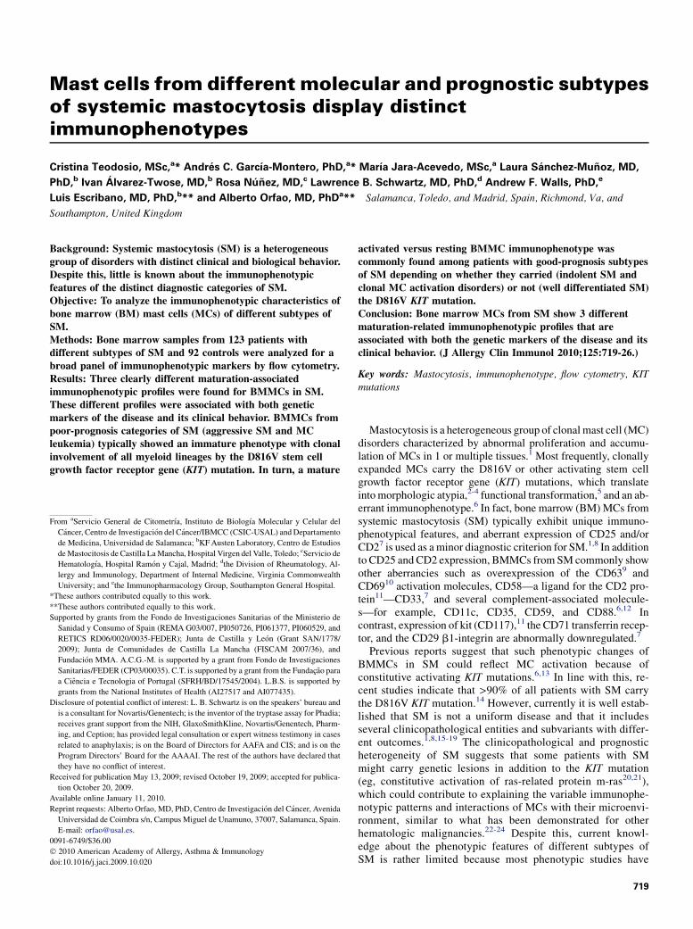
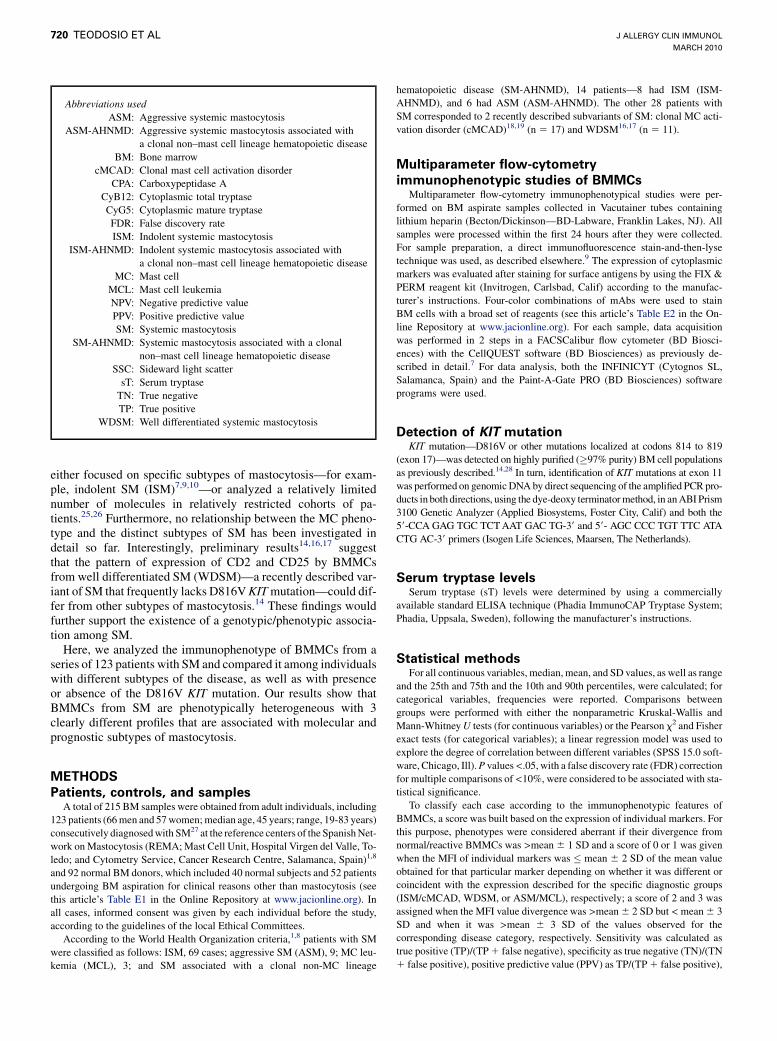
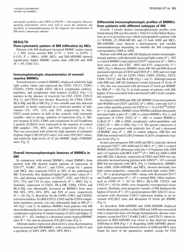
patients with SM showed similar patterns of expression ofCD117hi, FceRI1, HLA-I1, and CD34-, except for 1 patientwith MCL who expressed CD34 in 28% of the pathologicalMC. Conversely, they displayed higher light scatter values (P <.01), and aberrant expression of CD25hi, CD2, and CD123 in93%, 72%, and 73% of cases, respectively (P � .0001; Fig 1).Similarly, expression of CD22, HLA-DR, CD64, CD16, andHLA-DQ, was abnormally increased on BMMCs from mostSM: 96%, 85%, 84%, 69%, and 58% of cases, respectively(P � .02; Fig 2). Reactivity for the CD69, CD63, and CD203cactivation markers, FcgRII (CD32), CD45 and the CD59 comple-ment regulatory protein, was also abnormally high in SM (P <.05; Figs 1 and 2). In addition, BMMCs from patients with SMshowed abnormally low expression of CyB12 with both increasedcytoplasmic expression of mature tryptase (CyG5) and higher sTvalues (P 5 .02), leading to a decreased serum tryptase/BMMCratio (P 5 .01) and an increased sT/CyB12 ratio (Fig 3).
Aberrant marker expression allowed for a clear discriminationbetween normal and SM BMMCs, with a sensitivity of 98.1% anda specificity of 100% (PPV, 100%; NPV, 80%).
Differential immunophenotypic profiles of BMMCs
from patients with different subtypes of SMOverall, 3 clearly distinct immunophenotypic profiles were
found among SM (see this article’s Table E3 in the Online Repos-itory at www.jacionline.org), which corresponded to patients with(1) WDSM, (2) ISM/cMCAD, and (3) both ASM and MCL;SM-AHNMD cases showed a heterogeneous and variableimmunophenotype depending on whether the SM componentcorresponded to ISM or ASM.
Patients with ISM and cMCAD displayed similar immunophe-notypic patterns, with aberrantly increased light scatter (P� .003vs control BMMCs) and uniform CD25hi expression (P < .0001);most cases were also CD21 (88% and 81%, respectively; P <.0001; Fig 1). Moreover, BMMCs from cMCAD and ISM showedincreased expression of CD16 and CD45 (P < .005) and very highreactivity (P � .01) for CD59, CD63, CD69, CD203c, CD32,CD64, CD123, and HLA-DR (Figs 1 and 2). Although patientswith ISM and cMCAD displayed overall increased sT levels (P< .04), this was associated with decreased sT levels per BMMCfor ISM (P 5 .02; Fig 3); in both groups of patients with SM,higher sT was associated with a decreased CyB12 (total cytoplas-mic tryptase).
In contrast with ISM and cMCAD, BMMCs from most patientswith WDSM were CD25- and CD2- (P < .0001), with only 4 of 11cases either partially positive for CD25 (n 5 3) or CD2dim/CD25-
(n 5 1). In addition, BMMCs from WDSM exhibited abnormallyincreased light scatter (P � .006 vs control BMMCs; Fig 1), andexpression of CD16, CD22 (P 5 .003 vs control BMMCs),CyBcl2 (P 5 .006 vs ISM), cytoplasmic carboxypeptidase, andCyB12 (P 5 .003 vs ISM; Figs 1-3). The greater expression ofCyB12 was associated with relatively low sT and a decreasedsT/BMMC ratio (P � .008 vs control subjects, cMCAD, andISM) but normal total (CyB12)/mature (CyG5) cytoplasmic tryp-tase levels (Fig 3).
In turn, poor-prognosis SM (ASM and MCL) typically showedan aberrant CD251 (9/9 ASM and 2/3 MCL; P � .001 vs controlBMMCs) but CD2- phenotype with only 1 of 9 patients with ASMand 1 of 3 patients with MCL CD2dim (P < .0001 for ASM vs ISMand cMCAD). Likewise, expression of CD63 and CD69 was sig-nificantly increased among patients with ASM (P� .02 vs normalBM) but not patients with MCL (Fig 1). Furthermore, BMMCsfrom ASM, ASM-AHNMD, and MCL, showed aberrantly lowlight scatter properties—especially sideward light scatter (SSC;P � .02 vs good-prognosis SM)—along with decreased CD117and FceRI expression (P � .03 vs normal BM). Other markers,such as CD59 (P 5 .02 for MCL), HLA-DR, CD123, andCD32 (P < .02 for ASM), were frequently overexpressed versuscontrols. Similarly, poor-prognosis variants of SM displayed thehighest sT levels (P� .03 vs good-prognosis SM; Table E1) in as-sociation with decreased CyB12 expression leading to an in-creased sT/CyB12 ratio and decreased sT levels per BMMC(Fig 3).
As described, ISM-AHNMD and ASM-AHNMD displayedsimilar phenotypic profiles to their ISM and ASM non-associatedwith a clonal non-mast cell lineage hematopoietic disease coun-terparts, except for CD117, FceRI, CyB12, and CD123, whose ex-pression in ISM-AHNMD was closer to that observed amongASM than to ISM (Figs 1-3). Regarding ASM-AHNMD, pheno-typic features intermediate between those of ASM and MCL werefound for most of the parameters studied, except for CD2
J ALLERGY CLIN IMMUNOL
MARCH 2010
722 TEODOSIO ET AL
J ALLERGY CLIN IMMUNOL
VOLUME 125, NUMBER 3
TEODOSIO ET AL 723
(P 5 .04), CD16 (P 5 .03), and CyG5, which showed a higher andmore heterogeneous expression in ASM-AHNMD versus ASM(Figs 1-3).
Based on the expression of individual phenotypic markers,prediction of the specific subtype of SM (WDSM vs ISM/cMCAD vs ASM/MCL) could be achieved with a high sensitivity(67%, 86%, and 100%, respectively) and specificity (100%, 86%,and 88%, respectively; PPV, 100%, 94%, and 62%, respectively;NPV, 96%, 71%, and 100%, respectively). Associations betweenthe immunophenotype of clonal BMMCs from the distinctsubtypes of SM and the clinical features of the disease (sT levels,presence of hepatomegaly and/or splenomegaly) have also beenfound (see the Online Results section of this article in the OnlineRepository at www.jacionline.org).
Frequency and impact of KIT mutations on the
immunophenotype of BMMCs in SMMost patients with SM analyzed (97/109; 89%) displayed KIT
mutations, except for WDSM, for which only 3 of 10 cases carriedthe KIT mutation (the D816V mutation was positive in only 1 ofthese patients; P < .05 vs all SM variants except MCL; TableE1). Interestingly, the frequency of cases carrying KIT mutation re-stricted to the MC compartment was significantly higher in thegood-prognosis (ISM and cMCAD) versus poor-prognosis variants(ASM, MCL, or SM-AHNMD; P < .05 for ASM vs ISM andcMCAD and, ASM-AHNMD vs cMCAD), which also displayedKIT mutation in other nucleated BM myeloid cells (Table E1).
Among those patients who displayed KIT mutations, 95% car-ried the D816V KIT mutation whereas other KIT mutations weredetected in isolated cases (D816Y, I817V, V819Y, and V560Galong with a VI815-816 insertion, corresponding to a patientwith MCL, a patient with WDSM, and 2 patients with cMCAD,respectively). Overall, no significant phenotypic differences(P > .05) were found within each subtype of SM for D816V1
BMMCs versus BMMCs showing either other mutations in theactivating loop of KIT (n 5 4) or no KIT mutational changes atthe loci examined (n 5 6). However, the only patient withcMCAD showing the V560G KIT juxtamembrane mutation dis-played unique phenotypic features—CD25-/dim, CyBcl2hi,CD2-, and HLA-Idim—versus other cMCAD cases.
DISCUSSIONSystemic mastocytosis is a clinically and prognostically heter-
ogeneous group of disorders1,8,16-19 characterized by the clonalexpansion of immunophenotypically aberrant MCs in the pa-tients’ BM.6,7,9-11 However, little is known about the specific im-munophenotypic features of the distinct variants of SM. Here, weanalyzed the expression of a broad panel of functional proteins onBMMCs from a large cohort of patients with SM compared withnormal/reactive BMMCs. Overall, aberrant phenotypes wereidentified in all patients with SM, with 3 clearly distinct profilestypically associated with (1) the most common good-prognosiscategories of SM (cMCAD and ISM), (2) WDSM, and (3) caseswith poor-prognosis subtypes of SM (ASM and MCL).
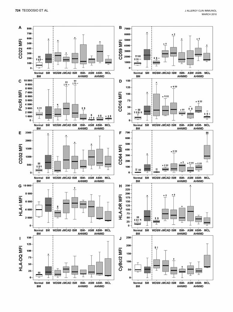
FIG 1. Light scatter (forward light scatter (FSC), A; side
characteristics (C-J) of BMMCs from adults with differe
gated as CD117hi/CD451 with intermediate-to-high scat
and FDR 10% versus *controls, �WDSM, �cMCAD, §IS
��SM, §§all other groups except SM, and jjjjall other g
:
Currently, aberrant expression of CD25 and/or CD2 onBMMCs represents the only immunophenotypical criterionused in the diagnostic work-up of SM.1,8 BMMCs from bothcMCAD and ISM showed a typically CD251/CD21 aberrant,mature (eg, FceRIhi) phenotype associated with overexpressionof the CD63, CD69, and CD203c activation markers and theCD64 high-affinity IgG Fc receptor (FcgRI). Interestingly,CD64 is normally absent in resting BMMCs29 but is expressedupon cytokine exposure (INF-g).30,31 Similarly, BMMCs frompatients with cMCAD and ISM also showed increased expressionof MHC class II molecules (HLA-DR and HLA-DQ) that are typ-ically negative in resting mouse and human MCs but upregulatedon activated MCs isolated from tissues infected with pathogensand/or stimulated with cytokines—for example, TNF-a andINFg—and LPS.32-34 Altogether, these results suggest thatBMMCs from patients with cMCAD and ISM display a pheno-typic profile similar to that of activated mature MCs, with aberrantexpression of CD2 and CD25. Because virtually every patientwithin these subgroups of SM carried the D816V KIT mutation,14
which leads to constitutive activation of kit,13 this mutation couldbe responsible for the aberrant activated phenotype of BMMCs inboth groups of SM. This hypothesis would be further supportedby the absence of phenotypic differences between BMMCsfrom patients with cMCAD with the D816V KIT mutation versusother mutations in the tyrosine kinase loop domain of KIT,whereas the only (cMCAD) patient carrying the V560G mutationin the juxtamembrane domain displayed a clearly different immu-nophenotypical profile.
In contrast with ISM and cMCAD, BMMCs from several othersubtypes of SM did not show a CD251/CD21 phenotype. Thus,BMMCs from WDSM were typically CD25-/CD2-, as previouslyreported in individual cases16 and small groups of patients.17 Fur-thermore, BMMCs from WDSM also showed normal expressionof the CD59, CD203c, and/or CD63 activation markers, which aretypically overexpressed by BMMCs from other subgroups ofSM.6,9,10,12 In fact, BMMCs from WDSM showed a phenotypesimilar to that of normal resting mature BMMCs, with strongexpression of CD117 and FceRI.6,35 In turn, aberrant phenotypesexpressed by WDSM were restricted to a few number of cytoplas-mic antigens (eg, CyBcl2, carboxypeptidase A [CPA], andtryptase). The increased expression of CyBcl2 (Fig 3) couldreflect an altered regulation of apoptosis associated with in-creased survival of MCs36 in WDSM, as previously suggestedfor cutaneous mastocytosis.37 In turn, the greater amount of cyto-plasmic enzymes (eg, tryptase and CPA) could contribute to thetypical hypergranulated morphologic appearance and the abnor-mally increased SSC of BMMCs in WDSM. Overexpression ofcytoplasmic tryptase (CyB12) in association with relatively lowsT could reflect an impaired secretion phenotype with a signifi-cantly decreased release of tryptase per BMMCs in WDSMversus normal BM. Of note, the overall increased cytoplasmiclevels of tryptase—pro and mature a/b-tryptase identified bythe B12 mAb—detected in WDSM were associated with a normaltotal/mature tryptase ratio (CyB12/CyG5). Altogether, theseresults suggest that spontaneous secretion of protryptase could
ward light scatter (SSC), B) and immunophenotypic
nt subtypes of SM versus normal/reactive BMMCs,
ter. MFI, Mean fluorescence intensity/BMMC. P <.05
M, jjISM-AHNMD, {ASM, **ASM-AHNMD, ��MCL,
roups except SM and WDSM.
J ALLERGY CLIN IMMUNOL
MARCH 2010
724 TEODOSIO ET AL
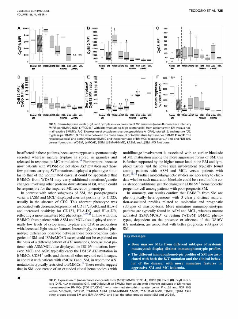
FIG 3. Serum tryptase levels (mg/L) and cytoplasmic expression of MC enzymes (mean fluorescence intensity
[MFI]) per BMMC (CD117hi/CD451 with intermediate-to-high scatter cells) from patients with SM versus nor-
mal/reactive BMMCs. A-C, Expression of cytoplasmic carboxypeptidase A (CPA), total (B12) and mature (G5)
tryptase per BMMC. D, The ratio between the mean amount of total/mature tryptase per BMMC. E and F, The
ratio between sT and both CyB12 per BMMC and the percentage of BMMCs, respectively. P <.05 and FDR 10%
versus *controls, �WDSM, �cMCAD, §ISM, jjISM-AHNMD, {ASM, and ��SM. ND, Not done.
J ALLERGY CLIN IMMUNOL
VOLUME 125, NUMBER 3
TEODOSIO ET AL 725
be affected in these patients, because protryptase is spontaneouslysecreted whereas mature tryptase is stored in granules andreleased in response to MC stimulation.38 Furthermore, becausemost patients with WDSM did not show KIT mutation and thosefew patients carrying KIT mutations displayed a phenotype simi-lar to that of the nonmutated cases, it could be speculated thatBMMCs from WDSM may carry additional mutations/geneticchanges involving other proteins downstream of kit, which couldbe responsible for the impaired MC secretion phenotype.
In contrast with other subgroups of SM, the poor-prognosisvariants (ASM and MCL) displayed aberrant positivity for CD25,usually in the absence of CD2. This aberrant phenotype wasassociated with decreased expression of CD117, FceRI, and HLA-Iand increased positivity for CD123, HLA-DQ, and HLA-DR,reflecting a more immature MC phenotype.6,39-41 In line with this,BMMCs from patients with ASM and MCL also displayed abnor-mally low levels of cytoplasmic tryptase and CPA in associationwith decreased light scatter features. Interestingly, the marked phe-notypic differences observed between these poor-prognosis cate-gories of SM and ISM/cMCAD cases could not be explained onthe basis of a different pattern of KIT mutations, because most pa-tients with ASM/MCL also displayed the D816V mutation; how-ever, MCL and ASM typically carry the D816V KIT mutation inBMMCs, CD341 cells, and almost all other myeloid cell lineages,in contrast with patients with cMCAD and ISM, in whom the KITmutation is typically restricted to BMMCs.14 These results suggestthat in SM, occurrence of an extended clonal hematopoiesis with
FIG 2. Expression of (mean fluorescence intensity [MF
tors (D-F), HLA molecules (G-I), and CyBcl2 (J) on BMM
normal/reactive BMMCs (CD117hi/CD451 with interme
versus *controls, �WDSM, �cMCAD, §ISM, jjISM-AHN
other groups except SM and ISM-AHNMD, and jjjjall th
:
multilineage involvement is associated with an earlier blockadeof MC maturation among the more aggressive forms of SM; thisis further supported by the higher tumor load in the BM and lym-phoid tissues and the lower skin involvement typically foundamong patients with ASM and MCL versus patients withISM.15,42 Further molecular/genetic studies are necessary to eluci-date whether such maturation blockade could be a result of the co-existence of additional genetic changes in a D816V1 hematopoieticprogenitor cell among patients with poor-prognosis SM.
In summary, our results confirm that BMMCs from SM arephenotypically heterogeneous with 3 clearly distinct matura-tion-associated profiles related to molecular and prognosticsubtypes of mastocytosis. More immature immunophenotypicpatterns are typically found in ASM and MCL, whereas matureactivated (ISM/cMCAD) or resting (WDSM) BMMC pheno-types, dependent on the presence or absence of the D816VKIT mutation, are associated with better prognostic subtypes ofSM.
Key messages
d Bone marrow MCs from different subtypes of systemicmastocytosis display distinct immunophenotypic profiles.
d The different immunophenotypic profiles of SM are asso-ciated with both the KIT mutation and the clinical behav-ior of the disease, with more immature features inaggressive SM and MC leukemia.
I]/BMMC) CD22 (A), CD59 (B), FceRI (C), FcgR recep-
Cs from adults with different subtypes of SM versus
diate-to-high scatter cells). P < .05 and FDR 10%
MD, {ASM, **ASM-AHNMD, ��MCL, ��SM, §§all
e other groups except SM and WDSM.
J ALLERGY CLIN IMMUNOL
MARCH 2010
726 TEODOSIO ET AL
REFERENCES
1. Valent P, Horny HP, Escribano L, Longley BJ, Li CY, Schwartz LB, et al. Diagnos-
tic criteria and classification of mastocytosis: a consensus proposal. Leuk Res
2001;25:603-25.
2. Sperr WR, Escribano L, Jordan JH, Schernthaner GH, Kundi M, Horny HP, et al.
Morphologic properties of neoplastic mast cells: delineation of stages of maturation
and implication for cytological grading of mastocytosis. Leuk Res 2001;25:529-36.
3. Stevens EC, Rosenthal NS. Bone marrow mast cell morphologic features and he-
matopoietic dyspoiesis in systemic mast cell disease. Hematopathology 2001;116:
117-82.
4. Samorapoompichit P, Schernthaner GH, Worda C, Wimazal F, Krauth MT, Sperr
WR, et al. Evaluation of neoplastic human mast cells by tryptase-immunoelectron
microscopy. Histopathology 2006;48:247-57.
5. Schernthaner GH, Jordan JH, Ghannadan M, Agis H, Bevec D, Nunez R, et al. Ex-
pression, epitope analysis, and functional role of the LFA-2 antigen detectable on
neoplastic mast cells. Blood 2001;98:3784-92.
6. Escribano L, Garcıa-Montero A, Nunez R, Orfao A. Flow cytometric analysis of
normal and neoplastic mast cells: role in diagnosis and follow-up of mast cell dis-
ease. Immunol Allergy Clin North Am 2006;26:535-47.
7. Escribano L, Orfao A, Dıaz-Agustin B, Villarrubia J, Cervero C, Lopez A, et al.
Indolent systemic mast cell disease in adults: immunophenotypic characterization
of bone marrow mast cells and its diagnostic implications. Blood 1998;91:2731-6.
8. Valent P, Horny H-P, Li CY, Longley JB, Metcalfe DD, Parwaresch RM, et al. Mas-
tocytosis (mast cell disease). In: Jaffe ES, Harris NL, Stein H, Vardiman JW, eds.
World Health Organization (WHO) classification of tumours. Pathology and genet-
ics of tumours of haematopoietic and lymphoid tissues. Lyon, France: IARC Press;
2001. p. 291-302.
9. Escribano L, Orfao A, Dıaz Agustın B, Cervero C, Herrero S, Villarrubia J, et al.
Human bone marrow mast cells from indolent systemic mast cell disease constitu-
tively express increased amounts of the CD63 protein on their surface. Cytometry
1998;34:223-8.
10. Dıaz-Agustın B, Escribano L, Bravo P, Herrero S, Nunez R, Navalon R, et al. The
CD69 early activation molecule is overexpressed in human bone marrow mast cells
from adults with indolent systemic mast cell disease. Br J Haematol 1999;106:400-5.
11. Escribano L, Dıaz-Agustın B, Nunez R, Prados A, Rodrıguez R, Orfao A. Abnor-
mal expression of CD antigens in mastocytosis. Int Arch Allergy Immunol 2002;
127:127-32.
12. Nunez-Lopez R, Escribano L, Schernthaner GH, Prados A, Rodrıguez-Gonzalez R,
Dıaz-Agustın B, et al. Overexpression of complement receptors and related anti-
gens on the surface of bone marrow mast cells in patients with systemic mastocy-
tosis. Br J Haematol 2003;120:257-65.
13. Orfao A, Garcia-Montero AC, Sanchez L, Escribano L. Recent advances in the un-
derstanding of mastocytosis: the role of KIT mutations. Br J Haematol 2007;138:
12-30.
14. Garcia-Montero AC, Jara-Acevedo M, Teodosio C, Sanchez ML, Nunez R, Prados A,
et al. KIT mutation in mast cells and other bone marrow hematopoietic cell lineages
in systemic mast cell disorders: a prospective study of the Spanish Network on Mas-
tocytosis (REMA) in a series of 113 patients. Blood 2006;108:2366-72.
15. Horny HP, Sotlar K, Valent P. Mastocytosis: state of the art. Pathobiology 2007;74:
121-32.
16. Akin C, Fumo G, Yavuz AS, Lipsky PE, Neckers L, Metcalfe DD. A novel form of
mastocytosis associated with a transmembrane c-kit mutation and response to im-
atinib. Blood 2004;103:3222-5.
17. Akin C, Escribano L, Nunez R, Garcıa-Montero A, Angulo M, Orfao A, et al. Well-
differentiated systemic mastocytosis: a new disease variant with mature mast cell
phenotype and lack of codon 816 c-Kit mutations [abstract]. J Allergy Clin Immu-
nol 2004;113(Suppl 2):S327.
18. Bonadonna P, Perbellini O, Passalacqua G, Caruso B, Colarossi S, Dal Fior D, et al.
Clonal mast cell disorders in patients with systemic reactions to Hymenoptera
stings and increased serum tryptase levels. J Allergy Clin Immunol 2009;123:
680-6.
19. Akin C, Scott LM, Kocabas CN, Kushnir-Sukhov N, Brittain E, Noel P, et al. Dem-
onstration of an aberrant mast-cell population with clonal markers in a subset of
patients with ‘‘idiopathic’’ anaphylaxis. Blood 2007;110:2331-3.
20. Guo X, Schrader KA, Xu Y, Schrader JW. Expression of a constitutively active mu-
tant of M-Ras in normal bone marrow is sufficient for induction of a malignant
mastocytosis/mast cell leukemia, distinct from the histiocytosis/monocytic leuke-
mia induced by expression of activated H-Ras. Oncogene 2005;24:2330-42.
21. Guo X, Stratton L, Schrader JW. Expression of activated M-Ras in hemopoietic
stem cells initiates leukemogenic transformation, immortalization and preferential
generation of mast cells. Oncogene 2006;25:4241-4.
22. De Zen L, Orfao A, Cazzaniga G, Masiero L, Cocito MG, Spinelli M, et al. Quan-
titative multiparametric immunophenotyping in acute lymphoblastic leukemia: cor-
relation with specific genotype, I: ETV6/AML1 ALLs identification. Leukemia
2000;14:1225-31.
23. Tabernero MD, Bortoluci AM, Alaejos I, Lopez-Berges MC, Rasillo A, Garcıa-
Sanz R, et al. Adult precursor B-ALL with BCR/ABL gene rearrangements dis-
plays a unique immunophenotype based on the pattern of CD10, CD34, CD13
and CD38 expression. Leukemia 2001;15:406-14.
24. Orfao A, Chillon MC, Bortoluci AM, Lopez-Berges MC, Garcıa-Sanz R, Gonzalez
M, et al. The flow cytometric pattern of CD34, CD15 and CD13 expression in acute
myeloblastic leukemia is highly characteristic of the presence of PML-RARalpha
gene rearrangements. Haematologica 1999;84:405-12.
25. Pardanani A, Kimlinger T, Reeder T, Li C-Y, Tefferi A. Bone marrow mast cell im-
munophenotyping in adults with mast cell disease: a prospective study of 33 pa-
tients. Leuk Res 2004;28:777-83.
26. Cervero C, Escribano L, San Miguel JF, Dıaz-Agustın B, Bravo P, Villarrubia J,
et al. Expression of Bcl-2 by human bone marrow mast cells and its overexpression
in mast cell leukemia. Am J Hematol 1999;60:191-5.
27. Escribano L, Alvarez-Twose I, Sanchez-Munoz L, Garcia-Montero A, Nunez R,
Almeida J, et al. Prognosis in adult indolent systemic mastocytosis: a long-term
study of the Spanish Network on Mastocytosis in a series of 145 patients. J Allergy
Clin Immunol 2009;124:514-21.
28. Sotlar K, Escribano L, Landt O, M€ohrle S, Herrero S, Torrelo A, et al. One-step
detection of c-kit point mutations using peptide nucleic acid-mediated polymer-
ase chain reaction clamping and hybridization probes. Am J Pathol 2003;162:
737-46.
29. Escribano L, Dıaz-Agustın B, Bellas C, Navalon R, Nunez R, Sperr WR, et al. Util-
ity of flow cytometric analysis of mast cells in the diagnosis and classification of
adult mastocytosis. Leuk Res 2001;25:563-70.
30. Woolhiser MR, Okayama Y, Gilfillan AM, Metcalfe DD. IgG-dependent activation
of human mast cells following up-regulation of FcgammaRI by IFN-gamma. Eur J
Immunol 2001;31:3298-307.
31. Marshall JS. Mast-cell responses to pathogens. Nat Rev Immunol 2004;4:787-99.
32. Henz BM, Maurer M, Lippert U, Worm M, Babina M. Mast cells as initiators of
immunity and host defense. Exp Dermatol 2001;10:1-10.
33. Galli SJ, Nakae S, Tsai M. Mast cells in the development of adaptive immune re-
sponses. Nat Immunol 2005;6:135-42.
34. Stelekati E, Orinska Z, Bulfone-Paus S. Mast cells in allergy: innate instructors of
adaptive responses. Immunobiology 2007;212:505-19.
35. Orfao A, Escribano L, Villarrubia J, Velasco JL, Cervero C, Ciudad J, et al.
Flow cytometric analysis of mast cells from normal and pathological human
bone marrow samples: identification and enumeration. Am J Pathol 1996;149:
1493-9.
36. Alfredsson J, Puthalakath H, Martin H, Strasser A, Nilsson G. Proapoptotic Bcl-2
family member Bim is involved in the control of mast cell survival and is induced to-
gether with Bcl-XL upon IgE-receptor activation. Cell Death Differ 2005;12:136-44.
37. Hartmann K, Artuc M, Baldus SE, Zirbes TK, Hermes B, Thiele J, et al. Expres-
sion of Bcl-2 and Bcl-xL in cutaneous and bone marrow lesions of mastocytosis.
Am J Pathol 2003;163 (Suppl 2):819-26.
38. Schwartz LB, Min HK, Ren S, Xia HZ, Hu J, Zhao W, et al. Tryptase precursors
are preferentially and spontaneously released, whereas mature tryptase is retained
by HMC-1 cells, Mono-Mac-6 cells, and human skin-derived mast cells. J Immu-
nol 2003;170:5667-73.
39. Schernthaner GH, Hauswirth AW, Baghestanian M, Agis H, Ghannadan M, Worda
C, et al. Detection of differentiation- and activation-linked cell surface antigens on
cultured mast cell progenitors. Allergy 2005;60:1248-55.
40. Matarraz S, Lopez A, Barrena S, Fernandez C, Jensen E, Flores J, et al. The im-
munophenotype of different immature, myeloid and B-cell lineage-committed
CD341 hematopoietic cells allows discrimination between normal/reactive and
myelodysplastic syndrome precursors. Leukemia 2008;22:1175-83.
41. Rottem M, Okada T, Goff JP, Metcalfe DD. Mast cells cultured from the peripheral
blood of normal donors and patients with mastocytosis originate from a CD341/Fc
epsilon RI- cell population. Blood 1994;84:2489-96.
42. Okayama Y, Kawakami T. Development, migration, and survival of mast cells.
Immunol Res 2006;34:97-115.

REFERENCE
E1. Schwartz LB. Diagnostic value of tryptase in anaphylaxis and mastocytosis.
Immunol Allergy Clin North Am 2006;26:451-63.
J ALLERGY CLIN IMMUNOL
VOLUME 125, NUMBER 3
TEODOSIO ET AL 726.e1
RESULTS
Association between the immunophenotype of
clonal BMMCs and other disease featuresPatients with ISM with sT levels �20 mg/L showed higher
BMMC counts (P 5 .0002) and lower reactivity for CD25 (P 5
.02) and FceRI (P 5 .01); in addition, a significant correlationwas found between sT and the mean amount of CD123 (r2 5
.31; P 5 .04) per BMMC. Likewise, patients with ISM displayinghepatomegaly and/or splenomegaly had lower expression ofFceRI (P 5 .005) and CD32 (P 5 .04), together with increasedtryptase levels (P 5 .009). Similarly, patients with cMCADwith high sT levels (�20 mg/L) showed increased forward light
scatter (P 5 .04), CD59 (P 5 .04), and CD69 (P 5 .02) expres-sion; within these patients, sT levels were also significantly asso-ciated with CD64 expression (r2 5 .55; P 5 .004).
Among patients with ASM, sT levels showed a significantcorrelation with the expression of CD63 (r2 5 .65; P 5 .03),CD69 (r2 5 .69; P 5 .01), and CyB12 (r2 5 .81; P 5 .006) perBMMC.
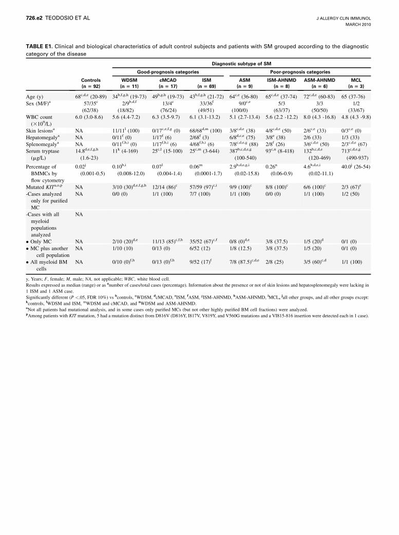
TABLE E1. Clinical and biological characteristics of adult control subjects and patients with SM grouped according to the diagnostic
category of the disease
Diagnostic subtype of SM
Good-prognosis categories Poor-prognosis categories
Controls
(n 5 92)
WDSM
(n 5 11)
cMCAD
(n 5 17)
ISM
(n 5 69)
ASM
(n 5 9)
ISM-AHNMD
(n 5 8)
ASM-AHNMD
(n 5 6)
MCL
(n 5 3)
Age (y) 68c,d,e (20-89) 34b,f,g,h (19-73) 49b,g,h (19-73) 43b,f,g,h (21-72) 64c,e (36-80) 65c,d,e (37-74) 72c,d,e (60-83) 65 (37-76)
Sex (M/F)a 57/35c
(62/38)
2/9b,d,f
(18/82)
13/4c
(76/24)
33/36f
(49/51)
9/0c,e
(100/0)
5/3
(63/37)
3/3
(50/50)
1/2
(33/67)
WBC count
(3109/L)
6.0 (3.0-8.6) 5.6 (4.4-7.2) 6.3 (3.5-9.7) 6.1 (3.1-13.2) 5.1 (2.7-13.4) 5.6 (2.2 -12.2) 8.0 (4.3 -16.8) 4.8 (4.3 -9.8)
Skin lesionsa NA 11/11l (100) 0/17c,e,f,g (0) 68/68d,m (100) 3/8c,d,e (38) 4/8c,d,e (50) 2/6c,e (33) 0/3c,e (0)
Hepatomegalya NA 0/11f (0) 1/17f (6) 2/68f (3) 6/8d,c,e (75) 3/8e (38) 2/6 (33) 1/3 (33)
Splenomegalya NA 0/11f,h,i (0) 1/17f,h,i (6) 4/68f,h,i (6) 7/8c,d,e,g (88) 2/8f (26) 3/6c,d,e (50) 2/3c,d,e (67)
Serum tryptase
(mg/L)
14.8d,e,f,g,h
(1.6-23)
11k (4-169) 25c,l (15-100) 25c,m (3-644) 387b,c,d,e,g
(100-540)
93c,n (8-418) 132b,c,d,e
(120-469)
713c,d,e,g
(490-937)
Percentage of
BMMCs by
flow cytometry
0.02j
(0.001-0.5)
0.10b,i
(0.008-12.0)
0.07l
(0.004-1.4)
0.06m
(0.0001-1.7)
2.9b,d,e,g,i
(0.02-15.8)
0.26n
(0.06-0.9)
4.6b,d,e,i
(0.02-11.1)
40.0j (26-54)
Mutated KITa,o,p NA 3/10 (30)d,e,f,g,h 12/14 (86)c 57/59 (97)c,i 9/9 (100)c 8/8 (100)c 6/6 (100)c 2/3 (67)e
-Cases analyzed
only for purified
MC
NA 0/0 (0) 1/1 (100) 7/7 (100) 1/1 (100) 0/0 (0) 1/1 (100) 1/2 (50)
-Cases with all
myeloid
populations
analyzed
NA
� Only MC NA 2/10 (20)d,e 11/13 (85)c,f,h 35/52 (67)c,f 0/8 (0)d,e 3/8 (37.5) 1/5 (20)d 0/1 (0)
� MC plus another
cell population
NA 1/10 (10) 0/13 (0) 6/52 (12) 1/8 (12.5) 3/8 (37.5) 1/5 (20) 0/1 (0)
� All myeloid BM
cells
NA 0/10 (0)f,h 0/13 (0)f,h 9/52 (17)f 7/8 (87.5)c,d,e 2/8 (25) 3/5 (60)c,d 1/1 (100)
y, Years; F, female; M, male; NA, not applicable; WBC, white blood cell.
Results expressed as median (range) or as anumber of cases/total cases (percentage). Information about the presence or not of skin lesions and hepatosplenomegaly were lacking in
1 ISM and 1 ASM case.
Significantly different (P <.05, FDR 10%) vs bcontrols, cWDSM, dcMCAD, eISM, fASM, gISM-AHNMD, hASM-AHNMD, iMCL, jall other groups, and all other groups except:kcontrols, lWDSM and ISM, mWDSM and cMCAD, and nWDSM and ASM-AHNMD.oNot all patients had mutational analysis, and in some cases only purified MCs (but not other highly purified BM cell fractions) were analyzed.pAmong patients with KIT mutation, 5 had a mutation distinct from D816V (D816Y, I817V, V819Y, and V560G mutations and a VI815-816 insertion were detected each in 1 case).
J ALLERGY CLIN IMMUNOL
MARCH 2010
726.e2 TEODOSIO ET AL
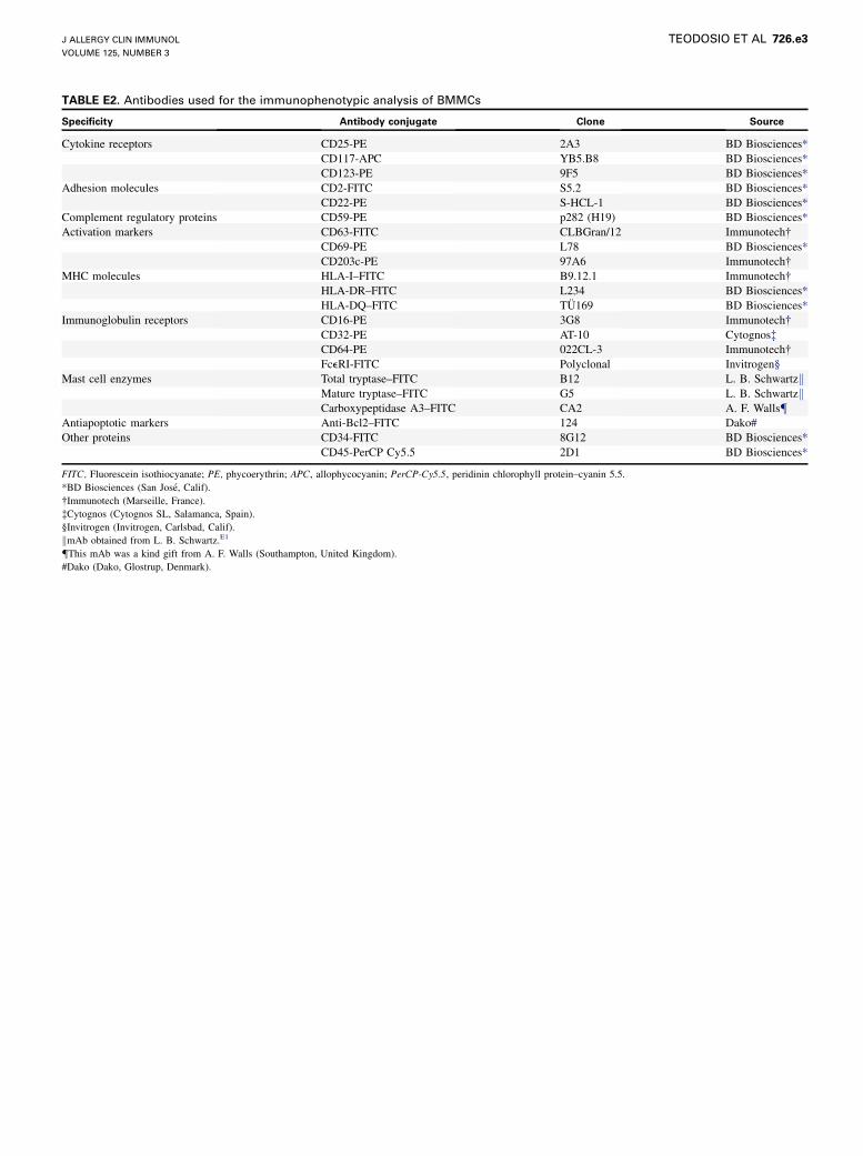
TABLE E2. Antibodies used for the immunophenotypic analysis of BMMCs
Specificity Antibody conjugate Clone Source
Cytokine receptors CD25-PE 2A3 BD Biosciences*
CD117-APC YB5.B8 BD Biosciences*
CD123-PE 9F5 BD Biosciences*
Adhesion molecules CD2-FITC S5.2 BD Biosciences*
CD22-PE S-HCL-1 BD Biosciences*
Complement regulatory proteins CD59-PE p282 (H19) BD Biosciences*
Activation markers CD63-FITC CLBGran/12 Immunotech�CD69-PE L78 BD Biosciences*
CD203c-PE 97A6 Immunotech�MHC molecules HLA-I–FITC B9.12.1 Immunotech�
HLA-DR–FITC L234 BD Biosciences*
HLA-DQ–FITC TU169 BD Biosciences*
Immunoglobulin receptors CD16-PE 3G8 Immunotech�CD32-PE AT-10 Cytognos�CD64-PE 022CL-3 Immunotech�FceRI-FITC Polyclonal Invitrogen§
Mast cell enzymes Total tryptase–FITC B12 L. B. SchwartzkMature tryptase–FITC G5 L. B. SchwartzkCarboxypeptidase A3–FITC CA2 A. F. Walls{
Antiapoptotic markers Anti-Bcl2–FITC 124 Dako#
Other proteins CD34-FITC 8G12 BD Biosciences*
CD45-PerCP Cy5.5 2D1 BD Biosciences*
FITC, Fluorescein isothiocyanate; PE, phycoerythrin; APC, allophycocyanin; PerCP-Cy5.5, peridinin chlorophyll protein–cyanin 5.5.
*BD Biosciences (San Jose, Calif).
�Immunotech (Marseille, France).
�Cytognos (Cytognos SL, Salamanca, Spain).
§Invitrogen (Invitrogen, Carlsbad, Calif).
kmAb obtained from L. B. Schwartz.E1
{This mAb was a kind gift from A. F. Walls (Southampton, United Kingdom).
#Dako (Dako, Glostrup, Denmark).
J ALLERGY CLIN IMMUNOL
VOLUME 125, NUMBER 3
TEODOSIO ET AL 726.e3
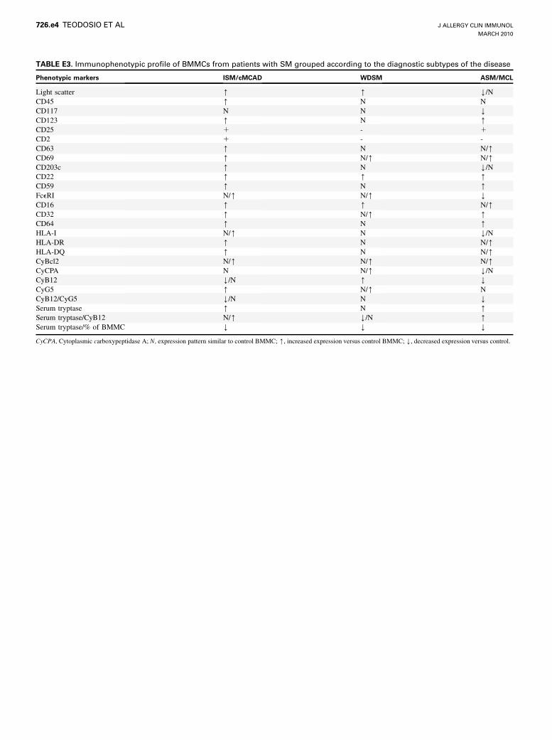
TABLE E3. Immunophenotypic profile of BMMCs from patients with SM grouped according to the diagnostic subtypes of the disease
Phenotypic markers ISM/cMCAD WDSM ASM/MCL
Light scatter [ [ Y/N
CD45 [ N N
CD117 N N YCD123 [ N [CD25 1 - 1
CD2 1 - -
CD63 [ N N/[CD69 [ N/[ N/[CD203c [ N Y/N
CD22 [ [ [CD59 [ N [FceRI N/[ N/[ YCD16 [ [ N/[CD32 [ N/[ [CD64 [ N [HLA-I N/[ N Y/N
HLA-DR [ N N/[HLA-DQ [ N N/[CyBcl2 N/[ N/[ N/[CyCPA N N/[ Y/N
CyB12 Y/N [ YCyG5 [ N/[ N
CyB12/CyG5 Y/N N YSerum tryptase [ N [Serum tryptase/CyB12 N/[ Y/N [Serum tryptase/% of BMMC Y Y Y
CyCPA, Cytoplasmic carboxypeptidase A; N, expression pattern similar to control BMMC; [, increased expression versus control BMMC; Y, decreased expression versus control.
J ALLERGY CLIN IMMUNOL
MARCH 2010
726.e4 TEODOSIO ET AL


![[Guidelines for the diagnosis, treatment and management of mastocytosis]](https://img.dokumen.tips/doc/110x75/635edba5dcf4a1629e036655/guidelines-for-the-diagnosis-treatment-and-management-of-mastocytosis.jpg)


