Embed Size (px)
Citation preview
Zhirong Fu, Srinivas Akula, Michael Thorpe and Lars Hellman*
Marked difference in efficiency of the digestiveenzymes pepsin, trypsin, chymotrypsin, andpancreatic elastase to cleave tightly foldedproteinshttps://doi.org/10.1515/hsz-2020-0386Received December 16, 2020; accepted April 30, 2021;published online May 12, 2021
Abstract: In order for the intestinal mucosa to absorb di-etary proteins they have to be digested into single aminoacids or very short peptides of a length of not more thanfour amino acids. In order to study the efficiency of thedigestive endopeptidases to digest folded proteins we haveanalyzed several target proteins under different condi-tions, native proteins, heat denatured and acid treated. Thethree pancreatic serine proteases, trypsin, chymotrypsin,and pancreatic elastase, were found to be remarkableinefficient in cleaving native folded proteins whereaspepsin, which acts at a very low pH (pH 1.2) wasmuchmoreefficient, possibly due to the denaturing conditions andthereby better accessibility to internal cleavage sites at thelow pH. Heat treatment improved the cleavage consider-ably by all three pancreatic enzymes, but acid treatmentfollowed by return to neutral pH did not have any majoreffect. Cleavage at the low pH when the protein is in adenatured state, is apparently very efficient. This indicatesthat pepsin is the prime enzyme cleaving the properlyfolded native proteins and that the pancreatic enzymesprimarily are involved in generating single amino acids orvery short peptides for efficient uptake by the intestinalmucosa.
Keywords: chymotrypsin; digestive enzymes; digestivesystem; pancreatic elastase; pepsin; trypsin.
Introduction
To efficiently use proteins as a food source they have to betransformed into single amino acids or very short peptidesof a length of not more than four amino acids. This isperformed by a number of different proteases of ourdigestive system. Polysaccharides start to be enzymaticallydigested already by the saliva, which contains the enzymeamylase, whereas proteins are first attacked by proteasesin the stomach (Chauncey et al. 1963; Janiak 2016; Meislerand Ting 1993). In the stomach the acidic environmentdenatures the proteins, whichmost likely has an importantrole in making internal peptide bonds more accessible forcleavage. The hydrochloric acid released by the acidglands of the stomach lowers the pH to below two. Theprotease of this intestinal compartment is pepsin, anaspartic protease with a pH optimum of between pH 1 and2, and therefore optimized for the environment in thestomach (Janiak 2016; Kageyama 2002). Following thedigestion in the stomach, the fully or partly digested foodenters the small intestine. There the pH is returned toneutral pH by the secretion of bicarbonate from duct cellsof the pancreas (Ishiguro et al. 2012). When the food passesthe duodenum a number of proteases also enter the in-testinal canal from the pancreas (Goettig et al. 2019;Guyonnet et al. 1999). There are enzymes with multiplespecificities including both endo- and exopeptidases. Theendopeptidases have specificities for different amino acidsat the P1 site, the amino acid after which the enzymecleaves. Trypsin cleaves after basic (positively charged)amino acids, chymotrypsin after aromatic amino acids andpancreatic elastase after aliphatic amino acids and afterpolar residues such as Ser and Thr. There are also severalcarboxypeptidases that cleave at the carboxy terminal endof the proteins.
In a recent study of proteases that are expressed byimmune cells, we have observed that they are stronglyaffected by the folding of the protein for efficient cleavage.Sites exposed on the surface were targeted by these en-zymes, whereas optimal sites were left uncleaved if hiddenin the structure (Fu et al. 2017). Our study therefore showed
*Corresponding author: Lars Hellman, Department of Cell andMolecular Biology, Uppsala University, Uppsala, The BiomedicalCenter, Box 596, S-751 24 Uppsala, Sweden,E-mail: [email protected]. https://orcid.org/0000-0003-1459-3815Zhirong Fu, Srinivas Akula and Michael Thorpe, Department of Celland Molecular Biology, Uppsala University, Uppsala, The BiomedicalCenter, Box 596, S-751 24 Uppsala, Sweden
Biol. Chem. 2021; 402(7): 861–867
Open Access.© 2021 Zhirong Fu et al., published by De Gruyter. This work is licensed under the Creative Commons Attribution 4.0 InternationalLicense.
that accessibility is a major factor for efficient cleavage bythese enzymes. Our question was therefore how similar ordissimilar the digestive enzymes are when it comes tofolding. These hematopoietic serine proteases belong tothe same protease family as the pancreatic serine pro-teases, trypsin, chymotrypsin, and pancreatic elastase. Wedecided to study four of the digestive enzymes for theircleavages of native properly folded proteins to see if thisfeature of these endopeptidases was a more general char-acteristic; a factor that may have a major impact on theevolution of an acidic environment and enzymes active at alow pH, as a prominent part of the digestive tract of mostmulticellular organisms.
Results
Target molecules
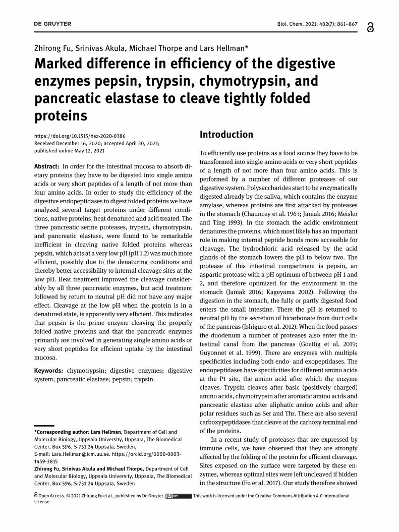
To study the importance of folding on the cleavage ofdietaryproteins we first used a type of target protein used to studycleavage specificity; a recombinant protein consistingof twocopies of the tightly folded Escherichia coli redox protein,thioredoxin (Trx) (Figure 1A). In a linker region betweenthese two copies a kinker regionwas inserted consisting of afew repeated Gly-Ser motifs and a nine amino acid regionwith a sequence susceptible for cleavage by an enzymewithtrypsin, chymotrypsin, or elastase specificity, respectively(Figure 1A and B). Following the second Trx, a region withsix His residues for easy purification of the recombinantprotein on IMAC Ni2+ chelating columns was inserted(Figure 1A). The benefit of this system is that one cansimultaneously analyze cleavage of an open and a closedstructurally-folded region in one experiment; the linker re-gion is open and linear whereas the Trx domains are tightlyfolded. This type of substrate has been very successful inobtaining quantitative information concerning the impor-tance of amino acids at and surrounding the cleavage site ofa number of hematopoietic serine proteases, and onthrombin (Gallwitz et al. 2010, 2012; Thorpe et al. 2012, 2016,2018a,b). As examples of a native folded protein the cleav-age of bovine serum albumin (BSA), chicken egg albumin(ovalbumin), and for the cleavagebypepsinalso a sample ofcow saliva were included.
Cleavage by pepsin at pH 1.2
We first analyzed the cleavage by pepsin at pH 1.2, which isthe physiological pH for this enzyme, of three different2xTrx substrates, one with a linker region with a trypsin
susceptible sequence, one with a chymotrypsin and onewith an elastase susceptible sequence (Figure 1C). Thetrypsin sequence has an Arg in central position, thechymotrypsin sequence a Phe in central position and theelastase sequence a Val in that position (Figure 1C).
Figure 1: Cleavage of target molecules by pepsin at pH 1.2.The efficiency in cleavage by pepsin at its physiologic pH wasanalyzed against three different 2xTrx substrates as well as BSA,ovalbumin and deglycosylated whole bovine saliva. The cleavagewas performed in glycine buffer at pH 1.2. (A) A schematic picture ofthe 2xTrx construct where the region inserted by double-strandedoligonucleotides is marked by ‘selected sequence’. The sequencesinserted and analyzed for cleavage by pepsin in panel C are listedabove each cleavage reaction, (B) a schematic picture of a standardcleavage reaction. Bands at ∼28 kDa represent uncleavedtwo-thioredoxin (Trx) protein and the two bands present at ∼14 kDarepresent cleaved protein, with the upper of the two bandsrepresenting the Trx containing the His6-tag. Sequences above thepanels in panel C represent the inserted octamers and numbersrepresent the reaction time in minutes, (C) analysis of the cleavageof three different 2xTrx substrates and of BSA by pepsin. The linkerregion of the 2xTrx substrates were almost completely cleaved afteronly 15 min and the tightly folded Trx domains were almostcompletely cleaved after 150 min. BSA was efficiently cleavedalready after 15 min, and (D) analysis of the cleavage of total bovinesaliva (deglycosylated) and of chicken egg ovalbumin by pepsin. Thesaliva was incubated with pepsin for 30 min and the ovalbumin for15, 45, and 150 min. As ovalbumin was only partially digested, BSAcleavedwith the same enzyme for 15 and 150minwas included in theassay to verify that the enzyme used to cleave ovalbuminwas active.
862 Z. Fu et al.: Cleavage of tightly folded proteins by intestinal proteases
The linker region was cleaved slightly faster thanthe tightly folded Trx sequences, however, both werealmost completely hydrolyzed after 150 min of incubation(Figure 1C). BSA was also very efficiently cleavedand almost no visible traces after 150 min of incubation(Figure 1C). Here, pepsin was shown to be very efficient incleaving both linear and tightly folded structures, which isa key characteristic of efficient food digestion. Pepsin wasalso analyzed for the cleavage of cow saliva that had beentreated with a panel of deglycosylation enzymes to allowthe proteins to more easily enter the gel as the content ofhighly glycosylated mucins make the saliva viscous. Thedeglycosylated saliva was the cleaved with pepsin for30 min and as can be seen from the figure almost all theproteins were almost completely digested (Figure 1D). Wealso analyzed the cleavage of ovalbumin by pepsin and toour surprise ovalbumin showed a quite different behavior.One part of the protein was rapidly digested whereas oneother part was remarkably stable against the action ofpepsin indicating that this part of the molecule has astructure quite compact and stable even at pH 1.2, showingthat some proteins may resist the cleavage by pepsin atpH 1.2. For these assays we used an enzyme to substrateratio of approximately 1–10 as also can be seen from the gel(Figure 1). The enzyme is visible as a faint band comparedto the more abundant targets (Figure 1).
Cleavage of 2xTrx substrates by trypsin,chymotrypsin, and pancreatic elastase at pH7.2
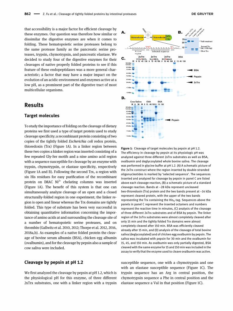
The cleavage of 2xTrx substrates with a preferred linkerregion for respective enzyme(s) was analyzed at the phys-iological pH of the duodenum, which is estimated to bearound pH 7. A trypsin substrate with a central Arg wasused for trypsin, a chymotrypsin substrate with a centralPhe for chymotrypsin and an elastase substrate with acentral Val for the pancreatic elastase were used to deter-mine the difference in cleavage activity on linear versustightly folded regions of the substrate (Figure 2A–C). Thesesubstrates were analyzed under three different conditions.In the first panel, the enzyme and substrate mixture wereincubated without any pre-treatment. In the second panel,the protein had first been heat denatured at 95 °C for 10minafter which the temperature was returned to 37 °C for thecleavage analysis. In the third panel, the target protein hadbeen incubated at pH 2.0 for 30min where after the sample
Figure 2: Cleavage of 2xTrx substrates by chymotrypsin, pancreaticelastase, and trypsin at pH 7.2, after different pretreatments of thesubstrate.Three different substrates, one each for the three differentpancreatic serine proteaseswereused todetermine thedifference incleavage efficiency of linear and tightly folded substrates by theseenzymes. The cleavage was performed without pretreatment, after a10min incubation at 95 °Cand after a 30min incubation at pH2.0. (A)Cleavage by trypsin, (B) cleavage by chymotrypsin, and (C) cleavageby pancreatic elastase. The linker regions were very efficientlycleaved after 15 min, whereas the tightly folded Trx domains werealmost untouched even after 150 min, both in the absence of pre-treatment and after a short pH drop to pH 2.0. However, the heatdenaturation seems to give a substantial improvement in thecleavage also of the Trx domains by chymotrypsin and elastase butonly aminor effect on the cleavageby trypsin. To show the specificityin target selection of the three enzymes we analyzed their cleavageof a panel of 2xTrx substrates with different P1 residues.
Z. Fu et al.: Cleavage of tightly folded proteins by intestinal proteases 863
was pH adjusted back to neutral pH at 7.2 for cleavageanalysis. The results showed the linker regions were effi-ciently cleaved by all three enzymes already at 15 min ofincubation, whereas the tightly folded regions were veryresistant to digestion by all three enzymes (Figure 2). Apreincubation at pH 2.0 for 30 min did not markedlychange this pattern (Figure 2B). However, heat denatur-ation significantly affected the cleavage, most likely due topartly denaturing the protein, which would open targetsites for more efficient cleavage. We also observed areduced cleavage of the linker region for both trypsin andchymotrypsin, possibly due to denaturation of the proteinmay have resulted in shielding of the open structure ofthe linker region. The ration between enzyme and targetwas approximately 500 times for trypsin, 300 times forchymotrypsin, and 25 for pancreatic elastase. These valueswere chosen based on the activity against the linker regionin initial experiments to obtain the best discriminatingconcentrations for the assay. This gives also indications tothe overall activity of the different enzymes indicatingconsiderably higher activity, per molar basis, of trypsinand chymotrypsin compared to pancreatic elastase andpepsin.
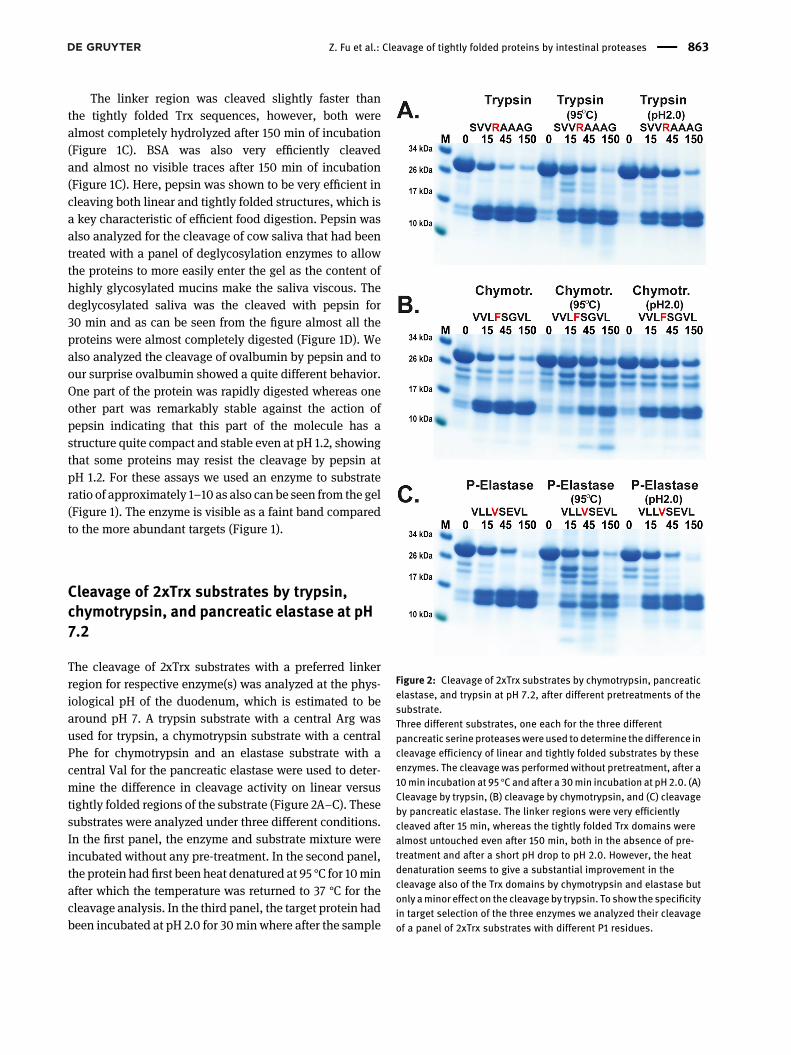
To confirm the correct cleavage sites within the linkerregion between the two Trx molecules all three enzymeswere analyzed with a panel of 2xTrx substrates withdifferent P1 residues. As can be seen from the figure trypsinonly cleaves the substrate with a centrally positionedArg and not the chymase nor the elastase substrates(Figure 3A). Similarly, chymotrypsin only cleaves thesubstrate with a centrally positioned aromatic amino acid(Figure 3B). In contrast, the pancreatic elastase was foundto be more unrestrictive and also cleave at other aminoacids than only the classical aliphatic residues, Val, Ile,and Ala or in the region of the linker close to the Trxsequences (Figure 3C).
Cleavage of BSA and ovalbumin bychymotrypsin, and by a mix of trypsin,chymotrypsin, and pancreatic elastase atpH 7.2
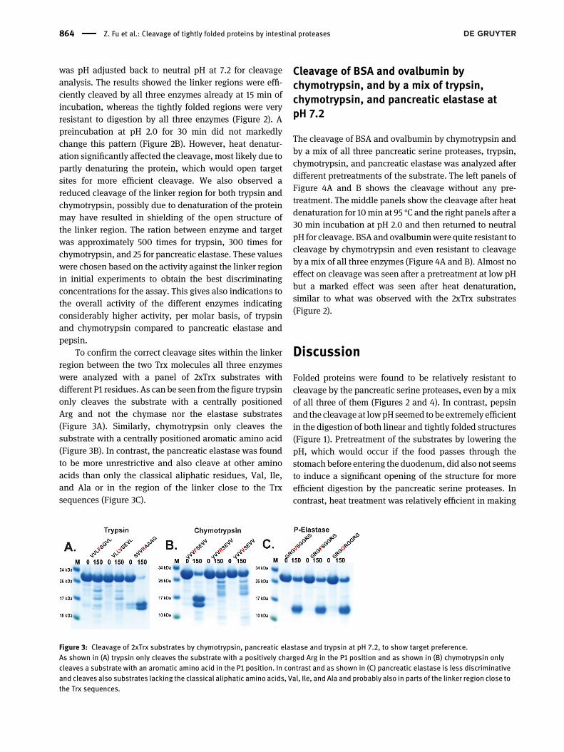
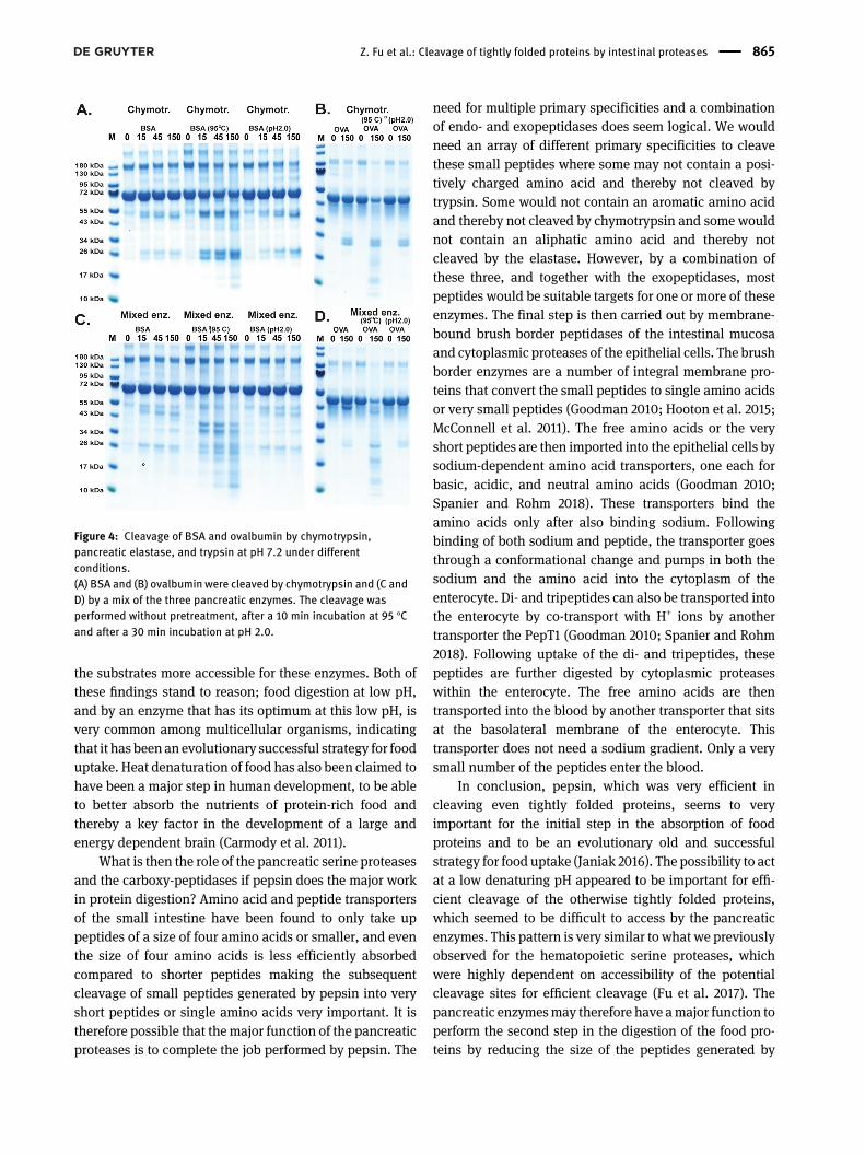
The cleavage of BSA and ovalbumin by chymotrypsin andby a mix of all three pancreatic serine proteases, trypsin,chymotrypsin, and pancreatic elastase was analyzed afterdifferent pretreatments of the substrate. The left panels ofFigure 4A and B shows the cleavage without any pre-treatment. The middle panels show the cleavage after heatdenaturation for 10min at 95 °C and the right panels after a30 min incubation at pH 2.0 and then returned to neutralpH for cleavage. BSA and ovalbuminwere quite resistant tocleavage by chymotrypsin and even resistant to cleavageby a mix of all three enzymes (Figure 4A and B). Almost noeffect on cleavage was seen after a pretreatment at low pHbut a marked effect was seen after heat denaturation,similar to what was observed with the 2xTrx substrates(Figure 2).
Discussion
Folded proteins were found to be relatively resistant tocleavage by the pancreatic serine proteases, even by a mixof all three of them (Figures 2 and 4). In contrast, pepsinand the cleavage at lowpH seemed to be extremely efficientin the digestion of both linear and tightly folded structures(Figure 1). Pretreatment of the substrates by lowering thepH, which would occur if the food passes through thestomach before entering the duodenum, did also not seemsto induce a significant opening of the structure for moreefficient digestion by the pancreatic serine proteases. Incontrast, heat treatment was relatively efficient in making
Figure 3: Cleavage of 2xTrx substrates by chymotrypsin, pancreatic elastase and trypsin at pH 7.2, to show target preference.As shown in (A) trypsin only cleaves the substrate with a positively charged Arg in the P1 position and as shown in (B) chymotrypsin onlycleaves a substrate with an aromatic amino acid in the P1 position. In contrast and as shown in (C) pancreatic elastase is less discriminativeand cleaves also substrates lacking the classical aliphatic amino acids, Val, Ile, and Ala and probably also in parts of the linker region close tothe Trx sequences.
864 Z. Fu et al.: Cleavage of tightly folded proteins by intestinal proteases
the substrates more accessible for these enzymes. Both ofthese findings stand to reason; food digestion at low pH,and by an enzyme that has its optimum at this low pH, isvery common among multicellular organisms, indicatingthat it has been an evolutionary successful strategy for fooduptake. Heat denaturation of food has also been claimed tohave been a major step in human development, to be ableto better absorb the nutrients of protein-rich food andthereby a key factor in the development of a large andenergy dependent brain (Carmody et al. 2011).
What is then the role of the pancreatic serine proteasesand the carboxy-peptidases if pepsin does the major workin protein digestion? Amino acid and peptide transportersof the small intestine have been found to only take uppeptides of a size of four amino acids or smaller, and eventhe size of four amino acids is less efficiently absorbedcompared to shorter peptides making the subsequentcleavage of small peptides generated by pepsin into veryshort peptides or single amino acids very important. It istherefore possible that the major function of the pancreaticproteases is to complete the job performed by pepsin. The
need for multiple primary specificities and a combinationof endo- and exopeptidases does seem logical. We wouldneed an array of different primary specificities to cleavethese small peptides where some may not contain a posi-tively charged amino acid and thereby not cleaved bytrypsin. Some would not contain an aromatic amino acidand thereby not cleaved by chymotrypsin and some wouldnot contain an aliphatic amino acid and thereby notcleaved by the elastase. However, by a combination ofthese three, and together with the exopeptidases, mostpeptides would be suitable targets for one or more of theseenzymes. The final step is then carried out by membrane-bound brush border peptidases of the intestinal mucosaand cytoplasmic proteases of the epithelial cells. The brushborder enzymes are a number of integral membrane pro-teins that convert the small peptides to single amino acidsor very small peptides (Goodman 2010; Hooton et al. 2015;McConnell et al. 2011). The free amino acids or the veryshort peptides are then imported into the epithelial cells bysodium-dependent amino acid transporters, one each forbasic, acidic, and neutral amino acids (Goodman 2010;Spanier and Rohm 2018). These transporters bind theamino acids only after also binding sodium. Followingbinding of both sodium and peptide, the transporter goesthrough a conformational change and pumps in both thesodium and the amino acid into the cytoplasm of theenterocyte. Di- and tripeptides can also be transported intothe enterocyte by co-transport with H+ ions by anothertransporter the PepT1 (Goodman 2010; Spanier and Rohm2018). Following uptake of the di- and tripeptides, thesepeptides are further digested by cytoplasmic proteaseswithin the enterocyte. The free amino acids are thentransported into the blood by another transporter that sitsat the basolateral membrane of the enterocyte. Thistransporter does not need a sodium gradient. Only a verysmall number of the peptides enter the blood.
In conclusion, pepsin, which was very efficient incleaving even tightly folded proteins, seems to veryimportant for the initial step in the absorption of foodproteins and to be an evolutionary old and successfulstrategy for food uptake (Janiak 2016). The possibility to actat a low denaturing pH appeared to be important for effi-cient cleavage of the otherwise tightly folded proteins,which seemed to be difficult to access by the pancreaticenzymes. This pattern is very similar to what we previouslyobserved for the hematopoietic serine proteases, whichwere highly dependent on accessibility of the potentialcleavage sites for efficient cleavage (Fu et al. 2017). Thepancreatic enzymesmay therefore have amajor function toperform the second step in the digestion of the food pro-teins by reducing the size of the peptides generated by
Figure 4: Cleavage of BSA and ovalbumin by chymotrypsin,pancreatic elastase, and trypsin at pH 7.2 under differentconditions.(A) BSA and (B) ovalbumin were cleaved by chymotrypsin and (C andD) by a mix of the three pancreatic enzymes. The cleavage wasperformed without pretreatment, after a 10 min incubation at 95 °Cand after a 30 min incubation at pH 2.0.
Z. Fu et al.: Cleavage of tightly folded proteins by intestinal proteases 865
pepsin. Both hematopoietic and pancreatic endopepti-dases thereby seem to show many similarities concerningthe effect of folding on the efficiency of substrate cleavage.As a third and fourth step, the brush border enzymes andthe cytoplasmic proteases of the enterocytes finish thesequence by cleaving the small peptides into single aminoacids for final transport into the blood.
We could also show that some proteins are remarkableresistant to the cleavage by pepsin at pH 1.2, as shown forone part of ovalbumin and for pepsin itself, so some pro-teins may have an amino acid composition that makesthem more compact and thereby more stable at low pH.However, we could also see that after heat treatmentovalbumin was relatively efficiently cleaved by a combi-nation of the pancreatic enzymes indicating that the com-bination of the different digestive enzymes, pepsin and thepancreatic enzymes, are of importance for the cleavage ofthe majority of proteins of the food.
It is also important to say that although pepsin is veryefficient in cleaving tightly folded proteins and that anacidic environment and proteases that are able to act effi-ciently at this low pH persons with complete gastrectomycan live a relatively normal life indicating pepsin hydro-lysis is not absolutely necessary for survival and that acombination of the pancreatic enzymes can be sufficientwhen acting together for our survival (Goodman 2010).
Materials and methods
Enzymes
Digestive enzymes used for analysis were all purchased fromSigma–Aldrich (Sigma–Aldrich Sweden AB, Stockholm, Sweden):Pepsin A from porcine gastric mucosa (Sigma P-6887), α-Chymotrypsinfrombovine pancreas (Sigma C-3142), Beta-trypsin frombovine pancreas(Sigma T-1426), and Elastase (Pancreatopeptidase E) from porcinepancreas (Sigma E-1250). Chymotrypsin and trypsin were dissolved withPBS, while pepsin was dissolved with 0.1 M HCl, 1% NaCl (pH 1.2).
Target molecules
A new type of recombinant substrate was used to study the importanceof folding on the cleavage of dietary proteins. Two copies of the E. coliTrx gene were inserted in tandem into the pET-21 vector. Between thetwo Trx molecules, a nine amino acid region was inserted with asequence susceptible for cleavage by trypsin, chymotrypsin, or elastaserespectively. For purification a His6-tag was also inserted in theC-terminal. The sequences of the individual clones were verified aftercloning by sequencing of both DNA strands. The plasmids were thentransformed into the E. coli Rosetta gami strain for protein expression(Novagen, Merck, Darmstadt, Germany). A 10 ml overnight culture oftransformed expression clone was diluted 10 times in LB + ampicillin
andgrown for 1–2 h at 37 °Cuntil theOD (600nm) reached0.5. IPTGwasthen added to a final concentration of 1 mM. The culture was subse-quently grown at 37 °C for an additional 3 h. After incubation, thebacteria were pelleted by centrifugation at 3000 rpm for 12 min. Thepelletwas thenwashedoncewith 25mlPBS+0.05%Tween 20and thenresuspended in 2 ml PBS. The pellet was sonicated 5 × 30 s to open thecells. The lysate was centrifuged at 13,000 rpm for 3 min and the su-pernatant was transferred to a new tube. Five hundred microliters ofNi-NTA slurry (50:50) (Qiagen, Hilden, Germany) was added and thesample was gently rotated for 45 min at 4 °C. The Ni-NTA beads werethen transferred to a 2ml columnandwerewashedwith 1ml, then 2 and2 ml of washing buffer (PBS + 0.05% Tween 20 + 20 mM imidazole).Proteinwas then eluted by adding 100 µl elution buffer followed by fiveadditional 200 µl volumes of elution buffer (PBS + 0.05% Tween20 + 100 mM imidazole). Each fraction was collected individually and10 µl from each elution fraction was then mixed with 2.5 µl of 4 × LDSloading buffer for SDS-PAGE analysis (Invitrogen, Carlsbad, CA, USA).The fractions containing the most protein were pooled together and theconcentration of the combined fractions was then determined by Bio-Rad DC Protein assay (Bio-Rad Laboratories Hercules, CA, USA).
Cleavage reactions
Approximately 25 µg of recombinant 2xTrx protein or BSA was addedto each 50 µl cleavage reaction (in PBS). For the cleavage analysis ofheat denatured proteins, 25 µg recombinant 2-Trx protein or BSA in50 µl PBS was pre-heated at 95 °C for 10min. For the cleavage analysisof acid denatured proteins, 25 µg recombinant 2xTrx protein in elutionbuffer or 25 µg BSA in H2Owas firstly adjusted with 0.1 MHCl to pH 2.0and incubated for 30min. Five µl of 10×PBS and0.1MNaOHwere thenadded to adjust the pH back to 7.2. Sterile H2O was finally added to50 µl in total. Native or denatured 2xTrx proteins, BSA, deglycosylatedcow saliva, or ovalbuminwas thenmixedwith active enzyme (48 ng oftrypsin or 80 ng of chymotrypsin or 2 µg of pancreatic elastase or 4.2 ug[or 16 U] of pepsin). Based on gel intensity the ratio between pepsinand targetmolecule seems to be close to 1–10which is a slightly highervalue compared to the value of the enzyme concentration given by thedistributing company. The reaction was kept at room temperatureduring the entire experiment and 10 µl of sample was removed at theindicated time points (0, 15, 45, and 150 min) and the reaction wasterminated by addition of 2.5 µl of 4× sample buffer and 0.5 ulβ-mercaptoethanol. The samples were then heat treated for 5 min at85 °C and analyzed on 4–12% pre-cast SDS-PAGE gels (Invitrogen,Carlsbad, CA, USA). The gels were stained overnight in colloidalCoomassie staining solution and de-stained with 25% of methanol forat least 3 h and subsequently with H2O until the backgroundwas clear(Neuhoff et al. 1988).
The cow saliva was deglycosylated by incubation with deglyco-sylation mix II (P6044) in deglycosylation buffer II according to theManufacturers recommendation by incubation at room temperaturefor 30 min followed by overnight incubation at 37 °C (New EnglandBiolabs, Ipswich, MA, USA).
Author contributions: All the authors have acceptedresponsibility for the entire content of this submittedmanuscript and approved submission.Research funding: This study was supported by the Knutand Alice Wallenberg Foundation (KAW 2017-0022).
866 Z. Fu et al.: Cleavage of tightly folded proteins by intestinal proteases
Conflict of interest statement: The authors declare thatthey have no conflict of interest regarding the contents ofthis article.
References
Carmody, R.N., Weintraub, G.S., and Wrangham, R.W. (2011).Energetic consequences of thermal and nonthermal foodprocessing. Proc. Natl. Acad. Sci. U.S.A. 108: 19199–19203.
Chauncey, H.H., Henrigues, B.L., and Tanzer, J.M. (1963). Comparativeenzyme activity of saliva from the sheep, hog, dog, rabbit, rat,and human. Arch. Oral Biol. 8: 615–627.
Fu, Z., Thorpe, M., Alemayehu, R., Roy, A., Kervinen, J., de Garavilla, L.,Abrink, M., and Hellman, L. (2017). Highly selective cleavage ofcytokines and chemokines by the human mast cell chymase andneutrophil cathepsin G. J. Immunol. 198: 1474–1483.
Gallwitz, M., Enoksson, M., Thorpe, M., Ge, X., and Hellman, L. (2010).The extended substrate recognition profile of the dog mast cellchymase reveals similarities and differences to the humanchymase. Int. Immunol. 22: 421–431.
Gallwitz, M., Enoksson, M., Thorpe, M., and Hellman, L. (2012). Theextended cleavage specificity of human thrombin. PloS One 7:e31756.
Goettig, P., Brandstetter, H., and Magdolen, V. (2019). Surface loopsof trypsin-like serine proteases as determinants of function.Biochimie 166: 52–76.
Goodman, B.E. (2010). Insights into digestion and absorption ofmajornutrients in humans. Adv. Physiol. Educ. 34: 44–53.
Guyonnet, V., Tluscik, F., Long, P.L., Polanowski, A., and Travis, J.(1999). Purification and partial characterization of the pancreaticproteolytic enzymes trypsin, chymotrypsin, and elastase fromthe chicken. J. Chromatogr. A 852: 217–225.
Hooton, D., Lentle, R.,Monro, J., Wickham,M., andSimpson, R. (2015).The secretion and action of brush border enzymes in themammalian small intestine. Rev. Physiol. Biochem. Pharmacol.168: 59–118.
Ishiguro, H., Yamamoto, A., Nakakuki, M., Yi, L., Ishiguro, M.,Yamaguchi, M., Kondo, S., and Mochimaru, Y. (2012). Physiology
andpathophysiology of bicarbonate secretion by pancreatic ductepithelium. Nagoya J. Med. Sci. 74: 1–18.
Janiak, M.C. (2016). Digestive enzymes of human and nonhumanprimates. Evol. Anthropol. 25: 253–266.
Kageyama, T. (2002). Pepsinogens, progastricsins, andprochymosins: structure, function, evolution, and development.Cell. Mol. Life Sci.: CMLS 59: 288–306,.
McConnell, R.E., Benesh, A.E., Mao, S., Tabb, D.L., and Tyska, M.J.(2011). Proteomic analysis of the enterocyte brush border. Am.J. Physiol. Gastrointest. Liver Physiol. 300: G914–G926.
Meisler, M.H. and Ting, C.N. (1993). The remarkable evolutionaryhistory of the human amylase genes. Crit. Rev. Oral Biol. Med. 4:503–509.
Neuhoff, V., Arold, N., Taube, D., and Ehrhardt, W. (1988). Improvedstaining of proteins in polyacrylamide gels including isoelectricfocusing gels with clear background at nanogram sensitivityusing Coomassie Brilliant Blue G-250 and R-250. Electrophoresis9: 255–262.
Spanier, B. and Rohm, F. (2018). Proton coupled oligopeptidetransporter 1 (PepT1) function, regulation, and influence on theintestinal homeostasis. Comp. Physiol. 8: 843–869.
Thorpe, M., Yu, J., Boinapally, V., Ahooghalandari, P., Kervinen, J.,Garavilla, L.D., and Hellman, L. (2012). Extended cleavagespecificity of the mast cell chymase from the crab-eatingmacaque (Macaca fascicularis): an interesting animal model forthe analysis of the function of the humanmast cell chymase. Int.Immunol. 12: 771–782.
Thorpe, M., Akula, S., and Hellman, L. (2016). Channel catfishgranzyme-like I is a highly specific serine protease with metaseactivity that is expressed by fish NK-like cells. Dev. Comp.Immunol. 63: 84–95.
Thorpe,M., Fu, Z., Albat, E., Akula, S., de Garavilla, L., Kervinen, J., andHellman, L. (2018a). Extended cleavage specificities of mast cellproteases 1 and 2 from golden hamster: classical chymase andan elastolytic protease comparable to rat andmouseMCP-5. PloSOne 13: e0207826.
Thorpe, M., Fu, Z., Chahal, G., Akula, S., Kervinen, J., de Garavilla, L.,and Hellman, L. (2018b). Extended cleavage specificity of humanneutrophil cathepsin G: a low activity protease with dualchymase and tryptase-type specificities. PloS One 13: e0195077.
Z. Fu et al.: Cleavage of tightly folded proteins by intestinal proteases 867


