Embed Size (px)
Citation preview
Nucleic Acids Research, 2015 1doi: 10.1093/nar/gkv070
Luzp4 defines a new mRNA export pathway in cancercellsNicolas Viphakone1, Marcus G. Cumberbatch1,2, Michaela J. Livingstone1, Paul R. Heath3,Mark J. Dickman4, James W. Catto2 and Stuart A. Wilson1,*
1Department of Molecular Biology and Biotechnology, The University of Sheffield, Firth Court, Western Bank,Sheffield, UK, 2Academic Urology Unit, The University of Sheffield, Beech Hill Road, Sheffield, UK, 3SheffieldInstitute for Translational Neuroscience, The University of Sheffield, 385a Glossop Road, Sheffield, UK and4Department of Chemical and Biological Engineering, The University of Sheffield, Mappin Street, Sheffield, UK
Received July 24, 2014; Revised January 12, 2015; Accepted January 19, 2015
ABSTRACT
Cancer testis antigens (CTAs) represented a poorlycharacterized group of proteins whose expression isnormally restricted to testis but are frequently up-regulated in cancer cells. Here we show that oneCTA, Luzp4, is an mRNA export adaptor. It asso-ciates with the TREX mRNA export complex sub-unit Uap56 and harbours a Uap56 binding motif, con-served in other mRNA export adaptors. Luzp4 bindsthe principal mRNA export receptor Nxf1, enhancesits RNA binding activity and complements Alyrefknockdown in vivo. Whilst Luzp4 is up-regulated in arange of tumours, it appears preferentially expressedin melanoma cells where it is required for growth.
INTRODUCTION
The transport of mRNA from the nucleus to the cytoplasmis an essential step in eukaryotic gene expression. It is sub-ject to rigorous quality control to ensure that only fully pro-cessed mature mRNA exits the nucleus (1). A major mRNAexport pathway used in human cells involves the multisub-unit TREX complex which comprises the THO complexand numerous additional subunits (2,3). TREX integratesinformation from transcription (4), splicing (5) and 3′ endprocessing (6,7) to mark mature mRNAs for export. TREXassembly requires Alyref, the THO complex, and is drivenby the Uap56 RNA helicase (3,8). A number of TREX sub-units including Alyref, Chtop and Uif harbour a specificpeptide motif (Uap56 binding motif, UBM) that allows di-rect interaction with Uap56 or its paralogue, Ddx39 (9,10).
Nxf1 is the major metazoan mRNA export receptorwhich functions as a heterodimer with Nxt1 (11). Free Nxf1sequesters its own N-terminal RNA binding domain butupon binding to TREX, the RNA binding domain of Nxf1is exposed allowing direct interactions with the mRNA(12,13). The key TREX subunits responsible for this con-
formational change in Nxf1 (to allow mRNA binding) areAlyref, which binds the N-terminal region of Nxf1, andThoc5 or Chtop, either of which can bind the NTF2-like(NTF2L) domain (aa 371-550) (10,13). A number of otherproteins that function in a manner similar to Alyref in bind-ing Nxf1 have been identified, including Uif, various SRproteins and U2af1 (9,14–16). These interactions probablyensure that multiple Nxf1 molecules are recruited to a sin-gle mRNA, ensuring efficient translocation of the mRNPacross the nuclear pore. Whilst Nxf1 is the major mRNAexport receptor, there exist several other Nxf genes in hu-mans (17) including Nxf2. Notably, Nxf2 destabilizes Nxf1mRNA in testis and neurons and thus Nxf2 may representthe major mRNA export receptor in these tissues (18).
There is increasing evidence that various proteins in-volved in mRNA export are dysregulated in cancer cells(19). These proteins include eIF4E which is up-regulatedin ∼30% of cancer cells. eIF4E is required for the exportof specific transcripts via the CRM1 pathway and also re-models the nuclear pore complex which promotes mRNAexport and oncogenic transformation (20). The TREX sub-unit Thoc5 is the target of leukaemogenic tyrosine kinases(21) and two other TREX subunits, Thoc1 (Hpr1) andAlyref, are dysregulated in cancer cells (22–24). Further-more, the correct packaging of mRNA by TREX is impor-tant to prevent R-loop formation and in TREX mutants, in-creased R-loop formation leads to genome instability (24).The TREX-2 complex, which is implicated in the transferof mRNA from the nuclear interior to the nuclear porecomplex (25), has also been shown to be associated withBRCA-2 and is implicated in maintaining genome stability(26). Together these studies highlight the close link betweenpackaging/export of mRNA and genome stability/cancer.
One important class of proteins implicated in cancer arethe cancer testis antigens (CTAs), which are a diverse collec-tion of proteins characterized by their largely restricted ex-pression in testis in the normal situation and up-regulationin one or more cancers. Many CTAs reside on the X-
*To whom correspondence should be addressed. Tel: +44 114 2222849; Email: [email protected]
C⃝ The Author(s) 2015. Published by Oxford University Press on behalf of Nucleic Acids Research.This is an Open Access article distributed under the terms of the Creative Commons Attribution License (http://creativecommons.org/licenses/by/4.0/), whichpermits unrestricted reuse, distribution, and reproduction in any medium, provided the original work is properly cited.

2 Nucleic Acids Research, 2015
chromosome and a key element in their induction in can-cer cells appears to be promoter demethylation (27). Theunique expression characteristics of CTAs means they rep-resent a promising group of targets both as biomarkers andtherapeutic targets in the treatment of cancer. However, de-spite the identification of a wide variety of CTAs, in generalthe molecular functions of this class of proteins are poorlycharacterized though Nxf2 is classified as a CTA (28). Here,we show that the CTA Luzp4 is an mRNA export factor re-quired for efficient growth of melanoma cells.
MATERIALS AND METHODS
Plasmids, antibodies and cell culture
Plasmids encoding CBP80-Myc, MS2-GFP, MS2-Alyref,MS2-Nxf1, pLUCSALRRE6MS2, GST-Uap56, GB1-Nxf1 and truncations are described previously (12) aswere FLAG-Nxf1-myc, FLAG-GFP, FLAG-Srsf1-myc,GST-Nxf1:p15 (13). FLAG-Uap56-myc was generatedby subcloning the Uap56 open reading frame into p3X-FLAG-Myc-CMV26 (Sigma). Luzp4 cDNA (NM 016383)used for all constructs was purchased from Source Bio-science (clone MGC:149218, IMAGE:40112435, locusBC128134). All Luzp4 constructs (wild-type or mutant)used for in vitro pulldowns and UV-crosslinking experi-ments were expressed as 6-His tagged fusions from pET24b(cloned NdeI/XhoI as PCR products). FLAG-Luzp4and Luzp4-myc used in co-immunoprecipitation (Co-IP)experiments were expressed from p3XFLAG-myc-CMV26(cloned HindIII/EcoRI) and pcDNA3.1-myc-HisB(cloned BamHI/XhoI), respectively. To make the FLAG-Luzp4 stable cell line, 3XFLAG-Luzp4 was amplifiedby PCR from p3XFLAG-Luzp4-myc (see above) andcloned NotI/XhoI into a modified pcDNA5-FRT-TO-His where the start codon of the N-terminal 6His tagwas destroyed by site-directed mutagenesis. The cellline was generated as previously described (9). A vectorpEGFPN1-Luzp4 (XhoI/BamHI) was built to monitorGFP-Luzp4 localisation in HeLa cells. This vector wasused as a template to generate the deletion mutant con-structs pEGFPN1-Luzp4(!N) (amino acids 119–313),pEGFPN1-Luzp4(! aa 22–40), pEGFPN1-Luzp4(! aa156–236), pEGFPN1-Luzp4(! aa 156–178) by divergentPCR. pEGFPN1-luzp4(!C) was made by amplifyingLuzp4 region encoding aa 1–240 by PCR and cloning theresulting PCR product into pEGFPN1 (XhoI/BamHI).For MS2 mRNA export assays, Luzp4 was amplified byPCR as an NheI/NotI product and cloned in pCINeo-MS2(XbaI/NotI), described previously (9). Antibodies wereobtained from the following suppliers Thoc1 (Hpr1),Nxf1 and Alyref (Abcam); Ars2 (Santa Cruz); Hnrnpa1(Millipore); FLAG, Tubulin (Sigma); GFP (Clontech). TheThoc5 and Uap56 antibodies have been described previ-ously (13). A rabbit polyclonal antibody was raised againstGST-luzp4(!N) (expressed from pGEX6P1-Luzp4(aa119–313)) and used for western analysis (Figure 5 andSupplementary Figure S8B). Cell lines used in functionalcomplementation experiments were built using the strategydepicted in Supplementary Figure S6B and describedpreviously (10), resulting in the expression of Luzp4 (orits mutant versions) with EmGFP fused to its C-terminus.
miRNAs hairpins also expressed from these constructs andtargeting Alyref and/or Uif were from (9). Growth curveswere performed as described previously (9). To determinestatistical differences in growth between the various celllines examined, we first log10-transformed the numberof cells obtained for each cell line from Day 4 (when thecurves start to scatter) for each independent experiment.The slopes of the linear regressions applied to these valueswere compiled and these growth rates were then comparedby a one-way ANOVA analysis followed by a Tukey test.
siRNA treatments and colony formation assays
For each RNAi condition, three wells of 6-well plateswith 50 000 cells/well were transfected twice with40 nM of the indicated siRNAs and 12 !l Interferin(PolyPlus-transfection R⃝)/well (renewed at 48 h). Con-trol siRNA, targeting dsRED sequence, and ALYREFsiRNA were 5′-CACCGUGAAGCUGAAGGUG-3′ and 5′-GGAACUCUUUGCUGAAUUU-3′, re-spectively (7). Uif knockdown was performed witha pool of two siRNAs purchased from Dharma-con: 5′-ACAUAAACAGUGUCGGAAA-3′, 5′-GCAAAGAGAACUCGUCAAU-3′. Luzp4 knock-down was performed with a pool of two siR-NAs: 5′-GCCUUCAAGACAGCAAUCA-3′, 5′-CAGAAGGAAAUCCGGACAA-3′. At 60–65 h posttransfection (seeding day), each well was phosphatebuffered saline (PBS) washed and treated with 100 !ltrypsin. All three wells of each RNAi condition were theninactivated and pooled with 2 ml of serum-containingmedium. Cell suspensions were homogenized, counted anddiluted to 200 cells/ml.
For each RNAi condition, a new 6-well plate wasseeded at 200 cells/well, therefore representing sixreplicates/condition. Each well was adjusted to 2 mlof culture medium and left to grow at 37◦C for 10 (293Tcells) to 14 days (MeWo cells). On the counting day, cellswere washed once with PBS and stained 5 min with 1ml of crystal violet (0.5 % giemsa powder in methanol)/ well. Each well was then washed twice with 1 ml ofdeionized water and left to dry for at least 15 min. Theentire experiment was repeated five times independentlyand only colonies containing at least 50 cells were counted.One-way ANOVA analysis followed by a Tukey test wasperformed to compare the effect of each siRNA-treatmenton MeWo and 293T cellular proliferation in Figure 6D andSupplementary Figure S8B, respectively.
Luzp4–6His protein expression and purification
pET24b-Luzp4–6His was transformed into Rosetta2 cells.Five litres of 37◦C pre-warmed terrific broth medium (50!g/ml Kanamycin) was inoculated from a 100 ml overnightculture (terrific broth, 50 !g/ml Kanamycin, 34 !g/mlChloramphenicol) to give a starting OD600 of 0.05. Thecells were grown at 37◦C up to OD600 = 0.5. Production ofLuzp4–6His was then induced with 0.5 mM IPTG for threeand a half hours at 37◦C. Cells were harvested and lysed bysonication on ice in binding buffer (50 mM Tris-HCl pH 8, 1M NaCl, 5 mM imidazol, 0.5% Triton-X100, 5% Glycerol,
Nucleic Acids Research, 2015 3
1 mM PMSF, 1X SigmaFASTTM Protease Inhibitors). Thelysate was cleared by centrifugation at 30 000xg for 30 minat 4◦C. The pellet of insoluble material was washed twicewith binding buffer and resuspended in 100 ml of denat-uration buffer (50 mM Tris-HCl pH 8, 500 mM NaCl, 8M Urea) for 20 min at room temperature. Denatured mate-rial was first cleared by centrifugation and then incubatedwith 5 ml of Cobalt beads (TALON R⃝) for 30 min. The col-umn was then washed three times with 20 ml of wash buffer(50 mM Tris-HCl pH 8, 1 M NaCl, 5 mM imidazole, 8 MUrea). Luzp4–6His was eluted with 6 × 5 ml of elutionbuffer (50 mM Tris-HCl pH 8, 1 M NaCl, 200 mM imidazol,8 M Urea). Glycerol and ethylenediaminetetraacetic acid(EDTA) were immediately added to the pooled fractions at10% and 1 mM final, respectively. Finally, the purified pro-tein was immediately dialysed twice against 5 l of dialysisbuffer (50 mM Tris-HCl pH 7.5, 0.5 M NaCl, 0.05% NP40,50 mM L-Arginine, 50 mM L-Glutamate, 10% glycerol). Intotal, ∼40 mg of soluble Luzp4–6His protein was obtainedand kept frozen at −80◦C as aliquots. GB1-Alyref-6His wasproduced and purified as described previously (10).
GST-pulldown experiments and Co-IP
GST-pulldown experiments and Co-IP experiments werecarried out as described previously (10,13) using RB100buffer for GST pulldowns (25 mM HEPES pH 7.5, 100 mMKOAc, 10 mM MgCl2, 1 mM dithiothreitol, 0.05% TritonX-100, 10% glycerol, 10 !g/ml RNase A) and IP lysis bufferfor Co-IPs (50 mM HEPES pH 7.5, 100 mM NaCl, 1 mMEDTA, 1 mM dithiothreitol, 0.5% Triton X-100 and 10%glycerol + (10 !g/ml RNase A) as indicated in figures). ThemRNP capture assays were carried out as described previ-ously (13).
Mass spectrometry analysis
For mass spectrometry analysis, 20 × 15 cm dishes of theFlp-InTM T-RExTM 293 FLAG-Luzp4 or control Flp-InTM
T-RExTM 293 FLAG stable cell lines were induced for 48h with 1 !g/ml of tetracycline and used for the IP withanti-FLAG MS2-agarose beads (Sigma). Interacting part-ners were eluted using 1 M Arginine-HCl (pH 3.5) and sub-sequently brought to pH 7.5 using 1.5 M Tris-HCl pH 8.8.In solution and in gel tryptic digestions were performedas previously described (29,30). Tryptic digests were resus-pended in 0.1% final concentration of Trifluoroacetic acid(TFA). Five microlitres was used for LC-MS/MS analy-sis. Peptides were separated using an Ultimate 3000 RSLCnano liquid chromatography system (ThermoFisher, UK),using a 150 mm × 75 !m I.D. PepMap reversed phasecolumn (ThermoFisher, UK). Linear gradient elution wasperformed from 95% buffer A (0.1% formic acid) to 40%buffer B (0.1% formic acid, 95% acetonitrile) at a flow rateof 300 nl/min in 60 min. MS/MS analysis was performedusing a maXis UHR TOF mass spectrometer (Bruker Dal-tonics) using an automated acquisition approach. MS andMS/MS scans (m/z 50–2000) were acquired in positiveion mode. Lock mass calibration was performed using HP1221.990364. Line spectra data were then processed into
peak list by Data Analysis (Bruker Daltonics) using the fol-lowing settings. The sum peak finder algorithm was usedfor peak detection using a signal-to-noise ratio of 10, a rela-tive to base peak intensity of 0.1% and an absolute intensitythreshold of 100. Spectra were deconvoluted and the peaklists exported as Mascot Generic Files (MGF) and searchedusing Mascot 2.2 server (Matrix Science) The Swiss-Protdatabase (Swiss-Prot Release 10.5, 20 April 2010, 516 604sequences) was searched using the following parameters(analysis peptide tolerance = ± 0.01 Da, MS/MS tolerance= ±0.01 Da and peptide charge 2+ and 3+). Search param-eters were as follows: enzyme; trypsin; variable modifica-tions: deamidation (NQ), oxidation (M); maximum missedcleavages = 1. A peptide ion score of ≤ 10 as a cut-off as cal-culated by Mascot was also used to filter. The false discov-ery rates were determined using the Mascot decoy databaseoption and were ≤1%. Protein identifications were basedon a minimum of two unique peptides. Mass spectrom-etry search results generated by Mascot (*.dat) were im-ported into ProHits Lite-VM 2.0.3 (http://prohitsms.com/)run via Virtual Box 4.3.6 (https://www.virtualbox.org/) onOSX 10.8.5. A comparison was then performed in Prohits(31) between the lists of proteins for Luzp4 IP and ControlIP. Filters used were: Artefacts proteins, Keratin, Albuminand Cytoskeleton. Proteins that passed the filtering and theanalysis are presented in Supplementary Table S1. The pro-teins present in Control IP were then removed from the listof Luzp4 interacting partners. The new filtered list of pro-teins was used to generate the cytoscape diagram (32) de-picted in Figure 2E.
MS2 mRNA export assays
Assays were carried out as described previously (9).
Nxf1 remodelling assays and UV-crosslinking experiments
The Nxf1 remodelling assay and UV cross-linkingexperiments were carried out as described previ-ously (10). The ssDNA used in some assays was 5′-CTCTAGATCAACCACTTTGT-3′. The integrated pixeldensities function of the software ImageJ (33) was used tomeasure and normalize the pixel densities of the phospho-rimage signals to the pixel densities of the correspondingCoomassie-stained gel signals. The radiolabelled RNA-crosslinking fold increase was calculated as the increaseof crosslinking observed for a given condition relative tothe crosslinking observed for the relevant bovine serumalbumin (BSA)-containing reactions (RNA or ssDNA).
Fluorescence in situ hybridisation and immunostaining
Fluorescence in situ hybridisation (FISH) and immunos-taining were carried out as described previously (13). Usingthe ImageJ software (NIH), the nuclear rim of cells in thepoly(A)+ panels of Figure 4A was determined by creatingoutline-masks from the corresponding DAPI images whichwere then applied onto the poly(A)+ signal images. The re-sulting images were used to perform fluorescence intensityscans with a line width of 5 pixels and a length of 56 pixels.To perform the quantifications displayed in Figure 4B and
4 Nucleic Acids Research, 2015
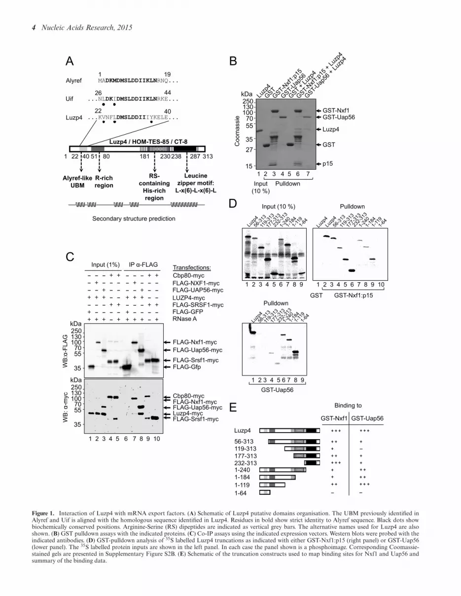
Figure 1. Interaction of Luzp4 with mRNA export factors. (A) Schematic of Luzp4 putative domains organisation. The UBM previously identified inAlyref and Uif is aligned with the homologous sequence identified in Luzp4. Residues in bold show strict identity to Alyref sequence. Black dots showbiochemically conserved positions. Arginine-Serine (RS) dipeptides are indicated as vertical grey bars. The alternative names used for Luzp4 are alsoshown. (B) GST pulldown assays with the indicated proteins. (C) Co-IP assays using the indicated expression vectors. Western blots were probed with theindicated antibodies. (D) GST-pulldown analysis of 35S labelled Luzp4 truncations as indicated with either GST-Nxf1:p15 (right panel) or GST-Uap56(lower panel). The 35S labelled protein inputs are shown in the left panel. In each case the panel shown is a phosphoimage. Corresponding Coomassie-stained gels are presented in Supplementary Figure S2B. (E) Schematic of the truncation constructs used to map binding sites for Nxf1 and Uap56 andsummary of the binding data.

Nucleic Acids Research, 2015 5
Supplementary Figure S9B, DAPI images of wide-field pic-tures (20x Leica objective) were thresholded, binary trans-formed, and cells were automatically counted using the Im-ageJ software. Pictures containing at least 200 cells (Fig-ure 4B) or at least 60 cells (Supplementary Figure S9B)were collected and their corresponding poly(A)+ signal im-ages were thresholded and used to only count cells dis-playing saturated poly(A)+-RNAs nuclear accumulation. Aone-way ANOVA analysis followed by a Fisher’s LSD testwas used to determine which cell lines were prone to dis-play a poly(A)+-RNA export block compared to the Con-trol RNAi cell line.
Luzp4 expression screening in tumour samples
Origene TissueScan Cancer 96-well plates that containedpre-normalized cDNA samples isolated from 18 differentbody tissues were used according to the manufacturer’s in-structions. Supplementary Table S2 provides patient detailsand tumour grades. The PCR primers used were as follows:Alyref forward 5′-GCCTGCACAGAGCGTAAACA-3′,Alyref reverse 5′-CTCGCATTATTATAGGCGTCCAG-3′, Luzp4 forward 5′-CGCCTTCAAGACAGCAATCA-3′, Luzp4 reverse 5′-CACTGCACTTCTCCTGCTCAA-3′, Uif forward 5′-AGCAGTGCAATGCCCAGTAA-3′
and Uif reverse 5′-ACCGCTCATTCAACGTCATC-3′. U1 forward 5′-ACCTGGCAGGGGAGA-3′ andU1 reverse 5′-GGGGAAAGCGCGAAC-3′ Expressionvalues plotted in Supplementary Figure S7 are shown as2−!CT using U1 snRNA as the normalizer.
RESULTS
Luzp4 associates with Nxf1 and TREX subunits
A BLAST search with the UBM from the N-terminal do-main of Alyref (9) identified Luzp4 (also called HOM-TES-85 or CT-8), a CTA with unknown function. Luzp4 is re-ported to reside within nuclear speckles, a site enriched infactors involved in pre-mRNA processing and mRNA ex-port (34). Luzp4 orthologues were clearly identifiable inother animals. However, analysis of the X-chromosome inmice revealed a gene expansion at the locus where Luzp4lies in other species and one Luzp4 paralogue in mice isknown as ovary and testis transcribed (Supplementary Fig-ure S1A). Luzp4 harbours a single UBM in its N-terminaldomain, a central domain rich in arginines, serine dipep-tides and histidines (RS-H) and a C-terminal leucine zip-per motif (Figure 1A and Supplementary Figure S1B). Wefound that following an internal deletion of amino acids (aa)156–236, which encompasses the RS-H region, GFP-Luzp4localized to the cytoplasm, whereas wild-type Luzp4 wasnuclear, suggesting the RS-H region contains the nuclear lo-calisation sequence of Luzp4 (Supplementary Figure S1C).Interestingly, deletion of the UBM region (aa 22–40) alteredthe nuclear distribution of GFP-Luzp4, leading to the for-mation of fewer and larger GFP-Luzp4 foci.
To establish whether Luzp4 directly associated withUap56 or Nxf1, we used GST pulldown assays with pu-rified Luzp4 protein produced in E. coli (Figure 1B). Wefound that both GST-Uap56 and GST-Nxf1 bound Luzp4whereas the GST control did not. We also observed the
Co-IP of Luzp4 with FLAG-Nxf1-myc and FLAG-Uap56-myc from human cells (Figure 1C). This interaction was notaffected by RNase treatment, indicating it was not RNA-dependent, whereas Co-IP of FLAG-Sfrs1 with Cbp80 wasRNA dependent (Figure 1C, lanes 9, 10) as reported pre-viously (13). Since the expression of Luzp4 is restricted totestis in normal human tissues (34) where Nxf2 is expressed,we used Co-IP analysis to establish that Luzp4 also bindsNxf2 (Supplementary Figure S2A). In order to map the re-gions of Luzp4 responsible for interaction with Uap56 andNxf1, a deletion series was designed and made on the basisof the protein sequence annotation in Figure 1 and Supple-mentary Figure S1, the secondary structure prediction inFigure 1, and constraints imposed by repetitive DNA mo-tifs within Luzp4 cDNA sequence restricting primers de-sign. These deletion mutants were used in GST-pulldownexperiments (Figure 1D, Supplementary Figure S2B). Nxf1associated strongly with the C-terminal domain of Luzp4and weakly with the N-terminus, whereas Uap56 associ-ated with aa 1–119 strongly, which encompasses the UBMbut only interacted weakly with C-terminal fragments ofLuzp4. We conclude that Luzp4 forms direct interactionswith both Uap56 and Nxf1 in vivo and thus has similaritieswith the mRNA export adaptors Alyref and Uif.
Luzp4 binds RNA, associates with TREX subunits and func-tions with Nxf1 in vivo
Because of its cellular localisation and its ability to bindNxf1 and Uap56, we thought that Luzp4 could be a newRNA-binding protein, despite a lack of obvious RNA-binding features in its protein sequence. To investigatewhether Luzp4 binds RNA, we used UV-crosslinking as-says and compared its ability to bind both single-strandedDNA and RNA with that for Alyref. Luzp4 crosslinkedstrongly with single-stranded RNA at low KCl concentra-tions and this crosslinking was dependent on UV irradi-ation (Figure 2A, Supplementary Figure S3A). However,the crosslinking was inhibited by higher KCl concentrationssuggesting an ionic interaction between Luzp4 and RNA.Alyref also showed inhibition of RNA binding at higherKCl concentrations suggesting an ionic interaction whichis consistent with the earlier observation that arginines areinvolved in the Alyref:RNA interaction (35). Alyref showedvery weak binding to single-stranded DNA, whereas Luzp4showed stronger binding which was also sensitive to increas-ing KCl. However the single-stranded DNA binding activ-ity of Luzp4 is only 28% of the RNA binding activity, in-dicating Luzp4 has clear preference for RNA over single-stranded DNA binding in this assay. We mapped the do-main responsible for interaction with RNA using trunca-tions of Luzp4 (Figure 2B and C) and found the majorRNA binding activity resided between aa 119–313 with aweaker RNA binding activity between aa 1–119. We con-firmed that GFP-Luzp4 associates with the mRNP in vivousing an mRNP capture assay (12) (Figure 2D, Supplemen-tary Figure S3B). We also observed that loss of the do-mains responsible for either Uap56 or Nxf1 interactions(aa1–119 or 241–313, respectively) prevented GFP-Luzp4from associating with the mRNP. Deletion of the Uap56and Nxf1 binding domains also altered the localisation of

6 Nucleic Acids Research, 2015
Figure 2. Luzp4 is an RNA binding protein that associates with TREX subunits. (A) UV-crosslinking assays using recombinant Alyref, Luzp4 or BSAcontrol and 32P-labelled single-stranded RNA or DNA oligonucleotides. Lanes 2, 4, 7, 10 had 100 mM KCl, lanes 8, 11 had 300 mM KCl and lanes 3,5, 9, 12 had 500 mM KCl in the reactions. The upper panel shows the phosphoimage of the same gel which is Coomassie stained in the lower panel. Thequantification shows the increase of crosslinking relative to the relevant BSA-containing reaction (ssDNA or ssRNA). Results where UV crosslinkingwas omitted were exposed and acquired at the same time and are presented in Supplementary Figure S3A. (B) UV crosslinking of 32P-labelled RNAoligonucleotide with the indicated purified truncations of Luzp4 in 100 mM NaCl. The upper panels show the phosphoimage of the Coomassie-stainedgels shown in the lower panels. (C) Schematic of the Luzp4 truncations used to map the RNA binding activity and summary of the RNA binding activityfor truncations measured using UV crosslinking. nd: not determined because the constructs are insoluble and precipitated repeatedly during refolding stepfollowing purification. (D) Luzp4 binds poly(A)+-RNAs in vivo. The indicated GFP fusions of Luzp4 were analysed for binding to poly(A)+-RNAs in vivousing UV crosslinking. Proteins were visualized by western blot with anti-GFP antibody. Results where UV crosslinking was omitted were exposed andacquired at the same time and are presented in Supplementary Figure S3B. (E) Summary of the interacting proteins identified using mass spectrometryfollowing IP of FLAG-Luzp4 stably expressed in Flp-InTM T-RExTM 293 cells (see Materials and Methods). (F) Validation of the mass spectrometrysamples. Proteins co-immunoprecipitating with FLAG or FLAG-Luzp4 were detected using western blotting with antibodies to the indicated proteins andFLAG-Luzp4 was detected using FLAG antibody.

Nucleic Acids Research, 2015 7
GFP-Luzp4 within the nucleus from a punctate to a morediffuse localisation (Supplementary Figure S3C).
To further characterize Luzp4, we immunoprecipitatedFLAG-Luzp4 stably expressed from a Flp-InTM T-RExTM
293 cell line and screened for binding partners by mass spec-trometry. This analysis identified numerous TREX com-ponents, together with other proteins involved in RNAmetabolism including components of the exon junctioncomplex and several SR proteins (Figure 2E, Supplemen-tary Table S1). Interestingly, Ars2/Srrt was present in theLuzp4 IP and has previously been shown to Co-IP withTREX (3). Some of these interacting proteins were vali-dated further by western blot performed on the samplesused for the mass spectrometry analysis (Figure 2F). Over-all, the Luzp4 interactome obtained here is consistent withits localisation in the nuclear speckles where splicing occurs(34,36) and is also consistent with a role in gene expressionand mRNA processing. These results further suggest thatLuzp4 assembles with TREX in vivo, like Alyref and Uif(9).
mRNA export adaptors such as Alyref and Uif preferen-tially bind to aa 1–198 of Nxf1 whereas co-adaptor proteinssuch as Chtop or Thoc5 mainly bind Nxf1 via the centralNTF2L domain. Pulldown assays established that Luzp4associates with aa 1–198 of Nxf1 (Figure 3A). On bind-ing aa 1–198 of Nxf1, adaptors such as Alyref hand theirbound mRNA over to Nxf1. Alyref binding also stimulatespartial exposure of the Nxf1 RNA binding domain whichleads to enhanced RNA binding by Nxf1 (12,13). We as-sessed the RNA binding activity of a GST-Nxf1-p15:Luzp4complex using UV cross-linking (Figure 3B) and found thaton binding Luzp4, the RNA crosslinking activity of Nxf1increased (Figure 3B, compare lanes 6 and 8). Furthermore,when Luzp4 is complexed with Nxf1, it shows significantlyreduced RNA cross-linking activity (Figure 3B, comparelanes 2 and 8). We conclude that upon binding to Nxf1,Luzp4, in common with other mRNA export adaptors, hasthe ability to enhance the RNA binding activity of Nxf1while simultaneously handing the RNA over to it.
To explore the functional relationship between Luzp4and Nxf1 in vivo, we utilized a tethered mRNA export as-say in which an mRNA export factor can overcome nu-clear retention of an inefficiently spliced pre-mRNA lead-ing to expression of luciferase retained within an intron(Figure 3C, Supplementary Figure S4) (3,14). As reportedpreviously (12), MS2-Nxf1 stimulated luciferase expres-sion strongly (∼75-fold), whereas MS2-Alyref stimulatedexpression by 5.8-fold, reflecting its requirement to subse-quently recruit Nxf1 prior to mRNA export (Figure 3C).MS2-Luzp4 showed a modest but reproducible ∼2-fold in-crease in reporter expression. Overexpression of untetheredFLAG-Nxf1 also led to a ∼2-fold increase in luciferase ex-pression and this effect has been reported previously witha similar reporter (37). Strikingly, when either Luzp4 orAlyref were tethered to the reporter mRNA via MS2, andFLAG-Nxf1 was simultaneously overexpressed, there wasa large increase in luciferase expression (10.2- and 22.2-fold activation, respectively) indicating that Luzp4 or Alyrefcan work synergistically with Nxf1 in this assay. The strongsynergistic effect observed on overexpression of Nxf1 indi-cates that endogenous Nxf1 levels may be limiting in this
assay. This could in part be due to inefficient recruitment ofNxf1 to a tethered export adaptor which may fail to asso-ciate with additional TREX subunits such as Thoc1, Chtopand Thoc5 which also mediate Nxf1 interactions (13,38).Increasing the concentration of Nxf1 may compensate forthe loss of interactions with additional TREX subunits withthe isolated tethered export adaptor, leading to enhancedmRNA export.
We investigated the impact of Luzp4 expression on Nxf1localisation in vivo (Figure 3D). Transiently transfectedFLAG-Nxf1 showed a diffuse nuclear staining and stainingat the nuclear rim as reported previously (39) whereas GFP-Luzp4 showed a punctate staining pattern, as observed pre-viously (34). When GFP-Luzp4 and Flag-Nxf1 were co-expressed, we observed loss of the nuclear rim staining forNxf1. These data are consistent with the earlier observationthat overexpression of Srsf1, which also binds and func-tions with Nxf1, can prevent its association with the nu-clear rim (40). Moreover, whilst inhibition of transcriptionby actinomycin D had no effect on FLAG-Nxf1 localisa-tion, it resulted in a diffuse nuclear distribution for GFP-Luzp4. Interestingly, this change in GFP-Luzp4 localisa-tion upon transcription inhibition was concomitant with apersistence of FLAG-Nxf1 staining at the nuclear rim ofcells co-expressing both proteins. We also examined GFP-Luzp4 and Nxf1 localisation in cells at different stages ofthe cell cycle and found that GFP-Luzp4 relocalizes to thecytoplasm in newly divided cells, whereas Nxf1 remains nu-clear and retains nuclear rim staining (Supplementary Fig-ure S5A). The leucine zipper domain of Luzp4 is involvedin this phenomenon since the mutant Luzp4!C(aa 1–240)is almost exclusively nuclear in newly divided cells (Sup-plementary Figure S5B). Together, these data indicate thatthere is a functional relationship between Nxf1 and Luzp4in vivo and Luzp4 can alter the steady-state localisation ofNxf1 in a transcription-dependent manner.
Luzp4 complements loss of Alyref in vivo
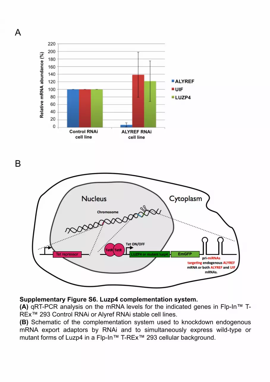
We noted that Alyref RNAi did not lead to up-regulationof endogenous Luzp4 expression in 293 cells (Supplemen-tary Figure S6A). This provided an opportunity to artifi-cially express Luzp4 and test whether it would complementthe mRNA export and growth defects observed followingAlyref RNAi in a 293 cell. To do this, we generated stableFlp-InTM T-RExTM 293 cell lines in which Luzp4 was ex-pressed as a GFP fusion in a transcript with miRNAs in its3′ UTR targeting either Alyref or Alyref+Uif (Supplemen-tary Figure S6B). We analysed these cell lines for mRNAexport defects using oligo(dT) FISH (Figure 4A and B).Alyref RNAi led to an mRNA export block as reportedpreviously with clear nuclear accumulation of poly(A)+-RNA (9). However, Luzp4 expression complemented thismRNA export defect, indicating that Luzp4 can functionas an mRNA export factor in place of Alyref. In contrast, aGFP fusion of Luzp4 missing aa 1–118 (Luzp4(!N)-GFP)which does not bind Uap56 (Figure 1D and E), or a GFPfusion of Luzp4 aa 1–240 (Luzp4(!C)-GFP), which bindsNxf1 and RNA poorly, did not complement the mRNA ex-port defect triggered by Alyref RNAi (Figure 4A and B).Following Alyref + Uif combined RNAi, cells exhibit an

8 Nucleic Acids Research, 2015
Figure 3. Luzp4 is an mRNA export adaptor. (A) The purified GB1-tagged Nxf1 proteins used in pulldown assays are shown (left panel). IgG-Sepharosepulldown assays using 35S-Luzp4 and the indicated GB1-tagged Nxf1 proteins (right panels). The upper panel is the phosphoimage of the same gel which isCoomassie stained in the lower panel. (B) Nxf1 remodelling assays. Luzp4 pre-incubated with a 32P-labelled RNA oligonucleotide was incubated with im-mobilized GST or GST-Nxf1:p15. The indicated complexes were then purified by GST-pulldown, eluted, UV-crosslinked and analysed by sodium dodecylsulphate-polyacrylamide gel electrophoresis. The upper panel is the phosphoimage of the same gel shown Coomassie stained in the lower panel. Input was1%. (C) Tethered mRNA export assays (also see Supplementary Figure S4A). The indicated MS2 fusion protein expression vectors were co-transfectedwith the MS2 reporter pLUCSALRRE6MS2, normally retained in the nucleus, together with a "-galactosidase expression vector. Luciferase activities weremeasured from the MS2 reporter and normalized for transfection efficiency with the "-galactosidase activities. The graph shows fold activation relative tothe levels seen with MS2-GFP and values represent averages from experiments carried out in triplicate on five separate occasions. (D) Immunofluorescenceimages of HeLa cells transfected with the indicated expression vectors. FLAG-Nxf1 was detected using FLAG antibody. When indicated, cells were treatedwith 5 !g/ml of actinomycin D for 3 h. Certain regions of images indicated by a white box are shown at higher magnification in the bottom left of a panelto observe the presence or reduction of FLAG-Nxf1 at the nuclear rim.
Nucleic Acids Research, 2015 9
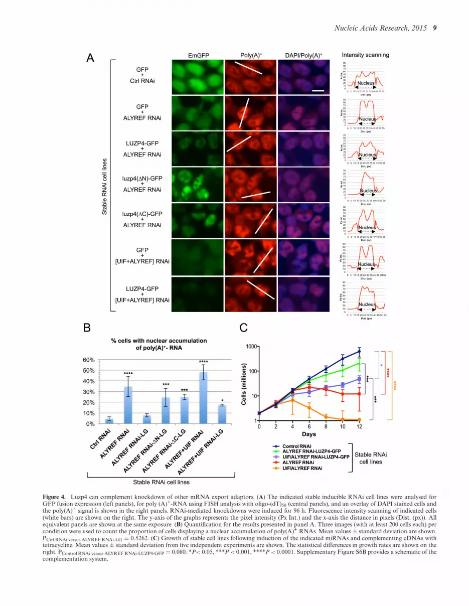
Figure 4. Luzp4 can complement knockdown of other mRNA export adaptors. (A) The indicated stable inducible RNAi cell lines were analysed forGFP fusion expression (left panels), for poly (A)+-RNA using FISH analysis with oligo-(dT)50 (central panels), and an overlay of DAPI stained cells andthe poly(A)+ signal is shown in the right panels. RNAi-mediated knockdowns were induced for 96 h. Fluorescence intensity scanning of indicated cells(white bars) are shown on the right. The y-axis of the graphs represents the pixel intensity (Px Int.) and the x-axis the distance in pixels (Dist. (px)). Allequivalent panels are shown at the same exposure. (B) Quantification for the results presented in panel A. Three images (with at least 200 cells each) percondition were used to count the proportion of cells displaying a nuclear accumulation of poly(A)+ RNAs. Mean values ± standard deviation are shown.PCtrl RNAi versus ALYREF RNAi-LG = 0.5262. (C) Growth of stable cell lines following induction of the indicated miRNAs and complementing cDNAs withtetracycline. Mean values ± standard deviation from five independent experiments are shown. The statistical differences in growth rates are shown on theright. PControl RNAi versus ALYREF RNAi-LUZP4-GFP = 0.080. *P< 0.05, ***P < 0.001, ****P < 0.0001. Supplementary Figure S6B provides a schematic of thecomplementation system.

10 Nucleic Acids Research, 2015
Figure 5. Luzp4 rescues the mRNA export pathway impaired in ALYREFRNAi cells. Western blot analysis of stable cell lines following induction ofthe indicated miRNAs and complementing cDNAs at the indicated timepoints. The antibodies used to detect the proteins are indicated on theright-hand side of each panel. Tetracycline is used to induce expression ofthe miRNA/complementing cDNA. Supplementary Figure S6B providesa schematic of the complementation system. LG = Luzp4-GFP.
extreme mRNA export block with robust nuclear accumu-lation of large areas of poly(A)+ material in the majorityof cells ((9) and Figure 4A and B). This mRNA exportblock was partially complemented by expression of GFP-Luzp4, with fewer cells showing strong nuclear accumula-tion of poly(A)+-RNA. We also examined the expressionof the GFP fusions in the stable cell lines (Figure 4A, leftpanels) and found that the levels of GFP protein were re-duced following Alyref RNAi and further reduced follow-ing Alyref + Uif RNAi. GFP-Luzp4 also showed reducedexpression following Alyref+Uif RNAi compared with justAlyref RNAi. This probably arises from reduced GFP orGFP-Luzp4 mRNA export in cell lines following Alyrefand Uif RNAi. Despite this, the lower levels of GFP-Luzp4following Alyref + Uif RNAi were still able to partiallycomplement mRNA export (Figure 4A and B). We furtherexamined the ability of Luzp4 to complement the growthdefect observed following Alyref and Uif RNAi using thestable cell lines generated for the FISH analysis. WhereasAlyref and Alyref+Uif combined RNAi cell lines showed aclear growth defect (Figure 4C), both cell lines showed sig-nificantly better growth when they expressed GFP-Luzp4.However, the cell growth was not completely restored tonormal levels, particularly when both Alyref and Uif wereknocked down by RNAi.
When mRNA export is blocked, cells respond by up-regulating a variety of mRNA export factors (9,11). Wetherefore also assessed what happens in cells followingAlyref or Alyref+Uif RNAi and the impact that expres-sion of GFP-Luzp4 had in such cells using the complemen-tation cell lines described above. Following Alyref RNAiwe saw a clear up-regulation of Uif, Nxf1 and Uap56 pro-tein levels (Figure 5, lanes 18–20). In contrast, an mRNPbinding factor not directly associated with mRNA export,Hnrnpa1, was not affected (Figure 5, lanes 18–20). WhenGFP-Luzp4 was co-expressed in the Alyref RNAi cell line,the up-regulation of Nxf1 and Uap56 was suppressed, par-ticularly at later time points of expression (Figure 5, com-pare lanes 20 and 24). However, Uif levels remained raisedfollowing Alyref RNAi and were not suppressed by GFP-Luzp4 expression. We also examined the ability of comple-menting cell lines expressing non-functional mutant formsof Luzp4 to suppress the up-regulation of export factors fol-lowing Alyref RNAi and found that neither mutant formsof Luzp4 could suppress this response (Figure 5, lanes 26–28, lanes 30–32). Finally, we examined the ability of GFP-Luzp4 expression to suppress up-regulation of export fac-tors following Alyref+Uif RNAi and found that it could not(Figure 5, lanes 34–36). Together, these results indicate thatLuzp4 functions as an mRNA export factor in human cellsthat is able to restore mRNA export function in cells com-promised by loss of mRNA export adaptors.
Luzp4 is preferentially expressed in melanomas
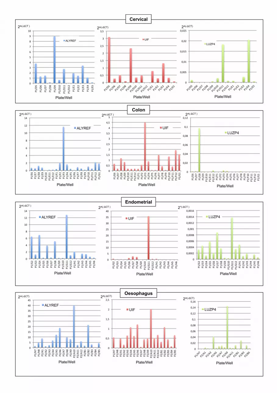
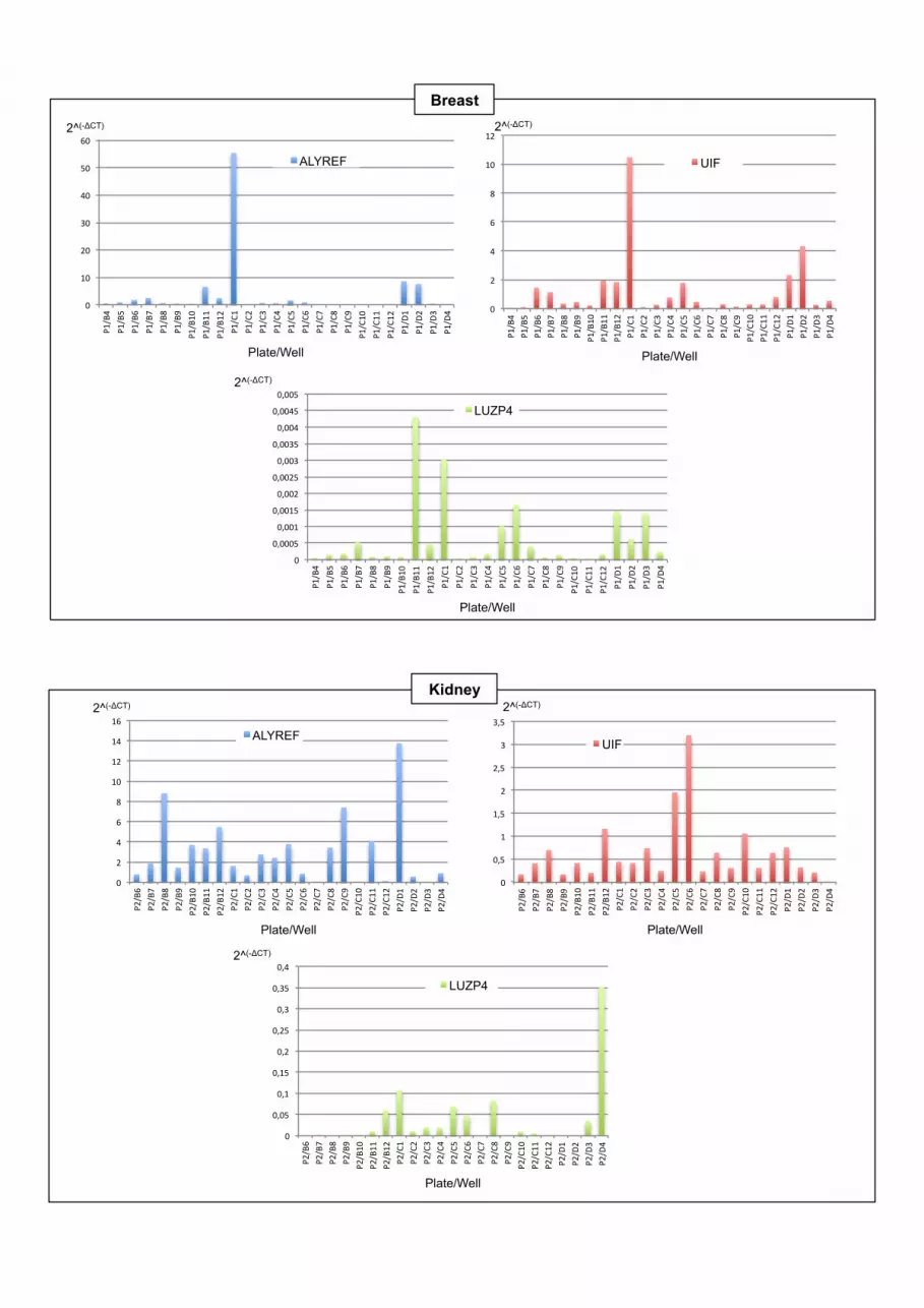
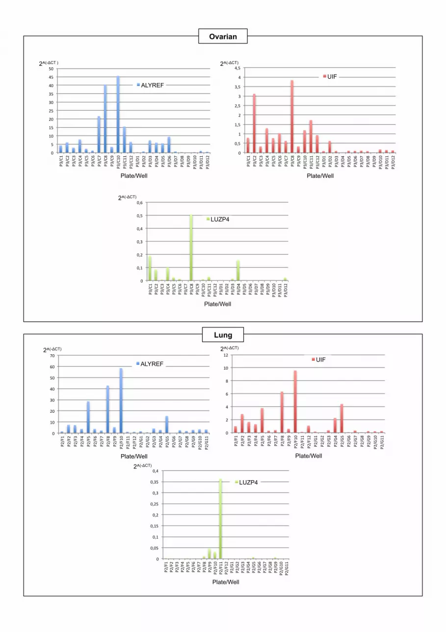
Since Luzp4 was originally described as a CTA we exam-ined Luzp4, Alyref and Uif expression in tumour biopsiesfrom 402 different patients, representing multiple differentclinical stages of tumours from a wide range of tissues (Fig-ure 6A, Supplementary Figure S7 and Supplementary Ta-ble S2). Generally, the expression levels for Luzp4 mRNA

Nucleic Acids Research, 2015 11
were low compared with Alyref and Uif, although we ob-served at least one tumour biopsy that showed increasedLuzp4 expression within most tissue types. The striking ex-ception was malignant melanoma where Luzp4 expressionwas very high for many patients. This is consistent with theearlier qualitative observation of Luzp4 expression in multi-ple melanoma samples (34). These data suggest that Luzp4expression is preferentially activated in melanoma.
To identify cells suitable for further studies on Luzp4, wescreened various cancer cell lines for expression of Luzp4and found that the melanoma cell line MeWo had read-ily detectable expression (Supplementary Figure S8A). Weconfirmed the expression of Luzp4 in MeWo cells usingqRT-PCR and found that the levels were significantly lowerthan those for both Uif and Alyref and found no detectableexpression of Luzp4 in 293T cells (Figure 6B). We alsoraised two separate antibodies to Luzp4 and screened forLuzp4 protein (Supplementary Figure S8B) in Mewo cells.Whilst the antibody was able to detect Luzp4 when tran-siently overexpressed we were unable to unambiguously de-tect the endogenous protein with our antibodies, suggestingthat it is present at low levels as indicated by the qRT-PCRanalysis.
We investigated whether Luzp4 RNAi led to a detectableblock in mRNA export in Mewo cells using oligo d(T) FISH(Supplementary Figure S9) and whilst we could see a clearblock of export following Alyref RNAi, no such block wasseen following Luzp4 RNAi. Therefore Luzp4 may be re-sponsible for the export of a restricted set of mRNAs. Wefurther analysed the effects of Luzp4, Alyref and Uif RNAion the growth of Mewo cells using a colony formation as-say (Figure 6D). Alyref RNAi led to a significant growthdefect in Mewo cells, whereas Uif RNAi did not, suggestingit was not essential for cellular proliferation, as previouslyobserved in 293T cells (9). Luzp4 RNAi led to a significantdrop in the number of Mewo cell colonies formed. Impor-tantly, Luzp4 siRNAs did not affect cellular proliferationof 293T cells (Supplementary Figure S8C), where Luzp4is not expressed, indicating they do not have off-target ef-fects which affect general cellular growth. We conclude thatLuzp4 is required for the growth of Mewo cells.
DISCUSSION
Luzp4 was first defined as a CTA 13 years ago but its molec-ular function has remained unknown until now. We haveshown that Luzp4 is a new RNA binding protein and wehave mapped two regions of the protein involved in this ac-tivity. One is an arginine-rich region located after the UBM,as is the case with Alyref. The second region involved inRNA binding maps to the leucine zipper motif and inter-estingly a helical wheel projection of the amino acids withinthe leucine zipper reveals a positively charged surface whichmay be required for RNA interactions (34). We have furtherdemonstrated that Luzp4 functions as an mRNA exportfactor, capable of complementing knockdown of Alyrefand partially complementing a double Alyref/Uif knock-down in 293 cells. Luzp4 has a UBM that allows associa-tion with Uap56. Given that Uap56 drives TREX assem-bly (3,10), this interaction probably ensures the incorpo-ration of Luzp4 in the TREX complex. Consistent with
this, the UBM is required for proper nuclear distributionof Luzp4 and we observed Co-IP of Luzp4 with TREXsubunits. A common property of mRNA export adaptorssuch as Alyref, Srsf3 and Srsf7 is their ability to bind the N-terminal region of Nxf1 and enhance its RNA binding ac-tivity (12). This involves release of the Nxf1 RNA bindingdomain from its interaction with the NTF2L domain (13).Here, we observe that Luzp4 also has the ability to enhancethe RNA binding activity of Nxf1 through an interactionwith the Nxf1 N-terminal region. Thus Luzp4 can be clas-sified as a new mRNA export adaptor.
Luzp4 was originally reported to be expressed in humantestis (34) and a more extensive survey of its tissue specificexpression by the Human Protein Atlas project (41) (http://www.proteinatlas.org/ENSG00000102021-LUZP4/tissue)confirms this restricted expression pattern. Interestingly,Luzp4 is able to interact with Nxf2 which may be themajor mRNA export receptor in this tissue. Whilsttestes express Luzp4, they also express Alyref (http://www.proteinatlas.org/ENSG00000183684/tissue/testis)and thus it is unlikely that Luzp4 acts as the sole mRNAexport adaptor in this tissue. Why the testis has evolveddistinct mRNA export factors remains unclear, though itmay be related to the meiotic gene expression programmein this tissue.
The ability of Luzp4 to complement the export defectseen following Alyref RNAi implies that it has the abil-ity to associate with a wide variety of mRNAs in a 293cell. This indicates there is functional redundancy betweenAlyref and Luzp4 as reported previously for Uif and Alyref(9). Indeed Alyref is clearly playing a major role in mRNAexport in Mewo cells, since its knockdown leads to a sig-nificant mRNA export block and prevents cellular prolif-eration. Interestingly in a number of high grade tumours,Alyref expression levels are reported to be diminished (24).Therefore, given the potential functional redundancy be-tween Alyref and Luzp4, it seems plausible that in certaintumours, Luzp4 may function to promote the export ofmRNAs which would normally utilize Alyref. Since Luzp4RNAi in Mewo cells does not lead to a strong accumulationof mRNA when assayed by FISH it is possible that the ex-port of a restricted set of mRNAs is dependent on Luzp4 inthese cells.
Luzp4 was originally discovered using the SEREX tech-nique, whereby circulating antibodies from a seminoma pa-tient were used for expression screening of testis cDNAlibraries. It was further shown to be expressed in a widevariety of cancers including melanoma, seminoma, ovar-ian, lung and glioma and its expression could be activatedin normal cells by addition of the DNA methylation in-hibitor, 5-aza-2′-deoxycytidine (34). Here, we have extendedthat analysis of the Luzp4 expression profile in cancer cellsand found it is expressed in a wide variety of cancer types,but particularly high levels of expression are observed inmelanoma cells. Recent work identified Luzp4 as commonlyup-regulated in multiple myeloma (MM) cell lines and thebone marrow of MM patients, a cell-type absent from ourscreen of tumours (42). Furthermore this study showedthat Luzp4 knockdown prevented colony formation for anMM cell line and sensitized the cell line to chemothera-peutic reagents (arsenic trioxide and bortezomib). These
12 Nucleic Acids Research, 2015
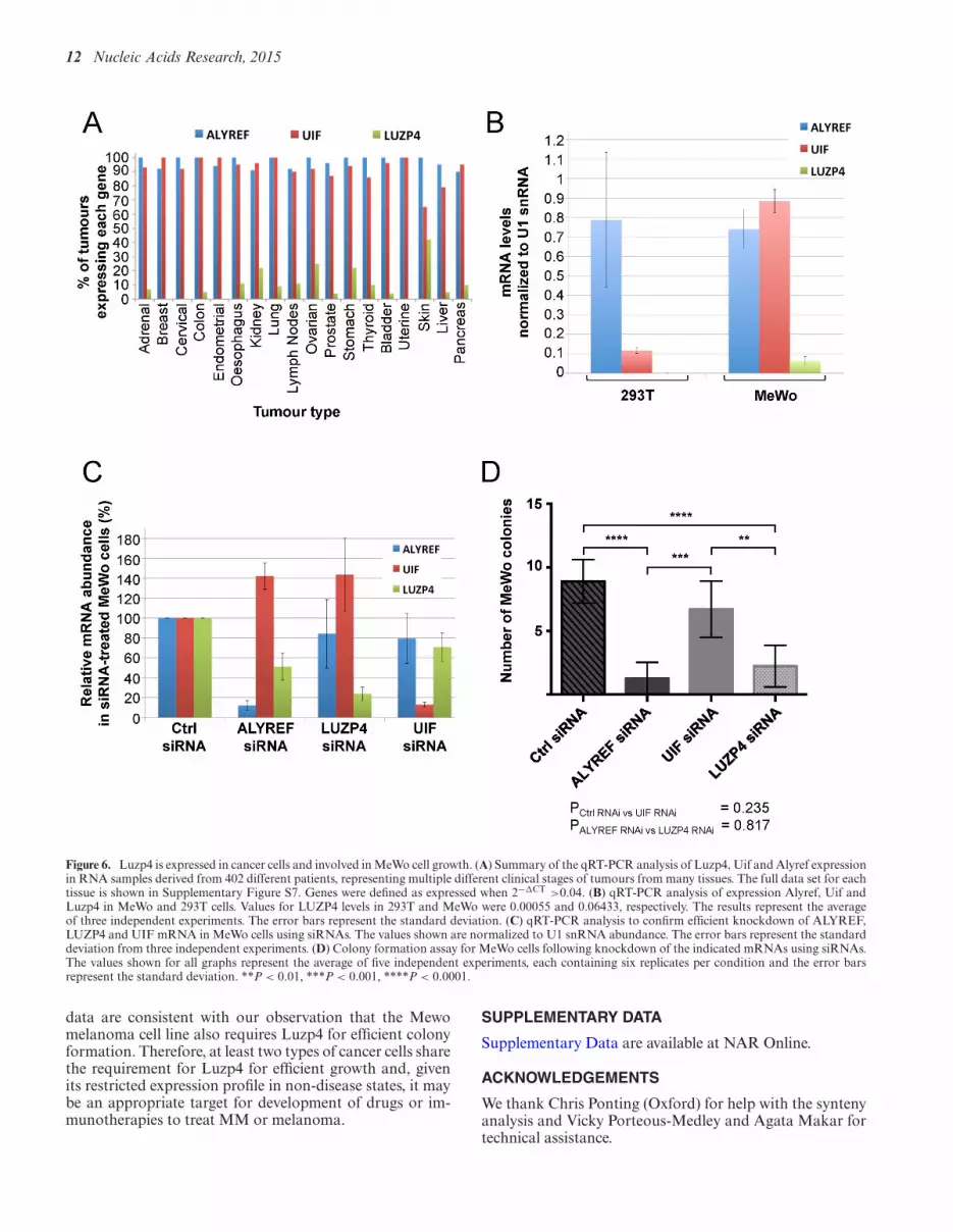
Figure 6. Luzp4 is expressed in cancer cells and involved in MeWo cell growth. (A) Summary of the qRT-PCR analysis of Luzp4, Uif and Alyref expressionin RNA samples derived from 402 different patients, representing multiple different clinical stages of tumours from many tissues. The full data set for eachtissue is shown in Supplementary Figure S7. Genes were defined as expressed when 2−!CT >0.04. (B) qRT-PCR analysis of expression Alyref, Uif andLuzp4 in MeWo and 293T cells. Values for LUZP4 levels in 293T and MeWo were 0.00055 and 0.06433, respectively. The results represent the averageof three independent experiments. The error bars represent the standard deviation. (C) qRT-PCR analysis to confirm efficient knockdown of ALYREF,LUZP4 and UIF mRNA in MeWo cells using siRNAs. The values shown are normalized to U1 snRNA abundance. The error bars represent the standarddeviation from three independent experiments. (D) Colony formation assay for MeWo cells following knockdown of the indicated mRNAs using siRNAs.The values shown for all graphs represent the average of five independent experiments, each containing six replicates per condition and the error barsrepresent the standard deviation. **P < 0.01, ***P < 0.001, ****P < 0.0001.
data are consistent with our observation that the Mewomelanoma cell line also requires Luzp4 for efficient colonyformation. Therefore, at least two types of cancer cells sharethe requirement for Luzp4 for efficient growth and, givenits restricted expression profile in non-disease states, it maybe an appropriate target for development of drugs or im-munotherapies to treat MM or melanoma.
SUPPLEMENTARY DATA
Supplementary Data are available at NAR Online.
ACKNOWLEDGEMENTS
We thank Chris Ponting (Oxford) for help with the syntenyanalysis and Vicky Porteous-Medley and Agata Makar fortechnical assistance.

Nucleic Acids Research, 2015 13
Author Contributions: N.V., M.C., M.L., P.H. and M.D. car-ried out the experiments. N.V., J.C. and S.W. designed theexperiments. N.V. and S.W. wrote the manuscript.
FUNDING
Biotechnology and Biological Sciences Research Coun-cil (BBSRC), UK [BB/E025293/1]; Wellcome Trust[083130/Z/07/Z]; Royal College of Surgeons of England[RS123]. M.C. is the recipient of a Wellcome Trust ClinicalTraining Fellowship and Royal College of Surgeons ofEngland Research Fellowship. Biotechnology and Bio-logical Sciences Research Council UK and Engineeringand Physical Sciences Research Council UK (to M.J.D.).Funding for open access charge: BBSRC and WellcomeTrust.Conflict of interest statement. None declared.
REFERENCES1. Natalizio,B.J. and Wente,S.R. (2013) Postage for the messenger:
designating routes for nuclear mRNA export. Trends Cell Biol., 23,365–373.
2. Strasser,K., Masuda,S., Mason,P., Pfannstiel,J., Oppizzi,M.,Rodriguez-Navarro,S., Rondon,A.G., Aguilera,A., Struhl,K.,Reed,R. et al. (2002) TREX is a conserved complex couplingtranscription with messenger RNA export. Nature, 417, 304–308.
3. Dufu,K., Livingstone,M.J., Seebacher,J., Gygi,S.P., Wilson,S.A. andReed,R. (2010) ATP is required for interactions between UAP56 andtwo conserved mRNA export proteins, Aly and CIP29, to assemblethe TREX complex. Genes Dev., 24, 2043–2053.
4. Li,Y., Wang,X., Zhang,X. and Goodrich,D.W. (2005) HumanhHpr1/p84/Thoc1 regulates transcriptional elongation andphysically links RNA polymerase II and RNA processing factors.Mol. Cell. Biol., 25, 4023–4033.
5. Masuda,S., Das,R., Cheng,H., Hurt,E., Dorman,N. and Reed,R.(2005) Recruitment of the human TREX complex to mRNA duringsplicing. Genes Dev., 19, 1512–1517.
6. Johnson,S.A., Cubberley,G. and Bentley,D.L. (2009)Cotranscriptional recruitment of the mRNA export factor Yra1 bydirect interaction with the 3′ end processing factor Pcf11. Mol. Cell,33, 215–226.
7. Katahira,J., Okuzaki,D., Inoue,H., Yoneda,Y., Maehara,K. andOhkawa,Y. (2013) Human TREX component Thoc5 affectsalternative polyadenylation site choice by recruiting mammaliancleavage factor I. Nucleic Acids Res., 41, 7060–7072.
8. Chi,B., Wang,Q., Wu,G., Tan,M., Wang,L., Shi,M., Chang,X. andCheng,H. (2013) Aly and THO are required for assembly of thehuman TREX complex and association of TREX components withthe spliced mRNA. Nucleic Acids Res., 41, 1294–1306.
9. Hautbergue,G.M., Hung,M.L., Walsh,M.J., Snijders,A.P., Chang,C.,Jones,R., Ponting,C.P., Dickman,M.J. and Wilson,S.A. (2009) UIF, anew mRNA export adaptor which works together with REF/ALY,requires FACT for recruitment to mRNA. Curr. Biol., 19, 1918–1924.
10. Chang,C.T., Hautbergue,G.M., Walsh,M.J., Viphakone,N., vanDijk,T.B., Philipsen,S. and Wilson,S.A. (2013) Chtop is a componentof the dynamic TREX mRNA export complex. EMBO J., 32,473–486.
11. Herold,A., Teixeira,L. and Izaurralde,E. (2003) Genome-wideanalysis of nuclear mRNA export pathways in Drosophila. Embo J,22, 2472–2483.
12. Hautbergue,G.M., Hung,M.L., Golovanov,A.P., Lian,L.Y. andWilson,S.A. (2008) Mutually exclusive interactions drive handover ofmRNA from export adaptors to TAP. Proc. Natl Acad. Sci. U.S.A.,105, 5154–5159.
13. Viphakone,N., Hautbergue,G.M., Walsh,M., Chang,C.T.,Holland,A., Folco,E.G., Reed,R. and Wilson,S.A. (2012) TREXexposes the RNA-binding domain of Nxf1 to enable mRNA export.Nat. Commun., 3, 1006.
14. Hargous,Y., Hautbergue,G.M., Tintaru,A.M., Skrisovska,L.,Golovanov,A.P., Stevenin,J., Lian,L.Y., Wilson,S.A. and Allain,F.H.(2006) Molecular basis of RNA recognition and TAP binding by theSR proteins SRp20 and 9G8. EMBO J., 25, 5126–5137.
15. Huang,Y., Gattoni,R., Stevenin,J. and Steitz,J.A. (2003) SR splicingfactors serve as adapter proteins for TAP-dependent mRNA export.Mol. Cell, 11, 837–843.
16. Zolotukhin,A.S., Tan,W., Bear,J., Smulevitch,S. and Felber,B.K.(2002) U2AF participates in the binding of TAP (NXF1) to mRNA.J. Biol. Chem., 277, 3935–3942.
17. Herold,A., Suyama,M., Rodrigues,J.P., Braun,I.C., Kutay,U.,Carmo-Fonseca,M., Bork,P. and Izaurralde,E. (2000) TAP (NXF1)belongs to a multigene family of putative RNA export factors with aconserved modular architecture. Mol. Cell. Biol., 20, 8996–9008.
18. Zhang,M., Wang,Q. and Huang,Y. (2007) Fragile X mentalretardation protein FMRP and the RNA export factor NXF2associate with and destabilize Nxf1 mRNA in neuronal cells. Proc.Natl Acad. Sci. U.S.A., 104, 10057–10062.
19. Siddiqui,N. and Borden,K.L. (2012) mRNA export and cancer.Wiley Interdiscip. Rev., 3, 13–25.
20. Culjkovic-Kraljacic,B., Baguet,A., Volpon,L., Amri,A. andBorden,K.L. (2012) The oncogene eIF4E reprograms the nuclearpore complex to promote mRNA export and oncogenictransformation. Cell Rep., 2, 207–215.
21. Griaud,F., Pierce,A., Gonzalez Sanchez,M.B., Scott,M.,Abraham,S.A., Holyoake,T.L., Tran,D.D., Tamura,T. andWhetton,A.D. (2013) A pathway from leukemogenic oncogenes andstem cell chemokines to RNA processing via THOC5. Leukemia, 27,932–940.
22. Li,Y., Lin,A.W., Zhang,X., Wang,Y., Wang,X. and Goodrich,D.W.(2007) Cancer cells and normal cells differ in their requirements forThoc1. Cancer Res., 67, 6657–6664.
23. Guo,S., Hakimi,M.A., Baillat,D., Chen,X., Farber,M.J.,Klein-Szanto,A.J., Cooch,N.S., Godwin,A.K. and Shiekhattar,R.(2005) Linking transcriptional elongation and messenger RNAexport to metastatic breast cancers. Cancer Res., 65, 3011–3016.
24. Dominguez-Sanchez,M.S., Saez,C., Japon,M.A., Aguilera,A. andLuna,R. (2011) Differential expression of THOC1 and ALY mRNPbiogenesis/export factors in human cancers. BMC Cancer, 11, 77.
25. Jani,D., Lutz,S., Hurt,E., Laskey,R.A., Stewart,M. andWickramasinghe,V.O. (2012) Functional and structuralcharacterization of the mammalian TREX-2 complex that linkstranscription with nuclear messenger RNA export. Nucleic AcidsRes., 40, 4562–4573.
26. Bhatia,V., Barroso,S.I., Garcia-Rubio,M.L., Tumini,E.,Herrera-Moyano,E. and Aguilera,A. (2014) BRCA2 prevents R-loopaccumulation and associates with TREX-2 mRNA export factorPCID2. Nature, 511, 362–365.
27. Simpson,A.J., Caballero,O.L., Jungbluth,A., Chen,Y.T. and Old,L.J.(2005) Cancer/testis antigens, gametogenesis and cancer. Nat. Rev.Cancer, 5, 615–625.
28. Kulkarni,P., Shiraishi,T., Rajagopalan,K., Kim,R., Mooney,S.M.and Getzenberg,R.H. (2012) Cancer/testis antigens and urologicalmalignancies. Nat. Rev., 9, 386–396.
29. Phillips,H.L., Williamson,J.C., van Elburg,K.A., Snijders,A.P.,Wright,P.C. and Dickman,M.J. (2010) Shotgun proteome analysisutilising mixed mode (reversed phase-anion exchangechromatography) in conjunction with reversed phase liquidchromatography mass spectrometry analysis. Proteomics, 10,2950–2960.
30. Cruz-Migoni,A., Hautbergue,G.M., Artymiuk,P.J., Baker,P.J.,Bokori-Brown,M., Chang,C.T., Dickman,M.J., Essex-Lopresti,A.,Harding,S.V., Mahadi,N.M. et al. (2011) A Burkholderiapseudomallei toxin inhibits helicase activity of translation factoreIF4A. Science, 334, 821–824.
31. Liu,G., Zhang,J., Choi,H., Lambert,J.P., Srikumar,T., Larsen,B.,Nesvizhskii,A.I., Raught,B., Tyers,M. and Gingras,A.C. (2012)Using ProHits to store, annotate, and analyze affinitypurification-mass spectrometry (AP-MS) data. Curr. Protoc.Bioinformatics, 2012, doi:10.1002/0471250953.bi0816s39.
32. Shannon,P., Markiel,A., Ozier,O., Baliga,N.S., Wang,J.T.,Ramage,D., Amin,N., Schwikowski,B. and Ideker,T. (2003)Cytoscape: a software environment for integrated models ofbiomolecular interaction networks. Genome Res., 13, 2498–2504.

14 Nucleic Acids Research, 2015
33. Schneider,C.A., Rasband,W.S. and Eliceiri,K.W. (2012) NIH Imageto ImageJ: 25 years of image analysis. Nat. Methods, 9, 671–675.
34. Tureci,O., Sahin,U., Koslowski,M., Buss,B., Bell,C., Ballweber,P.,Zwick,C., Eberle,T., Zuber,M., Villena-Heinsen,C. et al. (2002) Anovel tumour associated leucine zipper protein targeting to sites ofgene transcription and splicing. Oncogene, 21, 3879–3888.
35. Golovanov,A.P., Hautbergue,G.M., Tintaru,A.M., Lian,L.Y. andWilson,S.A. (2006) The solution structure of REF2-I revealsinterdomain interactions and regions involved in binding mRNAexport factors and RNA. RNA, 12, 1933–1948.
36. Girard,C., Will,C.L., Peng,J., Makarov,E.M., Kastner,B., Lemm,I.,Urlaub,H., Hartmuth,K. and Luhrmann,R. (2012)Post-transcriptional spliceosomes are retained in nuclear specklesuntil splicing completion. Nat. Commun., 3, 994.
37. Braun,I.C., Herold,A., Rode,M., Conti,E. and Izaurralde,E. (2001)Overexpression of TAP/p15 spliceosomes bypasses nuclear retentionand stimulates nuclear mRNA export. J. Biol. Chem., 276,20536–20543.
38. Katahira,J., Inoue,H., Hurt,E. and Yoneda,Y. (2009) Adaptor Alyand co-adaptor Thoc5 function in the Tap-p15-mediated nuclearexport of HSP70 mRNA. EMBO J., 28, 556–567.
39. Bachi,A., Braun,I.C., Rodrigues,J.P., Pante,N., Ribbeck,K., vonKobbe,C., Kutay,U., Wilm,M., Gorlich,D., Carmo-Fonseca,M. et al.(2000) The C-terminal domain of TAP interacts with the nuclear porecomplex and promotes export of specific CTE-bearing RNAsubstrates. RNA, 6, 136–158.
40. Lai,M.C. and Tarn,W.Y. (2004) Hypophosphorylated ASF/SF2binds TAP and is present in messenger ribonucleoproteins. J. Biol.Chem., 279, 31745–31749.
41. Uhlen,M., Oksvold,P., Fagerberg,L., Lundberg,E., Jonasson,K.,Forsberg,M., Zwahlen,M., Kampf,C., Wester,K., Hober,S. et al.(2010) Towards a knowledge-based human protein atlas. Nat.Biotechnol., 28, 1248–1250.
42. Wen,J., Li,H., Tao,W., Savoldo,B., Foglesong,J.A., King,L.C., Zu,Y.and Chang,C.C. (2014) High throughput quantitative reversetranscription PCR assays revealing over-expression of cancer testisantigen genes in multiple myeloma stem cell-like side population cells.Br. J. Haematol., 166, 711–719.

Leucine zipper motif: L-x(6)-L-x(6)-L
Similar to Uap56-Binding Motif found in Alyref R-rich region
RS-containing His-rich region
10 20 30 40 50 60 !MASFRKLTLS EKVPPNHPSR KKVNFLDMSL DDIIIYKELE GTNAEEEKNK RQNHSKKESP ! 70 80 90 100 110 120 !SRQQSKAHRH RHRRGYSRCR SNSEEGNHDK KPSQKPSGFK SGQHPLNGQP LIEQEKCSDN ! 130 140 150 160 170 180 !YEAQAEKNQG QSEGNQHQSE GNPDKSEESQ GQPEENHHSE RSRNHLERSL SQSDRSQGQL ! 190 200 210 220 230 240 !KRHHPQYERS HGQYKRSHGQ SERSHGHSER SHGHSERSHG HSERSHGHSK RSRSQGDLVD ! 250 260 270 280 290 300 !TQSDLIATQR DLIATQKDLI ATQRDLIATQ RDLIVTQRDL VATERDLINQ SGRSHGQSER ! 310 !HQRYSTGKNT ITT !!
B
Supplementary Figure S1. Bioinformatic analysis on Luzp4 and mapping of Luzp4 NLS. (A) Syntenic analysis of LUZP4 in vertebrates. LUZP4 genes are expanded in the mouse genome and include the paralogue OTT. (B) Amino acid sequence of Luzp4 with key motifs highlighted. (C) The UBM-like motif (aa 22-40) is important for Luzp4 nuclear distribution and the NLS of Luzp4 lies within the RS-containing His-rich (RS-H) region. Fluorescence images of the indicated GFP fusions expressed in HeLa cells. The scale bar corresponds to 10 µm.
A
C GFP-LUZP4 GFP-luzp4(∆22-40) GFP-luzp4(∆156-236) GFP-luzp4(∆156-178)
Luzp4/DNA luzp4(∆22-40/DNA luzp4(∆156-236/DNA luzp4(∆156-178/DNA
70 55 35
kDa 100 -!-!
-!-!-!-!
27
15 -!10
GST GST-Nxf1:p15
70 55 35
kDa 100 -!
-!-!-!-!-!
27
15
-!10
GST-Nxf1
p15
GST
GST-Uap56
70 55 35
kDa 100 -!
-!-!-!-!-!
27
15
-!10
GST-Uap56
1 2 3 4 5 6 7 8 9 1 2 3 4 5 6 7 8 9 10 1 2 3 4 5 6 7 8 9
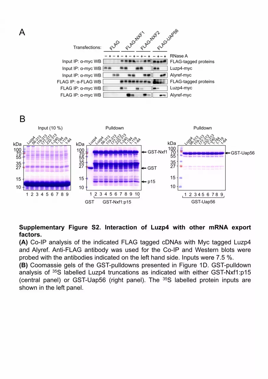
Supplementary Figure S2. Interaction of Luzp4 with other mRNA export factors. (A) Co-IP analysis of the indicated FLAG tagged cDNAs with Myc tagged Luzp4 and Alyref. Anti-FLAG antibody was used for the Co-IP and Western blots were probed with the antibodies indicated on the left hand side. Inputs were 7.5 %. (B) Coomassie gels of the GST-pulldowns presented in Figure 1D. GST-pulldown analysis of 35S labelled Luzp4 truncations as indicated with either GST-Nxf1:p15 (central panel) or GST-Uap56 (right panel). The 35S labelled protein inputs are shown in the left panel.
Input (10 %) Pulldown Pulldown B
A
_"
Input IP: α-myc WB FLAG IP: α-FLAG WB
Input IP: α-myc WB Input IP: α-myc WB
FLAG IP: α-myc WB FLAG IP: α-myc WB
FLAG-tagged proteins Luzp4-myc Alyref-myc FLAG-tagged proteins Luzp4-myc Alyref-myc
RNase A +" _" +" _" +" _" +"_" +"_" +" _" +"_"+"
Transfections:

Supplementary Figure S3. RNA binding activity of Luzp4. (A) Panels relevant to Figure 2A. In vitro RNA binding experiment where UV irradiation was omitted. The indicated phosphorimage was acquired at the same time as in Figure 2A. (B) mRNP capture assays of Luzp4 mutants. Panel relevant to Figure 2D. UV irradiation was here omitted. Proteins were visualised by Western blot with anti-GFP antibody. The image was acquired at the same time as Figure 2D. (C) Subcellular localisation of GFP-Luzp4 and truncations transiently expressed in Hela cells. The scale bar corresponds to 10 µm.
A
GFP
GFP/DNA Luzp4/DNA luzp4(∆N)/DNA luzp4(∆C)/DNA
GFP-LUZP4 GFP-luzp4(∆N) GFP-luzp4(∆C) C
B
- UV
1 2 3 4 5 6 7 8
70 55
35
27
kDa 100 -!
-!-!
-!-!
Input (1%)
+ - - - + - - - "- + - - - + - -"- - + - - - + -"- - - + - - - +"
oligo(dT) pulldown
- - - - - - - -"
WB
: α-G
FP!
GFP GFP-LUZP4 GFP-luzp4(∆N) GFP-luzp4(∆C) RNase A
Constructs expressed:
Coo
mas
sie -!
-!-!
-!-!
-!70 55
35
kDa
100
27
130 250 -!
Pho
spho
rimag
er
1 2 3 4 5 6
ssDNA
BS
A
Aly
ref
BS
A
Aly
ref
Luzp
4
Luzp
4
RNA
BSA
Luzp4 Alyref
- UV
250 130 100
70
55
35
27
15
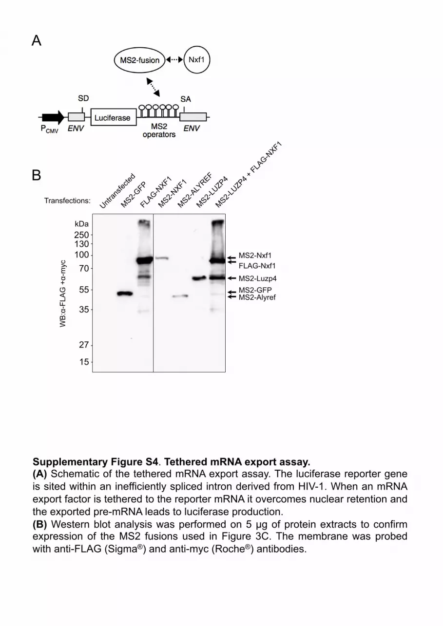
Supplementary Figure S4. Tethered mRNA export assay. (A) Schematic of the tethered mRNA export assay. The luciferase reporter gene is sited within an inefficiently spliced intron derived from HIV-1. When an mRNA export factor is tethered to the reporter mRNA it overcomes nuclear retention and the exported pre-mRNA leads to luciferase production. (B) Western blot analysis was performed on 5 µg of protein extracts to confirm expression of the MS2 fusions used in Figure 3C. The membrane was probed with anti-FLAG (Sigma®) and anti-myc (Roche®) antibodies.
kDa
MS2-Luzp4
FLAG-Nxf1
MS2-Alyref MS2-GFP
-!-!
-!-!
-!-!
-!-!
MS2-Nxf1
Transfections:
A
B
WB
:α-F
LAG
+α-
myc!

Supplementary Figure S5. Luzp4 relocalises to the cytoplasm in newly divided cells. (A) During cell division, Luzp4 is relocalised to the cytoplasm and is unable to trigger a depletion of Nxf1’s staining from the nuclear rim. GFP-LUZP4 and FLAG-NXF1 were transiently expressed in HeLa cells (B) This relocalisation phenomenon requires the leucine-zipper domain of Luzp4. In its absence, the majority of luzp4(∆C) mutant proteins stays in the nucleus. The indicated GFP fusions were transiently transfected in HeLa cells. The scale bars correspond to 10 µm.
GFP-LUZP4 +
FLAG-NXF1
DNA
Metaphase Prophase Meta-anaphase Interphase Telophase A
B
GFP- luzp4(∆N)
luzp4(∆C)!DNA
GFP- luzp4(∆C)
DNA luzp4(∆N)! luzp4(∆N)/DNA
luzp4(∆C)/DNA
B
Supplementary Figure S6. Luzp4 complementation system. (A) qRT-PCR analysis on the mRNA levels for the indicated genes in Flp-In™ T-REx™ 293 Control RNAi or Alyref RNAi stable cell lines. (B) Schematic of the complementation system used to knockdown endogenous mRNA export adaptors by RNAi and to simultaneously express wild-type or mutant forms of Luzp4 in a Flp-In™ T-REx™ 293 cellular background.
A
Control RNAi cell line
ALYREF RNAi cell line
Rel
ativ
e m
RN
A a
bund
ance
(%)
20 40 60 80
100
120
140 160
0
180
200
220

0!
10!
20!
30!
40!
50!
60!
70!
80!
90!
100!
P1/A1!
P1/A2!
P1/A3!
P1/A4!
P1/A5!
P1/A6!
P1/A7!
P1/A8!
P1/A9!
P1/A10!
P1/A11!
P1/A12!
P1/B1!
P1/B2!
P1/B3!
2^(-ΔCT )
Plate/Well
ALYREF
0!
0,1!
0,2!
0,3!
0,4!
0,5!
0,6!
0,7!
0,8!
0,9!
1!
P1/A1!
P1/A2!
P1/A3!
P1/A4!
P1/A5!
P1/A6!
P1/A7!
P1/A8!
P1/A9!
P1/A10!
P1/A11!
P1/A12!
P1/B1!
P1/B2!
P1/B3!
2^(-ΔCT)
Plate/Well
UIF
Adrenal
0!
0,01!
0,02!
0,03!
0,04!
0,05!
0,06!
0,07!
0,08!
0,09!
0,1!
P1/A1!
P1/A2!
P1/A3!
P1/A4!
P1/A5!
P1/A6!
P1/A7!
P1/A8!
P1/A9!
P1/A10!
P1/A11!
P1/A12!
P1/B1!
P1/B2!
P1/B3!
2^(-ΔCT )
Plate/Well
LUZP4
0!
0,2!
0,4!
0,6!
0,8!
1!
1,2!
P2/D5!
P2/D6!
P2/D7!
P2/D8!
P2/D9!
P2/D10!
P2/D11!
P2/D12!
P2/E1!
P2/E2!
P2/E3!
P2/E4!
P2/E5!
P2/E6!
P2/E7!
P2/E8!
P2/E9!
P2/E10!
P2/E11!
P2/E12!
2^(-ΔCT )
Plate/Well
UIF
0!
2!
4!
6!
8!
10!
12!
14!
P2/D5!
P2/D6!
P2/D7!
P2/D8!
P2/D9!
P2/D10!
P2/D11!
P2/D12!
P2/E1!
P2/E2!
P2/E3!
P2/E4!
P2/E5!
P2/E6!
P2/E7!
P2/E8!
P2/E9!
P2/E10!
P2/E11!
P2/E12!
2^(-ΔCT )
Plate/Well
ALYREF
0!
0,02!
0,04!
0,06!
0,08!
0,1!
0,12!
P2/D5!
P2/D6!
P2/D7!
P2/D8!
P2/D9!
P2/D10!
P2/D11!
P2/D12!
P2/E1!
P2/E2!
P2/E3!
P2/E4!
P2/E5!
P2/E6!
P2/E7!
P2/E8!
P2/E9!
P2/E10!
P2/E11!
P2/E12!
2^(-ΔCT )
Plate/Well
LUZP4
Liver
0!
1!
2!
3!
4!
5!
6!
7!
8!
9!
10!
P3/E1!
P3/E2!
P3/E3!
P3/E4!
P3/E5!
P3/E6!
P3/E7!
P3/E8!
P3/E9!
P3/E10!
P3/E11!
P3/E12!
P3/F1!
P3/F2!
P3/F3!
P3/F4!
P3/F5!
P3/F6!
P3/F7!
P3/F8!
P3/F9!
2^(-ΔCT)
Plate/Well
ALYREF
0!
0,5!
1!
1,5!
2!
2,5!
P3/E1!
P3/E2!
P3/E3!
P3/E4!
P3/E5!
P3/E6!
P3/E7!
P3/E8!
P3/E9!
P3/E10!
P3/E11!
P3/E12!
P3/F1!
P3/F2!
P3/F3!
P3/F4!
P3/F5!
P3/F6!
P3/F7!
P3/F8!
P3/F9!
2^(-ΔCT)
Plate/Well
UIF
0!
0,02!
0,04!
0,06!
0,08!
0,1!
0,12!
P3/E1!
P3/E2!
P3/E3!
P3/E4!
P3/E5!
P3/E6!
P3/E7!
P3/E8!
P3/E9!
P3/E10!
P3/E11!
P3/E12!
P3/F1!
P3/F2!
P3/F3!
P3/F4!
P3/F5!
P3/F6!
P3/F7!
P3/F8!
P3/F9!
2^(-ΔCT )
Plate/Well
LUZP4
Pancreas
Supplementary data 7. Relative expression of ALYREF, UIF and LUZP4 in cancerous tissues. qRT-PCR was used to evaluate the abundance of ALYREF, UIF, and LUZP4 at the RNA level using commercially purchased cDNA array plates from Origene. The graphs show delta CT values for each gene normalised to the U1 snRNA content for each cDNA taken from cancer tissues from a catalogue of patients. Wells can be cross-referenced to the Origene datasheet (Supplementary Table S2), ‘P’ stands for ‘plate’ and the following letter gives the well number.
0!
1!
2!
3!
4!
5!
6!
7!
8!
9!
10!
P1/D5!
P1/D6!
P1/D7!
P1/D8!
P1/D9!
P1/D10!
P1/D11!
P1/D12!
P1/E1!
P1/E2!
P1/E3!
P1/E4!
P1/E5!
2^(-ΔCT )
Plate/Well
ALYREF
0!
0,5!
1!
1,5!
2!
2,5!
3!
3,5!2^(-ΔCT)
Plate/Well
UIF
0!
0,005!
0,01!
0,015!
0,02!
0,025!2^(-ΔCT)
Plate/Well
LUZP4
Cervical
0!
2!
4!
6!
8!
10!
12!
14!
P1/E6!
P1/E7!
P1/E8!
P1/E9!
P1/E10!
P1/E11!
P1/E12!
P1/F1!
P1/F2!
P1/F3!
P1/F4!
P1/F5!
P1/F6!
P1/F7!
P1/F8!
P1/F9!
P1/F10!
P1/F11!
P1/F12!
P1/G
1!
2^(-ΔCT )
Plate/Well
ALYREF
0!
0,5!
1!
1,5!
2!
2,5!
3!
3,5!
4!
4,5!
5!P1
/E6!
P1/E7!
P1/E8!
P1/E9!
P1/E10!
P1/E11!
P1/E12!
P1/F1!
P1/F2!
P1/F3!
P1/F4!
P1/F5!
P1/F6!
P1/F7!
P1/F8!
P1/F9!
P1/F10!
P1/F11!
P1/F12!
P1/G
1!
2^(-ΔCT )
Plate/Well
UIF
0!
0,02!
0,04!
0,06!
0,08!
0,1!
0,12!
P1/E6!
P1/E7!
P1/E8!
P1/E9!
P1/E10!
P1/E11!
P1/E12!
P1/F1!
P1/F2!
P1/F3!
P1/F4!
P1/F5!
P1/F6!
P1/F7!
P1/F8!
P1/F9!
P1/F10!
P1/F11!
P1/F12!
P1/G
1!
2^(-ΔCT )
Plate/Well
LUZP4
Colon
0!
2!
4!
6!
8!
10!
12!
14!
P1/G
2!P1
/G3!
P1/G
4!P1
/G5!
P1/G
6!P1
/G7!
P1/G
8!P1
/G9!
P1/G
10!
P1/G
11!
P1/G
12!
P1/H1!
P1/H2!
P1/H3!
P1/H4!
P1/H5!
P1/H6!
2^(-ΔCT )
Plate/Well
ALYREF
0!
5!
10!
15!
20!
25!
30!
35!
40!
P1/G
2!P1
/G3!
P1/G
4!P1
/G5!
P1/G
6!P1
/G7!
P1/G
8!P1
/G9!
P1/G
10!
P1/G
11!
P1/G
12!
P1/H1!
P1/H2!
P1/H3!
P1/H4!
P1/H5!
P1/H6!
2^(-ΔCT )
Plate/Well
UIF
0!
0,0002!
0,0004!
0,0006!
0,0008!
0,001!
0,0012!
0,0014!
0,0016!
P1/G
2!P1
/G3!
P1/G
4!P1
/G5!
P1/G
6!P1
/G7!
P1/G
8!P1
/G9!
P1/G
10!
P1/G
11!
P1/G
12!
P1/H1!
P1/H2!
P1/H3!
P1/H4!
P1/H5!
P1/H6!
2^(-ΔCT )
Plate/Well
LUZP4
Endometrial
0!
5!
10!
15!
20!
25!
30!
35!
40!
45!
P1/H7!
P1/H8!
P2/A1!
P2/A2!
P2/A3!
P2/A4!
P2/A5!
P2/A6!
P2/A7!
P2/A8!
P2/A9!
P2/A10!
P2/A11!
P2/A12!
P2/B1!
P2/B2!
P2/B3!
P2/B4!
P2/B5!
2^(-ΔCT)
Plate/Well
ALYREF
0!
0,5!
1!
1,5!
2!
2,5!
P1/H7!
P1/H8!
P2/A1!
P2/A2!
P2/A3!
P2/A4!
P2/A5!
P2/A6!
P2/A7!
P2/A8!
P2/A9!
P2/A10!
P2/A11!
P2/A12!
P2/B1!
P2/B2!
P2/B3!
P2/B4!
P2/B5!
2^(-ΔCT)
Plate/Well
UIF
0!
0,02!
0,04!
0,06!
0,08!
0,1!
0,12!
0,14!
0,16!2^(-ΔCT)
Plate/Well
LUZP4
Oesophagus
0!
2!
4!
6!
8!
10!
12!
14!
16!
P2/B6!
P2/B7!
P2/B8!
P2/B9!
P2/B10!
P2/B11!
P2/B12!
P2/C1!
P2/C2!
P2/C3!
P2/C4!
P2/C5!
P2/C6!
P2/C7!
P2/C8!
P2/C9!
P2/C10!
P2/C11!
P2/C12!
P2/D1!
P2/D2!
P2/D3!
P2/D4!
2^(-ΔCT)
Plate/Well
ALYREF
0!
0,5!
1!
1,5!
2!
2,5!
3!
3,5!
P2/B6!
P2/B7!
P2/B8!
P2/B9!
P2/B10!
P2/B11!
P2/B12!
P2/C1!
P2/C2!
P2/C3!
P2/C4!
P2/C5!
P2/C6!
P2/C7!
P2/C8!
P2/C9!
P2/C10!
P2/C11!
P2/C12!
P2/D1!
P2/D2!
P2/D3!
P2/D4!
2^(-ΔCT)
Plate/Well
UIF
0!
0,05!
0,1!
0,15!
0,2!
0,25!
0,3!
0,35!
0,4!
P2/B6!
P2/B7!
P2/B8!
P2/B9!
P2/B10!
P2/B11!
P2/B12!
P2/C1!
P2/C2!
P2/C3!
P2/C4!
P2/C5!
P2/C6!
P2/C7!
P2/C8!
P2/C9!
P2/C10!
P2/C11!
P2/C12!
P2/D1!
P2/D2!
P2/D3!
P2/D4!
2^(-ΔCT)
Plate/Well
LUZP4
Kidney
0!
10!
20!
30!
40!
50!
60!
P1/B4!
P1/B5!
P1/B6!
P1/B7!
P1/B8!
P1/B9!
P1/B10!
P1/B11!
P1/B12!
P1/C1!
P1/C2!
P1/C3!
P1/C4!
P1/C5!
P1/C6!
P1/C7!
P1/C8!
P1/C9!
P1/C10!
P1/C11!
P1/C12!
P1/D1!
P1/D2!
P1/D3!
P1/D4!
2^(-ΔCT)
Plate/Well
ALYREF
0!
2!
4!
6!
8!
10!
12!
P1/B4!
P1/B5!
P1/B6!
P1/B7!
P1/B8!
P1/B9!
P1/B10!
P1/B11!
P1/B12!
P1/C1!
P1/C2!
P1/C3!
P1/C4!
P1/C5!
P1/C6!
P1/C7!
P1/C8!
P1/C9!
P1/C10!
P1/C11!
P1/C12!
P1/D1!
P1/D2!
P1/D3!
P1/D4!
2^(-ΔCT)
Plate/Well
UIF
0!
0,0005!
0,001!
0,0015!
0,002!
0,0025!
0,003!
0,0035!
0,004!
0,0045!
0,005!
P1/B4!
P1/B5!
P1/B6!
P1/B7!
P1/B8!
P1/B9!
P1/B10!
P1/B11!
P1/B12!
P1/C1!
P1/C2!
P1/C3!
P1/C4!
P1/C5!
P1/C6!
P1/C7!
P1/C8!
P1/C9!
P1/C10!
P1/C11!
P1/C12!
P1/D1!
P1/D2!
P1/D3!
P1/D4!
2^(-ΔCT)
Plate/Well
LUZP4
Breast
0!
10!
20!
30!
40!
50!
60!
70!
P2/F1!
P2/F2!
P2/F3!
P2/F4!
P2/F5!
P2/F6!
P2/F7!
P2/F8!
P2/F9!
P2/F10!
P2/F11!
P2/F12!
P2/G
1!P2
/G2!
P2/G
3!P2
/G4!
P2/G
5!P2
/G6!
P2/G
7!P2
/G8!
P2/G
9!P2
/G10!
P2/G
11!
2^(-ΔCT)
Plate/Well
ALYREF
0!
2!
4!
6!
8!
10!
12!
P2/F1!
P2/F2!
P2/F3!
P2/F4!
P2/F5!
P2/F6!
P2/F7!
P2/F8!
P2/F9!
P2/F10!
P2/F11!
P2/F12!
P2/G
1!P2
/G2!
P2/G
3!P2
/G4!
P2/G
5!P2
/G6!
P2/G
7!P2
/G8!
P2/G
9!P2
/G10!
P2/G
11!
2^(-ΔCT)
Plate/Well
UIF
0!
0,05!
0,1!
0,15!
0,2!
0,25!
0,3!
0,35!
0,4!
P2/F1!
P2/F2!
P2/F3!
P2/F4!
P2/F5!
P2/F6!
P2/F7!
P2/F8!
P2/F9!
P2/F10!
P2/F11!
P2/F12!
P2/G
1!P2
/G2!
P2/G
3!P2
/G4!
P2/G
5!P2
/G6!
P2/G
7!P2
/G8!
P2/G
9!P2
/G10!
P2/G
11!
2^(-ΔCT)
Plate/Well
LUZP4
Lung
0!
5!
10!
15!
20!
25!
30!
35!
40!
45!
50!
P3/C1!
P3/C2!
P3/C3!
P3/C4!
P3/C5!
P3/C6!
P3/C7!
P3/C8!
P3/C9!
P3/C10!
P3/C11!
P3/C12!
P3/D1!
P3/D2!
P3/D3!
P3/D4!
P3/D5!
P3/D6!
P3/D7!
P3/D8!
P3/D9!
P3/D10!
P3/D11!
P3/D12!
2^(-ΔCT )
Plate/Well
ALYREF
0!
0,5!
1!
1,5!
2!
2,5!
3!
3,5!
4!
4,5!
P3/C1!
P3/C2!
P3/C3!
P3/C4!
P3/C5!
P3/C6!
P3/C7!
P3/C8!
P3/C9!
P3/C10!
P3/C11!
P3/C12!
P3/D1!
P3/D2!
P3/D3!
P3/D4!
P3/D5!
P3/D6!
P3/D7!
P3/D8!
P3/D9!
P3/D10!
P3/D11!
P3/D12!
2^(-ΔCT)
Plate/Well
UIF
0!
0,1!
0,2!
0,3!
0,4!
0,5!
0,6!
P3/C1!
P3/C2!
P3/C3!
P3/C4!
P3/C5!
P3/C6!
P3/C7!
P3/C8!
P3/C9!
P3/C10!
P3/C11!
P3/C12!
P3/D1!
P3/D2!
P3/D3!
P3/D4!
P3/D5!
P3/D6!
P3/D7!
P3/D8!
P3/D9!
P3/D10!
P3/D11!
P3/D12!
2^(-ΔCT)
Plate/Well
LUZP4
Ovarian

0!
10!
20!
30!
40!
50!
60!
70!
80!
90!
100!
P2/G
12!
P2/H1!
P2/H2!
P2/H3!
P2/H4!
P2/H5!
P2/H6!
P2/H7!
P2/H8!
P2/H9!
P2/H10!
P2/H11!
P2/H12!
P3/A1!
P3/A2!
P3/A3!
P3/A4!
P3/A5!
P3/A6!
P3/A7!
P3/A8!
P3/A9!
P3/A10!
P3/A11!
P3/A12!
P3/B1!
P3/B2!
P3/B3!
P3/B4!
P3/B5!
P3/B6!
P3/B7!
P3/B8!
P3/B9!
P3/B10!
P3/B11!
P3/B12!
2^(-ΔCT )
Plate/Well
ALYREF
0!
1!
2!
3!
4!
5!
6!
7!
8!
9!
10!
P2/G
12!
P2/H1!
P2/H2!
P2/H3!
P2/H4!
P2/H5!
P2/H6!
P2/H7!
P2/H8!
P2/H9!
P2/H10!
P2/H11!
P2/H12!
P3/A1!
P3/A2!
P3/A3!
P3/A4!
P3/A5!
P3/A6!
P3/A7!
P3/A8!
P3/A9!
P3/A10!
P3/A11!
P3/A12!
P3/B1!
P3/B2!
P3/B3!
P3/B4!
P3/B5!
P3/B6!
P3/B7!
P3/B8!
P3/B9!
P3/B10!
P3/B11!
P3/B12!
2^(-ΔCT)
Plate/Well
UIF
0!
0,05!
0,1!
0,15!
0,2!
0,25!
P2/G
12!
P2/H1!
P2/H2!
P2/H3!
P2/H4!
P2/H5!
P2/H6!
P2/H7!
P2/H8!
P2/H9!
P2/H10!
P2/H11!
P2/H12!
P3/A1!
P3/A2!
P3/A3!
P3/A4!
P3/A5!
P3/A6!
P3/A7!
P3/A8!
P3/A9!
P3/A10!
P3/A11!
P3/A12!
P3/B1!
P3/B2!
P3/B3!
P3/B4!
P3/B5!
P3/B6!
P3/B7!
P3/B8!
P3/B9!
P3/B10!
P3/B11!
P3/B12!
2^(-ΔCT)
Plate/Well
LUZP4
Lymph Nodes
Urinary Bladder
0!
20!
40!
60!
80!
100!
120!
P4/F5!
P4/F6!
P4/F7!
P4/F8!
P4/F9!
P4/F10!
P4/F11!
P4/F12!
P4/G
1!P4
/G2!
P4/G
3!P4
/G4!
P4/G
5!P4
/G6!
P4/G
7!P4
/G8!
P4/G
9!P4
/G10!
P4/G
11!
P4/G
12!
P4/H1!
P4/H2!
P4/H3!
P4/H4!
2^(-ΔCT)
Plate/Well
ALYREF
0!
2!
4!
6!
8!
10!
12!
14!
16!
P4/F5!
P4/F6!
P4/F7!
P4/F8!
P4/F9!
P4/F10!
P4/F11!
P4/F12!
P4/G
1!P4
/G2!
P4/G
3!P4
/G4!
P4/G
5!P4
/G6!
P4/G
7!P4
/G8!
P4/G
9!P4
/G10!
P4/G
11!
P4/G
12!
P4/H1!
P4/H2!
P4/H3!
P4/H4!
2^(-ΔCT)
Plate/Well
UIF
0!
0,01!
0,02!
0,03!
0,04!
0,05!
0,06!
0,07!
0,08!
P4/F5!
P4/F6!
P4/F7!
P4/F8!
P4/F9!
P4/F10!
P4/F11!
P4/F12!
P4/G
1!P4
/G2!
P4/G
3!P4
/G4!
P4/G
5!P4
/G6!
P4/G
7!P4
/G8!
P4/G
9!P4
/G10!
P4/G
11!
P4/G
12!
P4/H1!
P4/H2!
P4/H3!
P4/H4!
2^(-ΔCT)
Plate/Well
LUZP4

0!
10!
20!
30!
40!
50!
60!
70!P4
/A1!
P4/A2!
P4/A3!
P4/A4!
P4/A5!
P4/A6!
P4/A7!
P4/A8!
P4/A9!
P4/A10!
P4/A11!
P4/A12!
P4/B1!
P4/B2!
P4/B3!
P4/B4!
P4/B5!
P4/B6!
2^(-ΔCT)
Plate/Well
ALYREF
0!
1!
2!
3!
4!
5!
6!
7!
8!
9!
P4/A1!
P4/A2!
P4/A3!
P4/A4!
P4/A5!
P4/A6!
P4/A7!
P4/A8!
P4/A9!
P4/A10!
P4/A11!
P4/A12!
P4/B1!
P4/B2!
P4/B3!
P4/B4!
P4/B5!
P4/B6!
2^(-ΔCT)
Plate/Well
UIF
0!
0,02!
0,04!
0,06!
0,08!
0,1!
0,12!
0,14!
0,16!
0,18!
P4/A1!
P4/A2!
P4/A3!
P4/A4!
P4/A5!
P4/A6!
P4/A7!
P4/A8!
P4/A9!
P4/A10!
P4/A11!
P4/A12!
P4/B1!
P4/B2!
P4/B3!
P4/B4!
P4/B5!
P4/B6!
2^(-ΔCT)
Plate/Well
LUZP4
Stomach
Thyroid
0!
10!
20!
30!
40!
50!
60!
70!
80!
P4/D8!
P4/D9!
P4/D10!
P4/D11!
P4/D12!
P4/E1!
P4/E2!
P4/E3!
P4/E4!
P4/E5!
P4/E6!
P4/E7!
P4/E8!
P4/E9!
P4/E10!
P4/E11!
P4/E12!
P4/F1!
P4/F2!
P4/F3!
P4/F4!
2^(-ΔCT)
Plate/Well
ALYREF
0!
1!
2!
3!
4!
5!
6!
7!
P4/D8!
P4/D9!
P4/D10!
P4/D11!
P4/D12!
P4/E1!
P4/E2!
P4/E3!
P4/E4!
P4/E5!
P4/E6!
P4/E7!
P4/E8!
P4/E9!
P4/E10!
P4/E11!
P4/E12!
P4/F1!
P4/F2!
P4/F3!
P4/F4!
2^(-ΔCT)
Plate/Well
UIF
0!
0,01!
0,02!
0,03!
0,04!
0,05!
0,06!
P4/D8!
P4/D9!
P4/D10!
P4/D11!
P4/D12!
P4/E1!
P4/E2!
P4/E3!
P4/E4!
P4/E5!
P4/E6!
P4/E7!
P4/E8!
P4/E9!
P4/E10!
P4/E11!
P4/E12!
P4/F1!
P4/F2!
P4/F3!
P4/F4!
2^(-ΔCT)
Plate/Well
LUZP4
Uterus
0!
20!
40!
60!
80!
100!
120!
P4/H5! P4/H6! P4/H7! P4/H8!
2^(-ΔCT)
Plate/Well
ALYREF
0!
2!
4!
6!
8!
10!
12!
14!
16!
P4/H5! P4/H6! P4/H7! P4/H8!
2^(-ΔCT)
Plate/Well
UIF
0!
0,005!
0,01!
0,015!
0,02!
0,025!
P4/H5! P4/H6! P4/H7! P4/H8!
2^(-ΔCT)
Plate/Well
LUZP4

Prostate
0!
20!
40!
60!
80!
100!
120!
P3/F10!
P3/F11!
P3/F12!
P3/G
1!P3
/G2!
P3/G
3!P3
/G4!
P3/G
5!P3
/G6!
P3/G
7!P3
/G8!
P3/G
9!P3
/G10!
P3/G
11!
P3/G
12!
P3/H1!
P3/H2!
P3/H3!
P3/H4!
P3/H5!
P3/H6!
P3/H7!
P3/H8!
2^(-ΔCT)
Plate/Well
ALYREF
0!
0,1!
0,2!
0,3!
0,4!
0,5!
0,6!
P3/F10!
P3/F11!
P3/F12!
P3/G
1!P3
/G2!
P3/G
3!P3
/G4!
P3/G
5!P3
/G6!
P3/G
7!P3
/G8!
P3/G
9!P3
/G10!
P3/G
11!
P3/G
12!
P3/H1!
P3/H2!
P3/H3!
P3/H4!
P3/H5!
P3/H6!
P3/H7!
P3/H8!
2^(-ΔCT)
Plate/Well
UIF
0!
0,01!
0,02!
0,03!
0,04!
0,05!
0,06!
0,07!
0,08!
0,09!
0,1!P3
/F10!
P3/F11!
P3/F12!
P3/G
1!P3
/G2!
P3/G
3!P3
/G4!
P3/G
5!P3
/G6!
P3/G
7!P3
/G8!
P3/G
9!P3
/G10!
P3/G
11!
P3/G
12!
P3/H1!
P3/H2!
P3/H3!
P3/H4!
P3/H5!
P3/H6!
P3/H7!
P3/H8!
2^(-ΔCT)
Plate/Well
LUZP4
0!
5!
10!
15!
20!
25!
P5/C1!
P5/C2!
P5/C3!
P5/C4!
P5/C5!
P5/C6!
P5/C7!
P5/C8!
P5/C9!
P5/C10!
P5/C11!
P5/C12!
P5/D1!
P5/D2!
P5/D3!
P5/D4!
P5/D5!
P5/D6!
P5/D7!
P5/D8!
P5/D9!
P5/D10!
P5/D11!
P5/D12!
P5/E1!
P5/E2!
P5/E3!
P5/E4!
P5/E5!
P5/E6!
P5/E7!
P5/E8!
P5/E9!
P5/E10!
P5/E11!
P5/E12!
P5/F1!
P5/F2!
P5/F3!
P5/F4!
P5/F5!
P5/F6!
P5/F7!
2^(-ΔCT)
Plate/Well
ALYREF
Skin
0!0,2!0,4!0,6!0,8!1!
1,2!1,4!1,6!1,8!2!
P5/C1!
P5/C2!
P5/C3!
P5/C4!
P5/C5!
P5/C6!
P5/C7!
P5/C8!
P5/C9!
P5/C10!
P5/C11!
P5/C12!
P5/D1!
P5/D2!
P5/D3!
P5/D4!
P5/D5!
P5/D6!
P5/D7!
P5/D8!
P5/D9!
P5/D10!
P5/D11!
P5/D12!
P5/E1!
P5/E2!
P5/E3!
P5/E4!
P5/E5!
P5/E6!
P5/E7!
P5/E8!
P5/E9!
P5/E10!
P5/E11!
P5/E12!
P5/F1!
P5/F2!
P5/F3!
P5/F4!
P5/F5!
P5/F6!
P5/F7!
2^(-ΔCT)
Plate/Well
UIF
0!
0,5!
1!
1,5!
2!
2,5!
3!
3,5!
4!
P5/C1!
P5/C2!
P5/C3!
P5/C4!
P5/C5!
P5/C6!
P5/C7!
P5/C8!
P5/C9!
P5/C10!
P5/C11!
P5/C12!
P5/D1!
P5/D2!
P5/D3!
P5/D4!
P5/D5!
P5/D6!
P5/D7!
P5/D8!
P5/D9!
P5/D10!
P5/D11!
P5/D12!
P5/E1!
P5/E2!
P5/E3!
P5/E4!
P5/E5!
P5/E6!
P5/E7!
P5/E8!
P5/E9!
P5/E10!
P5/E11!
P5/E12!
P5/F1!
P5/F2!
P5/F3!
P5/F4!
P5/F5!
P5/F6!
P5/F7!
2^(-ΔCT)
Plate/Well
LUZP4

Supplementary Figure S8. (A) Expression of Luzp4 in cancer cell lines. Various cancer cell lines were screened by non-saturating RT-PCR for LUZP4 expression and analysed by agarose gel electrophoresis. U1 RT-PCR was used to confirm efficient cDNA synthesis in each sample. LUZP4 RT-PCR primers were: forward 5’-GGAGAGGCTACTCAAGATGCAGAAGC-3’ and reverse 5’-GAGATCACTCTGAGTGTCCACAAGATCTCC-3’. U1 RT-PCR primers were: forward 5’-ACCTGGCAGGGGAGATACCA-3’ and reverse 5’-GGGGAAAGCGCGAACGCAGT-3’. (B) Western blot using our best anti-Luzp4 antibody, affinity purified, which recognizes recombinant or transfected Luzp4, but doesn’t detect any endogenous Luzp4. 40 µg of cell extracts and 20 ng of recombinant Luzp4-6His. Affinity-purified anti-Luzp4 used at 1:500. (C) LUZP4 RNAi has no effect on 293T cells proliferation. Colony formation assays performed on 293T cells treated with the indicated siRNAs. Colonies that were scored contained >50 cells; mean values for five independent experiments are shown and error bars represent the standard deviation. Related to Fig.6D. *:! P<0.05.! **:!P<0.01.!***:!P<0.001.!****:!P<0.0001.!
B
A
1 -!kb 3 -!
1 -!kb 3 -!
U1
LUZP4
Cancer cell lines:
Num
ber o
f col
onie
s 60
40
20
80 ****
**** ***
PCtrl RNAi vs UIF RNAi = 0.803 PCtrl RNAi vs LUZP4 RNAi = 0.942 PUIF RNAi vs LUZP4 RNAi = 0.480
C
WB: α-Luzp4 affinity-purified
Luzp4-6His!Luzp4!
-!-!-!-!-!
63 48 35
25
75
Luzp
4-6H
is
MeW
o
MeW
o
293T
293T
+pC
INeo
-LU
ZP4
kDa
-!-!-!-!-!
63 48 35
25
75 kDa
Ponceau
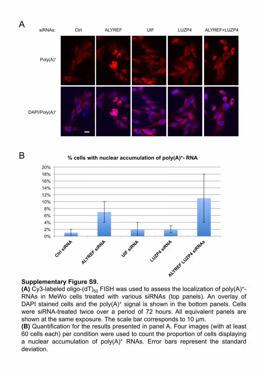
A
B
Supplementary Figure S9. (A) Cy3-labeled oligo-(dT)50 FISH was used to assess the localization of poly(A)+-RNAs in MeWo cells treated with various siRNAs (top panels). An overlay of DAPI stained cells and the poly(A)+ signal is shown in the bottom panels. Cells were siRNA-treated twice over a period of 72 hours. All equivalent panels are shown at the same exposure. The scale bar corresponds to 10 µm. (B) Quantification for the results presented in panel A. Four images (with at least 60 cells each) per condition were used to count the proportion of cells displaying a nuclear accumulation of poly(A)+ RNAs. Error bars represent the standard deviation.
Ctrl ALYREF
Poly(A)+
DAPI/Poly(A)+
siRNAs: UIF LUZP4 ALYREF+LUZP4
0% 2% 4% 6% 8%
10% 12% 14% 16% 18% 20%
% cells with nuclear accumulation of poly(A)+- RNA

Hit$Gene$Name Hit$Protein$ID Description Peptide$Number Unique$Peptide$Number Hit$Gene$Name Hit$Protein$ID Description Peptide$Number Unique$Peptide$NumberACTN4 ACTN4_HUMAN Alpha.actinin.43OS=Homo3sapiens3GN=ACTN43PE=13SV=2 372 62 ACTN4 ACTN4_HUMAN Alpha.actinin.43OS=Homo3sapiens3GN=ACTN43PE=13SV=2 18 10LIMA1 LIMA1_HUMAN LIM3domain3and3actin.binding3protein313OS=Homo3sapiens3GN=LIMA13PE=13SV=1 87 31 HIST2H4B H4_HUMAN Histone3H43OS=Homo3sapiens3GN=HIST1H4A3PE=13SV=2 50 12PPP1R12A MYPT1_HUMAN Protein3phosphatase313regulatory3subunit312A3OS=Homo3sapiens3GN=PPP1R12A3PE=13SV=1 30 16 HIST1H2AD H2A1D_HUMAN Histone3H2A3type31.D3OS=Homo3sapiens3GN=HIST1H2AD3PE=13SV=2 21 5SVIL SVIL_HUMAN Supervillin3OS=Homo3sapiens3GN=SVIL3PE=13SV=2 22 14 HIST1H2BC H2B1C_HUMAN Histone3H2B3type31.C/E/F/G/I3OS=Homo3sapiens3GN=HIST1H2BC3PE=13SV=4 26 5HIST2H4B H4_HUMAN Histone3H43OS=Homo3sapiens3GN=HIST1H4B3PE=13SV=2 66 14 RBMX HNRPG_HUMAN Heterogeneous3nuclear3ribonucleoprotein3G3OS=Homo3sapiens3GN=RBMX3PE=13SV=3 4 3TJP1 ZO1_HUMAN Tight3junction3protein3ZO.13OS=Homo3sapiens3GN=TJP13PE=13SV=3 17 12 CALM3 CALM_HUMAN Calmodulin3OS=Homo3sapiens3GN=CALM13PE=13SV=2 5 3LUZP4 LUZP4_HUMAN Leucine3zipper3protein343OS=Homo3sapiens3GN=LUZP43PE=23SV=1 39 11 HIST3H3 H31T_HUMAN Histone3H3.1t3OS=Homo3sapiens3GN=HIST3H33PE=13SV=3 16 5LUZP1 LUZP1_HUMAN Leucine3zipper3protein313OS=Homo3sapiens3GN=LUZP13PE=13SV=2 14 11 BANF1 BAF_HUMAN Barrier.to.autointegration3factor3OS=Homo3sapiens3GN=BANF13PE=13SV=1 7 4EIF4A3 IF4A3_HUMAN Eukaryotic3initiation3factor34A.III3OS=Homo3sapiens3GN=EIF4A33PE=13SV=4 17 10 H2AFY H2AY_HUMAN Core3histone3macro.H2A.13OS=Homo3sapiens3GN=H2AFY3PE=13SV=4 4 3PNN PININ_HUMAN Pinin3OS=Homo3sapiens3GN=PNN3PE=13SV=4 18 10 HIST1H2AB H2A1B_HUMAN Histone3H2A3type31.B/E3OS=Homo3sapiens3GN=HIST1H2AB3PE=13SV=2 14 4ACIN1 ACINU_HUMAN Apoptotic3chromatin3condensation3inducer3in3the3nucleus3OS=Homo3sapiens3GN=ACIN13PE=13SV=1 17 10AMOT AMOT_HUMAN Angiomotin3OS=Homo3sapiens3GN=AMOT3PE=13SV=1 13 9PDLIM1 PDLI1_HUMAN PDZ3and3LIM3domain3protein313OS=Homo3sapiens3GN=PDLIM13PE=13SV=4 24 9AIF1L AIF1L_HUMAN Allograft3inflammatory3factor31.like3OS=Homo3sapiens3GN=AIF1L3PE=13SV=1 16 9DDX39B UAP56_HUMAN Spliceosome3RNA3helicase3BAT13OS=Homo3sapiens3GN=BAT13PE=13SV=1 17 8ALYREF THOC4_HUMAN THO3complex3subunit343OS=Homo3sapiens3GN=THOC43PE=13SV=3 20 7ACTN1 ACTN1_HUMAN Alpha.actinin.13OS=Homo3sapiens3GN=ACTN13PE=13SV=2 100 7SRRT SRRT_HUMAN Serrate3RNA3effector3molecule3homolog3OS=Homo3sapiens3GN=SRRT3PE=13SV=1 12 6RPS13 RS13_HUMAN 40S3ribosomal3protein3S133OS=Homo3sapiens3GN=RPS133PE=13SV=2 15 6HIST1H2AD H2A1D_HUMAN Histone3H2A3type31.D3OS=Homo3sapiens3GN=HIST1H2AD3PE=13SV=2 33 6RNPS1 RNPS1_HUMAN RNA.binding3protein3with3serine.rich3domain313OS=Homo3sapiens3GN=RNPS13PE=13SV=1 22 6PAWR PAWR_HUMAN PRKC3apoptosis3WT13regulator3protein3OS=Homo3sapiens3GN=PAWR3PE=13SV=1 12 6SRSF7 SFRS7_HUMAN Splicing3factor;3arginine/serine.rich373OS=Homo3sapiens3GN=SFRS73PE=13SV=1 7 5HIST1H2BC H2B1C_HUMAN Histone3H2B3type31.C/E/F/G/I3OS=Homo3sapiens3GN=HIST1H2BC3PE=13SV=4 29 5SRSF1 SFRS1_HUMAN Splicing3factor;3arginine/serine.rich313OS=Homo3sapiens3GN=SFRS13PE=13SV=2 8 5RPS7 RS7_HUMAN 40S3ribosomal3protein3S73OS=Homo3sapiens3GN=RPS73PE=13SV=1 15 5BCLAF1 BCLF1_HUMAN Bcl.2.associated3transcription3factor313OS=Homo3sapiens3GN=BCLAF13PE=13SV=2 7 5CALD1 CALD1_HUMAN Caldesmon3OS=Homo3sapiens3GN=CALD13PE=13SV=2 11 5RBMX HNRPG_HUMAN Heterogeneous3nuclear3ribonucleoprotein3G3OS=Homo3sapiens3GN=RBMX3PE=13SV=3 12 5RBM8A RBM8A_HUMAN RNA.binding3protein38A3OS=Homo3sapiens3GN=RBM8A3PE=13SV=1 10 5HIST1H2BB H2B1B_HUMAN Histone3H2B3type31.B3OS=Homo3sapiens3GN=HIST1H2BB3PE=13SV=2 44 5KIAA1671 K1671_HUMAN Uncharacterized3protein3KIAA16713OS=Homo3sapiens3GN=KIAA16713PE=13SV=2 7 5CALM3 CALM_HUMAN Calmodulin3OS=Homo3sapiens3GN=CALM13PE=13SV=2 22 5MAGOH MGN_HUMAN Protein3mago3nashi3homolog3OS=Homo3sapiens3GN=MAGOH3PE=13SV=1 6 4TJP2 ZO2_HUMAN Tight3junction3protein3ZO.23OS=Homo3sapiens3GN=TJP23PE=13SV=1 5 4ABLIM1 ABLM1_HUMAN Actin.binding3LIM3protein313OS=Homo3sapiens3GN=ABLIM13PE=13SV=3 5 4SPECC1L CYTSA_HUMAN Cytospin.A3OS=Homo3sapiens3GN=CYTSA3PE=13SV=1 6 4HIST3H3 H31T_HUMAN Histone3H3.1t3OS=Homo3sapiens3GN=HIST3H33PE=13SV=3 13 4AKAP2 AKAP2_HUMAN A.kinase3anchor3protein323OS=Homo3sapiens3GN=AKAP23PE=13SV=3 7 4SHKBP1 SHKB1_HUMAN SH3KBP1.binding3protein313OS=Homo3sapiens3GN=SHKBP13PE=13SV=2 9 4STK38L ST38L_HUMAN Serine/threonine.protein3kinase338.like3OS=Homo3sapiens3GN=STK38L3PE=13SV=33 9 3SAP18 SAP18_HUMAN Histone3deacetylase3complex3subunit3SAP183OS=Homo3sapiens3GN=SAP183PE=13SV=1 8 3ERH ERH_HUMAN Enhancer3of3rudimentary3homolog3OS=Homo3sapiens3GN=ERH3PE=13SV=1 6 3POLDIP3 PDIP3_HUMAN Polymerase3delta.interacting3protein333OS=Homo3sapiens3GN=POLDIP33PE=13SV=2 6 3SRGAP2 FNBP2_HUMAN SLIT.ROBO3Rho3GTPase.activating3protein323OS=Homo3sapiens3GN=SRGAP23PE=13SV=2 4 3CSNK2A1 CSK21_HUMAN Casein3kinase3II3subunit3alpha3OS=Homo3sapiens3GN=CSNK2A13PE=13SV=1 4 3RPS15 RS15_HUMAN 40S3ribosomal3protein3S153OS=Homo3sapiens3GN=RPS153PE=13SV=2 5 3BANF1 BAF_HUMAN Barrier.to.autointegration3factor3OS=Homo3sapiens3GN=BANF13PE=13SV=1 7 3EFHD1 EFHD1_HUMAN EF.hand3domain.containing3protein3D13OS=Homo3sapiens3GN=EFHD13PE=13SV=1 6 3PLS3 PLST_HUMAN Plastin.33OS=Homo3sapiens3GN=PLS33PE=13SV=4 6 3FUS FUS_HUMAN RNA.binding3protein3FUS3OS=Homo3sapiens3GN=FUS3PE=13SV=1 6 3RPA1 RFA1_HUMAN Replication3protein3A3703kDa3DNA.binding3subunit3OS=Homo3sapiens3GN=RPA13PE=13SV=2 4 3PPP1R18 PHTNS_HUMAN Phostensin3OS=Homo3sapiens3GN=KIAA19493PE=13SV=1 7 3SRSF6 SFRS6_HUMAN Splicing3factor;3arginine/serine.rich363OS=Homo3sapiens3GN=SFRS63PE=13SV=2 3 2THOC5 THOC5_HUMAN THO3complex3subunit353homolog3OS=Homo3sapiens3GN=THOC53PE=13SV=2 3 2 UHR.TOF3MS/MS3spectra3for3these3peptides3were3manually3verifiedRPL27A RL27A_HUMAN 60S3ribosomal3protein3L27a3OS=Homo3sapiens3GN=RPL27A3PE=13SV=2 3 2DDX5 DDX5_HUMAN Probable3ATP.dependent3RNA3helicase3DDX53OS=Homo3sapiens3GN=DDX53PE=13SV=13 3 2PDLIM7 PDLI7_HUMAN PDZ3and3LIM3domain3protein373OS=Homo3sapiens3GN=PDLIM73PE=13SV=1 2 2SRSF10 SF13A_HUMAN Splicing3factor;3arginine/serine.rich313A3OS=Homo3sapiens3GN=SFRS13A3PE=13SV=1 2 2FAU RS30_HUMAN 40S3ribosomal3protein3S303OS=Homo3sapiens3GN=FAU3PE=13SV=1 4 2RPL26 RL26_HUMAN 60S3ribosomal3protein3L263OS=Homo3sapiens3GN=RPL263PE=13SV=1 3 2H2AFY H2AY_HUMAN Core3histone3macro.H2A.13OS=Homo3sapiens3GN=H2AFY3PE=13SV=4 3 2DDX1 DDX1_HUMAN ATP.dependent3RNA3helicase3DDX13OS=Homo3sapiens3GN=DDX13PE=13SV=2 2 2THOC1 THOC1_HUMAN THO3complex3subunit313OS=Homo3sapiens3GN=THOC13PE=13SV=1 2 2 UHR.TOF3MS/MS3spectra3for3these3peptides3were3manually3verifiedSRSF3 SFRS3_HUMAN Splicing3factor;3arginine/serine.rich333OS=Homo3sapiens3GN=SFRS33PE=13SV=1 6 2DDX21 DDX21_HUMAN Nucleolar3RNA3helicase323OS=Homo3sapiens3GN=DDX213PE=13SV=5 2 2RPL22 RL22_HUMAN 60S3ribosomal3protein3L223OS=Homo3sapiens3GN=RPL223PE=13SV=2 4 2RPSA RSSA_HUMAN 40S3ribosomal3protein3SA3OS=Homo3sapiens3GN=RPSA3PE=13SV=4 5 2RPL24 RL24_HUMAN 60S3ribosomal3protein3L243OS=Homo3sapiens3GN=RPL243PE=13SV=1 5 2THRAP3 TR150_HUMAN Thyroid3hormone3receptor.associated3protein333OS=Homo3sapiens3GN=THRAP33PE=13SV=23 5 2TMOD2 TMOD2_HUMAN Tropomodulin.23OS=Homo3sapiens3GN=TMOD23PE=13SV=1 2 2CORO1B COR1B_HUMAN Coronin.1B3OS=Homo3sapiens3GN=CORO1B3PE=13SV=1 4 2CSRP2 CSRP2_HUMAN Cysteine3and3glycine.rich3protein323OS=Homo3sapiens3GN=CSRP23PE=13SV=3 3 2RPL35 RL35_HUMAN 60S3ribosomal3protein3L353OS=Homo3sapiens3GN=RPL353PE=13SV=2 6 2PPP1CB PP1B_HUMAN Serine/threonine.protein3phosphatase3PP1.beta3catalytic3subunit3OS=Homo3sapiens3GN=PPP1CB3PE=13SV=3 5 2CFL2 COF2_HUMAN Cofilin.23OS=Homo3sapiens3GN=CFL23PE=13SV=1 21 2HIST1H1D H13_HUMAN Histone3H1.33OS=Homo3sapiens3GN=HIST1H1D3PE=13SV=2 34 2THOC2 THOC2_HUMAN THO3complex3subunit323OS=Homo3sapiens3GN=THOC23PE=13SV=2 1 1 UHR.TOF3MS/MS3spectra3for3these3peptides3were3manually3verifiedCHTOP/CA077 CA077_HUMAN Uncharacterized3protein3C1orf773OS=Homo3sapiens3GN=C1orf773PE=13SV=2 1 1 UHR.TOF3MS/MS3spectra3for3these3peptides3were3manually3verified
Control$IPLUZP4$IP