Embed Size (px)
Citation preview

The American Journal of Pathology, Vol. 179, No. 4, October 2011
Copyright © 2011 American Society for Investigative Pathology.
Published by Elsevier Inc. All rights reserved.
DOI: 10.1016/j.ajpath.2011.06.009
Biomarkers, Genomics, Proteomics, and Gene Regulation
Lung Gene Expression in a Rhesus Allergic AsthmaModel Correlates with Physiologic Parameters ofDisease and Exhibits Common and Distinct Pathways
with Human Asthma and a Mouse Asthma ModelAlexander R. Abbas,* Janet K. Jackman,†
Sherron L. Bullens,‡ Sarah M. Davis,§
David F. Choy,¶ Grazyna Fedorowicz,�
Martha Tan,** Bao-Tran Truong,**Y. Gloria Meng,** Lauri Diehl,†† Lisa A. Miller,§
Edward S. Schelegle,§ Dallas M. Hyde,§
Hilary F. Clark,* Zora Modrusan,�
Joseph R. Arron,¶ and Lawren C. Wu†
From the Departments of Bioinformatics,* Immunology,† Tumor
Biology and Angiogenesis,‡ ITGR Biomarker Discovery,¶
Molecular Biology,� Assay and Automation Technology,�� and
Pathology,†† Genentech Inc., South San Francisco; and the
Department of Anatomy, Physiology, and Cell Biology,§ School of
Veterinary Medicine and California National Primate Research
Center, University of California, Davis, California
Experimental nonhuman primate models of asthma ex-hibit multiple features that are characteristic of an eosi-nophilic/T helper 2 (Th2)-high asthma subtype, charac-terized by the increased expression of Th2 cytokinesand responsive genes, in humans. Here, we determinethe molecular pathways that are present in a house dustmite–induced rhesus asthma model by analyzing thegenomewide lung gene expression profile of the rhesusmodel and comparing it with that of human Th2-highasthma. We find that a prespecified human Th2 inflam-mation gene set from human Th2-high asthma is alsopresent in rhesus asthma and that the expression of thegenes comprising this gene set is positively correlatedin human and rhesus asthma. In addition, as in humanTh2-high asthma, the Th2 gene set correlates with phys-iologic markers of allergic inflammation and disease inrhesus asthma. Comparison of lung gene expressionprofiles from human Th2-high asthma, the rhesusasthma model, and a common mouse asthma modelindicates that genes associated with Th2 inflammationare shared by all three species. However, some patho-
physiologic aspects of human asthma (ie, subepithelialfibrosis, angiogenesis, neural biology, and immune hostdefense biology) are better represented in the gene ex-pression profile of the rhesus model than in the mousemodel. Further study of the rhesus asthma model mayyield novel insights into the pathogenesis of humanTh2-high asthma. (Am J Pathol 2011, 179:1667–1680; DOI:
10.1016/j.ajpath.2011.06.009)
Asthma is characterized by variable airflow obstruction, air-way hyperreactivity, and chronic airway inflammation. De-spite these common clinical features, asthma is a hetero-geneous disease that can be subclassified by a number ofdifferent measures, including the nature of the airway in-flammation.1,2 Airway eosinophilia, a hallmark of T helper 2(Th2) inflammation, defines a major subtype of severeasthma, with other major subtypes defined by neutrophilicand paucigranulocytic inflammation.1–4 Recent analyses ofairway gene expression in mild-to-moderate asthmatics notbeing treated with corticosteroids defines a molecular sig-nature of Th2 inflammation that correlates with airway andperipheral eosinophilia, airway hyperreactivity, subepithelialfibrosis, distinct patterns of mucin expression, and elevatedIgE.5,6 The Th2-high subtype of asthma in mild-to-moderateasthmatics is associated with the cytokines IL-5 and IL-13and is correlated with a clinical response to inhaled corti-costeroid treatment.
Supported by Genentech Inc. and NCRR grant RR00169.
Accepted for publication June 27, 2011.
A.R.A., J.K.J., S.L.B., D.F.C., G.F., M.T., B.-T.T., Y.G.M., L.D., H.F.C.,Z.M., J.R.A., and L.C.W. are employed by Genentech, Inc.
Supplemental material for this article can be found at http://ajp.amjpathol.org or at doi: 10.1016/j.ajpath.2011.06.009.
Current address of S.L.B., Department of Translational Biology, Bio-Marin Pharmaceutical Inc., Novato, CA.
Address reprint requests to Lawren C. Wu, Ph.D., Department of Im-munology, Genentech Inc., 1 DNA Way, South San Francisco, CA 94080.
E-mail: [email protected].1667

1668 Abbas et alAJP October 2011, Vol. 179, No. 4
Nonhuman primate models of asthma have been usedto study pathogenic mechanisms and the efficacy of ther-apies, given the close similarities between monkeys andhumans.7–13 Several features of lung and immune biologyin nonhuman primates more accurately model humanbiology than the mouse, which is the most commonlyused animal species for preclinical asthma studies.8 Forexample, in both humans and rhesus monkeys the pri-mary distal airway is the respiratory bronchiole, whereasin rodents it is the nonalveolarized bronchiole. Basal cellsare found throughout the tracheobronchial airways ofboth humans and rhesus monkeys but only in the tra-cheas of mice. In addition, Clara cells are found only inthe bronchioles of both humans and rhesus monkeys butthroughout the tracheobronchial airways of mice. Someunique features of asthma found in humans and rhesusmonkeys but not mice include intrinsic airway hyperreac-tivity (except for the A/J mouse strain), smooth musclehypertrophy in the more distal bronchi and respiratorybronchioles, exfoliation of epithelial sheets, and mast cellinfiltration of airway smooth muscle.11,14,15 Thus, thestudy of nonhuman primate models of asthma is an im-portant complement and supplement to studies of mouseasthma models in preclinical asthma research.
Gene expression profiling of disease tissues enablesthe genomewide assessment of molecular pathways thatare dysregulated in disease. Although several groupshave performed genomewide transcriptional profiling ofpreclinical mouse asthma models,16–24 and a few publi-cations have assessed gene expression in nonhumanprimate asthma models,10,25,26 data are limited ongenomewide gene expression in nonhuman primateasthma models, and we are not aware of any publicationsthat have directly compared the genomewide transcrip-tional profiles of human asthma with those of any preclin-ical asthma model, regardless of species.
A model of allergic asthma induced in rhesus monkeysby house dust mites (HDMs) exhibits multiple pathophys-iologic features of human allergic asthma and has beenused to assess the efficacy of immunomodulatory thera-pies.9–12 Here, we have conducted gene expression pro-filing of lung airway samples from this rhesus asthmamodel. We assess the relation between rhesus lung geneexpression and physiologic parameters of inflammationand disease. In addition, we compare lung gene expres-sion of human Th2-high asthma with that of the rhesusasthma model, as well as that of a commonly used mouseasthma model, to assess similarities and differences inlung gene expression between human Th2-high asthmaand these two animal models of asthma.
Materials and Methods
Human Subjects
Bronchial biopsy RNA from 27 nonsmoking patients withmild-to-moderate asthma and healthy nonsmoking sub-jects were obtained from the University of California SanFrancisco Airway Tissue Bank, a specimen biorepository
approved by the University of California San FranciscoCommittee on Human Research. Endobronchial biopsieshad been collected from a subset of patients for whomwe have previously described gene expression profilesof bronchial epithelial brushings.6,27 Three to six endo-bronchial biopsies were collected from the carinae of thesecond- to fourth-order bronchi. Informed consent wasobtained from all human subjects.
Rhesus Model of Allergic Asthma
An HDM-induced allergic asthma model in young adultrhesus monkeys (Macaca mulatta) has been describedpreviously.12 The model consisted of an 18-month dis-ease development phase, followed by a treatment phase.Data from the treatment phase was published previ-ously.12 Data from the 18-month disease developmentphase is presented in this article. The animal protocolwas approved by the Institutional Animal Care and UseCommittee Ethical Review Board at the University of Cal-ifornia Davis. All monkeys were selected from the Califor-nia National Primate Research Center’s breeding colonyon the basis of social rank, treated with ivermectin sub-cutaneously at 0.2 mg/kg for potential parasites, andisolated indoors for 1 month. Briefly, 12 adult rhesusmonkeys were sensitized with a subcutaneous injectionof HDM allergen extract followed by 3 biweekly intranasalinstillations of HDM and 6 weekly aerosol challenges ofHDM. Ten adult rhesus monkeys were subjected to con-trol treatment with PBS injections and mock aerosol chal-lenges. After the sensitization procedure, sensitized mon-keys were regularly exposed to aerosolized HDM for 2 to3 hours twice a week, for a total of 18 months, and alsoreceived additional subcutaneous and intranasal HDMboosts at weeks 56, 71, and 75. Although all 12 sensi-tized animals were enrolled and characterized for theduration of the study, only 4 of the 10 control animalswere kept and characterized beyond the postsensitiza-tion time point, because of cost constraints. The six con-trol animals that were dropped from the study were ran-domly selected for exclusion such that there were nodifferences in the mean group values of measured pa-rameters in the control group between the 4-animal sub-set and the entire group of 10 control animals. Data andsamples were collected over a 2-week period, starting atweeks �4 to �8 for the presensitization time point, week15 for the postsensitization time point, and week 75 forthe 18-month time point, whereby week 0 denotes thebeginning of the sensitization protocol. In the first week ofdata and sample collection, peripheral blood and serumsamples were obtained just before an aerosol challengefor determination of complete blood counts, flow cytomet-ric analysis, and serum ELISAs. Blood leukocyte valuesand differential counts were determined as describedpreviously.11 Forty-eight hours after the aerosol chal-lenge, pulmonary mechanics and airway hyperreactivityto methacholine were determined as described previ-ously.11 In the second week of data and sample collec-tion, bronchoalveolar lavage fluid (BALF) samples werecollected 48 hours after an aerosol challenge for deter-mination of leukocyte differentials and flow cytometric
analysis, as described previously.11 Immediately after
Lung Gene Expression in Rhesus Asthma 1669AJP October 2011, Vol. 179, No. 4
the BALF collection, five lung biopsies were obtainedfrom the subcarina at the lower or middle lobes of thelung by flexible bronchoscopy with the use of a 1-mmbiopsy forceps, after intubation with an appropriate-sized, cuffed endotracheal tube. Animals were deprivedof food 8 hours before the bronchoscopy to minimize therisk of aspiration during anesthesia. During the bronchos-copy procedure, oxygen saturation and heart rate moni-toring were provided continuously, and supplementaloxygen was routinely given. The bronchoscope (2.7 to3.6 mm in diameter; Olympus, Center Valley, PA) waspassed through the nose, 5 mg of lidocaine was instilledon the larynx for topical anesthesia, and the broncho-scope was directed into the trachea. A second dose of 5mg of lidocaine was administered through the broncho-scope for topical anesthesia and to reduce the coughreflex, and the bronchoscope was directed to the thinshelf of tissues dividing segmental or subsegmental air-ways. Three biopsies were preserved in RNAlater RNAstabilization reagent (Qiagen, Valencia, CA) for RNA ex-traction and gene expression profiling, and two biopsieswere reserved for histopathology analyses. Intradermalskin testing for reactivity to HDM antigen was performedas described previously11 at approximately 5 monthsbefore the start of sensitization for the presensitizationtime point and at week 5 for the postsensitization timepoint, whereby week 0 denotes the beginning of thesensitization protocol. During all data and sample collec-tion, animals were sedated with ketamine (10 mg/kg, i.m.)and then anesthetized with propofol (0.1 to 0.2 mg/kg/min, i.v.), with the dose adjusted as deemed necessaryby the attending veterinarian.
Flow Cytometry
Peripheral blood mononuclear cells and BALF cells wereprepared for immunofluoresence staining and analyzedby flow cytometry on a FACSCalibur flow cytometer (BDBiosciences, Franklin Lakes, NJ).
Total and HDM-Specific IgE ELISAs
Total IgE in rhesus monkey sera was measured by ELISAwith the use of monoclonal anti-IgE MAE11 (Genentech,South San Francisco, CA) for capture and peroxidase-conjugated goat anti-human IgE antibody (Kirkegaardand Perry Laboratories, Gaithersburg, MD) for detection.The standard range was 0.098 to 12.5 ng/mL for humanIgE. The minimum sample dilution was 1:10 to avoid anyinterference from sera in the assay. Rhesus monkey IgEconcentrations were calculated by dividing the concen-trations obtained on the basis of a human IgE standardcurve by a correlation factor of 0.029, which was deter-mined with purified cynomolgus monkey IgE (Genen-tech). HDM-specific IgE titers were measured by ELISAwith the use of monoclonal anti-IgE MAE11 for captureand biotinylated HDM allergen Der f1 (Indoor Biotechnol-ogies, Charlottesville, VA) for detection, followed byhorseradish peroxidase–conjugated streptavidin (GEHealthcare, Little Chalfont, Buckinghamshire, UK). For
calculation of titers, a cut point was set at twice theabsorbance of a 1:100 diluted blank rhesus monkey se-rum (Bioreclamation, Westbury, NY). The dilution factor atwhich an absorbance value equaled the cut point wascalculated from a linear interpolation of absorbance val-ues obtained from serial dilutions of samples. Titer isreported as the log10 of the dilution factor. Titers fornegative samples are reported as �1.52 because a min-imum sample dilution factor of 33.3 was used.
Histopathology
Biopsies were placed individually in cryomolds and sub-merged in optimal cutting temperature compound media(Sakura Finetek, Torrance, CA). Cryomolds were thenchilled to set optimal cutting temperature compound andstored at �80°C. All sections were cut 5-�m thick andstained with H&E for histologic analysis. All biopsies werescored in a blinded manner at the time of collection.
Microarray Gene Expression Profiling
Human lung airway biopsy RNA was isolated from ho-mogenized bronchial biopsies as described previ-ously.5,6 Total RNA was extracted from individual biopsysamples with the use of RNeasy Mini Kits (Qiagen), fol-lowing the manufacturer’s guidelines, and all biopsy RNAfrom each individual subject was pooled for further pro-cessing and analysis. RNA samples were quantified witha Nanodrop ND-1000 UV-spectrophotometer (ThermoScientific, West Palm Beach, FL), and RNA quality wasassessed with an Agilent 2100 Bioanalyzer (Agilent Tech-nologies, Palo Alto, CA). The quantity of total RNA used ina two-round amplification protocol ranged from 10 ng to50 ng per sample. First-round amplification and second-round cDNA syntheses were done with the MessageAmp II aRNA Amplification Kit (Applied Biosystems, Fos-ter City, CA). Cyanine-5 dye was incorporated with theQuick Amp Labeling kit (Agilent Technologies). Eachcyanine-5–labeled test sample (750 ng) was pooled withcyanine-3–labeled Universal Human Reference RNA(Stratagene, La Jolla, CA) and hybridized onto AgilentWhole Human Genome 4 � 44K arrays as described inthe manufacturer’s protocol. Arrays were washed, dried,and scanned on the Agilent scanner according to themanufacturer’s protocol. Microarray image files were an-alyzed with Feature Extraction software 9.5 (Agilent Tech-nologies). Human lung airway biopsy microarray datahave been deposited in Gene Expression Omnibus withthe accession code GSE23611.
Rhesus lung airway biopsies were recovered fromRNAlater and homogenized in RLT buffer (Qiagen) withthe use of an MM300 mixer mill (Retsch, Haan, Germany).RNA was isolated from homogenized tissue with the useof RNeasy Micro Kits (Qiagen) with on-column DNasetreatment, following the manufacturer’s guidelines, andconcentrated by ethanol precipitation. RNA sampleswere quantified with a Nanodrop ND-1000 UV-spectro-photometer (Thermo Scientific), and RNA quality was as-sessed with an Agilent 2100 Bioanalyzer (Agilent Tech-nologies). Subsequently, RNA was amplified with the Low
RNA Input Fluorescent Linear Amplification protocol (Agi-
1670 Abbas et alAJP October 2011, Vol. 179, No. 4
lent Technologies). A T7 RNA polymerase single round oflinear amplification was performed to incorporate Cya-nine-3 and Cyanine-5 label into cRNA. Each UniversalHuman Reference (Stratagene) cRNA labeled with Cya-nine-3 and test sample cRNA labeled with Cyanine-5(750 ng) was fragmented for 30 minutes at 60°C beforeloading onto Agilent Whole Human Genome microarrays(Agilent Technologies). Samples were hybridized for 18hours at 60°C with constant rotation. Microarrays werewashed, dried, and scanned on the Agilent scanner ac-cording to the manufacturer’s protocol. Microarray imagefiles were analyzed with Feature Extraction software ver-sion 7.5 with default parameters and Lowess normaliza-tion to yield summary ratio data (Agilent Technologies).Rhesus lung airway biopsy microarray data have beendeposited in Gene Expression Omnibus with the acces-sion code GSE23327.
Statistical and Bioinformatic Analyses
Rhesus model data were expressed as mean � SD.P values were calculated with JMP version 8.0.1 (SASInstitute, Cary, NC). A Wilcoxon/Kruskal–Wallis Test (ranksums) was used to compare the sensitized and nonsen-sitized groups. P values � 0.05 were considered signif-icant.
Figure 1. Sensitization to HDM antigen induces allergic inflammation and aira subcutaneous injection of HDM antigen extract and intranasal HDM, followof 18 months. Sensitized rhesus monkeys also received additional subcutanesubjected to control treatment with PBS injections and mock aerosol challen15 for the postsensitization time point, and week 75 for the 18-month time psensitized and all 10 nonsensitized monkeys, except for the 18-month timnonsensitized monkeys. BALF eosinophils (A), serum HDM IgE titers (B), peairway hyperreactivity to methacholine (F) in sensitized and nonsensitizehyperreactivity is expressed as EC150, the effective concentration of methach
lower EC150 indicates greater airway hyperreactivity. Data are expressed as mean �(rank sums).For analysis of human gene expression microarraydata, all statistical calculations were performed with R forWindows version 2.9.2, JMP version 8.0.1 (SAS Institute),and Partek Genomics Suite 6.5 (Partek Incorporated, StLouis, MO). Probes with features �50% present wereremoved before each statistical test. Differential geneexpression analysis between [Th2-high asthma] and[Th2-low asthma AND healthy control] was conducted byWelch’s t-test, because of unequal sample sizes (14 and26, respectively), and unequal variances were detectedby Bartlett’s test among array features.
For analysis of rhesus gene expression microarraydata, all statistical calculations were performed with the RProject software package, version 2.10.0. Linear model-ing was performed with the Limma package of Biocon-ductor. Control probes, probes exhibiting CVs in the low-est quartile, and probes without Entrez Gene annotationwere excluded. In cases in which multiple probesmapped to the same Entrez Gene record, the probe withthe highest CV was retained, and the others were ex-cluded. Moderated t-statistics from modeling were usedto calculate adjusted P values with the use of the methodof Benjamini–Hochberg. Probes were defined as differ-entially expressed when the false discovery rate was�20%. Correlation coefficients between gene expressionand physiologic parameters were performed with Spear-
erreactivity in rhesus monkeys. Twelve rhesus monkeys were sensitized withgular exposure to aerosolized HDM for 2 to 3 hours twice a week, for a totalintranasal HDM boosts at weeks 56, 71, and 75. Ten rhesus monkeys were
a were collected at week �4 to �8 for the presensitization time point, weekereby week 0 denotes the beginning of the sensitization protocol, for all 12, whereby data were collected for all 12 sensitized and only 4 of the 10blood (PB) eosinophils (C), BALF lymphocytes (D), total serum IgE (E), ands at presensitization, postsensitization, and 18-month time points. Airwayquired to increase airway resistance (Raw) to 150% of baseline, whereby a
way hyped by reous andges. Datoint, whe point
ripherald groupoline re
SD. *P � 0.05 versus nonsensitized animals, Wilcoxon/Kruskal-Wallis Test

Lung Gene Expression in Rhesus Asthma 1671AJP October 2011, Vol. 179, No. 4
man’s method. Nominal correlation P values were calcu-lated with algorithm AS 89 and adjusted with Storey’sq-value method. For comparison of human, rhesus, andmouse gene expression microarray data, expressiondata from murine studies were linked to orthologous hu-man genes with the use of National Center for Biotech-nology Information annotation: first, GenBank transcriptannotation for each human or murine probe was linked toa gene of the same species by GenBank annotation;second, human and mouse genes were linked byHomologene cluster data. Statistical thresholds for deter-mining differential expression in the animal models were
Figure 2. Sensitization to HDM antigen induces airway inflammation inrhesus monkeys. A: Inflammatory cell infiltration of the airway lamina pro-pria was assessed in lung biopsies from sensitized and nonsensitized mon-keys at presensitization, postsensitization, and 18-month time points. Origi-nal magnification, �40; H&E. Green arrows indicate lamina propria. Nodifference is seen in lamina propria cellularity between groups in the pre-sensitization biopsies. Increased lamina propria cellularity is seen in sensi-tized animals at the postsensitization and 18-month time points. B: Anincrease in inflammation is observed in the sensitized versus nonsensitizedgroup biopsies at the postsensitization and 18-month time points. Inflamma-tion was scored in the lamina propria underlying airway epithelium, whenassessable; 0 indicates no inflammatory infiltrate; 1, minimal inflammation/rare scattered immune cells; 2, mild inflammation/scattered immune cells; 3,moderate inflammation/immune cell clusters; and 4, severe inflammation/diffuse immune cell infiltrate. Individual animal scores are shown as individ-ual circles, with the mean inflammation score for a group of animals indi-cated as a line. *P � 0.0001 (mean inflammation score) versus nonsensitizedanimals, Dunnett’s test.
P value was �0.05 and fold change was �1.2.
Results
A Rhesus Asthma Model Exhibits Features ofAllergic/Th2 Inflammation
We developed a model of allergic asthma in rhesus mon-keys, as described previously,12 in which 12 adult rhesusmonkeys were sensitized to HDM allergen and 10 adultrhesus monkeys were subjected to mock sensitization.During 18 months of disease development and progres-sion, we assessed lung physiologic parameters (pulmo-nary mechanics and airway hyperreactivity to met-hacholine), lung inflammatory parameters (BALF cellcomposition and histology of lung airway biopsies), andsystemic parameters (total and HDM-specific serum IgE,complete peripheral blood count, and skin test reactivityto HDM) at presensitization, 15-week postsensitization,and 18-month time points. Overall, animals in the sensi-tized group developed several physiologic features ofasthma, including airway hyperreactivity, lung inflamma-tion, and systemic allergic sensitization, with some heter-ogeneity in disease parameters on an animal-to-animalbasis, as would be expected in an outbred population ofrhesus monkeys. These physiologic features of diseaseare comparable to those observed in previous studies ofthe rhesus asthma model9–11 and are also similar in mag-nitude to those observed in human asthma.
Figure 3. A Th2 inflammation gene set is shared and correlated in lung airwaybiopsies of human asthma and a rhesus asthma model. A: Each gene in apreviously defined human asthma epithelial three-gene set is increased in rhesusasthma biopsies. Microarray signal ratios for periostin (Agilent probe IDA_24_P347411), CLCA1 (Agilent probe ID A23_P153185), and serpinB2 (Agilentprobe ID A_23_P51217) are expressed as mean � SD. *P � 0.05 versus non-sensitized animals, t-test. B: A previously defined human asthma biopsy 79-geneset is correlated in human and rhesus asthma (Pearson’s correlation coefficient� � 0.495; P � 4.5 � 10�7). The fold change for each of 93 Agilent probescorresponding to 79 uniquely annotated genes is compared for human Th2-highasthma (versus Th2-low asthma and healthy controls) and rhesus asthma (versus
nonsensitized animals). Each point corresponds to one probe, but for clarity onlya subset of the 93 probes is labeled.
conce
1672 Abbas et alAJP October 2011, Vol. 179, No. 4
As shown in Figure 1, A and B, BALF eosinophils andHDM-specific serum IgE titers increased in sensitized ani-mals during the disease development phase at both thepostsensitization and 18-month time points. Sensitized an-imals also exhibited a statistically significant increase inBALF neutrophils and in the extent of activated CD4 T cellsin the BALF (see Supplemental Figure S1 at http://ajp.amjpathol.org). Peripheral blood eosinophils (Figure 1C),total BALF lymphocytes (Figure 1D), and total serum IgE(Figure 1E) were not significantly different between the sen-sitized and nonsensitized animals and did not change dur-ing the disease development period. Blinded scoring ofhistologic analysis of airway biopsies was consistent withthe development of airway inflammation in the sensitizedgroup. Although the small size of the rhesus monkeys cre-ated challenges for biopsy collection and quality, mild-to-moderate airway inflammation was apparent in most of thesensitized animals from which data could be obtained (Fig-ure 2, A and B; see also Supplemental Table S1 at http://ajp.amjpathol.org). In addition, all sensitized animals devel-oped skin test reactivity to HDM that was apparent at boththe postsensitization and the 18-month time points (see
Table 1. Correlation of Th2 Inflammation Signature Genes withRhesus Asthma Model
Entrezgene ID
Genesymbol Gene Name
10344 CCL26 Chemokine (C-C motif) ligand 2684171 LOXL4 Lysyl oxidase-like 44843 NOS2A Nitric oxide synthase 2A (inducible, he948 CD36 CD36 molecule (thrombospondin rece1012 CDH13 Cadherin 13, H-cadherin (heart)58494 JAM2 Junctional adhesion molecule 251751 HIGD1B HIG1 domain family, member 1B1179 CLCA1 Chloride channel, calcium activated, f7855 FZD5 Frizzled homolog 5 (Drosophila)1470 CST2 Cystatin SA3497 IGHE Immunoglobulin heavy chain epsilon522 ATP5J ATP synthase, H� transporting, subun246 ALOX15 Arachidonate 15-lipoxygenase26998 FETUB Fetuin B13 AADAC Arylacetamide deacetylase (esterase)84969 C20orf100 TOX high mobility group box family m116372 LYPD1 LY6/PLAUR domain containing 1116159 CYYR1 Cysteine/tyrosine-rich 19951 HS3ST4 Heparan sulfate (glucosamine) 3-O-su1469 CST1 Cystatin SN3881 KRT31 Keratin 317850 IL1R2 Interleukin 1 receptor, type II390010 NKX1-2 NK1 homeobox 279861 TUBAL3 Tubulin, alpha-like 35587 PRKD1 Protein kinase D1
*Mean fold increase in expression of a gene in the Th2-high subset oasthma subset and the healthy control subjects).
†Mean fold-increase in expression of a gene in sensitized rhesus mo‡Correlation coefficient.§Nominal P value from the Spearman’s correlation test.¶Airway hyperreactivity is expressed as EC150, whereby a lower EC1
indicate a positive correlation with airway hyperreactivity.�Data have passed threshold for significance (P � 0.069) at a false dPB, peripheral blood; AHR, airway hyperreactivity; EC150, the effective
of baseline.
Supplemental Table S2 at http://ajp.amjpathol.org).
The sensitized group was selected to have a greateraverage intrinsic airway hyperreactivity to methacholinethan the nonsensitized group at the presensitization timepoint, to model the intrinsic airway hyperreactivity observedin human asthma. With the use of a definition of airwayhyperreactivity of a methacholine EC150 value of �8 mg/mL,which is similar to the definition of airway hyperreactivity inhuman asthmatics,28 4 of the 12 animals in the sensitizedgroup and none of the animals in the nonsensitized groupwere hyperreactive before sensitization. Both sensitized andnonsensitized groups had increased airway hyperreactivity atthe postsensitization time point, compared with the presensiti-zation time point (Figure 1F). However, many more animals inthe sensitized group were hyperreactive at the postsensitiza-tion time point, compared with the nonsensitized group (9 ofthe 12 animals in the sensitized group were hyperreactive,whereas only 1 of the 10 animals in the nonsensitized groupwas hyperreactive). When we compared all 12 animals in thesensitized group with all 10 animals in the nonsensitized groupat the presensitization and postsensitization time points, wefound no statistically significant difference in the change inairway hyperreactivity between the groups, because of mini-
ogical Parameters of Th2 Inflammation and Disease in the
Humanbiopsy fold
change*
Rhesusbiopsy fold
change†
BALFeosinophil
�‡ P§
4.06 3.22 0.736� 0.001�
1.27 1.53 0.653� 0.005�
tes) 1.39 1.42 0.245 0.3281.66 1.33 0.211 0.4011.24 1.30 0.433 0.0731.40 1.29 0.513� 0.029�
1.54 1.18 0.159 0.528ember 1 5.76 1.17 0.311 0.209
1.42 1.16 0.384 0.1162.78 1.13 0.449� 0.062�
2.57 1.12 0.441� 0.067�
1.42 1.12 0.382 0.1181.44 1.12 �0.132 0.6023.34 1.12 0.602� 0.008�
1.48 1.10 �0.138 0.5852 1.44 1.08 0.518� 0.028�
1.44 1.08 0.583� 0.011�
1.24 1.07 0.306 0.444ferase 4 2.78 1.06 0.5� 0.035�
3.21 1.06 0.195 0.4381.41 1.05 0.449� 0.062�
1.33 1.02 0.315 0.2031.52 1.01 �0.008 0.9741.49 1.01 �0.057 0.8231.29 1.00 �0.011 0.967
(table continues)
n asthma (comparing the Th2-high asthma subset with both the Th2-low
ompared with nonsensitized rhesus monkeys.
ates greater hyperreactivity, such that negative correlations with EC150
y rate of 20%.ntration of methacholine required to increase airway resistance to 150%
Physiol
patocyptor)
amily m
it F6
ember
lfotrans
f huma
nkeys c
50 indic
iscover
mal changes or slight decreases in the hyperreactivity of the 4

Lung Gene Expression in Rhesus Asthma 1673AJP October 2011, Vol. 179, No. 4
animals in the sensitized group that were already hyperreac-tive at the presensitization time point. However, when we com-pared the 8 animals in the sensitized group and the 10 animalsin the nonsensitized group that were not hyperreactive beforesensitization and which had comparable levels of airway hy-perreactivity at the presensitization time point, we found thatthe sensitized group showed a greater increase in airwayhyperreactivity at the postsensitization time point than the non-sensitized group, indicating that the sensitization protocol in-duced airway hyperreactivity in the sensitized group (see Sup-plemental Figure S2 at http://ajp.amjpathol.org).
The Rhesus Asthma Model Exhibits a Th2Inflammation Gene Expression Signature in theLung Airways
To compare the molecular pathways and mediators in therhesus asthma model with those of human asthma, weanalyzed lung airway biopsies from all 12 sensitized and all10 nonsensitized animals at the postsensitization time point.RNA from these biopsies was prepared and analyzed on
Table 1. Continued
BALFlymphocyte
PBeosinophil
�‡ P§ �‡ P§ �‡
0.225 0.37 0.283 0.292 �0.075�0.022 0.966 0.405 0.128 �0.027
0.389 0.11 0.615� 0.007� 0.112�0.302 0.224 0.468� 0.062� 0.195
0.003 0.99 0.643� 0.005� �0.096�0.313 0.206 0.509� 0.037� 0.061�0.1 0.693 0.176 0.5 �0.015
0.457� 0.057� 0.046 0.843 0.0880.062 0.806 0.659� 0.004� 0.0070.098 0.699 �0.156 0.561 �0.1060.165 0.512 �0.148 0.572 �0.228
�0.065 0.799 0.681� 0.003� �0.026�0.104 0.681 0.256 0.31 0.24
0.369 0.131 0.145 0.579 0.0590.154 0.542 0.347 0.187 �0.148
�0.085 0.736 0.115 0.662 �0.146�0.095 0.709 0.327 0.222 0.051�0.229 0.569 0.575 0.132 0.015�0.019 0.941 0.141 0.622 0.187
0.018 0.944 �0.251 0.339 �0.226�0.056 0.825 �0.173 0.497 0.053�0.313 0.206 0.092 0.688 0.154
0.304 0.22 0.251 0.273 �0.3730.145 0.567 �0.251 0.349 �0.0710.149 0.556 �0.175 0.514 �0.148
Agilent Whole Human Genome expression microarray
chips, and genes were assessed for differential expressionby considering pairwise comparisons between sensitizedand control animals. We assessed the genes that weredifferentially expressed in the postsensitization time pointsamples from sensitized rhesus monkeys (versus nonsen-sitized) and compared them with the genes that were dif-ferentially expressed in lung airway biopsies from a previ-ously described cohort of mild-to-moderate asthmaticpatients who were not being treated with steroids (versushealthy controls).5,6
Our previous studies identified a set of three genesthat are specifically induced in human asthmatic epithe-lium, are directly regulated by the Th2 cytokine IL-13 invitro, and can serve as a surrogate marker of Th2 inflam-mation in the lung.6,27 These three genes (periostin, chlo-ride channel regulator 1/CLCA1, and serpin peptidaseinhibitor clade B member 2/serpinB2) are increased notonly in human asthmatic epithelial cell brushings but alsoin matched lung biopsies and define a Th2-high subset ofpatients who have airway eosinophilia, increased serumIgE, airway hyperreactivity, and subepithelial fibrosis.6
SerumHDM IgE
AHR(EC150)¶
P§ �‡ P§ �‡ P§
0.767 0.628� 0.005� �0.604� 0.008�
0.936 0.559� 0.02� �0.292 0.2630.656 0.238 0.341 �0.568� 0.014�
0.436 0.465� 0.052� �0.464� 0.052�
0.705 0.418 0.084 �0.322 0.1920.811 0.458� 0.056� �0.22 0.380.954 0.326 0.187 �0.587� 0.01�
0.729 0.231 0.356 �0.22 0.380.98 0.392 0.107 �0.224 0.3710.674 0.088 0.729 �0.066 0.7960.361 0.057 0.823 �0.172 0.4950.921 0.351 0.153 �0.208 0.4090.335 0.187 0.457 �0.462� 0.054�
0.818 0.268 0.283 �0.22 0.380.558 �0.072 0.776 �0.195 0.4380.563 0.282 0.257 �0.245 0.3270.843 0.441� 0.067� 0.103 0.6840.88 0.664� 0.042� �0.208 0.4170.456 0.377 0.123 �0.306 0.2180.366 0.062 0.806 0.041 0.8730.837 0.36 0.142 �0.268 0.2820.541 0.404 0.097 �0.233 0.3530.129 0.133 0.598 0.118 0.6410.78 0.023 0.929 �0.274 0.2710.558 �0.152 0.548 0.191 0.448
TotalIgE
Additional analysis of lung biopsies from Th2-high asth-

1674 Abbas et alAJP October 2011, Vol. 179, No. 4
matics identified a set of 93 probes corresponding to 79uniquely annotated genes that are highly intercorrelatedand that define a larger Th2 inflammation gene set in lungbiopsies, compared with the 3-gene set in lung epithe-lium.5 Several other groups have also defined Th2 inflam-mation gene sets, and many of the genes comprising ourepithelial 3-gene set and our biopsy 79-gene set havealso been identified by other groups in their Th2 inflam-mation gene sets and/or have been previously shownto be induced by the Th2 cytokines IL-4 and IL-13.5,19,20,25,26,29–34
We found that periostin, CLCA1, and serpinB2 areincreased in rhesus asthma lung biopsies (Figure 3A).Moreover, the human biopsy 79-gene set is correlatedbetween human asthma and rhesus asthma (Figure3B). Taken together, these data indicate that there is astrong Th2 inflammation gene signature in rhesusasthma and that the expression of Th2 inflammationgenes is similar between human Th2-high asthma andrhesus asthma.
Th2 Inflammation Genes Correlate withPhysiologic Measures of Th2 Inflammation andDisease in the Rhesus Asthma Model
Because our Th2 inflammation genes correlated with phys-iologic parameters of Th2 inflammation and disease in hu-man asthma,5,6 we investigated whether this was also truefor the rhesus asthma model. We assessed the correlationof our Th2 inflammation gene set at the postsensitizationtime point in the rhesus asthma model with BALF eosino-phils, BALF lymphocytes, peripheral blood eosinophils, totalserum IgE, HDM-specific serum IgE, and airway hyperre-activity to methacholine. To reduce the statistical penalty formultiple testing, we limited the genes tested to a subset of25 genes from the previously defined 79-gene set in humanasthmatics. This 25-gene set was generated by first apply-ing filters of a fold change � 1.2 and a q-value � 0.05 towhole-genome expression profiles of lung airway biopsiesfrom the Th2-high subset of asthmatic patients (comparingthe Th2-high asthma subset with both the Th2-low asthmasubset and the healthy control subjects) to generate a 35-gene set, followed by an additional filter of increased ex-pression (fold change � 1) in rhesus asthma lung airwaybiopsies compared with nonsensitized control rhesus lungairway biopsies to generate the final 25-gene set. Manyof the genes in the 25-gene set were modestly up-regulated in rhesus lung airway biopsies with respectto the mean fold change between sensitized and non-sensitized groups, because of the heterogeneity of theanimals in each group. Importantly, however, there wassufficient variability in gene expression and physio-logic parameters in individual animals to support test-ing for correlations.
With the use of a cutoff threshold corresponding to afalse discovery rate of 20%, we found that 10 of the 25genes were correlated with BALF eosinophils, 6 of the 25genes were correlated with HDM-specific serum IgE lev-els and peripheral blood eosinophils, and 5 of the 25
genes were correlated with airway hyperreactivity. Thiscorrelation between Th2 inflammation genes and diseaseparameters on an animal-to-animal basis in the sensitizedgroup is similar to that observed in human asthma.5,6
There was no correlation between the set of 25 genesand total serum IgE and very little correlation between theset of 25 genes and BALF lymphocytes, because only 1of the 25 genes was correlated with BALF lymphocytes. Asummary of the correlation of the 25 genes with the var-ious physiologic parameters is shown in Table 1, and aplot of the correlation of each gene in the 25-gene setwith BALF eosinophils is shown in Supplemental FigureS3 at http://ajp.amjpathol.org.
Comparison of Human, Rhesus, and MouseAsthma Gene Expression Profiles ShowsCommon and Distinct Pathophysiologies
The most commonly used species in preclinical animalmodels of asthma is the mouse. To assess which as-pects of human Th2 asthma are recapitulated in rhesusand mouse asthma models, we compared a publishedlung gene expression profile in a standard mouseasthma model with the airway biopsy gene expressionprofiles in rhesus asthma and human Th2 asthma. Wereanalyzed the lung gene expression microarray datafrom a published study of an acute ovalbumin-inducedasthma model in BALB/c mice (BALB/c OVA)21 with theuse of the same analytic methods that we applied toour rhesus and human data sets, and compared the
Figure 4. Human asthma Th2 inflammation genes are also up-regulated inrhesus and mouse models of asthma. Thirty-five genes that represent a Th2inflammation signature in the Th2-high subset of human asthma, defined byapplying filters of a fold change �1.2 and a q-value of �0.05 to whole-genomeexpression profiles of lung airway biopsies from the Th2-high subset of asth-matic patients (comparing the Th2-high asthma subset with both the Th2-lowasthma subset and the healthy control subjects), were tested for differentialexpression in the rhesus asthma model and an OVA-induced mouse asthmamodel. The fold change of each gene in each model species that meets a falsediscovery rate cutoff of 20% is plotted against the fold change of that gene in
human Th2-high asthma. The fold change of rhesus and mouse asthma modelgenes that do not meet the false discovery rate cutoff is plotted as 1.0.
Lung Gene Expression in Rhesus Asthma 1675AJP October 2011, Vol. 179, No. 4
results with the use of homology links between humanand mouse genes defined by Homologene.35 Althoughthere are multiple variations of mouse asthma modelsconsisting of different sensitization and challenge pro-tocols, as well as different antigens, the BALB/c OVAmouse asthma model whose gene expression we an-alyzed is the most commonly used version of mouseasthma in the literature. As expected, our analysis ofthe BALB/c OVA gene expression data set producedresults similar to those originally published.
When comparing genes that are commonly up-regu-lated in human, rhesus, and BALB/c OVA mouse asthma,we found that a dominant gene signature that is sharedamong all three species contains genes that are associ-ated with Th2 inflammation. Of the 35-gene set that rep-resents the Th2 inflammation signature in the Th2-highsubset of human asthma, as described above, many ofthe genes are also up-regulated in either sensitized rhe-sus or sensitized mice, although few of the genes areup-regulated in both species (Figure 4).
To more broadly assess the similarities and differencesbetween human, rhesus, and BALB/c OVA mouseasthma, all genes differentially expressed in human Th2-high asthma biopsies (comparing the Th2-high asthmasubset with both the Th2-low asthma subset and thehealthy control subjects) were examined for significantdifferential expression in rhesus and BALB/c OVAasthma. Most genes that were differentially expressed indisease were specific to a single species, with 309 genesin common between human and rhesus asthma and 217genes in common between human and BALB/c OVAasthma (Figure 5). Only 11 genes were up-regulated in allthree species (see Supplemental Table S3 at http://
Figure 5. A subset of genes is commonly up-regulated among human Th2-high asthma, rhesus asthma, and/or mouse asthma. The Venn diagram de-picts the total number of genes that are up-regulated in human Th2-highasthma, HDM-sensitized rhesus monkeys, and OVA-sensitized BALB/c mice.Intersections of the Venn diagram depict the total number of genes that areshared by two or three species.
ajp.amjpathol.org). Of these 11 genes, periostin has been
previously described to be associated with Th2 inflam-mation and is produced downstream of IL-4 and IL-13signaling.6,27
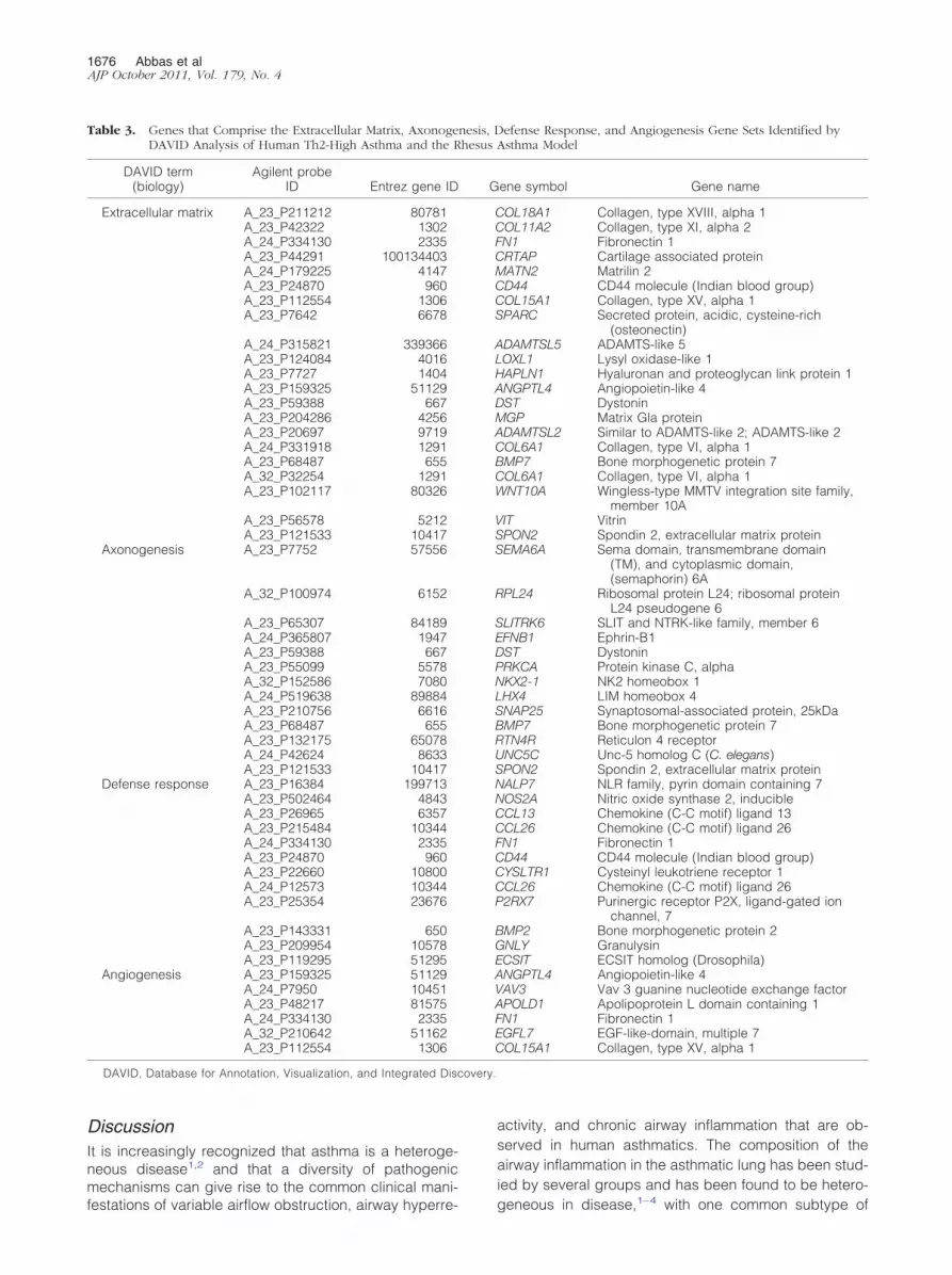
To determine which biological pathways in human Th2-high asthma are specifically represented in either rhesusor BALB/c OVA asthma, we used the Database for An-notation, Visualization, and Integrated Discovery36 toidentify sets of genes from the Gene Ontology, SwissProt/Protein Information Resource, KEGG, INTERPRO, andSMART databases that are enriched among the genesshared by human and rhesus asthma or human andmouse asthma (last accessed May 13, 2010). A total of30 groups with an adjusted P value of �10�1 are specificto human Th2-high asthma and rhesus asthma and de-scribe biological processes (Table 2). These groups areenriched for genes associated with extracellular matrixbiology, cell morphogenesis, nervous system biology,immune host defense biology, and angiogenesis (Table3). In contrast, only four groups with an adjusted P valueof �10�1 are specific to human Th2-high asthma andBALB/c OVA asthma and describe biological processes(Table 4). These groups include extracellular matrix biol-ogy, sequence-specific DNA-binding proteins, and pro-teins associated with developmental biology.
Table 2. Increased Expression of Biological Gene Sets in BothHuman Th2-High Asthma and the Rhesus AsthmaModel, as Identified by DAVID Analysis
DAVID term (biology) P*
Extracellular region part 4.19 � 10�5
Extracellular matrix 8.08 � 10�5
Proteinaceous extracellular matrix 1.90 � 10�4
Cellular component morphogenesis 3.77 � 10�4
Cell morphogenesis involved indifferentiation
5.73 � 10�4
Cell morphogenesis 0.00107Cell projection organization 0.00126Axonogenesis 0.00156Cell projection morphogenesis 0.00253Neuron projection morphogenesis 0.00267Cell morphogenesis involved in neuron
differentiation0.00294
Neuron projection development 0.00313Cell part morphogenesis 0.00313Extracellular matrix 0.00415Extracellular region 0.0193Biological adhesion 0.0410Cell motion 0.0423Axon guidance 0.0426Cell adhesion 0.0429Neuron development 0.0429Defense response 0.0452Response to extracellular stimulus 0.0453Neuron differentiation 0.0460Regulation of nervous system development 0.0577Angiogenesis 0.0619Extracellular space 0.0643Cell adhesion 0.0753Osteogenesis 0.0865Developmental protein 0.0911Cell junction 0.0920
*Corrected by the method of Benjamini-Hochberg for multiple com-parisons.
DAVID, Database for Annotation, Visualization, and Integrated Dis-covery.
covery.
1676 Abbas et alAJP October 2011, Vol. 179, No. 4
DiscussionIt is increasingly recognized that asthma is a heteroge-neous disease1,2 and that a diversity of pathogenicmechanisms can give rise to the common clinical mani-
Table 3. Genes that Comprise the Extracellular Matrix, AxonogeDAVID Analysis of Human Th2-High Asthma and the R
DAVID term(biology)
Agilent probeID Entrez gene ID
Extracellular matrix A_23_P211212 80781A_23_P42322 1302A_24_P334130 2335A_23_P44291 100134403A_24_P179225 4147A_23_P24870 960A_23_P112554 1306A_23_P7642 6678
A_24_P315821 339366A_23_P124084 4016A_23_P7727 1404A_23_P159325 51129A_23_P59388 667A_23_P204286 4256A_23_P20697 9719A_24_P331918 1291A_23_P68487 655A_32_P32254 1291A_23_P102117 80326
A_23_P56578 5212A_23_P121533 10417
Axonogenesis A_23_P7752 57556
A_32_P100974 6152
A_23_P65307 84189A_24_P365807 1947A_23_P59388 667A_23_P55099 5578A_32_P152586 7080A_24_P519638 89884A_23_P210756 6616A_23_P68487 655A_23_P132175 65078A_24_P42624 8633A_23_P121533 10417
Defense response A_23_P16384 199713A_23_P502464 4843A_23_P26965 6357A_23_P215484 10344A_24_P334130 2335A_23_P24870 960A_23_P22660 10800A_24_P12573 10344A_23_P25354 23676
A_23_P143331 650A_23_P209954 10578A_23_P119295 51295
Angiogenesis A_23_P159325 51129A_24_P7950 10451A_23_P48217 81575A_24_P334130 2335A_32_P210642 51162A_23_P112554 1306
DAVID, Database for Annotation, Visualization, and Integrated Dis
festations of variable airflow obstruction, airway hyperre-
activity, and chronic airway inflammation that are ob-served in human asthmatics. The composition of theairway inflammation in the asthmatic lung has been stud-ied by several groups and has been found to be hetero-
efense Response, and Angiogenesis Gene Sets Identified byAsthma Model
ene symbol Gene name
OL18A1 Collagen, type XVIII, alpha 1OL11A2 Collagen, type XI, alpha 2N1 Fibronectin 1RTAP Cartilage associated proteinATN2 Matrilin 2D44 CD44 molecule (Indian blood group)OL15A1 Collagen, type XV, alpha 1PARC Secreted protein, acidic, cysteine-rich
(osteonectin)DAMTSL5 ADAMTS-like 5OXL1 Lysyl oxidase-like 1APLN1 Hyaluronan and proteoglycan link protein 1NGPTL4 Angiopoietin-like 4ST DystoninGP Matrix Gla proteinDAMTSL2 Similar to ADAMTS-like 2; ADAMTS-like 2OL6A1 Collagen, type VI, alpha 1MP7 Bone morphogenetic protein 7OL6A1 Collagen, type VI, alpha 1NT10A Wingless-type MMTV integration site family,
member 10AIT VitrinPON2 Spondin 2, extracellular matrix proteinEMA6A Sema domain, transmembrane domain
(TM), and cytoplasmic domain,(semaphorin) 6A
PL24 Ribosomal protein L24; ribosomal proteinL24 pseudogene 6
LITRK6 SLIT and NTRK-like family, member 6FNB1 Ephrin-B1ST DystoninRKCA Protein kinase C, alphaKX2-1 NK2 homeobox 1HX4 LIM homeobox 4NAP25 Synaptosomal-associated protein, 25kDaMP7 Bone morphogenetic protein 7TN4R Reticulon 4 receptorNC5C Unc-5 homolog C (C. elegans)PON2 Spondin 2, extracellular matrix proteinALP7 NLR family, pyrin domain containing 7OS2A Nitric oxide synthase 2, inducibleCL13 Chemokine (C-C motif) ligand 13CL26 Chemokine (C-C motif) ligand 26N1 Fibronectin 1D44 CD44 molecule (Indian blood group)YSLTR1 Cysteinyl leukotriene receptor 1CL26 Chemokine (C-C motif) ligand 262RX7 Purinergic receptor P2X, ligand-gated ion
channel, 7MP2 Bone morphogenetic protein 2NLY GranulysinCSIT ECSIT homolog (Drosophila)NGPTL4 Angiopoietin-like 4AV3 Vav 3 guanine nucleotide exchange factorPOLD1 Apolipoprotein L domain containing 1N1 Fibronectin 1GFL7 EGF-like-domain, multiple 7OL15A1 Collagen, type XV, alpha 1
nesis, Dhesus
G
CCFCMCCS
ALHADMACBCW
VSS
R
SEDPNLSBRUSNNCCFCCCP
BGEAVAFEC
geneous in disease,1–4 with one common subtype of

Lung Gene Expression in Rhesus Asthma 1677AJP October 2011, Vol. 179, No. 4
asthma defined by eosinophilic lung inflammation that isassociated with Th2 inflammatory gene expression.5,6 Abetter understanding of the pathophysiology of eosino-philic/Th2-high asthma and other asthma subtypes willfacilitate the identification of novel asthma therapeutics,as well as the identification of the appropriate patientpopulations for treatment with these therapies.
Studies of nonhuman primate models of asthma haveyielded important insights into human asthma pathogen-esis, given the strong similarities between nonhuman pri-mates and humans. Although mice are the most com-monly used species in preclinical asthma research,studies of nonhuman primate models of asthma can com-plement and supplement studies of mouse models ofasthma, given the limitations of mouse asthma models inmodeling human disease.14,37–40 Here, we have as-sessed the genomewide lung gene expression profile ofan allergic asthma model induced in rhesus monkeys byHDM and have compared it with the lung gene expres-sion profiles of the eosinophilic/Th2-high subtype of hu-man asthma and a commonly used mouse asthmamodel. We find that a dominant gene expression signa-ture in all three species is a Th2 inflammation gene sig-nature. Significantly, there is a strong correlation betweena prespecified set of human Th2 inflammation genes inhuman Th2-high asthma and in the rhesus asthma model.In addition, similar to studies of this lung Th2 inflammationgene set in human asthma,5,6 we find that the Th2 inflam-mation gene set correlates with physiologic measures ofTh2 inflammation and disease in rhesus asthma, includ-ing BALF eosinophils, peripheral blood eosinophils,HDM-specific serum IgE, and airway hyperreactivity.Thus, consistent with previous reports,11 the rhesusasthma model exhibits many features of human Th2-highasthma. In support of our observations, others have alsoreported gene expression that is associated with Th2inflammation in the lungs or BALF of nonhuman primatemodels of asthma, including a previous study of theHDM-induced rhesus asthma model studied here.10,25,26
Differences in the specific genes detected in our studyversus previous studies of nonhuman primate asthmamodels may be due to i) differences in assessed genes(our study used a prespecified gene set that was gener-ated by applying statistical cutoffs to a human asthmabiopsy microarray gene expression data set, whereas
Table 4. Increased Expression of Biological Gene Sets in BothHuman Th2-High Asthma and the BALB/c OVAMouse Asthma Model, as Identified by DAVIDAnalysis
DAVID term (biology) P*
Extracellular structure organization 0.00210Sequence-specific DNA binding 0.0160Developmental protein 0.0540Embryonic organ development 0.0720
*Corrected by the method of Benjamini-Hochberg for multiple com-parisons.
DAVID, Database for Annotation, Visualization, and Integrated Dis-covery.
previously published studies assessed hand-curated
gene lists), ii) differences in assay sensitivity (we as-sessed gene expression levels by microarray, whereassome previously published studies assessed gene ex-pression levels by quantitative PCR), and/or iii) differ-ences in animal model samples (we assessed airwaybiopsy gene expression, whereas previously publishedstudies assessed either whole lung or BALF cell geneexpression).
Additional analysis of the lung gene expression pro-files of human, rhesus, and BALB/c OVA mouse asthmashowed that only a small subset of genes is sharedamong the three different species. The lack of significantgene overlap among the three species may be due toseveral reasons, including i) imperfect cross-hybridiza-tion between rhesus mRNA transcripts and human mi-croarray probes, which would reduce the number of rhe-sus genes that can be assessed by microarray; ii)incomplete orthology between human and mouse genes,which would reduce the number of mouse genes that canbe compared with human genes; and iii) limitations in theability of the animal models to fully mimic human diseasebiology. Imperfect cross-hybridization between rhesusmRNA transcripts and human microarray probes be-cause of differences in homology between rhesus andhuman gene sequences would lead to false-negative sig-nals. We calculate that 99% of human genes lie in regionspossessing synteny to a sequenced portion of the rhesusmacaque genome, and that 96% of the human probes onthe Agilent microarray platform are perfect matches insequence to the rhesus genes. In addition, analysis of ourhuman and rhesus microarray expression data indicatesthat the average expression intensity of each gene issimilar between the two species. Thus, imperfect cross-hybridization between rhesus mRNA transcripts and hu-man microarray probes is unlikely to be a main cause ofthe lack of significant gene overlap between rhesus andhuman gene expression. Ultimately, the main effect ofmicroarray platform and species orthology limitations onour conclusions is probably a potential underestimationof the similarities between human asthma and the rhesusand mouse asthma models.
When we compared the genes that are shared byhuman Th2-high asthma and rhesus asthma with thegenes that are shared by human Th2-high asthma andBALB/c OVA mouse asthma, we found that some of thebiological pathways that are present in the gene expres-sion profile of human Th2-high asthma are better repre-sented in the gene expression profile of the rhesusasthma model than that of the BALB/c OVA model. Theseinclude extracellular matrix biology, cell morphogenesis,nervous system biology, immune host defense biology,and angiogenesis. One limitation of our approach, whichmay have influenced the outcomes of our analyses, isthat we assessed airway biopsy gene expression fromhuman and rhesus and total lung homogenate gene ex-pression from mouse. Thus, the composition of the tis-sues being compared among human, rhesus, and mouseare different. However, a previously published study ofgene expression in total lung homogenate from an aller-gic asthma model induced in cynomolgus monkeys by
Ascaris suum antigen also identified Th2 inflammation
1678 Abbas et alAJP October 2011, Vol. 179, No. 4
and remodeling genes, although not nervous system orangiogenesis biology.26 Moreover, it should be notedthat the pattern of immune cell accumulation in the lungafter allergen challenge differs between rodents and pri-mates. Whereas in mice the immune cell accumulation isprimarily perivascular with diffuse peribronchial accumu-lation, in humans and rhesus the immune cell accumula-tion is primarily in the larger conducting airways. As such,the biological pathways present in the areas of the mouseairways that are comparable to the areas sampled bybiopsies in humans and rhesus monkeys are probablymore divergent than those present in total mouse lunghomogenate.
Subepithelial fibrosis of the lung, which consists ofextracellular matrix deposition in the basement mem-brane, is a pathological characteristic of asthma andmay contribute to irreversible lung obstruction.41,42 Aprevious study of the rhesus asthma model showedincreased thickening of the basement membrane, asassessed by histology.11 In contrast, commonly usedshort-term mouse models of asthma, such as theBALB/c OVA model that we analyzed in this study,21 donot develop subepithelial fibrosis and lung remodeling,although more chronic mouse models of asthma candevelop some features of fibrosis and remodeling.39
Neuronal dysfunction and dysregulation, encompass-ing activation of both the parasympathetic and sympa-thetic nervous systems, may contribute to the patho-genesis of asthma.43– 45 Neural mediators can havedirect effects on smooth muscle, airway glands, andalveolar walls; there is also substantial interaction be-tween the nervous system and the immune system inthe lungs of asthmatic persons. The lung vasculature isincreased in asthmatics, and the degree of vascularabnormalities in asthma is associated with asthma se-verity.46 – 48 Vascularization is not increased in nontrans-genic allergen-induced mouse asthma models,49,50 al-though overexpression of vascular endothelial growth factorin the mouse lung results in increased vascularization, Th2inflammation, and remodeling.49,51 In contrast, in the rhesusmonkey model of asthma, bronchial vascular density wasincreased at the mid-to-lower airway generations and wasindependent of changes in the interstitial compartment.7
Given the multiple variations of mouse asthma mod-els in the literature, comprising differences in sensiti-zation and challenge protocols, differences in aller-gens, and differences in mouse strains, it was notpossible for us to compare human Th2-high asthmagene expression with that of every mouse asthmamodel. We therefore chose to compare lung gene ex-pression in human asthma, rhesus asthma, and themost commonly used version of mouse asthma in theliterature, which is an acute OVA-induced mouseasthma model in the BALB/c strain. Some of the geneexpression features of human Th2-high asthma that areobserved in the rhesus asthma model, but not in themouse asthma model, may be present in other versionsof mouse asthma that incorporate a more chronic sen-sitization and challenge protocol or that use HDMantigen. To address this, we performed additional
comparisons of the gene expression profile of humanTh2-high asthma with those of a chronic OVA-inducedmouse asthma model16 and an acute HDM-inducedmouse asthma model,18 both of which are publicallyavailable. The chronic OVA-induced mouse asthmamodels share features of cell cycle/cell division, cellu-lar substructures, lymphocyte/T-cell activation and dif-ferentiation, immune response/inflammation, extracel-lular region, chemotaxis, and chitinase activity withhuman Th2-high asthma (see Supplemental Table S4at http://ajp.amjpathol.org). The acute HDM-inducedmouse asthma model shares features of cell cycle/celldivision, chemotaxis, extracellular region/extracellularmatrix, immune response/inflammation, and chitinaseactivity with human Th2-high asthma (see Supplemen-tal Table S5 at http://ajp.amjpathol.org). However, sim-ilar to the acute OVA-induced mouse asthma model,neither the chronic OVA-induced mouse asthma modelnor the acute HDM-induced mouse asthma model ex-hibits features of neural biology or angiogenesis, fea-tures that are shared between the rhesus asthmamodel and human Th2-high asthma.
Our study is the first to our knowledge to directlycompare genomewide human asthma lung gene ex-pression with that of any preclinical asthma model,regardless of species. We find that a lung Th2 inflam-mation gene signature that is present in the eosinophil-ic/Th2-high subtype of human asthma is modeled by aHDM antigen-induced allergic asthma model in rhesusmonkeys, as well as by a BALB/c OVA mouse asthmamodel. In addition, some gene profiles that are presentin human Th2-high asthma are better represented inthe gene expression profile of the rhesus asthmamodel, compared with that of the BALB/c OVA mouseasthma model. These include some inflammatory path-ways, subepithelial fibrosis and remodeling, angiogen-esis, and neural biology that may be associated withneurogenic inflammation. Further study of the rhesusasthma model may complement and supplement stud-ies of mouse asthma models, yield novel insights intothe pathogenesis of human Th2-high asthma, and aidin the identification of new therapeutic targets anddisease biomarkers for asthma, including potentiallysome of the genes reported in this study.
Acknowledgment
We thank the Primate Services Unit at the California Na-tional Primate Research Center for animal handling, care,and coordination, which were critical to this study.
References
1. Green RH, Brightling CE, Bradding P: The reclassification of asthmabased on subphenotypes. Curr Opin Allergy Clin Immunol 2007,7:43–50
2. Wenzel SE: Asthma: defining of the persistent adult phenotypes.Lancet 2006, 368:804–813
3. Douwes J, Gibson P, Pekkanen J, Pearce N: Non-eosinophilic
asthma: importance and possible mechanisms. Thorax 2002, 57:643–648
Lung Gene Expression in Rhesus Asthma 1679AJP October 2011, Vol. 179, No. 4
4. Haldar P, Pavord ID: Noneosinophilic asthma: a distinct clinical andpathologic phenotype. J Allergy Clin Immunol 2007, 119:1043–1052;quiz 1053–1044
5. Choy DF, Modrek B, Abbas AR, Kummerfeld S, Clark HF, Wu LC,Fedorowicz G, Modrusan Z, Fahy JV, Woodruff PG, Arron JR: Geneexpression patterns of Th2 inflammation and intercellular communi-cation in asthmatic airways. J Immunol 2011, 186:1861–1869
6. Woodruff PG, Modrek B, Choy DF, Jia G, Abbas AR, Ellwanger A,Koth LL, Arron JR, Fahy JV: T-helper type 2-driven inflammationdefines major subphenotypes of asthma. Am J Respir Crit Care Med2009, 180:388–395
7. Avdalovic MV, Putney LF, Schelegle ES, Miller L, Usachenko JL, TylerNK, Plopper CG, Gershwin LJ, Hyde DM: Vascular remodeling isairway generation-specific in a primate model of chronic asthma.Am J Respir Crit Care Med 2006, 174:1069–1076
8. Coffman RL, Hessel EM: Nonhuman primate models of asthma. J ExpMed 2005, 201:1875–1879
9. Fanucchi MV, Schelegle ES, Baker GL, Evans MJ, McDonald RJ,Gershwin LJ, Raz E, Hyde DM, Plopper CG, Miller LA: Immunostimu-latory oligonucleotides attenuate airways remodeling in allergic mon-keys. Am J Respir Crit Care Med 2004, 170:1153–1157
10. Miller LA, Hurst SD, Coffman RL, Tyler NK, Stovall MY, Chou DL,Putney LF, Gershwin LJ, Schelegle ES, Plopper CG, Hyde DM: Airwaygeneration-specific differences in the spatial distribution of immunecells and cytokines in allergen-challenged rhesus monkeys. Clin ExpAllergy 2005, 35:894–906
11. Schelegle ES, Gershwin LJ, Miller LA, Fanucchi MV, Van Winkle LS,Gerriets JP, Walby WF, Omlor AM, Buckpitt AR, Tarkington BK, WongVJ, Joad JP, Pinkerton KB, Wu R, Evans MJ, Hyde DM, Plopper CG:Allergic asthma induced in rhesus monkeys by house dust mite(Dermatophagoides farinae). Am J Pathol 2001, 158:333–341
12. Seshasayee D, Lee WP, Zhou M, Shu J, Suto E, Zhang J, Diehl L,Austin CD, Meng YG, Tan M, Bullens SL, Seeber S, Fuentes ME,Labrijn AF, Graus YM, Miller LA, Schelegle ES, Hyde DM, Wu LC,Hymowitz SG, Martin F: In vivo blockade of OX40 ligand inhibitsthymic stromal lymphopoietin driven atopic inflammation. J Clin Invest2007, 117:3868–3878
13. Van Scott MR, Hooker JL, Ehrmann D, Shibata Y, Kukoly C, Salleng K,Westergaard G, Sandrasagra A, Nyce J: Dust mite-induced asthma incynomolgus monkeys. J Appl Physiol 2004, 96:1433–1444
14. Boyce JA, Austen KF: No audible wheezing: nuggets and conun-drums from mouse asthma models. J Exp Med 2005, 201:1869–1873
15. Van Winkle LS, Baker GL, Chan JK, Schelegle ES, Plopper CG:Airway mast cells in a rhesus model of childhood allergic airwaysdisease. Toxicol Sci 2010, 116:313–322
16. Di Valentin E, Crahay C, Garbacki N, Hennuy B, Gueders M, Noel A,Foidart JM, Grooten J, Colige A, Piette J, Cataldo D: New asthmabiomarkers: lessons from murine models of acute and chronicasthma. Am J Physiol Lung Cell Mol Physiol 2009, 296:L185–197
17. Fulkerson PC, Zimmermann N, Hassman LM, Finkelman FD, Rothen-berg ME: Pulmonary chemokine expression is coordinately regulatedby STAT1. STAT6, and IFN-gamma, J Immunol 2004, 173:7565–7574
18. Kelada SN, Wilson MS, Tavarez U, Kubalanza K, Borate B, White-head G, Maruoka S, Roy MG, Olive M, Carpenter DE, Brass DM,Wynn TA, Cook DA, Evans CM, Schwartz DA, Collins FS: Strain-dependent genomic factors affect allergen-induced airway hyper-responsiveness in mice. Am J Respir Cell Mol Biol 2011, doi:10.1165/rcmb.2010-0315OC [Epub ahead of press]
19. Kuperman DA, Lewis CC, Woodruff PG, Rodriguez MW, Yang YH,Dolganov GM, Fahy JV, Erle DJ: Dissecting asthma using focusedtransgenic modeling and functional genomics. J Allergy Clin Immunol2005, 116:305–311
20. Lewis CC, Aronow B, Hutton J, Santeliz J, Dienger K, Herman N,Finkelman FD, Wills-Karp M: Unique and overlapping gene expres-sion patterns driven by IL-4 and IL-13 in the mouse lung. J Allergy ClinImmunol 2009, 123:795–804e798
21. Lewis CC, Yang JY, Huang X, Banerjee SK, Blackburn MR, Baluk P,McDonald DM, Blackwell TS, Nagabhushanam V, Peters W, Voeh-ringer D, Erle DJ: Disease-specific gene expression profiling in mul-tiple models of lung disease. Am J Respir Crit Care Med 2008,177:376–387
22. Park SG, Choi JW, Kim H, Roh GS, Bok J, Go MJ, Kwack K, Oh B, Kim
Y: Genome-wide profiling of antigen-induced time course expressionusing murine models for acute and chronic asthma. Int Arch AllergyImmunol 2008, 146:44–56
23. Rolph MS, Sisavanh M, Liu SM, Mackay CR: Clues to asthma patho-genesis from microarray expression studies. Pharmacol Ther 2006,109:284–294
24. Zimmermann N, King NE, Laporte J, Yang M, Mishra A, Pope SM,Muntel EE, Witte DP, Pegg AA, Foster PS, Hamid Q, Rothenberg ME:Dissection of experimental asthma with DNA microarray analysisidentifies arginase in asthma pathogenesis. J Clin Invest 2003, 111:1863–1874
25. Ayanoglu G, Desai B, Fick RB Jr, Grein J, de Waal Malefyt R, MattsonJ, McClanahan T, Olmstead S, Reece SP, Van Scott MR, Wardle RL:Modelling asthma in macaques: longitudinal changes in cellular andmolecular markers. Eur Respir J 2011, 37:541–552
26. Zou J, Young S, Zhu F, Gheyas F, Skeans S, Wan Y, Wang L, DingW, Billah M, McClanahan T, Coffman RL, Egan R, Umland S:Microarray profile of differentially expressed genes in a monkeymodel of allergic asthma. Genome Biol 2002, 3:research0020
27. Woodruff PG, Boushey HA, Dolganov GM, Barker CS, Yang YH,Donnelly S, Ellwanger A, Sidhu SS, Dao-Pick TP, Pantoja C, ErleDJ, Yamamoto KR, Fahy JV: Genome-wide profiling identifies ep-ithelial cell genes associated with asthma and with treatment re-sponse to corticosteroids. Proc Natl Acad Sci U S A 2007, 104:15858 –15863
28. Crapo RO, Casaburi R, Coates AL, Enright PL, Hankinson JL, IrvinCG, MacIntyre NR, McKay RT, Wanger JS, Anderson SD, CockcroftDW, Fish JE, Sterk PJ: Guidelines for methacholine and exercisechallenge testing-1999. This official statement of the American Tho-racic Society was adopted by the ATS Board of Directors, July 1999.Am J Respir Crit Care Med 2000, 161:309–329
29. ChuHW, Balzar S, Westcott JY, Trudeau JB, Sun Y, Conrad DJ,Wenzel SE: Expression and activation of 15-lipoxygenase pathway insevere asthma: relationship to eosinophilic phenotype and collagendeposition. Clin Exp Allergy 2002, 32:1558–1565
30. Gould HJ, Sutton BJ: IgE in allergy and asthma today. Nat RevImmunol 2008, 8:205–217
31. Komiya A, Nagase H, Yamada H, Sekiya T, Yamaguchi M, Sano Y,Hanai N, Furuya A, Ohta K, Matsushima K, Yoshie O, Yamamoto K,Hirai K: Concerted expression of eotaxin-1, eotaxin-2, and eo-taxin-3 in human bronchial epithelial cells. Cell Immunol 2003,225:91–100
32. Suresh V, Mih JD, George SC: Measurement of IL-13-induced iNOS-derived gas phase nitric oxide in human bronchial epithelial cells.Am J Respir Cell Mol Biol 2007, 37:97–104
33. Thurmond RL, Gelfand EW, Dunford PJ: The role of histamine H1 andH4 receptors in allergic inflammation: the search for new antihista-mines. Nat Rev Drug Discov 2008, 7:41–53
34. Zuo L, Fulkerson PC, Finkelman FD, Mingler M, Fischetti CA, Blan-chard C, Rothenberg ME: IL-13 induces esophageal remodeling andgene expression by an eosinophil-independent. IL-13R alpha 2-in-hibited pathway. J Immunol 2010, 185:660–669
35. Sayers EW, Barrett T, Benson DA, Bolton E, Bryant SH, Canese K, etal: Database resources of the National Center for BiotechnologyInformation. Nucleic Acids Res 38:D5-D16
36. Dennis G Jr, Sherman BT, Hosack DA, Yang J, Gao W, Lane HC,Lempicki RA: DAVID: database for Annotation, Visualization, andIntegrated Discovery. Genome Biol 2003, 4:P3
37. Finkelman FD, Wills-Karp M: Usefulness and optimization of mousemodels of allergic airway disease. J Allergy Clin Immunol 2008,121:603–606
38. Mestas J, Hughes CC: Of mice and not men: differences betweenmouse and human immunology. J Immunol 2004, 172:2731–2738
39. Taube C, Dakhama A, Gelfand EW: Insights into the pathogenesis ofasthma utilizing murine models. Int Arch Allergy Immunol 2004, 135:173–186
40. Wenzel S, Holgate ST: The mouse trap: It still yields few answers inasthma. Am J Respir Crit Care Med 2006, 174:1173–1178
41. Jeffery PK: Remodeling in asthma and chronic obstructive lung dis-ease. Am J Respir Crit Care Med 2001, 164:S28–S38
42. Pascual RM, Peters SP: Airway remodeling contributes to the pro-gressive loss of lung function in asthma: an overview. J Allergy ClinImmunol 2005, 116:477–486; quiz 487
43. Nockher WA, Renz H: Neurotrophins and asthma: novel insight intoneuroimmune interaction. J Allergy Clin Immunol 2006, 117:67–71

1680 Abbas et alAJP October 2011, Vol. 179, No. 4
44. Pisi G, Olivieri D, Chetta A: The airway neurogenic inflammation:clinical and pharmacological implications. Inflamm Allergy Drug Tar-gets 2009, 8:176–181
45. Undem BJ, Carr MJ: The role of nerves in asthma. Curr AllergyAsthma Rep 2002, 2:159–165
46. Hoshino M, Nakamura Y, Hamid QA: Gene expression of vascularendothelial growth factor and its receptors and angiogenesis in bron-chial asthma. J Allergy Clin Immunol 2001, 107:1034–1038
47. Li X, Wilson JW: Increased vascularity of the bronchial mucosa in mildasthma. Am J Respir Crit Care Med 1997, 156:229–233
48. Vrugt B, Wilson S, Bron A, Holgate ST, Djukanovic R, Aalbers R:
Bronchial angiogenesis in severe glucocorticoid-dependent asthma.Eur Respir J 2000, 15:1014–102149. Lee CG, Link H, Baluk P, Homer RJ, Chapoval S, Bhandari V, KangMJ, Cohn L, Kim YK, McDonald DM, Elias JA: Vascular endothelialgrowth factor (VEGF) induces remodeling and enhances TH2-mediated sensitization and inflammation in the lung. Nat Med2004, 10:1095–1103
50. Suzaki Y, Hamada K, Sho M, Ito T, Miyamoto K, Akashi S, Kashizuka H,Ikeda N, Nakajima Y, Iwase M, Homma I, Kobzik L, Kimura H: A potentantiangiogenic factor, endostatin prevents the development of asthma ina murine model. J Allergy Clin Immunol 2005, 116:1220–1227
51. Baluk P, Lee CG, Link H, Ator E, Haskell A, Elias JA, McDonald DM:Regulated angiogenesis and vascular regression in mice overex-
pressing vascular endothelial growth factor in airways. Am J Pathol2004, 165:1071–1085


