Embed Size (px)
Citation preview

Loss of STOP Protein Impairs Peripheral OlfactoryNeurogenesisKarelle Benardais1,2, Basem Kasem1,2, Alice Couegnas1,2, Brigitte Samama1,2,3*, Sebastien Fernandez2,
Christiane Schaeffer1,2,3, Maria-Cristina Antal1,2,3, Didier Job4, Annie Schweitzer4, Annie Andrieux4,
Anne Giersch1, Astrid Nehlig1, Nelly Boehm1,2,3
1 INSERM U666, Strasbourg, France, 2 Universite de Strasbourg, Faculte de Medecine, Institut d’Histologie, Strasbourg, France, 3 Hopitaux Universitaires de Strasbourg,
Strasbourg, France, 4 INSERM U836, Grenoble Institut of Neurosciences, Grenoble, France; iRTSV-GPC, CEA-Grenoble, France; Universite Joseph Fourrier, Grenoble, France
Abstract
Background: STOP (Stable Tubulin-Only Polypeptide) null mice show behavioral deficits, impaired synaptic plasticity,decrease in synaptic vesicular pools and disturbances in dopaminergic transmission, and are considered aneurodevelopmental model of schizophrenia. Olfactory neurons highly express STOP protein and are continually generatedthroughout life. Experimentally-induced loss of olfactory neurons leads to epithelial regeneration within two months,providing a useful model to evaluate the role played by STOP protein in adult olfactory neurogenesis.
Methodology/Principal Findings: Immunocytochemistry and electron microscopy were used to study the structure of theglomerulus in the main olfactory bulb and neurogenesis in the neurosensorial epithelia. In STOP null mice, olfactory neuronsshowed presynaptic swellings with tubulovesicular profiles and autophagic-like structures. In olfactory and vomeronasalepithelia, there was an increase in neurons turnover, as shown by the increase in number of proliferating, apoptotic andimmature cells with no changes in the number of mature neurons. Similar alterations in peripheral olfactory neurogenesishave been previously described in schizophrenia patients. In STOP null mice, regeneration of the olfactory epithelium didnot modify these anomalies; moreover, regeneration resulted in abnormal organisation of olfactory terminals within theolfactory glomeruli in STOP null mice.
Conclusions/Significance: In conclusion, STOP protein seems to be involved in the establishment of synapses in theolfactory glomerulus. Our results indicate that the olfactory system of STOP null mice is a well-suited experimental model (1)for the study of the mechanism of action of STOP protein in synaptic function/plasticity and (2) for pathophysiologicalstudies of the mechanisms of altered neuronal connections in schizophrenia.
Citation: Benardais K, Kasem B, Couegnas A, Samama B, Fernandez S, et al. (2010) Loss of STOP Protein Impairs Peripheral Olfactory Neurogenesis. PLoS ONE 5(9):e12753. doi:10.1371/journal.pone.0012753
Editor: Thomas Burne, University of Queensland, Australia
Received March 15, 2010; Accepted August 18, 2010; Published September 15, 2010
Copyright: � 2010 Benardais et al. This is an open-access article distributed under the terms of the Creative Commons Attribution License, which permitsunrestricted use, distribution, and reproduction in any medium, provided the original author and source are credited.
Funding: Source of funding: INSERM (Institut National de la Sante et de la Recherche Medicale). The funders had no role in study design, data collection andanalysis, decision to publish, or preparation of the manuscript.
Competing Interests: The authors have declared that no competing interests exist.
* E-mail: [email protected]
Introduction
STOP protein (Stable Tubulin-Only Polypeptide, for a
review, see [1]) is a microtubule-associated protein initially
isolated from preparations of rat brain cold-stable microtubules.
It is a calmodulin-regulated protein able to induce a high degree
of microtubule stability in cold-exposed cells [2]. Particularly
abundant in neurons, this protein has been shown to be
important for normal neurite formation during neuronal
differentiation in cultured neurons [3]. STOP null mice show
behavioral deficits (disorganized activity, social withdrawal,
impaired maternal behavior), hypersensitivity to amphetamine
in postpubertal mice, impaired synaptic plasticity, decrease in
hippocampal synaptic vesicular pools and disturbances in the
dopaminergic, glutamatergic and nicotinic neurotransmissions
[4–12] and have been proposed as a mouse model to explore the
neurodevelopmental and synaptic impairment hypothesis of
schizophrenia [4].
Although STOP null mice do not present major brain
anomalies, they show subtle modifications of the olfactory system
maturation [13]. As adults, they show cognitive deficits using novel
object recognition and olfactory discrimination tasks [14]. Since
olfactory and vomeronasal pathways highly express STOP
transcripts and protein [4,15,16], we hypothesized that STOP
protein deficiency may lead in adults to synaptic impairment in
this pathway. In rodents, there are two subdivisions in the
olfactory system: in the main olfactory system, neurosensory cells
(olfactory receptor neurons, ORNs) in the olfactory epithelium
(OE) send axons to the main olfactory bulb (OB) where they make
synapses with the dendrite of mitral/tufted cells in the OB
glomeruli; in the accessory olfactory system, axons arising from the
neurosensorial cells of the vomeronasal epithelium (VNE), lying in
the vomeronasal organ (VNO) make synapses with mitral/tufted
cells in the glomeruli of the accessory olfactory bulb (AOB). The
olfactory glomerulus represents a useful model system for synapse
analysis: its boundaries are sharply delineated; olfactory axons are
PLoS ONE | www.plosone.org 1 September 2010 | Volume 5 | Issue 9 | e12753
the unique input; olfactory presynaptic terminals are glutamater-
gic. The olfactory system is a highly plastic neuronal network.
Olfactory and vomeronasal neurosensorial cells constantly renew
life long [17–21]. There is a constant loss of neurosensorial cells,
which die by apoptosis; they are replaced by new neurons arising
from progenitors located in the basal compartment of the OE,
which consists of two distinct cell types: horizontal basal cells
(HBCs) directly attached to the basal lamina and globose basal
cells (GBCs) lying immediately above the HBC layer. GBCs are
associated with active proliferation and express early neuronal
differentiation markers whereas HBCs divide infrequently and
express cytokeratin 5 and 14, but not neuronal markers [22,23].
Immature neurons arising from cell division express GAP 43 and
doublecortin; they differentiate to fully mature neurons expressing
OMP (Olfactory Marker Protein) [24] and olfactory receptors at
the tip of their dendrite, when establishing synapses with the apical
dendrite of mitral/tufted cells in the OB. Experimentally-induced
loss of olfactory neurons leads to epithelial regeneration within two
months, providing a useful model to evaluate the role played by
STOP protein in adult olfactory neurogenesis [25].
In the present work, we first asked whether olfactory synapses
were morphologically disturbed in the absence of STOP protein,
as are hippocampal synapses. Second, do the synaptic modifica-
tions impair normal OE and VNE homeostasis? Third, to get
insight into STOP protein function in adult ORN biology, we
induced ORN regeneration and analysed both peripheral and
central levels at two ages, 3 and 10 months.
We show presynaptic anomalies and impaired neurogenesis,
some of the impairments recapitulating features observed in
schizophrenia patients. Regeneration of the OE did not modify
these anomalies in STOP null mice, but moreover induced
abnormal organisation of olfactory terminals within the olfactory
glomeruli.
Our results indicate that the olfactory system of STOP null mice
is a well-suited experimental model (1) for the study of the
mechanism of action of STOP protein in synaptic function/
plasticity and (2) for pathophysiological studies of the mechanisms
of altered neuronal connections in schizophrenia.
Results
Loss of STOP protein induces presynaptic anomalies inthe olfactory glomerulus
We first compared synaptic morphology in WT and STOP null
mice at 3 to 6 months of age. On semithin sections, no obvious
difference could be observed between the two genotypes. At the
ultrastructural level however, all STOP null mice displayed the
same modifications when compared to WT mice. In glomeruli,
olfactory fiber endings are easily recognized by their high electron
density (Figure 1A). In both OB and AOB glomeruli of STOP null
mice, some axons displayed terminal dilatations containing either
autophagic-like structures, (Figure 1B, E, F), complex smooth
canalicules (Figure 1C) or both (Figure 1D). These dilatations were
recognized as olfactory presynaptic endings by the presence of
synaptic vesicles or synaptic densities on the post-synaptic
membrane (Figure 1E, F) when few autophagic-like structures
were present. They were disseminated in the whole glomeruli layer
of the OB and AOB for each animal. They were specific to STOP
null mice, since never observed in WT mice (Figure 1A) and
different from lipofuscin, which was present in neuronal cell bodies
of the oldest mice of the two genotypes. The other layers of the OB
and AOB (olfactory fibers, vomeronasal fibers, plexiform, granule
and mitral cell layers) did not display any modification in STOP
null mice as compared to WT mice.
Neurogenesis in the olfactory and vomeronasal epitheliaAs olfactory and vomeronasal neurons are constantly renewed by
peripheral neurogenesis following apoptosis, we asked whether the
presynaptic modifications may lead to neurogenesis modifications.Neurogenesis in the OE. The OE was studied at six levels in
the rostro-caudal plan. In these six levels, all immunoreactive cells
present in the OE of both sides were counted.
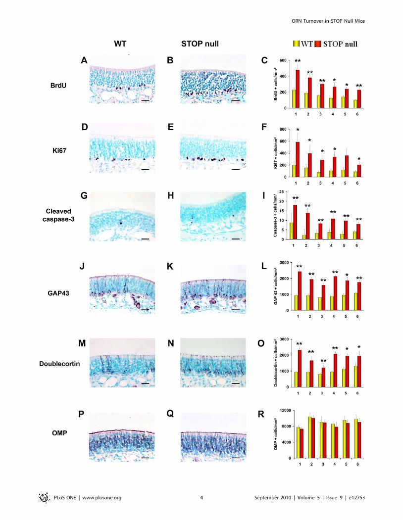
Proliferation was studied by evaluating the immunoexpression
of BrdU and Ki67. In the OE of both STOP null and WT mice,
positive nuclei were mainly present in the lowest layers, but few
superficial cells, either ORNs (round nuclei) or sustentacular cells
(elongated nuclei) were also labelled (Figure 2A, B, D, E). There
was a statistically significant increase of proliferating cells in STOP
null mice as compared to WT mice (Figure 2C, F). The specificity
of the increase in proliferation in the sensorial epithelium was
shown by the absence of difference in proliferation in the
respiratory epithelium between STOP null and WT mice as
shown by the mean number of Ki67 positive cells per mm2 in the
six areas studied for STOP null and WT mice respectively:
4.460.5 vs 4.260.3; 4.560.5 vs 4.760.4; 6.460.6 vs 5.0860.7;
14.761.2 vs 14.061.3; 18.360.5 vs 1762.
Apoptosis was evaluated by counting cleaved caspase 3 positive
cells (Figure 2G, H). There was a statistically significant increase in
the number of cleaved caspase 3 positive cells in all areas of OE
studied (Figure 2I) in STOP null mice when compared to WT
mice. Immature neurons were studied by their doublecortin and
GAP 43 immunoexpressions. They were localised in the lower part
of the epithelium. (Figure 2J, K, M, N). The number of
doublecortin and GAP 43 immunoreactive cells was statistically
greater in STOP null mice than in WT mice (Figure 2L, O).
There was no statistical difference in the number of mature OMP
expressing neurons in WT and STOP null mice (Figure 2P–R).
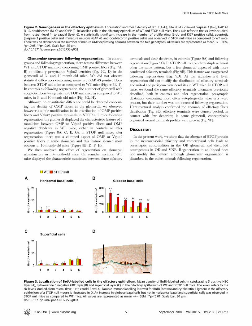
We then analysed the distribution of BrdU positive nuclei; cells
double-stained for BrdU and cytokeratin 5 were considered as
HBCs; nuclei, negative for cytokeratin 5 and localised just above
HBC layer were referred as GBCs, and the few remaining
superficial cells were considered as superficial cells (Figure 3D).
There was an increase in proliferation in GBC layer but not in
HBC and superficial cells layers in STOP null mice as compared
to WT mice (Figure 3A–C).Neurogenesis in the VNE. Three levels in the rostro-caudal
plan of the VNO were studied and all immunoreactive cells
present in these levels in the two VNOs of each animal were
counted.
As for the OE, there was a statistically significant increase in the
number of proliferating (Figure 4A–F), apoptotic (Figure 4G–I)
and immature cells (Figure 4J–O) but not mature neurons
(Figure 4P–R) in STOP null mice as compared to WT mice. In
the VNE of both WT and STOP null mice, proliferating and
immature cells were mainly but not exclusively located at the
margins of the neurosensorial epithelium (Figure 4A–E, J–N). We
then asked whether the difference in cell proliferation between the
two genotypes resulted from marginal, central or both prolifera-
tions. We observed that cells at the margins represented, in WT
and STOP null mice, 71%610 and 63%68 of the whole
proliferating cells, respectively; there was a statistically significant
increase in proliferating cells in STOP null mice as compared to
WT mice, both for marginal and central localisations (p,0.05).
Effect of olfactory regeneration on glomerularultrastructure and peripheral neurogenesis
To gain more insights in the mechanisms of synaptic and
neurogenesis disturbances in STOP null mice, we created a
complete degeneration of the OE of the right nasal cavity in 3- and
ORN Turnover in STOP Null Mice
PLoS ONE | www.plosone.org 2 September 2010 | Volume 5 | Issue 9 | e12753

10-month-old animals and analysed two months later epithelial
regeneration and glomerular structure. ZnSO4 infusion in the
right naris resulted in complete detachment of the OE 48 hours
later, without impairing the VNE.
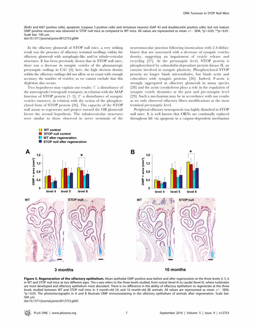
Epithelial regeneration and neurogenesis. We first evaluat-
ed the extent of OE regeneration at three levels of the nasal
cavity, where turbinates are most developed and OE most
abundant, and observed that there was no difference in the ability
of the OE to regenerate in the three levels, between WT and
STOP null mice, either for the 3 month-old (Fig. 5A) or the 10
month-old animals (Fig. 5B). This result lead us to consider the
mean values between these three levels for the analysis of
neurogenesis after regeneration.
In the 3-month-old groups, apoptotic, proliferating and
immature cells were more numerous in STOP null mice as
compared to WT mice in both control animals and after
regeneration, suggesting no effect of the regeneration itself
(Fig. 6A, C, E). The number of OMP positive cells did not differ
among the four groups (Fig. 6G). In the 10-month-old groups,
similar results were observed for apoptotic (Fig. 6D) and OMP
positive cells (Fig. 6H); however, there were no differences between
WT and STOP null mice either in controls or after regeneration
concerning proliferating (Fig. 6B) and immature cells (Fig. 6F).
We then analysed the OB to check the ability of olfactory
neurons axons to target the glomeruli in the absence of STOP
protein.
Figure 1. Ultrastructure of olfactory bulb glomeruli. Electron microscopy micrographs of olfactory bulb glomeruli in WT (A) and STOP null (B–F) mice at 3 to 6 months of age. In STOP null mice, olfactory axon endings are filled with autophagic-like structures (B, arrow), tubulovesicular profiles(C, arrowhead), or both (D). When few autophagic structures were present (arrows) (E, F), olfactory axons endings could be identified by the presenceof synaptic vesicles and postsynaptic densities (arrowheads) (E, F). De: dentrite; OA: olfactory axon. Scale bar: 0,5 mm (A, C, E, F); 1 mm (B, D).doi:10.1371/journal.pone.0012753.g001
ORN Turnover in STOP Null Mice
PLoS ONE | www.plosone.org 3 September 2010 | Volume 5 | Issue 9 | e12753
ORN Turnover in STOP Null Mice
PLoS ONE | www.plosone.org 4 September 2010 | Volume 5 | Issue 9 | e12753
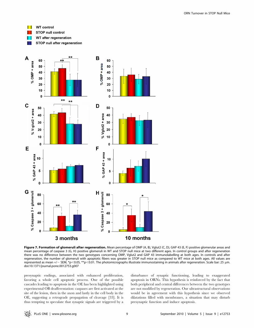
Glomerular structure following regeneration. In control
groups and following regeneration, there was no difference between
WT and STOP null mice concerning OMP positive fibers (Fig. 7A,
B) or olfactory presynaptic Vglut2 densities (Fig. 7C, D) in the
glomeruli of 3- and 10-month-old mice. We did not observe
statistical differences concerning immature GAP 43 positive fibers
between STOP null mice as compared to WT mice (Figure 7E, F).
In controls as following regeneration, the number of glomeruli with
apoptotic fibers was greater in STOP null mice as compared to WT
mice, in 3- and 10-month-old mice (Fig. 7G, H).
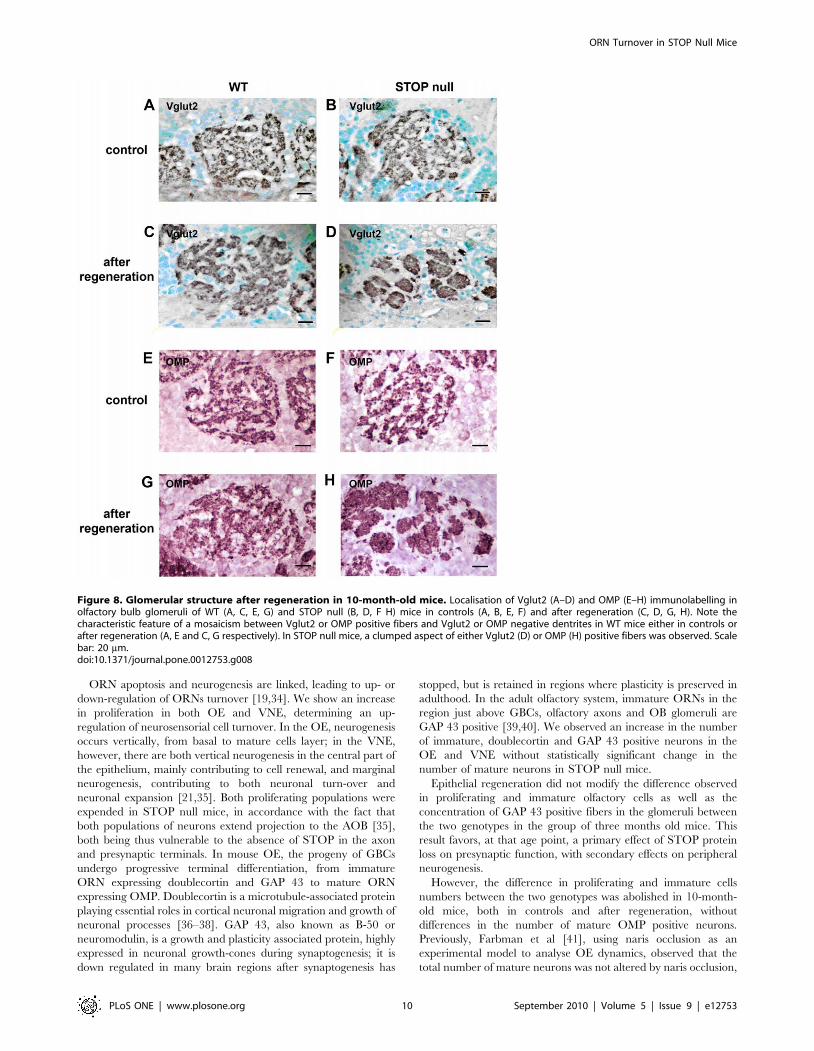
Although no quantitative difference could be detected concern-
ing the density of OMP fibers in the glomeruli, we observed
however a subtle modification in the distribution of OMP positive
fibers and Vglut2 positive terminals in STOP null mice following
regeneration: the glomeruli displayed the characteristic feature of a
mosaıcism between OMP or Vglut2 positive fibers and OMP
negative dendrites in WT mice, either in controls or after
regeneration (Figure 8A, C, E, G); in STOP null mice, after
regeneration, there was a clumped aspect of OMP or Vglut2
positive fibers in some glomeruli and this feature seemed most
obvious in 10-month-old mice (Figure 8B, D, F, H).
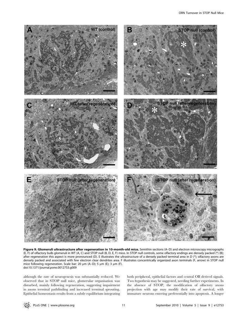
We then analysed the effect of regeneration on glomeruli
ultrastructure in 10-month-old mice. On semithin sections, WT
mice displayed the characteristic mosaıcism between dense olfactory
terminals and clear dendrites, in controls (Figure 9A) and following
regeneration (Figure 9C). In STOP null mice, controls displayed most
often the same pattern, but a few glomeruli appeared with more
condensed olfactory terminals (Fig. 9B). This feature was exaggerated
following regeneration (Fig. 9D). At the ultrastructural level,
regeneration did not modify the distribution of olfactory terminals
and mitral and periglomerular dendrites in WT mice. In STOP null
mice, we found the same olfactory terminals anomalies previously
described, both in controls and after regeneration: presynaptic
dilatations containing most often autophagic-like structures were
present, but their number was not increased following regeneration.
Ultrastructural analysis confirmed the anomaly of olfactory fibers
distribution (Fig. 9E): olfactory terminals were densely packed, in
contact with few dendrites; in some glomeruli, concentrically
organized axonal terminals profiles were present (Fig. 9F).
Discussion
In the present work, we show that the absence of STOP protein
in the neurosensorial olfactory and vomeronasal cells leads to
presynaptic abnormalities in the OB glomeruli and disturbed
neurogenesis in OE and VNE. Regeneration in adulthood does
not modify this pattern although glomerular organisation is
disturbed in the oldest animals following regeneration.
Figure 2. Neurogenesis in the olfactory epithelium. Localisation and mean density of BrdU (A–C), Ki67 (D–F), cleaved caspase 3 (G–I), GAP 43(J–L), doublecortin (M–O) and OMP (P–R) labelled cells in the olfactory epithelium of WT and STOP null mice. The x-axis refers to the six levels studied,from rostral (level 1) to caudal (level 6). A statistically significant increase in the number of proliferating (BrdU and Ki67 positive cells), apoptotic(caspase 3 positive cells) and immature neurons (GAP 43 and doublecortin positive cells) was observed in STOP null mice as compared to WT mice.There was no difference in the number of mature OMP expressing neurons between the two genotypes. All values are represented as mean +/2 SEM,*p,0.05, **p,0.01. Scale bar: 25 mm.doi:10.1371/journal.pone.0012753.g002
Figure 3. Localisation of BrdU-labelled cells in the olfactory epithelium. Mean density of BrdU-labelled cells in cytokeratine 5 positive HBClayer (A), cytokeratine 5 negative GBC layer (B) and superficial layer (C) in the olfactory epithelium of WT and STOP null mice. The x-axis refers to thesix levels studied, from rostral (level 1) to caudal (level 6). Double immunolabelling (arrows) for BrdU (brown) and cytokeratin 5 (green) in the olfactoryepithelium of a STOP null mouse is illustrated in D. An increase in globose basal cells but not in horizontal basal and superficial cells was observed inSTOP null mice as compared to WT mice. All values are represented as mean +/2 SEM, **p,0.01. Scale bar: 30 mm.doi:10.1371/journal.pone.0012753.g003
ORN Turnover in STOP Null Mice
PLoS ONE | www.plosone.org 5 September 2010 | Volume 5 | Issue 9 | e12753
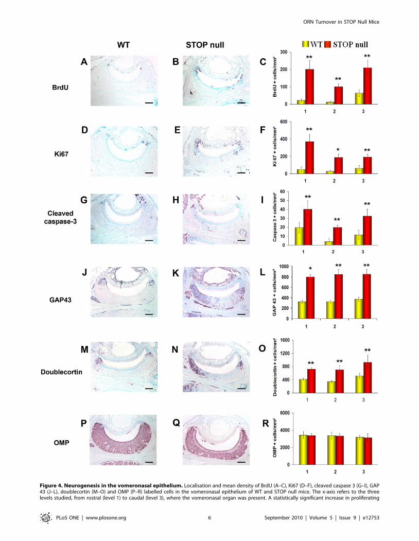
Figure 4. Neurogenesis in the vomeronasal epithelium. Localisation and mean density of BrdU (A–C), Ki67 (D–F), cleaved caspase 3 (G–I), GAP43 (J–L), doublecortin (M–O) and OMP (P–R) labelled cells in the vomeronasal epithelium of WT and STOP null mice. The x-axis refers to the threelevels studied, from rostral (level 1) to caudal (level 3), where the vomeronasal organ was present. A statistically significant increase in proliferating
ORN Turnover in STOP Null Mice
PLoS ONE | www.plosone.org 6 September 2010 | Volume 5 | Issue 9 | e12753
In the olfactory glomeruli of STOP null mice, a very striking
result was the presence of olfactory terminal swellings within the
olfactory glomeruli with autophagic-like and/or tubulo-vesicular
structures. It has been previously shown that in STOP null mice,
there was a decrease in synaptic vesicles of the glutamatergic
presynaptic endings in CA1 [4]; here, the high electron density
within the olfactory endings did not allow us to count with enough
accuracy the number of vesicles; so we cannot exclude that this
depletion also occurs.
Two hypotheses may explain our results: 1u a disturbance of
the anterograde/retrograde transport, in relation with the MAP
function of STOP protein [1–3]; 2u a disturbance of synaptic
vesicles turnover, in relation with the action of the phosphor-
ylated form of STOP protein [26]. The capacity of the STOP
null axons to regenerate and project toward the OB glomeruli
favors the second hypothesis. The tubulovesicular structures
were similar to those observed in nerve terminals of the
neuromuscular junction following intoxication with 2,4-dithio-
biuret that are associated with a decrease of synaptic vesicles
density, suggesting an impairment of vesicle release and
recycling [27]. At the presynaptic level, STOP protein is
phosphorylated by calmodulin-dependant protein kinase II, an
enzyme involved in synaptic plasticity. Phosphorylated STOP
protein no longer binds microtubules, but binds actin and
colocalizes with synaptic proteins [26]. Indeed, F-actin is
strongly aggregated in olfactory glomeruli in many species
[28] and the actin cytoskeleton plays a role in the regulation of
synaptic vesicle dynamics at the post and pre-synaptic level
[29]. Such a mechanism may be in accordance with our results
as we only observed olfactory fibers modifications at the most
terminal presynaptic level.
Peripheral olfactory neurogenesis was highly disturbed in STOP
null mice. It is well known that ORNs are continually replaced
throughout life via apoptosis in a caspase-dependent mechanism
(BrdU and Ki67 positive cells), apoptotic (caspase 3 positive cells) and immature neurons (GAP 43 and doublecortin positive cells), but not matureOMP positive neurons was observed in STOP null mice as compared to WT mice. All values are represented as mean +/2 SEM, *p,0.05, **p,0.01.Scale bar: 100 mm.doi:10.1371/journal.pone.0012753.g004
Figure 5. Regeneration of the olfactory epithelium. Mean epithelial OMP positive area before and after regeneration at the three levels 4, 5, 6in WT and STOP null mice at two different ages. The x-axis refers to the three levels studied, from rostral (level 4) to caudal (level 6), where turbinatesare most developed and olfactory epithelium most abundant. There is no difference in the ability of olfactory epithelium to regenerate at the threelevels studied between WT and STOP null mice in 3 month-old (A) and 10 month-old (B) animals. All values are represented as mean +/2 SEM,*p,0.05. The photomicrographs in A and B illustrate OMP immunostaining in the olfactory epithelium of animals after regeneration. Scale bar:500 mm.doi:10.1371/journal.pone.0012753.g005
ORN Turnover in STOP Null Mice
PLoS ONE | www.plosone.org 7 September 2010 | Volume 5 | Issue 9 | e12753

[30]. In STOP null mice, we observed an increase in the number
of cleaved caspase 3 positive neurosensorial cells and fiber endings
in the OB glomeruli. Caspase 3 is an effector caspase, which
mediates the terminal stages of apoptosis. It has been shown that
caspase 3 can be locally activated in synapses, triggering local
degeneration without initiating an irreversible apoptotic cascade
[31,32]. However, here we observed activated caspase 3
immunoreactivity in the whole ORN from pericaryon to
Figure 6. Neurogenesis in the olfactory epithelium after regeneration. Mean density of Ki 67 (A, B), caspase 3 (C, D), GAP 43 (E, F) and OMP(G, H) positive cells in the olfactory epithelium of WT and STOP null mice at two different ages. In the 3-month-old groups (A, C, E, G), apoptotic,proliferating and immature, but not mature neurons are more numerous in STOP null mice as compared to WT mice, both in control animals andafter regeneration. In the 10-month-old groups (B, D, F, H) only the number of caspase 3 positive neurons (D) was increased in STOP null mice ascompared to WT mice, in controls and after regeneration. All values are represented as mean +/2 SEM, *p,0.05, **p,0.01. The photomicrographsillustrate immunostaining in animals after regeneration. Scale bar: 25 mm.doi:10.1371/journal.pone.0012753.g006
ORN Turnover in STOP Null Mice
PLoS ONE | www.plosone.org 8 September 2010 | Volume 5 | Issue 9 | e12753
presynaptic endings, associated with enhanced proliferation,
favoring a whole cell apoptotic process. One of the possible
cascades leading to apoptosis in the OE has been highlighted using
experimental OB deafferentation: caspases are first activated at the
site of the lesion, then in the axon and lastly in the cell body in the
OE, suggesting a retrograde propagation of cleavage [33]. It is
thus tempting to speculate that synaptic signals are triggered by a
disturbance of synaptic functioning, leading to exaggerated
apoptosis in ORNs. This hypothesis is reinforced by the fact that
both peripheral and central differences between the two genotypes
are not modified by regeneration. Our ultrastructural observations
would be in agreement with this hypothesis since we observed
dilatations filled with membranes, a situation that may disturb
presynaptic function and induce apoptosis.
Figure 7. Formation of glomeruli after regeneration. Mean percentage of OMP (A, B), Vglut2 (C, D), GAP 43 (E, F) positive glomerular areas andmean percentage of caspase 3 (G, H) positive glomeruli in WT and STOP null mice at two different ages. In control groups and after regenerationthere was no difference between the two genotypes concerning OMP, Vglut2 and GAP 43 immunolabelling at both ages. In controls and afterregeneration, the number of glomeruli with apoptotic fibers was greater in STOP null mice as compared to WT mice at both ages. All values arerepresented as mean +/2 SEM, *p,0.05, **p,0.01. The photomicrographs illustrate immunostaining in animals after regeneration. Scale bar: 25 mm.doi:10.1371/journal.pone.0012753.g007
ORN Turnover in STOP Null Mice
PLoS ONE | www.plosone.org 9 September 2010 | Volume 5 | Issue 9 | e12753
ORN apoptosis and neurogenesis are linked, leading to up- or
down-regulation of ORNs turnover [19,34]. We show an increase
in proliferation in both OE and VNE, determining an up-
regulation of neurosensorial cell turnover. In the OE, neurogenesis
occurs vertically, from basal to mature cells layer; in the VNE,
however, there are both vertical neurogenesis in the central part of
the epithelium, mainly contributing to cell renewal, and marginal
neurogenesis, contributing to both neuronal turn-over and
neuronal expansion [21,35]. Both proliferating populations were
expended in STOP null mice, in accordance with the fact that
both populations of neurons extend projection to the AOB [35],
both being thus vulnerable to the absence of STOP in the axon
and presynaptic terminals. In mouse OE, the progeny of GBCs
undergo progressive terminal differentiation, from immature
ORN expressing doublecortin and GAP 43 to mature ORN
expressing OMP. Doublecortin is a microtubule-associated protein
playing essential roles in cortical neuronal migration and growth of
neuronal processes [36–38]. GAP 43, also known as B-50 or
neuromodulin, is a growth and plasticity associated protein, highly
expressed in neuronal growth-cones during synaptogenesis; it is
down regulated in many brain regions after synaptogenesis has
stopped, but is retained in regions where plasticity is preserved in
adulthood. In the adult olfactory system, immature ORNs in the
region just above GBCs, olfactory axons and OB glomeruli are
GAP 43 positive [39,40]. We observed an increase in the number
of immature, doublecortin and GAP 43 positive neurons in the
OE and VNE without statistically significant change in the
number of mature neurons in STOP null mice.
Epithelial regeneration did not modify the difference observed
in proliferating and immature olfactory cells as well as the
concentration of GAP 43 positive fibers in the glomeruli between
the two genotypes in the group of three months old mice. This
result favors, at that age point, a primary effect of STOP protein
loss on presynaptic function, with secondary effects on peripheral
neurogenesis.
However, the difference in proliferating and immature cells
numbers between the two genotypes was abolished in 10-month-
old mice, both in controls and after regeneration, without
differences in the number of mature OMP positive neurons.
Previously, Farbman et al [41], using naris occlusion as an
experimental model to analyse OE dynamics, observed that the
total number of mature neurons was not altered by naris occlusion,
Figure 8. Glomerular structure after regeneration in 10-month-old mice. Localisation of Vglut2 (A–D) and OMP (E–H) immunolabelling inolfactory bulb glomeruli of WT (A, C, E, G) and STOP null (B, D, F H) mice in controls (A, B, E, F) and after regeneration (C, D, G, H). Note thecharacteristic feature of a mosaicism between Vglut2 or OMP positive fibers and Vglut2 or OMP negative dentrites in WT mice either in controls orafter regeneration (A, E and C, G respectively). In STOP null mice, a clumped aspect of either Vglut2 (D) or OMP (H) positive fibers was observed. Scalebar: 20 mm.doi:10.1371/journal.pone.0012753.g008
ORN Turnover in STOP Null Mice
PLoS ONE | www.plosone.org 10 September 2010 | Volume 5 | Issue 9 | e12753
although the rate of neurogenesis was substantially reduced. We
observed that in STOP null mice, glomerular organisation was
disturbed, mainly following regeneration, suggesting impairment
in axons terminal pathfinding and increased terminal sprouting.
Epithelial homeostasis results from a subtle equilibrium integrating
both peripheral, epithelial factors and central OB derived signals.
Two hypothesis may be suggested, needing further experiments. In
the absence of STOP, the modification of olfactory axons
projection with age may modify their rate of survival, with
immature neurons entering preferentially into apoptosis. A longer
Figure 9. Glomeruli ultrastructure after regeneration in 10-month-old mice. Semithin sections (A–D) and electron microscopy micrographs(E, F) of olfactory bulb glomeruli in WT (A, C) and STOP null (B, D, E, F) mice. In STOP null controls, some olfactory endings are densely packed (*) (B);after regeneration this aspect is more pronounced (D). E illustrates the ultrastructure of a densely packed terminal area in D (*); olfactory axons aredensely packed and associated with few electron clear dendrites area. F illustrates concentrically organized axon terminals (F, arrow) in STOP nullmice following regeneration. Scale bar: 20 mm (A–D); 5 mm (E); 3 mm (F).doi:10.1371/journal.pone.0012753.g009
ORN Turnover in STOP Null Mice
PLoS ONE | www.plosone.org 11 September 2010 | Volume 5 | Issue 9 | e12753
survival time may then lead to lesser proliferation. In that respect,
recently, Sultan-Styne et al [42], showed that contrary to the
classic model where reduction in target (OB) support reduces
ORN life span, when OB neurons were selectively destroyed,
‘‘sensory population was surprisingly resilient when post-synaptic
neurons were depleted’’. A second hypothesis may be that absence
of STOP protein, in addition to the impairment of mature
neurons, may also progressively disturb stem cells and progenitors
biology in the OE. In vitro studies of ORN cultures would be
helpful for testing these two hypotheses.
STOP null mice have been proposed as a mouse model for the
analysis of synaptic dysconnexion in schizophrenia. Our results on
peripheral olfactory neurogenesis in these mice have striking
similarities with those observed in the OE of schizophrenia
patients. Indeed, Feron et al. [43] and McCurdy et al. [44] showed
an increase in cell proliferation in schizophrenia patients ORN
cultures. Microarray studies on OE have shown an enhanced
expression of genes related to proliferation in schizophrenia
patients as compared to controls [43]. Arnold et al. [45] observed
an increase in GAP 43 positive immature ORNs on histological
sections of schizophrenia patients OE.
In conclusion, we show (1) synaptic anomalies in a second brain
area in addition to hippocampus in STOP null mice, in
accordance with a role of STOP protein in synaptic function/
plasticity and (2) disturbed peripheral olfactory neurogenesis
paralleling observations in schizophrenia patients. The olfactory
pathway represents then a very useful neuronal circuit to test
hypothesis concerning (1) the mechanisms of STOP protein
functions at the synapse, (2) neuronal connectivity disturbances as
pathophysiological mechanisms involved in developmentally
induced synaptic connectivity disturbance and altered neurogen-
esis in schizophrenia, (3) new therapies for proof of concept for
future human treatment [46–48].
Materials and Methods
AnimalsSTOP null mice and their WT littermates were generated on a
mixed BALBc/129 SvPas and on a pure 129 SvPas background as
previously reported by Andrieux et al. [4]. All animals used in the
study underwent immunohistochemistry for the detection of
STOP protein, resulting in no staining in STOP null mice, and
genotyping by PCR as described by Andrieux et al. [4]. All mice
were kept under standard housing conditions with a 12-hour/12-
hour dark-light cycle. The experiments were carried out in
accordance with the European Communities Council Directive of
24 November 1986 (86/609/EEC), and the French Department
of Agriculture (License Nu 67-95). The protocol was approved by
the ethical Animal Research Committee of Louis Pasteur
University (CREMEAS #AL/01/19/10/07).
In a first experiment, we searched for differences in glomerular
ultrastructure and peripheral neurogenesis between WT and
STOP null mice: a first group of 24 mice, 3- to 6- month-old, was
used for ultrastructural study of the OBs; a second group of 13
animals was used to analyse proliferation and apoptosis in the OE
and VNE on paraffin sections in 3-month-old mice. In a second
experiment we analysed, in WT and STOP null mice, the effect of
OE regeneration on glomeruli structure and ultrastructure and on
peripheral neurogenesis at two age times (3 and 10 months).
BrdU injectionAnimals were given an intraperitoneal injection of the
thymidine analogue 5-Bromo-29deoxyuridine (BrdU; 100 mg/kg
body weight, Sigma, Saint-Quentin Fallavier, France; 10 mg/ml
diluted in saline solution) as a single dose, 24 hours before
sacrifice.
OE destruction by ZnSO4OE was destroyed according to the model described by Ducray
et al. [49] and Boehm et al. [50] with the following modifications.
Animals were anesthetized by i.p. injection of chloral hydrate and
local application of xylocaine on the bottom of the right naris.
Either ZnSO4 (lesioned animals) or physiological serum (control
animals) was injected in the right naris at the dose of 2610 ml for
the 3-month-old animals or 15 ml followed by 10 ml for the 10-
month-old animals. The two injections were realized at an interval
of 1 minute. Animals were kept under observation during one
hour until awakening.
Tissue preparation for morphological analysis by lightmicroscopy
All animals were anaesthetized with sodium pentobarbital and
perfused transcardially with freshly depolymerised 4% parafor-
maldehyde in 0.1 M phosphate buffer; the head was removed and
further fixed for 24 h in the same fixative. Heads were decalcified
for 8 days (3-month-old mice) or 15 days (10-month-old mice) in
15% EDTA, embedded in paraffin and 5 mm frontal sections were
cut. Every first section at 200 mm distance in the rostro-caudal
plan was stained with hematoxylin and eosin (H–E) in order to
standardize the levels to study. In the first experiment, six
consecutive levels (1 to 6), at 600 mm were analysed; the VNE was
present on the first three levels. In the second experiment, where
only the OE was of interest, levels 4 to 6 were analysed, level 4
corresponding to the end of the VNO.
ImmunocytochemistryThe following antibodies were used: polyclonal rabbit anti-STOP
protein [4], anti-GAP 43 (for the analyses of immature cells in the
OE and VNE; 1:5000, Chemicon, Abcys, Paris, France), anti-
cleaved caspase 3 (1:1000, Cell Signalling, Abcam, Cambrige, UK),
anti-doublecortin (1:5000, Abcam, Cambridge, UK), anti-cytoker-
atin 5 (1:5000, Abcam, Cambridge, UK), anti-Vglut2 (Synaptic
Systems, Gottingen, Germany), goat polyclonal anti-OMP (1:5000,
Wako Chemicals, Neuss, Germany), mouse monoclonal anti-GAP
43 (for the analysis in the OB; 1:5000, Sigma, Saint-Quentin
Fallavier, France), rat monoclonal anti-BrdU (1:1000, Abcam,
Cambridge, UK), rabbit monoclonal anti-Ki67 (1:500, Microm
Microtech, Francheville, France). Microwave unmasking in citrate
buffer (10 mM, pH 6) preceded incubation in the primary
antibody. After endogenous peroxidase blocking, secondary bioti-
nylated antibody incubation (1:200) for 2 h was followed by
incubation in avidin-biotin complex (Vectastain Elite kit, Vector
Laboratories, Abcys, Paris, France). The peroxidase reaction
product was revealed by VIP (Vector Laboratories, Abcys, Paris,
France). For BrdU detection, incubation in the primary antibody
was preceded by DNA denaturation in 2N HCl. The primary
antibody was omitted in negative controls.
Double immunostaining was used to distinguish, in the OE, the
HBC (cytokeratin 5 positive) from the GBC (cytokeratin 5
negative) proliferating compartment. BrdU was revealed in a first
step using DAB as a chromogen, followed by cytokeratin 5
detection using Histogreen (Vector Laboratories, Abcys, Paris,
France) as a chromogen.
Electron microscopyAnimals were anaesthetized with sodium pentobarbital and
perfused with glutaraldehyde (2.5%) for 10 min. The OBs were
ORN Turnover in STOP Null Mice
PLoS ONE | www.plosone.org 12 September 2010 | Volume 5 | Issue 9 | e12753
fixed for 12 additional hours in the same fixative, rinsed in
cacodylate buffer (0.1 M, pH 7.4); OBs were sliced, post-fixed in
osmium tetroxid and embedded in epon 812. Ultrathin sections
were cut on a Leica ultramicrotome and stained with lead citrate
and uranyl acetate. Grids were examined on a Siemens
transmission electron microscope.
QuantificationsAll slides and grids were blind coded until completion of data
analysis. For all cell counts, only stained cell bodies or nuclei were
considered as positive. For apoptotic cell count, only cytoplasmic
labeling ranging from 5 to 15 mm was considered.
For the study of neurogenesis in the first experiment, all BrdU,
Ki67, activated caspase 3, doublecortin, GAP 43 and OMP
immunoreactive cells were counted in the OE and VNE of the two
naris cavities, at the six levels selected in the rostro-caudal plan.
For each animal and each marker, three sections corresponding to
each one of these six levels were analysed and a mean number of
cells was calculated from these counts. The area covered by the
epithelia was measured using ImageJ software (WS Rasband,
Image J, US National Institutes of Health, Bethesda, MD, USA;
http://rsb.info.nih.gov/ij/). Results were expressed as a mean
number of positive nuclei or cells per mm2 of epithelium +/2
SEM. The areas of the epithelia for each level did not statistically
differ between STOP null mice and WT mice (Student’s t-test).
Concerning the VNE, proliferation occurs in two subpopulations
of cells, marginal and central cells [51]; we then counted the two
populations of Ki67 positive cells in the second of the six levels
studied, where the VNO is typically C-shaped. The VNE was
divided in angles of 20u, 90u, 160u and 180u (for details see ref 51);
marginal and central cells were counted in the most external (0u to
20u and 160u to 180u) and the central (20 to 160u) segments
respectively. We calculated the percentage represented by the
marginal compartment of proliferating cells and compared for
each localisation, marginal and central, the number of prolifer-
ating cells between STOP and WT mice.
In the second experiment, following regeneration, only the OE
of the right naris, where ZnSO4 was injected, was considered.
Epithelium regeneration was analysed by comparing, in WT and
STOP null mice, the OMP-positive surface of epithelium lining
the right naris at levels 4 to 6. Since there was no difference in the
ability of the OE to regenerate at these three levels between WT
and STOP null mice (see Results), we considered the mean values
between these three levels for the analysis of neurogenesis after
regeneration. For each animal and each marker, three sections at
levels 4–6 were analysed and a mean value was calculated for each
level. For each marker, results are expressed as a mean number of
positive cells per mm2 of epithelium +/2 SEM.
For the studies of glomeruli, the surface labeled by each marker
was measured after threshold using ImageJ software. Results are
expressed as a percentage of labelled area per glomerular surface.
Statistical analysisStatistical differences between the experimental groups were
performed using ANOVA, followed by the Scheffe posthoc test.
The data are expressed as mean values +/2 SEM.
Acknowledgments
We are grateful to Joseph Descamps, Josiane Meder and Roland Bury for
excellent technical assistance, to Pr Meyer for help in statistical analysis.
Author Contributions
Conceived and designed the experiments: DJ AA AN NB. Performed the
experiments: KB BK AC BS SF CS AS AA NB. Analyzed the data: KB
BK AC BS SF CS MCA AS NB. Contributed reagents/materials/analysis
tools: DJ AG. Wrote the paper: KB BS MCA AG AN NB.
References
1. Bosc C, Andrieux A, Job D (2003) Stop proteins. Biochemistry 42:
12125–12132.
2. Job D, Fischer EH, Margolis RL (1981) Rapid disassembly of cold-stablemicrotubules by calmodulin. Proc Natl Acad Sci USA 78: 4679–4682.
3. Guillaud L, Bosc C, Fourest-Lieuvin A, Denarier E, Pirollet F, et al. (1998)STOP proteins are responsible for the high degree of microtubule stabilization
observed in neuronal cells. J Cell Biol 142: 167–179.
4. Andrieux A, Salin PA, Vernet M, Kujala P, Baratier J, et al. (2002) Thesuppression of brain cold-stable microtubules in mice induces synaptic defects
associated with neuroleptic-sensitive behavioural disorders. Genes Dev 16:2350–2364.
5. Brun P, Begou M, Andrieux A, Mouly-Badina L, Clerget M (2005)
Dopaminergic transmission in STOP null mice. J Neurochem 94: 63–73.
6. Fradley EL, O’Meara GF, Newman RJ, Andrieux A, Job D, et al. (2005) STOP
knockout and NMDA NR1 hypomorphic mice exhibit deficits in sensorimotorgating. Behav Brain Res 163: 257–264.
7. Begou M, Brun P, Bertrand JB, Job D, Schweitzer A, et al. (2007) Post-pubertal
emergence of alterations in locomotor activity in STOP null mice. Synapse 61:689–697.
8. Bouvrais-Veret C, Weiss S, Andrieux A, Schweitzer A, McIntosh JM, et al.
(2007) Sustained increase of alpha7 nicotinic receptors and choline-inducedimprovement of learning deficit in STOP knock-out mice. Neuropharmacology
52: 1691–1700.
9. Brenner E, Sonnewald U, Schweitzer A, Andrieux A, Nehlig A (2007)
Hypoglutamatergic activity in the STOP knockout mouse: A potential modelfor chronic untreated schizophrenia. J Neurosci Res 85: 3487–3493.
10. Bouvrais-Veret C, Weiss S, Hanoun N, Andrieux A, Schweitzer A, et al. (2008)
Microtubule-associated STOP protein deletion triggers restricted changes indopaminergic neurotransmission. J Neurochem 104: 745–756.
11. Hanaya R, Koning E, Ferrandon A, Nehlig A (2008) The role of the inherited
genetic background on the consequences of lithium-pilocarpine status epilepti-cus: Study in Genetic Absence Epilepsy Rats from Strasbourg and Wistars
audiogenic rats. Neurobiol Dis 31: 451–458.
12. Delotterie D, Ruiz G, Brocard J, Schweitzer A, Roucard C, et al. (2010) Chronic
administration of atypical antipsychotics improves behavioral and synapticdefects of STOP null mice. Psychopharmacology 208: 131–141.
13. Richard M, Sacquet J, Jany M, Schweitzer A, Jourdan F, et al. (2009) STOP
proteins contribute to the maturation of the olfactory system. Mol Cell Neurosci41: 120–134.
14. Powell KJ, Hori SE, Leslie R, Andrieux A, Schellinck H (2007) Cognitiveimpairments in the STOP null mouse model of schizophrenia. Behav Neurosci
121: 826–835.
15. Couegnas A, Schweitzer A, Andrieux A, Ghandour MS, Boehm N (2007)
Expression pattern of lacZ reporter gene in adult and developing mouse brain.J Neurosci Res 85: 1515–1527.
16. Eastwood SL, Lyon L, George L, Andrieux A, Job D, et al. (2007) Altered
expression of synaptic protein mRNAs in STOP (MAP6) mutant mice.J Psychopharmacol 21: 635–644.
17. Barber PC, Raisman G (1978) Cell division in the vomeronasal organ of theadult mouse. Brain Res 141: 57–66.
18. Graziadei GA, Graziadei PP (1979) Neurogenesis and neuron regeneration in
the olfactory system of mammals. I. Morpholigical aspects of differentiation and
structural organization of the olfactory sensory neurons. J Neurocytol 8: 1–18.
19. Carr VM, Farbman AI (1992) Ablation of the olfactory bulb up-regulates therate of neurogenesis and induces precocious cell death in olfactory epithelium.
Exp Neurol 115: 55–59.
20. Calof AL, Hagiwara N, Holcomb JD, Mumm JS, Shou J (1996) Neurogenesis
and cell death in olfactory epithelium. J Neurobiol 30: 67–81.
21. Martinez-Marcos A, Jia C, Quan W, Halpern M (2005) Neurogenesis, migration,
and apoptosis in the vomeronasal epithelium of adult mice. J Neurobiol 63: 173–187.
22. Beites CL, Kawauchi S, Crocker CE, Calof AL (2005) Identification andmolecular regulation of neural stem cells in the olfactory epithelium. Exp Cell
Res 306: 309–316.
23. Murdoch B, Roskams AJ (2007) Olfactory epithelium progenitors: insights from
transgenic mice and in vitro biology. J Mol Histol 38: 581–599.
24. Margolis FL (1982) Olfactory marker protein, (OMP). Scand J Immunol Suppl:181–199.
25. Schwob JE (2002) Neural regeneration and the peripheral olfactory system. AnatRec 269: 33–49.
26. Baratier J, Peris L, Brocard J, Gory-Faure S, Dufour F, et al. (2006)
Phosphorylation of microtubule-associated protein by calmodulin kinase II.
J Biol Chem 281: 19561–19569.
ORN Turnover in STOP Null Mice
PLoS ONE | www.plosone.org 13 September 2010 | Volume 5 | Issue 9 | e12753
27. Rheuben MB, Autio DM, Xu YF, Atchinson WD (2004) Morphometric
characterization of the neuromuscular junction of rodents intoxicated with 2,4-dithiobiuret: evidence that terminal nerve recycling processes contribute to
muscle weakness. Toxicol Appl Pharmacol 196: 266–286.
28. Rossler W, Kuduz J, Schurmann FW, Schild D (2002) Aggregation of F-actin inolfactory glomeruli: a common feature of glomeruli across phyla. Chem Senses
27: 803–810.29. Dillon C, Goda Y (2005) The actin cytoskeleton: integrating form and function
at the synapse. Annu Rev Neurosci 28: 25–55.
30. Cowan CM, Roskams AJ (2002) Apoptosis in the mature and developingolfactory neuroepithelium. Microsc Res Tech 58: 204–215.
31. Mattson MP, Keller JN, Begley JG (1998) Evidence for synaptic apoptosis. ExpNeurol 153: 35–48.
32. Garden GA, Budd SL, Tsai E, Hanson L, Kaul M, et al. (2002) Caspasecascades in human immunodeficiency virus-associated neurodegeneration.
J Neurosci 22: 4015–4024.
33. Cowan CM, Thai J, Krajewski S, Reed JC, Nicholson DW, et al. (2001)Caspases 3 and 9 send a pro-apoptotic signal from synapse to cell body in
olfactory receptor neurons. J Neurosci 21: 7099–7109.34. Fung KM, Peringa J, Venkatachalam S, Lee VMY, Trojanowski JQ (1997)
Coordinate reduction in cell proliferation and cell death in mouse olfactory
epithelium from birth to maturity. Brain Res 761: 347–351.35. De La Rosa-Prieto C, Saiz-Sanchez D, Ubeda-Banon I, Argandona-Palacios L,
Garcia Munozguren S, Martinez-Marcos A (2009) Fate of marginal neuroblastsin the vomeronasal epithelium of adult mice. J Comp Neurol 517: 723–736.
36. Francis F, Koulakoff A, Boucher D, Chafey P, Schaar B, et al. (1999)Doublecortin is a developmentally regulated, microtubule-associated protein
expressed in migrating and differentiating neurons. Neuron 23: 247–256.
37. Gleeson JG, Lin PT, Flanagan LA, Walsh CA (1999) Doublecortin is amicrotubule-associated protein and is expressed widely by migrating neurons.
Neuron 23: 257–271.38. Friocourt G, Koulakoff A, Chafey P, Boucher D, Fauchereau F, et al. (2003)
Doublecortin functions at the extremities of growing neuronal processes. Cereb
Cortex 13: 620–626.39. Verhaagen J, Oestreicher AB, Gispen WH, Margolis FL (1989) The expression
of the growth associated protein B50/GAP 43 in the olfactory system of neonataland adult rats. J Neurosci 9: 683–691.
40. Weiler E, Benali A (2005) Olfactory epithelia differentially express neuronal
markers. J Neurocytol 34: 217–240.
41. Farbman AI, Brunjes PC, Rentfro L, Michas J, Ritz S (1988) The effect of
unilateral naris occlusion on cell dynamics in the developing rat olfactory
epithelium. J Neurosci 8: 3290–3295.
42. Sultan-Styne K, Toledo R, Walker C, Kallkopf A, Riback CE, Guthrie KM
(2009) Long-term survival of olfactory sensory neurons after target depletion.
J Comp Neurol 515: 696–710.
43. Feron F, Perry C, Hirning MH, McGrath J, Mackay-Sim A (1999) Altered
adhesion, proliferation and death in neural cultures from adults with
schizophrenia. Schizophr Res 40: 211–218.
44. McCurdy RD, Feron F, Perry C, Chant DC, McLean D, et al. (2006) Cell cycle
alterations in biopsied olfactory neuroepithelium in schizophrenia and bipolar I
disorder using cell culture and gene expression analyses. Schizophr Res 82:
163–173.
45. Arnold SE, Han LY, Moberg PJ, Turetsky BI, Gur RE, et al. (2001)
Dysregulation of olfactory receptor neuron lineage in schizophrenia. Arch
Gen Psychiatry 58: 829–835.
46. Cascella N, Takaki M, Lin S, Sawa A (2007) Neurodevelopmental involvement
in schizophrenia: the olfactory epithelium as an alternative model for research.
J Neurochem 102: 587–594.
47. Robertson GS, Hori SE, Powell KJ (2006) Schizophrenia: an integrative
approach to modeling a complex disorder. J Psychiatry Neurosci 31: 157–167.
48. Turetsky BI, Hahn CG, Borgmann-Winter K, Moberg PJ (2009) Scents and
nonsense: olfactory dysfunction in schizophrenia. Schizophr Bull 35: 1117–31.
49. Ducray A, Bondier JR, Michel G, Bon K, Millot JL, Propper A, et al. (2002)
Recovery following peripheral destruction of olfactory neurons in young and
adult mice. Eur J Neurosci 15: 1907–1917.
50. Boehm N, Lazarus C, Aron C (1991) Interactions of testosterone with the
olfactory system in the display of mounting behavior in the female rat. Physiol
Behav 50: 1001–1006.
51. Martinez-Marcos A, Ubeda-Banon I, Deng L, Halpern M (2000) Neurogenesis
in the vomeronasal epithelium of adult rats: evidence for different mechanisms of
growth and neuronal turnover. J Neurobiol 44: 423–435.
ORN Turnover in STOP Null Mice
PLoS ONE | www.plosone.org 14 September 2010 | Volume 5 | Issue 9 | e12753


