Embed Size (px)
Citation preview

Loss of non-phosphorylated neurofilament immunoreactivity intemporal cortical areas in Alzheimer’s disease
Ramasamy Thangavelb,e, Shailendra K. Sahub,c, Gary W. Van Hoesenb,d, and AsgarZaheera,b,e
a Veterans Affairs Medical Center, Iowa City, IA 52242, USA
b Department of Neurology, University of Iowa College of Medicine, 200 Hawkins Drive, Iowa City, IA 52242,USA
c Department of Neurosurgery, University of Iowa College of Medicine, 200 Hawkins Drive, Iowa City, IA52242, USA
d Department of Anatomy and Cell Biology, University of Iowa College of Medicine, Bowen Science Building,51 Newton Road, Iowa City, IA 52242, USA, University of Iowa, Iowa City, IA 52242, USA
AbstractThe distribution of immunoreactive neurons with non-phosphorylated neurofilament protein(SMI32) was studied in temporal cortical areas in normal subjects and in patients with Alzheimer’sdisease (AD). SMI32 immunopositive neurons were localized mainly in cortical layers II, III, V andVI, and were medium to large sized pyramidal neurons. Patients with AD had prominent degenerationof SMI32 positive neurons in layers III and V of Brodmann areas 38, 36, 35, 20; in layers II and IVof the entorhinal cortex (Brodmann area 28); and hippocampal neurons. Neurofibrillary tangles(NFTs) were stained with thioflavin-S and with an antibody (AT8) against hyperphosphorylated tau.The NFT distribution was compared to that of the neuronal cytoskeletal marker SMI32 in thesetemporal cortical regions. The results showed that the loss of SMI32 immunoreactivity in temporalcortical regions of AD brain is paralleled by an increase in NFTs and AT8 immunoreactivity inneurons. The SMI32 immunoreactivity was drastically reduced in the cortical layers where tangle-bearing neurons are localized. A strong SMI32 immunoreactivity was observed in numerous neuronscontaining NFTs by double-immunolabelling with SMI32 and AT8. However, few neurons werelabeled by AT8 and SMI32. These results suggest that the development of NFTs in some neuronsresults from some alteration in SMI32 expression, but does not account for all, particularly, earlyNFT related changes. Also, there is a clear correlation of NFTs with selective population of pyramidalneurons in the temporal cortical areas and these pyramidal cells are specifically prone to formationof paired helical filaments. Furthermore, these pyramidal neurons might represent a significantportion of the neurons of origin of long corticocortical connection, and consequently contribute tothe destruction of memory-related input to the hippocampal formation.
eAddress correspondence to: Asgar Zaheer, Department of Neurology, The University of Iowa, 200 Hawkins Drive, Iowa City, IA 52242,Tel. 319-335-8529; Fax 319-335-6821, E-Mail: E-mail: [email protected] Or Ramasamy Thangavel, Department of Neurology,The University of Iowa, 200 Hawkins Drive, Iowa City, IA 52242, Tel. 319-335-8520; Fax 319-335-8528, E-Mail: E-mail: [email protected]'s Disclaimer: This is a PDF file of an unedited manuscript that has been accepted for publication. As a service to our customerswe are providing this early version of the manuscript. The manuscript will undergo copyediting, typesetting, and review of the resultingproof before it is published in its final citable form. Please note that during the production process errors may be discovered which couldaffect the content, and all legal disclaimers that apply to the journal pertain.
NIH Public AccessAuthor ManuscriptNeuroscience. Author manuscript; available in PMC 2010 May 5.
Published in final edited form as:Neuroscience. 2009 May 5; 160(2): 427–433. doi:10.1016/j.neuroscience.2009.02.037.
NIH
-PA Author Manuscript
NIH
-PA Author Manuscript
NIH
-PA Author Manuscript

KeywordsAlzheimer’s disease; non-phosphorylated neurofilament protein; neurofibrillary tangles; entorhinalcortex; perirhinal cortex; hyperphosphorylated tau protein
IntroductionAlzheimer’s disease (AD) is a neurodegenerative disease affecting the cognitive memoryfunction due to specific neuropathological changes, including the shrinkage and loss of neuronsin the cerebral cortex. The two hallmark lesions of this disease, neurofibrillary tangles (NFTs)and senile plaques were described first by Alzheimer in 1907. The lesions are usually foundin regions of the hippocampal CA1, entorhinal cortex, perirhinal cortex and other limbicstructures. Neurofibrillary tangles contain the abnormally hyperphosphorylated forms of tauprotein that invade and destroy cortical neurons in AD. A large number of studies have revealedthat NFTs correlated well with the clinical expression of dementia in AD (Arriagada et al.,1992, Bierer et al., 1995, Gomez-Isla et al., 1997, Mitchell et al., 2002, Guillozet et al.,2003). However, NFTs do not affect AD brain uniformly. NFTs occur earlier and with muchhigher density in the superficial layers of perirhinal and entorhinal cortex than in most areasof the neocortex (Arnold et al., 1991a). Earlier studies also have shown the selective regionalvariability’s of NFT density within the hippocampal CA1, where the NFTs are in a largernumber (Bobinski et al., 1997, Fukutani et al., 2000). NFTs target selective populations ofneurons, and particularly, specific layers of the cortex. Numerous studies have demonstrateda drastic loss of SMI32 immunoreactive pyramidal cells in the frontal, inferior temporal andvisual cortices in AD (Hof et al., 1990, Hof and Morrison, 1990, Bussiere et al., 2003a, Bussiereet al., 2003b, Giannakopoulos et al., 2003, Ayala-Grosso et al., 2006). They also suggestneurofilament protein associated changes in neuronal cytoskeleton lead to NFT pathology inAD (Morrison et al., 1987, Hof et al., 1990). In addition, the loss of pyramidal neurons,containing non-phosphorylated neurofilament protein, is associated with the brain atrophicchanges in AD (Hof et al., 1990, Morrison and Hof, 2002) and has been correlated with memoryand cognitive impairment in the disease progression.
In the present study, we performed immunohistochemical methods using antibodies thatrecognize both non-phosphorylated neurofilaments (SMI32) and abnormally phosphorylatedtau protein (AT8) to identify SMI32 containing pyramidal neurons as the vulnerable cell-population in the temporal lobe of AD. In addition, we also determined if AT8 positive NFTswere present in the vulnerable SMI32 containing neurons in AD.
EXPERIMENTAL PROCEDURESIn this study, we examined the temporal cortical areas according to Brodmann’scytoarchitectural nomenclature. In addition, Brodmann’s area 36 and 20 were also included.AD brain tissue sections were processed following the procedures described by (Thangavel etal., 2008a). Briefly, free-floating sections of the temporal lobe of AD were stainedimmunohistochemically using AT8 and SMI32 monoclonal antibodies. No immunostainingwas observed in control sections where the AT8 antibody was omitted.
Brain tissue samplesAD brains were obtained from 6 individuals at autopsy (University of Iowa Deeded BodyProgram, Iowa City, IA, USA) with duration of dementia from 5 to 12 years (AD cases aresummarized in Table 1) and age-matched control brains were obtained at routine autopsy, frompatients dying without any history of neurological or psychiatric illness.
Thangavel et al. Page 2
Neuroscience. Author manuscript; available in PMC 2010 May 5.
NIH
-PA Author Manuscript
NIH
-PA Author Manuscript
NIH
-PA Author Manuscript

Double ImmunohistochemistryAs described previously (Thangavel et al., 2008a), the brain sections were sequentiallyimmunolabeled with the primary antibodies to AT8 and SMI32 then visualized by the avidin-biotin peroxidase complex (ABC) method using chromogens, diaminobenzidine (DAB) andnickel-enhanced diaminobenzidine (DAB/Ni). In brief, the tissue sections were firstimmunostained with AT8 antibody using the DAB/Ni to develop black-gray color reactionproducts. In the second cycle, the sections were processed following ABC-method for SMI32immunoreactivity and the brown color reaction product was developed by DAB without nickelammonium sulfate.
Thioflavin-S stainingThe brain sections were stained with 1-% thioflavin-S (Thangavel et al., 2008a, Thangavel etal., 2008b) to study AD pathology and to identify the laminar distribution pattern, density ofNFTs, and APs in the temporal cortical areas of AD. The sections were pretreated in a 1:1mixture of chloroform (CHCl3)/absolute ethanol (EtOH) for 10 min, immersed in 95% EtOHand 70% EtOH for 5 min each, rinsed quickly in water, then incubated in 0.1% thioflavin-Sfor 5 min at room temperature in the dark. Finally, the sections were briefly differentiated in80% EtOH solution and rinsed in water, and mounted with Aqua mount. Also, some SMI32immunostained sections were incubated with 1% thioflavin-S for double staining. Both lightand fluorescent microscopic observations were performed simultaneously to examine the co-localization of SMI32 immunoreactive neurons and NFTs.
Fluorescence ImmunohistochemistrySequential double-immunofluorescence labeling was performed essentially as described by(Su et al., 1996). Briefly, the temporal lobe sections were rinsed (3×10 minutes) with phosphatebuffer saline (PBS, pH 7.4). To quench endogenous peroxidase activity sections were treatedwith 0.3% hydrogen peroxide in PBS for 20 minutes. Sections were incubated with 10 % normalgoat serum for 1 hour at room temperature to block non-specific staining followed by overnightincubation at 4° C with SMI32 antibody (1:5000 dilutions). Sections were rinsed again withPBS-Triton-X 100 and incubated with FITC-conjugated IgG for 1 hour at room temperature.Sections were rinsed with PBS (3× 10 minutes) and the second blocking step at roomtemperature in the dark was carried out. After this step, sections were again incubated overnight with AT8 antibody (1:1000 dilutions) at 4° C. Finally the sections were washed with PBSand incubated with CY3 labeled IgG (red fluorescence, Jackson ImmunoResearch, WestGrove, PA) for 1 hour at room temperature in the dark.
ResultsSMI32 immunoreactive pyramidal neurons are comprised of long corticocortical projectionswhose functional impairment is thought to contribute to dementia in AD. Our results show asignificant reduction of SMI32 immunoreactive neurons in AD brains. SMI32immunoreactivity showed in the somata and dendrites of pyramidal neurons and SMI32neurons were severely degenerated in entorhinal layer II islands and perirhinal column of theAD group in comparison to the normal cases (Fig. 1). Loss of SMI32 immunoreactivity wasobserved in the AD brain, mainly where AT8 positive NFTs were distributed. NFTs were notuniformly distributed in AD. In early AD cases, some of the SMI32 immunoreactive neuronswere not completely altered in entorhinal layer II islands and perirhinal cortex (Fig. 2).
Expression of AT8 and SMI32In tissue sections double-labeled with AT8 and SMI32 antibodies, AT8-immunopositiveneurons, dystrophic neurites and plaques were found in the hippocampus, areas 28, 38, 35, 36
Thangavel et al. Page 3
Neuroscience. Author manuscript; available in PMC 2010 May 5.
NIH
-PA Author Manuscript
NIH
-PA Author Manuscript
NIH
-PA Author Manuscript

and 20. We found numerous AT8-immunoreactive NFTs and high intensity AT8 staining wasassociated with cytoskeletal changes.
Immunoreactivity for SMI32 was observed in pyramidal neurons of hippocampus and othertemporal cortical regions. A higher number of AT8/SMI32 double-labeled neurons were seenin layers III and V of temporal cortex. We found a significant inverse association between AT8and SMI32 staining in most of the AD cases since, there were high populations of AT8-stainedcell bodies and neuropil threads in the presence of low numbers of SMI32 positive neurons.By contrast, AT8 immunoreactivity was normal in SMI32 labeling regions not affected by theneuropathological process of AD.
NFT formation and SMI32 expression were most intense in selective layers and specific areasin the temporal lobe of AD brain. The AT8 and SMI32 immunostaining overlaps were in theentorhinal, perirhinal cortical areas (Fig. 3) and in the hippocampus (Fig. 4). SMI32 aberrantlyimmunostained the pyramidal cells in the hippocampus. Some pyramidal neurons appearedrelatively normal in morphology. Even within the affected areas, there were clear differencesbetween adjacent sub-populations of neurons. AT8 immunoreactivity was seen in associationwith SMI32 positive cells. SMI32 is distributed in many NFTs. However, SMI32immunoreactivity was not seen in all NFT bearing neurons. SMI32 expression levels are alteredin AD.
Thioflavin-S counterstainingWe investigated the relevance of NFT distribution to the SMI32 immunoreactive neurons inthe temporal cortical regions by using the brain sections double stained with SMI32 antibodyand thioflavin-S histochemistry. Some SMI32 stained pyramidal neurons contained thioflavin-S stained NFTs (Fig. 5). The entorhinal, perirhinal regions and hippocampus showed moredense distribution of NFTs and the loss of SMI32 immunoreactive pyramidal neurons. In oursevere AD case, the SMI32 containing pyramidal neurons were dramatically reduced in theentorhinal and perirhinal regions.
Double immunofluorescence stainingThe antibodies SMI32 and AT8 were used to visualize the co-localization of the non-phosphorylated neurofilament protein and tau in the temporal cortex of AD using doubleimmunofluorescence technique. Results show that the SMI32 staining was stronger inpyramidal neurons (Fig. 6). NFTs were mainly occurred in the SMI32 positive pyramidalneurons. In addition, the high expression of SMI32 neurons was more vulnerable in AD. NFTslabeled with AT8 antibody were present in the cell body of SMI32 labeled pyramidal neuronsand neuritic processes. Some neurons with AT8 positive staining also showed weak stainingwith SMI32.
DiscussionWe compared early and advanced cases of AD brains with the age matched non-AD controlbrains. Our results clearly showed a loss of large pyramidal neurons in AD brains. We notedthat SMI32 immunoreactive neurons decreased moderately in some areas of the hippocampusand severely in the entorhinal/perirhinal region of the AD group. These results are consistentwith the reported loss of SMI32 immunoreactive neurons in the temporal neocortex of ADbrain (Hof et al., 1990, Hof and Morrison, 1995, Ayala-Grosso et al., 2006), extensive loss ofneurons in the layer II of entorhinal cortex in early and severe AD (Gomez-Isla et al., 1996),and the formation of clustering pattern of NFTs in SMI32 immunoreactive pyramidal neuronsthat comprise the corticocortical connections in the neocortex (Radenahmad et al., 2003).
Thangavel et al. Page 4
Neuroscience. Author manuscript; available in PMC 2010 May 5.
NIH
-PA Author Manuscript
NIH
-PA Author Manuscript
NIH
-PA Author Manuscript

Several investigators have described the hierarchical pattern of NFT pathology in some brainareas and different cortical layers exhibiting pathological changes prior to others in AD (Arnoldet al., 1991b, Bouras et al., 1993, Thangavel et al., 2008b). In early AD, NFTs are found firstin layer II of the entorhinal cortex, perirhinal cortex, the CA1 area of the hippocampus, andthe inferior temporal cortex.
Abnormally phosphorylated tau (AT8-immunoreactive), one of the major components of theNFTs in AD, recognizes the earliest stage of hyperphosphorylation in NFTs. Earlier studieshave shown AT8 immunostained NFT formation in the entorhinal layers II, III, IV, perirhinallayers II, III, V, hippocampal CA1, and temporal neocortical layers III and V in AD(Augustinack et al., 2002). We also observed the preferential AT8 immunostained NFTs inspecific layers and areas of the AD brain. In the early AD group, we observed AT8immunoreactive NFT formations in layer III and severe alterations of SMI32 immunoreactivityin the perirhinal cortex and the inferior temporal cortex. (Bouras et al., 1993), made a similarobservation. Such AT8 immunoreactive NFTs formation in selected layers and loss of SMI32immunoreacitive neurons in these temporal cortical regions were reported to reflect the majordisruption of hippocampal circuits and cortico-cortical connections in AD (Bussiere et al.,2003a). The loss of large pyramidal SMI32 immunoreacitive neurons is common to AD andother neurological and neurodegenerative disorders. Supporting this notion, a number ofstudies have shown the selective loss of large pyramidal neurons in deep cortical layers III, Vand VI in Huntington and in psychiatric diseases (Sieradzan and Mann, 2001, Macdonald andHalliday, 2002, Law and Harrison, 2003). Similarly, loss of SMI32 positive large pyramidalneurons in the cerebral neocortex and cortico-cortical projection system in AD (Lewis et al.,1987, Hof et al., 1990); a marked reduction in SMI32-containing pyramidal neurons in thetemporal neocortex and cortico-cortical projections during the progression of Lowy BodyVariant disease (Wakabayashi et al., 1995). Additionally, pyramidal neurons enriched inSMI32 immunopositive neurofilaments connecting the superior temporal and prefrontalcortices of the association neorcortex are selectively and severely affected in dementia (Hof etal., 1990, Duong and Gallagher, 1994, Bussiere et al., 2003a, Bussiere et al., 2003b). Therefore,the susceptibility of SMI32 positive pyramidal neurons in AD is an expected reality (Hof etal., 1990, Duong and Gallagher, 1994, Vickers et al., 1994, Morrison and Hof, 2002). Incontrast, some of the studies have shown that SMI32-immunoreactive pyramidal neurons werenot completely degenerated and a number of neurons were not altered in AD (Shepherd et al.,2001, Radenahmad et al., 2003). Such non-homogenous results are very likely since, SMI32immunoreactivity is expressed in subpopulations of pyramidal neurons in non-human primatesand human brain (Campbell and Morrison, 1989, Hof and Morrison, 1995, Hof et al., 1996a,Hof et al., 1996b, Elston and Rosa, 1997). In this study, we found that degenerating neuronswere immunoreactive for AT8 and SMI32 while some neurons were positive for SMI32 onlyand others stained only for AT8.
In brief, our results show that non-phosphorylated (SMI32 positive) neurofilaments in temporalcortical areas were susceptible to early degeneration in AD and the vulnerability of these SMI32positive subpopulations of pyramidal cells in AD was associated with co-expression ofabnormally phosphorylated (AT8 positive) tau protein.
ConclusionThe regional and laminar NFT distribution in the cortex of AD reveals that NFTs target neuronsthat give rise to corticocortical association axons and pathology is coupled closely withdementia. Our results showed a loss of long projecting SMI32 immunopositive pyramidalneurons in the temporal cortical regions with associated morphological changes. The temporalcortical neuronal loss suggests early involvement of corticocortical association pathways inAD.
Thangavel et al. Page 5
Neuroscience. Author manuscript; available in PMC 2010 May 5.
NIH
-PA Author Manuscript
NIH
-PA Author Manuscript
NIH
-PA Author Manuscript

AcknowledgementsThis research study was supported by the National Institute of Neurological Disorders and Stroke grants NS 14944(to G.W.VH), NS 47145 (to A.Z) and by the Department of Veterans Affairs Merit Review award (to A.Z.). We thankPaul Reimann for photography and Darrell Wilkins for brain tissue acquisition from the University of Iowa DeededBody Program, Iowa City, IA 52242, USA).
AbbreviationsAD
Alzheimer’s disease
SMI32 non-phosphorylated neurofilament protein
AT8 abnormally hyperphosphorylated tau protein
NFTs neurofibrillary tangles
ABC avidin-biotin peroxidase complex
DAB/Ni nickel enhanced diaminobenzidine
EtOH ethanol
PBS phosphate buffer saline
IgG immunoglobulin
ReferencesArnold SE, Hyman BT, Flory J, Damasio AR, Van Hoesen GW. The topographical and neuroanatomical
distribution of neurofibrillary tangles and neuritic plaques in the cerebral cortex of patients withAlzheimer’s disease. Cereb Cortex 1991a;1:103–116. [PubMed: 1822725]
Arnold SE, Hyman BT, Van Hoesen GW, Damasio AR. Some cytoarchitectural abnormalities of theentorhinal cortex in schizophrenia. Archives of general psychiatry 1991b;48:625–632. [PubMed:2069493]
Arriagada PV, Growdon JH, Hedley-Whyte ET, Hyman BT. Neurofibrillary tangles but not senile plaquesparallel duration and severity of Alzheimer’s disease. Neurology 1992;42:631–639. [PubMed:1549228]
Augustinack JC, Schneider A, Mandelkow EM, Hyman BT. Specific tau phosphorylation sites correlatewith severity of neuronal cytopathology in Alzheimer’s disease. Acta neuropathologica 2002;103:26–35. [PubMed: 11837744]
Ayala-Grosso C, Tam J, Roy S, Xanthoudakis S, Da Costa D, Nicholson DW, Robertson GS. Caspase-3cleaved spectrin colocalizes with neurofilament-immunoreactive neurons in Alzheimer’s disease.Neuroscience 2006;141:863–874. [PubMed: 16750894]
Bierer LM, Hof PR, Purohit DP, Carlin L, Schmeidler J, Davis KL, Perl DP. Neocortical neurofibrillarytangles correlate with dementia severity in Alzheimer’s disease. Archives of neurology 1995;52:81–88. [PubMed: 7826280]
Thangavel et al. Page 6
Neuroscience. Author manuscript; available in PMC 2010 May 5.
NIH
-PA Author Manuscript
NIH
-PA Author Manuscript
NIH
-PA Author Manuscript

Bobinski M, Wegiel J, Tarnawski M, Bobinski M, Reisberg B, de Leon MJ, Miller DC, Wisniewski HM.Relationships between regional neuronal loss and neurofibrillary changes in the hippocampalformation and duration and severity of Alzheimer disease. Journal of neuropathology and experimentalneurology 1997;56:414–420. [PubMed: 9100672]
Bouras C, Hof PR, Morrison JH. Neurofibrillary tangle densities in the hippocampal formation in a non-demented population define subgroups of patients with differential early pathologic changes.Neuroscience letters 1993;153:131–135. [PubMed: 8327187]
Bussiere T, Giannakopoulos P, Bouras C, Perl DP, Morrison JH, Hof PR. Progressive degeneration ofnonphosphorylated neurofilament protein-enriched pyramidal neurons predicts cognitive impairmentin Alzheimer’s disease: stereologic analysis of prefrontal cortex area 9. The Journal of comparativeneurology 2003a;463:281–302. [PubMed: 12820162]
Bussiere T, Gold G, Kovari E, Giannakopoulos P, Bouras C, Perl DP, Morrison JH, Hof PR. Stereologicanalysis of neurofibrillary tangle formation in prefrontal cortex area 9 in aging and Alzheimer’sdisease. Neuroscience 2003b;117:577–592. [PubMed: 12617964]
Campbell MJ, Morrison JH. Monoclonal antibody to neurofilament protein (SMI-32) labels asubpopulation of pyramidal neurons in the human and monkey neocortex. The Journal of comparativeneurology 1989;282:191–205. [PubMed: 2496154]
Duong T, Gallagher KA. Immunoreactivity patterns in neurofibrillary tangles of the inferior temporalcortex in Alzheimer disease. Molecular and chemical neuropathology/sponsored by the InternationalSociety for Neurochemistry and the World Federation of Neurology and research groups onneurochemistry and cerebrospinal fluid 1994;22:105–122. [PubMed: 7916770]
Elston GN, Rosa MG. The occipitoparietal pathway of the macaque monkey: comparison of pyramidalcell morphology in layer III of functionally related cortical visual areas. Cereb Cortex 1997;7:432–452. [PubMed: 9261573]
Fukutani Y, Cairns NJ, Shiozawa M, Sasaki K, Sudo S, Isaki K, Lantos PL. Neuronal loss andneurofibrillary degeneration in the hippocampal cortex in late-onset sporadic Alzheimer’s disease.Psychiatry and clinical neurosciences 2000;54:523–529. [PubMed: 11043800]
Giannakopoulos P, Herrmann FR, Bussiere T, Bouras C, Kovari E, Perl DP, Morrison JH, Gold G, HofPR. Tangle and neuron numbers, but not amyloid load, predict cognitive status in Alzheimer’sdisease. Neurology 2003;60:1495–1500. [PubMed: 12743238]
Gomez-Isla T, Hollister R, West H, Mui S, Growdon JH, Petersen RC, Parisi JE, Hyman BT. Neuronalloss correlates with but exceeds neurofibrillary tangles in Alzheimer’s disease. Annals of neurology1997;41:17–24. [PubMed: 9005861]
Gomez-Isla T, Price JL, McKeel DW Jr, Morris JC, Growdon JH, Hyman BT. Profound loss of layer IIentorhinal cortex neurons occurs in very mild Alzheimer’s disease. J Neurosci 1996;16:4491–4500.[PubMed: 8699259]
Guillozet AL, Weintraub S, Mash DC, Mesulam MM. Neurofibrillary tangles, amyloid, and memory inaging and mild cognitive impairment. Archives of neurology 2003;60:729–736. [PubMed:12756137]
Hof PR, Cox K, Morrison JH. Quantitative analysis of a vulnerable subset of pyramidal neurons inAlzheimer’s disease: I. Superior frontal and inferior temporal cortex. The Journal of comparativeneurology 1990;301:44–54. [PubMed: 2127598]
Hof PR, Morrison JH. Quantitative analysis of a vulnerable subset of pyramidal neurons in Alzheimer’sdisease: II. Primary and secondary visual cortex. The Journal of comparative neurology 1990;301:55–64. [PubMed: 1706358]
Hof PR, Morrison JH. Neurofilament protein defines regional patterns of cortical organization in themacaque monkey visual system: a quantitative immunohistochemical analysis. The Journal ofcomparative neurology 1995;352:161–186. [PubMed: 7721988]
Hof PR, Rosenthal RE, Fiskum G. Distribution of neurofilament protein and calcium-binding proteinsparvalbumin, calbindin, and calretinin in the canine hippocampus. Journal of chemical neuroanatomy1996a;11:1–12. [PubMed: 8841885]
Hof PR, Ungerleider LG, Webster MJ, Gattass R, Adams MM, Sailstad CA, Morrison JH. Neurofilamentprotein is differentially distributed in subpopulations of corticocortical projection neurons in the
Thangavel et al. Page 7
Neuroscience. Author manuscript; available in PMC 2010 May 5.
NIH
-PA Author Manuscript
NIH
-PA Author Manuscript
NIH
-PA Author Manuscript

macaque monkey visual pathways. The Journal of comparative neurology 1996b;376:112–127.[PubMed: 8946287]
Law AJ, Harrison PJ. The distribution and morphology of prefrontal cortex pyramidal neurons identifiedusing anti-neurofilament antibodies SMI32, N200 and FNP7. Normative data and a comparison insubjects with schizophrenia, bipolar disorder or major depression. Journal of psychiatric research2003;37:487–499. [PubMed: 14563380]
Lewis DA, Campbell MJ, Terry RD, Morrison JH. Laminar and regional distributions of neurofibrillarytangles and neuritic plaques in Alzheimer’s disease: a quantitative study of visual and auditorycortices. J Neurosci 1987;7:1799–1808. [PubMed: 2439665]
Macdonald V, Halliday G. Pyramidal cell loss in motor cortices in Huntington’s disease. Neurobiologyof disease 2002;10:378–386. [PubMed: 12270698]
Mitchell TW, Mufson EJ, Schneider JA, Cochran EJ, Nissanov J, Han LY, Bienias JL, Lee VM,Trojanowski JQ, Bennett DA, Arnold SE. Parahippocampal tau pathology in healthy aging, mildcognitive impairment, and early Alzheimer’s disease. Annals of neurology 2002;51:182–189.[PubMed: 11835374]
Morrison JH, Hof PR. Selective vulnerability of corticocortical and hippocampal circuits in aging andAlzheimer’s disease. Progress in brain research 2002;136:467–486. [PubMed: 12143403]
Morrison JH, Lewis DA, Campbell MJ, Huntley GW, Benson DL, Bouras C. A monoclonal antibody tonon-phosphorylated neurofilament protein marks the vulnerable cortical neurons in Alzheimer’sdisease. Brain research 1987;416:331–336. [PubMed: 3113670]
Radenahmad N, Neal JW, Wilcock G, Pearson RC. A neurofilament antibody recognizes a subset ofpyramidal cells in the human neocortex that are preserved in Alzheimer’s disease. Neuropathologyand applied neurobiology 2003;29:316–320. [PubMed: 12787329]
Shepherd CE, Thiel E, McCann H, Halliday GM. Neurofilament-immunoreactive neurons are notselectively vulnerable in Alzheimer’s disease. Neurobiology of disease 2001;8:136–146. [PubMed:11162247]
Sieradzan KA, Mann DM. The selective vulnerability of nerve cells in Huntington’s disease.Neuropathology and applied neurobiology 2001;27:1–21. [PubMed: 11298997]
Su JH, Cummings BJ, Cotman CW. Plaque biogenesis in brain aging and Alzheimer’s disease. I.Progressive changes in phosphorylation states of paired helical filaments and neurofilaments. Brainresearch 1996;739:79–87. [PubMed: 8955927]
Thangavel R, Sahu SK, Van Hoesen GW, Zaheer A. Modular and laminar pathology of Brodmann’s area37 in Alzheimer’s disease. Neuroscience 2008a;152:50–55. [PubMed: 18222045]
Thangavel R, Van Hoesen GW, Zaheer A. The Abnormally Phosphorylated Tau Lesion of EarlyAlzheimer’s Disease. Neurochemical research. 2008b
Vickers JC, Riederer BM, Marugg RA, Buee-Scherrer V, Buee L, Delacourte A, Morrison JH. Alterationsin neurofilament protein immunoreactivity in human hippocampal neurons related to normal agingand Alzheimer’s disease. Neuroscience 1994;62:1–13. [PubMed: 7816192]
Wakabayashi K, Hansen LA, Masliah E. Cortical Lewy body-containing neurons are pyramidal cells:laser confocal imaging of double-immunolabeled sections with anti-ubiquitin and SMI32. Actaneuropathologica 1995;89:404–408. [PubMed: 7618438]
Thangavel et al. Page 8
Neuroscience. Author manuscript; available in PMC 2010 May 5.
NIH
-PA Author Manuscript
NIH
-PA Author Manuscript
NIH
-PA Author Manuscript

Fig. 1.Images showing SMI32 immunoreactivity in the entorhinal cortex of age-matched control (A),AD (B) brain, and (C) higher magnification area from (B). Note, the darkly stained layer IIisland cells, somata and dendrites of pyramidal neurons in layers III, V and VI of the entorhinalcortex (A). Also, note the loss of SMI32 immunoreactive pyramidal neurons in entorhinalcortex of AD brain (B, C). Abbreviation: EC, entorhinal cortex. Cortical layers are indicatedby Roman numerals.
Thangavel et al. Page 9
Neuroscience. Author manuscript; available in PMC 2010 May 5.
NIH
-PA Author Manuscript
NIH
-PA Author Manuscript
NIH
-PA Author Manuscript
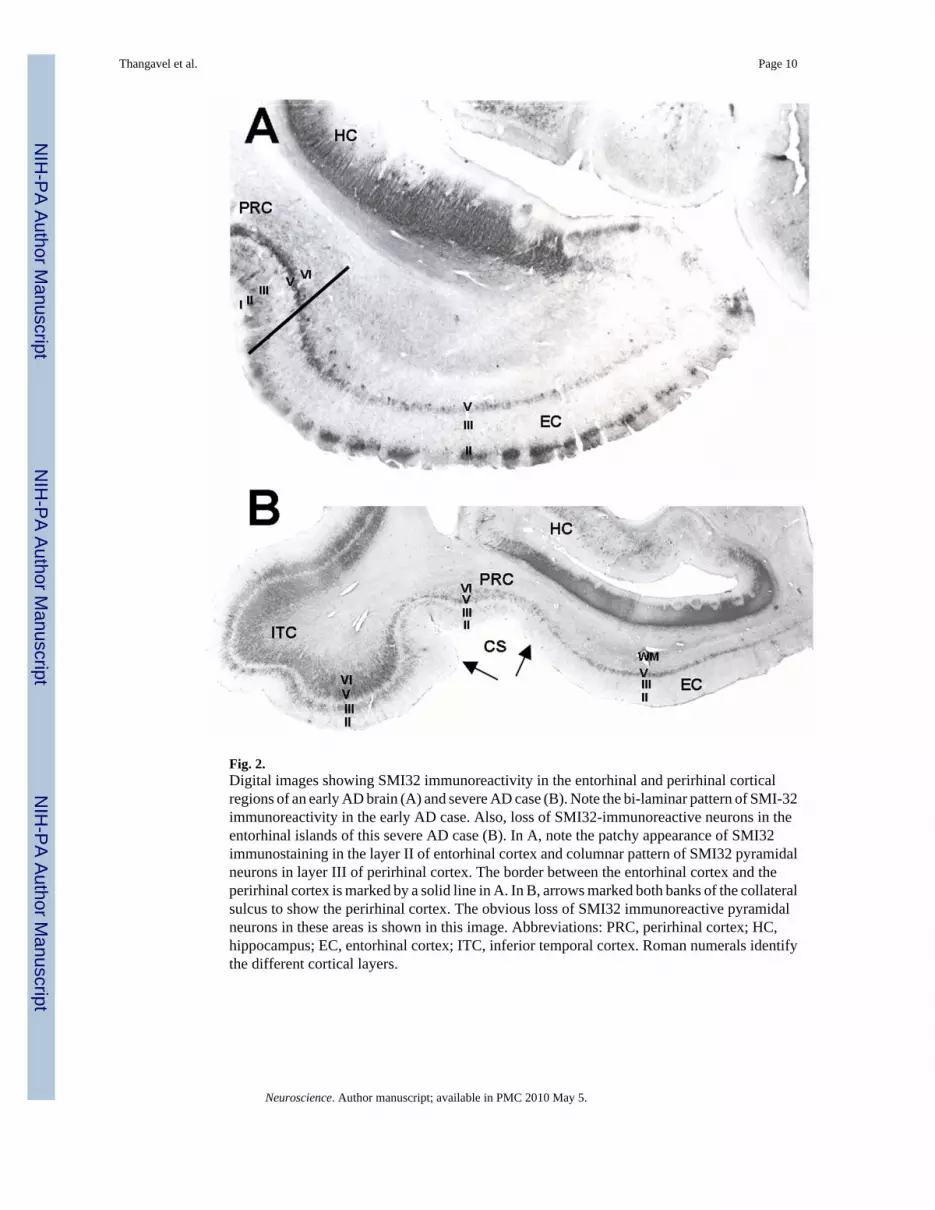
Fig. 2.Digital images showing SMI32 immunoreactivity in the entorhinal and perirhinal corticalregions of an early AD brain (A) and severe AD case (B). Note the bi-laminar pattern of SMI-32immunoreactivity in the early AD case. Also, loss of SMI32-immunoreactive neurons in theentorhinal islands of this severe AD case (B). In A, note the patchy appearance of SMI32immunostaining in the layer II of entorhinal cortex and columnar pattern of SMI32 pyramidalneurons in layer III of perirhinal cortex. The border between the entorhinal cortex and theperirhinal cortex is marked by a solid line in A. In B, arrows marked both banks of the collateralsulcus to show the perirhinal cortex. The obvious loss of SMI32 immunoreactive pyramidalneurons in these areas is shown in this image. Abbreviations: PRC, perirhinal cortex; HC,hippocampus; EC, entorhinal cortex; ITC, inferior temporal cortex. Roman numerals identifythe different cortical layers.
Thangavel et al. Page 10
Neuroscience. Author manuscript; available in PMC 2010 May 5.
NIH
-PA Author Manuscript
NIH
-PA Author Manuscript
NIH
-PA Author Manuscript

Fig. 3.Image showing double immunostaining of AT8 (black) and SMI32 (brown) immunoreactivecells in the perirhinal cortex of an early AD brain (A) and higher magnification area (B). SMI32immunoreactive neurons (brown) and AT8 immunoreactive NFTs (black) are distributed likea columnar pattern in the perirhinal cortex from an AD case. SMI32 immunoreactive pyramidalneurons are co-localized with AT8 antibody indicated by arrowhead. Some neurons stainedonly with AT8 antibody highlighted by arrowhead. Somata of the pyramidal neurons and thedendritic processes are stained darkly with SMI32 antibody (brown) indicated by arrow.Abbreviation: PRC, perirhinal cortex. Cortical layers are highlighted by Roman numerals.
Thangavel et al. Page 11
Neuroscience. Author manuscript; available in PMC 2010 May 5.
NIH
-PA Author Manuscript
NIH
-PA Author Manuscript
NIH
-PA Author Manuscript

Fig. 4.Double immunostaining of SMI32 immunoreactivity (grey/black; small arrows) and AT8stained NFTs (brown; large arrows) in the hippocampus of a severe AD case. Low (A) andhigh (B) magnification images. Note, weak or less numbers of SMI32 positive cells are locatedin region showing more NFTs and many SMI32 containing immunoreactive neurons are in thearea showing a low density of NFTs. Arrowhead shows the presence of NFTs in some of theSMI32 immunoreactive neurons.
Thangavel et al. Page 12
Neuroscience. Author manuscript; available in PMC 2010 May 5.
NIH
-PA Author Manuscript
NIH
-PA Author Manuscript
NIH
-PA Author Manuscript

Fig. 5.Double labeling for thioflavin-S and SMI32 immunostaining in the entorhinal cortex of ADbrain. Note the presence of thioflavin-S stained NFTs in the layer II island of the entorhinalcortex (small arrow). Some of the NFTs are labeled with SMI32 antibody. Arrowheadshighlight thioflavin-S stained NFT contained SMI32 positive processes. Abbreviation: EC,entorhinal cortex.
Thangavel et al. Page 13
Neuroscience. Author manuscript; available in PMC 2010 May 5.
NIH
-PA Author Manuscript
NIH
-PA Author Manuscript
NIH
-PA Author Manuscript

Fig. 6.Double labeled fluoresence images of the temporal cortex pyramidal neurons in an AD caseare shown in A, SMI32/FITC green; B, AT8/CY3 red; and C (merged). Note that some of theSMI32 labeled cell bodies are immunoreactive to AT8 positive neurons, also labeled withSMI32 (thin arrow), are yellow in merged image (C), while some of the neuronal processeslacked AT8 immunoreactivity. Some neurites, positive only for AT8, are pointed by arrowhead(C). Asterisk shows only for SMI32.
Thangavel et al. Page 14
Neuroscience. Author manuscript; available in PMC 2010 May 5.
NIH
-PA Author Manuscript
NIH
-PA Author Manuscript
NIH
-PA Author Manuscript

NIH
-PA Author Manuscript
NIH
-PA Author Manuscript
NIH
-PA Author Manuscript
Thangavel et al. Page 15
Table 1Summary of cases
Case Age (Yrs) Sex Duration of illness (Yrs)
1 85 F 5
2 73 F 10+
3 87 F 10
4 83 M 12+
5 87 M 10+
6 84 M 8
7 92 F Non-AD
8 87 F Non-AD
9 89 M Non-AD
10 85 M Non-AD
Neuroscience. Author manuscript; available in PMC 2010 May 5.


