Embed Size (px)
Citation preview
Lm
TCP
a
ARRA
KSCEcN
Dxs1mThmE2daLq
S
s1
P
t
0d
Journal of Steroid Biochemistry & Molecular Biology 121 (2010) 546–555
Contents lists available at ScienceDirect
Journal of Steroid Biochemistry and Molecular Biology
journa l homepage: www.e lsev ier .com/ locate / j sbmb
iquid chromatography–mass spectrometry (LC–MS) of steroid hormoneetabolites and its applications�
revor M. Penning ∗, Seon-Hwa Lee1, Yi Jin, Alejandro Gutierrez2, Ian A. Blairenters of Excellence in Environmental Toxicology and Cancer Pharmacology, Department of Pharmacology, University of Pennsylvania School of Medicine,hiladelphia, PA 19104-6084, USA
r t i c l e i n f o
rticle history:eceived 27 September 2009eceived in revised form 9 January 2010ccepted 11 January 2010
eywords:teroid conjugatesatechol estrogenslectron capture atmospheric pressure
a b s t r a c t
Advances in liquid chromatography–mass spectrometry (LC–MS) can be used to measure steroid hor-mone metabolites in vitro and in vivo. We find that LC–electrospray ionization (ESI)-MS using a LCQion trap mass spectrometer in the negative ion mode can be used to monitor the product profilethat results from 5�-dihydrotestosterone (DHT)-17�-glucuronide, DHT-17�-sulfate, and tibolone-17�-sulfate reduction catalyzed by human members of the aldo–keto reductase (AKR) 1C subfamily and assignkinetic constants to these reactions. We also developed a stable isotope dilution LC–electron captureatmospheric pressure chemical ionization (ECAPCI)-MS method for the quantitative analysis of estrone(E1) and its metabolites as pentafluorobenzyl (PFB) derivatives in human plasma in the attomole range.
hemical ionizationormal-phase HPLC
The limit of detection for E1-PFB was 740 attomole on column. Separations can be performed usingnormal-phase LC because ionization takes place in the gas phase rather than in solution. This permitsefficient separation of the regioisomeric 2- and 4-methoxy-E1. The method was validated for the simulta-neous analysis of plasma E2 and its metabolites: 2-methoxy-E2, 4-methoxy-E2, 16�-hydroxy-E2, estrone(E1), 2-methoxy-E1, 4-methoxy-EI, and 16�-hydroxy-E1 from 5 pg/mL to 2000 pg/mL. Our LC–MS meth-ods have sufficient sensitivity to detect steroid hormone levels in prostate and breast tumors and should
osis a
aid their molecular diagnAbbreviations: 3�-Diol-17G, 3�-hydroxy-5�-androstane-17�-glucuronide; 3�-iol-17G, 3�-hydroxy-5�-androstane-17�-glucuronide; 3�-Diol-17S, 3�-hydro-y-5�-androstane-17�-sulfate; 3�-Diol-17S, 3�-hydroxy-5�-androstane-17�-ulfate; DHT, dihydrotestosterone; DHTG, DHT-17�-glucuro- nide; DHTS, DHT-7�-sulfate; E1, estrone; E2, 17�-estradiol; Tibolone, [7�,17�]-17-hydroxy-7-ethyl-19-norpregn-5(10)-en-20-yn-3-one; TibS, tibolone-17�-sulfate; 3�-OH-
ib, 3�-hydroxy-tibolone; 3�-OH-Tib, 3�-hydroxy-tibolone; 3�-OH-TibS, 3�-ydroxy-tibolone-17�-sulfate; 3�-OH-TibS, 3�-hydroxy-tibolone-17�-sulfate; 2-ethoxy-E1, 2-methoxy-estrone; 4-methoxy-E1, 4-methoxy-estrone; 2-methoxy-
2, 2-methoxy-17�-estradiol; 4-methoxy-E2, 4-methoxy-17�-estradiol; 2-OH-E1,,3-dihydroxy-estrone; 2-OH-E2, 2,3-dihydroxy-17�-estradiol; 4-OH-E1, 3,4-ihydroxyestrone; 4-OH-E2, 3,4-dihydroxy-17�-estradiol; AcN, acetonitrile; AKR,ldo–keto reductase; PFB, pentafluorobenzyl; HQC, high quality control sample;LQC, lower limit of quantitation; LQC, lower quality control sample; MQC, middleuality control sample; QC, quality control sample.� Article from special issue on “Steroid profiling and analytics: going towardsterome”.∗ Corresponding author at: Department of Pharmacology, University of Penn-
ylvania, 130C John Morgan Building, 3620 Hamilton Walk, Philadelphia, PA9104-6084, USA. Tel.: +1 215 898 9445; fax: +1 215 573 2236.
E-mail address: [email protected] (T.M. Penning).1 Current address: Department of Bioanalytical Chemistry, Graduate School of
harmaceutical Science, Tohoku University, Aoba-ku, Sendai 980-8578, Japan.2 Current address: Department of Pediatric Oncology, Dana-Farber Cancer Insti-
ute, Boston, MA 02115, USA.
960-0760/$ – see front matter © 2010 Elsevier Ltd. All rights reserved.oi:10.1016/j.jsbmb.2010.01.005
nd treatment.© 2010 Elsevier Ltd. All rights reserved.
1. Introduction
Radioimmunoassay or ELISA based methods were once consid-ered state-of-the-art methods for measuring steroid metabolitesin biospecimens. These approaches now appear to be fraught withdifficultly. First, they can only measure one analyte at a time, thusmultiple assays are required to measure all the metabolites froma single steroid hormone. Second, steroid metabolites have highlyrelated structures and in a biospecimen mixtures of stereoisomers,regioisomers or compounds that differ by only the substitutionof a carbonyl group for an alcohol exist. It is thus not possibleto control for interference in the immunoassay from both knownand unknown structurally related steroids that may be present inthe biological matrix. Third, these immunological approaches donot give any structural validation of the analyte. Fourth, in manyinstances antisera do not exist for all the steroid metabolites ofinterest to allow immunodetection in the first place. This is cer-tainly true for the detection of conjugated steroids.
The reliability of radioimmunoassays for steroid hormones has
also been questioned by position papers which have documentedthe large inter-laboratory variability that exists in measuringplasma testosterone [1–3] and the difficulty in measuring 17�-estradiol (E2) and its metabolites in plasma and urine [4–6]. Incontrast, gas chromatography–mass spectrometry (GC–MS) cou-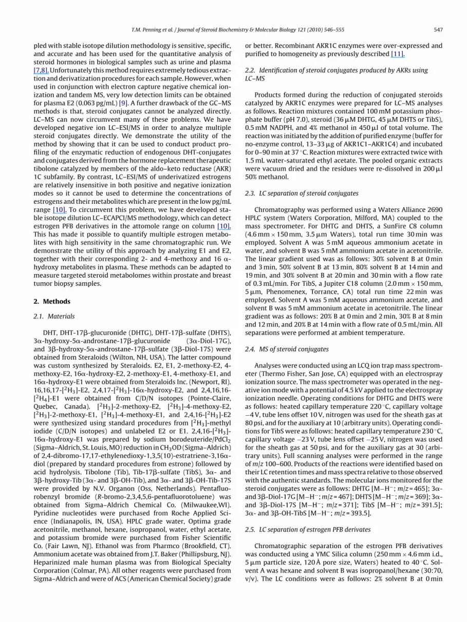
emistr
pas[tuifmLdsmfiat1amerbeTldthmt
2
2
3aowm11[Q[wi1(oda3wroPeaaCAHCS
T.M. Penning et al. / Journal of Steroid Bioch
led with stable isotope dilution methodology is sensitive, specific,nd accurate and has been used for the quantitative analysis ofteroid hormones in biological samples such as urine and plasma7,8]. Unfortunately this method requires extremely tedious extrac-ion and derivatization procedures for each sample. However, whensed in conjunction with electron capture negative chemical ion-
zation and tandem MS, very low detection limits can be obtainedor plasma E2 (0.063 pg/mL) [9]. A further drawback of the GC–MS
ethods is that, steroid conjugates cannot be analyzed directly.C–MS can now circumvent many of these problems. We haveeveloped negative ion LC–ESI/MS in order to analyze multipleteroid conjugates directly. We demonstrate the utility of theethod by showing that it can be used to conduct product pro-
ling of the enzymatic reduction of endogenous DHT-conjugatesnd conjugates derived from the hormone replacement therapeuticibolone catalyzed by members of the aldo–keto reductase (AKR)C subfamily. By contrast, LC–ESI/MS of underivatized estrogensre relatively insensitive in both positive and negative ionizationodes so it cannot be used to determine the concentrations of
strogens and their metabolites which are present in the low pg/mLange [10]. To circumvent this problem, we have developed sta-le isotope dilution LC–ECAPCI/MS methodology, which can detectstrogen PFB derivatives in the attomole range on column [10].his has made it possible to quantify multiple estrogen metabo-ites with high sensitivity in the same chromatographic run. Weemonstrate the utility of this approach by analyzing E1 and E2,ogether with their corresponding 2- and 4-methoxy and 16 �-ydroxy metabolites in plasma. These methods can be adapted toeasure targeted steroid metabolomes within prostate and breast
umor biopsy samples.
. Methods
.1. Materials
DHT, DHT-17�-glucuronide (DHTG), DHT-17�-sulfate (DHTS),�-hydroxy-5�-androstane-17�-glucuronide (3�-Diol-17G),nd 3�-hydroxy-5�-androstane-17�-sulfate (3�-Diol-17S) werebtained from Steraloids (Wilton, NH, USA). The latter compoundas custom synthesized by Steraloids. E2, E1, 2-methoxy-E2, 4-ethoxy-E2, 16�-hydroxy-E2, 2-methoxy-E1, 4-methoxy-E1, and
6�-hydroxy-E1 were obtained from Steraloids Inc. (Newport, RI).6,16,17-[2H3]-E2, 2,4,17-[2H3]-16�-hydroxy-E2, and 2,4,16,16-
2H4]-E1 were obtained from C/D/N isotopes (Pointe-Claire,uebec, Canada). [2H3]-2-methoxy-E2, [2H3]-4-methoxy-E2,
2H3]-2-methoxy-E1, [2H3]-4-methoxy-E1, and 2,4,16-[2H3]-E2ere synthesized using standard procedures from [2H3]-methyl
odide (C/D/N isotopes) and unlabeled E2 or E1. 2,4,16-[2H3]-6�-hydroxy-E1 was prepared by sodium borodeuteride/PdCl2Sigma–Aldrich, St. Louis, MO) reduction in CH3OD (Sigma–Aldrich)f 2,4-dibromo-17,17-ethylenedioxy-1,3,5(10)-estratriene-3,16�-iol (prepared by standard procedures from estrone) followed bycid hydrolysis. Tibolone (Tib), Tib-17�-sulfate (TibS), 3�- and�-hydroxy-Tib (3�- and 3�-OH-Tib), and 3�- and 3�-OH-Tib-17Sere provided by N.V. Organon (Oss, Netherlands). Pentafluo-
obenzyl bromide (R-bromo-2,3,4,5,6-pentafluorotoluene) wasbtained from Sigma–Aldrich Chemical Co. (Milwaukee,WI).yridine nucleotides were purchased from Roche Applied Sci-nce (Indianapolis, IN, USA). HPLC grade water, Optima gradecetonitrile, methanol, hexane, isopropanol, water, ethyl acetate,nd potassium bromide were purchased from Fisher Scientific
o. (Fair Lawn, NJ). Ethanol was from Pharmco (Brookfield, CT).mmonium acetate was obtained from J.T. Baker (Phillipsburg, NJ).eparinized male human plasma was from Biological Specialtyorporation (Colmar, PA). All other reagents were purchased fromigma–Aldrich and were of ACS (American Chemical Society) gradey & Molecular Biology 121 (2010) 546–555 547
or better. Recombinant AKR1C enzymes were over-expressed andpurified to homogeneity as previously described [11].
2.2. Identification of steroid conjugates produced by AKRs usingLC–MS
Products formed during the reduction of conjugated steroidscatalyzed by AKR1C enzymes were prepared for LC–MS analysesas follows. Reaction mixtures contained 100 mM potassium phos-phate buffer (pH 7.0), steroid (36 �M DHTG, 45 �M DHTS or TibS),0.5 mM NADPH, and 4% methanol in 450 �l of total volume. Thereaction was initiated by the addition of purified enzyme (buffer forno-enzyme control, 13–33 �g of AKR1C1–AKR1C4) and incubatedfor 0–90 min at 37 ◦C. Reaction mixtures were extracted twice with1.5 mL water-saturated ethyl acetate. The pooled organic extractswere vacuum dried and the residues were re-dissolved in 200 �l50% methanol.
2.3. LC separation of steroid conjugates
Chromatography was performed using a Waters Alliance 2690HPLC system (Waters Corporation, Milford, MA) coupled to themass spectrometer. For DHTG and DHTS, a SunFire C8 column(4.6 mm × 150 mm, 3.5 �m Waters), total run time 30 min wasemployed. Solvent A was 5 mM aqueous ammonium acetate inwater, and solvent B was 5 mM ammonium acetate in acetonitrile.The linear gradient used was as follows: 30% solvent B at 0 minand 3 min, 50% solvent B at 13 min, 80% solvent B at 14 min and19 min, and 30% solvent B at 20 min and 30 min with a flow rateof 0.3 mL/min. For TibS, a Jupiter C18 column (2.0 mm × 150 mm,5 �m, Phenomenex, Torrance, CA) total run time 22 min wasemployed. Solvent A was 5 mM aqueous ammonium acetate, andsolvent B was 5 mM ammonium acetate in acetonitrile. The lineargradient was as follows: 20% B at 0 min and 2 min, 30% B at 8 minand 12 min, and 20% B at 14 min with a flow rate of 0.5 mL/min. Allseparations were performed at ambient temperature.
2.4. MS of steroid conjugates
Analyses were conducted using an LCQ ion trap mass spectrom-eter (Thermo Fisher, San Jose, CA) equipped with an electrosprayionization source. The mass spectrometer was operated in the neg-ative ion mode with a potential of 4.5 kV applied to the electrosprayionization needle. Operating conditions for DHTG and DHTS wereas follows: heated capillary temperature 220 ◦C, capillary voltage−4 V, tube lens offset 10 V, nitrogen was used for the sheath gas at80 psi, and for the auxiliary at 10 (arbitrary units). Operating condi-tions for TibS were as follows: heated capillary temperature 230 ◦C,capillary voltage −23 V, tube lens offset −25 V, nitrogen was usedfor the sheath gas at 50 psi, and for the auxiliary gas at 30 (arbi-trary units). Full scanning analyses were performed in the rangeof m/z 100–600. Products of the reactions were identified based ontheir LC retention times and mass spectra relative to those observedwith the authentic standards. The molecular ions monitored for thesteroid conjugates were as follows: DHTG [M−H−; m/z = 465]; 3�-and 3�-Diol-17G [M−H−; m/z = 467]; DHTS [M−H−; m/z = 369]; 3�-and 3�-Diol-17S [M−H−; m/z = 371]; TibS [M−H−; m/z = 391.5];3�- and 3�-OH-TibS [M−H−; m/z = 393.5].
2.5. LC separation of estrogen PFB derivates
Chromatographic separation of the estrogen PFB derivativeswas conducted using a YMC Silica column (250 mm × 4.6 mm i.d.,5 �m particle size, 120 Å pore size, Waters) heated to 40 ◦C. Sol-vent A was hexane and solvent B was isopropanol/hexane (30:70,v/v). The LC conditions were as follows: 2% solvent B at 0 min

5 emistr
as3otDm
2
it2sTtifisaww4T
2
aAtf4d4sTaddot(TespT(ttmtw1d
2
taht
48 T.M. Penning et al. / Journal of Steroid Bioch
nd 2 min, 10% solvent B at 5 min, 20% solvent B at 12 min, 50%olvent B at 22 min, and 24 min, 2% solvent B at 28 min and5 min with a flow rate of 0.9 mL/min. A post-column flow-ratef 0.5 mL/min was used to prevent the source from becoming con-aminated with carbon deposits. Pre-column filters (2 �m; Alltech,eerfield, IL) were used to protect the column from particulateatter.
.6. ECAPCI of estrogen PFB derivates
A TSQ 7000 (Thermo Fisher) mass spectrometer was operatedn the APCI negative ion mode under the following condi-ions: vaporizer temperature 500 ◦C, heated capillary temperature00 ◦C, corona needle discharge current 15 �A, sheath gas pres-ure (nitrogen) 20 psi, auxiliary gas (nitrogen) 15 (arbitrary units).he parent and daughter resolution settings were of 0 V forhe methoxy compounds and 10 V for all other analytes andnternal standards. The [M−PFB]− ions of the analytes wereltered through the first quadrupole, and collision-induced dis-ociation (CID) was performed using argon as the collision gast 3.0 mTorr in the second (rf-only) quadrupole. Product ionsere then detected in the third quadrupole. Collision energiesere optimized for each analyte, and ranged from 25 eV to
5 eV (Table 3). MRM transitions were performed as shown inable 3.
.7. Sample preparation for LC–ECAPCI/MS analysis
Heparinized male human plasma was allowed to thaw. Plasmaliquots (1 mL) were transferred to 10 mL glass centrifuge tubes.n aliquot (10 �l) of analyte solution of the appropriate concen-
ration was added to each sample using a dedicated microsyringe,ollowed by vortex mixing. This was followed by an aliquot (10 �l;ng) of internal standard solution, which was added using anotheredicated microsyringe. Samples were vortex mixed for 1 min, andmL of ethyl acetate was added. This mixture was mechanically
haken for 1 h, followed by centrifugation at 4000 RPM for 5 min.he samples were then placed at −20 ◦C and the lower phase wasllowed to freeze, after which the supernatant was transferred byecantation into a new 5 mL glass centrifuge tube and evaporated toryness under nitrogen at 37 ◦C using an N-Evap Analytical Evap-rator (Organomation, Berlin, MA). Methanol (200 �l) was addedo the dried samples, followed by vortex mixing for 2 min. Water800 �l) was then added, and the samples were vortex-mixed again.he reconstituted solution was then placed on a C18 solid phasextraction (SPE) cartridge (7 mm/3 mL Empore C18 Standard Den-ity Extraction Disk Cartridges, 3 M, St. Paul, MN), which had beenre-conditioned with 0.5 mL of methanol and 0.5 mL of water.his cartridge was placed inside a 15 mL plastic centrifuge tubeFisher Scientific). All samples and washes were eluted throughhe SPE cartridge by centrifugation at 1800 RPM for 3 min. The car-ridge was washed sequentially with 1 mL of water and 0.3 mL of
ethanol/water (20:80, v/v). The cartridges were then transferredo a new 15 mL plastic centrifuge tube and the analytes were elutedith 0.3 mL of methanol. The resulting eluate was transferred to a
.8 mL plastic centrifuge tube (Fisher Scientific), and evaporated toryness under nitrogen at 37 ◦C.
.8. PFB derivatization
After drying, samples were reconstituted in 100 �l of acetoni-rile, and were treated with 100 �l of pentafluorobenzyl bromide incetonitrile (1:19, v/v), followed by 100 �l of ethanolic potassiumydroxide (2:100, w/v). After vortex mixing, samples were allowedo react at room temperature for 15 min, evaporated to dryness
y & Molecular Biology 121 (2010) 546–555
under nitrogen, and reconstituted in 200 �l of isopropanol/hexane(2:98, v/v), 90 �l of which was injected on the LC.
2.9. Data analysis
Calibration curves ranged from 5 pg/mL to 1000 pg/mL. For eachcurve, eight different concentrations, distributed over the entireconcentration range, were used. Peak height ratios between theanalytes and their respective internal standard were calculatedfrom each sample using Finnigan LCquan version 1.2 software.The data were fit to a linear least-squares regression curve witha weighting index of 1/x. A water blank, plasma blank, and plasmablank spiked with internal standard were also processed with eachcalibration curve.
2.10. Accuracy and precision
Five replicates of each quality control (QC) sample at each con-centration level were processed and analyzed along with the 8standard curve samples. The lower quality control sample (LQC),middle quality control sample (MQC), and high quality control sam-ple (HQC) were analyzed on three separate days, while the lowerlimit of quantitation (LLOQ) samples were analyzed on 1 day only.Assay accuracy was assessed by comparing means of the measuredanalyte concentrations with the theoretical concentrations in theQC samples. Within-day precision was expressed as percent rela-tive standard deviation (% RSD) and was obtained by calculatingthe percent ratio between the relative standard deviation of fivereplicates (n = 5) and their mean at each concentration within thesame validation run. Inter-day precision, defined as percent rela-tive standard deviation (% RSD) of three different validation runs(n = 3) was also assessed.
2.11. Determination of steady state kinetic parameters
Initial rates of the NADPH dependent reduction of ketosteroidsand their conjugates catalyzed by AKR1C were measured usinga Hitachi F-2500 fluorescence spectrophotometer (Hitachi Amer-ica Ltd., New York, NY) by monitoring the change in fluorescenceemission of NADPH. Excitation and emission wavelengths wereset at 340 nm and 450 nm, respectively. Changes in fluorescenceunits were converted to nanomoles of cofactor by using standardcurves of fluorescence emission versus known NADPH concentra-tions. Data were analyzed by nonlinear least-squares fitting to theequation:
� = kcat[E][S]Km + [S]
where � is the initial velocity, [E] and [S] are the total molarconcentrations of the enzyme and steroid substrate, respectively,kcat (s−1) is the turnover number, and Km (�M) is the apparentMichaelis–Menten constant for the steroid substrate.
3. Results
3.1. Metabolism of endogenous steroid conjugates by aldo–ketoreductase (AKR) 1C subfamily enzymes
The four aldo–keto reductase (AKR) 1C subfamily membersfound in humans (AKR1C1–AKR1C4) have been shown to catalyzethe NADPH dependent reduction of 3-ketosteroids to yield 3�-
or 3�-hydroxysteroids with different stereochemical preferencesbased on steroid substrate. For example, AKR1C2 is predominatelya 3�-HSD with DHT; while AKR1C1 is predominately a 3�-HSD withthe same substrate [12]. By contrast the same two enzymes con-vert tibolone (a hormone replacement therapeutic pro-drug) only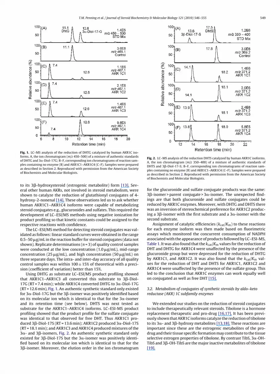
T.M. Penning et al. / Journal of Steroid Biochemistry & Molecular Biology 121 (2010) 546–555 549
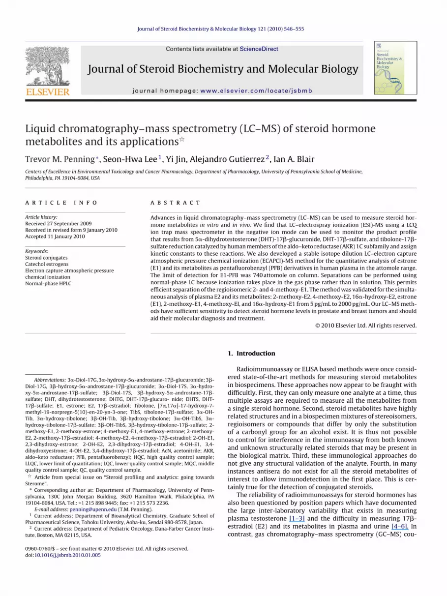
Fig. 1. LC–MS analysis of the reduction of DHTG catalyzed by human AKR1C iso-forms. A, the ion chromatogram (m/z 450–500) of a mixture of authentic standardsopao
teshhsdpr
i0swctcs
t1(foaspwd(3efi3
Fig. 2. LC–MS analysis of the reduction DHTS catalyzed by human AKR1C isoforms.A, the ion chromatogram (m/z 350–400) of a mixture of authentic standards of
important since these are the estrogenic metabolites of the pro-
f DHTG and 3�-Diol-17G; B–F, corresponding ion chromatograms of reaction sam-les containing no enzyme (B) and AKR1C1–AKR1C4 (C–F). Samples were prepareds described in Section 2. Reproduced with permission from the American Societyf Biochemists and Molecular Biologists.
o its 3�-hydroxysteroid (estrogenic metabolite) form [13]. Sev-ral other human AKRs, not involved in steroid metabolism, werehown to catalyze the reduction of glutathionyl conjugates of 4-ydroxy-2-nonenal [14]. These observations led us to ask whetheruman AKR1C1–AKR1C4 isoforms were capable of metabolizingteroid conjugates e.g., glucuronides and sulfates. This required theevelopment of LC–ESI/MS methods using negative ionization forroduct profiling so that kinetic constants could be assigned to theespective reactions with confidence.
The LC–ESI/MS method for detecting steroid conjugates was val-dated as follows: linear standard curves were obtained in the range.5–50 �g/mL in the reaction buffer for steroid conjugates (data nothown). Replicate determinations (n = 3) of quality control samplesere conducted at the low concentration (1.0 �g/mL), mid-range
oncentration (25 �g/mL), and high concentration (50 �g/mL) onhree separate days. The intra- and inter-day accuracy of all qualityontrol samples was within 100 ± 15% of theoretical with a preci-ion (coefficient of variation) better than 15%.
Using DHTG as substrate LC–ESI/MS product profiling showedhat AKR1C1–AKR1C3 all converted this substrate to 3�-Diol-7G (RT = 7.4 min); while AKR1C4 converted DHTG to 3�-Diol-17GRT = 12.6 min), Fig. 1. An authentic synthetic standard only existedor 3�-Diol-17G but the 3�-isomer was positively identified basedn its molecular ion which is identical to that for the 3�-isomernd its retention time (see below). DHTS was next tested asubstrate for the AKR1C1–AKR1C4 isoforms. LC–ESI-MS productrofiling showed that the product profile for the sulfate conjugateas identical to that observed for free DHT. Thus AKR1C1 pro-uced 3�-Diol-17S (RT = 13.6 min); AKR1C2 produced 3�-Diol-17SRT = 18.1 min); and AKR1C3 and AKR1C4 produced mixtures of the
�- and 3�-isomers, Fig. 2. An authentic synthetic standard onlyxisted for 3�-Diol-17S but the 3�-isomer was positively identi-ed based on its molecular ion which is identical to that for the�-isomer. Moreover, the elution order in the ion chromatogramDHTS and 3�-Diol-17-S; B–F, corresponding ion chromatograms of reaction sam-ples containing no enzyme (B) and AKR1C1–AKR1C4 (C–F). Samples were preparedas described in Section 2. Reproduced with permission from the American Societyof Biochemists and Molecular Biologists.
for the glucuronide and sulfate conjugate products was the same:3�-isomer > parent conjugate > 3�-isomer. The unexpected find-ings are that both glucuronide and sulfate conjugates could bereduced by AKR1C enzymes. Moreover, with DHTG and DHTS therewas an inversion of stereochemical preference for AKR1C2 produc-ing a 3�-isomer with the first substrate and a 3�-isomer with thesecond substrate.
Assignment of catalytic efficiencies (kcat/Km) to these reactionsfor each enzyme isoform was then made based on fluorimetricassays which monitored the concurrent consumption of NADPHassociated with the appearance of products followed by LC–ESI-MS,Table 1. It was also found that the kcat/Km values for the reduction ofDHT and DHTG for AKR1C4 were unaffected by the presence of theglucuronide group but were depressed for the reduction of DHTGby AKR1C1, and AKR1C2. It was also found that the kcat/Km val-ues for the reduction of DHT and DHTS for AKR1C1, AKR1C2 andAKR1C4 were unaffected by the presence of the sulfate group. Thisled to the conclusion that AKR1C enzymes can work equally wellon conjugated as well as free DHT [15].
3.2. Metabolism of conjugates of synthetic steroids by aldo–ketoreductase (AKR) 1C subfamily enzymes
We extended our studies on the reduction of steroid conjugatesto include therapeutically relevant steroids. Tibolone is a hormonereplacement therapeutic and pro-drug [16,17]. It has been previ-ously shown that AKR1C isoforms catalyze the reduction of tiboloneto its 3�- and 3�-hydroxy metabolites [13,18]. These reactions are
drug and their tissue specific formation may contribute to the tissueselective estrogen properties of tibolone. By contrast TibS, 3�-OH-TibS and 3�-OH-TibS are the major inactive metabolites of tibolone[19].
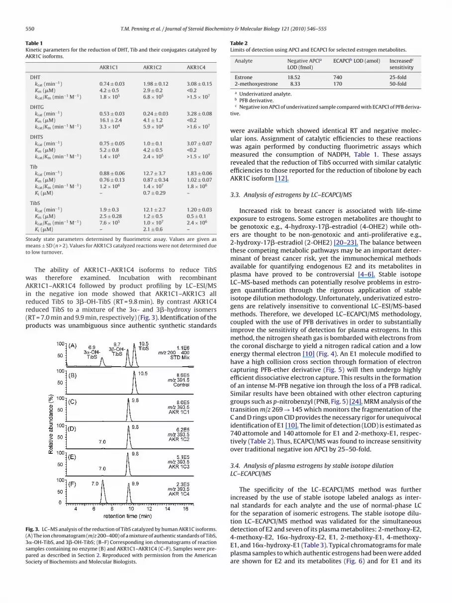
550 T.M. Penning et al. / Journal of Steroid Biochemistry & Molecular Biology 121 (2010) 546–555
Table 1Kinetic parameters for the reduction of DHT, Tib and their conjugates catalyzed byAKR1C isoforms.
AKR1C1 AKR1C2 AKR1C4
DHTkcat (min−1) 0.74 ± 0.03 1.98 ± 0.12 3.08 ± 0.15Km (�M) 4.2 ± 0.5 2.9 ± 0.2 <0.2kcat/Km (min−1 M−1) 1.8 × 10⁵ 6.8 × 10⁵ >1.5 × 10⁷
DHTGkcat (min−1) 0.53 ± 0.03 0.24 ± 0.03 3.28 ± 0.08Km (�M) 16.1 ± 2.4 4.1 ± 1.2 <0.2kcat/Km (min−1 M−1) 3.3 × 10⁴ 5.9 × 10⁴ >1.6 × 10⁷
DHTSkcat (min−1) 0.75 ± 0.05 1.0 ± 0.1 3.07 ± 0.07Km (�M) 5.2 ± 0.8 4.2 ± 0.5 <0.2kcat/Km (min−1 M−1) 1.4 × 10⁵ 2.4 × 10⁵ >1.5 × 10⁷
Tibkcat (min−1) 0.88 ± 0.06 12.7 ± 3.7 1.83 ± 0.06Km (�M) 0.76 ± 0.13 0.87 ± 0.34 1.02 ± 0.07kcat/Km (min−1 M−1) 1.2 × 10⁶ 1.4 × 10⁷ 1.8 × 10⁶
Ki (�M) – 0.7 ± 0.29 –
TibSkcat (min−1) 1.9 ± 0.3 12.1 ± 2.7 1.20 ± 0.03Km (�M) 2.5 ± 0.28 1.2 ± 0.5 0.5 ± 0.1kcat/Km (min−1 M−1) 7.6 × 10⁵ 1.0 × 10⁷ 2.4 × 10⁶
K (�M) – 2.1 ± 0.6 –
Smt
wAirr(p
F(3spS
Table 2Limits of detection using APCI and ECAPCI for selected estrogen metabolites.
Analyte Negative APCIa
LOD (fmol)ECAPCIb LOD (amol) Increasedc
sensitivity
Estrone 18.52 740 25-fold2-methoxyestrone 8.33 170 50-fold
i
teady state parameters determined by fluorimetric assay. Values are given aseans ± SD (n > 2). Values for AKR1C3 catalyzed reactions were not determined due
o low turnover.
The ability of AKR1C1–AKR1C4 isoforms to reduce TibSas therefore examined. Incubation with recombinantKR1C1–AKR1C4 followed by product profiling by LC–ESI/MS
n the negative ion mode showed that AKR1C1–AKR1C3 alleduced TibS to 3�-OH-TibS (RT = 9.8 min). By contrast AKR1C4educed TibS to a mixture of the 3�- and 3�-hydroxy isomersRT = 7.0 min and 9.9 min, respectively) (Fig. 3). Identification of theroducts was unambiguous since authentic synthetic standards
ig. 3. LC–MS analysis of the reduction of TibS catalyzed by human AKR1C isoforms.A) The ion chromatogram (m/z 200–400) of a mixture of authentic standards of TibS,�-OH-TibS, and 3�-OH-TibS; (B–F) Corresponding ion chromatograms of reactionamples containing no enzyme (B) and AKR1C1–AKR1C4 (C–F). Samples were pre-ared as described in Section 2. Reproduced with permission from the Americanociety of Biochemists and Molecular Biologists.
a Underivatized analyte.b PFB derivative.c Negative ion APCI of underivatized sample compared with ECAPCI of PFB deriva-
tive.
were available which showed identical RT and negative molec-ular ions. Assignment of catalytic efficiencies to these reactionswas again performed by conducting fluorimetric assays whichmeasured the consumption of NADPH, Table 1. These assaysrevealed that the reduction of TibS occurred with similar catalyticefficiencies to those reported for the reduction of tibolone by eachAKR1C isoform [12].
3.3. Analysis of estrogens by LC–ECAPCI/MS
Increased risk to breast cancer is associated with life-timeexposure to estrogens. Some estrogen metabolites are thought tobe genotoxic e.g., 4-hydroxy-17�-estradiol (4-OHE2) while oth-ers are thought to be non-genotoxic and anti-proliferative e.g.,2-hydroxy-17�-estradiol (2-OHE2) [20–23]. The balance betweenthese competing metabolic pathways may be an important deter-minant of breast cancer risk, yet the immunochemical methodsavailable for quantifying endogenous E2 and its metabolites inplasma have proved to be controversial [4–6]. Stable isotopeLC–MS-based methods can potentially resolve problems in estro-gen quantification through the rigorous application of stableisotope dilution methodology. Unfortunately, underivatized estro-gens are relatively insensitive to conventional LC–ESI/MS-basedmethods. Therefore, we developed LC–ECAPCI/MS methodology,coupled with the use of PFB derivatives in order to substantiallyimprove the sensitivity of detection for plasma estrogens. In thismethod, the nitrogen sheath gas is bombarded with electrons fromthe coronal discharge to yield a nitrogen radical cation and a lowenergy thermal electron [10] (Fig. 4). An E1 molecule modified tohave a high collision cross section through formation of electroncapturing PFB-ether derivative (Fig. 5) will then undergo highlyefficient dissociative electron capture. This results in the formationof an intense M-PFB negative ion through the loss of a PFB radical.Similar results have been obtained with other electron capturinggroups such as p-nitrobenzyl (PNB, Fig. 5) [24]. MRM analysis of thetransition m/z 269 → 145 which monitors the fragmentation of theC and D rings upon CID provides the necessary rigor for unequivocalidentification of E1 [10]. The limit of detection (LOD) is estimated as740 attomole and 140 attomole for E1 and 2-methoxy-E1, respec-tively (Table 2). Thus, ECAPCI/MS was found to increase sensitivityover traditional negative ion APCI by 25–50-fold.
3.4. Analysis of plasma estrogens by stable isotope dilutionLC–ECAPCI/MS
The specificity of the LC–ECAPCI/MS method was furtherincreased by the use of stable isotope labeled analogs as inter-nal standards for each analyte and the use of normal-phase LCfor the separation of isomeric estrogens. The stable isotope dilu-tion LC–ECAPCI/MS method was validated for the simultaneous
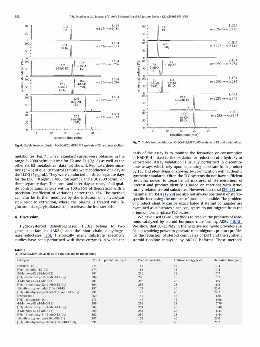
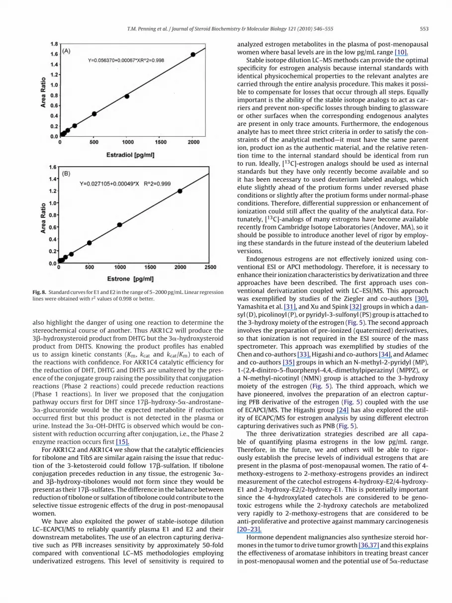
detection of E2 and seven of its plasma metabolites: 2-methoxy-E2,4-methoxy-E2, 16�-hydroxy-E2, E1, 2-methoxy-E1, 4-methoxy-E1, and 16�-hydroxy-E1 (Table 3). Typical chromatograms for maleplasma samples to which authentic estrogens had been were addedare shown for E2 and its metabolites (Fig. 6) and for E1 and its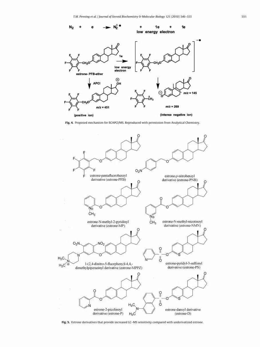
T.M. Penning et al. / Journal of Steroid Biochemistry & Molecular Biology 121 (2010) 546–555 551
Fig. 4. Proposed mechanism for ECAPCI/MS. Reproduced with permission from Analytical Chemistry.
Fig. 5. Estrone derivatives that provide increased LC–MS sensitivity compared with underivatized estrone.
552 T.M. Penning et al. / Journal of Steroid Biochemistry & Molecular Biology 121 (2010) 546–555
F
mrottftipcsg
4
gns
TL
ig. 6. Stable isotope dilution LC–ECAPCI/MRM/MS analysis of E2 and metabolites.
etabolites (Fig. 7). Linear standard curves were obtained in theange 5–2000 pg/mL plasma for E2 and E1 (Fig. 8) as well as thether six E2 metabolites (data not shown). Replicate determina-ions (n = 5) of quality control samples were conducted one day athe LLOQ (5 pg/mL). They were conducted on three separate daysor the LQC (10 pg/mL), MQC (50 pg/mL), and HQC (500 pg/mL) onhree separate days. The intra- and inter-day accuracy of all qual-ty control samples was within 100 ± 15% of theoretical with arecision (coefficient of variation) better than 15%. The methodan also be further modified by the inclusion of a hydrolysistep prior to extraction, where the plasma is treated with �-lucuronidase/arylsulfatase step to release the free steroids.
. Discussion
Hydroxysteroid dehydrogenases (HSDs) belong to twoene superfamilies (AKRs) and the short-chain dehydroge-ase/reductases [25]. Historically many substrate specificitytudies have been performed with these enzymes in which the
able 3C–ECAPCI/MRM/MS analysis of estradiol and its metabolites.
Estrogen [M−PFB] parent ion (m/z)
Estradiol (E2) 271[2H3]-estradiol (E2-D3) 2742-Methoxy-E2 (2-MeO-E2) 301[2H3]-2-methoxy-E2 (2-MeO-E2-D3) 3044-Methoxy-E2 (4-MeO-E2) 301[2H3]-4-methoxy-E2 (4-MeO-E2-D3) 30416�-Hydroxy-estradiol (16�-OH-E2) 287[2H4]-16�-hydroxy-estradiol (16�-OH-E2-D4) 291Estrone (E1) 269[2H4]-estrone (E1-D4) 2734-Methoxy-E1 (4-MeO-E1) 299[2H3]-4-methoxy-E1 (4-MeO-E1-D3) 3022-Methoxy-E1 (2-MeO-E1) 299[2H3]-2-methoxy-E1 (2-MeO-E1-D3) 30216�-Hydroxy-estrone (16�-OH-E1) 287[2H4]-16�-hydroxy-estrone (16�-OH-E1-D4) 291
Fig. 7. Stable isotope dilution LC–ECAPCI/MRM/MS analysis of E1 and metabolites.
basis of the assay is to monitor the formation or consumptionof NAD(P)H linked to the oxidation or reduction of a hydroxy orketosteroid. Assay validation is usually performed in discontin-uous assays which rely upon separating substrate from productby TLC and identifying unknowns by co-migration with authenticsynthetic standards. Often the TLC systems do not have sufficientresolving power to separate all mixtures of stereoisomers ofinterest and product identify is based on reactions with struc-turally related steroid substrates. However, bacterial [26–28] andmammalian HSDs [12,29] are also not always positional or stereo-specific increasing the number of products possible. The problemof product identity can be exacerbated if steroid conjugates areexamined as substrates since conjugates do not migrate from theorigin of normal-phase TLC plates.
We have used LC–MS methods to resolve the products of reac-
tions catalyzed by steroid hormone transforming AKRs [15,18].We show that LC–ESI/MS in the negative ion mode provides suf-ficient resolving power to generate umambiguous product profilesfor the reduction of steroid conjugates of DHT and the syntheticsteroid tibolone catalyzed by AKR1C isoforms. These methodsProduct ion (m/z) Collision energy (eV) Retention time (min)
183 43 17.4185 43 17.4286 28 17.7286 28 17.7286 28 18.5286 28 18.5171 40 22.6173 40 22.7145 45 6.95147 45 6.96284 28 7.39284 28 7.40284 28 8.37284 28 8.44171 40 22.6173 40 22.7
T.M. Penning et al. / Journal of Steroid Biochemistr
Fl
as3putter(p3ouse
ftcaprsw
Ldtcu
[20–23].
ig. 8. Standard curves for E1 and E2 in the range of 5–2000 pg/mL. Linear regressionines were obtained with r2 values of 0.998 or better.
lso highlight the danger of using one reaction to determine thetereochemical course of another. Thus AKR1C2 will produce the�-hydroxysteroid product from DHTG but the 3�-hydroxysteroidroduct from DHTS. Knowing the product profiles has enableds to assign kinetic constants (Km, kcat and kcat/Km) to each ofhe reactions with confidence. For AKR1C4 catalytic efficiency forhe reduction of DHT, DHTG and DHTS are unaltered by the pres-nce of the conjugate group raising the possibility that conjugationeactions (Phase 2 reactions) could precede reduction reactionsPhase 1 reactions). In liver we proposed that the conjugationathway occurs first for DHT since 17�-hydroxy-5�-androstane-�-glucuronide would be the expected metabolite if reductionccurred first but this product is not detected in the plasma orrine. Instead the 3�-OH-DHTG is observed which would be con-istent with reduction occurring after conjugation, i.e., the Phase 2nzyme reaction occurs first [15].
For AKR1C2 and AKR1C4 we show that the catalytic efficienciesor tibolone and TibS are similar again raising the issue that reduc-ion of the 3-ketosteroid could follow 17�-sulfation. If tiboloneonjugation precedes reduction in any tissue, the estrogenic 3�-nd 3�-hydroxy-tibolones would not form since they would beresent as their 17�-sulfates. The difference in the balance betweeneduction of tibolone or sulfation of tibolone could contribute to theelective tissue estrogenic effects of the drug in post-menopausalomen.
We have also exploited the power of stable-isotope dilutionC–ECAPCI/MS to reliably quantify plasma E1 and E2 and their
ownstream metabolites. The use of an electron capturing deriva-ive such as PFB increases sensitivity by approximately 50-foldompared with conventional LC–MS methodologies employingnderivatized estrogens. This level of sensitivity is required toy & Molecular Biology 121 (2010) 546–555 553
analyzed estrogen metabolites in the plasma of post-menopausalwomen where basal levels are in the low pg/mL range [10].
Stable isotope dilution LC–MS methods can provide the optimalspecificity for estrogen analysis because internal standards withidentical physicochemical properties to the relevant analytes arecarried through the entire analysis procedure. This makes it possi-ble to compensate for losses that occur through all steps. Equallyimportant is the ability of the stable isotope analogs to act as car-riers and prevent non-specific losses through binding to glasswareor other surfaces when the corresponding endogenous analytesare present in only trace amounts. Furthermore, the endogenousanalyte has to meet three strict criteria in order to satisfy the con-straints of the analytical method—it must have the same parention, product ion as the authentic material, and the relative reten-tion time to the internal standard should be identical from runto run. Ideally, [13C]-estrogen analogs should be used as internalstandards but they have only recently become available and soit has been necessary to used deuterium labeled analogs, whichelute slightly ahead of the protium forms under reversed phaseconditions or slightly after the protium forms under normal-phaseconditions. Therefore, differential suppression or enhancement ofionization could still affect the quality of the analytical data. For-tunately, [13C]-analogs of many estrogens have become availablerecently from Cambridge Isotope Laboratories (Andover, MA), so itshould be possible to introduce another level of rigor by employ-ing these standards in the future instead of the deuterium labeledversions.
Endogenous estrogens are not effectively ionized using con-ventional ESI or APCI methodology. Therefore, it is necessary toenhance their ionization characteristics by derivatization and threeapproaches have been described. The first approach uses con-ventional derivatization coupled with LC–ESI/MS. This approachwas exemplified by studies of the Ziegler and co-authors [30],Yamashita et al. [31], and Xu and Spink [32] groups in which a dan-syl (D), picolinoyl (P), or pyridyl-3-sulfonyl (PS) group is attached tothe 3-hydroxy moiety of the estrogen (Fig. 5). The second approachinvolves the preparation of pre-ionized (quaternized) derivatives,so that ionization is not required in the ESI source of the massspectrometer. This approach was exemplified by studies of theChen and co-authors [33], Higashi and co-authors [34], and Adamecand co-authors [35] groups in which an N-methyl-2-pyridyl (MP),1-(2,4-dinitro-5-fluorphenyl-4,4,-dimethylpiperazinyl (MPPZ), ora N-methyl-nicotinyl (NMN) group is attached to the 3-hydroxymoiety of the estrogen (Fig. 5). The third approach, which wehave pioneered, involves the preparation of an electron captur-ing PFB derivative of the estrogen (Fig. 5) coupled with the useof ECAPCI/MS. The Higashi group [24] has also explored the util-ity of ECAPC/MS for estrogen analysis by using different electroncapturing derivatives such as PNB (Fig. 5).
The three derivatization strategies described are all capa-ble of quantifying plasma estrogens in the low pg/mL range.Therefore, in the future, we and others will be able to rigor-ously establish the precise levels of individual estrogens that arepresent in the plasma of post-menopausal women. The ratio of 4-methoxy-estrogens to 2-methoxy-estrogens provides an indirectmeasurement of the catechol estrogens 4-hydroxy-E2/4-hydroxy-E1 and 2-hydroxy-E2/2-hydroxy-E1. This is potentially importantsince the 4-hydroxylated catechols are considered to be geno-toxic estrogens while the 2-hydroxy catechols are metabolizedvery rapidly to 2-methoxy-estrogens that are considered to beanti-proliferative and protective against mammary carcinogenesis
Hormone dependent malignancies also synthesize steroid hor-mones in the tumor to drive tumor growth [36,37] and this explainsthe effectiveness of aromatase inhibitors in treating breast cancerin post-menopausal women and the potential use of 5�-reductase

5 emistr
ihosmlptdpg
ge2odThkrbokzcnorto
A
PHRP
R
[
[
[
[
[
[
[
[
[
[
[
[
[
[
[
[
[
[
[
[
[
[
54 T.M. Penning et al. / Journal of Steroid Bioch
nhibitors in treating prostate cancer [38–42]. Considerable effortas been made in diagnosing these tumors in terms of the presencef nuclear receptors, real-time-PCR to measure transcript levels ofteroidogenic enzymes, and immunohistochemical approaches toeasure both receptor and enzyme levels. A characteristic property
ess developed which is a pre-requisite to complete the molecularathology of these tumors are robust methods to measure intra-umoral levels of estrogens and androgens. The methods we haveeveloped offer promise to measure steroid metabolomes withinrostate and breast tumor biopsy samples and the informationleaned could be used to determine treatment paradigm.
The regulation of ligand occupancy of nuclear receptors is oftenoverned by pairs of HSDs. For example regulation of the ER is gov-rned by type 1 17�-HSD (which reduces E1 to E2) and by type/4 17�-HSD (which oxidizes E2 to E1) [43–46], while regulationf the AR is governed by AKR1C2 (which reduces 5�-DHT to 3�-iol) and by HSD17B6 (which oxidizes 3�-diol to DHT) [47,48].hus a component of measuring intra-tumoral levels of steroidormones is to distinguish between the intracrine formation ofetosteroids from hydroxysteroids. The LC–ECAPCI-MS methodequires derivatization to an electron capturing group. This cane accomplished using the pentafuorobenzyl bromide or pentaflu-robenzylcarboxymethoxime derivative, for hydroxysteroids andetosteroids, respectively. The derivatization with pentafluoroben-ylcarboxymethoxime was developed originally for EC negativehemical ionization gas chromatography–MS of ketosteroids Alter-atively, conventional derivatives such as N-hydroxy-oximes [49]r pre-ionized derivatives such as those formed by the Girard Teagent [50] could be employed to improve sensitivity of ketos-eroid detection by ESI/MS or APCI/MS [51,52]. The next phase ofur work will be to implement such methods.
cknowledgements
This work was supported by the following research grants:30ES013587 and R01CA90744 from the National Institutes ofealth and a Prostate Cancer Foundation Challenge Grant (TMP),01CA091016 (IAB), and a Pilot-Project Grant awarded with30ES13587 (YJ).
eferences
[1] V. Moal, E. Mathieu, P. Reynier, Y. Malthièry, Y. Gallois, Low serum testosteroneassayed by liquid chromatography–tandem mass spectrometry. Comparisonwith five immunoassay techniques, Clin. Chim. Acta 386 (1–2) (2007) 12–19.
[2] L.M. Thienpont, K. Van Uytfanghe, S. Blincko, C.S. Ramsay, H. Xie, R.C. Doss, B.G.Keevil, L.J. Owen, A.L. Rockwood, M.M. Kushnir, K.Y. Chun, D.W. Chandler, H.P.Field, P.M. Sluss, State-of-the-art of serum testosterone measurement by iso-tope dilution–liquid chromatography–tandem mass spectrometry, Clin. Chem.54 (8) (2008) 1290–1297.
[3] C. Wang, C.H. Catlin, L.M. Demers, B. Starcevic, R.S. Swerdloff, Measurementof total serum testosterone in adult men: comparison of current laboratorymethods versus liquid chromatography–tandem mass spectrometry, J. Clin.Endocrinol. Metab. 89 (6) (2004) 534–543.
[4] R. Giese, Measurement of endogenous estrogens: analytical challenges andrecent advances, J. Chromatogr. A. 1000 (1–2) (2003) 401–412.
[5] F. Stanczyk, J.S. Lee, R.J. Santen, Standardization of steroid hormone assays:why, how and when? Cancer Epidemiol. Biomarkers Prev. 16 (9) (2007)1713–1719.
[6] X. Xu, J.M. Roman, H.J. Issaq, L.K. Keefre, T.D. Veenstra, R.G. Ziegler, Quantitativemeasurement of endogenous estrogens and estrogen metabolites in humanserum by liquid chromatography–tandem mass spectrometry, Anal. Chem. 79(20) (2007) 7813–7821.
[7] H. Adlercreutz, M.J. Tikkanen, D.H. Hunneman, Mass fragmentographic deter-mination of eleven estrogens in the body fluids of pregnant and nonpregnantsubjects, J. Steroid Biochem. 5 (3) (1974) 211–217.
[8] H. Adlercreutz, P. Kiuru, S. Rasku, K. Wähälä, T. Fotsis, An isotope dilution gaschromatographic–mass spectrometric method for the simultaneous assay of
estrogens and phytoestrogens in urine, J. Steroid Biochem. Mol. Biol. 92 (5)(2004) 399–411.[9] R.J. Santen, L. Demers, S. Ohorodnik, J. Settlage, P. Langecker, D. Blanchett, P.E.Goss, S. Wang, Superiority of gas chromatography/tandem mass spectrometryassay (GC/MS/MS) for estradiol for monitoring of aromatase inhibitory therapy,Steroids 72 (8) (2007) 666–671.
[
[
y & Molecular Biology 121 (2010) 546–555
10] G. Singh, A. Gutierrez, K. Xu, I.A. Blair, Liquid chromatography/electron cap-ture atmospheric pressure chemical ionization/mass spectrometry: analysis ofpentafluorobenzyl derivatives of biomolecules and drugs in the attomole range,Anal. Chem. 72 (14) (2000) 3007–3013.
11] M.E. Burczynski, R.G. Harvey, T.M. Penning, Expression and characteri-zation of four recombinant human dihydrodiol dehydrogenase isoforms:oxidation of trans-7,8-dihydroxy-7,8-dihydrobenzo[a]pyrene to the activatedo-quinone metabolite benzo[a]pyrene-7,8-dione, Biochemistry 37 (32) (1998)6781–6790.
12] S. Steckelbroeck, Y. Jin, S. Gopishetty, B. Oyesanmi, T.M. Penning, Humancytosolic 3�-hydroxysteroid dehydrogenases of the aldo–keto reductasesuperfamily display significant 3�-hydroxysteroid dehydrogenase activity:implications for steroid hormone metabolism and action, J. Biol. Chem. 279(11) (2003) 10784–10795.
13] S. Steckelbroeck, Y. Jin, B. Oyesanmi, H.J. Kloosterboer, T.M. Penning, Tiboloneis metabolized by the 3�/3�-hydroxysteroid dehydrogenase activities of thefour human isozymes of the aldo–keto reductase 1C subfamily: inversion ofstereospecificity with a �5(10)-3-ketosteroid, Mol. Pharmacol. 66 (6) (2004)1702–1711.
14] K. Ramana, B.L. Dixit, S. Srivastava, G.K. Balendiran, S.K. Sivastava, A. Bhat-nagar, Selective recognition of glutathiolated aldehydes by aldose reductase,Biochemistry 39 (40) (2000) 12172–12180.
15] Y. Jin, L. Duan, S.H. Lee, H.J. Kloosterboer, I.A. Blair, T.M. Penning, Humancytosolic hydroxysteroid dehydrogenases of the aldo–keto reductase super-family catalyze reduction of conjugated steroids: implications for phase I andphase II steroid hormone metabolism, J. Biol. Chem. 284 (15) (2009) 10013–10022.
16] P. Albertazzi, R. di Micco, E. Zanardi, Tibolone: a review, Maturitas 30 (3) (1998)295–305.
17] R. Moore, Livial: a review of clinical studies, Br. J. Obstet. Gynaecol. 106 (Suppl.19) (1999) 1–21.
18] S. Stecklebroeck, B. Oyesanmi, Y. Jin, S.-H. Lee, H.J. Kloosterboer, T.M. Penning,Tibolone metabolism in human liver is catalyzed by 3�/3�-hydroxysteroiddehydrogenase activities of the four isoforms of the aldo–keto reduc-tase (AKR)1C subfamily, J. Pharmacol. Exp. Therap. 316 (3) (2006) 1300–1309.
19] R.M. Vos, S.F. Krebbers, C.H. Verhoeven, L.P. Delbressine, The in vivo humanmetabolism of tibolone, Drug Metab. Dispos. 30 (2) (2002) 106–112.
20] J.Q. Chen, T.R. Brown, J.D. Yager, Mechanisms of hormone carcinogenesis: evo-lution of views, role of mitochondria, Adv. Exp. Med. Biol. 630 (2008) 1–18.
21] A.H. Eliassen, S.A. Missmer, S.S. Tworoger, S.E. Hankinson, Circulating 2-hydroxy- and 16�-hydroxy estrone levels and risk of breast cancer amongpostmenopausal women, Cancer Epidemiol. Biomarkers Prev. 17 (8) (2008)2029–2035.
22] J. Russo, M. Hasan Lareef, G. Balogh, S. Guo, I.H. Russo, Estrogen and its metabo-lites are carcinogenic agents in human breast epithelial cells, J. Steroid Biochem.Mol. Biol. 87 (1) (2003) 1–25.
23] R. Santen, E. Cavalieri, E. Rogan, J. Russo, J. Guttenplan, J. Ingle, W. Yue, Estrogenmediation of breast tumor formation involves estrogen receptor-dependent,as well as independent, genotoxic effects, Ann. N.Y. Acad. Sci. 1155 (2009)132–140.
24] T. Higashi, N. Takayama, T. Nishio, E. Taniguchi, K. Shimada, Procedure forincreasing the detection responses of estrogens in LC–MS based on introductionof a nitrobenzene moiety followed by electron capture atmospheric pressurechemical ionization, Anal. Bioanal. Chem. 386 (3) (2006) 658–665.
25] T.M. Penning, Molecular endocrinology of hydroxysteroid dehydrogenases,Endocr. Rev. 18 (3) (1997) 281–305.
26] J.H. Abalain, S. Di Stefano, M.L. Abalain-Colloc, H.H. Floch, Cloning, sequencingand expression of Pseudomonas testosteroni gene encoding 3�-hydroxysteroiddehydrogenase, J. Steroid Biochem. Mol. Biol. 55 (2) (1995) 233–238.
27] D. Ghosh, Z. Wawrzak, C.M. Weeks, W.L. Duax, M. Erman, The refined three-dimensional structure of 3�,20�-hydroxysteroid dehydrogenase and possibleroles of the residues conserved in short-chain dehydrogenases, Structure 15(10) (1994) 629–640.
28] R. Strickler, D.F. Covey, B. Tobias, Study of 3�,20 �-hydroxysteroid dehydro-genase with an enzyme-generated affinity alkylator: dual enzyme activity at asingle active site, Biochemistry 19 (22) (1980) 4950–4954.
29] T.M. Penning, M.E. Burczynski, J.M. Jez, C.-F. Hung, H.-K. Lin, H. Ma, M. Moore,N. Palackal, K. Ratnam, Human 3�-hydroxysteroid dehydrogenase isoforms(AKR1C1–AKR1C4) of the aldo–keto reductase superfamily: functional plas-ticity and tissue distribution reveals roles in the inactivation and formation ofmale and female sex hormones, Biochem. J. 351 (Pt 1) (2000) 67–77.
30] X. Xu, T.D. Veenstra, S.D. Fox, J.M. Roman, H.J. Issaq, R. Falk, J.E. Saavedra, L.K.Keefer, R.G. Ziegler, Measuring fifteen endogenous estrogens simultaneouslyin human urine by high-performance liquid chromatography–mass spectrom-etry, Anal. Chem. 77 (20) (2005) 6646–6654.
31] K. Yamashita, M. Okuyama, Y. Watanabe, S. Honma, S. Kobayashi, M.Numazawa, Highly sensitive determination of estrone and estradiol in humanserum by liquid chromatography–electrospray ionization-tandem mass spec-trometry, Steroids 72 (11–12) (2007) 819–827.
32] L. Xu, D.C. Spink, Analysis of steroidal estrogens as pyridine-3-sulfonyl deriva-tives by liquid chromatography–electrospray tandem mass spectrometry, Anal.Biochem. 375 (1) (2008) 105–114.
33] Y.H. Lin, C.Y. Chen, G.S. Wang, Analysis of steroid estrogens in water using liquidchromatography/tandem mass spectrometry with chemical derivatizations,Rapid Commun. Mass Spectrom. 21 (13) (2007) 1973–1983.

emistr
[
[
[
[
[
[
[
[
[[
[
[
[
[
[
[
[
[
sue: influence of androgen deprivation therapy on its level, Steroids 71 (11–12)(2006) 1007–1013.
T.M. Penning et al. / Journal of Steroid Bioch
34] T. Nishio, T. Higashi, A. Funaishi, J. Tanaka, K. Shimada, Development and appli-cation of electrospray-active derivatization reagents for hydroxysteroids, J.Pharm. Biomed. Anal. 44 (3) (2007) 786–795.
35] W.C. Yang, F.E. Regnier, D. Sliva, J. Adamec, Stable isotope-coded quaternizationfor comparative quantification of estrogen metabolites by high-performanceliquid chromatography–electrospray ionization mass spectrometry, J. Chro-matogr. B: Analyt. Technol. Biomed. Life Sci. 870 (2) (2008) 233–240.
36] F. Labrie, A. Belanger, J. Simard, Intracrinology. Autonomy and freedom ofperipheral tissues, Ann. Endocrinol. 56 (1) (1995) 23–29.
37] F. Labrie, V. Luu-The, S.X. Lin, J. Simard, C. Labrie, M. El-Alfy, G. Pelletier, A.Belanger, Intracrinology: role of the family of 17�-hydroxysteroid dehydro-genases in human physiology and disease, J. Mol. Endocrinol. 25 (1) (2000)1–16.
38] A. Brodie, Q. Liu, Y. Liu, B. Long, Aromatase inhibitors and their antitumor effectsin model systems, Endocr. Relat. Cancer 6 (2) (1999) 205–210.
39] A. Brodie, W.M. Garrett, J.R. Hendrickson, C.H. Tsai-Morris, P.A. Marcotte, C.H.Robinson, Inactivation of aromatase in vitro by 4-hydroxy-4-androstene-3,17-dione and 4-acetoxy-4-androstene-3,17-dione and sustained effects, Steroids38 (6) (1981) 693–702.
40] H.G. Bull, M. Garcia-Calvo, S. Andersson, W.E. Baginsky, H.K. Chan, D.E.Ellsworth, R.R. Miller, R.A. Stearns, R. Bakshi, G.H. Rasmusson, R.L. Tolmna,R.W. Myers, J.W. Kozarich, G.S. Harris, Mechanism-based inhibition of humansteroid 5�-reductase by finasteride: enzyme-catalyzed formation of NADP+-dihydrofinasteride, a potent bisubstrate analog, J. Am. Chem. Soc. 118 (10)(1996) 2359–2365.
41] G.J. Gormley, 5�-Reductase inhibitors in prostate cancer, Endocr. Relat. Cancer
3 (1) (1996) 57–67.42] W.R. Miller, Aromatase Inhibitors, Endocr. Relat. Cancer 3 (1) (1996) 65–79.43] J. Adamski, T. Normand, F. Leenders, D. Monte, A. Begue, D. Stehelin, P.W. Jung-
blut, Y. de, Launoit, Molecular cloning of a novel widely expressed human80 kDa 17�-hydroxysteroid dehydrogenase IV, Biochem. J. 311 (Pt. 2) (1995)437–443.
[
y & Molecular Biology 121 (2010) 546–555 555
44] S. Andersson, 17�-Hydroxysteroid dehydrogenase: isozymes and mutations, J.Endocrinol. 146 (2) (1995) 197–200.
45] S. Andersson, W.M. Geissler, S. Patel, L. Wu, The molecular biology of androgenic17�-hydroxysteroid dehydrogenases, J. Steroid Biochem. Mol. Biol. 53 (1–6)(1995) 37–39.
46] F. Labrie, V. Luu-The, S.-X. Lin, C. Labrie, J. Simard, R. Breton, A. Belanger, The keyrole of 17�-hydroxysteroid dehydrogenases in sex steroid biology, Steroids 62(1) (1997) 148–158.
47] D.R. Bauman, S. Steckelbroeck, M.V. Williams, D.M. Peehl, T.M. Penning, Iden-tification of the major oxidative 3�-hydroxysteroid dehydrogenase in humanprostate that converts 5�-androstane-3�,17�-diol to 5�-dihydrotestosterone:a potential therapeutic target for androgen dependent disease, Mol. Endocrinol.20 (2) (2006) 444–458.
48] T.M. Penning, D. Bauman, Y. Jin, T.L. Rizner, Identification of the molecularswitch that regulates access of 5�-DHT to the androgen receptor, Mol. CellEndocrinol. 265–266 (2007) 77–82.
49] S.-H. Lee, I.A. Blair, Characterization of 4-oxo-2-nonenal as a novel product oflipid peroxidation, Chem. Res. Toxicol. 13 (2000) 698–702.
50] W.J. Griffiths, S. Liu, G. Alvelius, J. Sjovall, Derivatization for the charac-terisation of neutral oxosteroids by electrospray and matrix-assisted laserdesorption/ionisation tandem mass spectrometry: the Girard P derivative,Rapid Commun. Mass Spectrom. 17 (9) (2003) 924–935.
51] T. Higashi, N. Takayama, M. Kyutoku, K. Shimada, E. Koh, M. Namiki, Liquidchromatography–mass spectrometric assay of androstenediol in prostatic tis-
52] T. Higashi, A. Yamauchi, K. Shimada, E. Koh, A. Mizokami, M. Namiki,Determination of prostatic androgens in 10 mg of tissue using liquidchromatography–tandem mass spectrometry with charged derivatization,Anal. Bioanal. Chem. 382 (4) (2005) 1035–1043.


