Embed Size (px)
Citation preview

1831
J. Exp. Med.
The Rockefeller University Press • 0022-1007/97/12/1831/11 $2.00Volume 186, Number 11, December 1, 1997 1831–1841http://www.jem.org
Lipopolysaccharide Induces Disseminated Endothelial Apoptosis Requiring Ceramide Generation
By Adriana Haimovitz-Friedman,
‡
Carlos Cordon-Cardo,
§
Shariff Bayoumy,
*
Mark Garzotto,
*
i
Maureen McLoughlin,
‡
Ruth Gallily,
‡
Carl K. Edwards III,
¶
Edward H. Schuchman,
**
Zvi Fuks,
‡
and Richard Kolesnick
*
From the
*
Laboratory of Signal Transduction, the
‡
Department of Radiation Oncology, the
§
Department of Pathology, and the
i
Department of Surgery, Memorial Sloan Kettering Cancer Center, New York 10021; the
¶
Department of Pharmacology, Inflammation Research, Amgen Inc., Boulder, Colorado 80301-2546; and the
**
Department of Human Genetics, Mount Sinai School of Medicine, New York 10029
Summary
The endotoxic shock syndrome is characterized by systemic inflammation, multiple organdamage, circulatory collapse and death. Systemic release of tumor necrosis factor (TNF)-
a
andother cytokines purportedly mediates this process. However, the primary tissue target remainsunidentified. The present studies provide evidence that endotoxic shock results from dissemi-nated endothelial apoptosis. Injection of lipopolysaccharide (LPS), and its putative effectorTNF-
a
, into C
57
BL/6 mice induced apoptosis in endothelium of intestine, lung, fat and thy-mus after 6 h, preceding nonendothelial tissue damage. LPS or TNF-
a
injection was followedwithin 1 h by tissue generation of the pro-apoptotic lipid ceramide. TNF-binding protein,which protects against LPS-induced death, blocked LPS-induced ceramide generation and en-dothelial apoptosis, suggesting systemic TNF is required for both responses. Acid sphingomy-
elinase knockout mice displayed a normal increase in serum TNF-
a
in response to LPS, yetwere protected against endothelial apoptosis and animal death, defining a role for ceramide inmediating the endotoxic response. Furthermore, intravenous injection of basic fibroblastgrowth factor, which acts as an intravascular survival factor for endothelial cells, blocked LPS-induced ceramide elevation, endothelial apoptosis and animal death, but did not affect LPS-inducedelevation of serum TNF-
a
. These investigations demonstrate that LPS induces a disseminatedform of endothelial apoptosis, mediated sequentially by TNF and ceramide generation, andsuggest that this cascade is mandatory for evolution of the endotoxic syndrome.
E
ndotoxic shock is a potentially lethal complication ofsystemic infection by gram-negative bacteria (1, 2).
The toxin responsible for the induction of endotoxic shockis the glycolipid LPS, the only lipid present in the outermembrane of gram-negative bacteria. Release of LPS intothe circulation activates a series of tissue responses that intheir most severe forms lead to septic shock and death. Ma-jor events in the pathogenesis of the LPS syndrome includeneutrophil, monocyte, and macrophage inflammatory re-sponses, intravascular coagulopathy resulting from activa-tion of plasma complement and clotting cascades, endothe-lial cell damage, and hypotension. Death of patients resultsfrom extensive tissue injury, multiple organ failure, and cir-culatory collapse.
Although a number of cytokines, including IL-1
b
, IL-6,and IL-8 are released by LPS-activated inflammatory cellsduring the onset of the endotoxic response (3), mountingevidence points to TNF-
a
as a primary mediator of this
event (4–6). Not only are substantial quantities of TNF-
a
rapidly released into the circulation, but intravenous injec-tion of TNF-
a
produces a systemic response very similar toLPS. Furthermore, approaches to interfere with TNF ac-tion, such as using neutralizing antibodies (4–6) or TNFbinding proteins (TNF-bps), abrogate experimental endo-toxic shock (7–11). Perhaps the most compelling evidencefor a role for TNF-
a
is the attenuation of endotoxic shockobserved in mice lacking the 55-kD TNF receptor (12, 13).
Although TNF-
a
was originally defined as a cytokinecapable of inducing necrosis of tumors in vivo, recent stud-ies suggest that in most instances TNF-
a
initiates an apo-ptotic form of cell death. In this regard, numerous studieshave linked activation of the sphingomyelin pathway to theinduction of apoptosis by TNF-
a
. The sphingomyelinpathway is an ubiquitous, evolutionarily conserved signal-ing system analogous to the cAMP and phosphoinositidepathways. Sphingomyelin (
N
-acylsphingosin-1-phospho-
on Decem
ber 13, 2015jem
.rupress.orgD
ownloaded from
Published December 1, 1997

1832
LPS Induces Endothelial Apoptosis via Ceramide
choline) is a phospholipid preferentially concentrated in theplasma membrane of mammalian cells (14). Sphingomyelincatabolism occurs via the action of sphingomyelin-specificforms of phospholipase C, termed sphingomyelinases, whichhydrolyze the phosphodiester bond of sphingomyelin,yielding ceramide and phosphorylcholine. Several forms ofsphingomyelinase exist, distinguished by their pH optima(15). Human and murine acid sphingomyelinase (ASMase;pH optimum 4.5–5.0) have been cloned and determined tobe the products of a conserved gene, while Mg
2
1
-depen-dent or -independent neutral SMases (pH optimum 7.4)have yet to be molecularly characterized. ASMase knock-out mice retain NSMase activity, indicating that the neutralforms are products of a distinct gene or genes (16).
Signaling through the sphingomyelin pathway is medi-ated via generation of ceramide, which acts as a secondmessenger in stimulating a variety of cellular functions (forreview see references 17–19). Receptors distinct as CD28,CD95, and the TNF-
a
, IL-1
b
, progesterone,
g
-interferon,and glucocorticoid receptors signal via the sphingomyelinpathway after ligand binding. Thus ceramide signals pleio-tropic cellular functions, including proliferation of fibro-blasts, differentiation of promyelocytes, inhibition of therespiratory burst in human neutrophils, survival of T9glioma cells, and apoptosis, to list a few.
Studies on the involvement of the sphingomyelin signal-ing system in apoptosis revealed that several cytokines andenvironmental stresses, including TNF-
a
(20–22), CD95/Fas/APO-1 (23–25), ionizing radiation, ultraviolet-C, heat,and oxidative stress (26–28) induce rapid ceramide genera-tion while effecting an apoptotic response. Furthermore,cell-permeable ceramide analogues, but not analogues ofother lipid second messengers, mimicked the effect of cy-tokines and stress to induce apoptosis. Ceramide action wasstereospecific, as analogues of the naturally occurring dihy-droceramide, failed to initiate the apoptotic program. Thesestudies suggested, but did not provide conclusive evidence,that ceramide mediates cytokine- and stress-induced apoptosis.
Definitive evidence for role of ASMase and ceramide insignaling one form of stress-induced apoptosis was derivedfrom studies using genetic models of ASMase deficiency.Santana et al. (29) reported that lymphoblasts from patientswith Niemann-Pick disease (NPD),
1
an inherited deficiencyof ASMase, manifested defects in ceramide generation andthe apoptotic response to ionizing radiation. These abnor-malities were reversible upon restoration of ASMase activ-ity by retroviral transfer of human ASMase cDNA. Fur-thermore, ASMase knockout mice failed to generateceramide and develop typical apoptotic lesions in the pul-monary endothelium after exposure to total body irradia-tion. The apoptotic response in the thymus, however, waspreserved. The exact opposite occurred in irradiated p53knockout mice. Whereas the thymus of the p53 knockoutmouse was protected against radiation-induced apoptosis,
the lung endothelium was not. Differences were observedin other tissues as well. While these studies demonstratedthat radiation is capable of activating two apparently dis-tinct and independent signaling mechanisms for inductionof apoptosis, they also suggested a specific sensitivity of en-dothelial cells towards the ASMase-mediated signaling sys-tem for initiating apoptosis in response to stress.
Since both TNF-
a
and endothelial cell damage are criti-cally involved in the pathogenesis of the endotoxic syn-drome, we explored whether ceramide-mediated endothe-lial cell apoptosis plays a role in the LPS-induced responsein vivo. Genetic and pharmacologic manipulations allowedfor molecular ordering of the early and critical events in theprogression of this syndrome. The data show that injectionof LPS into C
57
BL/6 mice resulted in a disseminated formof microvascular endothelial apoptosis, mediated sequen-tially by TNF and ceramide generation, and suggested thatthis cascade plays a mandatory role in the evolution of LPS-induced death.
Materials and Methods
LPS Treatment.
Using a 26-gauge needle, C
57
BL/6 mice wereinjected intraperitoneally with LPS (
Salmonella typhimurium
; West-phal purified, Difco Laboratories, MI) resuspended in sterile wa-ter. For TNF-
a
and TNF-bp injections, mice were first anesthetizedwith pentobarbital (50 mg/kg) intraperitoneally. After obtainingadequate anesthesia, recombinant human TNF-
a
or TNF-bp (Am-gen, Boulder, CO) was injected intravenously with a 28-gaugeneedle via a retro-orbital approach. Sham-injected animals re-ceived diluent. For studies measuring survival, animals were moni-tored for up to 2 wk. Survival as the end point in these experi-ments was calculated from the time of treatment using theproduct limit Kaplan-Meier method (30). Calculations of thedose leading to 50% lethality (LD
50
) at a given time after LPStreatment was performed using probit analysis. For studies evalu-ating histology or tissue ceramide content, mice were killed byhypercapnia asphyxiation.
Mice were housed in a pathogen-free environment in our ani-mal facility. This facility is approved by the American Associationfor Accreditation of Laboratory Animal Care and is maintained inaccordance with the regulations and standards of the UnitedStates Department of Agriculture and the Department of Healthand Human Services, National Institutes of Health.
Lipid Studies.
For studies measuring tissue ceramide levels,abdominal contents of killed animals were immediately exposedthrough a midline incision, and the gastric pylorus was identified.The duodenum was transected and the proximal 3–4 cm of smallintestine were excised and placed on ice. Using a Nikon SMZ-2Bdissecting microscope set at 10
3
magnification, the antimesen-teric border of the bowel was incised, exposing the mucosal sur-face of the bowel. The bowel was irrigated with cold PBS and themucosa bluntly dissected from the underlying muscularis propriawith curved tissue forceps. Mucosa were homogenized in 8 vol(vol/vol) of ice-cold PBS. Homogenate (0.6 ml) was transferredto 16
3
100-mm glass tubes and lipids were extracted with 3 mlof chloroform/methanol (2:1 vol/vol). After mild alkaline hy-drolysis to remove glycerophospholipids ceramide was quantifiedusing
E. coli
diacylglycerol kinase (Calbiochem Novabiochem, LaJolla, CA) as described
(27).
Apoptosis.
Apoptosis in vivo was assessed by the DNA termi-
1
Abbreviations used in this paper:
MPT, mitochondrial membrane perme-ability transition; NPD, Niemann-Pick disease; PECAM, platelet endo-thelial cell adhesion molecule-1.
on Decem
ber 13, 2015jem
.rupress.orgD
ownloaded from
Published December 1, 1997

1833
Haimovitz-Friedman et al.
nal transferase nick-end translation method (also termed theTUNEL assay), as described (31). In brief, tissue specimens werefixed overnight in 4% buffered formaldehyde and embedded inparaffin blocks. 5-
m
m-thick tissue sections, adherent to polyly-sine-treated slides, were deparaffinized by heating at 90
8
C for 10min and then at 60
8
C for 5 min. Tissue-mounted slides were firstwashed with 90% and then 80% ethanol (3 min each) and rehy-drated. The slides were incubated in 10 mM Tris-HCl, pH 8, for5 min, digested with 0.1% pepsin, rinsed in distilled water, andtreated with 3% H
2
O
2
in PBS for 5 min at 22
8
C to inactivate en-dogenous peroxidase. After three washes in PBS, the slides wereincubated for 15 min at 22
8
C in buffer (140 mM Na-cacodylate,pH 7.2, 30 mM Trizma base, 1 mM CoCl
2
) and then for 30 minat 37
8
C in reaction mixture (0.2 U/
m
l terminal deoxynucleotidyltransferase, 2 nM biotin-11-dUTP, 100 mM Na-cacodylate, pH7.0, 0.1 mM DTT, 0.05 mg/ml bovine serum albumin, and 2.5mM CoCl
2
). The reaction was stopped by transferring the slicesto a bath of 300 mM NaCl, 30 mM Na citrate for 15 min at22
8
C. The slides were washed in PBS, blocked with 2% humanserum albumin in PBS for 10 min, rewashed, and then incubatedwith avidin-biotin peroxidase complexes. After 30 min at 22
8
C,cells were stained with the chromogen 3,3
9
diamonobenzidine tetra-chloride and counterstained with hematoxylin. Nuclei of apoptoticcells appear brown and granular, while normal nuclei stain blue.
Some studies employed double staining with TUNEL to assessapoptosis followed by immunostaining with a rat monoclonalanti-CD 31 antibody to identify endothelial cells. For these stud-ies, TUNEL-stained sections were incubated with normal rabbitserum (10% in PBS-bovine serum albumin; Cappel Labs., Cochran-ville, PA) and subsequently with a primary rat anti-CD31 anti-body at 4
8
C (1:500 dilution; PharMingen, San Diego, CA). A ratmAb of the same subclass as the primary antibody was used as anegative control at a similar working dilution. Biotinylated rabbitanti–rat antibodies were applied for 1 h (1:100 dilution; VectorLaboratories, Burlingame, CA), followed by avidin-biotin perox-idase complexes for 30 min (1:25 dilution; Vector Laboratories).True Blue Peroxidase substrate was used as the final chromogen(KPL Laboratories, Gaithersburg, MD) and nuclear fast red (Vec-tor Labs. Inc., Burlingame, CA) was used as the nuclear counter-stain. Cell surface immunoreactivity was identified as a dark bluestaining. Double staining was considered positive when specificcells displayed a brown nuclear stain in the context of a surround-ing or superimposed blue-to-black membrane immunoreactivepattern. The scoring of stained tissue was conducted indepen-dently by two investigators. The areas scored were always selectedrandomly and counts by each of the investigators were carried outin a blinded fashion, unveiling the code at the end of the study.
Serum TNF-
a
Levels.
Blood was obtained from anesthetizedmice through an abdominal incision by aspiration from the infe-rior vena cava using a 28-gauge needle (Becton Dickinson, Ruth-erford, NJ). Serum TNF-
a
levels were measured by ELISA ac-cording to the manufacturer’s instructions (Biosource International,Camarillo, CA).
Statistical Analysis.
Statistical analysis were performed by Stu-dent’s
t
test and Chi Square test. Differences in product limit KaplanMeier survival curves were evaluated by the Mantel log-rank testfor censored data (32).
Results
Initial studies examined the time course and dose depen-dence of LPS-induced death of C
57
BL/6 mice. For thesestudies,
S. typhimurium
LPS or diluent were injected intra-
peritoneally. Death was detected as early as 16 h after a maxi-mal dose of LPS (270
m
g/25g mouse) and all of the micewere dead after 48 h. As little as 60
m
g of LPS/25g mousewas effective and the LD
50
was
z
90
m
g of LPS/25g mouse.To explore whether endothelial cell apoptosis is associ-
ated with the LPS response, C
57
BL/6 mice were injectedwith 90
m
g of LPS/25 g of mouse body weight and multi-ple tissues were evaluated for an apoptotic response usingthe TUNEL method. Fig. 1 shows that LPS induced anapoptotic response in microvascular endothelial cells of in-testinal crypts, the lung, pericolic fat, and thymus. Crypts ofthe intestinal mucosa are comprised of a layer of columnarepithelial cells on the intestinal luminal surface and a centralnetwork of capillaries in the lamina propria. Intestinalcrypts from sham injected animals demonstrated minimalapoptosis (Fig. 1
A
,
left
). Apoptotic cells display an intensebrown nuclear stain, whereas the nuclei of unaffected cellsare visualized blue due to the hematoxylin counterstaining.LPS-injected animals, however, demonstrated diffuse en-dothelial apoptosis with little if any changes in the epithelialcell layer (Fig. 1
A
,
middle
). This effect was maximal at 6 hand preceded the onset of apoptosis in the epithelial cells ofthe crypt, which became apparent after 8–10 h (data notshown). Similarly, the lungs of sham-treated animals dis-played little apoptosis in either capillary endothelial cells orin tissue pneumocytes (Fig. 1
A
,
left
). Substantial and selec-tive apoptotic damage was detected, however, in the pul-monary microvascular endothelium in response to LPS in-jection by 6 h (Fig. 1
A
,
middle
). In both these tissues,hematoxylin- and eosin-stained sections from LPS-treatedanimals revealed large numbers of endothelial cells withshrunken pyncnotic nuclei, many of which were frag-mented (data not shown). These apoptotic cells appeared tobe phagocytized by neighboring cells in some sections.Apoptotic damage to the endothelium of pericolic fat tissuewas similarly detected by 6–8 h after LPS injection, whileadipocytes and fibroblasts, seen on the periphery of Fig. 1
A,
middle
, were spared. This effect was also observed inmediastinal and subcutaneous fat tissue (data not shown). Inall of these organs, the extent of endothelial, and the subse-quent nonendothelial, tissue damage was dose-dependent,increasing from 60 to 175
m
g of LPS/25 g of mouse bodyweight (data not shown).
Apoptosis was also observed in thymic tissue by 6 h afterLPS injection. Apoptotic cells, as assessed by the TUNELassay, appeared in the thymus as discrete foci manifesting aconfiguration reminiscent of a vascular formation (data notshown). However, the dense packing of cells within thistissue precluded histologic and morphologic identificationof the apoptotic cells as endothelium. To determine whetherthese apoptotic cells were of endothelial origin, we devel-oped a double staining technique. Thymic tissue, stainedby the TUNEL method to detect apoptotic nuclei, werecostained immunohistochemically with an antibody to theendothelial cell surface antigen CD31, also known as plate-let endothelial cell adhesion molecule (PECAM)-1 (33).Normal endothelium of thymic microvessels were identi-fied by dark blue staining of the cell membrane, whereas
on Decem
ber 13, 2015jem
.rupress.orgD
ownloaded from
Published December 1, 1997

1834
LPS Induces Endothelial Apoptosis via Ceramide
thymocytes lacked this stain and manifested only a light rednuclear color resulting from the use of fast red as counter-stain (Fig. 1
B
,
left
). In specimens from LPS-treated mice,apoptotic endothelial cells displayed a central brown nu-clear core surrounded by a blue-black perimeter (Fig. 1
B
,
middle
). It should be noted that the microvessels identifiedin the thymus are comprised of only two to four endothe-lial cells and thus appear smaller than those in the lung andfat, which were frequently comprised of five to eight en-dothelial cells. Using this double staining technique, wewere able to demonstrate that virtually all of the apoptoticcells present in the thymus at 6 h after LPS stimulation rep-resented endothelial cells in microvessels, the lumens ofwhich were partially or completely collapsed (Fig. 1
B
,
mid-dle
). It should also be noted that endothelial apoptosis oc-curred in all of these tissues in the absence of an inflamma-tory response, which was subsequently detected at 10–12 h.Taken together, these studies indicate that intraperitonealinjection of LPS induces a disseminated form of endothelialapoptosis, which precedes nonendothelial parenchymal tis-sue damage.
The extent of microvascular involvement was quantified.Table 1 shows that 71% of the intestinal villae and 64–79%of the microvessels of the lung, fat and thymus displayedapoptotic damage at 6 hours after a dose of 90
m
g of LPS/25g mouse. Similar effects were observed at 8 h after injec-tion of 90 mg of LPS/25g mouse and with 175 mg of LPS/25g mouse (data not shown). It should be noted that apoptosiswas detected in less than 5% of microvessels in tissues fromcontrol sham-treated animals (data not shown).
To determine whether ceramide generation plays a rolein LPS-induced apoptosis, C57BL/6 mice were treated with175 mg of LPS/25g mouse and at various periods of timethereafter, the intestinal mucosa was dissected away fromthe muscularis layer. Ceramide content of the intestinalmucosa significantly increased from a basal level of 1,200pmol/mg tissue by 1 h after LPS injection and peaked attwofold by 2 h (P ,0.001 vs. control; Fig. 2 A). As little as60 mg/25g mouse was effective and a maximal effect oc-curred with 175 mg/25g mouse (Fig. 2 B). Similar cera-mide elevation was detected in the lung of C57BL/6 micewithin the first hour after LPS injection (n 5 3; data notshown). In contrast, the level of the lipid second messenger1,2-diacylglycerol was not elevated (data not shown).These studies demonstrate that ceramide generation pre-cedes the apoptotic response.
Since TNF-a is a primary mediator of the septic shockresponse to LPS (4–6), and since ceramide has been de-scribed as a mediator of TNF-induced apoptosis in numer-ous cellular systems (17–19), we investigated the effect ofTNF-a on tissue ceramide generation, endothelial apopto-sis, and survival of C57BL/6 mice. Recombinant humanTNF-a, when injected intravenously, induced time- and
toxylin and fast red counterstains, respectively. Original magnifications:intestine 3400; lung, pericolic fat and thymus 31,000. This experimentrepresents one of three similar studies.
Figure 1. LPS induces, and TNF-bp blocks, apoptosis in the endothe-lium of (A) intestine, lung, pericolic fat, and (B) thymus. C57BL/6 micewere injected intraperitoneally with 90 mg of S. typhimurium LPS/25 g ofmouse body weight or diluent (PBS), and after 6 h were killed by hyper-capnia asphyxiation. For studies using TNF-bp, animals were injectedwith 75 mg of TNF-bp/25 g of mouse body weight or with diluent (PBS)2 h before LPS. Tissue specimens were fixed overnight in 4% bufferedformaldehyde and apoptosis assessed as in Materials and Methods byTUNEL assay (A) or a combination of TUNEL and immunohistochemi-cal staining for the cell surface antigen CD31 (B). Nuclei of apoptoticcells appear brown and granular, and in B are surrounded by a blue-blackperimeter. Normal nuclei in A stain blue and in B stain red due to hema-
on Decem
ber 13, 2015jem
.rupress.orgD
ownloaded from
Published December 1, 1997
1835 Haimovitz-Friedman et al.
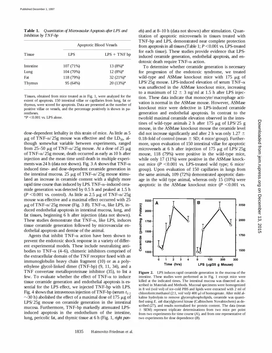
dose-dependent lethality in this strain of mice. As little as 5mg of TNF-a/25g mouse was effective and the LD50, al-though somewhat variable between experiments, rangedfrom 25–50 mg of TNF-a/25g mouse. At a dose of 25 mgof TNF-a/25g mouse, death occurred as early as 10 h afterinjection and the mean time until death in multiple experi-ments was 24 h (data not shown). Fig. 3 A shows that TNF-ainduced time- and dose-dependent ceramide generation inthe intestinal mucosa. 25 mg of TNF-a/25g mouse stimu-lated an increase in ceramide content with a slightly morerapid time course than induced by LPS. TNF-a–induced cera-mide generation was detected by 0.5 h and peaked at 1.5 h(P ,0.001 vs. control). As little as 2.5 mg of TNF-a/25gmouse was effective and a maximal effect occurred with 25mg of TNF-a/25g mouse (Fig. 3 B). TNF-a, like LPS, in-duced endothelial apoptosis in intestinal mucosa, lung, andfat tissues, beginning 6 h after injection (data not shown).These studies demonstrate that TNF-a, like LPS, inducestissue ceramide generation followed by microvascular en-dothelial apoptosis and demise of the animal.
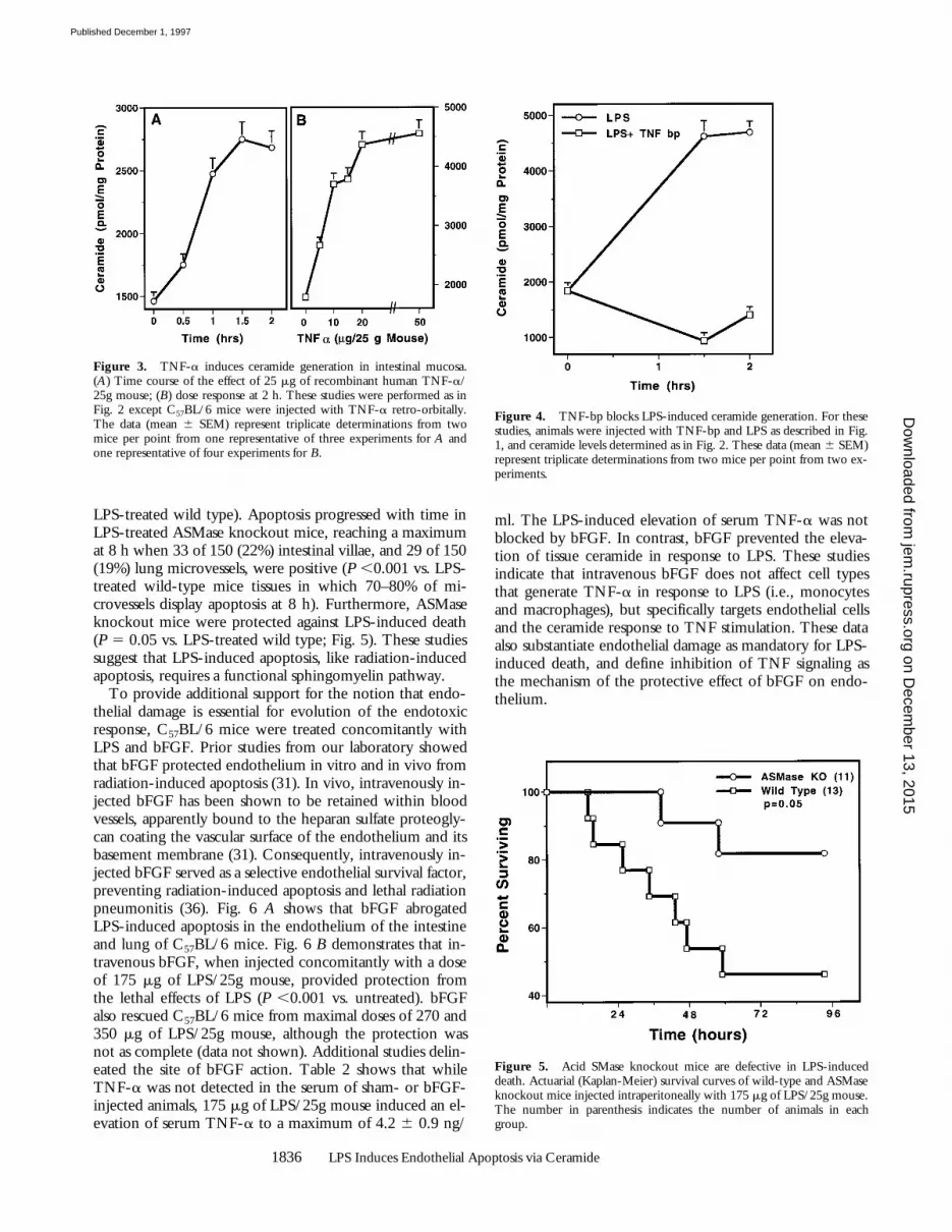
Agents that inhibit TNF-a action have been shown toprevent the endotoxic shock response in a variety of differ-ent experimental models. These include neutralizing anti-bodies to TNF-a (4–6), chimeric inhibitors comprised ofthe extracellular domain of the TNF receptor fused with animmunoglobulin heavy chain fragment (10) or as a poly-ethylene glycol-linked dimer (TNF-bp) (9, 11, 34), and aTNF convertase metalloproteinase inhibitor (35), to list afew. To evaluate whether the effect of TNF-a to inducetissue ceramide generation and endothelial apoptosis is es-sential for the LPS effect, we injected TNF-bp with LPS.Fig. 4 shows that intravenous injection of TNF-bp (serum t1/2
z30 h) abolished the effect of a maximal dose of 175 mg ofLPS/25g mouse on ceramide generation in the intestinalmucosa. Furthermore, TNF-bp markedly attenuated LPS-induced apoptosis in the endothelium of the intestine,lung, pericolic fat, and thymic tissue at 6 h (Fig. 1, right pan-
els) and at 8–10 h (data not shown) after stimulation. Quan-titation of apoptotic microvessels in tissues treated withTNF-bp and LPS, demonstrated near complete protectionfrom apoptosis in all tissues (Table 1; P ,0.001 vs. LPS-treatedfor each tissue). These studies provide evidence that LPS-induced ceramide generation, endothelial apoptosis, and en-dotoxic death require TNF-a action.
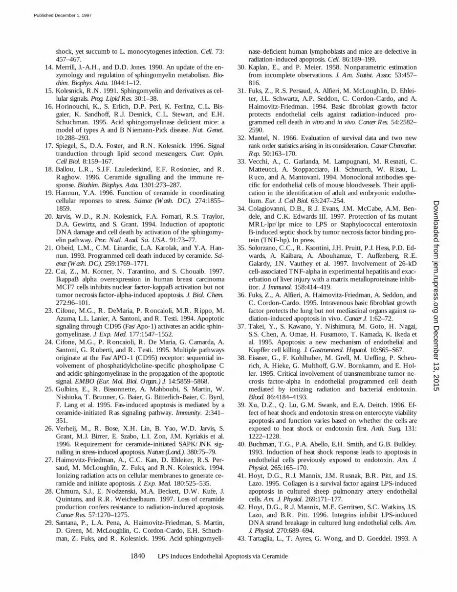
To determine whether ceramide generation is necessaryfor progression of the endotoxic syndrome, we treatedwild-type and ASMase knockout mice with 175 mg ofLPS/25g mouse. LPS-induced elevation of serum TNF-awas unaffected in the ASMase knockout mice, increasingto a maximum of 12 6 3 ng/ml at 1.5 h after LPS injec-tion. These data indicate that monocyte/macrophage acti-vation is normal in the ASMase mouse. However, ASMaseknockout mice were defective in LPS-induced ceramidegeneration and endothelial apoptosis. In contrast to thetwofold maximal ceramide elevation observed in the intes-tines of wild-type animals 2 h after 175 mg of LPS/25 gmouse, in the ASMase knockout mouse the ceramide leveldid not increase significantly and after 2 h was only 1.27 60.18-fold of control (mean 6 SD; 4 mice/group). Further-more, upon evaluation of 150 intestinal villae for apoptoticmicrovessels at 6 h after injection of 175 mg of LPS/25gmouse, 118 (79%) were positive in the wild-type mice,while only 17 (11%) were positive in the ASMase knock-out mice (P ,0.001 vs. LPS-treated wild type; 6 mice/group). Upon evaluation of 150 capillaries in lungs fromthe same animals, 109 (72%) demonstrated apoptotic dam-age in the wild type animal, whereas only 15 (10%) wereapoptotic in the ASMase knockout mice (P ,0.001 vs.
Table 1. Quantitation of Microvascular Apoptosis after LPS and Inhibition by TNF-bp
Apoptotic Blood Vessels
Tissue LPS LPS 1 TNF bp
Intestine 107 (71%) 13 (8%)*Lung 104 (70%) 12 (8%)*Fat 118 (79%) 32 (21%)*Thymus 95 (64%) 20 (13%)*
Tissues, obtained from mice treated as in Fig. 1, were analyzed for theextent of apoptosis. 150 intestinal villae or capillaries from lung, fat orthymus, were scored for apoptosis. Data are presented as the number ofpositive villae or vessels, and the percentage positively is shown in pa-rentheses.*P ,0.001 vs. LPS alone.
Figure 2. LPS induces rapid ceramide generation in the mucosa of theintestine. These studies were performed as in Fig. 1 except mice werekilled at the indicated times. The intestinal mucosa was dissected as de-scribed in Materials and Methods. Mucosal specimens were homogenizedin 8 vol (vol/vol) of ice-cold PBS and lipids were extracted with 2 ml ofchloroform:methanol (2:1, vol/vol)/400 ml of homogenate. After mild al-kaline hydrolysis to remove glycerophospholipids, ceramide was quanti-fied using E. coli diacylglycerol kinase (Calbiochem Novabiochem) as de-scribed (27), and results normalized for protein content. The data (mean6 SEM) represent triplicate determinations from two mice per pointfrom two experiments for time course (A), and from one representative oftwo experiments for dose dependence (B).
on Decem
ber 13, 2015jem
.rupress.orgD
ownloaded from
Published December 1, 1997
1836 LPS Induces Endothelial Apoptosis via Ceramide
LPS-treated wild type). Apoptosis progressed with time inLPS-treated ASMase knockout mice, reaching a maximumat 8 h when 33 of 150 (22%) intestinal villae, and 29 of 150(19%) lung microvessels, were positive (P ,0.001 vs. LPS-treated wild-type mice tissues in which 70–80% of mi-crovessels display apoptosis at 8 h). Furthermore, ASMaseknockout mice were protected against LPS-induced death(P 5 0.05 vs. LPS-treated wild type; Fig. 5). These studiessuggest that LPS-induced apoptosis, like radiation-inducedapoptosis, requires a functional sphingomyelin pathway.
To provide additional support for the notion that endo-thelial damage is essential for evolution of the endotoxicresponse, C57BL/6 mice were treated concomitantly withLPS and bFGF. Prior studies from our laboratory showedthat bFGF protected endothelium in vitro and in vivo fromradiation-induced apoptosis (31). In vivo, intravenously in-jected bFGF has been shown to be retained within bloodvessels, apparently bound to the heparan sulfate proteogly-can coating the vascular surface of the endothelium and itsbasement membrane (31). Consequently, intravenously in-jected bFGF served as a selective endothelial survival factor,preventing radiation-induced apoptosis and lethal radiationpneumonitis (36). Fig. 6 A shows that bFGF abrogatedLPS-induced apoptosis in the endothelium of the intestineand lung of C57BL/6 mice. Fig. 6 B demonstrates that in-travenous bFGF, when injected concomitantly with a doseof 175 mg of LPS/25g mouse, provided protection fromthe lethal effects of LPS (P ,0.001 vs. untreated). bFGFalso rescued C57BL/6 mice from maximal doses of 270 and350 mg of LPS/25g mouse, although the protection wasnot as complete (data not shown). Additional studies delin-eated the site of bFGF action. Table 2 shows that whileTNF-a was not detected in the serum of sham- or bFGF-injected animals, 175 mg of LPS/25g mouse induced an el-evation of serum TNF-a to a maximum of 4.2 6 0.9 ng/
ml. The LPS-induced elevation of serum TNF-a was notblocked by bFGF. In contrast, bFGF prevented the eleva-tion of tissue ceramide in response to LPS. These studiesindicate that intravenous bFGF does not affect cell typesthat generate TNF-a in response to LPS (i.e., monocytesand macrophages), but specifically targets endothelial cellsand the ceramide response to TNF stimulation. These dataalso substantiate endothelial damage as mandatory for LPS-induced death, and define inhibition of TNF signaling asthe mechanism of the protective effect of bFGF on endo-thelium.
Figure 3. TNF-a induces ceramide generation in intestinal mucosa.(A) Time course of the effect of 25 mg of recombinant human TNF-a/25g mouse; (B) dose response at 2 h. These studies were performed as inFig. 2 except C57BL/6 mice were injected with TNF-a retro-orbitally.The data (mean 6 SEM) represent triplicate determinations from twomice per point from one representative of three experiments for A andone representative of four experiments for B.
Figure 4. TNF-bp blocks LPS-induced ceramide generation. For thesestudies, animals were injected with TNF-bp and LPS as described in Fig.1, and ceramide levels determined as in Fig. 2. These data (mean 6 SEM)represent triplicate determinations from two mice per point from two ex-periments.
Figure 5. Acid SMase knockout mice are defective in LPS-induceddeath. Actuarial (Kaplan-Meier) survival curves of wild-type and ASMaseknockout mice injected intraperitoneally with 175 mg of LPS/25g mouse.The number in parenthesis indicates the number of animals in eachgroup.
on Decem
ber 13, 2015jem
.rupress.orgD
ownloaded from
Published December 1, 1997

1837 Haimovitz-Friedman et al.
Discussion
The present studies define a set of early biochemical andbiological responses to LPS using a standard model of en-dotoxic shock. Fig. 7 orders these events. Within 1 h of in-traperitoneal injection of LPS, elevation of tissue ceramidecontent was detected in the intestinal mucosa and lung. Al-though our evidence supports endothelium as the primarysource of the increase in ceramide, it remains formally pos-sible that cells other than endothelium contribute to the ce-ramide elevation. Ceramide elevation appeared dependenton TNF action since TNF mimicked the LPS effect, andTNF-bp blocked the LPS-induced increase in tissue cera-mide. Elevation of ceramide preceded the appearance of ageneralized form of apoptosis, expressed initially in the mi-crovascular endothelium of a variety of organs, beginningat 6 h after LPS injection. Both ceramide elevation and en-dothelial apoptosis preceded damage to nonendothelial pa-renchymal tissue and the death of the animal, which be-came apparent at 16 h after a dose of 175 mg LPS/25gmouse. Endothelial apoptosis appeared mandatory for the
progression of the endotoxic response, since intravenousinjection of bFGF, which specifically protects the endothe-lium against stress-induced apoptosis, prevented death.Furthermore, ceramide appeared to be a key intracellularmediator of this response, as the ASMase knockout mouse,which is defective in ceramide generation but not in TNF-aproduction, exhibited decreased endothelial apoptosis anddeath. Nevertheless, additional models of endotoxic shockmust be studied before it can be concluded that these ob-servations are representative of this process in general, assignificant species differences have been observed (3).
Previous studies reported that LPS induces endothelialdamage in vivo and under some conditions in vitro. Mi-crovascular injury occurs in numerous tissues during sepsis,including the lung, gut and liver, and this event has beengenerally considered an important element in the patho-genesis of the septic shock syndrome (1). The mechanismof microvascular injury and its relevance to the evolution ofthe septic shock syndrome have been a subject of substan-
Figure 6. Basic FGF blocks LPS-induced endothelial apoptosis and animal death. (A) C57BL/6 mice injected intravenously with 800 ng bFGF 30 min before, and 5 min, 1 and 2 h after, anintraperitoneal injection of 175 mg of LPS/25g mouse. Endothelial apoptosis was assessed as inFig. 1 by TUNEL assay. (B) Actuarial (Kaplan-Meier) survival curves of C57BL/6 treated as in A.The number in parenthesis indicates the number of animals in each group.
Table 2. Basic FGF Blocks LPS-induced Tissue Ceramide Generation but Not Serum TNF-a Elevation
Control bFGF LPS LPS 1 bFGF
TNFa (ng/ml) n.d. n.d. 4.2 6 0.9 7.9 6 2.0Ceramide (pmol/mg prot) 1942 6 97 1352 6 113 3175 6 274* 1810 6 108
Blood and intestinal mucosa were obtained from anesthetized mice (3 per group) 1.5 h after intraperitoneal injection of 175 mg of LPS/25g mousewith or without a single intravenous injection of 800 ng bFGF, as in Fig. 6 a. Serum TNF-a levels were measured in triplicate by ELISA as describedin Materials and Methods. Ceramide content was measured in triplicate as described in Fig. 2. n.d., not detected. *P ,0.01 vs. LPS 1 bFGF and control.
on Decem
ber 13, 2015jem
.rupress.orgD
ownloaded from
Published December 1, 1997

1838 LPS Induces Endothelial Apoptosis via Ceramide
tial debate. Disseminated intravascular thrombosis, exten-sive endothelial necrosis, and humoral microvascular dys-function have all been ascribed a role as mediating vascularcollapse (1). Generalized endothelial apoptosis has not hith-erto been reported, although apoptosis of liver endothe-lium ex vivo was recognized subsequent to induction ofTNF-a on Kupfer cells by LPS (37). The large majority ofstudies reported that LPS did not induce apoptosis in pri-mary cultures of endothelial cells (37–40) unless a secondstress such as heat shock or cycloheximide was applied sub-sequently (39, 40). However, one group has argued thatLPS can induce direct DNA damage leading to apoptosis inprimary cultures of sheep pulmonary endothelial cells (41, 42).
In the present studies, endothelial apoptosis appeared tobe preferentially increased in tissues known to play promi-nent roles in the pathogenesis of endotoxic shock. In thisregard, the microvascular endothelium of the bowel andlung were markedly affected. However, even the endothe-lium of tissues which play no overt role in the endotoxicresponse, such as the pericolic fat and the thymus, seemedto be affected. Endothelial damage preceded nonendothe-lial damage suggesting that loss of vascular integrity mayplay a role in the parenchymal tissue damage and the multi-organ failure that characterizes the endotoxic syndrome. Itis reasonable to suggest that the generalized nature of theapoptotic response in the microvascular endothelium mayaccount, in part, for the circulatory failure that is a majorfactor in the progression of the endotoxic response. Whetherendothelial apoptosis in the lung is the critical lesion lead-ing to the asphyxiation that results in the ultimate demiseof affected mice (5) cannot be ascertained from the presentinformation.
The critical role of endothelial cell apoptosis in thepathogenesis of endotoxic shock is similar to its role in theevolution of the inflammatory phase of radiation-inducedpneumonitis. As in the case of the LPS response, microvas-
cular endothelial apoptosis preceded the expression ofother histopathological manifestations of lethal radiation-induced pneumonitis, and intravenous injections of bFGFabrogated the evolution of pneumonitis and death afterwhole lung irradiation (31, 36). Furthermore, both LPS-and radiation-induced endothelial apoptosis in vivo ap-peared initiated by activation of ASMase. Thus, the sphin-gomyelin pathway may integrate diverse responses to signaldeath in stressed endothelial cells. Consistent with this hy-pothesis, the prevention of ceramide generation by bFGFsuggests that the anti-apoptotic survival function of bFGFmay be mediated, in part, via this mechanism.
The present investigations establish a role for TNF-a inLPS-induced generation of ceramide and apoptosis in vivo.Recent studies have clarified the mechanism by which the55-kD TNF receptor signals the apoptotic response (43–52). This receptor contains a carboxy-terminal death do-main which appears to be required for transmission of theapoptotic signal. Binding of TNF-a to the receptor triggersformation of a multiprotein complex in which cytoplasmicproteins and the receptor interact through their respectivedeath domain motifs. Upon TNF stimulation, the receptordeath domain binds to the death domain of a cytoplasmicprotein known as TRADD (TNF receptor 1–associateddeath domain), which in turn binds the death domain ofanother cytoplasmic protein, termed FADD/MORT-1.The latter protein also contains a death effector domain(DED) motif, which binds the DED of the ICE/Ced-3protease FLICE/MACH-1 (Caspase 8). It has been sug-gested that activation of FLICE/MACH-1 initiates activa-tion of a cascade of caspases, which serves as the effectorsystem for the apoptotic destruction of the cell.
This model suggests that ligand binding to the TNF re-ceptor is capable of activating the final death effector path-way without apparent involvement of lipid second messen-gers. However, several recent studies demonstrated a rolefor ceramide in TNF-induced cell death, in some systems.In this regard, activation of the death domain system of the55-kD TNF and CD95 receptors has been shown to cou-ple to ASMase (24, 53). This notion was based on the ob-servation that mutations in these receptor death domainsthat abolished apoptosis also abolished ceramide generation.Furthermore, dominant negative FADD/MORT-1 blockedceramide generation and apoptosis in BJAB B lymphoma cells,but not apoptosis induced by ceramide analogues. WhetherASMase activation might couple to FLICE/MACH-1 acti-vation is presently uncertain. However, Pronk et al. (54),using peptide inhibitors of ICE/Ced-3 proteases, molecu-larly ordered ceramide generation downstream of an unde-fined CPP32-like protease during REAPER-induced apo-ptosis in Drosophila Schneider L2 cells. In the presentstudies, TNF appeared essential for ceramide generationduring the evolution of the endotoxic syndrome. Whetherthe TNF receptor death domain adaptor protein system isinvolved in LPS-induced ceramide generation via TNF-ain vivo, remains uncertain.
Although the present studies define ceramide as critical
Figure 7. Proposed schemafor progression of the endotoxicresponse. LPS, released by gramnegative bacteria, interacts withinflammatory cells leading togeneration of TNF-a and othercytokines. TNF-a, acting uponendothelium, stimulates sphin-gomyelin hydrolysis to ceramidevia an ASMase. Apoptosis of theendothelium ensues, which canbe blocked by bFGF via inhibi-tion of ceramide generation. Wefurther propose that endothelialapoptosis results in generalizedmicrovascular dysfunction suffi-cient to compromise the circula-tion to major organs, leading tononendothelial tissue damage,circulatory collapse, and death.
on Decem
ber 13, 2015jem
.rupress.orgD
ownloaded from
Published December 1, 1997

1839 Haimovitz-Friedman et al.
for the induction of endothelial apoptosis by LPS, its pre-cise role in signaling apoptosis is as yet unknown. Kroemerand coworkers (55) have provided evidence that ceramideacts upstream of mitochondria to initiate apoptosis. Theirinvestigations suggested that ceramide, once generated, sig-nals mitochondrial membrane permeability transition (MPT),a committed step in the apoptotic process. MPT may signalapoptosis via release of an apoptosis-initiating factor, a Z-VAD-but not DEVD-inhibitable ICE-like protease (55). Consis-tent with this paradigm, Pastroini et al. (56) showed thatTNF-a– and ceramide-stimulated MPT was not inhibitedby the protein synthesis inhibitor cycloheximide. Alterna-tively, ceramide-initiated MPT may involve the release ofcytochrome C from mitochondria and activation of aCPP32-like protease (Caspase 3) (57–59). Either scenario isconsistent with the inhibition of ceramide-mediated apo-ptosis by Bcl-2 that has been reported in numerous systems(55, 60–62). Whether ceramide-mediated mitochondrialdamage is linked to the SAPK/JNK signaling system, also
reported to be critically involved in TNF-mediated apoptosisin endothelial cells (26), remains unknown.
The present investigations enhance the understanding ofthe mechanism of the endotoxic syndrome, defining up-stream elements of the LPS signaling system and their mo-lecular ordering, as well as the early tissue responses thattrigger its pathogenesis. The identification of biochemicalpathways that signal pro- and anti-apoptosis during the LPSresponse, and the characterization of the primary tissue tar-get for the endotoxic syndrome, provide a molecular andcellular context for testable experimental hypotheses, and abasis for developing strategies for pharmacologic interven-tion, with potential for clinical application. In particular,the ability of bFGF to inhibit ceramide generation suggeststhat treatment with bFGF may affect the progression of theLPS syndrome in gram-negative septicemia with evidenceof rising serum TNF, or in patients already manifestingsymptoms of septic shock.
This work was supported by grant CA52462 to Z. Fuks from the National Institutes of Health.
Correspondence should be addressed to Richard Kolesnick, Laboratory of Signal Transduction, MemorialSloan-Kettering Cancer Center, 1275 York Ave., New York, NY 10021. Tel.: (212) 639-8573; Fax: (212)639-2767.
Received for publication 27 June 1997 and in revised form 23 September 1997.
References1. Morrison, D.C., and J.L. Ryan. 1987. Endotoxins and disease
mechanisms. Ann. Rev. Med. 38:417–432.2. Bone, R.C. 1991. Sepsis, the sepsis syndrome, multi-organ
failure: a plea for comparable definitions. Ann. Int. Med. 114:332–333.
3. Tracey, K.J., and S.F. Lowry. 1990. The role of cytokine me-diators in septic shock. Adv. Surg. 23:21–56.
4. Beutler, B., I.W. Milsark, and A.C. Cerami. 1985. Passiveimmunization against cachectin/tumor necrosis factor pro-tects mice from lethal effect of endotoxin. Science (Wash.DC). 229:869–871.
5. Tracey, K.J., B. Beutler, S.F. Lowry, J. Merryweather, S.Wolpe, I.W. Milsark, R.J. Hariri, T.J. Fahey III, A. Zentella,J.D. Albert et al. 1986. Shock and tissue injury induced by re-combinant human Cachectin. Science (Wash. DC). 234:470–474.
6. Mathison, J.C., E. Wolfson, and R.J. Ulevitch. 1988. Partici-pation of tumor necrosis factor in the mediation of gram neg-ative bacterial lipopolysaccharide-induced injury in rabbits. J.Clin. Invest. 81:1925–1937.
7. Lesslauer, W., H. Tabuchi, R. Gentz, M. Brockhaus, E.J.Schlaeger, G. Grau, P.F. Piguet, P. Pointaire, P. Vassalli, andH. Loetscher. 1991. Recombinant soluble tumor necrosisfactor receptor proteins protect mice from lipopolysaccha-ride-induced lethality. Eur. J. Immunol. 21:2883–2886.
8. Hale, K.K., C.G. Smith, S.L. Baker, R.W. Vanderslice, C.H.Squires, T.M. Gleason, K.K. Tucker, T. Kohno, and D.A.Russell. 1995. Multifunctional regulation of the biological ef-
fects of TNF-alpha by the soluble type I and type II TNF re-ceptors. Cytokine. 7:26–38.
9. Russell, D.A., K.K. Tucker, N. Chinookoswong, R.C. Thomp-son, and T. Kohno. 1995. Combined inhibition of interleu-kin-1 and tumor necrosis factor in rodent endotoxemia: im-proved survival and organ function. J. Infect. Dis. 171:1528–1538.
10. Van Zee, K., L. Moldawer, H. Oldenburg, W. Thompson, S.Stackpole, W. Montegut, M. Rogy, C. Meschter, H. Gallati,C. Schiller et al. 1996. Protection against lethal Escherichiacoli bacteremia in baboons (Papio anubis) by pretreatmentwith a 55-kD TNF receptor (CD120a)-Ig fusion protein, Ro45-2081. J. Immunol. 156:2221–2230.
11. Espat, N.J., J.C. Cendan, E.A. Beierle, T.A. Auffenberg, J.Rosenberg, D. Russell, J.S. Kenney, E. Fischer, W. Montegut,S.F. Lowry et al. 1995. PEG-BP-30 monotherapy attenuatesthe cytokine-mediated inflammatory cascade in baboon Es-cherichia coli septic shock. J. Surg. Res. 59:153–158.
12. Rothe, J., W. Lesslauer, H. Lotscher, Y. Lang, P. Koebel, F.Kontgen, A. Althage, R. Zinkernagel, M. Steinmetz, and H.Bluethmann. 1993. Mice lacking the tumour necrosis factorreceptor 1 are resistant to TNF-mediated toxicity but highlysusceptible to infection by Listeria monocytogenes. Nature(Lond.). 364:798–802.
13. Pfeffer, K., T. Matsuyama, T.M. Kundig, A. Wakeham, K.Kishihara, A. Shahinian, K. Wiegmann, P.S. Ohashi, M.Kronke, and T.W. Mak. 1993. Mice deficient for the 55 kdtumor necrosis factor receptor are resistant to endotoxic
on Decem
ber 13, 2015jem
.rupress.orgD
ownloaded from
Published December 1, 1997
1840 LPS Induces Endothelial Apoptosis via Ceramide
shock, yet succumb to L. monocytogenes infection. Cell. 73:457–467.
14. Merrill, J.-A.H., and D.D. Jones. 1990. An update of the en-zymology and regulation of sphingomyelin metabolism. Bio-chim. Biophys. Acta. 1044:1–12.
15. Kolesnick, R.N. 1991. Sphingomyelin and derivatives as cel-lular signals. Prog. Lipid Res. 30:1–38.
16. Horinouchi, K., S. Erlich, D.P. Perl, K. Ferlinz, C.L. Bis-gaier, K. Sandhoff, R.J. Desnick, C.L. Stewart, and E.H.Schuchman. 1995. Acid sphingomyelinase deficient mice: amodel of types A and B Niemann-Pick disease. Nat. Genet.10:288–293.
17. Spiegel, S., D.A. Foster, and R.N. Kolesnick. 1996. Signaltranduction through lipid second messengers. Curr. Opin.Cell Biol. 8:159–167.
18. Ballou, L.R., S.J.F. Laulederkind, E.F. Rosloniec, and R.Raghow. 1996. Ceramide signalling and the immune re-sponse. Biochim. Biophys. Acta. 1301:273–287.
19. Hannun, Y.A. 1996. Function of ceramide in coordinatingcellular reponses to stress. Science (Wash. DC). 274:1855–1859.
20. Jarvis, W.D., R.N. Kolesnick, F.A. Fornari, R.S. Traylor,D.A. Gewirtz, and S. Grant. 1994. Induction of apoptoticDNA damage and cell death by activation of the sphingomy-elin pathway. Proc. Natl. Acad. Sci. USA. 91:73–77.
21. Obeid, L.M., C.M. Linardic, L.A. Karolak, and Y.A. Han-nun. 1993. Programmed cell death induced by ceramide. Sci-ence (Wash. DC). 259:1769–1771.
22. Cai, Z., M. Korner, N. Tarantino, and S. Chouaib. 1997.IkappaB alpha overexpression in human breast carcinomaMCF7 cells inhibits nuclear factor-kappaB activation but nottumor necrosis factor-alpha-induced apoptosis. J. Biol. Chem.272:96–101.
23. Cifone, M.G., R. DeMaria, P. Roncaioli, M.R. Rippo, M.Azuma, L.L. Lanier, A. Santoni, and R. Testi. 1994. Apoptoticsignaling through CD95 (Fas/Apo-1) activates an acidic sphin-gomyelinase. J. Exp. Med. 177:1547–1552.
24. Cifone, M.G., P. Roncaioli, R. De Maria, G. Camarda, A.Santoni, G. Ruberti, and R. Testi. 1995. Multiple pathwaysoriginate at the Fas/APO-1 (CD95) receptor: sequential in-volvement of phosphatidylcholine-specific phospholipase Cand acidic sphingomyelinase in the propagation of the apoptoticsignal. EMBO (Eur. Mol. Biol. Organ.) J. 14:5859–5868.
25. Gulbins, E., R. Bissonnette, A. Mahboubi, S. Martin, W.Nishioka, T. Brunner, G. Baier, G. Bitterlich-Baier, C. Byrd,F. Lang et al. 1995. Fas-induced apoptosis is mediated by aceramide-initiated Ras signaling pathway. Immunity. 2:341–351.
26. Verheij, M., R. Bose, X.H. Lin, B. Yao, W.D. Jarvis, S.Grant, M.J. Birrer, E. Szabo, L.I. Zon, J.M. Kyriakis et al.1996. Requirement for ceramide-initiated SAPK/JNK sig-nalling in stress-induced apoptosis. Nature (Lond.). 380:75–79.
27. Haimovitz-Friedman, A., C.C. Kan, D. Ehleiter, R.S. Per-saud, M. McLoughlin, Z. Fuks, and R.N. Kolesnick. 1994.Ionizing radiation acts on cellular membranes to generate ce-ramide and initiate apoptosis. J. Exp. Med. 180:525–535.
28. Chmura, S.J., E. Nodzenski, M.A. Beckett, D.W. Kufe, J.Quintans, and R.R. Weichselbaum. 1997. Loss of ceramideproduction confers resistance to radiation-induced apoptosis.Cancer Res. 57:1270–1275.
29. Santana, P., L.A. Pena, A. Haimovitz-Friedman, S. Martin,D. Green, M. McLoughlin, C. Cordon-Cardo, E.H. Schuch-man, Z. Fuks, and R. Kolesnick. 1996. Acid sphingomyeli-
nase-deficient human lymphoblasts and mice are defective inradiation-induced apoptosis. Cell. 86:189–199.
30. Kaplan, E., and P. Meier. 1958. Nonparametric estimationfrom incomplete observations. J. Am. Statist. Assoc. 53:457–816.
31. Fuks, Z., R.S. Persaud, A. Alfieri, M. McLoughlin, D. Ehlei-ter, J.L. Schwartz, A.P. Seddon, C. Cordon-Cardo, and A.Haimovitz-Friedman. 1994. Basic fibroblast growth factorprotects endothelial cells against radiation-induced pro-grammed cell death in vitro and in vivo. Cancer Res. 54:2582–2590.
32. Mantel, N. 1966. Evaluation of survival data and two newrank order statistics arising in its consideration. Cancer Chemother.Rep. 50:163–170.
33. Vecchi, A., C. Garlanda, M. Lampugnani, M. Resnati, C.Matteucci, A. Stoppacciaro, H. Schnurch, W. Risau, L.Ruco, and A. Mantovani. 1994. Monoclonal antibodies spe-cific for endothelial cells of mouse bloodvessels. Their appli-cation in the identification of adult and embryonic endothe-lium. Eur. J. Cell Biol. 63:247–254.
34. Colagiovanni, D.B., R.J. Evans, J.M. McCabe, A.M. Ben-dele, and C.K. Edwards III. 1997. Protection of fas mutantMRL-lpr/lpr mice to LPS or Staphylococcal enterotoxinB-induced septic shock by tumor necrosis factor binding pro-tein (TNF-bp). In press.
35. Solorzano, C.C., R. Ksontini, J.H. Pruitt, P.J. Hess, P.D. Ed-wards, A. Kaibara, A. Abouhamze, T. Auffenberg, R.E.Galardy, J.N. Vauthey et al. 1997. Involvement of 26-kDcell-associated TNF-alpha in experimental hepatitis and exac-erbation of liver injury with a matrix metalloproteinase inhib-itor. J. Immunol. 158:414–419.
36. Fuks, Z., A. Alfieri, A. Haimovitz-Friedman, A. Seddon, andC. Cordon-Cardo. 1995. Intravenous basic fibroblast growthfactor protects the lung but not mediastinal organs against ra-diation-induced apoptosis in vivo. Cancer J. 1:62–72.
37. Takei, Y., S. Kawano, Y. Nishimura, M. Goto, H. Nagai,S.S. Chen, A. Omae, H. Fusamoto, T. Kamada, K. Ikeda etal. 1995. Apoptosis: a new mechanism of endothelial andKupffer cell killing. J. Gastroenterol. Hepatol. 10:S65–S67.
38. Eissner, G., F. Kohlhuber, M. Grell, M. Ueffing, P. Scheu-rich, A. Hieke, G. Multhoff, G.W. Bornkamm, and E. Hol-ler. 1995. Critical involvement of transmembrane tumor ne-crosis factor-alpha in endothelial programmed cell deathmediated by ionizing radiation and bacterial endotoxin.Blood. 86:4184–4193.
39. Xu, D.Z., Q. Lu, G.M. Swank, and E.A. Deitch. 1996. Ef-fect of heat shock and endotoxin stress on enterocyte viabilityapoptosis and function varies based on whether the cells areexposed to heat shock or endotoxin first. Arch. Surg. 131:1222–1228.
40. Buchman, T.G., P.A. Abello, E.H. Smith, and G.B. Bulkley.1993. Induction of heat shock response leads to apoptosis inendothelial cells previously exposed to endotoxin. Am. J.Physiol. 265:165–170.
41. Hoyt, D.G., R.J. Mannix, J.M. Rusnak, B.R. Pitt, and J.S.Lazo. 1995. Collagen is a survival factor against LPS-inducedapoptosis in cultured sheep pulmonary artery endothelialcells. Am. J. Physiol. 269:171–177.
42. Hoyt, D.G., R.J. Mannix, M.E. Gerritsen, S.C. Watkins, J.S.Lazo, and B.R. Pitt. 1996. Integrins inhibit LPS-inducedDNA strand breakage in cultured lung endothelial cells. Am.J. Physiol. 270:689–694.
43. Tartaglia, L., T. Ayres, G. Wong, and D. Goeddel. 1993. A
on Decem
ber 13, 2015jem
.rupress.orgD
ownloaded from
Published December 1, 1997

1841 Haimovitz-Friedman et al.
novel domain within the 55 kd TNF receptor signals celldeath. Cell. 74:845–853.
44. Hsu, H., J. Xiong, and D.V. Goeddel. 1995. The TNF re-ceptor 1-associated protein TRADD signals cell death andNF-kappa B activation. Cell. 81:495–504.
45. Hsu, H., H.B. Shu, M.G. Pan, and D.V. Goeddel. 1996.TRADD-TRAF2 and TRADD-FADD interactions definetwo distinct TNF receptor 1 signal transduction pathways.Cell. 84:299–308.
46. Chinnaiyan, A.M., K. O’Rourke, M. Tewari, and V.M.Dixit. 1995. FADD, a novel death domain-containing pro-tein, interacts with the death domain of Fas and initiates apo-ptosis. Cell. 81:505–512.
47. Chinnaiyan, A.M., C.G. Tepper, M.F. Seldin, K. O’Rourke,F.C. Kischkel, S. Hellbardt, P.H. Krammer, M.E. Peter, andV.M. Dixit. 1996. FADD/MORT1 is a common mediatorof CD95 (Fas/APO-1) and tumor necrosis factor receptor-induced apoptosis. J. Biol. Chem. 271:4961–4965.
48. Boldin, M.P., T.M. Goncharov, Y.V. Goltsev, and D.Wallach. 1996. Involvement of MACH, a novel MORT1/FADD-interacting protease, in Fas/APO-1- and TNF recep-tor-induced cell death. Cell. 85:803–15.
49. Boldin, M.P., I.L. Mett, E.E. Varfolomeev, I. Chumakov, Y.Shemer-Avni, J.H. Camonis, and D. Wallach. 1995. Self-associ-ation of the “death domains” of the p55 tumor necrosis factor(TNF) receptor and Fas/APO1 prompts signaling for TNFand Fas/APO1 effects. J. Biol. Chem. 270:387–391.
50. Chinnaiyan, A.M., K. Orth, K. O’Rourke, H. Duan, G.G.Poirier, and V.M. Dixit. 1996. Molecular ordering of the celldeath pathway. Bcl-2 and Bcl-xL function upstream of theCED-3-like apoptotic proteases. J. Biol. Chem. 271:4573–4576.
51. Muzio, M., A.M. Chinnaiyan, F.C. Kischkel, K. Orourke, A.Shevchenko, J. Ni, C. Scaffidi, J.D. Bretz, M. Zhang, R.Gentz et al. 1996. FLICE, a novel FADD-homologous ICE/CED-3-like protease, is recruited to the CD95 (Fas/APO-1)death-inducing signaling complex. Cell. 85:817–827.
52. Kischkel, F.C., S. Hellbardt, I. Behrmann, M. Germer, M.Pawlita, P.H. Krammer, and M.E. Peter. 1995. Cytotoxicity-dependent APO-1 (Fas/CD95)-associated proteins form adeath-inducing signaling complex (DISC) with the receptor.
EMBO (Eur. Mol. Biol. Organ.) J. 14:5579–5588.53. Wiegmann, K., S. Schutze, T. Machleidt, D. Witte, and M.
Kronke. 1994. Functional dichotomy of neutral and acidicsphingomyelinases in tumor necrosis factor signaling. Cell.78:1005–1015.
54. Pronk, G.J., K. Ramer, P. Amiri, and L.T. Williams. 1996.Requirement of an ICE-like protease for induction of apoptosisand ceramide generation by REAPER. Science (Wash. DC).271:808–810.
55. Susin, S.A., N. Zamzami, M. Castedo, T. Hirsch, P. Mar-chetti, A. Macho, E. Daugas, M. Geuskens, and G. Kroemer.1996. Bcl-2 inhibits the mitochondrial release of an apopto-genic protease. J. Exp. Med. 184:1331–1341.
56. Pastorino, J., G. Simbula, K. Yamamoto, P.J. Glascott, R.Rothman, and J. Farber. 1996. The cytotoxicity of tumor ne-crosis factor depends on induction of the mitochondrial per-meability transition. J. Biol. Chem. 271:29792–29798.
57. Liu, X.S., C.N. Kim, J. Yang, R. Jemmerson, and X.D.Wang. 1996. Induction of apoptotic program in cell-free ex-tracts - requirement for datp and cytochrome C. Cell. 86:147–157.
58. Kluck, R.M., E. Bossywetzel, D.R. Green, and D.D. New-meyer. 1997. The release of cytochrome C from mitochon-dria - a primary site for Bcl-2 regulation of apoptosis. Science(Wash. DC). 275:1132–1136.
59. Yang, J., X.S. Liu, K. Bhalla, C.N. Kim, A.M. Ibrado, J.Y.Cai, T.I. Peng, D.P. Jones, and X.D. Wang. 1997. Preven-tion of apoptosis by Bcl-2 - release of cytochrome C frommitochondria blocked. Science (Wash. DC). 275:1129–1132.
60. Castedo, M., T. Hirsch, S. Susin, N. Zamzami, P. Marchetti,A. Macho, and G. Kroemer. 1996. Sequential acquisition ofmitochondrial and plasma membrane alterations during earlylymphocyte apoptosis. J. Immunol. 157:512–521.
61. Zhang, K.Z., J.A. Westberg, E. Holtta, and L.C. Andersson.1996. Bcl-2 regulates neural differentiation. Proc. Natl. Acad.Sci. USA. 93:4504–4508.
62. Martin, S.J., S. Takayama, A.J. McGahon, T. Miyashita, J.Corbeil, R.N. Kolesnick, J.C. Reed, and D.R. Green. 1995.Inhibition of ceramide-induced apoptosis by Bcl-2. CellDeath Differ. 2:253–257.
on Decem
ber 13, 2015jem
.rupress.orgD
ownloaded from
Published December 1, 1997