Embed Size (px)
Citation preview
RESEARCH Open Access
Lipid accelerating the fibril of islet amyloidpolypeptide aggravated the pancreatic isletinjury in vitro and in vivoXiao-Dan Mo1, Li-Ping Gao3, Qing-Jun Wang3, Jie Yin1 and Yu-Hong Jing1,2*
Abstract
Background: The fibrillation of islet amyloid polypeptide (IAPP) triggered the amyloid deposition, then enhanced theloss of the pancreatic islet mass. However, it is not clear what factor is the determinant in development of the fibrilformation. The aim of this study is to investigate the effects of lipid on IAPP fibril and its injury on pancreatic islet.
Methods: The fibril form of human IAPP (hIAPP) was tested using thioflavin-T fluorescence assay and transmission electronmicroscope technology after incubated with palmitate for 5 h at 25 °C. The cytotoxicity of fibril hIAPP was evaluated in INS-1cells through analyzing the leakage of cell membrane and cell apoptosis. Type 2 diabetes mellitus (T2DM) animal model wasinduced with low dose streptozotocin combined the high-fat diet feeding for two months in rats. Plasma biochemistryparameters were measured before sacrificed. Pancreatic islet was isolated to evaluate their function.
Results: The results showed that co-incubation of hIAPP and palmitate induced more fibril form. Fibril hIAPP induced celllesions including cell membrane leakage and cell apoptosis accompanied insulin mRNA decrease in INS-1 cell lines. In vivo,Plasma glucose, triglyceride, rIAPP and insulin increased in T2DM rats compared with the control group. In addition, IAPPand insulin mRNA increased in pancreatic islet of T2DM rats. Furthermore, T2DM induced the reduction of insulin receptorexpression and cleaved caspase-3 overexpression in pancreatic islet.
Conclusions: Results in vivo and in vitro suggested that lipid and IAPP plays a synergistic effect on pancreatic islet celldamage, which implicated in enhancing the IAPP expression and accelerating the fibril formation of IAPP.
Keywords: IAPP, Lipid, Pancreatic islet, T2DM, Insulin, Palmitate
BackgroundType 2 diabetes (T2DM) is characterized by the hyper-lipidemia and aberrant metabolism. Aberrant metabol-ism caused the serials of symptoms in diabetic patientsincluding overweight or obesity, unbalance of anabolismand catabolism in adipocyte, chronic inflammationwhich induced the insulin resistance. Amyloid depos-ition consisted of islet amyloid polypeptide (IAPP) canbe observed in pancreatic biopsy of patient with T2DM.IAPP is a 37-amino acid peptide of the calcitonin genefamily. It is the most abundant component of pancreatic
amyloid [1, 2]. IAPP has been suggested to be toxic to β-cells and to be involved in the development of T2DM [3,4]. It has been proposed that overexpression of IAPPcontributes to pancreatic amyloid formation and devel-opment of T2DM, this viewpoint supported by trans-genic mouse and rat studies involving the overexpressionof human IAPP (hIAPP) in islets of Langerhans [5]. Clin-ical studies indicate that hIAPP preferentially forms theamyloid deposition that implicates the pathology of islethyalinization which is the pathological characteristic ofT2DM [6–8]. Amyloid deposition is observed not only inpatients with T2DM but also in some overweight ornon-diabetic individuals [9–11]. Therefore, evidence sug-gests that some unidentified factors are involved in theregulation of IAPP secretion and amyloid deposition.Several studies have reported that the high expression ofIAPP can be induced by lipid condition [4, 12–14]. It is
* Correspondence: [email protected] of Anatomy and Histology & Embryology, Neuroscience, School ofBasic Medical Sciences, Lanzhou University, No. 199 of Donggang West Road,Lanzhou City, Gansu Province 730000, People’s Republic of China2Key Laboratory of Preclinical Study for New Drugs of Gansu province,Lanzhou University, No. 199 of Donggang West Road, Lanzhou City, GansuProvince 730000, People’s Republic of ChinaFull list of author information is available at the end of the article
© The Author(s). 2018 Open Access This article is distributed under the terms of the Creative Commons Attribution 4.0International License (http://creativecommons.org/licenses/by/4.0/), which permits unrestricted use, distribution, andreproduction in any medium, provided you give appropriate credit to the original author(s) and the source, provide a link tothe Creative Commons license, and indicate if changes were made. The Creative Commons Public Domain Dedication waiver(http://creativecommons.org/publicdomain/zero/1.0/) applies to the data made available in this article, unless otherwise stated.
Mo et al. Lipids in Health and Disease (2018) 17:42 https://doi.org/10.1186/s12944-018-0694-8

known to cause the typical characteristic of T2DM [15,16]. But how and what accelerating amyloid depositionby lipid is not clear.Amyloid deposition is initiated by changed of protein
conformation. Especially, the fibril form is preference toaccumulate, which damage the cell membrane and in-duced the cell apoptosis [17–21]. The aim of the presentstudy is to explore the effects of lipid on fibril formationof IAPP and the toxicity of fibril IAPP on pancreatic isletcells. The T2DM animal model was established by feed-ing with high fat diet or streptozotocin (STZ) injection,the IAPP levels and the pancreatic islet pathology andfunction were investigated. High dose STZ badly impairsthe insulin secretion by inducing β-cell death mimickingtype 1 diabetes. Low-dose STZ has been known to in-duce a mild impairment of insulin secretion which issimilar to the feature of the later stage of type 2 diabetes[22, 23]. The investigators have been developed a ratmodel by feeding the animal with high-fat diet followinglow-dose STZ that closely mimic the metabolic charac-teristics of human type 2 diabetes [24].
MethodsReagentsThe hIAPP was synthesized using t-boc chemistry andpurified by reverse phase high-performance liquid chro-matography (Shanghai Zi Yu Biotech Co. Ltd., Shanghai,China). 1,1,1,3,3,3-Hexafluoro-2-propanol (HFIP), palmi-tate (PA), thioflavin-T (ThT) and STZ were purchasedfrom Sigma (St. Louis, MO, USA). Enzymatic diagnostickits for plasma glucose and triglyceride were purchasedfrom Randox (Crumlin Co., Antrim, UK). The lactate de-hydrogenase (LDH) assay kit and TUNEL kit were pur-chased from Roche (Roche, USA). The sandwich enzyme-linked immunosorbent assay (ELISA) kits of insulin andrIAPP were obtained from R&D (R&D, IL, USA). Theanti-IAPP, and IRβ antibodies were purchased fromAbcam (Cambridge, UK). Anti-Cleaved caspase3 antibodywas purchased from Cell signaling (Cell signaling Tech-nology, MA, USA). The anti-glyceraldehyde 3-phosphatedehydrogenase (GAPDH) antibody was obtained fromSanta Cruz (Santa Cruz, CA, USA). The RPMI-1640medium and the fetal bovine serum (FBS) were purchasedfrom Gibco BRL (Gaithersburg, MD, USA). The waterused in all experiments was ultrapure, and supplied by aMilli-Q water purification system from Millipore.
ThT fluorescence assaysThT based fluorescence assays were performed to evalu-ate the hIAPP fibril form. Briefly, hIAPP was first dis-solved in HFIP and sonicated for 2 min to homogenizethe sample. hIAPP was then diluted in 25 mM PBS(pH 7.4) containing 50 mM NaCl and 1% HFIP to a finalconcentration of 20 μM. The hIAPP were co-incubated
with 200 μM PA at 25 °C for 5 h. Samples were ali-quoted at designated time intervals and ThT based fluor-escence assays were used to detect the fibril formationof hIAPP; the fluorescence emission experiments wereperformed on a Hitachi FL-2700 fluorometer (Toyko,Japan) with the excitation and emission wavelengths setat 450 and 482 nm, respectively. All experiments wererepeated for at least three times.
Transmission electron microscopy (TEM)20 μM hIAPP were co-incubated with 200 μM PA at 25 °C for 5 h, then Five microliters of samples to be imagedwere spotted on a 300 mesh Formvar-carbon coated cop-per grid (Shanghai, China) and stained with 1% freshlyprepared uranyl formate. Samples were air dried and ob-served under a JEM2100 TEM (JEMO, Toyko, Japan) op-erating at an accelerating voltage of 100 kV.
Cell culture and treatmentThe rat insulinoma cell line (INS-1) was purchased fromthe Institute of Biochemistry and Cell Biology of Shang-hai. The cells were briefly maintained in an RPMI-1640medium supplemented with 10% FBS, 2 mmol/L L-glu-tamine, 1 mmol/L sodium pyruvate, 10 mmol/L HEPES,50 mmol/L mercaptoethanol, 100 U/mL penicillin, and100 U/mL streptomycin at 37 °C in a humid atmosphere(95% relative humidity, 5% CO2). For the peptide treat-ment, lyophilized hIAPP was dissolved in HFIP, whichwas removed by evaporation under N2. The hIAPP incu-bation with palmitate for 5 h at 25 °C, then used to treatcells. INS-1 cells were incubated with 20 μM and 50 μMfibril hIAPP for 24 h to evaluate the LDH leakage, levelsof insulin mRNA and apoptosis.
LDH assayThe LDH release indicates the change of membrane perme-ability, which can reflect the damage extent of the cell mem-brane. The cells were precipitated by centrifugation (1500×g)for 10 min at room temperature at the end of the treatments.The supernatants were transferred to a 96-well plate. TheLDH activity was assayed using the cytotoxicity detection kitaccording to the manufacturer’s instructions. The cellstreated with 1% Triton X-100 were used as high control,while media without cells served as a low control. The re-sults were expressed as % LDH leakage [(experimental value− low control) / (high control − low control) × 100] [25].
Animals and treatmentA total of 48 healthy male Sprague Dawley (SD) rats weigh-ing 200-220 g were purchased from the Experimental Ani-mal Center of Lanzhou University. The animals were housedin a standard environment at 20–25 °C and 50–70% humid-ity and maintained under a 12 h light–dark cycle with foodand water ad libitum. All animal experimental protocols
Mo et al. Lipids in Health and Disease (2018) 17:42 Page 2 of 11
were approved by the institutional Animal Ethics Commit-tee, Lanzhou University (permission number: SCXK Gan2009–0004). The animals were randomly divided into fourgroups as follows: control group (single injection with equalvolume of 0.1 M citrate buffer through peritonea, feedingwith regular diet), STZ group (single injection with STZthrough peritonea, 30 mg/kg, STZ dissolved in 0.1 M citratebuffer, feeding with regular diet), HD group (single injectionwith equal volume of 0.1 M citrate buffer through peritonea,feeding with high-fat diet), and STZ+HD group (single in-jection with STZ through peritonea, 30 mg/kg, STZ dis-solved in 0.1 M citrate buffer, feeding with regular diet).Diets (Ke Ao Co. Ltd., Beijing) contained 15% (low fat) or36% (high fat) calories derived from fat. Fat was provided ascorn oil and hydrogenated coconut oil, with the ratio of satu-rated to unsaturated fatty acids being 1:3 in each diet. Theprogressively increasing amounts of fat were balanced by de-creasing amounts of carbohydrate (65 kcal % and 44 kcal %in low- and high-fat diets, respectively) and constantamounts of protein (20 kcal%).
Examination of plasma rIAPP, insulin, triglyceride andglucose in diabetic ratsPlasma was isolated by low-speed centrifugation of bloodat 4 °C. The plasma glucose and triglyceride were mea-sured with respective enzymatic diagnostic kits accordingto the manual instruction. Plasma insulin and rIAPP weremeasured by respective ELISA kit according to the man-ual instruction. All data were obtained from two inde-pendent measurements, each with triplicate incubations.
Glucose tolerance test (GTT) and insulin sensitivity tests(IST)For the glucose tolerance and insulin sensitivity tests,the rats were intraperitoneally injected with glucose(2 g/kg) or subcutaneously injected with human regularinsulin (0.75 unit/ kg) after 12 h of fasting, respectively.Blood samples were collected from the tail vein at0 min, 30 min, 60 min, 90 min, and 120 min after treat-ment. The blood glucose levels were measured.
Histology examination of pancreatic isletAfter two months, four rats, which were randomly selectedfrom each group, were anesthetized. The pancreatic tailwas isolated (in a size of 0.5 × 0.5 × 0.5 cm) and performedon formalin-fixed. The paraffin-embedded pancreas speci-mens were cut at a 5 μm thickness. Sections were collectedand stained by hematoxylin-Eosin (H-E) to evaluate thehistopathological changes of pancreatic islet. To identify thepancreatic islet, sections were stained with insulin antibody(1:100) overnight at 4 °C. Then the sections were rinsedwith 0.01 M PBS and incubated with FITC-IgG (1:50). Thesections were observed under fluorescence microscope andscanned in dark field. The other sections were used to
incubate using the rIAPP primary antibody (1:100), over-night at 4 °C. The sections were rinsed with 0.01 M PBSand incubated with the corresponding second antibodies at37 °C for 1 h, then rinsed and incubated with streptavidin-conjugated horseradish peroxidase (1:100) at 37 °C for 1 h.The immunoreactivity was visualized with 0.05% a diami-nobenzidine (DAB) as a chromogen. The sections were ob-served under a microscope and scanned in bright field. Thepancreatic islets were manually outlined. The pancreaticislet area was calculated using ImageJ software. The opticaldensity was used to quantify the rIAPP expressed by nor-malized with unit area of pancreatic islet.
TUNEL assayThe paraffin-embedded pancreas specimens were cut at a5 μm thickness. And terminal deoxynucleotidyl transferase-mediated deoxyuridine triphosphate biotin nick end label-ing (TUNEL) assay was performed on the sections. Sectionswere treated with 0.2% H2O2 for 10 min, rinsed in 0.1 MPBS, and incubated with Proteinase K Solution (contain200 mmol Tris, pH, 7.4, 0.5 mmol EDTA, and proteinase K1 mg/ml) for 20 min at room temperature. Then the sec-tions were incubated in TUNEL reaction mixture using theIn Situ Cell Death Detection Kit for 1 h at 37 °C, thenrinsed in 0.1 M PBS three times for 5 min and incubated inConverter peroxidase (POD) for 30 min at 37 °C, rinsed in0.1 M PBS three times for 5 min, and color-developed withSIGMA FAST, a DAB POD substrate. The pancreatic isletswere manually outlined. The TUNEL positive cells werecounted and normalized with unit area of pancreatic islet.
Procedure of pancreatic islet isolationAfter two months, eight rats, which were randomly se-lected from each group, were anesthetized. And the pan-creas was isolated. After enzymatic digestion of thepancreas, islets of Langerhans were purified using a dis-continuous density gradient of ficoll solutions as previ-ously reported [26], with minor adjustments. Briefly, thecommon bile duct was cannulated and 1 ml of digestionsolution (0.2 mg/ml collagenase in HBSS containing10 mmol HEPES) was injected to distend the pancreas.The pancreas was removed and placed for 15 min at37 °C in a Petri dish filled with 5 ml of digestion solutionto isolate the islet.
RNA extraction and Q-RT-PCRThe total RNA was extracted from the isolated pancreaticislet or INS-1 cells using the RNAiso plus reagent (TakaraBiotech, Co., Ltd., Dalian, China) and depleted of contaminat-ing DNA with RNase-free DNase according to the manufac-turers’ instructions. cDNA was synthesized from 1 μg ofRNA with M-MuLV reverse transcriptase and random hex-amer according to the manufacturer’s instructions (Fermen-tas, Burlington, Canada). Q-RT-PCR was performed using
Mo et al. Lipids in Health and Disease (2018) 17:42 Page 3 of 11

the PIKoREAl96 detector (Thermo Scientific, USA). Theprimers for the rIAPP were 5′-GCCCACTGAAAGG-GATCTTG-3 (forward) and 5′-GCACTTCCGTTTGTC-CACCT-3′ (reverse). The primers for the rat preproinsulinwere: 5′-CAGCACCTTTGTGGTTCTCACTT-3′ (forward)and 5′-CTCCACCCAGCTCCAGTTGT-3′ (reverse). Therat GAPDH primers were 5′-GGCACAGTCAAGGCTGA-GAATG-3′ (forward) and 5′-ATGGTGGTGAAGACGC-CAGTA-3′ (reverse). The assays were initiated for 5 min at95 °C and 40 cycles of 15 s at 94 °C and 1 min at 60 °C. Therelative levels of amylin or preproinsulin mRNA expressionwere calculated using the 2ΔCT method.
Protein extraction and western blot analysisThe total proteins were extracted from pancreatic islet orINS-1 cells using RIPA buffer that contains protease inhibi-tors. The proteins (50 μg) were fractionated on 10% sodiumdodecyl sulfate polyacrylamide gel electrophoresis, and thentransferred into polyvinylidene fluoride membranes. Themembranes were blotted with anti-IRβ (1:1000), anti-caspase-3 (1:1000), and anti-GAPDH (1:5000) antibodies, aswell as with horseradish peroxidase-conjugated second anti-body (1:5000). The immunoreactive protein bands were visu-alized by enhanced chemiluminescence.
Statistical analysisThe data were expressed as mean ± SEM. Statistical ana-lysis was performed using SPSS statistical program at17.0version. The difference between two groups was ana-lyzed by Student’s t-test, whereas that among three ormore groups was analyzed by one-way or two way analysisof variance with least significant difference test. The differ-ence with P < 0.05 was considered statistically significant.
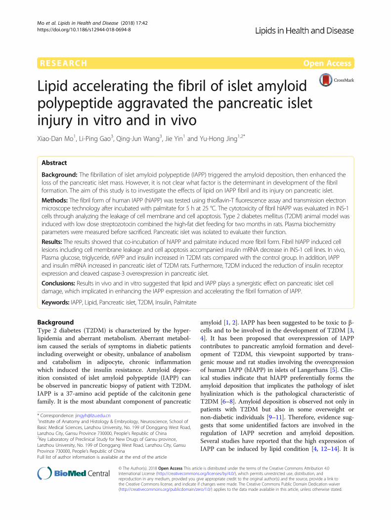
ResultsEffects of lipid on fibrillation of hIAPPTwenty μM hIAPP incubated with 200 μM palmitate at25 °C, and then ThT-based fluorescence assays were per-formed. hIAPP exhibited maximum ThT emission with along lag time of 4.3 ± 0.6 h (Fig. 1a) compared withpalmitate treated-hIAPP (1.5 ± 0.2 h, Fig. 1a). This resultsuggested palmitate accelerated the formation of fibrilhIAPP. Under TEM, a mesh of typical long linear fibrilswas detected for palmitate treated-hIAPP incubated for5 h (Fig. 1c). In contrast, only a few linear fibrils to-gether with significant amount of amorphous aggregateswere observed in untreated hIAPP (Fig. 1b), which isconsistent with the results by ThT assay.
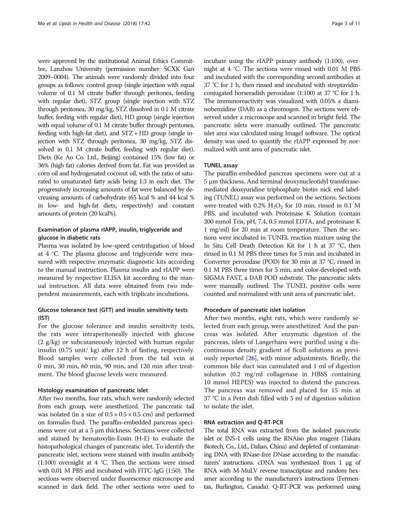
Effects of fibril hIAPP on membrane permeability ofpancreatic cellsThe INS-1 cell, which was the cell line of insulinomacharacterized by insulin secretion were incubated withexogenous fibril hIAPP for 24 h at concentrations of
20 μM and 50 μM to further confirm the direct effecton the INS-1 cells. The results showed that the insulinmRNA levels decreased about 50% after treatment withfibril hIAPP (Fig. 2a). These findings also suggested thatfibril hIAPP may repress the transcription of insulingene. Membrane permeability was evaluated with the ra-tio of the LDH leakage. As shown in Fig. 2b, the LDHleakage increased to 6–8 folds after incubation with fibrilhIAPP. This result suggested that hIAPP aggravated thecell damage in a dose-dependent manner. In addition,the hIAPP treatment induced the caspase-3 high expres-sion, which indicated the initiation of DNA damage andcell apoptosis (Fig. 2c and d).
Changes of body weight, GTT and IST in diabetic ratsAs shown in Fig. 3b, the body weight gain in high-fat dietgroup is more than in the control. Alternatively, bodyweight gain in STZ treated group is less than in the control.And the extent of body weight gain in STZ +HD group issimilar as the control. The glucose tolerance increasedabout one fold and the insulin sensitivity decreased about20% in the HD and HD+ STZ groups, respectively, com-pared with that in the control group (Fig. 3c and d).
Changes of plasma rIAPP, insulin, triglyceride and glucosein diabetic ratsAs shown in Fig. 4a, Plasma glucose was higher in the STZ(about 250 mg/dl) and STZ +HD (about 220 mg/dl) groupsthan that in the control group after two months of treat-ment. Among them, plasma glucose increased significantlyin the STZ group. Plasma triglyceride was higher in the HD(about 70 mg/dl) and STZ +HD (about 60 mg/dl) groupsthan that in the control group. Among them, plasma trigly-ceride significantly increased in the HD group (Fig. 4b).The plasma insulin decreased in STZ group, and increasedin HD and HD+ STZ group compared with the controlgroup (Fig. 4c). Plasma rIAPP significantly increased in HD+ STZ group compared with the control group (p < 0.01,Fig. 4d). Also, rIAPP increased in STZ and HD groupscompared with the control group (p < 0.05, Fig. 4d).
Changes of pancreatic islet pathology in diabetic ratsTo confirm the pancreatic islet morphology and excludethe inflammation which induced by STZ injection, we ob-served the pancreatic islet by H-E staining under micro-scope. As shown in Fig. 5a, the boundary is obviousbetween islet and acinus. We did not find inflammatory cellinfiltration in islet. To prove islet function, the insulin ex-pression in islet was detected by immunofluorescence. Re-sults showed insulin immunoreaction is positive in isletfrom control, HD, STZ and STZ +HD groups (Fig. 5b).The rIAPP in the islet were evaluated using immunohisto-chemistry and semi-quantitative methods. As shown in Fig.5c and d, the rIAPP expression increased in the islet of the
Mo et al. Lipids in Health and Disease (2018) 17:42 Page 4 of 11
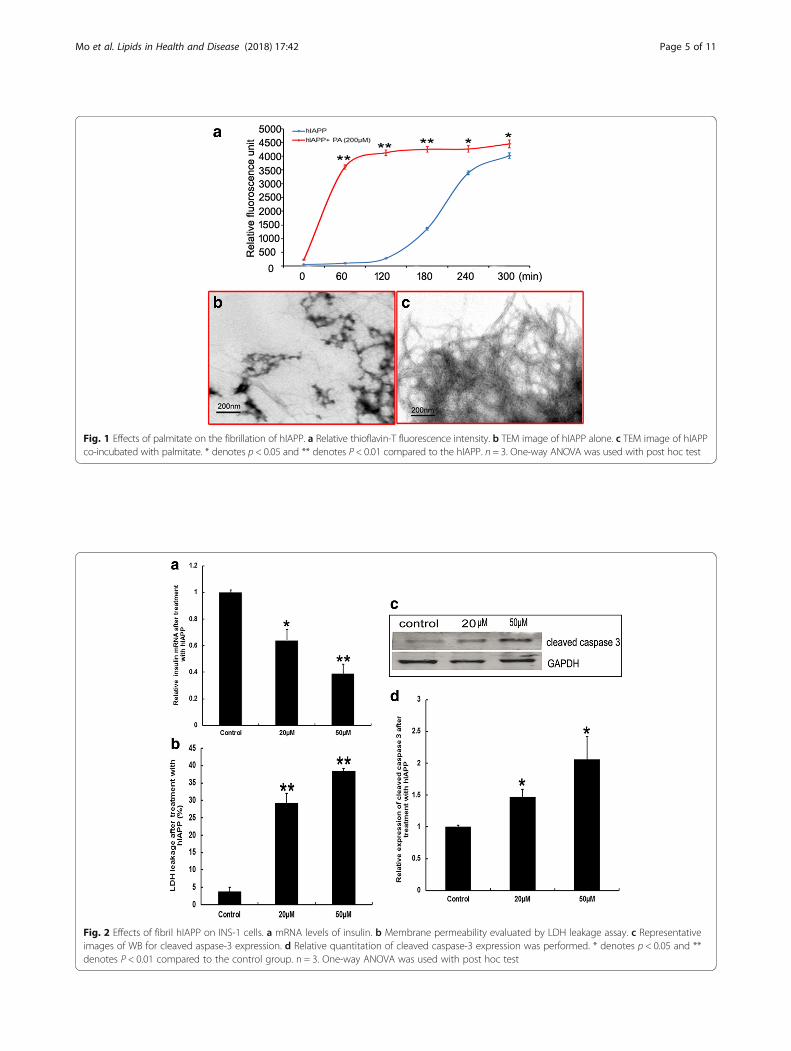
Fig. 1 Effects of palmitate on the fibrillation of hIAPP. a Relative thioflavin-T fluorescence intensity. b TEM image of hIAPP alone. c TEM image of hIAPPco-incubated with palmitate. * denotes p < 0.05 and ** denotes P < 0.01 compared to the hIAPP. n = 3. One-way ANOVA was used with post hoc test
Fig. 2 Effects of fibril hIAPP on INS-1 cells. a mRNA levels of insulin. b Membrane permeability evaluated by LDH leakage assay. c Representativeimages of WB for cleaved aspase-3 expression. d Relative quantitation of cleaved caspase-3 expression was performed. * denotes p < 0.05 and **denotes P < 0.01 compared to the control group. n = 3. One-way ANOVA was used with post hoc test
Mo et al. Lipids in Health and Disease (2018) 17:42 Page 5 of 11

HD and HD+ STZ groups compared with the controlgroup. No significant difference was found in the STZgroup compared with the control group. To evaluate thecell apoptosis, we detected the number of TUNEL positivecells in islet. As shown in Fig. 5e and f, numbers of TUNELpositive cell increased in the HD, STZ, and HD+ STZgroups compared with the control group.
Effects of HD on isolated pancreatic isletTo quantify the rIAPP expression in pancreatic islet, theislet was isolated from pancreas. The rIAPP mRNA in-creased in HD and STZ +HD groups compared with thecontrol, but no difference in STZ group (Fig. 6a). Simi-larly, the insulin mRNA in HD and STZ +HD groups,but not STZ group increased compared with the control(Fig. 6b). Total protein was extracted from isolated pan-creatic islet, and the levels of IR-β and caspase-3 weremeasured by western blot. The results showed IR-β ex-pression decreased in HD and STZ +HD groups
compared with the control (Fig. 6c and d). Additionally,the caspase-3 increased in STZ, HD and STZ +HDgroups compared with the control (Fig. 6e and f).
DiscussionAmyloid deposition plays a critical role in many differenthuman diseases, including Huntington’s disease, Parkin-son’s disease (PD), Alzheimer’s disease (AD) and T2DM.Among these diseases, T2DM together with AD are lead-ing causes of morbidity and mortality in the elderly. Bothdiseases share common clinical and biochemical features[27], including functional tissue loss due to accumulationand aggregation of small peptides, such as IAPP in thepancreas of T2DM patients, or beta amyloid in AD pa-tients. Specifically, IAPP fibrils arise following initial in-creased production of IAPP which leads to oligomericaggregation of IAPP molecules that then assemble intoamyloid fibrils in pancreatic islets, eventually resulting inpancreatic beta cell loss [28–30]. More secretion of IAPP
Fig. 3 Body weight, fast glucose tolerance and insulin sensitivity after treating with high-fat diet, STZ injection, and STZ injection combined withhigh-fat diet. a Flow chart of animal experiment. b The extent of body weight gain duration of two months. * denotes p < 0.05 in the HD groupvs. control group; ## denotes p < 0.01 in the STZ group vs. control group. n = 12. c Glucose tolerance is evaluated after 12 h fasting. * denotesp < 0.05 in the HD group vs. control group, n = 12. d Insulin sensitivity was evaluated after 12 h fasting, * denotes p < 0.05 in the HD group vs.control group. n = 12. Two-way ANOVA was used with post hoc test
Mo et al. Lipids in Health and Disease (2018) 17:42 Page 6 of 11

is the precondition, but not the determinant factor inamyloid deposition. Increasing literatures have reportedIAPP fibrils play the dramatic roles in the development ofamyloid deposition. Therefore, facilitation of fibril forma-tion accelerates the amyloid deposition. In our presentstudy, we found incubation of hIAPP with palmitate in-creased the fibril form which caused the increase of mem-brane permeability and apoptosis in INS-1 cells. In type 2diabetic rats induced by feeding with high fat diet, we fundrIAPP expression increased accompanied pancreatic isletcell apoptosis, but no typical amyloid deposition. In iso-lated pancreatic islet tissue, we fund rIAPP mRNA and in-sulin mRNA increased, but insulin receptor decreased indiabetic rats, which may implicate the abnormal insulinsignals. Given the insulin signals play pivotal roles in cellsurvival, pancreatic cell apoptosis partially contributed tothe reduction of insulin signaling.IAPP is co-localized with insulin in the islet beta-cells
and is co-secreted with insulin in response to beta-cellstimulation by both glucose and non-glucose secretagoguesagents, such as arginine [31]. Therefore, therapies that alterendogenous insulin secretion are likely to cause parallelchanges in IAPP secretion. Both insulin and IAPP gene ex-pression and release by pancreatic islets are regulated byglucose [13, 32–34]. In vitro, the study showed that palmi-tate and oleate cause transcriptional induction of amylingene in beta-cells and murine islets. This induction is medi-ated by the Ca2+-PKC signaling pathway and de novo syn-thesized proteins. However, the effect of lipid on theexpression and release of IAPP by pancreatic islet in vivo is
not clear. Our present studies indicate high fat diet com-bined with the low dose STZ injection induced the featuresof T2DM characterized by decrease of insulin sensitivityand increase of plasma insulin, glucose and triglyceride, aswell plasma rIAPP augment and islet cell apoptosis. Toconfirm the changes of pancreatic islet under high fat dietconsumption, pancreatic islet was isolated and its functionwas analyzed. These results suggested the transcription ofinsulin and amylin were affected under high fat diet condi-tion. Together with all data in vivo, we think simultaneouschanges of IAPP expression, glucolipid metabolism and in-sulin sensitivity caused islet cell apoptosis which contrib-uted to the onset of T2DM.The most widely accepted hypothesis is that IAPP-
induced cytotoxicity occurs via a membrane disruptionmechanism. The experimental evidence suggested that thepeptide Aβ, involved in Alzheimer’s disease, could formcation-selective channels in planar lipid bilayers [13]. Simi-lar experiments showed that hIAPP could also form cation-selective channels and ultimately disrupt the membranes[35]. These channels have been also observed for otheramyloidogenic proteins suggesting that the toxicity of amyl-oid proteins seems to be linked to their shared potential toform pores in membrane [36, 37]. Many studies suggesthIAPP could induce membrane damage. But the exactmechanism of hIAPP-induced membrane disruption is farfrom clear. And numerous models have been describedduring recent years [38–41]. A report concluded that sol-uble oligomers from several types of amyloids, includinghIAPP, specifically increase lipid bilayer conductance, while
Fig. 4 Blood biochemical parameters of rats after treating with high-fat diet, STZ injection, and STZ injection combined with high-fat diet for2 months. a Plasma glucose. b Plasma triglyceride. c Plasma insulin. d Plasma rIAPP. * denotes p < 0.05 compared with the control group;** denotes p < 0.01 compared with the control group. n = 12. Two-way ANOVA was used with post hoc test
Mo et al. Lipids in Health and Disease (2018) 17:42 Page 7 of 11

the soluble low molecular weight species have no effect,suggesting that this may represent the common primarymechanism of pathogenesis in amyloid-related diseases,such as AD, PD and diabetes [42].The lipid condition accelerated the IAPP toxicity not
only based on its high expression but also on the associ-ated IAPP conformation. The dynamics and extent ofIAPP oligomerization and aggregation were shown to beimportant parameters of IAPP toxicity [40, 43, 44]. How-ever, the absence of the islet amyloid in several trans-genic mouse strains with very high amylin productioncontradicted this suggestion. Therefore, additional fac-tors need to be considered. The islet amyloid occurredafter a persistent intake of a high-fat diet in transgenicmouse models [42, 45]. Finding the effects of lipid onIAPP toxicity was a critical issue. A previous study sug-gested that free fatty acids (FFAs) can act as direct po-tent stimulators of amylin fibrillogenesis [46]. Our study
showed that insulin resistance and rIAPP production oc-curred in rats feeding with high-fat diet. These led to achronic overproduction of rIAPP. Another study indi-cated that FFAs not only enhanced fibril formation fromsynthetic hIAPP in vitro, but also rapidly led to the ap-pearance of an abnormal intragranular material in cellsof cultivated hIAPP transgenic mouse islets [47–49].Cell damage with IAPP was caused by the increase of
membrane permeability. Accordingly, evidence showedthat various sizes of the hIAPP-induced membrane poresor openings, ranging from Ca2+-permeable to permeablefor fluorescent dyes with a size larger than 1 kDa [50–52]. Soluble hIAPP and amyloid oligomers generallycould have characteristics of pore-forming protein toxinslike α-hemolysin, and might have a similar mechanismof action [53–55]. Our results also confirmed that cellmembrane permeability increased after incubation withhIAPP in a dose-dependent manner.
Fig. 5 Morphology of pancreatic islet after treatment with STZ injection, high-fat diet, and STZ injection combined with high-fat diet in rats. aRepresentative images of H-E staining to show the islet by dashed lines. b Representative images of insulin immunoflurorescence staining toshow the islet by dashed lines. c Representative images of amylin expression in the islet (outlined by dashed lines). d Levels of amylin expressioncalculated with ImageJ software in sixteen islets from four rats each group. e Representative images of TUNEL staining in the islet (outlined bydashed lines). f Numbers of TUNEL positive cells in the islet were calculated and normalized by unit area of pancreatic islet. * denotes p < 0.05compared with the control group. n = 4. Two-way ANOVA was used with post hoc test
Mo et al. Lipids in Health and Disease (2018) 17:42 Page 8 of 11

ConclusionsFibril hIAPP directly damaged the pancreatic cells char-acterized by increase of permeability of cell membrane,inhibition of insulin secretion, and triggering cell apop-tosis. Furthermore, high fat diet induced endogenousrIAPP secretion accompanied the insulin secretion, butin isolated pancreatic islet, high fat diet reduced the in-sulin receptor expression, suggesting the insulin signal-ing is inhibited. Insulin resistance and IAPP expressioninduced by fatty acids aggravated the pancreatic islet cellapoptosis in vivo. Together with evidence from in vivoand in vitro, suggested fatty acid and amylin plays thesynergistic effect on pancreatic islet cell damage whichinvolved in amylin high expression and fibril formationof IAPP, cell membrane leakage and cell apoptosis. Also,the limitation and deficiency in our experiment is non-blinded methods, which may produce the bias.
Abbreviationsaa: amino acid; DAB: Diaminobenzidine; FAs: Fatty acids; FBS: Fetal bovineserum; GAPDH: Glyceraldehyde 3-phosphate dehydrogenase; HD: High-fatdiet; HFIP: 1,1,1,3,3,3-Hexafluoro-2-propanol; hIAPP: human islet amyloidpolypeptide; LDH: The lactate dehydrogenase; MTT: thiazolyl blue tetrazolium
bromide; PA: Palmitate; Q-RT-PCR: Quantitative real-time PCR; rIAPP: rodentislet amyloid polypeptide; STZ: Streptozotocin; T2DM: Type 2 diabetesmellitus; TEM: Transmission Electronic Microscopy; ThT: thioflavin-T
AcknowledgmentsWe would like to acknowledge Miss Bei Wang for help with ElectronicMicroscopy technology and Miss Xiao-Xia Xi and Miss Jing Sun for animalcare and sample collection.
FundingThis work is partly supported by National Natural Science Foundation ofChina (No. 81370448 and 81570725) to Jing Yu Hong.
Availability of data and materialsThe datasets used and/or analyzed during the current study are availablefrom the corresponding author on reasonable request.
Authors’ contributionsXDM and YHJ planned experiments, interpreted data, and approved theversion to be published. XDM and YHJ performed most of the experimentsand analyzed data and YHJ wrote the paper. QJW and JY participated in theanimal experiment. LPG participated in acquisition of the study specimens.All authors read and approved the final paper.
Ethics approvalThis animal study was approved by the institutional Animal EthicsCommittee, Lanzhou University (permit number: SCXK Gan 2009–0004).
Fig. 6 Isolated pancreatic islet function was evaluated after treatment with STZ injection, high-fat diet, and STZ injection combined with high-fatdiet in rats. a mRNA levels of amylin. b mRNA levels of insulin. c Representative images of WB for IRβ expression. Relative quantitation of IRβ ex-pression was performed and shown in (d). e Representative images of WB for cleaved caspase-3 expression. f Relative quantitation of cleavedcaspase-3 expression was performed. * denotes p < 0.05 compared with the control group; ** denotes p < 0.01 compared with the control group.n = 4. Two-way ANOVA was used with post hoc test
Mo et al. Lipids in Health and Disease (2018) 17:42 Page 9 of 11

Consent for publicationNot applicable
Competing interestsThe authors declare that they have no competing interests.
Publisher’s NoteSpringer Nature remains neutral with regard to jurisdictional claims inpublished maps and institutional affiliations.
Author details1Institute of Anatomy and Histology & Embryology, Neuroscience, School ofBasic Medical Sciences, Lanzhou University, No. 199 of Donggang West Road,Lanzhou City, Gansu Province 730000, People’s Republic of China. 2KeyLaboratory of Preclinical Study for New Drugs of Gansu province, LanzhouUniversity, No. 199 of Donggang West Road, Lanzhou City, Gansu Province730000, People’s Republic of China. 3Institute of Biochemistry and MolecularBiology, School of Basic Medical Sciences, Lanzhou University, No. 199 ofDonggang West Road, Lanzhou City, Gansu Province 730000, People’sRepublic of China.
Received: 3 January 2018 Accepted: 2 March 2018
References1. Cooper GJ, Willis AC, Clark A, Turner RC, Sim RB, Reid KB. Purification and
characterization of a peptide from amyloid-rich pancreases of type 2diabetic patients. Proc Natl Acad Sci U S A. 1987;84:8628–32.
2. Westermark P, Wernstedt C, O'Brien TD, Hayden DW, Johnson KH. Isletamyloid in type 2 human diabetes mellitus and adult diabetic cats containsa novel putative polypeptide hormone. Am J Pathol. 1987;127:414–7.
3. Mukherjee A, Soto C. Prion-like protein aggregates and type 2 diabetes.Cold Spring Harb Perspect Med. 2017;7(5):a024315.
4. Hoppener JW, Jacobs HM, Wierup N, Sotthewes G, Sprong M, de Vos P,Berger R, Sundler F, Ahren B. Human islet amyloid polypeptide transgenicmice: in vivo and ex vivo models for the role of hIAPP in type 2 diabetesmellitus. Exp Diabetes Res. 2008;2008:697035.
5. Matveyenko AV, Butler PC. Islet amyloid polypeptide (IAPP) transgenicrodents as models for type 2 diabetes. ILAR J. 2006;47:225–33.
6. Schneider HM, Storkel S, Will W. Amyloid of islets of Langerhans and its relationto diabetes mellitus (author's transl). Dtsch Med Wochenschr. 1980;105:1143–7.
7. Rocken C, Linke RP, Saeger W. Immunohistology of islet amyloidpolypeptide in diabetes mellitus: semi-quantitative studies in a post-mortemseries. Virchows Arch A Pathol Anat Histopathol. 1992;421:339–44.
8. Clark A, Wells CA, Buley ID, Cruickshank JK, Vanhegan RI, Matthews DR,Cooper GJ, Holman RR, Turner RC. Islet amyloid, increased A-cells, reducedB-cells and exocrine fibrosis: quantitative changes in the pancreas in type 2diabetes. Diabetes Res. 1988;9:151–9.
9. Bell ET. Hyalinization of the islets of Langerhans in nondiabetic individuals.Am J Pathol. 1959;35:801–5.
10. Ehrlich JC, Ratner IM. Amyloidosis of the islets of Langerhans. A restudy of islethyalin in diabetic and non-diabetic individuals. Am J Pathol. 1961;38:49–59.
11. Westermark P. Quantitative studies on amyloid in the islets of Langerhans.Ups J Med Sci. 1972;77:91–4.
12. Hull RL, Andrikopoulos S, Verchere CB, Vidal J, Wang F, Cnop M, Prigeon RL,Kahn SE. Increased dietary fat promotes islet amyloid formation and beta-cell secretory dysfunction in a transgenic mouse model of islet amyloid.Diabetes. 2003;52:372–9.
13. Meier DT, Morcos M, Samarasekera T, Zraika S, Hull RL, Kahn SE. Isletamyloid formation is an important determinant for inducing isletinflammation in high-fat-fed human IAPP transgenic mice. Diabetologia.2014;57:1884–8.
14. Ma Z, Westermark GT. Effects of free fatty acid on polymerization of isletamyloid polypeptide (IAPP) in vitro and on amyloid fibril formation incultivated isolated islets of transgenic mice overexpressing human IAPP.Mol Med. 2002;8:863–8.
15. Jeong HR, An SS. Causative factors for formation of toxic islet amyloid polypeptideoligomer in type 2 diabetes mellitus. Clin Interv Aging. 2015;10:1873–9.
16. Xin A, Mizukami H, Inaba W, Yoshida T, Takeuchi YK, Yagihashi S. Pancreasatrophy and islet amyloid deposition in patients with elderly-onset type 2diabetes. J Clin Endocrinol Metab. 2017;102:3162–71.
17. Han S, Kollmer M, Markx D, Claus S, Walther P, Fandrich M. Amyloid plaquestructure and cell surface interactions of beta-amyloid fibrils revealed byelectron tomography. Sci Rep. 2017;7:43577.
18. Oskarsson ME, Singh K, Wang J, Vlodavsky I, Li JP, Westermark GT. Heparansulfate proteoglycans are important for islet amyloid formation and isletamyloid polypeptide-induced apoptosis. J Biol Chem. 2015;290:15121–32.
19. Guo SM, Bag N, Mishra A, Wohland T, Bathe M. Bayesian total internalreflection fluorescence correlation spectroscopy reveals hIAPP-induced plasmamembrane domain organization in live cells. Biophys J. 2014;106:190–200.
20. Radovan D, Opitz N, Winter R. Fluorescence microscopy studies on isletamyloid polypeptide fibrillation at heterogeneous and cellular membraneinterfaces and its inhibition by resveratrol. FEBS Lett. 2009;583:1439–45.
21. Misumi Y, Ueda M, Obayashi K, Jono H, Yamashita T, Ando Y. Interactionbetween amyloid fibril formation and extracellular matrix in theproceedings of VIIIth international symposium on familial Amyloidoticpolyneuropathy. Amyloid. 2012;19(Suppl 1):8–10.
22. Reed MJ, Meszaros K, Entes LJ, Claypool MD, Pinkett JG, Gadbois TM, ReavenGM. A new rat model of type 2 diabetes: the fat-fed, streptozotocin-treatedrat. Metab Clin Exp. 2000;49:1390–4.
23. Srinivasan K, Viswanad B, Asrat L, Kaul CL, Ramarao P. Combination of high-fat diet-fed and low-dose streptozotocin-treated rat: a model for type 2diabetes and pharmacological screening. Pharmacol Res. 2005;52:313–20.
24. Sahin K, Onderci M, Tuzcu M, Ustundag B, Cikim G, Ozercan IH, Sriramoju V,Juturu V, Komorowski JR. Effect of chromium on carbohydrate and lipidmetabolism in a rat model of type 2 diabetes mellitus: the fat-fed,streptozotocin-treated rat. Metab Clin Exp. 2007;56:1233–40.
25. Sutton R, Peters M, McShane P, Gray DW, Morris PJ. Isolation of rat pancreaticislets by ductal injection of collagenase. Transplantation. 1986;42:689–91.
26. Xu S, Kim JH, Hwang KH, Das R, Quan X, Nguyen TT, Kim SJ, Cha SK, Park KS.Autocrine insulin increases plasma membrane K(ATP) channel via PI3K-VAMP2pathway in MIN6 cells. Biochem Biophys Res Commun. 2015;468:752–7.
27. Gotz J, Ittner LM, Lim YA. Common features between diabetes mellitus andAlzheimer's disease. Cell Mol Life Sci. 2009;66:1321–5.
28. Haataja L, Gurlo T, Huang CJ, Butler PC. Islet amyloid in type 2 diabetes, andthe toxic oligomer hypothesis. Endocr Rev. 2008;29:303–16.
29. Konarkowska B, Aitken JF, Kistler J, Zhang S, Cooper GJ. The aggregationpotential of human amylin determines its cytotoxicity towards islet beta-cells. FEBS J. 2006;273:3614–24.
30. Goldsbury C, Goldie K, Pellaud J, Seelig J, Frey P, Muller SA, Kistler J, CooperGJ, Aebi U. Amyloid fibril formation from full-length and fragments ofamylin. J Struct Biol. 2000;130:352–62.
31. Li X, Ma L, Zheng W, Chen T. Inhibition of islet amyloid polypeptide fibrilformation by selenium-containing phycocyanin and prevention of beta cellapoptosis. Biomaterials. 2014;35:8596–604.
32. Verchere CB, D'Alessio DA, Prigeon RL, Hull RL, Kahn SE. The constitutivesecretory pathway is a major route for islet amyloid polypeptide secretionin neonatal but not adult rat islet cells. Diabetes. 2000;49:1477–84.
33. Sjolander J, Byman E, Kulak K, Nilsson SC, Zhang E, Krus U, Westermark GT, StormP, King BC, Renstrom E, Blom AM. C4b-binding protein protects beta-cells fromislet amyloid polypeptide-induced cytotoxicity. J Biol Chem. 2016;291:21644–55.
34. Zhang S, Liu H, Chuang CL, Li X, Au M, Zhang L, Phillips AR, Scott DW,Cooper GJ. The pathogenic mechanism of diabetes varies with the degreeof overexpression and oligomerization of human amylin in the pancreaticislet beta cells. FASEB J. 2014;28:5083–96.
35. Alarcon C, Verchere CB, Rhodes CJ. Translational control of glucose-inducedislet amyloid polypeptide production in pancreatic islets. Endocrinology.2012;153:2082–7.
36. Delgado DA, Doherty K, Cheng Q, Kim H, Xu D, Dong H, Grewer C, QiangW. Distinct membrane disruption pathways are induced by 40-residue beta-amyloid peptides. J Biol Chem. 2016;291:12233–44.
37. Sciacca MF, Milardi D, Messina GM, Marletta G, Brender JR, Ramamoorthy A,La Rosa C. Cations as switches of amyloid-mediated membrane disruptionmechanisms: calcium and IAPP. Biophys J. 2013;104:173–84.
38. Ho CS, Khadka NK, She F, Cai J, Pan J. Polyglutamine aggregates impair lipidmembrane integrity and enhance lipid membrane rigidity. Biochim BiophysActa. 2016;1858:661–70.
39. Cao P, Raleigh DP. In vitro studies of membrane permeability induced byAmyloidogenic polypeptides using large Unilamellar vesicles. Methods MolBiol. 2016;1345:283–90.
40. Zhang X, St Clair JR, London E, Raleigh DP. Islet amyloid polypeptide membraneinteractions: effects of membrane composition. Biochemistry. 2017;56:376–90.
Mo et al. Lipids in Health and Disease (2018) 17:42 Page 10 of 11

41. Sciacca MF, Lolicato F, Di Mauro G, Milardi D, D'Urso L, Satriano C,Ramamoorthy A, La Rosa C. The role of cholesterol in driving IAPP-membrane interactions. Biophys J. 2016;111:140–51.
42. Cao P, Abedini A, Wang H, Tu LH, Zhang X, Schmidt AM, Raleigh DP. Isletamyloid polypeptide toxicity and membrane interactions. Proc Natl AcadSci U S A. 2013;110:19279–84.
43. Gurlo T, Ryazantsev S, Huang CJ, Yeh MW, Reber HA, Hines OJ, O'Brien TD,Glabe CG, Butler PC. Evidence for proteotoxicity in beta cells in type 2diabetes: toxic islet amyloid polypeptide oligomers form intracellularly inthe secretory pathway. Am J Pathol. 2010;176:861–9.
44. Miller-Thomas MM, Sipe AL, Benzinger TL, McConathy J, Connolly S,Schwetye KE. Multimodality review of amyloid-related diseases of thecentral nervous system. Radiographics. 2016;36:1147–63.
45. Engel MF, Khemtemourian L, Kleijer CC, Meeldijk HJ, Jacobs J, Verkleij AJ, deKruijff B, Killian JA, Hoppener JW. Membrane damage by human isletamyloid polypeptide through fibril growth at the membrane. Proc NatlAcad Sci U S A. 2008;105:6033–8.
46. Ritzel RA, Meier JJ, Lin CY, Veldhuis JD, Butler PC. Human islet amyloidpolypeptide oligomers disrupt cell coupling, induce apoptosis, and impairinsulin secretion in isolated human islets. Diabetes. 2007;56:65–71.
47. Westermark GT, Gebre-Medhin S, Steiner DF, Westermark P. Islet amyloiddevelopment in a mouse strain lacking endogenous islet amyloidpolypeptide (IAPP) but expressing human IAPP. Mol Med. 2000;6:998–1007.
48. Verchere CB, D'Alessio DA, Palmiter RD, Weir GC, Bonner-Weir S, Baskin DG,Kahn SE. Islet amyloid formation associated with hyperglycemia intransgenic mice with pancreatic beta cell expression of human islet amyloidpolypeptide. Proc Natl Acad Sci U S A. 1996;93:3492–6.
49. Rochet JC, Lansbury PT Jr. Amyloid fibrillogenesis: themes and variations.Curr Opin Struct Biol. 2000;10:60–8.
50. Groop LC, Saloranta C, Shank M, Bonadonna RC, Ferrannini E, DeFronzo RA.The role of free fatty acid metabolism in the pathogenesis of insulinresistance in obesity and noninsulin-dependent diabetes mellitus. J ClinEndocrinol Metab. 1991;72:96–107.
51. Ohsawa H, Kanatsuka A, Yamaguchi T, Makino H, Yoshida S. Islet amyloidpolypeptide inhibits glucose-stimulated insulin secretion from isolated ratpancreatic islets. Biochem Biophys Res Commun. 1989;160:961–7.
52. Poitout V, Hagman D, Stein R, Artner I, Robertson RP, Harmon JS. Regulationof the insulin gene by glucose and fatty acids. J Nutr. 2006;136:873–6.
53. Anguiano M, Nowak RJ, Lansbury PT Jr. Protofibrillar islet amyloidpolypeptide permeabilizes synthetic vesicles by a pore-like mechanism thatmay be relevant to type II diabetes. Biochemistry. 2002;41:11338–43.
54. Demuro A, Mina E, Kayed R, Milton SC, Parker I, Glabe CG. Calciumdysregulation and membrane disruption as a ubiquitous neurotoxicmechanism of soluble amyloid oligomers. J Biol Chem. 2005;280:17294–300.
55. Kagan BL. Amyloidosis and protein folding. Science (New York, NY). 2005;307:42–3. author reply 42-43
• We accept pre-submission inquiries
• Our selector tool helps you to find the most relevant journal
• We provide round the clock customer support
• Convenient online submission
• Thorough peer review
• Inclusion in PubMed and all major indexing services
• Maximum visibility for your research
Submit your manuscript atwww.biomedcentral.com/submit
Submit your next manuscript to BioMed Central and we will help you at every step:
Mo et al. Lipids in Health and Disease (2018) 17:42 Page 11 of 11


