Embed Size (px)
Citation preview

REVIEW ARTICLES
Studies on osteoporosis in Saudi Arabia 152Lina Fahmi Hammad
ORIGINAL ARTICLES
Comparison of the prognostic value of neutrophil to lymphocyte ratios at different time points in patients with ST segment elevation myocardial infarction by five-year follow-up 163Gurbet Ozge Mert, Muhammet Dural, Kadir Ugur Mert, Muzaffer Bilgin, Bulent GorenekEvaluation on predictive values of risk scores in non-variceal upper gastrointestinal bleeding 172Nam-Hun Jong, Hye-Song Kim, Hak-Chol Ri, Song-Il Rim, Jong-Nam Kang Electromechanical delay and 4-chamber longitudinal strain in patients with obstructive sleep apnea 180Ferdi Kahraman, Sema Avci, Gokhan PerincekSingle tertiary center experience from Turkey regarding the experience for cardiac implantable electrical devices 188Emre Ozdemir, Mert Pehlivan Altin, Sadik Volkan Emren, Cem Nazli, Mehmet Tokac Evaluation of facilities for diagnostic imaging at the primary health care centres in Al-Madinah, Saudi Arabia 196Moustafa E Radwan, Tareef Sahal Daqqaq, Mujahed A Turjoman, Moayad A Karbouji, Abdullah A Al Ahmadi, Rizq A BadawiThe management of mucinous appendiceal lesions: Case series and review of the literature 202Hasan Dagmura, Emin Daldal, Fatih Dasiran, Ahmet Akbas, Ismail OkanThe effects of ‘Adequacy of Anesthesia’ monitorization in general anesthesia on hemodynamics, recovery, and the cost of anesthetic drugs 208Yilmaz Resul, Topal Ahmet, Arican Sule, Hacibeyoglu Gulcin, Turk Seyda End-tidal carbon dioxide levels under surgical drapes during local eye surgery: Retrospective study 215Ilknur Suidiye Yorulmaz, Ali Umit Esbah, Onur Ozlu, Kuddusi Teberik, Muhammet Uzeyir Sozer, Murat KayaEfficacy of lamotrigine for seizures, dysexecutive symptoms, anger and sadness rumination in patients with epilepsy 221Amara Gul, Saima MehreenArthroscopic assisted percutaneous figure of eight tension band wiring of patellar fractures 227Cumhur Deniz Davulcu, Mehmet Can Unlu, Yusuf Pirincci, Taha Demir, Aybars Kivrak, Mahmut Kursat OzsahinComputed tomography findings of organizing pneumonia 233Yeliz Dadali, Sercan Ozkacmaz, Yurdanur Erdogan, Havva Akmaz Unlu, Funda Demirag, Ilke BursaliInfluence of neonatal care advancement on mortality and the incidence of bronchopulmonary dysplasia in very low birth weight (VLBW) Saudi infants: two period’s comparison 244Badr Hasan Sobaih
CASE REPORTS
Cesarean scar pregnancy: Review and case reports 249Mansour B Alorf, Hala F Elsharaky, Muntaha H Al-SalemSuccessful management of patient with fulminant myocarditis related refractory ventricular fibrillation by veno-arterial extracorporeal membrane oxygenation 254Asiye Yavuz, Behiye Deniz Kosovali, Mustafa Kemal Bayar
JUNE 2022VOLUME 54 NUMBER 2
The Official Journal of The Kuwait Medical Association
KU ISSN 0023-5776 Continued inside
KUWAIT MEDICAL JOURNAL
ة تيَّكوَي
ةال َّةالطبيَّ اجَلمعي
Open access for articles at
http: www.kmj.org.kw
Indexed and abstracted in:
SCOPUS
EMBASE
(The Excerpta Medica Database)
Science Citation Index Expanded
(also known as SciSearch®)
Journal Citation Reports/Science Edition
IMEMR Current Contents
(Index Medicus for the Eastern Mediterranean Region)
available online at: www.emro.who.int/EMRJorList/online.aspx

KUWAIT MEDICAL JOURNAL 3June 2022 JUNE 2022Vol. 54 No. 2
C O N T E N T S
KUWAIT MEDICAL JOURNAL
Continued from cover
THE PUBLICATION OF ADVERTISEMENTS IN THE KUWAIT MEDICAL JOURNAL DOES NOT CONSTITUTE ANY GUARANTEE OR ENDORSEMENT BY THE KUWAIT MEDICAL ASSOCIATION OR THE EDITORIAL BOARD OF THIS JOURNAL, OF THE ADVERTISED PRODUCTS, SERVICES, OR CLAIMS MADE BY THE ADVERTISERS. THE PUBLICATION OF ARTICLES AND OTHER EDITORIAL MATERIAL IN THE JOURNAL DOES NOT NECESSARILY REPRESENT POLICY RECOMMENDATIONS OR ENDORSEMENT BY THE ASSOCIATION.
PUBLISHER: The Kuwait Medical Journal (KU ISSN-0023-5776) is a quarterly publication of THE KUWAIT MEDICAL ASSOCIATION. Address: P.O. Box 1202, 13013 Safat, State of Kuwait; Telephone: 1881181 Fax: 25317972, 25333276. E-mail : [email protected]: The Kuwait Medical Journal. All rights reserved. No part of this publication may be reproduced without written permission from the publisher. Printed in Kuwait.INSTRUCTIONS FOR AUTHORS: Authors may submit manuscripts prepared in accordance with the Uniform Requirements for Manuscripts Submitted to Biomedical Journals. These Requirements are published in each issue of the Kuwait Medical Journal.CHANGE OF ADDRESS: Notice should be sent to the Publisher six weeks in advance of the effective date. Include old and new addresses with mail codes.KUWAIT MEDICAL JOURNAL (previously The Journal of the Kuwait Medical Association) is added to the list of journals adhering to the “Uniform Requirements for Manuscripts Submitted to Biomedical Journals”, American College of Physicians, Independence Mall West, Sixth Street at Race, Philadelphia, PA 19106-1572, USA, and can be located at http://www.icmje.org/jrnlist.html
First case of bloodstream infection caused by Clostridium saccharolyticumin in the Middle East 258Aarti Chadha, Wafaa Jamal, Vincent O RotimiAdenomatoid tumor of the adrenal gland synchronous with clear cell papillary renal cell carcinoma: Report of an unusual case 262Abdulkadir Yasir Bahar, Murat Sahin Breast hamartoma: A series of variable clinical presentation 269Amal Abdullah AbdulkareemThyroglossal cyst fistulized to the skin accompanying multinodular goiter in an adult: A case report 274Ozkan Gorgulu, Mehmet Nuri Kosar
SHORT COMMUNICATION
Understanding the context of COVID-19 pandemic; lessons learnt for a long-term sustainable healthcare system 277Zainab Al Lawati, Alaa Al LawatiHow to understand the “Stealth” Omicron subvariant and prepare for its challenge 283HyokJu Ri, GunHyok Kim, HyonSu Jo, CholSik Ri
SELECTED ABSTRACTS OF ARTICLES PUBLISHED ELSEWHERE BY AUTHORS IN KUWAIT 287
FORTHCOMING CONFERENCES AND MEETINGS 291
WHO-FACTS SHEET 298
1. Dracunculiasis (guinea-worm disease)2. Hypertension3. Monkeypox4. Oral health5. Rehabilitation

Kuwait Medical Journal Published by the Kuwait Medical Association
Previously known as The Journal of the Kuwait Medical Association (Est. 1967)
Honorary President: Abdulaziz Al-Babtain
EDITORIAL BOARDEditor-in-Chief:Fuad Abdulla M Hasan, Kuwait
Editor:Adel Khader Ayed, KuwaitInternational Editor:Pawan K Singal, Canada
Associate Editors:Adel A Alzayed, KuwaitIgnacio Rodriguez, USAMichael Redmond, USA
Mousa Khoursheed, KuwaitMustafa M Ridha, KuwaitNasser Behbehani, KuwaitNoura Al-Sweih, Kuwait
INTERNATIONAL ADVISORY BOARDAnanda S Prasad, USAAnders Lindstrand, Sweden Andrew J Rees, UKBelle M Hegde, IndiaBengt Jeppsson, SwedenCharles A Dinarello, USAChristian Imielinski, PolandElizabeth Dean, CanadaFiona J Gilbert, UKFrank D Johnston, UKGeorge Russell, UKGraeme RD Catto, UK
Peter RF Bell, UKPhilip M Moody, USARaymond M Kirk, UKSamuel Dagogo-Jack, USAS Muralidharan, IndiaStig Bengmark, SwedenTulsi D Chugh, IndiaWilliam A Tweed, CanadaWilliam B Greenough, USAZoheir Bshouty, Canada
REGIONAL ADVISORY BOARDAbdulla BehbehaniAbeer K Al-BahoAlexander E OmuAli Al-MukaimiAli Al-SayeghAsmahan Al-ShubailiChacko MathewEiman M Mokaddas
Faisal A Al-KandariHabib Abul Joseph C LongeneckerKefaya AM AbdulmalekKhalid Al-JarallahMazen Al EssaMohamed AA MoussaMousa Khadadah
Mustafa Al-Mousawi Nasser J HayatNawaf Al-MutairiNebojsa RajacicSoad Al-BaharSukhbir Singh UppalWaleed AlazmiWaleed A Aldhahi
EDITORIAL OFFICE
Editorial Manager : Vineetha Elizabeth Mammen
EDITORIAL ADDRESS
P.O. Box: 1202, 13013-Safat, KuwaitTelephone: (00-965) 1881181(Ext. 114, 115) - Fax: (00-965) 25317972, 25333276
E-mail: [email protected]: www.kmj.org.kw
Giuseppe Botta, ItalyJames W Roach, USAJan T Christenson, SwitzerlandJohn V Forester, UKJulian Little, CanadaKostadin L Karagiozov, JapanLewis D Ritchie, UKMechael M Meguid, USAMohammed Zayer, SwedenNeva E Haites, UKNirmal K Ganguli, IndiaOleg Eremin, UK
Formerly known as ‘The Journal of the Kuwait Medical Association’, the Kuwait Medical Journal (KMJ) was established in the year 1967. It is the official publication of the Kuwait Medical Association and is published quarterly and regularly every March, June, September and December.
KMJ aims to publish peer-reviewed manuscripts of international interest. Submissions on clinical, scientific or laboratory investigations of relevance to medicine and health science come within the scope of its publication. Original articles, case reports, brief communications, book reviews, insights and letters to the editor are all considered. Review articles are solicited. Basic medical science articles are published under the section ‘Experimental Medicine’.
The Kuwait Medical Journal follows the guidelines set down in “Recommendations for the Conduct, Reporting, Editing, and Publication of Scholarly Work in Medical Journals” developed by the International Committee of Medical Journal Editors (ICMJE). The official and most recent version of the recommendations are available at www.icmje.org.
Journal Policies
Ethics in PublishingWhere human investigations are part of the study, the
research must be conducted ethically in accordance with the Declaration of Helsinki, and the design of the work has to be approved by a local ethics committee and informed written consent must be obtained from all subjects. Documented review and approval from the Institutional Review Board or Ethics Committee must be submitted along with any studies involving people, medical records and human tissues. A relevant statement of approval should be added in the ‘Subjects and Methods’ section of the manuscript.
Authors should also consult guidelines for the reporting of specific study types (e.g., the CONSORT guidelines for the reporting of randomized trials); see http://equator-network.org.
CopyrightThe publisher reserves copyright on the Journal’s contents.
No part may be reproduced, translated or transmitted in any form by any means, electronic or mechanical, including scanning, photocopying, recording or any other information storage and retrieval system without prior permission from the publisher. The publisher shall not be held responsible for any inaccuracy of the information contained therein.
Conflict of InterestPotential conflicts of interest for all authors must be
identified in a ‘Conflict of interest’ statement at the end of the manuscript. An electronic cover letter from the corresponding author is acceptable. Authors of research articles should disclose any affiliation with any organization with a financial interest, direct or indirect, in the subject matter or materials discussed in the manuscript (e.g., consultancies, employment, expert testimony, honoraria, retainers, stock) that may affect the conduct or reporting of the work submitted. If uncertain as to what might be considered a potential conflict of interest, authors should err on the side of full disclosure. Because reviews and editorials are based on selection and interpretation of the literature, the Journal expects that authors of such articles will not have any financial interest in a company (or its competitor) that makes a product discussed in the article. Information about potential conflict of interest will be made available to reviewers and will be published with the manuscript at the discretion of
the editors. If there is no conflict of interest, please state: “The authors declare no conflicts of interest.”
Peer ReviewAll submitted manuscripts are reviewed by the editorial
staff to ensure adherence to the guidelines of the Journal. Manuscripts that are considered suitable for review are sent to a peer in the relevant field of study as part of a double- blinded peer review. The reviewer may recommend the manuscript be accepted as is, undergo revision, or be rejected. If a reviewer recommends revision of a manuscript, the revised version must be re-submitted to the Journal within 3 months from the date when the review report is sent to the corresponding author.
AuthorsTo be named as an author on a submission, the following 4
criteria are followed:1. Substantial contributions to the conception or design of the
work; or the acquisition, analysis, or interpretation of data for the work; AND
2. Drafting the work or revising it critically for important intellectual content; AND
3. Final approval of the version to be published; AND 4. Agreement to be accountable for all aspects of the work in
ensuring that questions related to the accuracy or integrity of any part of the work are appropriately investigated and resolved.In addition to being accountable for the parts of the work
he/she has done, an author should be able to identify which co-authors are responsible for specific other parts of the work. Authors should also have confidence in the integrity of the contributions of their co-authors. Specific contributions made by each author to the article must be clearly stated at the end of the document. Those who do not meet all four criteria should be mentioned in the Acknowledgment section of the submission.
Once a paper has been accepted, the Journal does not consider requests to add, delete or rearrange the sequence of the authors. If the corresponding author requests to add, remove or rearrange the authors’ names after manuscript submission, the journal will seek justification for the requested change. Written confirmation signed by all authors, attesting that they agree to the addition, removal, or rearrangement of names is required. In the case of the addition or removal of authors, the author being added or removed must confirm assent. Requests that are not sent by the corresponding author will not be considered.
The corresponding author is responsible for communication with the journal during the manuscript submission, peer review, and publication process, and must ensure that all the journal’s administrative requirements are properly completed. He/she should also be available throught out the submission and peer review process to respond to editorial queries in a timely manner. It is also the corresponding authors responsibility to ensure all the co-authors are made aware of the most recent status of their submission.
FeesPublication in the Kuwait Medical Journal is free of charge.
PlagiarismThe Journal defines plagiarism as the use of others’
published and unpublished ideas or words without prior consent, and presenting them as new and original, whether intentional or not. If an accepted or published paper is found to
Guidelines for Authors
Guidelines for Authors
be plagiarised, the manuscript will be retracted and the author will be blacklisted from submitting to the journal.
Preparing your manuscript
Article typesOriginal Articles: Original Articles include laboratory
and clinical investigations as well as research not previously published or being considered for publication elsewhere. The text should contain a Title page, Abstract (in structured format) of not more than 250 words, Key Words (no more than five), Introduction, Subjects (or Materials) and Methods, Results, Discussion, Conclusion, Acknowledgment/s (if any) and References, Figure Legends, Tables, and Figures in this order. Details of the section contents are explained below for further adherence.
Review Articles (solicited only): Review articles should contain separate sections such as Title Page, Abstract (preferably in structured format) of no more than 250 words, Key Words (no more than five), Introduction, Methods/History (if applicable), Literature Review, Conclusion, Acknowledgment/s (if any) and References followed by (if relevant), Legends to figures, Tables, and Figures.
Case Reports: These should contain separate sections such as Title page, Abstract (a short summary of not more than 200 words), Key Words (no more than five), Introduction, Case history/report, Discussion, Conclusion, Acknowledgment/s (if any) and References followed by (if relevant), Legends to figures, Tables, and Figures.
Short Communications: Short communications are concise articles that aim to report new ideas, significant improvements to existing methods, a new practical application, or a new tool or resource. Short communications do not cover in detail background information about the problems treated, rather they provide key pointers to the reader. The work reported needs to be technically sound, innovative and significantly unique, advancing the state of the art. Short communication is not intended to publish preliminary results. Short communications should be similar to a research article, but with briefer Materials and Methods and Discussion.
Letters to the Editor: Letters may comment on recently published KMJ articles, novel cases or topics of current interest to the public. They should be concise and to the point, with a maximum of 1000 words and 2 authors. Letters commenting on previously published articles must be received within 6 months of publication of the relevant article.
Title PageThe title page of the submitted manuscript should provide
a clear title of the study followed by full names of all authors, the highest academic degree and affiliations if any, the name and address of the institution(s) where the work was done including the department, the name and complete address of the corresponding author to whom proofs and correspondences shall be sent, duly supported with contacts such as telephone, mobile and the e-mail address. This page must also contain any disclaimers, sources of support and a conflict of interest declaration.
Structured abstractA structured abstract (no more than 250 words) is required
for studies under the section “Original Articles”. It must provide an overview of the entire paper, and should contain succinct statements on the following, where appropriate: Objective(s), Design, Setting, Subjects, Intervention(s), Main
Outcome Measure(s), Result(s), and Conclusion(s). (See: Haynes RB, Mulrow CD, Huth AJ, Altman DG, Gardner MJ. More informative abstracts revisited. Annals of Internal Medicine 1990; 113:69-76). Abstracts for all other category of submissions shall be a short summary followed by Key words and the report or review.
Preparation of the manuscript The manuscript should be typed as ‘normal text’ with
no hyphenation and no hard-returns within paragraphs (use automatic wordwrap) on A4 size (29.7 x 21 cm) paper in single column format, preferably in font size 12. Cell format for paragraphs, artwork and/or special effects for the text and/or table(s) are not acceptable. Italics shall be used only for foreign/Latin expressions and/or special terminologies such as names of micro-organisms. Maintain a minimum of 2 cm margin on both sides of the text and a 3 cm margin at the top and bottom of each page. No part of the manuscript other than abbreviations and/or subtitles should be written in upper case. Header/foot notes, end notes, lines drawn to separate the paragraphs or pages etc. are not acceptable. Do not submit articles written/saved in ‘Track-change’ mode.
More than six authors are not appreciated for a research article and if listed, the authors may be asked to justify the contribution of each individual author. For case reports, not more than three authors are acceptable. Regarding contributions of authors over the limit mentioned above, please read the ‘Acknowledgment’ section.
Key wordsKey Words (maximum five) should be preferably MeSH
terms, and shall not duplicate words already in the manuscript title. MeSH terms can be checked at: http://www.nlm.nih.gov/mesh/.
TablesTables typed on separate pages using table format (MS Word
or Excel) should follow the list of references. Tables must be numbered consecutively using Arabic numerals and provided with appropriate titles. Contents of the table should be simple, and information therein not duplicated, but duly referred to, in the main text. Tables recording only a few values are not appreciated, since such information can be more accurately, usefully and concisely presented in a sentence or two in the manuscript.
Design of the workThis should be stated clearly. The rationale behind the choice
of sample size should be given. Those about to begin randomized controlled studies may wish to study the CONSORT statement (JAMA 1996; 276:637-639).
IllustrationsAll illustrations including figures should be numbered as
Fig 1, Fig 2, etc in running sequence and submitted as separate attachments along with the manuscript. Photographs should fit within a print area of 164 x 235 mm. In the case of figures where patient’s identity is not concealed, authors need to submit a written consent of the patient or of the patient’s guardian, in case of minors. Figure legends should be listed separately after the ‘References’ section. If any of the tables, illustrations or photomicrographs have been published elsewhere previously, a written consent for re-production is required from the copyright holder along with the manuscript. When charts are submitted, the numerical data on which they were based should be supplied.
KUWAIT MEDICAL JOURNAL
AbbreviationsExcept for units of measurement, abbreviations should be
defined on their first use in the abstract and in the text and then applied consistently throughout the article. Non-standard abbreviations or those appearing fewer than three times are not accepted. Use abbreviated units of measure, only when used with numbers. Abbreviations used as legends in Tables and/or figures should be duly defined below the respective item.
Numbers and unitsMeasurements of length, height, weight and volume
must be reported in metric units (meter, kilogram, liter etc.) or their decimal multiples. Temperature should be given in degrees Celsius, Blood pressure in mmHg, and hematological and biochemical measurements in Système International (SI) units. For decimal values, use a point, and not a comma, e.g., 5.7. Use a comma for numbers > 10,000 (i.e., 103) and do not use a comma for numbers < 9999, (e.g., 6542).
Drug names Non-proprietary (generic) names of product should be
employed. If a brand name for a drug is used, the British or international non-proprietary (approved) name should be given in parentheses. The source of any new or experimental preparation should also be given.
AcknowledgmentContributors who meet fewer than all 4 of the afore-
mentioned criteria for authorship should only be listed in this section. Contributions of others who have involved in the study, such as statisticians, radiologists etc. and/or those who have assisted in the preparation of the manuscript being submitted could also be included in this section. The corresponding author must obtain written permission to be acknowledged from all acknowledged individuals.
ReferencesIndicate references in the text in sequence using Arabic
numerals within square brackets and as superscripts (e.g.,[1, 3-5] etc). Do not quote additional data (like part of the title, year of publication etc.) from the references, with citations in the text, unless very important. In the References section, list them in the same sequence as they appeared in the text. Include the names and initials of all authors if not more than six (< 6). Write the last name of authors followed by the initials with no punctuation other than a comma to separate the names. In references where authorship exceeds six, use et al after six author names. Do not use automatic numbering, end notes or footnotes for references. References to manuscripts either in preparation or submitted for publication, personal communications, unpublished data, etc. are not acceptable.
References should be limited to those relating directly to the contents of the paper and should be set out in the style outlined by the International Committee of Medical Journal Editors (ICMJE), as shown in the examples below. Additional examples are in the ICMJE sample references. https://www.nlm.nih.gov/bsd/uniform_requirements.html
ExamplesArticle: Rose ME, Huerbin MB, Melick J, Marion DW,
Palmer AM, Schiding JK, et al. Regulation of interstitial excitatory amino acid concentrations after cortical contusion injury. Brain Res. 2002;935(1-2):40-6.Book: Murray PR, Rosenthal KS, Kobayashi GS, Pfaller MA.
Medical microbiology. 4th ed. St. Louis: Mosby; 2002.Book chapter: Meltzer PS, Kallioniemi A, Trent JM. Chromosome alterations in human solid tumors. In: Vogelstein B, Kinzler KW, editors. The genetic basis of human cancer. New York: McGraw-Hill; 2002. p. 93-113.
Weblinks: eatright.org [Internet]. Chicago: Academy of Nutrition and Dietetics; c2016 [cited 2016 Dec 27]. Available from: http://www.eatright.org/.
Manuscript submissionTo present your original work for consideration, one
complete set of the manuscript written in English, accompanied by tables and one set of figures (if applicable) should be submitted to the Editor by e-mail to “[email protected]” as attachment files.
The manuscript submitted by e-mail should be in MS Word document (.doc) format, together with a scanned copy or PDF version of the signed consent letter of the author(s) (see the section ‘Authorship and Consent Form’ for details). Figures or photographs, if any, need to be presented as separate attachments in JPG or BMP format with a resolution of 300 dpi and illustrations such as graphs, charts etc., as Excel format files. Incomplete/improper submissions will not be processed, and will be returned. Author(s) will receive a formal acknowledgment letter with a permanent reference number towards each successful submission.
Following a peer review process, the corresponding author will be advised of the status; acceptance or recommendation for revision or rejection of the paper, in a formal letter sent through e-mail. A galley proof will be forwarded to the corresponding author by e-mail at the time of publication of the accepted paper, which must be returned to the journal office within 48 hours with specific comments or corrections, if any. Such corrections in the galley proof must be limited to typographical errors or missing contents from the finally accepted version.
Authorship and consent form All authors must give their signed consent for publication in
a letter of submission, which should accompany the manuscript. This letter should contain the following statement:
“This manuscript (write the title) is an unpublished work which is not under consideration elsewhere and the results contained in this paper have not been published previously in whole or part, except in abstract form. In consideration of the KMJ accepting my/our submission for publication, the author(s) undersigned hereby assign all copyrights ownership to the KMJ and shall have no right to withdraw its publication. It is expressly certified that I/we, have done/actively participated in this study and agree to the accuracy of contents of this manuscript. It was conducted in accordance with current ethical considerations and meets with the committee’s approval. I/all of us agree to its publication in KMJ and to the authorship as expressed in this declaration and in the title page of our manuscript”.
The consent form must also contain the names of all authors, along with their signatures.
Manuscripts should be submitted to:The Editor,Kuwait Medical JournalP.O. Box: 1202Code-13013-SafatKuwait.Telephone: (965) 1881181, 25333920 extn. 114E-mail: [email protected] Website: www.kma.org.kw/KMJ
KUWAIT MEDICAL JOURNAL June 2022152
Review Article
Kuwait Medical Journal 2022; 54 (2): 152 - 162
Studies on osteoporosis in Saudi Arabia
ABSTRACT
KEY WORDS: bone mineral density, dual-energy X-ray absorptiometry, osteopenia, osteoporosis, Saudi population
Lina Fahmi HammadDepartment of Radiological Science, College of Applied Medical Sciences, King Saud University, Riyadh, Saudi Arabia
Background: Osteoporosis is a major health problem both worldwide and in Saudi Arabia. It is distinguished by decreased bone mass with modification in microarchitecture leading to bone fragility and enhanced risk of fracture. Osteoporosis risk factors include age, gender, genetic history and menopause. Furthermore, various diseases affect the prevalence of osteoporosis such as diabetes mellitus, hyperparathyroidism and vitamin D deficiency. Multiple studies have addressed different aspects of osteoporosis within Saudi Arabia.Objective: The purpose of this review is to highlight and summarize studies investigating the degree, the effects and factors affecting osteoporosis in Saudi Arabia and exposes their recommendations.
Discussion & conclusion: Several studies have exposed the prevalence of osteoporosis in the Saudi population. Other studies discussed risk factors, introduced methods of measurement, examined genetic influence and studied different types of treatment on an animal model. Within Saudi Arabia, osteoporosis detection and measurements started in 1993 and continues to date. It was found to be more common in females than in males. Low sun exposure, vitamin D deficiency and low physical activity are some factors, among others, that are suggested to contribute to the high prevalence of the disease in Saudi Arabia. Additional research is required, and prevention and management of osteoporosis studies are essential.
Address correspondence to:Dr. Lina F Hammad, MSc, Ph.D., College of Applied Medical Sciences, King Saud University, Riyadh, Saudi Arabia. Tel: +966 504788992; E-mail: [email protected]
INTRODUCTIONOsteoporosis is a silent disease, which goes
undetected and is considered a serious public health disease. Osteoporosis provokes painful fractures, causing diminished mobility and reduced quality of life; these fractures are frequently developed among elderly. There are some common sites where osteoporotic fractures take place such as hip, pelvis, forearm, spine and ankle[1]. Due to its prevalence worldwide, osteoporosis has attracted a lot of attention in the last two decades[2].
According to the International Osteoporosis Foundation, osteoporosis targeted about 75 million patients in USA, Europe and Japan causing more than 8.9 million fractures annually[3], and around 200 million women will be affected by osteoporosis[4]. In Saudi Arabia, studies in 2012 reported the incidence of osteoporosis as 30.7% and 34% in men and women
above 50 years[5]. It was calculated that the cost of managing one patient with osteoporotic fractures equaled to SR 48,712 (US$ 12,989.90)[6]. With 7528 fractures in a population of 1,300,336 people who are 55 years or older, it was estimated that the cost to the health system will be approximately SR 564.75 million ($ 150.60 million)[7].
METHODSLiterature search on PubMed and Google scholar
for published literature using keywords osteopenia, osteoporosis, dual-energy X-ray absorptiometry (DXA), bone mineral density (BMD) and quantitative sonography (QUS) in Saudi Arabia was conducted. Attention was directed to evaluate the prevalence of osteoporosis among the Saudi Arabian population in the last few decades. Studies evaluated included the studies discussing risk factors, methods of
KUWAIT MEDICAL JOURNAL 153June 2022
measurement, genetic effects and the effect of treatments in animal models[2,8-10]. Studies that investigated the effect of education were not included in this review. The review summarizes the previously mentioned studies and their recommendations. Studies have been classified and tabulated in to four sub-sections (Tables 1-4), which included osteoporosis detection and measurements, the effect of various diseases on bone mineral density, genetic influence on the prevalence of osteoporosis and finally, animal studies in Saudi Arabia.
LITERATURE REVIEWMethods of measurement of bone strength
BMD was defined by the World Health Organization as a typical tool in the estimation of bone strength in medical practice[11], and is measured at different skeletal sites using DXA. Bone material features are the key element for bone strength, with osteoporotic bones having abnormal bone matrix[12]. Although bone strength depends on bone mass, bone geometry and composition, larger bones are stronger than smaller ones even with the same BMD. This is due to the difference in bone diameter; as it grows radially, the strength of bone is enhanced by the radius of the participated bone elevated to the fourth power[12].
Although DXA is the gold standard for the measurement of osteoporosis, some vital factors couldn’t be observed by DXA which include intrinsic properties of bony tissue, cortical thickness and trabecular bone morphology. Other methods can be used for evaluating bone strength and stiffness[13], which include QUS; a technique suggested to provide information regarding bone structure. QUS offers stiffness index through measurements of broadband ultrasound attenuation and speed of sound values in the calcaneus region[14]. The advantages of this technique include being portable, simple, inexpensive and non-invasive with no ionizing radiation, but the significant low correlation between BMD of the lumbar spine and QUS of the heel, with QUS detecting significantly less osteoporosis than DXA scan[15] and the moderate positive correlation between QUS T-score and DXA T-score[15] limited the use of QUS as a replacement technique for BMD measurements despite the high reliability of QUS and precision[16].
Various other techniques used to evaluate the quantity of minerals present in bone include mineral and calcium percent determination, gravimetric determination of ash weight, conventional radiography, single photon and x-ray absorptiometry, quantitative computed tomography and quantitative magnetic resonance[17,18].
Osteoporosis detection and measurement studies in Saudi population
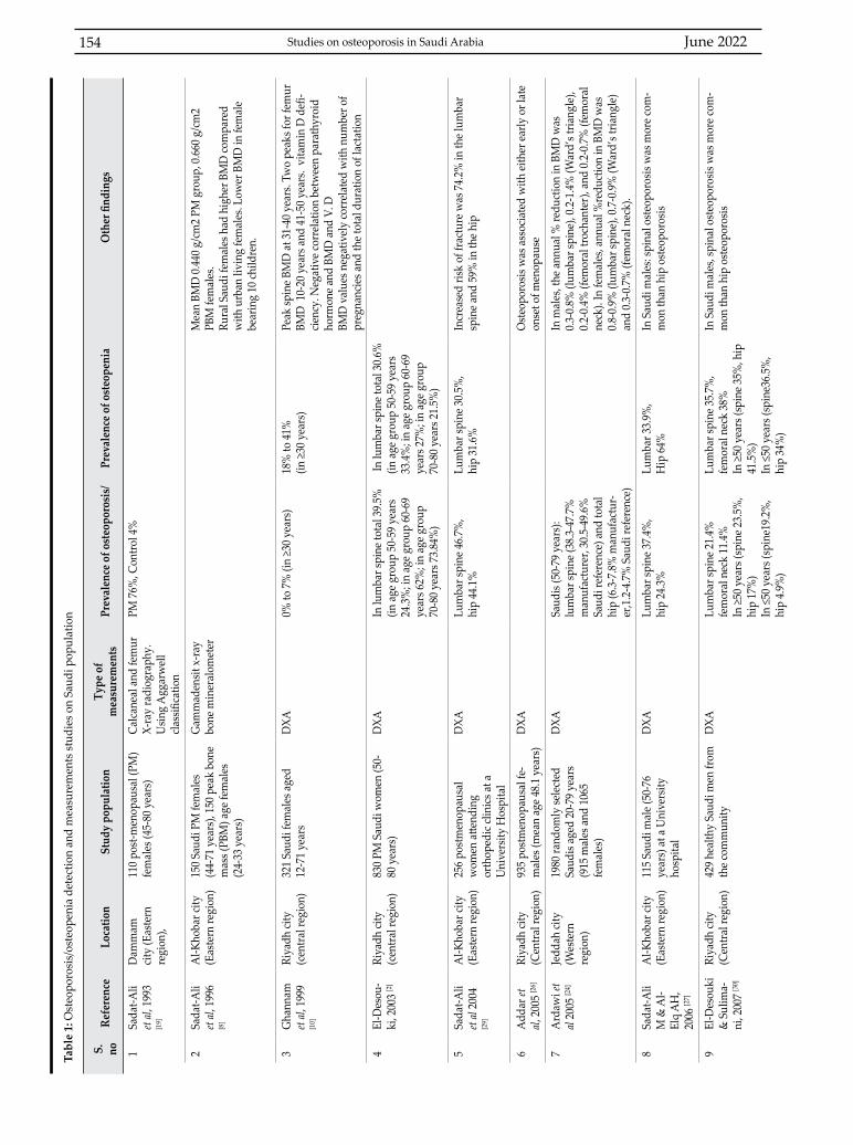
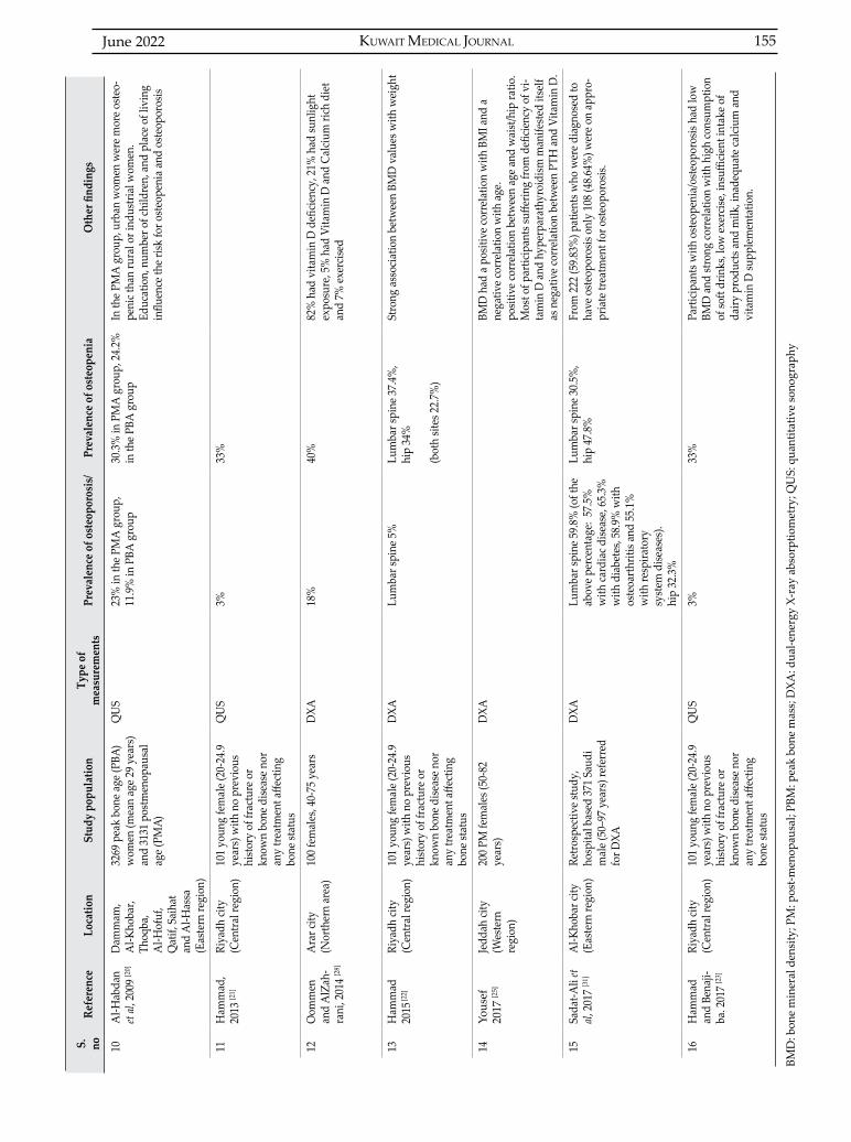
There are 16 documented studies that investigated the presence of osteopenia and osteoporosis in the Saudi population (Table 1) arranged chronologically, not including studies that investigated secondary osteoporosis in the Saudi population. Table 1 documented the city/area in which the study was conducted, the technique used, sample size and sex, percentage of osteoporosis and osteopenia and any other findings. Techniques incorporated in the above studies varied from the use of calcaneal and femur X-ray radiography (1 study) and Gammadensit x-ray bone mineralometer (1 study) to the use of QUS (3 studies) and DXA (11 studies).
Calcaneal X-ray radiography as a tool to examine the presence or absence of osteoporosis was unable to detect bone loss of less than 30%, and positioning in radiography was found to alter the results[19]. In Saudi females, Ghannam et al described one peak spine BMD to occur at 31-40 years and the presence of two peaks for femur, the first peak BMD in femur occurring at 10 to 20 years and second occurring between 41 to 50 years. In addition to that, mean BMD measured in post-menopausal females were less than BMD in peak bone mass females[10]. BMD in healthy Saudi females aged 12-71 years (n=321) were found to be lower than their USA counterparts; these results were justified partially by the high number of pregnancies accompanied by longer lactation duration. Other findings include vitamin D deficiency and calcidiol, 25-hydroxycholecalciferol levels and several BMD measurements negatively correlated with parathyroid hormone (PTH) levels[10].
Using QUS, the occurrence of osteopenia and osteoporosis in the calcaneus region were reported to be 24.2% and 11.9% respectively in peak bone age women (mean age: 29 years)[20]. When using the same technique, the percentage of osteopenia was higher and osteoporosis was lower (33% and 3% respectively) in young (mean age: 21.35 years, age range: 20-29 years) university Saudi females. The differences between the two studies might be attributed to demographic and educational differences, with the sample in the later study being younger and university educated[21].
In young Saudi females, the occurrence of osteopenia equaled to 37.4% in the lumbar spine, 34% in the femur, with 5% osteoporosis in the lumbar spine with DXA. A positive correlation between BMD and weight was noted[22]. High consumption of soft drinks, low exercise, insufficient intake of dairy products and milk, and inadequate calcium and vitamin D supplementation were found in young females with osteopenia or osteoporosis[23].
June 2022154 Studies on osteoporosis in Saudi Arabia
Tabl
e 1:
Ost
eopo
rosi
s/os
teop
enia
det
ectio
n an
d m
easu
rem
ents
stud
ies o
n Sa
udi p
opul
atio
n
S.
noRe
fere
nce
Loca
tion
Stud
y po
pula
tion
Type
of
mea
sure
men
tsPr
eval
ence
of o
steo
poro
sis/
Prev
alen
ce o
f ost
eope
nia
Oth
er fi
ndin
gs
1Sa
dat-A
li et
al, 1
993
[19]
Dam
mam
cit
y (E
aste
rn
regi
on),
110
post
-men
opau
sal (
PM)
fem
ales
(45-
80 y
ears
)Ca
lcane
al a
nd fe
mur
X-
ray
radi
ogra
phy.
U
sing
Agg
arw
ell
class
ifica
tion
PM 7
6%, C
ontro
l 4%
2Sa
dat-A
li et
al, 1
996
[8]
Al-K
hoba
r city
(E
aste
rn re
gion
)15
0 Sa
udi P
M fe
mal
es
(44-
71 y
ears
), 15
0 pe
ak b
one
mas
s (PB
M) a
ge fe
mal
es
(24-
33 y
ears
)
Gam
mad
ensit
x-ra
y bo
ne m
iner
alom
eter
Mea
n BM
D 0
.440
g/c
m2
PM g
roup
, 0.6
60 g
/cm
2 PB
M fe
mal
es.
Rura
l Sau
di fe
mal
es h
ad h
ighe
r BM
D co
mpa
red
with
urb
an li
ving
fem
ales
. Low
er B
MD
in fe
mal
e be
arin
g 10
child
ren.
3G
hann
am
et al,
199
9 [1
0]
Riya
dh ci
ty
(cen
tral r
egio
n)32
1 Sa
udi f
emal
es a
ged
12-7
1 ye
ars
DXA
0% to
7%
(in
≥30
year
s)18
% to
41%
(in ≥
30 y
ears
)Pe
ak sp
ine B
MD
at 3
1-40
yea
rs. T
wo
peak
s for
fem
ur
BMD
10-
20 y
ears
and
41-
50 y
ears
. vi
tam
in D
defi
-cie
ncy.
Neg
ativ
e cor
rela
tion
betw
een
para
thyr
oid
horm
one a
nd B
MD
and
V. D
BMD
val
ues n
egat
ivel
y co
rrel
ated
with
num
ber o
f pr
egna
ncie
s and
the t
otal
dur
atio
n of
lact
atio
n
4El
-Des
ou-
ki, 2
003
[2]
Riya
dh ci
ty
(cen
tral r
egio
n)83
0 PM
Sau
di w
omen
(50-
80 y
ears
)D
XAIn
lum
bar s
pine
tota
l 39.
5%
(in a
ge g
roup
50-
59 y
ears
24
.3%
; in
age g
roup
60-
69
year
s 62%
; in
age g
roup
70
-80
year
s 73.
84%
)
In lu
mba
r spi
ne to
tal 3
0.6%
(in a
ge g
roup
50-
59 y
ears
33
.4%
; in
age g
roup
60-
69
year
s 27%
; in
age g
roup
70
-80
year
s 21.
5%)
5Sa
dat-A
li et
al 20
04
[29]
Al-K
hoba
r city
(E
aste
rn re
gion
)25
6 po
stm
enop
ausa
lw
omen
atte
ndin
gor
thop
edic
clini
cs a
t aU
nive
rsity
Hos
pita
l
DXA
Lum
bar s
pine
46.
7%,
hip
44.1
%Lu
mba
r spi
ne 3
0.5%
,hi
p 31
.6%
Incr
ease
d ris
k of
frac
ture
was
74.
2% in
the l
umba
r sp
ine a
nd 5
9% in
the h
ip
6A
ddar
et
al, 2
005
[26]
Riya
dh ci
ty
(Cen
tral r
egio
n)93
5 po
stm
enop
ausa
l fe-
mal
es (m
ean
age 4
8.1
year
s)D
XAO
steo
poro
sis w
as a
ssoc
iate
d w
ith ei
ther
early
or l
ate
onse
t of m
enop
ause
7A
rdaw
i et
al 20
05 [2
4]Je
ddah
city
(W
este
rn
regi
on)
1980
rand
omly
sele
cted
Saud
is ag
ed 2
0-79
yea
rs(9
15 m
ales
and
106
5fe
mal
es)
DXA
Saud
is (5
0-79
yea
rs):
lum
bar s
pine
(38.
3-47
.7%
m
anuf
actu
rer,
30.5
-49.
6%
Saud
i ref
eren
ce) a
nd to
tal
hip
(6.3
-7.8
% m
anuf
actu
r-er
,1.2
-4.7
% S
audi
refe
renc
e)
In m
ales
, the
ann
ual %
redu
ctio
n in
BM
D w
as
0.3-
0.8%
(lum
bar s
pine
), 0.
2-1.
4% (W
ard’
s tria
ngle
), 0.
2-0.
4% (f
emor
al tr
ocha
nter
), an
d 0.
2-0.
7% (f
emor
al
neck
). In
fem
ales
, ann
ual %
redu
ctio
n in
BM
D w
as
0.8-
0.9%
(lum
bar s
pine
), 0.
7-0.
9% (W
ard’
s tria
ngle
) an
d 0.
3-0.
7% (f
emor
al n
eck)
.
8Sa
dat-A
li M
& A
l-El
q A
H,
2006
[27]
Al-K
hoba
r city
(E
aste
rn re
gion
)11
5 Sa
udi m
ale (
50-7
6 ye
ars)
at a
Uni
vers
ityho
spita
l
DXA
Lum
bar s
pine
37.
4%,
hip
24.3
%Lu
mba
r 33.
9%,
Hip
64%
In S
audi
mal
es: s
pina
l ost
eopo
rosis
was
mor
e com
-m
on th
an h
ip o
steo
poro
sis
9El
-Des
ouki
&
Sul
ima-
ni, 2
007
[30]
Riya
dh ci
ty
(Cen
tral r
egio
n)42
9 hea
lthy
Saud
i men
from
the c
omm
unity
DXA
Lum
bar s
pine
21.
4%fe
mor
al n
eck
11.4
%In
≥50
yea
rs (s
pine
23.
5%,
hip
17%
)In
≤50
yea
rs (s
pine
19.2
%,
hip
4.9%
)
Lum
bar s
pine
35.
7%,
fem
oral
nec
k 38
%In
≥50
yea
rs (s
pine
35%
, hip
41
.5%
)In
≤50
yea
rs (s
pine
36.5
%,
hip
34%
)
In S
audi
mal
es, s
pina
l ost
eopo
rosis
was
mor
e com
-m
on th
an h
ip o
steo
poro
sis
KUWAIT MEDICAL JOURNAL 155June 2022
S.
noRe
fere
nce
Loca
tion
Stud
y po
pula
tion
Type
of
mea
sure
men
tsPr
eval
ence
of o
steo
poro
sis/
Prev
alen
ce o
f ost
eope
nia
Oth
er fi
ndin
gs
10A
l-Hab
dan
et al,
200
9 [2
0]D
amm
am,
Al-K
hoba
r, Th
oqba
, A
l-Hof
uf,
Qat
if, S
aiha
t an
d A
l-Has
sa
(Eas
tern
regi
on)
3269
pea
k bo
ne a
ge (P
BA)
wom
en (m
ean
age 2
9 ye
ars)
an
d 31
31 p
ostm
enop
ausa
l ag
e (PM
A)
QU
S23
% in
the P
MA
gro
up,
11.9
% in
PBA
gro
up30
.3%
in P
MA
gro
up, 2
4.2%
in
the P
BA g
roup
In th
e PM
A g
roup
, urb
an w
omen
wer
e mor
e ost
eo-
peni
c tha
n ru
ral o
r ind
ustri
al w
omen
.Ed
ucat
ion,
num
ber o
f chi
ldre
n, a
nd p
lace
of l
ivin
g in
fluen
ce th
e risk
for o
steo
peni
a an
d os
teop
oros
is
11H
amm
ad,
2013
[21]
Riya
dh ci
ty
(Cen
tral r
egio
n)10
1 yo
ung
fem
ale (
20-2
4.9
year
s) w
ith n
o pr
evio
us
hist
ory
of fr
actu
re o
rkn
own
bone
dise
ase n
or
any
treat
men
t affe
ctin
g bo
ne st
atus
QU
S3%
33%
12O
omm
en
and
AlZ
ah-
rani
, 201
4 [2
8]
Ara
r city
(N
orth
ern
area
)10
0 fe
mal
es, 4
0-75
yea
rsD
XA18
%40
%82
% h
ad v
itam
in D
defi
cienc
y, 2
1% h
ad su
nlig
ht
expo
sure
, 5%
had
Vita
min
D a
nd C
alciu
m ri
ch d
iet
and
7% ex
ercis
ed
13H
amm
ad
2015
[22]
Riya
dh ci
ty
(Cen
tral r
egio
n)10
1 yo
ung
fem
ale (
20-2
4.9
year
s) w
ith n
o pr
evio
us
hist
ory
of fr
actu
re o
rkn
own
bone
dise
ase n
or
any
treat
men
t affe
ctin
g bo
ne st
atus
DXA
Lum
bar s
pine
5%
Lum
bar s
pine
37.
4%,
hip
34%
(bot
h sit
es 2
2.7%
)
Stro
ng a
ssoc
iatio
n be
twee
n BM
D v
alue
s with
wei
ght
14Yo
usef
20
17 [2
5]Je
ddah
city
(W
este
rn
regi
on)
200
PM fe
mal
es (5
0-82
ye
ars)
DXA
BMD
had
a p
ositi
ve co
rrel
atio
n w
ith B
MI a
nd a
ne
gativ
e cor
rela
tion
with
age
. po
sitiv
e cor
rela
tion
betw
een
age a
nd w
aist
/hip
ratio
.M
ost o
f par
ticip
ants
suffe
ring
from
defi
cienc
y of
vi-
tam
in D
and
hyp
erpa
rath
yroi
dism
man
ifest
ed it
self
as n
egat
ive c
orre
latio
n be
twee
n PT
H a
nd V
itam
in D
.
15Sa
dat-A
li et
al, 2
017
[31]
Al-K
hoba
r city
(E
aste
rn re
gion
)Re
trosp
ectiv
e stu
dy,
hosp
ital b
ased
371
Sau
di
mal
e (50
–97
year
s) re
ferr
ed
for D
XA
DXA
Lum
bar s
pine
59.
8% (o
f the
ab
ove p
erce
ntag
e: 5
7.5%
w
ith ca
rdia
c dise
ase,
65.3
%
with
dia
bete
s, 58
.9%
with
os
teoa
rthrit
is an
d 55
.1%
w
ith re
spira
tory
syst
em d
iseas
es).
hip
32.3
%
Lum
bar s
pine
30.
5%,
hip
47.8
%Fr
om 2
22 (5
9.83
%) p
atie
nts w
ho w
ere d
iagn
osed
to
have
ost
eopo
rosis
onl
y 10
8 (4
8.64
%) w
ere o
n ap
pro-
pria
te tr
eatm
ent f
or o
steo
poro
sis.
16H
amm
ad
and
Bena
ji-ba
. 201
7 [2
3]
Riya
dh ci
ty
(Cen
tral r
egio
n)10
1 yo
ung
fem
ale (
20-2
4.9
year
s) w
ith n
o pr
evio
us
hist
ory
of fr
actu
re o
rkn
own
bone
dise
ase n
or
any
treat
men
t affe
ctin
g bo
ne st
atus
QU
S3%
33%
Parti
cipan
ts w
ith o
steo
peni
a/os
teop
oros
is ha
d lo
w
BMD
and
stro
ng co
rrel
atio
n w
ith h
igh
cons
umpt
ion
of so
ft dr
inks
, low
exer
cise,
insu
fficie
nt in
take
of
dairy
pro
duct
s and
milk
, ina
dequ
ate c
alciu
m a
nd
vita
min
D su
pple
men
tatio
n.
BMD
: bon
e m
iner
al d
ensi
ty; P
M: p
ost-m
enop
ausa
l; PB
M: p
eak
bone
mas
s; D
XA: d
ual-e
nerg
y X-
ray
abso
rptio
met
ry; Q
US:
qua
ntita
tive
sono
grap
hy
June 2022156 Studies on osteoporosis in Saudi Arabia
S.
noRe
fere
nce
Loca
tion
Prim
ary
dise
ase
Stud
y po
pula
tion
Type
of
mea
sure
men
tsPr
eval
ence
of
oste
opor
osis
Prev
alen
ce o
f os
teop
enia
Oth
er fi
ndin
gs
1H
urai
b et
al,
1993
[33]
Riya
dh ci
ty
(Cen
tral r
egio
n)H
aem
odia
lysis
Mul
ticen
ter s
tudy
, 209
hae
mod
i-al
ysis
patie
nts m
ean
dura
tion
on
dial
ysis
was
3.5
±1.5
yea
rs. m
ean
age 3
9.4±
14 (1
8-70
yea
rs),
128
mal
es
and
81 fe
mal
es
Dua
l–ph
oton
abs
orp-
tiom
etry
65%
low
bon
e min
eral
den
sity,
92%
hy
perp
arat
hyro
idism
chan
ges,
66%
pu
re h
yper
para
thyr
oidi
sm, 6
0% w
ith
varia
ble d
egre
es o
f AL
into
xica
tion
2A
l-Maa
touq
et
al, 2
004
[9]
Riya
dh ci
ty
(Cen
tral r
egio
n)Ty
pe-2
dia
bete
s m
ellit
us (T
2D)
104
T2D
PM
Sau
di fe
mal
e (m
ean
age=
63
year
s) a
nd 1
01 n
on-d
iabe
tic
PM S
audi
fem
ale (
mea
n ag
e = 6
0 ye
ars)
DXA
T2D
gro
up (L
umba
r sp
ine 4
6.8%
, hip
19
.8%
)Co
ntro
l gro
up
(Lum
ber s
pine
22.
2%
& h
ip 1
2.1%
)
T2D
(Lum
bar s
pine
43
.68%
, hip
44.
7%)
Cont
rol g
roup
(Lum
-be
r 46.
5% sp
ine H
ip
49.5
%)
A re
duct
ion
in B
MD
in T
2D co
mpa
red
to n
on-d
iabe
tic g
roup
3A
l-Elq
&
Sada
t-Ali
2006
[3
5]
Al-K
hoba
r city
(E
aste
rn re
gion
)T2
D15
4 Sa
udi m
ale (
57 T
2D m
ale (
mea
n ag
e= 5
9.76
yea
rs),
34 p
atie
nts (
mea
n ag
e= 6
0.90
yea
rs) w
ith im
paire
d fa
stin
g gl
ucos
e and
63
norm
al
gluc
ose l
evel
pat
ient
s (m
ean
age=
62
.53
year
s))
DXA
Lum
bar s
pine
40.
7%hi
p 27
.9%
Lum
bar s
pine
35%
hip
53.2
%N
o sig
nific
ant d
iffer
ence
s bet
wee
n th
e thr
ee g
roup
s in
BMD
, T-s
core
and
Z-
scor
e
4M
alab
u an
d Fo
unda
, 200
7 [3
2]
Riya
dh ci
ty
(Cen
tral r
egio
n)Pr
imar
y hy
per-
para
thyr
oidi
smN
= 46
, Fem
ale=
35, M
ale=
11,
DXA
83%
of p
atie
nts h
ad o
steo
peni
a/os
te-
opor
osis.
45.
7% co
mpl
aine
d of
Bon
e pa
ins,
15.2
% h
ad re
nal s
tone
s, 6.
5%
poly
uria
, 4.3
% h
ad d
epre
ssio
n an
d co
nstip
atio
n.
5Sa
dat-A
li et
al,
2008
[34]
Al-K
hoba
r city
(E
aste
rn re
gion
)A
dole
scen
t idi
o-pa
thic
scol
iosis
(AIS
)
32 g
irls w
ith A
IS (m
ean
age o
f 18
.42±
5.71
, 14-
26 y
ears
) and
thei
r sib
lings
(N=2
7, (m
ean
age o
f 17
.65±
4.5;
14-
25ye
ars)
DXA
AIS
lum
bar s
pine
&
hip
62.5
%A
IS lu
mba
r spi
ne &
hi
p 28
.1%
Girl
s with
nor
mal
spin
e had
sign
ifi-
cant
hig
her B
MI a
nd B
MD
than
AIS
sib
lings
. The
deg
ree o
f Cob
b an
gle
affec
ting
the s
ever
ity o
f dise
ase
6Sa
dat-A
li et
al,
2008
[42]
Al-K
hoba
r city
(E
aste
rn re
gion
)Si
ckle
cell
anem
ia10
3 pa
tient
s (45
M &
58F
)D
XA35
% (1
5M+2
1F)
30%
(13M
+18F
)A
redu
ctio
n in
test
oste
rone
and
estra
-di
ol in
ost
eopo
rotic
SCD
fem
ale a
nd
low
er es
tradi
ol in
SCD
ost
eopo
rotic
/os
teog
enic
mal
es
7Sa
dat-A
li et
al,
2009
[37]
Al-K
hoba
r city
(E
aste
rn re
gion
)G
luco
corti
coid
th
erap
yRe
trosp
ectiv
e stu
dy, 1
65 p
atie
nts
pres
crib
ed ≥
7.5
mg
ofPr
edni
solo
ne/d
ay, ≥
6 m
onth
s (26
pa
tient
s had
DXA
, 21F
, 5M
)
DXA
50%
, N=1
3(10
F &
3M)
50%
, N=1
3 (1
1F &
2 2M
)Ca
lcium
and
Vita
min
D w
ere p
re-
scrib
ed to
the m
ajorit
y of
pat
ient
s bu
t non
e had
ant
ireso
rptiv
e/an
abol
ic th
erap
y
8A
lAm
ri an
d Sa
dat-A
li, 20
09
[44]
Al-K
hoba
r city
(E
aste
rn re
gion
)Ch
emot
hera
py
treat
ed ca
ncer
pa
tient
N=7
1 pa
tient
s, m
ean
age=
49.
29
year
s, ag
e ran
ge 2
9-67
yea
rsD
XASp
ine 2
5.8%
& h
ip
22.6
%Sp
ine 3
3.87
% &
hip
29
%O
steo
peni
a an
d os
teop
oros
is fo
und
in
65.6
% p
atie
nts ≤
50 y
ears
, com
pare
d to
56
.4%
pat
ient
s ≥51
yea
rs o
ld.
Nor
mal
BM
D in
pat
ient
s with
48.
68 ±
27
.35 m
onth
s ear
lier l
ast c
hem
othe
rapy
cy
cle co
mpa
red
to m
ore o
steo
poro
sis
in p
atie
nts w
ho re
ceiv
ed la
st ch
emo-
ther
apy
for l
ess t
han
2 ye
ars.
Tabl
e 2:
Det
ectio
n an
d m
easu
rem
ents
of se
cond
ary
oste
opor
osis/
oste
open
ia in
the S
audi
pop
ulat
ion
KUWAIT MEDICAL JOURNAL 157June 2022
S.
noRe
fere
nce
Loca
tion
Prim
ary
dise
ase
Stud
y po
pula
tion
Type
of
mea
sure
men
tsPr
eval
ence
of
oste
opor
osis
Prev
alen
ce o
f os
teop
enia
Oth
er fi
ndin
gs
9A
l-Tur
ki 2
009
[43]
Al-K
hoba
r city
(Eas
tern
regi
on)
Preg
nanc
y in
sick
-le
cell
anem
iaN
=38
(20
paro
us, m
ean
age=
27
.55±
4.9
year
s and
18
nulli
paro
us,
mea
n ag
e= 2
6.33
±2.1
yea
rs)
DXA
65%
in p
arou
s SCD
, 27
.7%
in n
ullip
arou
s SC
D
20%
in p
arou
s SCD
, 38
.9%
in n
ullip
arou
s SC
D
Ost
eopo
rosis
in b
oth
grou
ps w
ere
high
er a
t lum
bar s
pine
com
pare
d to
th
e hip
regi
on (P
= 0.
001)
. BM
D w
as
low
er in
par
ous w
omen
whe
n co
m-
pare
d to
the n
ullip
arou
s wom
en.
10Sa
dat-A
li et
al,
2011
[40]
Al-K
hoba
r city
(E
aste
rn re
gion
)Vi
tam
in D
defi
-cie
ncy
in si
ckle
ce
ll an
emia
Sick
le ce
ll gr
oup
(86M
, 100
F)Co
ntro
l gro
up (1
00M
, 100
F)D
XASi
ckle
cell
fem
ale
(spi
ne 7
8% &
hip
74
%).
Sick
le ce
ll m
ale
(spi
ne 7
2% &
hip
71
%).
Cont
rols
fem
ale
(spi
ne 2
6% &
hip
22%
). Co
ntro
ls m
ale
(spi
ne 1
3% &
hip
12
%).
Sick
le ce
ll fe
mal
e (s
pine
15%
& h
ip
17%
). Si
ckle
cell
mal
e (s
pine
12%
& h
ip
15%
).Co
ntro
l fem
ale
(spi
ne 1
2% &
hip
11
%).
Cont
rol m
ale
(spi
ne 1
0% &
hip
9%
).
Patie
nts w
ith S
CD h
ad si
gnifi
cant
ly
elev
ated
seru
m a
lkal
ine p
hosp
hata
se
and
PTH
leve
ls in
the p
rese
nce o
f low
vi
tam
in D
stat
us
11Sa
dat-A
li et
al,
2011
[38]
A
l-Kho
bar c
ity
(Eas
tern
regi
on)
Vita
min
D d
efi-
cienc
y 40
0 Sa
udi m
en a
nd w
omen
DXA
Nor
mal
25O
HD
(>30
pg
/mL)
8.8
%,
insu
fficie
ncy
of
25O
HD
(21-
29 p
g/m
L) 3
3.67
%, d
eficie
n-cy
of 2
5OH
D (<
20
pg/m
L) 6
7.31
%
Nor
mal
25O
HD
(>30
pg
/mL)
24.
4%,
insu
fficie
ncy
of
25O
HD
(21-
29 p
g/m
L) 5
0%, d
eficie
ncy
of 2
5OH
D (<
20 p
g/m
L) 3
2.69
%
Posit
ive r
elat
ions
hip
betw
een
vita
min
D
seru
m le
vel a
nd B
MD
, low
BM
D in
su
bjec
ts w
ith V
itam
in D
defi
cienc
y an
d sig
nific
ant n
egat
ive c
orre
latio
ns b
e-tw
een
BMD
and
par
athy
roid
hor
mon
e.
12A
rdaw
i et a
l, 20
12 [3
9]Je
ddah
city
(W
este
rn re
gion
)Vi
tam
in D
defi
-cie
ncy
834M
(20–
74 y
ears
)D
XAD
eficie
ncy
(25(
OH
)D<5
0 nm
ol/L
) and
in
suffi
cienc
y (≥
50–7
5 nm
ol/L
) wer
e pr
esen
t in
87.8
% a
nd 9
.7%
, pos
itive
co
rrel
atio
n be
twee
n an
d se
rum
25(
OH
)D
leve
ls
13Kh
oshh
al et
al,
2015
[36]
Al-M
adin
ah
Al-M
onaw
arah
(W
este
rn re
gion
)
Type
1 d
iabe
tes
(T1D
)36
T1D
child
ren
& 3
9 co
ntro
lQ
US
In T
1D (1
3.9%
with
Z-
scor
e ≤-1
). Co
ntro
ls (0
% w
ith Z
-sco
re
≤-1)
.
Sign
ifica
ntly
low
er le
vels
of o
steo
cal-
cin a
nd p
roco
llage
n N
-term
inal
pept
ide a
nd si
gnifi
cant
ly h
ighe
r ser
umle
vels
of b
one r
esor
ptio
n m
arke
rs w
ere
foun
d in
the d
iabe
tic g
roup
.
14A
lham
ad a
nd
Nad
ama,
201
5
[45]
Riya
dh ci
ty
(Cen
tral r
egio
n)In
ters
titia
l lun
g di
seas
e (IL
D)
196
new
ly d
iagn
osed
ILD
pat
ient
sD
XASp
ine 3
3%, h
ip 1
3%,
dist
al ra
dius
20%
(5
5% o
ne si
de in
-vo
lvem
ent,
21%
had
tw
o sit
es in
volv
e-m
ent,
24%
had
thre
e sit
es in
volv
emen
t).
Spin
e 41%
,H
ip 4
0%, D
istal
ra
dius
32%
(39%
had
on
e site
invo
lvem
ent,
17%
had
two
sites
, 44
% h
ad th
ree s
ites
invo
lvem
ent)
The n
eed
for s
cree
ning
and
agg
ress
ive
treat
men
t with
var
ious
ant
i-bon
e re
sorp
tive t
hera
pies
for p
atie
nts w
ith
ILD
15A
l-Om
ran
et al,
201
6 [4
6]A
l-Kho
bar c
ity
(Eas
tern
regi
on)
Ant
ipsy
chot
ic dr
ugs
120M
& 2
5F (m
ean
age 4
0.75
±7.
16,
age r
ange
26-
56ye
ars)
Mea
n du
ratio
n of
ant
ipsy
chot
ic m
edica
tion
= 8.
45±5
.4ye
ars)
DXA
Spin
e 44%
hip
14.2
%Sp
ine 4
5.7%
hip
59.4
2%A
ttrib
uted
to h
yper
prol
acte
mia
in
duce
d by
ant
ipsy
chot
ic m
edica
tion,
ot
her f
acto
rs su
ch a
s lev
els o
f Vita
min
D
wer
e not
cont
rolle
d
T2D
: Typ
e-2
diab
etes
mel
litus
; ILD
: int
erst
itial
lung
dis
ease
; DXA
: dua
l-ene
rgy
X-ra
y ab
sorp
tiom
etry
; QU
S: q
uant
itativ
e so
nogr
aphy
June 2022158
In Saudi females aged 20-79 years, the annual percentage reduction in BMD was 0.8-0.9% in the lumbar spine, 0.7-0.9% in the Ward’s triangle and 0.3-0.7% in the femoral neck[24]. In addition to that, the presence of lower BMD in urban living females compared to rural Saudi females were reported[8]. BMD values were found to correlate positively with body mass index[25] and negatively with number of pregnancies and total duration of lactation[8,10]. Osteoporosis was associated with either early or late onset of menopause, possibly due to genetic factors, as Saudi women had similar age of menopause to Arabic ladies but lower than western countries[26].
In postmenopausal women, an increase in osteoporosis was found to occur with age[2]; in subjects below 60 years, 33.4% had osteopenia and 24.3% had osteoporosis, with the incidence of osteopenia and osteoporosis being increased (27% osteopenic and 62% osteoporotic) in ladies between 60-69 years old. In ladies of older age (70-79 years), 21.5% had osteopenia and 73.8% had osteoporosis[2]. In other areas within Saudi Arabia, osteoporosis/osteopenia percentage varied from 24.3% to 64% in Al-Khobar city (Eastern region)[27] and 18% to 40% in Arar city (northern area)[28]. The risk of fracture was calculated as 74.2% in the lumbar spine and 59% in the hip in postmenopausal women[29].
In Saudi males, the annual percentage reduction in BMD was 0.3-0.8% in the lumbar spine, 0.2-1.4% in Ward’s triangle, 0.2-0.4% in the femoral trochanter, and 0.2-0.7% in the femoral neck[24]. Spinal osteoporosis was found to exist in 21.4-37.4% of the population studied compared to 11.4-24.3% with hip osteoporosis[27,30]. A positive correlation was found between osteoporosis/osteopenia and age in the Saudi male population[30]. In a hospital based retrospective study on 371 Saudi males (age range: 50-97 years) referred for DXA, percentage of osteoporosis in the lumbar spine and hip area equaled to 59.8% and 32.3% respectively. Of the 222 (59.83%) patients who were diagnosed to have osteoporosis, only 108 (48.64%) were on appropriate treatment for osteoporosis[31].
Effect of various diseases on bone mineral density in Saudi Arabian population
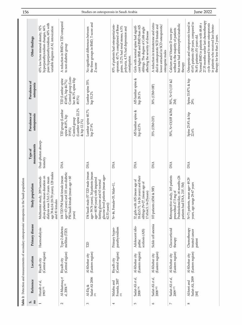
Secondary osteoporosis occurs as a result of some diseases, or from hormonal or drug inducing changes in bones. Table 2 summarizes the studies conducted in Saudi Arabia to investigate changes in BMD secondary to these diseases. There are 15 documented studies that reported the presence of secondary osteopenia and osteoporosis in the Saudi population arranged chronologically. The table documented the city/area at which the study was conducted, the main disease,
sample size and sex, technique used, percentage of osteoporosis and osteopenia found and any other findings. Efforts are required to formulate policies to diagnose and treat osteoporosis secondary to these diseases.
Bone pain was reported by 45.7% of patients with primary hyperparathyroidism, attributed to skeletal and renal manifestation of the primary hyperparathyroidism[32], whereas low BMD was found in hemodialytic patients with 66% of patients suffering from pure hyperparathyroidism and 92% of patients from hyperparathyroidism changes[33]. In adolescent idiopathic scoliosis, lower body mass index and BMD were found in subjects with scoliosis compared to their siblings, and the degree of Cobb angle was found to affect the severity of disease[34].
A reduction in BMD was found among post-menopausal Saudi women with type-2 diabetes mellitus (T2D) compared to non-diabetic group. The reduction was attributed to the presence of T2D as both groups had similar life style factors[9]. In contrast to the previous study, T2D male patients demonstrated no differences in BMD, T-score and Z-score compared to both patients with impaired fasting glucose and normal glucose level patients[35]. The relationship between type 1 diabetes (T1D) and bone quality in Saudi children investigated using QUS, biochemical bone markers and bone alkaline phosphatase activity revealed the presence of significant high serum levels of bone resorption markers and the presence of Z-score below -1 in 13.9% of the T1D children compared to none in the non-diabetic children. These findings indicate that children with T1D may have low bone formation with high bone resorption markers leading to an increase in both osteoporosis and bone fracture susceptibility in the future[36].
In a retrospective study investigating glucocorticoid-induced osteoporosis (n=26), 47.6% females and 50% males were osteoporotic and 52.4% females and 40% males had osteopenia. Calcium and vitamin D were prescribed to the majority of patients but none had antiresorptive/anabolic therapy. It was recommended that proper investigation and treatment are required for the management of glucocorticoid-induced osteoporosis[37].
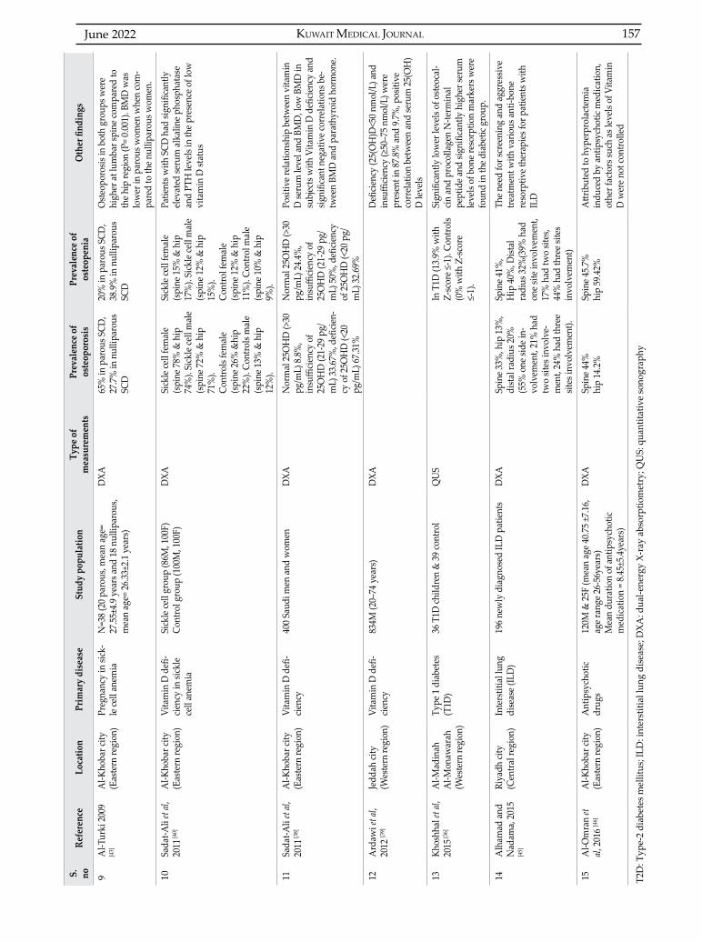
Vitamin D deficiency had a positive relationship with low BMD and PTH. The studies attributed low BMD and vitamin D deficiency to lifestyle, age, obesity and low exposure to sun light and suggested the importance of evaluating hypovitaminosis during the management of low bone mass[38-40].
The risk of osteoporosis and osteopenia in Saudi patients with sickle cell disease (SCD) was reported as a reduction in BMD values[41] associated with lower vitamin D and higher alkaline phosphatase
Studies on osteoporosis in Saudi Arabia
KUWAIT MEDICAL JOURNAL 159June 2022
and PTH values[38]. Possible causes for the reduction in vitamin D were suggested to include malabsorption, insufficient vitamin D consumption, reduction in activity related to repeated crisis and lower sun exposure[38]. A reduction in testosterone and estradiol in osteoporotic females with SCD and estradiol in SCD osteoporotic/osteogenic males convey the important role that sex steroids hormone plays in the development of secondary osteoporosis[42]. When the influence of pregnancy on bone mass was investigated in 38 sickle cell anemic Saudi females, a significant increase in osteoporosis within the parous group (n=13/20) compared to nulliparous group (n=5/18) was found. The presence of no significant difference in calcium level, phosphorus level, hemoglobin electrophoresis and parathyroid hormone found between the two groups suggested that pregnancy was the possible cause of osteoporosis, though no comparison with non-sickle cell controls were made[43].
In another context, time of the last chemotherapy cycle was found to impact bone mass, with normal BMD found in patients who got their last chemotherapy cycle 48.68±27.35 months earlier as opposed to more osteoporosis in patients who received chemotherapy within less than 2 years. Surprisingly, osteopenia and osteoporosis were found to be more common (65.6%) in patients with an age of ≤50 years, compared to 56.4% in patients of ≥51 years[44].
The need for screening and treatment with various anti-bone resorptive therapies for patients with interstitial lung disease was suggested based on the low bone mass observed in 196 newly diagnosed subjects with interstitial lung disease. In addition to that, the diagnosis of usual interstitial pneumonia was more frequently observed in patients with osteoporosis than in those without osteoporosis[45].
Negative effects on bone mass were found in patients taking antipsychotic drugs. Although the significant reduction in bone mass compared to controls were attributed to hyperprolactemia induced by antipsychotic medication, other factors such as levels of vitamin D were not controlled[46].
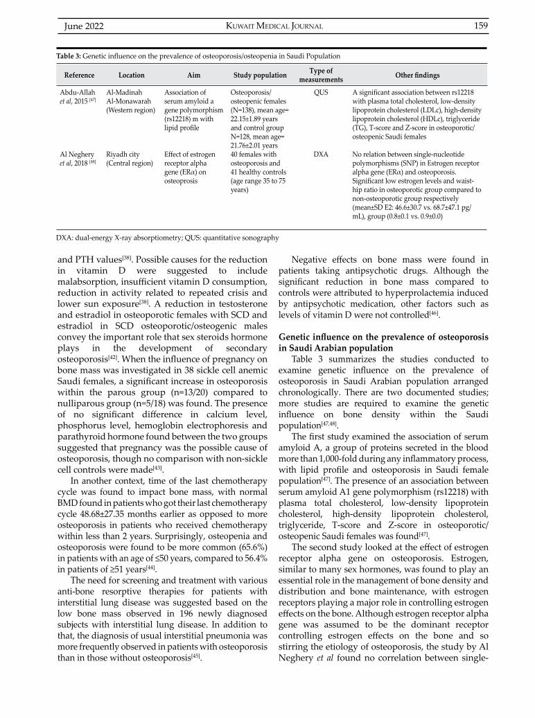
Genetic influence on the prevalence of osteoporosis in Saudi Arabian population
Table 3 summarizes the studies conducted to examine genetic influence on the prevalence of osteoporosis in Saudi Arabian population arranged chronologically. There are two documented studies; more studies are required to examine the genetic influence on bone density within the Saudi population[47,48].
The first study examined the association of serum amyloid A, a group of proteins secreted in the blood more than 1,000-fold during any inflammatory process, with lipid profile and osteoporosis in Saudi female population[47]. The presence of an association between serum amyloid A1 gene polymorphism (rs12218) with plasma total cholesterol, low-density lipoprotein cholesterol, high-density lipoprotein cholesterol, triglyceride, T-score and Z-score in osteoporotic/osteopenic Saudi females was found[47].
The second study looked at the effect of estrogen receptor alpha gene on osteoporosis. Estrogen, similar to many sex hormones, was found to play an essential role in the management of bone density and distribution and bone maintenance, with estrogen receptors playing a major role in controlling estrogen effects on the bone. Although estrogen receptor alpha gene was assumed to be the dominant receptor controlling estrogen effects on the bone and so stirring the etiology of osteoporosis, the study by Al Neghery et al found no correlation between single-
Reference Location Aim Study population Type of measurements Other findings
Abdu-Allah et al, 2015 [47]
Al Neghery et al, 2018 [48]
Al-Madinah Al-Monawarah (Western region)
Riyadh city (Central region)
Association of serum amyloid a gene polymorphism (rs12218) m with lipid profile
Effect of estrogen receptor alpha gene (ERα) on osteoprosis
Osteoporosis/osteopenic females (N=138), mean age= 22.15±1.89 years and control group N=128, mean age= 21.76±2.01 years40 females with osteoporosis and41 healthy controls (age range 35 to 75 years)
QUS
DXA
A significant association between rs12218 with plasma total cholesterol, low-density lipoprotein cholesterol (LDLc), high-density lipoprotein cholesterol (HDLc), triglyceride (TG), T-score and Z-score in osteoporotic/osteopenic Saudi females
No relation between single-nucleotide polymorphisms (SNP) in Estrogen receptor alpha gene (ERα) and osteoporosis. Significant low estrogen levels and waist-hip ratio in osteoporotic group compared to non-osteoporotic group respectively(mean±SD E2: 46.6±30.7 vs. 68.7±47.1 pg/mL), group (0.8±0.1 vs. 0.9±0.0)
DXA: dual-energy X-ray absorptiometry; QUS: quantitative sonography
Table 3: Genetic influence on the prevalence of osteoporosis/osteopenia in Saudi Population
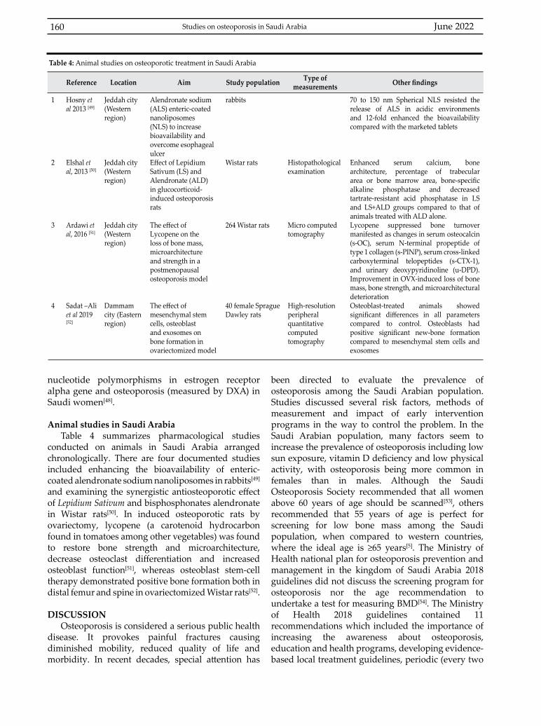
June 2022160
nucleotide polymorphisms in estrogen receptor alpha gene and osteoporosis (measured by DXA) in Saudi women[48].
Animal studies in Saudi ArabiaTable 4 summarizes pharmacological studies
conducted on animals in Saudi Arabia arranged chronologically. There are four documented studies included enhancing the bioavailability of enteric-coated alendronate sodium nanoliposomes in rabbits[49] and examining the synergistic antiosteoporotic effect of Lepidium Sativum and bisphosphonates alendronate in Wistar rats[50]. In induced osteoporotic rats by ovariectomy, lycopene (a carotenoid hydrocarbon found in tomatoes among other vegetables) was found to restore bone strength and microarchitecture, decrease osteoclast differentiation and increased osteoblast function[51], whereas osteoblast stem-cell therapy demonstrated positive bone formation both in distal femur and spine in ovariectomized Wistar rats[52].
DISCUSSIONOsteoporosis is considered a serious public health
disease. It provokes painful fractures causing diminished mobility, reduced quality of life and morbidity. In recent decades, special attention has
been directed to evaluate the prevalence of osteoporosis among the Saudi Arabian population. Studies discussed several risk factors, methods of measurement and impact of early intervention programs in the way to control the problem. In the Saudi Arabian population, many factors seem to increase the prevalence of osteoporosis including low sun exposure, vitamin D deficiency and low physical activity, with osteoporosis being more common in females than in males. Although the Saudi Osteoporosis Society recommended that all women above 60 years of age should be scanned[53], others recommended that 55 years of age is perfect for screening for low bone mass among the Saudi population, when compared to western countries, where the ideal age is ≥65 years[5]. The Ministry of Health national plan for osteoporosis prevention and management in the kingdom of Saudi Arabia 2018 guidelines did not discuss the screening program for osteoporosis nor the age recommendation to undertake a test for measuring BMD[54]. The Ministry of Health 2018 guidelines contained 11 recommendations which included the importance of increasing the awareness about osteoporosis, education and health programs, developing evidence-based local treatment guidelines, periodic (every two
Studies on osteoporosis in Saudi Arabia
Reference Location Aim Study population Type of measurements Other findings
1
2
3
4
Hosny et al 2013 [49]
Elshal et al, 2013 [50]
Ardawi et al, 2016 [51]
Sadat –Ali et al 2019 [52]
Jeddah city (Western region)
Jeddah city (Western region)
Jeddah city (Western region)
Dammam city (Eastern region)
Alendronate sodium (ALS) enteric-coated nanoliposomes (NLS) to increase bioavailability and overcome esophageal ulcerEffect of Lepidium Sativum (LS) andAlendronate (ALD) in glucocorticoid-induced osteoporosis rats
The effect of Lycopene on the loss of bone mass, microarchitecture and strength in a postmenopausalosteoporosis model
The effect of mesenchymal stem cells, osteoblast and exosomes on bone formation in ovariectomized model
rabbits
Wistar rats
264 Wistar rats
40 female Sprague Dawley rats
Histopathological examination
Micro computed tomography
High-resolution peripheral quantitative computed tomography
70 to 150 nm Spherical NLS resisted the release of ALS in acidic environments and 12-fold enhanced the bioavailability compared with the marketed tablets
Enhanced serum calcium, bone architecture, percentage of trabecular area or bone marrow area, bone-specific alkaline phosphatase and decreased tartrate-resistant acid phosphatase in LS and LS+ALD groups compared to that of animals treated with ALD alone.Lycopene suppressed bone turnover manifested as changes in serum osteocalcin (s-OC), serum N-terminal propeptide of type 1 collagen (s-PINP), serum cross-linked carboxyterminal telopeptides (s-CTX-1), and urinary deoxypyridinoline (u-DPD). Improvement in OVX-induced loss of bone mass, bone strength, and microarchitectural deteriorationOsteoblast-treated animals showed significant differences in all parameters compared to control. Osteoblasts had positive significant new-bone formation compared to mesenchymal stem cells and exosomes
Table 4: Animal studies on osteoporotic treatment in Saudi Arabia
KUWAIT MEDICAL JOURNAL 161June 2022
years) updates to guidelines, developing local fracture risk assessment tool in addition to regulation of post-fracture care to control secondary fractures[54].
CONCLUSION Further studies are needed not only for diagnosis
and treatment purposes, but as well as on topics related to prevention, education, management and the use of herbal medicine for prevention and treatment. A local fracture risk assessment tool or other programs to evaluate the 10-year probability of bone fracture risk by integrating clinical risk factors and bone mineral density are required.
ACKNOWLEDGMENT Funding: NoneConflict of Interest: None.Authors’ contributions: Lina Fahmi Hammad drafted this review paper manuscript, read and approved the final version of the manuscript.
REFERENCES
1. Ensrud KE. Epidemiology of fracture risk with advancing age. J Gerontol A Biol Sci Med Sci 2013; 68(10):1236-42.
2. El-Desouki MI. Osteoporosis in postmenopausal Saudi women using dual x-ray bone densitometry. Saudi Med J 2003; 24(9):953-6.
3. Who are candidates for prevention and treatment for osteoporosis? Osteoporos Int 1997; 7(1):1-6.
4. Kanis JA. Assessment of osteoporosis at the primary health care level. WHO Collaborating Centre for Metabolic Bone Diseases. WHO Collaborating Centre for Metabolic Bone Diseases. 2007.
5. Sadat-Ali M, Al-Habdan IM, Al-Turki HA, Azam MQ. An epidemiological analysis of the incidence of osteoporosis and osteoporosis-related fractures among the Saudi Arabian population. Ann Saudi Med 2012; 32(6):637-41.
6. Bubshait D, Sadat-Ali M. Economic implications of osteoporosis-related femoral fractures in Saudi Arabian society. Calcif Tissue Int 2007; 81(6):455-8.
7. Sadat-Ali M, Al-Dakheel DA, Azam MQ, Al-Bluwi MT, Al-Farhan MF, AlAmer HA, et al. Reassessment of osteoporosis-related femoral fractures and economic burden in Saudi Arabia. Arch Osteoporos 2015; 10:37.
8. Sadat-Ali M, Al-Habdan I, Marwah S. Bone mineral density measurements of distal radius in Saudi Arabian females. Ann Saudi Med 1996; 16(4):414-6.
9. Al-Maatouq MA, El-Desouki MI, Othman SA, Mattar EH, Babay ZA, Addar M. Prevalence of osteoporosis among postmenopausal females with diabetes mellitus. Saudi Med J 2004; 25(10):1423-7.
10. Ghannam NN, Hammami MM, Bakheet SM, Khan BA. Bone mineral density of the spine and femur in healthy Saudi females: relation to vitamin D status, pregnancy, and lactation. Calcif Tissue Int 1999; 65(1):23-8.
11. World Health Organization. Assessment of fracture risk and its application to screening for postmenopausal osteoporosis : report of a WHO study group [ meeting held in Rome from 22 to 25 June 1992] . Geneva: World Health Organization; 1994.
12. Clarke B. Normal bone anatomy and physiology. Clin J Am Soc Nephrol 2008; 3(Suppl 3):S131-9.
13. Faulkner KG. Clinical use of bone densitometry. Osteoporosis: Academic Press; 2001. p. 433-58.
14. Gregg EW, Kriska AM, Salamone LM, Roberts MM, Anderson SJ, Ferrell RE, et al. The epidemiology of quantitative ultrasound: a review of the relationships with bone mass, osteoporosis and fracture risk. Osteoporos Int 1997; 7(2):89-99.
15. El-Desouki MI, Sherafzal MS, Othman SA. Comparison of bone mineral density with dual energy x-ray absorptiometry, quantitative ultrasound and single energy x-ray absorptiometry. Saudi Med J 2005; 26(9):1346-50.
16. Hammad LF. Measurements of bone mineral density and stiffness index in young Saudi females. Pak J Med Sci 2016; 32(2):399-402.
17. Jergas M, Genant HK. Current methods and recent advances in the diagnosis of osteoporosis. Arthritis Rheum 1993; 36(12):1649-62.
18. Boskey AL. Bone composition: relationship to bone fragility and antiosteoporotic drug effects. Bonekey Rep 2013; 2:447.
19. Sadat-Ali M, El-Hassan AY, Ibrahim EM, Al-Frehi H, Al-Muhanna F. Postmenopausal osteoporosis in Saudi women: a pilot screening. Ann Saudi Med 1993; 13(3):272-4.
20. Alhabdan IM, Sadat-Ali M, Al-Muhanna FA, Al-Elq AH, Al-Mulhim AA. Bone mass measurement using quantitative ultrasound in healthy Saudi women. A cross-sectional screening. Saudi Med J 2009; 30(11):1426-31.
21. Hammad LF. Quantitative ultrasound measurements of stiffness index in young adult females. Clin Pract 2013; 5:1.
22. Hammad LF. Bone mineral density in University aged Saudi females. Pak J Med Sci 2015; 31(3):556-60.
23. Hammad LF, Benajiba N. Lifestyle factors influencing bone health in young adult women in Saudi Arabia. Afr Health Sci 2017; 17(2):524-31.
24. Ardawi MSM, Maimany AA, Bahksh TM, Nasrat HAN, Milaat WA, Al-Raddadi RM. Bone mineral density of the spine and femur in healthy Saudis. Osteoporos Int 2005; 16(1):43-55.
25. Yousef FM. Associations factors affecting on osteoporosis in postmenopausal women in Saudi Arabian, Jeddah. International Journal of Pharmaceutical Research & Allied Sciences 2017; 6(2):204-212.
26. Addar M, El Desouki M, Babay Z. Correlates of age at menopause and osteoporosis in Saudi women. Clin Exp Obstet Gynecol 2005; 32(2):135-7.
27. Sadat-Ali M, AlElq A. Osteoporosis among male Saudi Arabs: a pilot study. Ann Saudi Med 2006; 26(6):450-4.
June 2022162
28. Oommen A, AlZahrani I. Prevalence of osteoporosis and factors associated with osteoporosis in women above 40 years in the Northern Part of Saudi Arabia. Int J Res Med Sci 2014; 2(1):274.
29. Sadat-Ali M, Al-Habdan IM, Al-Mulhim FA, El-Hassan AY. Bone mineral density among postmenopausal Saudi women. Saudi Med J 2004; 25(11):1623-5.
30. El-Desouki MI, Sulimani RA. High prevalence of osteoporosis in Saudi men. Saudi Med J 2007; 28(5):774-7.
31. Sadat-Ali M, Almomen AW, AlOmar HK, AlAlwan SA, Gullenpet AH, AlAnii FM. The current issues on osteoporosis among male Saudi Arabians. Journal of Men’s Health 2017; 13(2):53-9.
32. Malabu UH, Founda MA. Primary hyperparathyroidism in Saudi Arabia: a review of 46 cases. Med J Malaysia 2007; 62(5):394-7.
33. Huraib S, Souqqiyeh MZ, Aswad S, Al-Swailem AR. Pattern of renal osteodystrophy in haemodialysis patients in Saudi Arabia. Nephrol Dial Transplant 1993; 8(7):603-8.
34. Sadat-Ali M, Al-Othman A, Bubshait D, Al-Dakheel D. Does scoliosis causes low bone mass? A comparative study between siblings. Eur Spine J 2008; 17(7):944-7.
35. Al-Elq AH, Sadat-Ali M. Diabetes mellitus and male osteoporosis. Is there a relationship? Saudi Med J 2006; 27(11):1729-33.
36. Khoshhal KI, Sheweita SA, Al-Maghamsi MS, Habeb AM. Does type 1 diabetes mellitus affect bone quality in prepubertal children? Journal of Taibah University Medical Sciences 2015; 10(3):300-5.
37. Sadat-Ali M, AlElq AH, AlShafei BA, Al-Turki HA, AbuJubara MA. Osteoporosis prophylaxis in patients receiving chronic glucocorticoid therapy. Ann Saudi Med 2009; 29(3):215-8.
38. Sadat-Ali M, Al Elq AH, Al-Turki HA, Al-Mulhim FA, Al-Ali AK. Influence of vitamin D levels on bone mineral density and osteoporosis. Ann Saudi Med 2011; 31(6):602-8.
39. Ardawi M-S, Sibiany AM, Bakhsh TM, Qari MH, Maimani AA. High prevalence of vitamin D deficiency among healthy Saudi Arabian men: relationship to bone mineral density, parathyroid hormone, bone turnover markers, and lifestyle factors. Osteoporos Int 2012; 23(2):675-86.
40. Sadat-Ali M, Al-Elq A, Al-Turki H, Sultan O, Al-Ali A, AlMulhim F. Vitamin D level among patients with sickle cell anemia and its influence on bone mass. Am J Hematol 2011; 86(6):506-7.
41. Sadat-Ali M, Al Elq AH. Sickle cell anaemia: is it a cause for secondary osteoporosis? West Afr J Med 2007; 26(2):134-7.
42. Sadat-Ali M, Al-Elq A, Sultan O, Al-Turki H. Secondary osteoporosis due to sickle cell anemia: do sex steroids play a role? Indian J Med Sci 2008; 62(5):193-8.
43. Al-Turki H. Influence of pregnancy on bone mass in sickle cell anemia. West Afr J Med 2009; 28(3):169-72.
44. Al Amri A, Sadat-Ali M. Cancer chemotherapy-induced osteoporosis: How common is it among Saudi Arabian cancer survivors. Indian J Cancer 2009; 46(4):331-4.
45. Alhamad EH, Nadama R. Bone mineral density in patients with interstitial lung disease. Sarcoidosis Vasc Diffuse Lung Dis 2015; 32(2):151-9.
46. Al-Omran AS, Abu-Madini MS, Sadat-Ali M, Alfaraidy MH, Shihada WK. Low bone mass secondary to antipsychotic medications. Saudi J Med Med Sci 2016; 4(3):202-5.
47. Abdu-Allah AM, El Tarhouny SA, Baghdadi HH. Serum amyloid a gene polymorphism and its association with lipid profile in Saudi females with osteoporosis. Pak J Med Sci 2015; 31(5):1124-9.
48. Al Neghery LM, Daghestani M, Sherbeeni S, Ajaj S, Daghestani MH, Eldali A. Relationship between polymorphism in Exon 8 of estrogen receptor alpha gene and osteoporosis in Saudi women. Eur Exp Biol 2018; 8(1):3.
49. Hosny KM, Ahmed OAA, Al-Abdali RT. Enteric-coated alendronate sodium nanoliposomes: a novel formula to overcome barriers for the treatment of osteoporosis. Expert Opin Drug Deliv 2013; 10(6):741-6.
50. Elshal MF, Almalki AL, Hussein HK, Khan JA. Synergistic antiosteoporotic effect of Lepidium sativum and alendronate in glucocorticoid-induced osteoporosis in Wistar rats. Afr J Tradit Complement Altern Med 2013; 10(5):267-73.
51. Ardawi M-SM, Badawoud MH, Hassan SM, Rouzi AA, Ardawi JM, AlNosani NM, et al. Lycopene treatment against loss of bone mass, microarchitecture and strength in relation to regulatory mechanisms in a postmenopausal osteoporosis model. Bone 2016; 83:127-40.
52. Sadat-Ali M, Al-Dakheel DA, AlMousa SA, AlAnii FM, Ebrahim WY, AlOmar HK, et al. Stem-cell therapy for ovariectomy-induced osteoporosis in rats: a comparison of three treatment modalities. Stem Cells Cloning 2019; 12:17-25.
53. Al-Saleh Y, Sulimani R, Sabico S, Raef H, Fouda M, Alshahrani F, et al. 2015 Guidelines for Osteoporosis in Saudi Arabia: Recommendations from the Saudi Osteoporosis Society. Ann Saudi Med 2015; 35(1):1-12.
54. Ministry of Health. National Plan for Osteoporosis Prevention and Management in the Kingdom of Saudi Arabia. 2018.
Studies on osteoporosis in Saudi Arabia
KUWAIT MEDICAL JOURNAL 163June 2022
Original Article
Kuwait Medical Journal 2022; 54 (2): 163 - 171
Comparison of the prognostic value of neutrophil to lymphocyte ratios at different time
points in patients with ST segment elevation myocardial infarction by five-year follow-up
ABSTRACT
KEY WORDS: mortality, myocardial infarction, neutrophil to lymphocyte ratio, outcomes
Gurbet Ozge Mert1, Muhammet Dural2, Kadir Ugur Mert2, Muzaffer Bilgin3, Bulent Gorenek 2
1Department of Cardiology, Eskişehir Yunus Emre State Hospital, Eskişehir, Turkey 2Department of Cardiology, Faculty of Medicine, Eskişehir Osmangazi University, Eskişehir, Turkey 3Department of Biostatistics, Faculty of Medicine, Eskişehir Osmangazi University, Eskişehir, Turkey
Objective: Previous studies have shown that the neutrophil to lymphocyte ratio (NLR), an indicator of systemic inflammation, is associated with mortality in patients with ST-segment elevation myocardial infarction (STEMI). The association between the timing of NLR measurements and short- and long-term outcomes is not clear. Design: Prospective studySetting: Tertiary university hospitalSubjects: One hundred and ten consecutive patients with STEMI who were treated with primary percutaneous coronary intervention (PCI) were enrolled prospectively in the study in a tertiary hospital over 9 months. Interventions: NLRs at admission and 4, 24 and 48 hours after admission were compared to determine outcomes in
patients with STEMI who were treated with primary PCI. Main Outcome measure: In this study with 5-year follow-up, we aimed to determine the appropriate timing of NLR to predict 30-day and 5-year mortality in patients with STEMI undergoing primary PCI.Results: Remarkably, in our study, NLR at 48 hours after admission was more accurate for predicting major adverse cardiac events (MACE) within 30 days, as well as 5-year mortality. Nevertheless, we found that NLR, when time was not taken into consideration, was also associated with mortality at 30-day and 5-year follow-up and MACE at 30-day follow-up. Conclusion: Our findings indicate that NLR at 48 hours after admission or before discharge is more accurate for predicting clinical outcomes in patients with STEMI.
Address correspondence to:Gurbet Ozge Mert, MD, Department of Cardiology, Eskişehir Yunus Emre State Hospital, Uluönder Mah. Fevzi Çakmak Cad., No:1 26190 Tepebaşı, Eskişehir, Turkey. Tel: 0222 3352041; E-mail: [email protected]
INTRODUCTIONInflammatory processes that are the initial cause
in the progression of atherosclerosis also have an important role in the development of myocardial infarction (MI), which is caused by destabilization and rupture of plaques[1]. An elevated leukocyte count, an important indicator of inflammation, has been shown to be associated with worse clinical outcomes in patients with acute coronary syndromes[2,3] and acute ST-segment elevation
myocardial infarction (STEMI)[4-6]. Previous studies have shown an increase in neutrophil and monocyte counts in patients with MI[7-9]. Lymphocytopenia, as well as high neutrophil counts, was found to be associated with worse clinical outcomes in patients with acute STEMI[10-13].
The distribution of leukocyte subtypes plays an important role in modulating the inflammatory response. Hence, the neutrophil to lymphocyte ratio (NLR) can also be an indicator of systemic
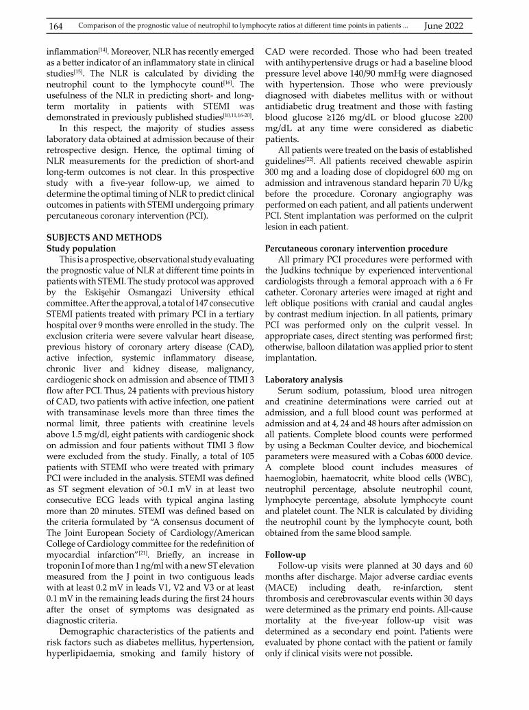
June 2022164
inflammation[14]. Moreover, NLR has recently emerged as a better indicator of an inflammatory state in clinical studies[15]. The NLR is calculated by dividing the neutrophil count to the lymphocyte count[16]. The usefulness of the NLR in predicting short- and long-term mortality in patients with STEMI was demonstrated in previously published studies[10,11,16-20].
In this respect, the majority of studies assess laboratory data obtained at admission because of their retrospective design. Hence, the optimal timing of NLR measurements for the prediction of short-and long-term outcomes is not clear. In this prospective study with a five-year follow-up, we aimed to determine the optimal timing of NLR to predict clinical outcomes in patients with STEMI undergoing primary percutaneous coronary intervention (PCI).
SUBJECTS AND METHODSStudy population
This is a prospective, observational study evaluating the prognostic value of NLR at different time points in patients with STEMI. The study protocol was approved by the Eskişehir Osmangazi University ethical committee. After the approval, a total of 147 consecutive STEMI patients treated with primary PCI in a tertiary hospital over 9 months were enrolled in the study. The exclusion criteria were severe valvular heart disease, previous history of coronary artery disease (CAD), active infection, systemic inflammatory disease, chronic liver and kidney disease, malignancy, cardiogenic shock on admission and absence of TIMI 3 flow after PCI. Thus, 24 patients with previous history of CAD, two patients with active infection, one patient with transaminase levels more than three times the normal limit, three patients with creatinine levels above 1.5 mg/dl, eight patients with cardiogenic shock on admission and four patients without TIMI 3 flow were excluded from the study. Finally, a total of 105 patients with STEMI who were treated with primary PCI were included in the analysis. STEMI was defined as ST segment elevation of >0.1 mV in at least two consecutive ECG leads with typical angina lasting more than 20 minutes. STEMI was defined based on the criteria formulated by “A consensus document of The Joint European Society of Cardiology/American College of Cardiology committee for the redefinition of myocardial infarction”[21]. Briefly, an increase in troponin I of more than 1 ng/ml with a new ST elevation measured from the J point in two contiguous leads with at least 0.2 mV in leads V1, V2 and V3 or at least 0.1 mV in the remaining leads during the first 24 hours after the onset of symptoms was designated as diagnostic criteria.
Demographic characteristics of the patients and risk factors such as diabetes mellitus, hypertension, hyperlipidaemia, smoking and family history of
CAD were recorded. Those who had been treated with antihypertensive drugs or had a baseline blood pressure level above 140/90 mmHg were diagnosed with hypertension. Those who were previously diagnosed with diabetes mellitus with or without antidiabetic drug treatment and those with fasting blood glucose ≥126 mg/dL or blood glucose ≥200 mg/dL at any time were considered as diabetic patients.
All patients were treated on the basis of established guidelines[22]. All patients received chewable aspirin 300 mg and a loading dose of clopidogrel 600 mg on admission and intravenous standard heparin 70 U/kg before the procedure. Coronary angiography was performed on each patient, and all patients underwent PCI. Stent implantation was performed on the culprit lesion in each patient.
Percutaneous coronary intervention procedureAll primary PCI procedures were performed with
the Judkins technique by experienced interventional cardiologists through a femoral approach with a 6 Fr catheter. Coronary arteries were imaged at right and left oblique positions with cranial and caudal angles by contrast medium injection. In all patients, primary PCI was performed only on the culprit vessel. In appropriate cases, direct stenting was performed first; otherwise, balloon dilatation was applied prior to stent implantation.
Laboratory analysisSerum sodium, potassium, blood urea nitrogen
and creatinine determinations were carried out at admission, and a full blood count was performed at admission and at 4, 24 and 48 hours after admission on all patients. Complete blood counts were performed by using a Beckman Coulter device, and biochemical parameters were measured with a Cobas 6000 device. A complete blood count includes measures of haemoglobin, haematocrit, white blood cells (WBC), neutrophil percentage, absolute neutrophil count, lymphocyte percentage, absolute lymphocyte count and platelet count. The NLR is calculated by dividing the neutrophil count by the lymphocyte count, both obtained from the same blood sample.
Follow-upFollow-up visits were planned at 30 days and 60
months after discharge. Major adverse cardiac events (MACE) including death, re-infarction, stent thrombosis and cerebrovascular events within 30 days were determined as the primary end points. All-cause mortality at the five-year follow-up visit was determined as a secondary end point. Patients were evaluated by phone contact with the patient or family only if clinical visits were not possible.
Comparison of the prognostic value of neutrophil to lymphocyte ratios at different time points in patients ...

KUWAIT MEDICAL JOURNAL 165June 2022
Statistical analysisStatistical analyses were performed using the IBM
statistics 21.0 MedCalc 11.0 software package. Patient characteristics are presented as the mean ± standard deviation or median (interquartile range) for continuous variables when appropriate and as percentages for discrete variables. The variables were tested for a normal distribution by the Shapiro-Wilk normality test. Differences in categorical variables were analysed by Pearson’s chi-square test or Fisher’s exact test. An independent-sample t-test was used for the analysis of normally distributed variables and a Mann-Whitney U-test was used for the analysis of non-normally distributed variables.
Via MedCalc11.0, complete sensitivity/specificity reports were created with receiver operating characteristic (ROC) curves and the area under the curve (AUC) and cut-off points of the parameters measured. We used the method of Deong et al[23] to calculate the standard error of the AUC and of the difference between AUCs. The diagnostic performance of the tests to discriminate endpoints were revealed by
backward stepwise logistic regression analysis and also by comparing AUCs. A two-tailed P <0.05 was considered to indicate statistical significance.
RESULTSA total of 105 patients [81% male (n: 85), mean age:
58.36±13.01] with STEMI who were treated with stent implantation were included in the analysis. MI was revealed as follows: 35 (33.3%) anterior MI, 13 (12.4%) anteroseptal MI, 32 (30.5%) inferior MI, 23 (21.9%) inferior and right ventricular MI and two (1.9%) lateral MI. The culprit coronary artery was detected as the left main coronary artery in one (1%), left anterior descending artery in 50 (47.6%), circumflex artery in 12 (11.4%) and the right coronary artery in 42 (40%) patients.
The total WBC count was elevated at admission and decreased after the 24-hour plateau. The neutrophil count reached its maximum value four hours after admission and decreased until the 48th hour. Conversely, lymphocytes followed an inverse curve to that of neutrophils, reaching their lowest value four
Fig 1: Changes in white blood cells, neutrophils, lymphocytes and NLR over time.
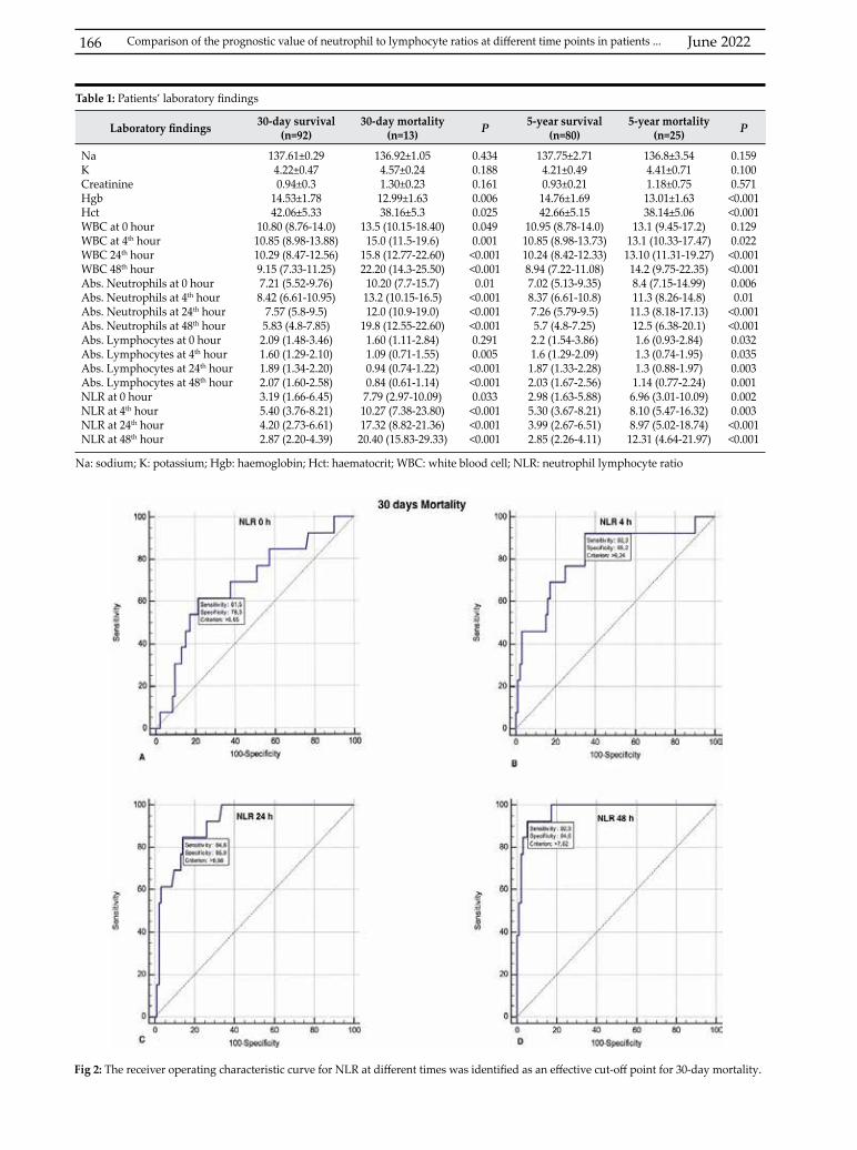
June 2022166
Laboratory findings 30-day survival (n=92)
30-day mortality (n=13) P 5-year survival
(n=80)5-year mortality
(n=25) P
NaKCreatinineHgbHctWBC at 0 hourWBC at 4th hourWBC 24th hourWBC 48th hourAbs. Neutrophils at 0 hourAbs. Neutrophils at 4th hourAbs. Neutrophils at 24th hourAbs. Neutrophils at 48th hourAbs. Lymphocytes at 0 hourAbs. Lymphocytes at 4th hourAbs. Lymphocytes at 24th hourAbs. Lymphocytes at 48th hourNLR at 0 hourNLR at 4th hourNLR at 24th hourNLR at 48th hour
137.61±0.294.22±0.470.94±0.3
14.53±1.7842.06±5.33
10.80 (8.76-14.0)10.85 (8.98-13.88)10.29 (8.47-12.56)9.15 (7.33-11.25)7.21 (5.52-9.76)8.42 (6.61-10.95)
7.57 (5.8-9.5)5.83 (4.8-7.85)2.09 (1.48-3.46)1.60 (1.29-2.10)1.89 (1.34-2.20)2.07 (1.60-2.58)3.19 (1.66-6.45)5.40 (3.76-8.21)4.20 (2.73-6.61)2.87 (2.20-4.39)
136.92±1.054.57±0.241.30±0.2312.99±1.6338.16±5.3
13.5 (10.15-18.40)15.0 (11.5-19.6)
15.8 (12.77-22.60)22.20 (14.3-25.50)
10.20 (7.7-15.7)13.2 (10.15-16.5)12.0 (10.9-19.0)
19.8 (12.55-22.60)1.60 (1.11-2.84)1.09 (0.71-1.55)0.94 (0.74-1.22)0.84 (0.61-1.14)7.79 (2.97-10.09)10.27 (7.38-23.80)17.32 (8.82-21.36)20.40 (15.83-29.33)
0.4340.1880.1610.0060.0250.0490.001<0.001<0.0010.01
<0.001<0.001<0.0010.2910.005<0.001<0.0010.033<0.001<0.001<0.001
137.75±2.714.21±0.490.93±0.2114.76±1.6942.66±5.15
10.95 (8.78-14.0)10.85 (8.98-13.73)10.24 (8.42-12.33)8.94 (7.22-11.08)7.02 (5.13-9.35)8.37 (6.61-10.8)7.26 (5.79-9.5)5.7 (4.8-7.25)2.2 (1.54-3.86)1.6 (1.29-2.09)1.87 (1.33-2.28)2.03 (1.67-2.56)2.98 (1.63-5.88)5.30 (3.67-8.21)3.99 (2.67-6.51)2.85 (2.26-4.11)
136.8±3.544.41±0.711.18±0.7513.01±1.6338.14±5.06
13.1 (9.45-17.2)13.1 (10.33-17.47)13.10 (11.31-19.27)14.2 (9.75-22.35)8.4 (7.15-14.99)11.3 (8.26-14.8)11.3 (8.18-17.13)12.5 (6.38-20.1)1.6 (0.93-2.84)1.3 (0.74-1.95)1.3 (0.88-1.97)1.14 (0.77-2.24)6.96 (3.01-10.09)8.10 (5.47-16.32)8.97 (5.02-18.74)12.31 (4.64-21.97)
0.1590.1000.571<0.001<0.0010.1290.022<0.001<0.0010.0060.01
<0.001<0.0010.0320.0350.0030.0010.0020.003<0.001<0.001
Table 1: Patients’ laboratory findings
Na: sodium; K: potassium; Hgb: haemoglobin; Hct: haematocrit; WBC: white blood cell; NLR: neutrophil lymphocyte ratio
Fig 2: The receiver operating characteristic curve for NLR at different times was identified as an effective cut-off point for 30-day mortality.
Comparison of the prognostic value of neutrophil to lymphocyte ratios at different time points in patients ...
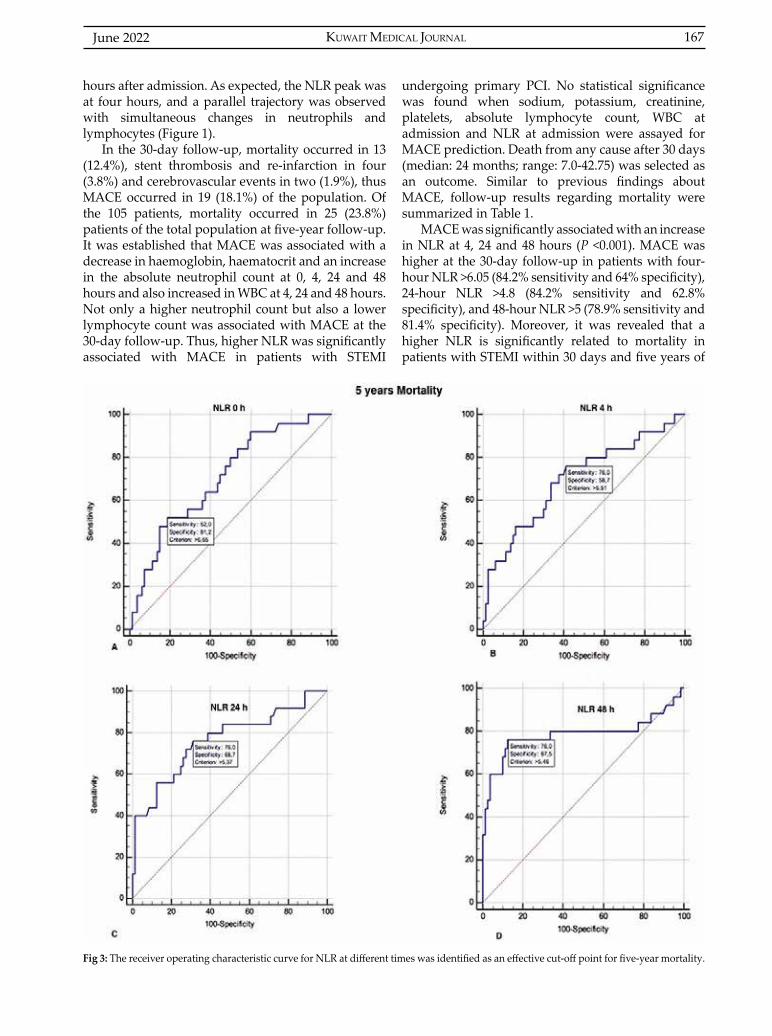
KUWAIT MEDICAL JOURNAL 167June 2022
hours after admission. As expected, the NLR peak was at four hours, and a parallel trajectory was observed with simultaneous changes in neutrophils and lymphocytes (Figure 1).
In the 30-day follow-up, mortality occurred in 13 (12.4%), stent thrombosis and re-infarction in four (3.8%) and cerebrovascular events in two (1.9%), thus MACE occurred in 19 (18.1%) of the population. Of the 105 patients, mortality occurred in 25 (23.8%) patients of the total population at five-year follow-up. It was established that MACE was associated with a decrease in haemoglobin, haematocrit and an increase in the absolute neutrophil count at 0, 4, 24 and 48 hours and also increased in WBC at 4, 24 and 48 hours. Not only a higher neutrophil count but also a lower lymphocyte count was associated with MACE at the 30-day follow-up. Thus, higher NLR was significantly associated with MACE in patients with STEMI
undergoing primary PCI. No statistical significance was found when sodium, potassium, creatinine, platelets, absolute lymphocyte count, WBC at admission and NLR at admission were assayed for MACE prediction. Death from any cause after 30 days (median: 24 months; range: 7.0-42.75) was selected as an outcome. Similar to previous findings about MACE, follow-up results regarding mortality were summarized in Table 1.
MACE was significantly associated with an increase in NLR at 4, 24 and 48 hours (P <0.001). MACE was higher at the 30-day follow-up in patients with four-hour NLR >6.05 (84.2% sensitivity and 64% specificity), 24-hour NLR >4.8 (84.2% sensitivity and 62.8% specificity), and 48-hour NLR >5 (78.9% sensitivity and 81.4% specificity). Moreover, it was revealed that a higher NLR is significantly related to mortality in patients with STEMI within 30 days and five years of
Fig 3: The receiver operating characteristic curve for NLR at different times was identified as an effective cut-off point for five-year mortality.
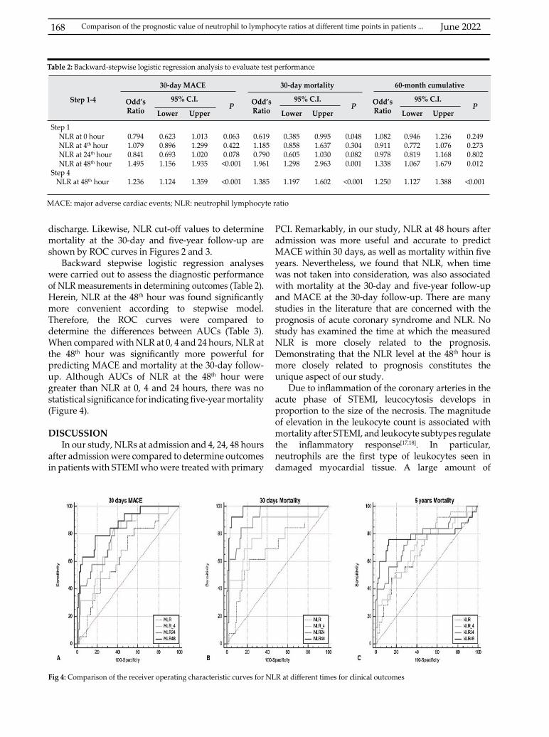
June 2022168
discharge. Likewise, NLR cut-off values to determine mortality at the 30-day and five-year follow-up are shown by ROC curves in Figures 2 and 3.
Backward stepwise logistic regression analyses were carried out to assess the diagnostic performance of NLR measurements in determining outcomes (Table 2). Herein, NLR at the 48th hour was found significantly more convenient according to stepwise model. Therefore, the ROC curves were compared to determine the differences between AUCs (Table 3). When compared with NLR at 0, 4 and 24 hours, NLR at the 48th hour was significantly more powerful for predicting MACE and mortality at the 30-day follow-up. Although AUCs of NLR at the 48th hour were greater than NLR at 0, 4 and 24 hours, there was no statistical significance for indicating five-year mortality (Figure 4).
DISCUSSIONIn our study, NLRs at admission and 4, 24, 48 hours
after admission were compared to determine outcomes in patients with STEMI who were treated with primary
PCI. Remarkably, in our study, NLR at 48 hours after admission was more useful and accurate to predict MACE within 30 days, as well as mortality within five years. Nevertheless, we found that NLR, when time was not taken into consideration, was also associated with mortality at the 30-day and five-year follow-up and MACE at the 30-day follow-up. There are many studies in the literature that are concerned with the prognosis of acute coronary syndrome and NLR. No study has examined the time at which the measured NLR is more closely related to the prognosis. Demonstrating that the NLR level at the 48th hour is more closely related to prognosis constitutes the unique aspect of our study.
Due to inflammation of the coronary arteries in the acute phase of STEMI, leucocytosis develops in proportion to the size of the necrosis. The magnitude of elevation in the leukocyte count is associated with mortality after STEMI, and leukocyte subtypes regulate the inflammatory response[17,18]. In particular, neutrophils are the first type of leukocytes seen in damaged myocardial tissue. A large amount of
Step 1-4
30-day MACE 30-day mortality 60-month cumulative
Odd’s Ratio
95% C.I.P Odd’s
Ratio95% C.I.
P Odd’s Ratio
95% C.I.P
Lower Upper Lower Upper Lower Upper
Step 1 NLR at 0 hourNLR at 4th hourNLR at 24th hourNLR at 48th hour
Step 4 NLR at 48th hour
0.7941.0790.8411.495
1.236
0.6230.8960.6931.156
1.124
1.0131.2991.0201.935
1.359
0.0630.4220.078<0.001
<0.001
0.6191.1850.7901.961
1.385
0.3850.8580.6051.298
1.197
0.9951.6371.0302.963
1.602
0.0480.3040.0820.001
<0.001
1.0820.9110.9781.338
1.250
0.9460.7720.8191.067
1.127
1.2361.0761.1681.679
1.388
0.2490.2730.8020.012
<0.001
Table 2: Backward-stepwise logistic regression analysis to evaluate test performance
MACE: major adverse cardiac events; NLR: neutrophil lymphocyte ratio
Fig 4: Comparison of the receiver operating characteristic curves for NLR at different times for clinical outcomes
Comparison of the prognostic value of neutrophil to lymphocyte ratios at different time points in patients ...
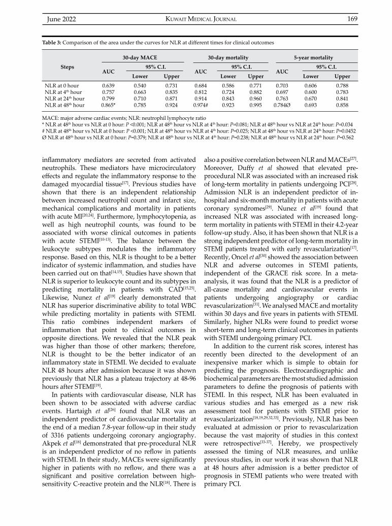
KUWAIT MEDICAL JOURNAL 169June 2022
inflammatory mediators are secreted from activated neutrophils. These mediators have microcirculatory effects and regulate the inflammatory response to the damaged myocardial tissue[17]. Previous studies have shown that there is an independent relationship between increased neutrophil count and infarct size, mechanical complications and mortality in patients with acute MI[20,24]. Furthermore, lymphocytopenia, as well as high neutrophil counts, was found to be associated with worse clinical outcomes in patients with acute STEMI[10-13]. The balance between the leukocyte subtypes modulates the inflammatory response. Based on this, NLR is thought to be a better indicator of systemic inflammation, and studies have been carried out on that[14,15]. Studies have shown that NLR is superior to leukocyte count and its subtypes in predicting mortality in patients with CAD[15,25]. Likewise, Nunez et al[19] clearly demonstrated that NLR has superior discriminative ability to total WBC while predicting mortality in patients with STEMI. This ratio combines independent markers of inflammation that point to clinical outcomes in opposite directions. We revealed that the NLR peak was higher than those of other markers; therefore, NLR is thought to be the better indicator of an inflammatory state in STEMI. We decided to evaluate NLR 48 hours after admission because it was shown previously that NLR has a plateau trajectory at 48-96 hours after STEMI[19].
In patients with cardiovascular disease, NLR has been shown to be associated with adverse cardiac events. Hartaigh et al[26] found that NLR was an independent predictor of cardiovascular mortality at the end of a median 7.8-year follow-up in their study of 3316 patients undergoing coronary angiography. Akpek et al[18] demonstrated that pre-procedural NLR is an independent predictor of no reflow in patients with STEMI. In their study, MACEs were significantly higher in patients with no reflow, and there was a significant and positive correlation between high-sensitivity C-reactive protein and the NLR[18]. There is
also a positive correlation between NLR and MACEs[27]. Moreover, Duffy et al showed that elevated pre-procedural NLR was associated with an increased risk of long-term mortality in patients undergoing PCI[28]. Admission NLR is an independent predictor of in-hospital and six-month mortality in patients with acute coronary syndromes[29]. Nunez et al[19] found that increased NLR was associated with increased long-term mortality in patients with STEMI in their 4.2-year follow-up study. Also, it has been shown that NLR is a strong independent predictor of long-term mortality in STEMI patients treated with early revascularization[17]. Recently, Oncel et al[30] showed the association between NLR and adverse outcomes in STEMI patients, independent of the GRACE risk score. In a meta-analysis, it was found that the NLR is a predictor of all-cause mortality and cardiovascular events in patients undergoing angiography or cardiac revascularization[31]. We analysed MACE and mortality within 30 days and five years in patients with STEMI. Similarly, higher NLRs were found to predict worse short-term and long-term clinical outcomes in patients with STEMI undergoing primary PCI.
In addition to the current risk scores, interest has recently been directed to the development of an inexpensive marker which is simple to obtain for predicting the prognosis. Electrocardiographic and biochemical parameters are the most studied admission parameters to define the prognosis of patients with STEMI. In this respect, NLR has been evaluated in various studies and has emerged as a new risk assessment tool for patients with STEMI prior to revascularization[18,19,29,32,33]. Previously, NLR has been evaluated at admission or prior to revascularization because the vast majority of studies in this context were retrospective[33-37]. Hereby, we prospectively assessed the timing of NLR measures, and unlike previous studies, in our work it was shown that NLR at 48 hours after admission is a better predictor of prognosis in STEMI patients who were treated with primary PCI.
Steps
30-day MACE 30-day mortality 5-year mortality
AUC95% C.I.
AUC95% C.I.
AUC95% C.I.
Lower Upper Lower Upper Lower Upper
NLR at 0 hourNLR at 4th hourNLR at 24th hourNLR at 48th hour
0.6390.7570.7990.865*
0.5400.6630.7100.785
0.7310.8350.8710.924
0.6840.8120.9140.974#
0.5860.7240.8430.923
0.7710.8820.9600.995
0.7030.6970.763
0.784Ø
0.6060.6000.6700.693
0.7880.7830.8410.858
MACE: major adverse cardiac events; NLR: neutrophil lymphocyte ratio* NLR at 48th hour vs NLR at 0 hour: P <0.001; NLR at 48th hour vs NLR at 4th hour: P=0.081; NLR at 48th hour vs NLR at 24th hour: P=0.034# NLR at 48th hour vs NLR at 0 hour: P <0.001; NLR at 48th hour vs NLR at 4th hour: P=0.025; NLR at 48th hour vs NLR at 24th hour: P=0.0452Ø NLR at 48th hour vs NLR at 0 hour: P=0.379; NLR at 48th hour vs NLR at 4th hour: P=0.238; NLR at 48th hour vs NLR at 24th hour: P=0.562
Table 3: Comparison of the area under the curves for NLR at different times for clinical outcomes
June 2022170
Study limitationsIn our study, we did not evaluate in-hospital MACE
and mortality because we excluded early complications and patients with cardiogenic shock to obtain blood counts until 48 hours from each patient. Also, we did not take into consideration five-year MACE because of the study design. A well-designed (prospective, randomized, and multi-centre) study with larger study populations is needed to evaluate this reasonable marker in order to confirm our findings.
CONCLUSION It can be concluded from our findings that the NLR
is a marker which predicts short- and long-term outcomes in STEMI patients treated with primary PCI. To the best of our knowledge, this is the first study that identifies the prognostic importance of the timing of NLR measures to predict clinical outcomes. It is logical to postulate that NLR at 48 hours after admission or before discharge is more accurate for predicting clinical outcomes in patients with STEMI.
ACKNOWLEDGMENTConflict of interest statement: The authors report no conflict of interest.
This research received no specific grant from any funding agency in the public, commercial, or not for-profit sectors.Author contribution: Gurbet Ozge Mert: study design, data collection, literature review and wrote manuscript; Muhammet Dural: critical review; Kadir Ugur Mert: study design and supervision, data collection, analysis and interpretation, and wrote manuscript; Muzaffer Bilgin: data analysis and interpretation, and literature review; Bulent Gorenek: study design, supervision and critical review.
REFERENCES
1. Zairis MN, Lyras AG, Bibis GP, Patsourakos NG, Makrygiannis SS, Kardoulas AD, et al. Association of inflammatory biomarkers and cardiac troponin I with multifocal activation of coronary artery tree in the setting of non-ST-elevation acute myocardial infarction. Atherosclerosis 2005; 182:161-7.
2. Furman MI, Gore JM, Anderson FA, Budaj A, Goodman SG, Avezum A, et al. Elevated leukocyte count and adverse hospital events in patients with acute coronary syndromes: findings from the Global Registry of Acute Coronary Events (GRACE). Am Heart J 2004; 147(1):42-8.
3. Bhatt DL, Chew DP, Lincoff AM, Simoons ML, Harrington RA, Ommen SR, et al. Effect of revascularization on mortality associated with an elevated white blood cell count in acute coronary syndromes. Am J Cardiol 2003; 92(2):136-40.
4. Cannon CP, McCabe CH, Wilcox RG, Bentley JH, Braunwald E. Association of white blood cell count with increased mortality in acute myocardial infarction and unstable angina pectoris. OPUS-TIMI 16 Investigators. Am J Cardiol 2001; 87(5):636-9, A10.
5. Furman MI, Becker RC, Yarzebski J, Savegeau J, Gore JM, Goldberg RJ. Effect of elevated leukocyte count on in-hospital mortality following acute myocardial infarction. Am J Cardiol 1996; 78(8):945-8.
6. Barron HV, Cannon CP, Murphy SA, Braunwald E, Gibson CM. Association between white blood cell count, epicardial blood flow, myocardial perfusion, and clinical outcomes in the setting of acute myocardial infarction: a thrombolysis in myocardial infarction 10 substudy. Circulation 2000; 102(19):2329-34.
7. Coller BS. Leukocytosis and ischemic vascular disease morbidity and mortality: is it time to intervene? Arterioscler Thromb Vasc Biol 2005; 25(4):658-70.
8. Thomson SP, Gibbons RJ, Smars PA, Suman VJ, Pierre RV, Santrach PJ, et al. Incremental value of the leukocyte differential and the rapid creatine kinase-MB isoenzyme for the early diagnosis of myocardial infarction. Ann Intern Med 1995; 122(5):335-41.
9. Maekawa Y, Anzai T, Yoshikawa T, Asakura Y, Takahashi T, Ishikawa S, et al. Prognostic significance of peripheral monocytosis after reperfused acute myocardial infarction:a possible role for left ventricular remodeling. J Am Coll Cardiol 2002; 39(2):241-6.
10. Chia S, Nagurney JT, Brown DF, Raffel OC, Bamberg F, Senatore F, et al. Association of leukocyte and neutrophil counts with infarct size, left ventricular function and outcomes after percutaneous coronary intervention for ST-elevation myocardial infarction. Am J Cardiol 2009; 103(3):333-7.
11. Dragu R, Huri S, Zuckerman R, Suleiman M, Mutlak D, Agmon Y, et al. Predictive value of white blood cell subtypes for long-term outcome following myocardial infarction. Atherosclerosis 2008; 196(1):405-12.
12. Widmer A, Linka AZ, Attenhofer Jost CH, Buergi B, Brunner-La Rocca HP, Salomon F, et al. Mechanical complications after myocardial infarction reliably predicted using C-reactive protein levels and lymphocytopenia. Cardiology 2003; 99(1):25-31.
13. Zouridakis EG, Garcia-Moll X, Kaski JC. Usefulness of the blood lymphocyte count in predicting recurrent instability and death in patients with unstable angina pectoris. Am J Cardiol 2000; 86(4):449-51.
14. Balta S, Celik T, Mikhailidis DP, Ozturk C, Demirkol S, Aparci M, et al. The Relation Between Atherosclerosis and the Neutrophil-Lymphocyte Ratio. Clin Appl Thromb Hemost 2016; 22(5):405-11.
15. Kaya MG. [Inflammation and coronary artery disease: as a new biomarker neutrophil/lymphocyte ratio]. Turk Kardiyol Dern Ars 2013; 41(3):191-2.
16. Arbel Y, Finkelstein A, Halkin A, Birati EY, Revivo M, Zuzut M, et al. Neutrophil/lymphocyte ratio is related to the severity of coronary artery disease and clinical outcome in patients undergoing angiography. Atherosclerosis 2012; 225(2):456-60.
Comparison of the prognostic value of neutrophil to lymphocyte ratios at different time points in patients ...
KUWAIT MEDICAL JOURNAL 171June 2022
17. Shen XH, Chen Q, Shi Y, Li HW. Association of neutrophil/lymphocyte ratio with long-term mortality after ST elevation myocardial infarction treated with primary percutaneous coronary intervention. Chin Med J (Engl) 2010; 123(23):3438-43.
18. Akpek M, Kaya MG, Lam YY, Sahin O, Elcik D, Celik T, et al. Relation of neutrophil/lymphocyte ratio to coronary flow to in-hospital major adverse cardiac events in patients with ST-elevated myocardial infarction undergoing primary coronary intervention. Am J Cardiol 2012; 110(5):621-7.
19. Nunez J, Nunez E, Bodi V, Sanchis J, Minana G, Mainar L, et al. Usefulness of the neutrophil to lymphocyte ratio in predicting long-term mortality in ST segment elevation myocardial infarction. Am J Cardiol 2008; 101(6):747-52.
20. O’Donoghue M, Morrow DA, Cannon CP, Guo W, Murphy SA, Gibson CM, et al. Association between baseline neutrophil count, clopidogrel therapy, and clinical and angiographic outcomes in patients with ST-elevation myocardial infarction receiving fibrinolytic therapy. Eur Heart J 2008; 29(8):984-91.
21. Antman E, Bassand J-P, Klein W, Ohman M, Lopez Sendon JL, Rydén L, et al. Myocardial infarction redefined—a consensus document of The Joint European Society of Cardiology/American College of Cardiology committee for the redefinition of myocardial infarction: The Joint European Society of Cardiology/ American College of Cardiology Committee. J Am Coll Cardiol 2000; 36(3):959-69.
22. Steg PG, James SK, Atar D, Badano LP, Blomstrom-Lundqvist C, Borger MA, et al. ESC Guidelines for the management of acute myocardial infarction in patients presenting with ST-segment elevation. Eur Heart J 2012; 33(20):2569-619.
23. DeLong ER, DeLong DM, Clarke-Pearson DL. Comparing the areas under two or more correlated receiver operating characteristic curves: a nonparametric approach. Biometrics 1988; 44(3):837-45.
24. Kirtane AJ, Bui A, Murphy SA, Barron HV, Gibson CM. Association of peripheral neutrophilia with adverse angiographic outcomes in ST-elevation myocardial infarction. Am J Cardiol 2004; 93(5):532-6.
25. Horne BD, Anderson JL, John JM, Weaver A, Bair TL, Jensen KR, et al. Which white blood cell subtypes predict increased cardiovascular risk? J Am Coll Cardiol 2005; 45(10):1638-43.
26. o Hartaigh B, Bosch JA, Thomas GN, Lord JM, Pilz S, Loerbroks A, et al. Which leukocyte subsets predict cardiovascular mortality? From the LUdwigshafen RIsk and Cardiovascular Health (LURIC) Study. Atherosclerosis 2012; 224(1):161-9.
27. Azab B, Zaher M, Weiserbs KF, Torbey E, Lacossiere K, Gaddam S, et al. Usefulness of neutrophil to lymphocyte
ratio in predicting short- and long-term mortality after non-ST-elevation myocardial infarction. Am J Cardiol 2010; 106(4):470-6.
28. Duffy BK, Gurm HS, Rajagopal V, Gupta R, Ellis SG, Bhatt DL. Usefulness of an elevated neutrophil to lymphocyte ratio in predicting long-term mortality after percutaneous coronary intervention. Am J Cardiol 2006; 97(7):993-6.
29. Tamhane UU, Aneja S, Montgomery D, Rogers EK, Eagle KA, Gurm HS. Association between admission neutrophil to lymphocyte ratio and outcomes in patients with acute coronary syndrome. Am J Cardiol 2008; 102(6):653-7.
30. Oncel RC, Ucar M, Karakas MS, Akdemir B, Yanikoglu A, Gulcan AR, et al. Relation of neutrophil-to-lymphocyte ratio with GRACE risk score to in-hospital cardiac events in patients with ST-segment elevated myocardial infarction. Clin Appl Thromb Hemost 2015; 21(4):383-8.
31. Wang X, Zhang G, Jiang X, Zhu H, Lu Z, Xu L. Neutrophil to lymphocyte ratio in relation to risk of all-cause mortality and cardiovascular events among patients undergoing angiography or cardiac revascularization: a meta-analysis of observational studies. Atherosclerosis 2014; 234(1):206-13.
32. Cho KH, Jeong MH, Ahmed K, Hachinohe D, Choi HS, Chang SY, et al. Value of early risk stratification using hemoglobin level and neutrophil-to-lymphocyte ratio in patients with ST-elevation myocardial infarction undergoing primary percutaneous coronary intervention. Am J Cardiol 2011; 107(6):849-56.
33. Acet H, Ertaş F, Ata Akıl M, Özyurtlu F, Yıldız A, Polat N, et al. Novel predictors of infarct-related artery patency for ST-segment elevation myocardial infarction: Platelet-to-lymphocyte ratio, uric acid, and neutrophil-to-lymphocyte ratio. Anatol J Cardiol 2016; 15(8):648-56.
34. Akyel A, Yayla Ç, Erat M, Çimen T, Doğan M, Açıkel S, et al. Neutrophil-to-lymphocyte ratio predicts hemodynamic significance of coronary artery stenosis. Anatol J Cardiol 2015; 15(12):1002-7.
35. Bahadır A, Baltacı D, Türker Y, Türker Y, Iliev D, Öztürk S, et al. Is the neutrophil-to-lymphocyte ratio indicative of inflammatory state in patients with obesity and metabolic syndrome? Anatol J Cardiol 2015; 15(10):816-22.
36. Aykan AÇ, Hatem E, Kalaycıoğlu E, Karabay CY, Zehir R, Gökdeniz T, et al. Neutrophil-to-lymphocyte ratio may be a marker of peripheral artery disease complexity. Anatol J Cardiol 2016; 16(7):497-503.
37. MK Akboğa, Akyel A, Şahinarslan A, Yayla Ç, Alsancak Y, Gökalp G, et al. Neutrophil-to-lymphocyte ratio is increased in patients with rheumatic mitral valve stenosis? Anatol J Cardiol 2015; 15(5):380-4.
KUWAIT MEDICAL JOURNAL June 2022172
Original Article
Kuwait Medical Journal 2022; 54 (2): 172 - 179
Evaluation on predictive values of risk scores in non-variceal upper gastrointestinal bleeding
ABSTRACT
KEY WORDS: mortality, non-variceal upper gastrointestinal bleeding, re-bleeding, risk score
Nam-Hun Jong, Hye-Song Kim, Hak-Chol Ri, Song-Il Rim, Jong-Nam KangDepartment of Gastroenterology, Pyongyang Medical College Hospital, Kim Il Sung University, Democratic People’s
Republic of Korea
Objective: To determine which is the best score in predicting re-bleeding and mortality for patients with acute non-variceal upper gastrointestinal bleeding (NVUGIB) among seven scores (Forrest, full Rockall score (RS), admission Rockall, Glasgow-blatchford score (GBS), Cedars-Sinai medical center predictive index (CMCPI), T score, AIMS65 score (AIMS65), Progetto nazionale emorragia digestive (PNED)).Design: Prospective studySetting: Pyongyang medical University Hospital, Gastroenterology Department from 1/2015 to 4/2019Subjects: This study included 404 adult patients with NVUGIBInterventions: Fluid resuscitation, blood transfusion, pharmacologic therapy proton pump inhibitor (intravenous or oral), endoscopic hemostasis and surgery were considered according to national guidelines for management of upper gastrointestinal bleedingMain outcome measures: Main clinical outcomes were re-
bleeding and 30-day mortality. Predictive ability of all scores were estimated using area under the receiver operating curve (AUROC).Results: The largest AUROC (0.846) was obtained from the Forrest score, followed by T score (AUROC: 0.818), full RS (AUROC: 0.813), CMCPI (AUROC: 0.799) and GBS (AUROC: 0.794) when predicting re-bleeding. Admission RS (AUROC: 0.716) and AIMS65 (AUROC: 0.691) were less predictive than other scores. Regarding mortality, PNED (AUROC: 0.955) score had the highest predictive ability followed by full RS (Rockall score) (AUROC: 0.919), admission RS (AUROC: 0.917), AIMS65 (AUROC: 0.899), CMCPI (AUROC: 0.872) GBS (AUROC: 0.854), T score (AUROC: 0.851). Forrest score (AUROC: 0.590) had the worst performance at predicting death.Conclusion: Forrest score is the best at predicting re-bleeding and PNED score predicts mortality for patients with NVUGIB with high accuracy.
Address correspondence to:Hye-Song Kim, Ryonhwa Dong, Pyongyang, Democratic People’s Republic of Korea. Tel: 02-551-2232; E-mail: [email protected]
INTRODUCTIONNonvariceal upper gastrointestinal bleeding
(NVUGIB) is a common gastrointestinal emergency with an annual incidence of 50 to 150 per 100,000 adults. Mortality from upper gastrointestinal bleeding (UGIB) is around 10% and may reach 35% in aged patients with comorbidities[1]. Although 80% of UGIB stop spontaneously, the remaining patients re-bleed or continue bleeding. Re-bleeding or continuous bleeding is an independent risk of mortality[2].
Therefore, early identification of patients at high risk of re-bleeding or death is an essential part in the
management of NVUGIB helping clinicians guide appropriate treatment. Identifying patients who are at low risk of poor outcome is also important to reduce hospital stay and medical resources.
To date, several clinical scores have been developed to predict re-bleeding, mortality, clinical intervention and urgent endoscopy for patients with NVUGIB. Recent international recommendations regarding management of NVUGIB encourage gastroenterologists to perform these scoring systems for risk stratification. These scores vary from simple classification (Forrest score) to complex model based
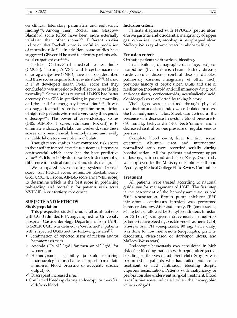
KUWAIT MEDICAL JOURNAL 173June 2022
on clinical, laboratory parameters and endoscopic finding[3-9]. Among them, Rockall and Glasgow-Blachford score (GBS) have been more externally validated than other scores[4,5]. Different studies indicated that Rockall score is useful in prediction of mortality risk[10,11]. In addition, some studies have suggested GBS could be used to identify patients who need outpatient care[12,13].
Besides Cedars-Sinai medical center index (CMCPI), T score, AIMS65 and Progetto nazionale emorragia digestive (PNED) have also been described and these scores require further evaluation[6-9]. Marmo R et al developed Italian PNED score and they concluded it was superior to Rockall score in predicting mortality[9]. Some studies reported AIMS65 had better accuracy than GBS in predicting in-patient mortality and the need for emergency intervention[14,15]. It was also suggested that T score is helpful for the prediction of high-risk patients who need a very early therapeutic endoscopy[16]. The power of pre-endoscopy scores (GBS, AIMS65, T score, admission Rockall) is to eliminate endoscopist’s labor on weekend, since these scores only use clinical, haemodynamic and easily available laboratory variables to calculate.
Though many studies have compared risk scores in their ability to predict various outcomes, it remains controversial which score has the best predictive value[17-20]. It is probably due to variety in demography, difference in medical care level and study design.
We compared seven scoring systems (Forrest score, full Rockall score, admission Rockall score, GBS, CMCPI, T score, AIMS65 score and PNED score) to determine which is the best score in predicting re-bleeding and mortality for patients with acute NVUGIB in our tertiary care center.
SUBJECTS AND METHODSStudy population
This prospective study included all adult patients with UGIB admitted to Pyongyang medical University Hospital, Gastroenterology Department from 1/2015 to 4/2019. UGIB was defined as ‘confirmed’ if patients with suspected UGIB met the following criteria[21]:• Combination of reported signs of melena and/or
hematemesis withüAnemia (Hb <13.0g/dl for men or <12.0g/dl for
women), orüHemodynamic instability (a state requiring
pharmacologic or mechanical support to maintain a normal blood pressure or adequate cardiac output), or
üDiscrepant increased urea• Confirmed bleeding during endoscopy or manifest
old/fresh blood
Inclusion criteriaPatients diagnosed with NVUGIB (peptic ulcer,
erosive gastritis and duodenitis, malignancy of upper gastrointestinal tract, esophagitis, esophageal ulcer, Mallory-Weiss syndrome, vascular abnormalities)
Exclusion criteriaCirrhotic patients with variceal bleeding.
In all patients, demographic data (age, sex), co-morbidities (liver disease, chronic kidney disease, cardiovascular disease, cerebral disease, diabetes, pulmonary disease, malignancy of other tract), previous history of peptic ulcer, UGIB and use of medication (non-steroid anti-inflammatory drug, oral anti-coagulants, corticosteroids, acetylsalicylic acid, clopidogrel) were collected by taking history.
Vital signs were measured through physical examination and shock index was calculated to assess the haemodynamic status. Shock was defined as the presence of a decrease in systolic blood pressure to <90 mmHg, tachycardia >100 beats/minute, and a decreased central venous pressure or jugular venous pressure.
Complete blood count, liver function, serum creatinine, albumin, urea and international normalized ratio were recorded serially during hospitalization. All the patients underwent upper endoscopy, ultrasound and chest X-ray. Our study was approved by the Ministry of Public Health and Pyongyang Medical College Ethic Review Committee.
TreatmentAll patients were treated according to national
guidelines for management of UGIB. The first step is the assessment of the hemodynamic status and fluid resuscitation. Proton pump inhibitor (PPI) intravenous continuous infusion was performed before endoscopy. After endoscopy, PPI (omeprazole, 80 mg bolus, followed by 8 mg/h continuous infusion for 72 hours) was given intravenously in high-risk patients (active bleeding, visible vessel, adherent clot) whereas oral PPI (omeprazole, 80 mg, twice daily) was done for low risk lesions (esophagitis, gastritis, duodenitis, clean-based or dark-spot ulcers, and Mallory-Weiss tears)
Endoscopic hemostasis was considered in high risk of re-bleeding patients with peptic ulcer (active bleeding, visible vessel, adherent clot). Surgery was performed in patients who had failed endoscopic treatment or had continuous bleeding despite vigorous resuscitation. Patients with malignancy or perforation also underwent surgical treatment. Blood transfusions were indicated when the hemoglobin value is <7 g/dL.
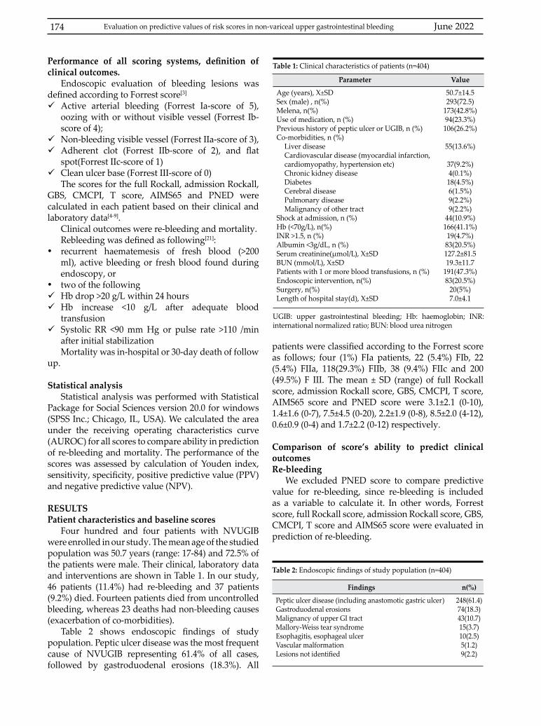
June 2022174
Performance of all scoring systems, definition of clinical outcomes.
Endoscopic evaluation of bleeding lesions was defined according to Forrest score[3]
üActive arterial bleeding (Forrest Ia-score of 5), oozing with or without visible vessel (Forrest Ib-score of 4);
üNon-bleeding visible vessel (Forrest IIa-score of 3),üAdherent clot (Forrest IIb-score of 2), and flat
spot(Forrest IIc-score of 1)üClean ulcer base (Forrest III-score of 0)
The scores for the full Rockall, admission Rockall, GBS, CMCPI, T score, AIMS65 and PNED were calculated in each patient based on their clinical and laboratory data[4-9].
Clinical outcomes were re-bleeding and mortality.Rebleeding was defined as following[21]:
· recurrent haematemesis of fresh blood (>200 ml), active bleeding or fresh blood found during endoscopy, or
· two of the followingüHb drop >20 g/L within 24 hoursüHb increase <10 g/L after adequate blood
transfusionüSystolic RR <90 mm Hg or pulse rate >110 /min
after initial stabilizationMortality was in-hospital or 30-day death of follow
up.
Statistical analysisStatistical analysis was performed with Statistical
Package for Social Sciences version 20.0 for windows (SPSS Inc.; Chicago, IL, USA). We calculated the area under the receiving operating characteristics curve (AUROC) for all scores to compare ability in prediction of re-bleeding and mortality. The performance of the scores was assessed by calculation of Youden index, sensitivity, specificity, positive predictive value (PPV) and negative predictive value (NPV).
RESULTSPatient characteristics and baseline scores
Four hundred and four patients with NVUGIB were enrolled in our study. The mean age of the studied population was 50.7 years (range: 17-84) and 72.5% of the patients were male. Their clinical, laboratory data and interventions are shown in Table 1. In our study, 46 patients (11.4%) had re-bleeding and 37 patients (9.2%) died. Fourteen patients died from uncontrolled bleeding, whereas 23 deaths had non-bleeding causes (exacerbation of co-morbidities).
Table 2 shows endoscopic findings of study population. Peptic ulcer disease was the most frequent cause of NVUGIB representing 61.4% of all cases, followed by gastroduodenal erosions (18.3%). All
Parameter Value
Age (years), X±SDSex (male) , n(%)Melena, n(%)Use of medication, n (%)Previous history of peptic ulcer or UGIB, n (%)Co-morbidities, n (%)
Liver diseaseCardiovascular disease (myocardial infarction, cardiomyopathy, hypertension etc)Chronic kidney diseaseDiabetesCerebral diseasePulmonary diseaseMalignancy of other tract
Shock at admission, n (%)Hb (<70g/L), n(%)INR >1.5, n (%)Albumin <3g/dL, n (%)Serum creatinine(μmol/L), X±SDBUN (mmol/L), X±SDPatients with 1 or more blood transfusions, n (%)Endoscopic intervention, n(%)Surgery, n(%)Length of hospital stay(d), X±SD
50.7±14.5293(72.5)
173(42.8%)94(23.3%)106(26.2%)
55(13.6%)
37(9.2%)4(0.1%)18(4.5%)6(1.5%)9(2.2%)9(2.2%)
44(10.9%)166(41.1%)19(4.7%)83(20.5%)127.2±81.519.3±11.7
191(47.3%)83(20.5%)
20(5%)7.0±4.1
Table 1: Clinical characteristics of patients (n=404)
UGIB: upper gastrointestinal bleeding; Hb: haemoglobin; INR: international normalized ratio; BUN: blood urea nitrogen
patients were classified according to the Forrest score as follows; four (1%) FIa patients, 22 (5.4%) FIb, 22 (5.4%) FIIa, 118(29.3%) FIIb, 38 (9.4%) FIIc and 200 (49.5%) F III. The mean ± SD (range) of full Rockall score, admission Rockall score, GBS, CMCPI, T score, AIMS65 score and PNED score were 3.1±2.1 (0-10), 1.4±1.6 (0-7), 7.5±4.5 (0-20), 2.2±1.9 (0-8), 8.5±2.0 (4-12), 0.6±0.9 (0-4) and 1.7±2.2 (0-12) respectively.
Comparison of score’s ability to predict clinical outcomesRe-bleeding
We excluded PNED score to compare predictive value for re-bleeding, since re-bleeding is included as a variable to calculate it. In other words, Forrest score, full Rockall score, admission Rockall score, GBS, CMCPI, T score and AIMS65 score were evaluated in prediction of re-bleeding.
Findings n(%)
Peptic ulcer disease (including anastomotic gastric ulcer)Gastroduodenal erosionsMalignancy of upper GI tractMallory-Weiss tear syndromeEsophagitis, esophageal ulcerVascular malformationLesions not identified
248(61.4)74(18.3)43(10.7)15(3.7)10(2.5)5(1.2)9(2.2)
Table 2: Endoscopic findings of study population (n=404)
Evaluation on predictive values of risk scores in non-variceal upper gastrointestinal bleeding
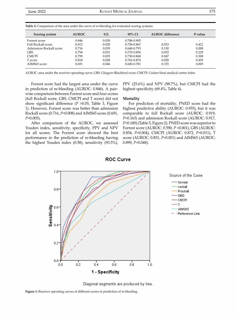
KUWAIT MEDICAL JOURNAL 175June 2022
Forrest score had the largest area under the curve in prediction of re-bleeding (AUROC: 0.846). A pair-wise comparison between Forrest score and four scores (full Rockall score, GBS, CMCPI and T score) did not show significant difference (P >0.05, Table 3, Figure 1). However, Forrest score was better than admission Rockall score (0.716, P=0.008) and AIMS65 score (0.691, P=0.005).
After comparison of the AUROC, we assessed Youden index, sensitivity, specificity, PPV and NPV for all scores. The Forrest score showed the best performance in the prediction of re-bleeding having the highest Youden index (0.58), sensitivity (93.5%),
PPV (25.6%) and NPV (98.7%), but CMCPI had the highest specificity (69.4%, Table 4).
MortalityFor prediction of mortality, PNED score had the
highest predictive ability (AUROC: 0.955), but it was comparable to full Rockall score (AUROC: 0.919, P=0.163) and admission Rockall score (AUROC: 0.917, P=0.180) (Table 5, Figure 2). PNED score was superior to Forrest score (AUROC: 0.590, P <0.001), GBS (AUROC: 0.854, P=0.004), CMCPI (AUROC: 0.872, P=0.011), T score (AUROC: 0.851, P=0.001) and AIMS65 (AUROC: 0.899, P=0.048).
Scoring system AUROC S.E. 95% CI AUROC difference P-value
Forrest scoreFull Rockall scoreAdmission Rockall scoreGBSCMCPIT scoreAIMS65 score
0.8460.8130.7160.7940.7990.8180.691
0.0300.0280.0390.0310.0350.0280.046
0.788-0.9050.758-0.8670.640-0.7930.733-0.8560.730-0.8680.762-0.8740.600-0.781
-0.0330.1300.0520.0470.0280.155
-0.4210.0080.2290.3080.4950.005
Table 3: Comparison of the area under the curve of re-bleeding for evaluated scoring systems
AUROC: area under the receiver operating curve; GBS: Glasgow-Blachford score; CMCPI: Cedars-Sinai medical center index
Figure 1: Receiver operating curves of different scores in prediction of re-bleeding.
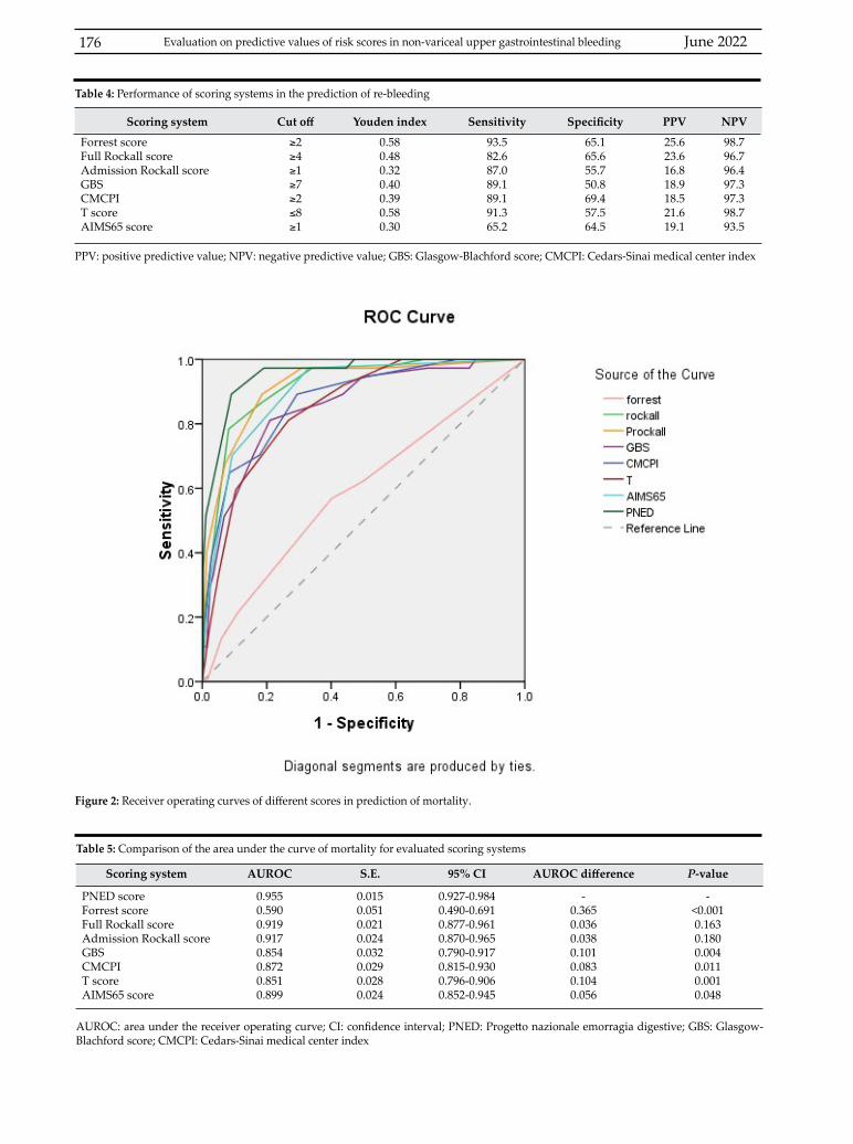
June 2022176
Scoring system Cut off Youden index Sensitivity Specificity PPV NPV
Forrest scoreFull Rockall scoreAdmission Rockall scoreGBSCMCPIT scoreAIMS65 score
≥2≥4≥1≥7≥2≤8≥1
0.580.480.320.400.390.580.30
93.582.687.089.189.191.365.2
65.165.655.750.869.457.564.5
25.623.616.818.918.521.619.1
98.796.796.497.397.398.793.5
Table 4: Performance of scoring systems in the prediction of re-bleeding
PPV: positive predictive value; NPV: negative predictive value; GBS: Glasgow-Blachford score; CMCPI: Cedars-Sinai medical center index
Scoring system AUROC S.E. 95% CI AUROC difference P-value
PNED scoreForrest scoreFull Rockall scoreAdmission Rockall scoreGBSCMCPIT scoreAIMS65 score
0.9550.5900.9190.9170.8540.8720.8510.899
0.0150.0510.0210.0240.0320.0290.0280.024
0.927-0.9840.490-0.6910.877-0.9610.870-0.9650.790-0.9170.815-0.9300.796-0.9060.852-0.945
-0.3650.0360.0380.1010.0830.1040.056
-<0.0010.1630.1800.0040.0110.0010.048
Table 5: Comparison of the area under the curve of mortality for evaluated scoring systems
AUROC: area under the receiver operating curve; CI: confidence interval; PNED: Progetto nazionale emorragia digestive; GBS: Glasgow-Blachford score; CMCPI: Cedars-Sinai medical center index
Figure 2: Receiver operating curves of different scores in prediction of mortality.
Evaluation on predictive values of risk scores in non-variceal upper gastrointestinal bleeding
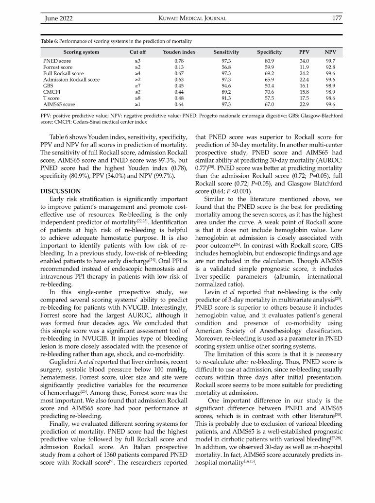
KUWAIT MEDICAL JOURNAL 177June 2022
Scoring system Cut off Youden index Sensitivity Specificity PPV NPV
PNED scoreForrest scoreFull Rockall scoreAdmission Rockall scoreGBSCMCPIT scoreAIMS65 score
≥3≥2≥4≥2≥7≤2≥8≥1
0.780.130.670.630.450.440.480.64
97.356.897.397.394.689.291.397.3
80.959.969.265.950.470.657.567.0
34.011.924.222.416.115.817.522.9
99.792.899.699.698.998.998.699.6
PPV: positive predictive value; NPV: negative predictive value; PNED: Progetto nazionale emorragia digestive; GBS: Glasgow-Blachford score; CMCPI: Cedars-Sinai medical center index
Table 6: Performance of scoring systems in the prediction of mortality
Table 6 shows Youden index, sensitivity, specificity, PPV and NPV for all scores in prediction of mortality. The sensitivity of full Rockall score, admission Rockall score, AIMS65 score and PNED score was 97.3%, but PNED score had the highest Youden index (0.78), specificity (80.9%), PPV (34.0%) and NPV (99.7%).
DISCUSSIONEarly risk stratification is significantly important
to improve patient’s management and promote cost-effective use of resources. Re-bleeding is the only independent predictor of mortality[22,23]. Identification of patients at high risk of re-bleeding is helpful to achieve adequate hemostatic purpose. It is also important to identify patients with low risk of re-bleeding. In a previous study, low-risk of re-bleeding enabled patients to have early discharge[24]. Oral PPI is recommended instead of endoscopic hemostasis and intravenous PPI therapy in patients with low-risk of re-bleeding.
In this single-center prospective study, we compared several scoring systems’ ability to predict re-bleeding for patients with NVUGIB. Interestingly, Forrest score had the largest AUROC, although it was formed four decades ago. We concluded that this simple score was a significant assessment tool of re-bleeding in NVUGIB. It implies type of bleeding lesion is more closely associated with the presence of re-bleeding rather than age, shock, and co-morbidity.
Guglielmi A et al reported that liver cirrhosis, recent surgery, systolic blood pressure below 100 mmHg, hematemesis, Forrest score, ulcer size and site were significantly predictive variables for the recurrence of hemorrhage[25]. Among these, Forrest score was the most important. We also found that admission Rockall score and AIMS65 score had poor performance at predicting re-bleeding.
Finally, we evaluated different scoring systems for prediction of mortality. PNED score had the highest predictive value followed by full Rockall score and admission Rockall score. An Italian prospective study from a cohort of 1360 patients compared PNED score with Rockall score[9]. The researchers reported
that PNED score was superior to Rockall score for prediction of 30-day mortality. In another multi-center prospective study, PNED score and AIMS65 had similar ability at predicting 30-day mortality (AUROC: 0.77)[20]. PNED score was better at predicting mortality than the admission Rockall score (0.72; P=0.05), full Rockall score (0.72; P=0.05), and Glasgow Blatchford score (0.64; P <0.001).
Similar to the literature mentioned above, we found that the PNED score is the best for predicting mortality among the seven scores, as it has the highest area under the curve. A weak point of Rockall score is that it does not include hemoglobin value. Low hemoglobin at admission is closely associated with poor outcome[26]. In contrast with Rockall score, GBS includes hemoglobin, but endoscopic findings and age are not included in the calculation. Though AIMS65 is a validated simple prognostic score, it includes liver-specific parameters (albumin, international normalized ratio).
Levin et al reported that re-bleeding is the only predictor of 3-day mortality in multivariate analysis[23]. PNED score is superior to others because it includes hemoglobin value, and it evaluates patient’s general condition and presence of co-morbidity using American Society of Anesthesiology classification. Moreover, re-bleeding is used as a parameter in PNED scoring system unlike other scoring systems.
The limitation of this score is that it is necessary to re-calculate after re-bleeding. Thus, PNED score is difficult to use at admission, since re-bleeding usually occurs within three days after initial presentation. Rockall score seems to be more suitable for predicting mortality at admission.
One important difference in our study is the significant difference between PNED and AIMS65 scores, which is in contrast with other literature[20]. This is probably due to exclusion of variceal bleeding patients, and AIMS65 is a well-established prognostic model in cirrhotic patients with variceal bleeding[27,28]. In addition, we observed 30-day as well as in-hospital mortality. In fact, AIMS65 score accurately predicts in-hospital mortality[14,15].
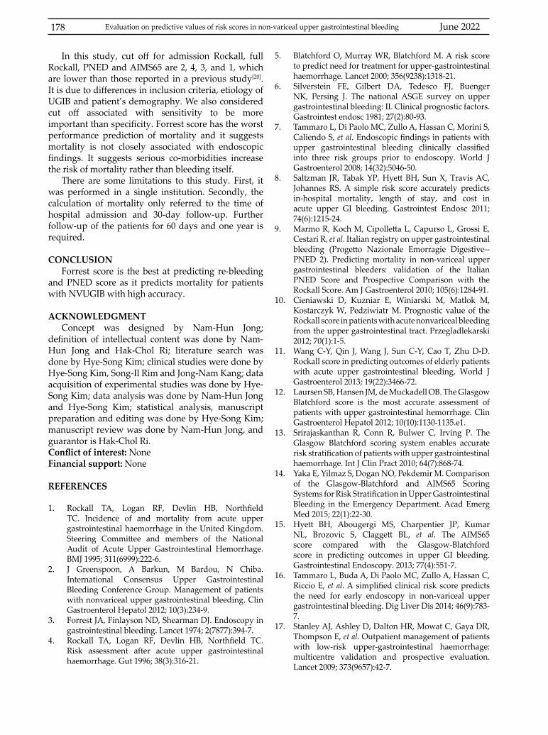
June 2022178
In this study, cut off for admission Rockall, full Rockall, PNED and AIMS65 are 2, 4, 3, and 1, which are lower than those reported in a previous study[20]. It is due to differences in inclusion criteria, etiology of UGIB and patient’s demography. We also considered cut off associated with sensitivity to be more important than specificity. Forrest score has the worst performance prediction of mortality and it suggests mortality is not closely associated with endoscopic findings. It suggests serious co-morbidities increase the risk of mortality rather than bleeding itself.
There are some limitations to this study. First, it was performed in a single institution. Secondly, the calculation of mortality only referred to the time of hospital admission and 30-day follow-up. Further follow-up of the patients for 60 days and one year is required.
CONCLUSIONForrest score is the best at predicting re-bleeding
and PNED score as it predicts mortality for patients with NVUGIB with high accuracy.
ACKNOWLEDGMENT Concept was designed by Nam-Hun Jong;
definition of intellectual content was done by Nam-Hun Jong and Hak-Chol Ri; literature search was done by Hye-Song Kim; clinical studies were done by Hye-Song Kim, Song-Il Rim and Jong-Nam Kang; data acquisition of experimental studies was done by Hye-Song Kim; data analysis was done by Nam-Hun Jong and Hye-Song Kim; statistical analysis, manuscript preparation and editing was done by Hye-Song Kim; manuscript review was done by Nam-Hun Jong, and guarantor is Hak-Chol Ri. Conflict of interest: None Financial support: None
REFERENCES
1. Rockall TA, Logan RF, Devlin HB, Northfield TC. Incidence of and mortality from acute upper gastrointestinal haemorrhage in the United Kingdom. Steering Committee and members of the National Audit of Acute Upper Gastrointestinal Hemorrhage. BMJ 1995; 311(6999):222-6.
2. J Greenspoon, A Barkun, M Bardou, N Chiba. International Consensus Upper Gastrointestinal Bleeding Conference Group. Management of patients with nonvariceal upper gastrointestinal bleeding. Clin Gastroenterol Hepatol 2012; 10(3):234-9.
3. Forrest JA, Finlayson ND, Shearman DJ. Endoscopy in gastrointestinal bleeding. Lancet 1974; 2(7877):394-7.
4. Rockall TA, Logan RF, Devlin HB, Northfield TC. Risk assessment after acute upper gastrointestinal haemorrhage. Gut 1996; 38(3):316-21.
5. Blatchford O, Murray WR, Blatchford M. A risk score to predict need for treatment for upper-gastrointestinal haemorrhage. Lancet 2000; 356(9238):1318-21.
6. Silverstein FE, Gilbert DA, Tedesco FJ, Buenger NK, Persing J. The national ASGE survey on upper gastrointestinal bleeding: II. Clinical prognostic factors. Gastrointest endosc 1981; 27(2):80-93.
7. Tammaro L, Di Paolo MC, Zullo A, Hassan C, Morini S, Caliendo S, et al. Endoscopic findings in patients with upper gastrointestinal bleeding clinically classified into three risk groups prior to endoscopy. World J Gastroenterol 2008; 14(32):5046-50.
8. Saltzman JR, Tabak YP, Hyett BH, Sun X, Travis AC, Johannes RS. A simple risk score accurately predicts in-hospital mortality, length of stay, and cost in acute upper GI bleeding. Gastrointest Endosc 2011; 74(6):1215-24.
9. Marmo R, Koch M, Cipolletta L, Capurso L, Grossi E, Cestari R, et al. Italian registry on upper gastrointestinal bleeding (Progetto Nazionale Emorragie Digestive--PNED 2). Predicting mortality in non-variceal upper gastrointestinal bleeders: validation of the Italian PNED Score and Prospective Comparison with the Rockall Score. Am J Gastroenterol 2010; 105(6):1284-91.
10. Cieniawski D, Kuzniar E, Winiarski M, Matlok M, Kostarczyk W, Pedziwiatr M. Prognostic value of the Rockall score in patients with acute nonvariceal bleeding from the upper gastrointestinal tract. Przegladlekarski 2012; 70(1):1-5.
11. Wang C-Y, Qin J, Wang J, Sun C-Y, Cao T, Zhu D-D. Rockall score in predicting outcomes of elderly patients with acute upper gastrointestinal bleeding. World J Gastroenterol 2013; 19(22):3466-72.
12. Laursen SB, Hansen JM, de Muckadell OB. The Glasgow Blatchford score is the most accurate assessment of patients with upper gastrointestinal hemorrhage. Clin Gastroenterol Hepatol 2012; 10(10):1130-1135.e1.
13. Srirajaskanthan R, Conn R, Bulwer C, Irving P. The Glasgow Blatchford scoring system enables accurate risk stratification of patients with upper gastrointestinal haemorrhage. Int J Clin Pract 2010; 64(7):868-74.
14. Yaka E, Yilmaz S, Dogan NO, Pekdemir M. Comparison of the Glasgow-Blatchford and AIMS65 Scoring Systems for Risk Stratification in Upper Gastrointestinal Bleeding in the Emergency Department. Acad Emerg Med 2015; 22(1):22-30.
15. Hyett BH, Abougergi MS, Charpentier JP, Kumar NL, Brozovic S, Claggett BL, et al. The AIMS65 score compared with the Glasgow-Blatchford score in predicting outcomes in upper GI bleeding. Gastrointestinal Endoscopy. 2013; 77(4):551-7.
16. Tammaro L, Buda A, Di Paolo MC, Zullo A, Hassan C, Riccio E, et al. A simplified clinical risk score predicts the need for early endoscopy in non-variceal upper gastrointestinal bleeding. Dig Liver Dis 2014; 46(9):783-7.
17. Stanley AJ, Ashley D, Dalton HR, Mowat C, Gaya DR, Thompson E, et al. Outpatient management of patients with low-risk upper-gastrointestinal haemorrhage: multicentre validation and prospective evaluation. Lancet 2009; 373(9657):42-7.
Evaluation on predictive values of risk scores in non-variceal upper gastrointestinal bleeding

KUWAIT MEDICAL JOURNAL 179June 2022
18. Stanley AJ, Dalton HR, Blatchford O, Ashley D, Mowat C, Cahill A, et al. Multicentre comparison of the Glasgow Blatchford and Rockall Scores in the prediction of clinical end-points after upper gastrointestinal haemorrhage. Aliment Pharmacol Ther 2011; 34(4):470-5.
19. Pang SH, Ching JYL, Lau JYW, Sung JJY, Graham DY, Chan FKL. Comparing the Blatchford and pre-endoscopic Rockall score in predicting the need for endoscopic therapy in patients with upper GI hemorrhage. Gastrointest Endosc 2010; 71(7):1134-40.
20. Stanley AJ, Laine L, Dalton HR, Ngu JH, Schultz M, Abazi R, et al. Comparison of risk scoring systems for patients presenting with upper gastrointestinal bleeding: international multicentre prospective study. BMJ 2017; 356:i6432.
21. Sung JJY, Mössner J, Barkun A, Kuipers EJ, Lau J, Jensen D, et al. Intravenous esomeprazole for prevention of peptic ulcer rebleeding: rationale/design of Peptic Ulcer Bleed study. Aliment Pharmacol Ther 2008; 27(8):666-77.
22. Barkun AN, Bardou M, Kulpers EJ, Sung J, Hunt RH, Martel M, et al. International consensus recommendations on the management of patients with nonvariceal upper gastrointestinal bleeding. Ann Intern Med 2010; 152(2):101-13.
23. Levin DA, Watermeyer GA, Deetlefs E, Metz DC, Thomson SR. The efficacy of endoscopic therapy in bleeding peptic ulcer patients. S Afr Med J 2012; 102(5):290-3.
24. Hearnshaw SA, Logan RFA, Lowe D, Travis SPL, Murphy MK, Palme KL. Acute upper gastrointestinal bleeding in the UK: patient characteristics, diagnoses and outcomes in the 2007 UK audit. Gut 2011; 60(10):1327-35.
25. Guglielmi A, Ruzzenente A, Sandri M, Kind R, Lombardo F, Rodella L, et al. Risk assessment and prediction of rebleeding in bleeding gastroduodenal ulcer. Endoscopy 2002; 34(10):778-86.
26. Romagnuolo J, Barkun AN, Enns R, Armstrong D, Gregor J. Simple clinical predictors may obviate urgent endoscopy in selected patients with nonvariceal upper gastrointestinal tract bleeding. Arch Intern Med 2007; 167(3):265-70.
27. Mohammad AN, Morsy KH, Ali MA. Variceal bleeding in cirrhotic patients: What is the best prognostic score? Turk J Gastroenterol 2016; 27(5):464-9.
28. Motola-Kuba M, Escobedo-Arzate A, Tellez-Avila F, Altamirano J, Aguilar-Olivos N, Gonzalez-Angulo A, et al. Validation of prognostic scores for clinical outcomes in cirrhotic patients with acute variceal bleeding. Ann Hepatol 2016; 15(6):895-901.

KUWAIT MEDICAL JOURNAL June 2022180
Original Article
Kuwait Medical Journal 2022; 54 (2): 180 - 187
Electromechanical delay and 4-chamber longitudinal strain in patients with obstructive sleep apnea
ABSTRACT
KEY WORDS: atrial electromechanical delay, longitudinal strain, obstructive sleep apnea
Ferdi Kahraman1, Sema Avci2, Gokhan Perincek3
1Department of Cardiology, Gebze Fatih State Hospital, Kocaeli, Turkey2Department of Emergency Medicine, Amasya University Sabuncuoglu Serefeddin Research and Training Hospital,
Amasya, Turkey3Department of Pulmonology, Kars Harakani State Hospital, MD, Kars, Turkey
Objectives: The aim of this study was to evaluate atrial electromechanical delay (AEMD), apical 4-chamber longitudinal strain (4C-LS) and echocardiographic changes in patients with obstructive sleep apnea (OSA).Design: Prospective cross-sectional studySetting: Secondary care hospitalSubjects: Forty-six patients (32 male, 14 female) who were diagnosed as mild-to-severe OSA (apnea hypopnea index ≥5 events/h) and control group consisted of 35 healthy subjects (18 male, 17 female)Intervention: Polysomnography, blood samples and transthoracic echocardiograhy (TTE) were evaluatedMain outcome measures: TTE was used to evaluate echocardiographic changes, AEMD and 4C-LSResults: Left ventricle end-diastolic and end-systolic
diameter, interventricular septum and posterior wall thickness were significantly higher; left ventricular ejection fraction and Ea/Aa mitral ratio were lower; right ventricle basal, mid and vertical diameters, Emax, Amax, and Ea tricuspid, tricuspid regurgitan velocity, systolic pulmonary artery pressure, and systolic motion tricuspid were significantly higher in the OSA group. Tricuspid annular plane systolic excursion (TAPSE) was significantly lower and AEMD lateral/tricuspid, lateral/mitral and septal were significantly higher in the OSA group. Mid anterolateral, apicolateral, apex, apical septal strains and 4C-LS were decreased significantly in the OSA groupConclusion: Right-left ventricular systolic-diastolic functions were impaired in patients with OSA. In these patients, apical 4C-LS was lower and AEMD was prolonged.
Address correspondence to:Dr. Sema Avci, MD, Amasya University Sabuncuoglu Serefeddin Research and Training Hospital, Amasya, Turkey. Tel: +90 5308431363; E-mail: [email protected]; [email protected]
INTRODUCTIONObstructive sleep apnea (OSA) is a serious, life-
threatening and common chronic disease affecting particularly middle-aged men in the populations[1,2]. OSA is characterized by repetitive surcease episodes of breathing while sleeping, by the reason of complete or partial airway obstruction[1,2]. Hypoxia and hypercapnia caused by OSA increases the arousal of the patient throughout the night[3]. These arousals associated with hypoxia and hypercapnia stimulate the sympathetic nervous system, increase catecholamine release and myocardial oxygen consumption[3]. Increased myocardial oxygen
consumption causes cardiac ischemia, myocardial infarction, hypertrophy, hypertension, increase in left ventricle wall tension, congestive heart failure, arrhythmias such as atrial fibrillation and stroke[3,4]. Cardiovascular complications are the most common complications of OSA. Echocardiography is the gold standard noninvasive imaging method for evaluating myocardial functions of these type of patients and assessing the effects of OSA on heart[5].
Atrial electromechanical delay (AEMD) is the time interval between the beginning of P wave on surface electrocardiography and beginning of the late diastolic wave on Tissue Doppler Imaging (TDI)[6]. The

KUWAIT MEDICAL JOURNAL 181June 2022
structural changes of atrial tissue cause delay between the electrical stimulation and mechanical contraction[6]. Atrial tissue changes can cause prolongation of P wave on surface electrocardiogram[7]. Prolongation of P wave can be seen in patients undergoing coronary artery bypass surgery, patients with hypertrophic cardiomyopathy, right atrial dilatation, atrial septal defect hypertension and chronic obstructive pulmonary disease due to affected atrial tissue[7].
Two-dimensional speckle tracking echocardiography is a strain measurement method to obtain the size of regional myocardial deformations[8,9]. Myocardial deformations can be obtained by this easily applied method in the longitudinal, circumferential and radial pointing[8,9].
The aim of this study was to evaluate AEMD, apical 4-chamber longitudinal strain (4C-LS) and echocardiographic changes in patients with OSA. We especially focused on AEMD, longitudinal strain and diastolic-systolic functions of right-left heart in these patients.
SUBJECTS AND METHODSStudy population
This prospective study was conducted with the approval of the Caucasian University Medical Faculty ethics committee between April and August 2018. The patient group consisted of 46 patients (32 male, 14 female) who were referred to the Sleep Disorders Center of Caucasian University Hospital and were diagnosed as mild-to-severe OSA (apnea hypopnea index (AHI) ≥5 events/h) on polysomnographic evaluation. The control group consisted of 35 healthy subjects (18 male, 17 female) who were found not to have OSA (AHI <5 events/h) on polysomnographic evaluation.
The exclusion criteria were as follows: chronic obstructive pulmonary disease on pulmonary function tests, patients with valvular and structural heart disease, wall-motion abnormality, coronary artery disease, acute coronary syndrome, heart failure, atrioventricular conduction abnormalities, previous history of atrial fibrillation, ejection fraction <50%, use of drugs that affect atrioventricular conduction system, uncontrolled hypertension, pulmonary embolism, pneumonia, insulin dependent diabetes mellitus, history of cerebrovascular disease, history of continuous positive airway pressure, renal impairment, hypo-hyperthyroidism, anemia, electrolyte disorders, acid-base disorders, malignancy, patients using two or more oral antidiabetic drugs, systemic inflammatory response syndrome, poor echocardiographic view.
The following parameters of all patients were evaluated: age, gender, body mass index, comorbidity,
blood glucose, electrolytes, liver function tests, renal function tests, complete blood count, transthoracic echocardiography and polysomnography. All patients were informed about the study. An informed and signed consent was obtained for all procedures.
Blood samplesAll blood samples were drawn from the vein in
the forearm and collected into 2 mL Lavender (EDTA) top tube and 5 mL Yellow top tube were analyzed with Pentra DF Nexus, Horiba Medical, Japan with Automated Cell Counter Methodology and Cobas C 501, Roche. The complete blood samples were stabilized optimally when run within four hours of collection, stable for 24 hours at room temperature, and stable for 36 hours at 2-8 °C. The biochemical blood samples were stabilized optimally when run within two hours of collection, stable for 24 hours at +4º C.
PolysomnographyOvernight polysomnography was performed
in all patients using conventional and analysis according to the American Academy of Sleep Medicine[10]. All patients spent one entire night in the sleep laboratory with the aim of capturing a typical night’s sleep. The wakefulness, sleep stages, respiration, cardiopulmonary functions and body movements of patients were evaluated. Electroencephalography, electro-oculography and chin muscle electromyography channels were used to monitor sleep stages. Airflow and respiratory effort channels were used to assess sleep-disordered breathing. Arterial oxygen saturation was measured with finger pulse oximetry channel. Movement changes of the chest and abdomen during breathing were recorded by using respiratory inductive plethysmography. Limb electromyogram channels were placed on the legs (tibialis anterior muscle) and evaluated periodical limb movements. An oronasal flow cannula attached to pneumotachograph and apneas-hypopneas. Apneas were defined as the cut-off of airflow for more than ≥10 s. Hypopnea were defined as peak signal excursion drop by ≥30% of pre-event baseline using nasal pressure, duration of the ≥30% drop in signal excursion is ≥10 seconds and ≥3% oxygen desaturation from pre-event baseline and/or the event is associated with an arousal[11]. The AHI was obtained by dividing the total number of apneas and hypopneas during the entire sleeping time. OSA classification was made according to American Academy of Sleep Medicine: mild OSA was defined as AHI of 5-15, moderate OSA was defined as AHI of 15-30 and severe OSA was defined AHI of more than 30[10].

June 2022182 Electromechanical delay and 4-chamber longitudinal strain in patients with obstructive sleep apnea
EchocardiographyTransthoracic echocardiography (Epiq 7; Philips)
was evaluated by a practitioner in a standard protocol in all patients. A 2.5 MHz probe was used for the Doppler measurements and 2.5-3.5 MHz probe for tissue Doppler measurements. Patients were monitored using electrocardiographic leads and were placed in the left lateral decubitus position. Echocardiographic images were obtained from the parasternal views (long axis, short axis), the apical four-chamber view and the subcostal view. Echocardiographic measurements were performed at the end of expiration according to the recommendations of the American Society of Echocardiography/European Association of Echocardiography[12]. 1) diameters of right ventricle (RV) measured in apical view; 2) left ventricle (LV) diameter and wall thickness were measured in the parasternal view; 3) left atrial diameter measured in the parasternal view; 4) aortic root diameters, measured at the sinus of Valsalva; 5) LV ejection fraction measured in apical 4-chamber view by modified Simpson method; 6) RV and LV functions were evaluated as follows: a) maximal peak velocity of early diastolic flow (Emax), maximal peak velocity of atrial contraction (Amax), and ratio of these (Emax/Amax), measured over the mitral and tricuspid valves; b) TDI measured in the mitral and tricuspid lateral annulus at early diastole (Ea), atrium systole (Aa) and ratio of these (Ea/Aa); c) the ratio of Emax/Ea; 7) aortic, tricuspid, mitral and pulmonary valvular evaluation; 8) tricuspid regurgitant velocity (TRV) recorded by continuous wave Doppler.
AEMD was calculated from colored-TDI recordings. AEMD was determined as the time interval between the beginning of echocardiographic P wave to the initial of late diastolic wave in TDI recordings. AEMD was measured from lateral/tricuspid, lateral/mitral and septal anulus from apical 4-chamber views.
The echocardiographic examinations and standard 2D measurements for strain were acquired according to American Society of Echocardiography recommendations[12]. The images were digitally stored and measurements were performed by the same practitioner. Images were obtained at a frame rate of 50 to 70 per second, and digital loops were saved onto optical disc for off-line analysis. The cardiac cycle with the best image quality and without any artefacts was selected for reporting results. Two and three-chambers images were not used due to intense artefacts. Apical 4-chamber longitudinal strain images were obtained. The practitioner identified three points on each view (two borders of the mitral annulus and the apex). Speckles were tracked frame by-frame throughout the LV wall during the cardiac cycle and basal, mid, and apical regions of interest were created.
Clinical ProfileOSA (n=46)Mean±SD /
n (%)
Controls (n=35)Mean±SD /
n (%)P-value
AgeGender
MaleFemale
BMI (kg/m2)Comorbidities
HypertensionDiabetes MellitusHyperlipidemia
49.3±10.5
32 (69.6%)14 (30.4%)35.67±5.77
28 (60.9%)-
6 (13%)
48.3±5.1
18 (51.4%)17 (48.6%)28.41±4.1
3 (8.6%)1 (2.9%)1 (2.9%)
NS
NSNS
<0.001
<0.001NSNS
OSA: obstructive sleep apnea; continuous variables are expressed as mean ± standard deviation; NS: non-significant; BMI: body mass index.
Table 1: Clinical characteristics of the study population
Biochemical parameters
OSA(Mean±SD)
Controls(Mean±SD) P-value
Glucose UreaCreatinineUric acidHDLTriglycerideC-reactive proteinAlbuminASTALTLDHCalciumSodiumPotassiumALPTotal BilirubinProtein
108.1±15.535.6±9.20.87±0.166.22±1.342.6±8.2176±61
0.59±0.874.31±0.3324.5±8.233.6±15.2188±36.69.5±0.3
140.7±3.24.49±0.3582.4±24.80.43±0.217.07±1.68
98.5±11.830±7.4
0.72±0.194.89±1.5748.9±12.6170.6±80.70.37±0.334.59±0.2420.1±8.621.6±14.3198.9±40.1
9±0.4140.5±1.94.42±0.34100.5±13.40.46±0.247.34±0.45
0.0070.0010.001<0.0010.048NSNS
<0.0010.018<0.001
NSNSNSNSNSNSNS
OSA: obstructive sleep apnea; continuous variables are expressed as mean ± standard deviation; NS: non-significant; HDL: high density lipoprotein; AST: aspartate aminotransferase; ALT: alanine aminotransferase; LDH: lactate dehydrogenase; ALP: alkaline phosphatase.
Table 2: Biochemical parameters of the study population
Statistical analysisAll statistical calculations were performed
with SPSS 23.0 (SPSS for Windows, Chicago, IL, SA). All continuous variables were expressed as mean±standard deviation; categoric variables were defined as percentages (%). The categorical parameters were compared with Chi Square test and Fischer’s exact test. The normal distribution was determined by histogram and Kolmogorov-Smirnov test. Mean values of continuous variables were compared between the groups using Mann-Whitney U test. All tests were applied as two tailed; the statistical significance level was P <0.05.
RESULTSBaseline clinical characteristics of the study
population are presented in Table 1. Body mass index

KUWAIT MEDICAL JOURNAL 183June 2022
and number of patients with hypertension were higher in the OSA group (P <0.001).
Biochemical blood parameters of the study population are presented in Table 2. Glucose levels, urea, creatinine, uric acid, aspartate transaminase and alanine aminotransferase were higher significantly in the OSA group.
Complete blood counts of the study population are presented in Table 3. Hemoglobin, hematocrit, eosinophil count and percentage were significantly higher in the OSA group.
Conventional and tissue Doppler echocardiographic parameters of left heart and septum for two groups are shown in Table 4. LV end-diastolic diameter, end-systolic diameter, interventricular septum and posterior wall thickness were significantly higher in the OSA group (P <0.05). LV ejection fraction and Ea/Aa mitral ratio were lower in the OSA group.
Conventional and tissue Doppler echocardiographic parameters of right heart for two groups are shown in Table 5. Right ventricle basal, mid
Biochemical parameters
OSA(Mean±SD)
Controls(Mean±SD) P-value
Hemoglobin (mg/dL)HematocritWBCMPVPlatelet countLymphocyte countLymphocyte percentPLRNeutrophil countNeutrophil percentNLREosinophil countEosinophil percentRDWPCT
15.9±1.4448.05±4.47.86±1.878.44±0.58262.4±58.82.36±0.7331.03±8.95122±47.324.61±1.3658.91±7.812.17±1.080.26±0.153.16±1.4215.4±1.980.22±0.06
15.02±1.2645.15±3.57.38±2.188.91±0.79259.4±572.34±0.6332.47±7.12
118.83±41.944.26±1.7
56.82±8.131.91±0.850.21±0.292.77±2.9915.12±1.230.23±0.04
0.0110.002NS
0.006NSNSNSNSNSNSNS
0.0020.009NSNS
OSA: obstructive sleep apnea; continuous variables are expressed as mean ± standard deviation; NS: non-significant; WBC: white blood cell; MPV: mean platelet volume; PLR: platelet-to-lymphocyte ratio; NLR: neutrophil-to-lymphocyte ratio; RDW: red blood cell distribution width; PCT: plateletcrit
Table 3: Complete blood counts of the study population
Left heart and septum OSA(Mean±SD)
Controls(Mean±SD) P-value
Left heartDimensionsLeft atrium (parasternal long axis)
Diameter (mm)Left ventricle (parasternal long axis)
End-diastolic diameter (mm)End-systolic diameter (mm)
Left ventricle wall thickness Interventricular septum (mm)Posterior wall (mm)
Ventricular functionSystolic function
LV ejection fraction (%)Diastolic function
Emax mitral (cm/s)Amax mitral (cm/s)Emax/Amax mitralEa (tissue doppler lateral mitral) (cm/s)Aa (tissue doppler lateral mitral) (cm/s)Ea/Aa mitralEmax/AaEmax/Ea
Mitral E wave decelaration time (ms)Aortic root diameter (cm)
SeptumEa (tissue doppler septal) (cm/s) Aa (tissue doppler septal) (cm/s)Ea/Aa septalEmax/Ea
Systolic motion mitral Systolic motion septal
Heart rate, beats/min
38±0.38
49.4±0.4231±0.36
11.8±1.410.5±1.4
58.27±5.54
74.59±16.5775.96±12.331.01±0.2710.93±3.4812.34±3.280.65±0.240.97±0.457.32±2.13153.9±21.63.52±0.25
7.41±1.6311±2.12
0.71±0.271.08±0.549.91±3.118.42±1.4776.6±8.8
35.2±3.1
46.2±3.926.6±0.31
10.7±1.39.6±1.2
61.73±3.31
70.18±13.6270.43±19.131.33±1.9411.25±3.0611.38±2.460.78±0.291.05±0.426.69±2.48145.1±24.73.41±0.33
7.7±2.710.77±2.60.76±0.351.01±0.718.99±1.987.78±1.3573.6±10.1
0.007
0.004<0.001
<0.0010.003
0.008
NSNSNSNSNS
<0.001NSNSNSNS
NSNSNSNSNSNSNS
Table 4: Echocardiographic findings of the left heart and septum in both groups
OSA: obstructive sleep apnea; continuous variables are expressed as mean ± standard deviation; NS: non-significant; LV: left ventricle.
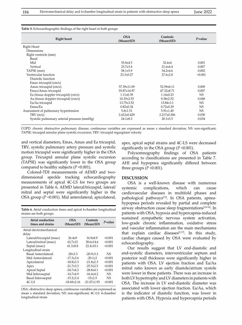
June 2022184
and vertical diameters, Emax, Amax and Ea tricuspid, TRV, systolic pulmonary artery pressure and systolic motion tricuspid were significantly higher in the OSA group. Tricuspid annular plane systolic excursion (TAPSE) was significantly lower in the OSA group compared to healthy subjects (P <0.001).
Colored-TDI measurements of AEMD and two-dimensional speckle tracking echocardiography measurements of apical 4C-LS for two groups are presented in Table 6. AEMD lateral/tricuspid, lateral/mitral and septal were significantly higher in the OSA group (P <0.001). Mid anterolateral, apicolateral,
Right heart OSA(Mean±SD)
Controls(Mean±SD) P-value
Right HeartDimensionsRight ventricle (mm)
BasalMidVertical
TAPSE (mm)Ventricular function
Diastolic functionEmax tricuspid (cm/s)Amax tricuspid (cm/s)Emax/Amax tricuspidEa (tissue doppler tricuspid) (cm/s)Aa (tissue doppler tricuspid) (cm/s)Ea/Aa tricuspidEmax/Ea
Assessment of pulmonary hypertensionTRV (m/s)Systolic pulmonary arterial pressure (mmHg)
35.8±4.523.7±3.458.1±5.923.3±0.27
57.39±11.0955.87±14.971.11±0.3910.35±2.5513.75±3.520.82±0.345.8±1.51
2.412±0.42924.1±8.3
32.4±421.6±4.454.2±4.627.6±2.8
52.59±8.1147.32±8.711.14±0.239.38±2.5213.84±3.10.72±0.295.91±1.49
2.217±0.30620.1±5.5
0.0010.0070.002<0.001
0.0090.007NS
0.048NSNSNS
0.0300.034
Table 5: Echocardiographic findings of the right heart in both groups
COPD: chronic obstructive pulmonary disease; continuous variables are expressed as mean ± standard deviation; NS: non-significant; TAPSE: tricuspid annular plane systolic excursion; TRV: tricuspid regurgitant velocity
Atrial conduction times and strains
OSA(Mean±SD)
Controls(Mean±SD) P-value
Atrial electromechanical delay
Lateral/tricuspid (msec) Lateral/mitral (msec)Septal (msec)
Longitudinal strainBasal AnterolateralMid AnterolateralApicolateral Apex Apical SeptalMid Inferoseptal Basal Inferoseptal4C-LS
26.4±962.7±1241.5±8.8
-20±3.3-17.3±3.6-18.8±3.3-21.7±3.3-24.7±4.2-14.7±4.9-15.2±2.4
-18.69±2.16
16.9±8.938.6±14.622.4±10.1
-20.9±3.3-20.1±3
-21.8±2.3-25.5±2.5-28.8±4.1-16.4±4.2-15±2.5
-21.07±1.93
<0.001<0.001<0.001
NS<0.001<0.001<0.001<0.001
NSNS
<0.001
Table 6. Atrial conduction times and apical 4-chamber longitudinal strains are both groups
OSA: obstructive sleep apnea; continuous variables are expressed as mean ± standard deviation; NS: non-significant; 4C-LS: 4-chamber longitudinal strain
apex, apical septal strains and 4C-LS were decreased significantly in the OSA group (P <0.001).
Polysomnography findings of OSA patients according to classifications are presented in Table 7. AHI and hypopnea significantly differed between three groups (P <0.001).
DISCUSSIONOSA is a well-known disease with numerous
systemic complications, which can cause cardiovascular diseases in multifold phases and pathological pathways[13]. In OSA patients, apnea-hypopnea periods revealed by partial and complete airway obstruction cause sleep fragmentations[14-16]. In patients with OSA, hypoxia and hypercapnia-induced sustained sympathetic nervous system activation, low-grade chronic inflammation, oxidative stress and vascular inflammation are the main mechanisms that explain cardiac diseases[2,17]. In this study, cardiac changes caused by OSA were evaluated by echocardiography.
Our results suggest that LV end-diastolic and end-systolic diameters, interventricular septum and posterior wall thickness were significantly higher in patients with OSA. LV ejection fraction and Ea/Aa mitral ratio known as early diastole/atrium systole were lower in these patients. There was an increase in both LV hypertrophy and LV diameters in patients with OSA. The increase in LV end-diastolic diameter was associated with lower ejection fraction. Ea/Aa, which is the indicator of diastolic function, was lower in patients with OSA. Hypoxia and hypercapnia periods
Electromechanical delay and 4-chamber longitudinal strain in patients with obstructive sleep apnea
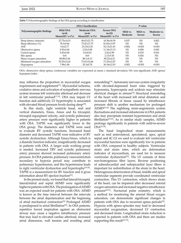
KUWAIT MEDICAL JOURNAL 185June 2022
may influence the proportion in myocardial oxygen requirement and supplement[17]. Myocardial ischemia, oxidative stress and activation of sympathetic nervous system increase left ventricular afterload and decrease in left ventricular preload[17,18]. OSA may impair LV function and additively LV hypertrophy is associated with elevated blood pressure levels during sleep[18].
In this study, right ventricle basal, mid and vertical diameters, Emax, Amax and Ea tricuspid, tricuspid regurgitant velocity, and systolic pulmonary artery pressure were significantly higher in patients with OSA. TAPSE was significantly lower in the OSA group. The diameters and TAPSE were used to evaluate RV systolic functions. Increased basal diameter and decreased TAPSE were indicative of RV systolic dysfunction. Although Emax/Amax, which is a diastolic function indicator, insignificantly decreased in patients with OSA. A larger scale working group is needed. Increased TRV and systolic pulmonary artery pressure showed increased pulmonary artery pressure. In OSA patients, pulmonary vasoconstriction secondary to hypoxia period may contribute to pulmonary hypertension, and this hypertension leads to right ventricular dysfunction and hypertrophy[19,20]. TAPSE is a measurement for RV function and it gives information about RV ejection fraction[21].
In the present study, we found that lateral/tricuspid, lateral/mitral and septal AEMD were significantly higher in patients with OSA. The prolongation of AEMD was an expected result for patients with OSA. AEMD is known as the time interval between the intervals between the atrial depolarization and the beginning of atrial mechanical contraction[22]. Prolonged AEMD is predisposed to atrial fibrillation[22]. In OSA patients, repetitive forced inspiration against an obstructed airway may cause a negative intrathoracic pressure that may lead to elevated cardiac afterload, increased atrial dimension, wall tension and resulting atrial
remodeling[14]. Autonomic nervous system irregularity with elevated-depressed heart rates triggered by hypoxemia, hypercapnia and acidosis may stimulate electrical changes in atrium[14]. Structural remodeling of the heart with increased left atrial dilatation and increased fibrosis of tissue caused by intrathoracic pressure shift is another mechanism for prolonged AEMD[23,24]. The nightlong renin-angiotensin system fluctuations and increased aldosterone in OSA patients also may precipitate resistant hypertension and atrial fibrillation[25,26]. As in similar study samples, AEMD prolongs significantly in patients with mild-to-severe OSA[27,28].
The basal longitudinal strain measurements such as mid anterolateral, apicolateral, apex, apical septal and 4C-LS we used to evaluate left ventricular myocardial function were significantly low in patients with OSA compared to healthy subjects. Ventricular strain and strain rates, which are deformation indicator of myocardium, are used for to measure ventricular dysfunction[29]. The LV consists of three non-homogenous fiber layers. Reverse positioning of subendocardial and subepicardial layer fibers is important for redistribution of the strain in the heart. Heterogenous deterioration of basal, middle and apical ventricular segments provide coordinated ventricular contraction. This LV contraction, which shows strain of the heart, can be impaired after decreased arterial oxygen saturation and increased negative intrathoracic pressure[30,31]. Nocturnal pulse oximetry, which is a method for monitoring the arterial blood oxygen saturation, can demonstrate significant changes in patients with OSA due to recurrent apnea periods[32]. Hypoxia with apnea episodes may lead to decreased myocardial oxygenation, decreased LV contraction and decreased strain. Longitudinal strain reduction is expected in patients with OSA and there are studies supporting this result[5,8,33].
Polysomnographic findings
OSA Classification P-value
Mild OSA(n=8)
Mean±SD / n (%)
Moderate OSA(n=6)
Mean±SD / n (%)
Severe OSA(n=32)
Mean±SD / n (%)
Mild vs. Moderate
Mild vs.Severe
Moderate vs. Severe
Sleep latency (minute)Sleep efficiency (%)AHIObstructive apneaCentral apneaMix apneaMean oxygen saturationMinimum oxygen saturationHypoapnea
27.75±14.5178.3±9.669.6±2.770.93±0.460.43±0.46
0±088.08±3.4470.25±12.41
7.98±1.88
49.67±21.7573.47±17.5723.33±3.252.53±3.080.5±0.63
0±088.1±4.13
73.67±14.4620.3±0.78
18.38±18.3981.83±13.9752.13±21.8111.34±15.132.42±2.892.13±5.4589.22±4.2371.25±12.0736.18±12.93
0.037NS
0.002NSNSNSNSNS
0.002
NSNS
<0.0010.0040.0100.026NSNS
<0.001
0.002NS
<0.0010.0450.016NSNSNS
<0.001
Table 7: Polysomnographic findings of the OSA group according to classification
OSA: obstructive sleep apnea; continuous variables are expressed as mean ± standard deviation; NS: non-significant; AHI: apnea/hypopnea index
June 2022186
The small number of patients and the presence of longitudinal strain images in only 4-chamber due to artefacts are the main limitations of the study. The patients were enrolled from only one sleep center, hence limiting the variegation of the patients.
CONCLUSIONThis study revealed that OSA is a disease that
can impair the left-right ventricular systolic-diastolic functions of the heart and it may cause structural changes in the heart. Moreover, OSA may cause prolongation of atrial conduction times and it may decrease the contraction forces of the heart.
This prospective clinical study is important because of the combined evaluation right-left ventricular functions, AEMD and longitudinal strain.
ACKNOWLEDGMENTConflict of interest: None Financial disclosures: NoneAuthors Contribution: Ferdi Kahraman did data collection and designed the manuscript; Sema Avci and Gokhan Perincek did analysis and wrote the manuscript; Ferdi Kahraman and Sema Avci reviewed the final version of manuscript.
REFERENCES
1. Spicuzza L, Caruso D, Di Maria G. Obstructive sleep apnea syndrome and its management. Ther Adv Chronic Dis 2015; 6(5):273-85.
2. Jean-Louis G, Zizi F, Clark LT, Brown CD, McFarlane SI. Obstructive sleep apnea and cardiovascular disease: role of the metabolic syndrome and ıts components. J Clin Sleep Med 2008; 4(3):261-72.
3. Dorasamy P. Obstructive sleep apnea and cardiovascular risk. Ther Clin Risk Manag 2007; 3(6):1105-11.
4. Gilat H, Vinker S, Buda I, Soudry E, Shani M, Bachar G. Obstructive sleep apnea and cardiovascular comorbidities: a large epidemiologic study. Medicine (Baltimore) 2014; 93(9):e45.
5. Farahani MM, Vahedi E, Lotfian I, Motashaker-Arani M. Echocardiographic Abnormalities in Patients with Sleep Apnea Syndrome. Arch Cardiovasc Imaging 2014; 2(1):e14534.
6. Ari H, Ari S, Akkaya M, Aydın C, Emlek N, Sarigul OY, et al. Predictive value of atrial electromechanical delay for atrial fibrillation recurrence. Cardiol J 2013; 20(6):639-47.
7. Caglar IM, Dasli T, Caglar FN, Teber MK, Ugurlucan M, Ozmen G. Evaluation of atrial conduction features with tissue Doppler imaging in patients with chronic obstructive pulmonary disease. Clin Res Cardiol 2012; 101(8):599-606.
8. Altekin RE, Yanikoglu A, Baktir AO, Karakas MS, Ozel D, Cilli A, et al. Assessment of subclinical left ventricular dysfunction in obstructive sleep apnea patients with
speckle tracking echocardiography. Int J Cardiovasc Imaging 2012; 28(8):1917-30.
9. Altekin RE, Yanıkoğlu A, Karakaş MS, Ozel D, Yıldırım AB, Mehmet Kabukçu. Evaluation of subclinical left ventricular systolic dysfunction in patients with obstructive sleep apnea by automated function imaging method; an observational study. Anadolu Kardiyol Derg 2012; 12(4):320-30.
10. Kapur VK, Auckley DH, Chowdhuri S, Kuhlmann DC, Mehra R, Ramar K, et al. Clinical Practice Guideline for Diagnostic Testing for Adult Obstructive Sleep Apnea: An American Academy of Sleep Medicine Clinical Practice Guideline. J Clin Sleep Med 2017; 13(3):479-504.
11. American Academy of Sleep Medicine. AASM clarifies hypopnea scoring criteria, 2013. (Accessed December 26, 2018, at https://aasm.org/aasm-clarifies-hypopnea-scoring-criteria/).
12. Lang RM, Bierig M, Devereux RB, Flachskampf FA, Foster E, Pellikka PA, et al. Recommendations for Chamber Quantification: A Report from the American Society of Echocardiography’s Guidelines and Standards Committee and the Chamber Quantification Writing Group, Developed in Conjunction with the European Association of Echocardiography, a Branch of the European Society of Cardiology. J Am Soc Echocardiogr 2005; 18:1440-63.
13. De Torres-Alba F, Gemma D, Armada-Romero E, Rey-Blas JR, López-de-Sá E, López-Sendon LJ. Obstructive sleep apnea and coronary artery disease: from pathophysiology to clinical implications. Pulm Med 2013; 2013:768064.
14. Somers VK, White DP, Amin R, Abraham WT, Costa F, Culebras A, et al. Sleep apnea and cardiovascular disease: an American Heart Association/American College of Cardiology Foundation Scientific Statement From the American Heart Association Council for High Blood Pressure Research Professional Education Committee, Council on Clinical Cardiology, Stroke Council, and Council on Cardiovascular Nursing. Circulation 2008; 118(10):1080-11.
15. Zamarrón C, Cuadrado LV, Álvarez-Sala R. Pathophysiologic mechanisms of cardiovascular disease in obstructive sleep apnea syndrome. Pulm Med 2013;521087.
16. Kohli P, Balachandran JS, Malhotra A. Obstructive sleep apnea and the risk for cardiovascular disease. Curr Atheroscler Rep 2011; 13(2):138-46.
17. Danica LP, Krotin M, Zdravkovic M, Soldatovic I, Zdravkovic D, Brajkovic M, et al. Early left ventricular systolic and diastolic dysfunction in patients with newly diagnosed obstructive sleep apnoea and normal left ventricular ejection fraction. Sci World J 2014; 2014:898746.
18. Mannarino MR, Di Filippo F, Pirro M. Obstructive sleep apnea syndrome. Eur J Int Med 2012; 23(7):586-93.
19. Bradley TD. Right and left ventricular functional impairment and sleep apnea. Clin Chest Med 1992; 13(3):459-79.
Electromechanical delay and 4-chamber longitudinal strain in patients with obstructive sleep apnea
KUWAIT MEDICAL JOURNAL 187June 2022
20. Maripov A, Mamazhakypov A, Sartmyrzaeva M, Akunov A, Muratali Uulu K, Duishobaev M, et al. Right ventricular remodeling and dysfunction in obstructive sleep apnea: A systematic review of the literature and meta-analysis. Can Respir J 2017; 2017:1587865.
21. Schmid E, Hilberath JN, Blumenstock G, Shekar PS, Kling S, Shernan SK, et al. Tricuspid annular plane systolic excursion (TAPSE) predicts poor outcome in patients undergoing acute pulmonary embolectomy. Heart Lung Vessel 2015; 7(2):151-58.
22. Lee DH, Choi SY, Park JS, Seo JM, Choi JH, Cho YR, et al. Comparison of prolonged atrial electromechanical delays with different definitions in the discrimination of patients with non-valvular paroxysmal atrial fibrillation. Korean Circ J 2015; 45(6):479-85.
23. Iwasaki YK, Kato T, Xiong F, Shi YF, Naud P, Maguy A, et al. Atrial fibrillation promotion with long-term repetitive obstructive sleep apnea in a rat model. J Am Coll Cardiol 2014; 64(19):2013-23.
24. Ramos P, Rubies C, Torres M, Batlle M, Farre R, Brugada J, et al. Atrial fibrosis in a chronic murine model of obstructive sleep apnea: mechanisms and prevention by mesenchymal stem cells. Respir Res 2014; 15(1):54.
25. Logan AG, Tkacova R, Perlikowski S, Leung RS, Tisler A, Floras JS, et al. Refractory hypertension and sleep apnea: effect of CPAP on blood pressure and baroreflex. Eur Respir J 2003; 21(2):241-7.
26. Ozaydin M, Varol E, Türker Y, Peker O, Erdogan D, Dogan A, et al. Association between renin-angiotensin-aldosterone system blockers and postoperative atrial fibrillation in patients with mild and moderate left ventricular dysfunction. Anadolu Kardiyol Derg 2010; 10(2):137-42.
27. Yagmur J, Yetkin O, Cansel M, Acikgoz N, Ermis N,
Karakus Y, et al. Assessment of atrial electromechanical delay and influential factors in patients with obstructive sleep apnea. Sleep Breath 2012; 16(1):83-8.
28. Karabag T, Aydin M, Altin R, Dogan SM, Cil C, Buyukuysal C, et al. Evaluation of atrial electromechanical delay and left atrial mechanical function in patients with obstructive sleep apnea: Cardiac involvement in patients with OSA. Wien Klin Wochenschr 2012; 124(13-14):444-52.
29. Haruki N, Takeuchi M, Nakai H, Kanazawa Y, Tsubota N, Shintome R, et al. Overnight sleeping induced daily repetitive left ventricular systolic and diastolic dysfunction in obstructive sleep apnea: quantitative assessment using tissue Doppler imaging. Eur J Echocardiogr 2009; 10(6):769-75.
30. Vendelin M, Bovendeerd PH, Engelbrecht J, Arts T. Optimizing ventricular fibers: uniform strain or stress, but not ATP consumption, leads to high efficiency. Am J Physiol Heart Circ Physiol 2002; 283(3):H1072-81.
31. Orban M, Bruce CJ, Pressman GS, Leinveber P, Romero-Corral A, Korinek J, et al. Dynamic changes of left ventricular performance and left atrial volume induced by the Mueller maneuver in healthy young adults and implications for obstructive sleep apnea, atrial fibrillation, and heart failure. Am J Cardiol 2008; 102(11):1557-61.
32. Alvarez D, Hornero R, Marcos JV, Campo F. Multivariate analysis of blood oxygen saturation recordings in obstructive sleep apnea diagnosis. IEEE Trans Biomed Eng 2010; 57(12):2816-24.
33. Altekin RE, Karakas MS, Yanikoglu A, Ozel D, Ozbudak O, Demir I, et al. Determination of right ventricular dysfunction using the speckle tracking echocardiography method in patients with obstructive sleep apnea. Cardiol J 2012; 19(2):130-9.

KUWAIT MEDICAL JOURNAL June 2022188
Original Article
Kuwait Medical Journal 2022; 54 (2): 188 - 195
Single tertiary center experience from Turkey regarding the experience for cardiac implantable electrical devices
ABSTRACT
KEY WORDS: cardiac implantable electrical devices, complication, implantable cardioverter defibrillator, pacemaker
Emre Ozdemir, Mert Pehlivan Altin ,Sadik Volkan Emren, Cem Nazli , Mehmet TokacDepartment of Cardiology, Katip Celebi University, Ataturk Education and Research Hospital, Izmir, Turkey
Objective: To present our experience of cardiac implantable electrical devices (CIED) implantation and complications in a tertiary referral centre and to compare these data with literatureDesign: Retrospective observational studySetting: Izmir Katip Çelebi Universty, Ataturk Research and Education Hospital, Turkey Subjects: One thousand two hundred and nine de-novo CIED patients and 50 patients who underwent revision and battery exchange proceduresIntervention: Hospital records were scanned.Main outcome measures: Compared to most other studies in literature, the population of this study was younger. The complication rate of cardiac resynchronization therapy (CRT) implantation was low, but the actual cause of six of the 10 mortalities could not be determined. Results: 38.6% (n=486) of patients had a cardiac pacemaker (CPM), 53.6% (n=675) an implantable cardioverter-defibrillators (ICD), and 7.8% (n=98) had CRT-D. The
mean age of patients was 63.9±14.8 years and 66.8% (n=842) were male. The main indication for ICDs was primary prevention in 715 (56.8%) patients. The main indications for CPM implantation were as follows: 234 (18.6%) complete atrioventricular block and 83 (6.6%) sick sinus syndrome. The mean left ventricular ejection fraction value was 39.5%±16.5 before CIED implantation and 40.1%±%16.1 after CIED implantation. Device infection was determined at 2.1% (n=27); pocket infection at 1.7% (21), pneumothorax at 0.6% (n=7) and tamponade at 30.3% (n=3). The rate of pocket hematoma was 3.4% (n=42). Mortality occurred in 10 patients. Subclavian thrombus developed in three patients, shock in 773 and lead dysfunction in 70 patients.Conclusion: In contrast to general knowledge, no gender difference was determined in terms of complication rates, which were also lower than literature rates, despite operation complexity. Therefore, these data present different knowledge from the data available in literature.
Address correspondence to:Emre Ozdemir, MD, Katip Celebi University, Ataturk Education and Research Hospital, Izmir, Turkey. Tel: +90 2322444444; E-mail: [email protected]
INTRODUCTIONUnder appropriate indications, cardiac pacemakers
(CPM), implantable cardioverter-defibrillators (ICD) and cardiac resynchronization therapy (CRT), under the collective term of cardiac implantable electrical devices (CIED), have been applied in increasing numbers due to low associated risks and favorable outcomes. With increasing ageing populations worldwide, there has been a parallel increase in the prevalence of CIEDs[1]. As a result of the rapidly expanding number of patients and developments in technology, ~1 million transvenous CIEDs are now implanted annually worldwide[2].
Since CPM was first introduced in 1958, CIEDs have undergone great technological advances[3]. Nevertheless, despite the increasing complexity and the development of numerous features, the issues of CIEDs have remained similar. Indications for CIED are chronotropic incompetence or prevention of malignant arryhthmia, and complications may be acute, related to implantation, or long-term, related to the pulse generator or lead[4].
The aim of this study was to present the experience of CIED implantation and associated problems during follow-up in a tertiary health centre in Turkey.

KUWAIT MEDICAL JOURNAL 189June 2022
SUBJECTS AND METHODSPatient selection
A retrospective review was made of patients who underwent CIED implantation between January 2007 and June 2018. The cases in the study cohort were predominantly those implanted with a first cardiac device, and cases of revision and battery exchange procedures were a minority. All patient data were retrieved from the hospital medical report system archive.
For each patient, a record was made of gender, age at pacemaker implantation, indication, type and working mode of CIED, complication rates with associated causes, treatment and results, pocket hematomas, comorbid coronary arterial disease (CAD), arterial hypertension (HT), diabetes mellitus (DM), and left vetricular ejection fraction (LVEF) values before and after CIED implantation. CAD was defined as a history of coronary stent, coronary arterial by-pass grafting, non-critical CAD (<50% lesion on coronary angiography), and no CAD was defined as normal coronary artery on coronary angiography. Comorbid HT and DM were defined as regulated blood pressure or blood glucose, respectively, with the use of at least one drug.
Anticoagulant therapies were classified as warfarin, rivaroxaban, apixaban, dabigatran or edoxaban, and anti-aggregant therapies as acetylsalicylic acid, clopidogrel, ticagrelor, prasugrel and acetylsalicylic acid combination therapies.
Data were recorded with regard to application of therapy on ICD-enabled devices and the details of which arrhythmias (ventricular tachycardia (VT), ventricular fibrillation (VF), supraventricular tachycardia (SVT), atrial fibrillation (AF)) were treated before any shock therapy, and a separate record was
made of appropriate (VT-VF) or inappropriate shocking (SVT-AF).
The LVEF values before CIED were accepted as persistent LVEF rate for at least three months, and LVEF of patients with a minimum value of one year after the procedure was accepted as the value after the CIED. Patients with no LVEF record at one year after the procedure were excluded from the study.
Data were also collected related to pocket hematomas, lead dysfunctions, pocket or lead infections, upgrade (CPM to ICD, CPM or ICD to CRT-D), revision (lead or pocket problems) or extraction procedures of CIEDs.
Approval for the study was granted by the Local Ethics Committee with decision date 26-09-2019 and number 400. All procedures were performed following patient verbal and/or written informed consent.
Statistical analysisData obtained in the study were analysed
statistically using SPSS version 15.0 software (SPSS Inc., Chicago, IL, USA). Continuous variables were stated as mean ± standard deviation, median and range. Categorical variables were stated as number (n) and percentage (%). A value of P <0.05 was considered statistically significant.
RESULTSThe study sample comprised 1209 patients with
first application of CIED and 50 patients who underwent revision and battery exchange procedures.
In the total study population, 38.6% (n=486) of patients had a CPM, 53.6% (n=675) had an ICD, and 7.8% (n=98) had a biventricular ICD (CRT-D) (Table 1). In the distribution of these CIEDs, 15.1% (n=190) of patients had dual chamber CPM (DDD-CPM),
VariableCIED without
defibrillation function CIED with defibrillation functionTotal P-value
CPMs ICDs CRT-Ds
Number of patientsMean age (years)Gender (Male/Female)HTDMCAD
Normal coronaryNon-critical CADOperated CABG Inoperable multiple vessel-serious CAD
48658.9±12.1234/252
396115
123102136198
67571.1±15.1540/135
30687
64753361
9860.0±10.4
68/307719
29261618
125963.9 ± 14.8
842/417779221
216203185277
<0.001<0.0010.070.94
<0.001
CIED: cardiac implantable electronic device; CPM: cardiac pacemaker; ICD: implantable cardioverter defibrillator; CRT: cardiac resynchronization therapy; HT: arterial hypertension; DM: diabetes mellitus, CAD: coronary arterial disease; CABG: coronary arterial by-pass grafting
Table 1: General data and comorbid diseases of patients

June 2022190 Single tertiary center experience from Turkey regarding the experience for cardiac implantable electrical...
10.2% (n=129) had single chamber CPM (VVI-CPM), 13.3% (n=167) had single chamber with atrial sense (VDD-CPM), 53.3% (n=462) had single ventricular chamber ICD (VVI-ICD), 16.6% (n=209) had dual chamber ICD (DDD-ICD), and 0.3% (n=4) of patients had single chamber ICD with atrial sense (VDD-ICD). The mean number of leads for each patient was 1.47±0.63 (Table 2).
The mean age of the whole cohort was 63.9±14.8 years, and male gender was dominant at 66.8% (n=842). The mean age was 58.9±12.1 years in the CPM group, 71.1±15.1 years in the ICD group and 60.0±10.4 years in the CRT group. The mean age was determined to be similar in the CPM and CRT groups, and a statistically significant difference was determined in the ICD group (P <0.001) (Table 1).
CIED Number, percentage
VVI-CPMDDD-CPMVDD-CPMVVI-ICD
DDD-ICDVDD-ICDCRT-ICD
n:129; 10.2n:190; 15.1 n:167; 13.3n:462; 53.3n:209: 16.6
n:4; 0.3n:98; 7.8
Table 2: Mode of CIEDs
CIED: cardiac implantable electronic device; CPM: cardiac pacemaker; ICD: implantable cardioverter defibrillator
Of the patients with ICD implantation, 56.8% (n=715) had an indication for primary prevention of malignant arrhythmia and 4.0% (n=50) had an indication for secondary prevention after a documented malignant arrhythmia.
The CPM implantation indications were as follows: 18.6% (n=234) complete atrioventricular (AV) block, 6.6% (n=83) sick sinus syndrome (SSS), 3.6% (n=46) symptomatic sinus bradycardia, 3.3% (n=41) Mobitz type 2 AV block, 1.4% (n=17) junctional rhythm, 0.8% (n:10) symptomatic atrial fibrillation with slow ventricular response, 0.4% (n=5) cardio-inhibitory syncope, 0.3% (n=4) symptomatic carotid sinus hypersensitivity, and 0.3% (n=4) Mobitz type 1 AV block (Table 3). Of this CPM group, complete AV block persisted in two patients after percutaneous coronary intervention, in two patients after electrophysiology, and in two patients after myocardial infarction.
In these indications, there were some intersecting sets. In the CRT-D group, implantation indication was left ventricular bundle block + LVEF≤ 35 + symptomatic sinus bradycardia in two patients and complete AV block with indication for primary prevention in five patients. Due to coronary sinus stenosis, four patients underwent coronary sinus balloon dilation.
In the whole CIED group, 2.5% (n=32) of patients underwent a pacemaker revision procedure and 1.4% (n=18) of patients underwent CIED battery exchange due to replacement indication (Table 3).
The mean LVEF value before CIED implantation was 39.5±16.5%, and mean LVEF value at one year after CIED implantation was 40.1±16.1% for all groups. There was no statistical difference. For the ICD and CRT-D patients, who had a CIED implantation for primary prevention, the mean LVEF value was 28.07±9.73% before CIED implantation, and the mean LVEF increased to 29.22±10.09% after CIED implantation (P<0.001). There was a statistically significant difference regarding LVEF increase for these groups, but not at a level to indicate changing the ICD implantation.
Arterial hypertension was the most frequent comorbid disease at 61.9% (n=779), DM was detected at the rate of 17.6% (n=221), and CAD at 59.2% (n=745). The clinical distribution of CAD was as follows: non-critical CAD 16.1% (n=203), coronary arterial by-pass grafting history 14.7% (n=185), coronary stent implantation 22% (n=277), and inoperable multiple vessel, serious CAD history 6.4% (n=80). In 17.2% (n=216) of patients, the coronary artery was observed to be normal on coronary angiography. No statistically significant difference was determined between all the groups (CPMs, ICDs, CRT-Ds) in respect of DM comorbidity, but for CAD and HT, there were statistically low rates in the CPM group (P=0.94 for DM, P <0.001 for CAD, P=0.07 for HT) (Table 1).
In respect of complications, local infections, such as device infections needing extraction of all the leads, the generator and antibiotic therapy were seen in 2.1% (n=27) of the patients. Pocket infection requiring
Table 3: CIED procedure indications
AV: atrioventrıcular; ICD: implantable cardioverter defibrillator; EOL: end of life; ERI: elective replacement indication; CIED: cardiac implantable electronic devices
Variable Number Percentage
Complete AV blockICD primary preventionSick sinus syndromeICD secondary preventionSymptomatic sinus bradycardiaMobitz type 2 AV blockCIED revisionERI/EOL. CIED ReplacementJunctional rhythmSymptomatic atrial fibrillation with slow ventricular responseCardio-inhibitory syncopeMobitz type 1 AV blockCarotid sinus hypersensitivityTotal
23471583504641321817
10544
1259
18.656.86.64.03.63.32.51.41.4
0.80.40.30.3
100.0

KUWAIT MEDICAL JOURNAL 191June 2022
hospitalization with treatment of IV or oral antibiotherapy only was determined in 1.7% (n=21) of patients, pneumothorax in 0.6% (n=7) and pericardial/cardiac tamponade in 0.3% (n=3). No statistically significant difference was determined between the groups in respect of pneumothorax and pericardial/cardiac tamponade complications (CPMs, ICDs, CRT-Ds) (P=0.75 for pneumothorax, P=0.32 for tamponade). Both local and device infections were determined at statistically significantly lower rates in the CRT-D group (P=0.001 for local infections, P=0.001 for device infections) (Table 4).
Battery pocket hematoma was detected in 3.4% (n=42) of patients, 32 of which were treated with the application of a heavy object such as a sandbag on the battery pocket, and 10 were treated with percutaneous draining of the hematoma from the pocket with a syringe. No statistically significant difference was determined between the groups (CPMs, ICDs, CRT-Ds) in respect of the rate of pocket hematomas (P=0.44).
Mortality developed in 10 patients during the hospitalisation period after CIED implantation. The reason for mortality could not be detected in six cases, in all of which the general condition deteriorated after the procedure and death occurred in intensive care follow-up. In the other three mortality cases, three occured due to pneumosepsis and one due to pericardial/cardiac tamponade.
Table 4: CIED complications
CIED: cardiac implantable electronic device; CPM: cardiac pacemaker; ICD: implantable cardioverter defibrillator; CRT: cardiac resynchronization therapy
Variable
CIED without defibrillation
function
CIED with defibrillation
function P-value
CPMs ICDs CRT-Ds
Device infectionsLocal/Pocket infectionPneumothoraxTamponadePocket hematomaSubclavian thrombusLead dysfunction
201632
141
43
6531
272
18
1011109
0.0010.0010.750.320.44
-<0.001
During the follow-up period after CIED implantation, three patients developed subclavian thrombus. All three cases developed unilateral upper extremity edema. One patient was VVI-ICD and thrombus was detected in the first week. One of these was DDD-ICD and thrombus was detected in the fourth week, and the other was VVI-CPM and thrombus was detected in the second month. The first case with thrombus detected in the first week was
treated with low-dose alteplase infusion (25 mg for 25 hours), and the other two were administered anticoagulants of coumadin-warfarin with 2-3 INR value.
During the follow-up period of CIED implantation, some ICD patients received therapeutic shock; 4.5% (n=35) of patients received appropriate shocks for ventricular tachycardia and 0.8% (n=6) for ventricular fibrillation. Inappropriate shocks were received by 2.5% (n=19) of patients for supraventricular tachycardia, by 1.8% (n=14) for atrial fibrillation, and by 0.2% (n=2) for external noise (Table 5).
No ICD shocks were reported by 90.2% (n=697) of patients during follow-up. In the total CIEDs, lead dysfunction was determined during the follow-up period in 5.6% (n=70) of patients, because of symptoms such as inappropriate shocks, CPM dysfunction or muscle stimulation due to pacing (Table 4,5). Lead revision was applied to 32 cases, and in the other 38 cases, the problem was resolved with re-programming of the CIED. Statistically significant differences were determined between the CPM and ICD/CRT-D groups in respect of lead dysfunction (P <0.001) and the difference was greater in the CPM group (n=531 vs. 657).
Two patients underwent left ventricular assist device implantation, and three patients underwent cardiac transplantation during the follow-up period.
DISCUSSIONFollowing the first successful cardiac electro-
stimulation, the development of modern CIEDs started in the early 19th century[5]. In 1869, rhythm control could be performed via external electrical energy application to the precordium of a patient with tachycardia. This was possibly the first report of external cardioversion[6].
Table 5: Shock record/status of defibrillator CIEDs during the follow-up period
Shock status Number Percentage
NoneAppropriate shocks for VTAppropriate shocks for VFInappropriate shocks for SVTInappropriate shocks for AFInappropriate shocks for any external noises*Total
6973561914
21259
55.32.80.51.51.1
0.2100.0
VT: ventricular tachycardia; VF: venticular fibrillation; SVT: supra-ventricular tachycardia; AF: atrial fibrillation, * External noise: an oversensed wave due to external electromagnetic energy, far-field R-waves or electrical activity of the musculoskeletal system noise which is secondary to lead or battery dysfunction .

June 2022192
Over the years, temporary pacing in humans became successful[7]. In 1952, Madsen et al described a precordial plate and oesophageal electrode which could pace the heart for emergent cases[8]. In 1958, Furman et al described an externally powered transvenous bipolar catheter[9].
With advancements in techology, Larsson et al presented the first epicardial leads and self-contained pacemaker[10].
After all these steps in CIED technologies, current modern cardiology can manage to treat symptomatic bradycardia, prevent sudden cardiac death, and reduce heart failure symptoms with the use of CPMs, ICDs, and CRT-Ds.
The increasing aging population has led to increasing numbers of CIEDs used in modern cardiology over recent decades[11].
According to the data of this study, our hospital has a year-on-year increasing number of CIED implantations, revisions and battery replacements. These numbers are expected to increase further due to changes in the healthcare reimbursement procedures, and that more patients from all over the Aegean region in Turkey are being referred to our hospital.
In this study, a retrospective examination was made of 1259 patients with cardiac device (CPM, ICD and CRT-D) implantation, revision or replacement applied between January 2007 and June 2018. The aim of the study was to present the experiences of our CIED procedures, patient features and complication rates/results. Male gender was seen to be predominant overall (66.8%, n:841), which was consistent with the findings of other studies[12-14]. However, there were more females in the ICD group with 252 female to 234 male patients. This could be partly related to the overall increase in referral rates of female patients from other hospitals to avoid the known higher complication rates[15] and concern for higher hospital costs. The gender distribution of the other groups was compatible with the male predominance of the total patient population (539 male to 132 female in CPM group, 68 male to 30 female in CRT-D group). The overall mean age of the patients was 63.9±14.8 years, which was younger than ages reported in other studies. In those previous studies, ~ 80% of CIEDs were implanted in patients aged >70 years, with a mean age of 74 years[14,15]. The younger mean age could be explained by the overall younger population age, and due to limited medical resources, perhaps only the younger patients have access to such therapy. The overall mean age of the current study is similiar to the 63±14 years reported in a study of pocket hematomas in Turkey by Demir et al[16]. The mean age
of patients in the CPM and CRT groups was determined to be statistically similar, but in the ICD group an older mean age was identified (CPM: 58.9±12.1 years; ICD: 71.1±15.1 years; CRT: 60.0±10.4 years; P<0.01).
Of the total patients in the current study, 38.6% (n=486) had a CPM, 53.6% (n=675) had an ICD, and 7.8% (n=98) had a CRT-D. The mean age was 71.1±15.1 years in the ICD group, 61.6±10.2 years in the CRT-D group, and 58.9±12.9 years in the CRT-D group. As CAD and low LVEF are known to increase with aging, it is to be expected that the ICD group would be older than the CPM group and the CRT-D group would be younger than the ICD group, as life expectancy is longer for young patients and younger patients were more frequently selected for CRT-D implantation.
The most frequent comorbid disease in the current cohort was found to be HT (61.9%, n: 77), which was similiar to the rate of 72.8% reported by Demir et al[16].
In the current study, indications for CPM implantation were as follows: 18.6% (n=234) complete AV block, 6.6% (n=83) SSS, 3.6% (n=46) symptomatic sinus bradycardia, 3.3% (n=41) Mobitz type 2 AV block, 1.4% (n=17) junctional rhythm, 0.8% (n=10) symptomatic atrial fibrillation with slow ventricular response, 0.4% (n=5) cardio-inhibitory syncope, 0.3% (n=4) symptomatic carotid sinus hypersensitivity, and 0.3% (n=4) Mobitz type 1 AV block. This distribution was similiar to the rates reported by Proclemer et al, of 45% AV conduction disturbances, and 25% SSS[14]. In the current study, atrioventricular synchrony mode (DDD-CPM 15.1%, n=190, and VDD-CPM 13.3%, n=167) was preferred more frequently than asynchronous mode (VVI-CPM 10.2%, n=129), which was consistent with the findings of Coma et al[17], but unlike those in the study by Proclemer et al[14] and Aktoz et al[12]. In those two studies, there was a higher rate of VVI mode than DDD mode.
In the ICD group, primary prevention indication was more frequent than secondary prevention indication (56.8%, n=715 vs. 4.0%, n=50), and 14 patients underwent ICD implantation for hypertrophic cardiomyopathy. One patient in the ICD group had Brugada syndrome, one had arrhythmogenic right ventricular dysplasia, and one had permanent low LVEF after Takotsubo syndrome. These data were compatible with those of the study by Proclemer et al[18], where more frequent primary prevention rates were also determined. There could also be an association with the aim of clinicians to protect the societally more active young population from sudden cardiac events. In the current study, there was a higher rate of VVI-ICD than DDD-ICD
Single tertiary center experience from Turkey regarding the experience for cardiac implantable electrical...

KUWAIT MEDICAL JOURNAL 193June 2022
(53.3%, n=462 vs. 16.6%, n=209) as VVI-ICD is preferred in our centre for primary prevention of malignant arrhythmias, and this finding was compatible with the Israeli ICD Registry result[19]. In the Israeli ICD Registry, there was reported to be no difference between VVI-ICD and DDD-ICD in reducing mortality rates or heart failure episodes and inappropriate shock application.
The total infection rate in the current study was 3.8%, including device infections (2.1%, n=27) and local infections (1.7%, n=21). This rate is higher than the data of previous studies. According to literature, the risk of CIED infection is 0.5% after new implantation, and ~1–5 % after replacement or revision[20,21]. The hematoma rate was 3.4% (n=42), pneumothorax 0.6% (n=7) and pericardial/cardiac tamponade 0.3% (n=3). No significant difference was determined between the genders in respect of all the complications (device infection p=0.98; local infection p=0.98; hematoma p=0.28; pneumothorax p=0.17; tamponade p=0.47). According to literature, females are at higher risk of developing pneumothorax and tamponade[15]. No statistically significant difference was determined between all the groups (CPMs, ICDs, CRT-Ds) in respect of pocket hematomas, pneumothorax and pericardial/cardiac tamponade (p=0.44 for hematomas, p=0.75 for pneumothorax, p=0.32 for tamponade). Both local and device infections had statistically significantly lower rates in the CRT-D group (p=0.001 for local infections, p=0.001 for device infections). This differences originated between CPM via CRT-D (p=0.075). It has been reported in literature that larger and more rigid defibrillator leads constitute an increased risk for complications[22]. In this context, the current study data are controversial as lower complication rates were determined in the CRT-D group, but this can be attributed to more experienced operators performing CRT-D implantation in our center.
Hematoma was detected in 3.4% (n=42) of patients, with no statistically significant difference determined between all the groups (CPMs, ICDs, CRT-Ds) in respect of pocket hematomas (P=0.24). Although this finding is similiar to the rate reported by Demir et al[16], larger series such as in the study by Sridhar et al[23] with 1677 hematomas, have shown that the complexity of CIED is associated with more frequent hematomas (CRT>DDD>VVI)
On the defibrillator CIEDs, a total of 41 (5.3%) patients received appropriate shock (35 patients for VT and 6 patients for VF) and 35 (4.5%) received inappropriate shock (19 patients for SVT, 14 patients for AF and two patients for external noise or T wave
oversensing). Wide ranges have been reported in literature, with a meta-analysis stating inappropriate shocks in 10% - 24% of patients and appropriate ICD therapies in 17-64% of patients[24]. The rates in the current study were low in comparison to previously reported values. This can be associated with shocks not felt by the patient or it could be related to patient follow-up continuing at a different center.
Lead dysfunction was determined in 5.6% (n=70) of patients in the current study through the detection of inappropriate shocks, pacing dysfunction or diaphragmatic stimulation of cardiac pacemaker. Lead revision was required in 32 cases, and in the other 38 patients the problem was resolved by re-programming. There is a reporting bias about lead dysfunction, with a range of 0.28% to 1.14% reported in one study[25] and up to 40% in another[26]. Statistically significant differences were determined between the CPM and ICD/CRT-D groups in respect of lead dysfunction (P<0.001) and the difference was greater in the CPM group (n=531 vs. 657).
A previous study in Japan reported that only 0.3% of patients (one patient in 330 transvenous permanent cases) suffered upper extremity thrombosis due to pacemaker implantation[26]. In the current study, three patients were determined with subclavian thrombus after CIED (one VVI-ICD, one DDD-ICD, one VVI-CPM). Of these cases, two were treated with anticoagulation and one with low dose fibrinolytic therapy.
Mortality following CIED implantation is rare and has been reported at rates of 0.08-1.1%[27]. When mortality occurs, it is frequently due to cardiac or major vascular perforation or massive hemopneumothorax. In the current study, mortality occurred in 10 (0.79%) patients after CIED implantation; three because of pneumosepsis, one because of pericardial/cardiac tamponade, and in the other six patients, the exact cause of mortalitiy could not be determined, although with a deteroration in the general condition after the procedure, there could have been an association with pulmonary embolization.
Limitations of the study are mostly related to the retrospective nature of the study and that the data were collected from a single center database. Therefore, the findings do not reflect the experience of other pacing centres.
CONCLUSIONThis is a single center experience for overall CIED
implantations. Some of the complications rates in this study are lower compared to published data, which can be attributed to the fact that complex
June 2022194
CIED implantations are performed by more experienced operators in our centre. The mortality rate was within the published range. In contrast to the current general knowledge, no difference was determined between the genders in respect of complication rates and our center had a younger population than that reported in most studies.
AKNOWLEDGMENTAll authors declare that there is no funding or
conflict of interest. All procedures were performed following patient informed consent.
Author contributions: Emre Ozdemir did data design and collection, and manuscript writing; Mert Pehlivan Altin did data collection; Sadik Volkan Emren did statistical analysis; Cem Nazli and Mehmet Tokac reviewed the manuscript.
REFERENCES
1. Afolabi BA, Kusumoto FM. Remote monitoring of patients with implanted cardiac devices: a review. Eur Cardiol 2012; 8(2):88-93.
2. Mond HG, Proclemer A. The 11th world survey of cardiac pacing and implantable cardioverter-defibrillators: calendar year 2009—a World Society of Arrhythmia’s project. Pacing Clin Electrophysiol 2011; 34(8):1013-27.
3. Elmqvist R. Review of early pacemaker development. Pacing Clin Electrophysiol 1978; 1:535-6.
4. Udo EO, Zuithoff NP, van Hemel NM, de Cock CC, Hendriks T, Doevendans PA, et al. Incidence and predictors of short- and long-term complications in pacemaker therapy: the FOLLOWPACE study. Heart Rhythm 2012; 9(5):728-35.
5. Harken DE. Pacemakers, past-makers, and the paced: an informal history from A to Z (Aldini to Zoll). Biomed Instrum Technol 1991; 25(4):299-321. Erratum in: Biomed Instrum Technol 1991; 25(6):456.
6. Bloomfield DK. Early work on external electrical defibrillation. N Engl J Med 1980; 303:1366-7.
7. McWilliam JA. Electrical stimulation of the heart in man. BMJ 1889; 1(1468):348-50.
8. Madsen JK, Meibom J, Videbak R, Pedersen F, Grande P. Transcutaneous pacing: experience with the Zoll noninvasive temporary pacemaker. Am Heart J 1988; 116(1 Pt 1):7-10.
9. Furman S, Robinson G. The use of an intracardiac pacemaker in the correction of total heart block. Surg Forum 1958; 9:245-8.
10. Larsson B, Elmqvist H, Ryden L, Schuller H. Lessons from the first patient with an implanted pacemaker: 1958-2001. Pacing Clin Electrophysiol 2003; 26(1 Pt 1): 114-24.
11. Authors/Task Force Members, Brignole M, Auricchio A, Baron-Esquivias G, et al. 2013 ESC Guidelines on cardiac pacing and cardiac resynchronization therapy: the Task Force on cardiac pacing and resynchronization
therapy of the European Society of Cardiology (ESC). Developed in collaboration with the European Heart Rhythm Association (EHRA). Eur Heart J 2013; 34(29):2281-2329.
12. Aktoz M, Uçar MF, Yılmaztepe MA, Taylan G, Altay S. Gender differences and demographics and type of cardiac device over a 10-year period. Niger J Clin Pract 2018; 21(1):27-32.
13. Cantillon DJ, Dukkipati SR, Ip JH, Exner DV, Niazi IK, Banker RS, et al. Comparative study of acute and mid-term complications with leadless and transvenous cardiac pacemakers. Heart Rhythm 2018; 15(7):1023-30.
14. Proclemer A, Ghidina M, Gregori D, Facchin D, Rebellato L, et al. Trend of the main clinical characteristics and pacing modality in patients treated by pacemaker: Data from the Italian Pacemaker Registry for the quinquennium 2003-07. Europace 2010; 12(2):202-9.
15. Kirkfeldt RE, Johansen JB, Nohr EA, Jørgensen OD, Nielsen JC. Complications after cardiac implantable electronic device implantations: an analysis of a complete, nationwide cohort in Denmark. Eur Heart J 2013; 35(18):1186-94.
16. Demir GG, Guler GB, Guler E, Gunes HM, Kizilirmak F, Karaca IO, et al. Pocket haematoma after cardiac electronic device implantation in patients receiving antiplatelet and anticoagulant treatment: a single-centre experience. Acta Cardiol 2017; 72(1):47-52.
17. Samartin RC, de Carranza MJ, Mateas FR, Gonzalez JL, Andres ML. Spanish Pacemaker Registry. Ninth official report of the Spanish Society of Cardiology Working Group on Cardiac Pacing (2011). Revista Espanola de Cardiologia 2012; 65(12):1117-32.
18. Proclemer A, Zecchin M, D’Onofrio A, et al. Registro Italiano Pacemaker e Defibrillatori: Bollettino Periodico 2016 Associazione Italiana di Aritmologia e Cardiostimolazione 2018: 119-131.
19. Konstantino Y, Haim M, Boxer J, Goldenberg I, Feldman A, Michowitz Y, et al. Clinical outcomes of single- versus dualchamber implantable cardioverter defibrillators: Lessons from the Israeli ICD Registry. J Cardiovasc Electrophysiol 2016; 27(6):718-23.
20. Uslan DZ, Sohail MR, St Sauver JL, Friedman PA, Hayes DL, Stones SM, et al. Permanent pacemaker and implantable cardioverter defibrillator infection: a population-based study. Arch Intern Med 2007; 167(7):669-75.
21. Poole JE, Gleva MJ, Mela T, Chung MK, Uslan DZ, Borge R, et al. Complication rates associated with pacemaker or implantable cardioverter-defibrillator generator replacements and upgrade procedures: results from the REPLACE registry. Circulation 2010; 122(16):1553-61.
22. Schuchert A, Muto C, Maounis T, Frank R, Boulogne E, Polauck A, et al. Lead complications, device infections, and clinical outcomes in the first year after implantation of cardiac resynchronization therapy-defibrillator and cardiac resynchronization therapy-pacemaker. Europace 2013; 15(1):71-76.
Single tertiary center experience from Turkey regarding the experience for cardiac implantable electrical...

KUWAIT MEDICAL JOURNAL 195June 2022
23. Sridhar ARM, Yarlagadda V, Yeruva MR, Kanmanthareddy A, Vallakati A, Dawn B, et al. Impact of haematoma after pacemaker and CRT device implantation on hospitalization costs, length of stay, and mortality: a population-based study. Europace 2015; 17(10):1548-54.
24. Germano JJ, Reynolds M, Essebag V, Josephson ME. Frequency and causes of implantable cardioverter-defibrillator therapies: is device therapy proarrhythmic? Am J Cardiol 2006: 97(8):1255-61.
25. Liu J, Brumberg G, Rattan R, Patel D, Adelstein E, Jain s, et al. Longitudinal follow-up of implantable cardioverter leads. Am J Cardiol 2014; 113(1):103-6.
26. Kleemann T, Becker T, Doenges K, Vater M, Senges J, Schneider S, et al. Annual rate of transvenous defibrillation lead defects in implantable cardioverter-defibrillators over a period of >10 years. Circulation 2007; 115(19):2474-80.
26. Noma M, Ueno Y, Mizushima A, Sese A, Kikuchi Y. Recurrent forearm thrombophlebitis after transvenous permanent pacemaker insertion. Jpn Heart J 1993; 34(6):809-813.
27. van Rees JB, de Bie MK, Thijssen J, Borleffs CJW, Schalij MJ, van Erven L. Implantation-related complications of implantable cardioverter-defibrillators and cardiac resynchronization therapy devices: a systematic review of randomized clinical trials. J Am Coll Cardiol 2011; 58(10):995-1000.
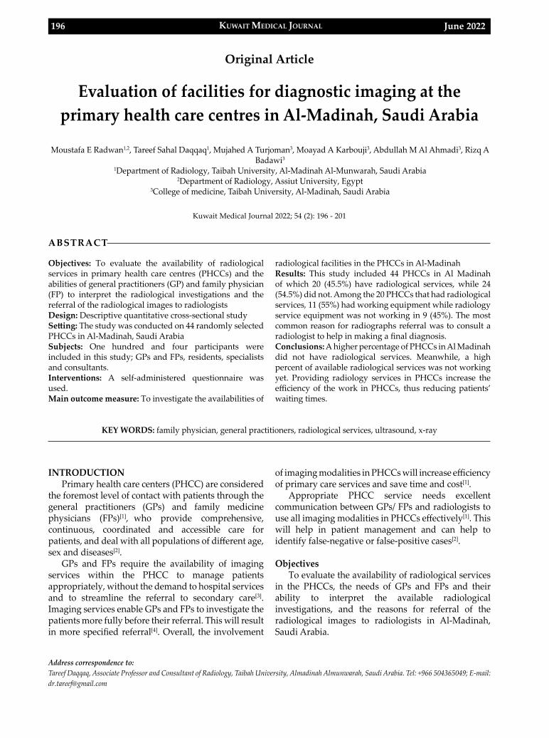
KUWAIT MEDICAL JOURNAL June 2022196
Original Article
Kuwait Medical Journal 2022; 54 (2): 196 - 201
Evaluation of facilities for diagnostic imaging at theprimary health care centres in Al-Madinah, Saudi Arabia
ABSTRACT
KEY WORDS: family physician, general practitioners, radiological services, ultrasound, x-ray
Moustafa E Radwan1,2, Tareef Sahal Daqqaq1, Mujahed A Turjoman3, Moayad A Karbouji3, Abdullah M Al Ahmadi3, Rizq A Badawi3
1Department of Radiology, Taibah University, Al-Madinah Al-Munwarah, Saudi Arabia2Department of Radiology, Assiut University, Egypt
3College of medicine, Taibah University, Al-Madinah, Saudi Arabia
Objectives: To evaluate the availability of radiological services in primary health care centres (PHCCs) and the abilities of general practitioners (GP) and family physician (FP) to interpret the radiological investigations and the referral of the radiological images to radiologistsDesign: Descriptive quantitative cross-sectional studySetting: The study was conducted on 44 randomly selected PHCCs in Al-Madinah, Saudi ArabiaSubjects: One hundred and four participants were included in this study; GPs and FPs, residents, specialists and consultants. Interventions: A self-administered questionnaire was used.Main outcome measure: To investigate the availabilities of
radiological facilities in the PHCCs in Al-MadinahResults: This study included 44 PHCCs in Al Madinah of which 20 (45.5%) have radiological services, while 24 (54.5%) did not. Among the 20 PHCCs that had radiological services, 11 (55%) had working equipment while radiology service equipment was not working in 9 (45%). The most common reason for radiographs referral was to consult a radiologist to help in making a final diagnosis. Conclusions: A higher percentage of PHCCs in Al Madinah did not have radiological services. Meanwhile, a high percent of available radiological services was not working yet. Providing radiology services in PHCCs increase the efficiency of the work in PHCCs, thus reducing patients’ waiting times.
Address correspondence to:Tareef Daqqaq, Associate Professor and Consultant of Radiology, Taibah University, Almadinah Almunwarah, Saudi Arabia. Tel: +966 504365049; E-mail: [email protected]
INTRODUCTIONPrimary health care centers (PHCC) are considered
the foremost level of contact with patients through the general practitioners (GPs) and family medicine physicians (FPs)[1], who provide comprehensive, continuous, coordinated and accessible care for patients, and deal with all populations of different age, sex and diseases[2].
GPs and FPs require the availability of imaging services within the PHCC to manage patients appropriately, without the demand to hospital services and to streamline the referral to secondary care[3]. Imaging services enable GPs and FPs to investigate the patients more fully before their referral. This will result in more specified referral[4]. Overall, the involvement
of imaging modalities in PHCCs will increase efficiency of primary care services and save time and cost[1].
Appropriate PHCC service needs excellent communication between GPs/ FPs and radiologists to use all imaging modalities in PHCCs effectively[1]. This will help in patient management and can help to identify false-negative or false-positive cases[2].
Objectives To evaluate the availability of radiological services
in the PHCCs, the needs of GPs and FPs and their ability to interpret the available radiological investigations, and the reasons for referral of the radiological images to radiologists in Al-Madinah, Saudi Arabia.
KUWAIT MEDICAL JOURNAL 197June 2022
MATERIALS AND METHODSThis cross-sectional study was conducted on 44
randomly selected PHCCs in Al-Madinah, Saudi Arabia. One hundred and four participants including GPs, FPs, residents, specialists and consultants were included in this study.
Study toolsThe tool used in this study was developed by
researchers after extensive review of the relevant and current literature. A self-administered questionnaire was used. It is composed of four parts of 11 questions, targeting the research objectives.
1. To investigate the availabilities of radiological facilities in the PHCCs in Al-Madinah through three questions: a. Are there any imaging (radiology) services in
the PHCCs? b. What are the types of available radiology
services? c. Are the currently available radiology devices
working?2. To explore the importance of radiological
investigations in helping FPs for the diagnosis of certain diseases in PHCCs through five questions: a. What was the extent of your need as a GP/
FP for the presence of radiology service in the PHCC to facilitate the diagnosis of certain diseases?
b. Could the radiographic findings of cases change or affect the patients’ diagnosis or their management plan?
c. Does the provision of radiology services in PHCCs decrease the referral of cases to tertiary centers?
d. Would the availability of radiology services increase the efficacy of your work?
e. In your opinion, what is the most important type of radiology services that can be useful for you in diagnosing and developing a management plan for patients?
3. To know if FPs can interpret radiological images by themselves or if they need to refer to a radiologist through two questions: a. Did you read the radiograph issued by the
PHCCs by yourself? b. If you need to communicate with radiologists,
how did you contact them?4. To know the reasons for referral of the
radiological images to a radiologist through one question: a. What are the reasons behind sending the
radiographs to be read by the radiologist?
Administrative and ethical considerationThis prospective research was conducted after
approval of the institutional board of ethics numbered 071-1439. The assurance of anonymity of participants was provided by asking the participants not to put their names on the questionnaire and all information remained confidential. Participants were reassured that participation in this study was voluntary and withdrawal from the study can be done at any time if they wish. Confidentiality and privacy were maintained by data coding to eliminate identifying data with personal information. Researchers provided the participants with the questionnaire involving covering page that included the ethical considerations, the purpose of the study and the right to accept or refuse to participate. The researchers published the link of electronic questionnaires in each healthcare centre for the period June to August 2018.
Data analysisData were gathered and analyzed using IBM SPSS
software (release 25), SPSS Inc., for Windows (Microsoft), United States of America). Statistical methods included descriptive analysis, such as number and percentage, and frequencies. Microsoft word and Excel have been used to generate graphs and tables.
RESULTSThis study included 44 PHCC’s in Al Madinah. The
availabilities of radiological facilities showed that 20 (45.5%) of the total PHCC’s had radiological services, while 24 (54.5%) did not.
Among the 20 PHCCs that had radiological services, 10 (50%) had both x-rays and ultrasound, while 9 (45%) had only x-rays and one center (5%) had only ultrasound. Out of the 20 PHCCs that have available radiological services, 11 (55%) PHCCs had working equipment while radiology service equipment was not working in 9 (45%) PHCCs.
Overall, 104 participants were enrolled in this study and they were split into two groups; Group 1 included the participants who work in PHCCs with available radiological services (n=56) while Group 2 included the participants who work in PHCCs with no available radiological services (n=48). Nine of the participants (8.7%) were FP consultants (six in centers with radiological services and three in centers with no radiological services), 32 (30.8%) were FP specialists (20 in centers with radiological services and 12 in centers with no radiological services), 18 (17.3%) were FP residents (12 in centers with radiological services and six in centers with no radiological services) and 45 (43.3%) were GPs (18 in centers with radiological
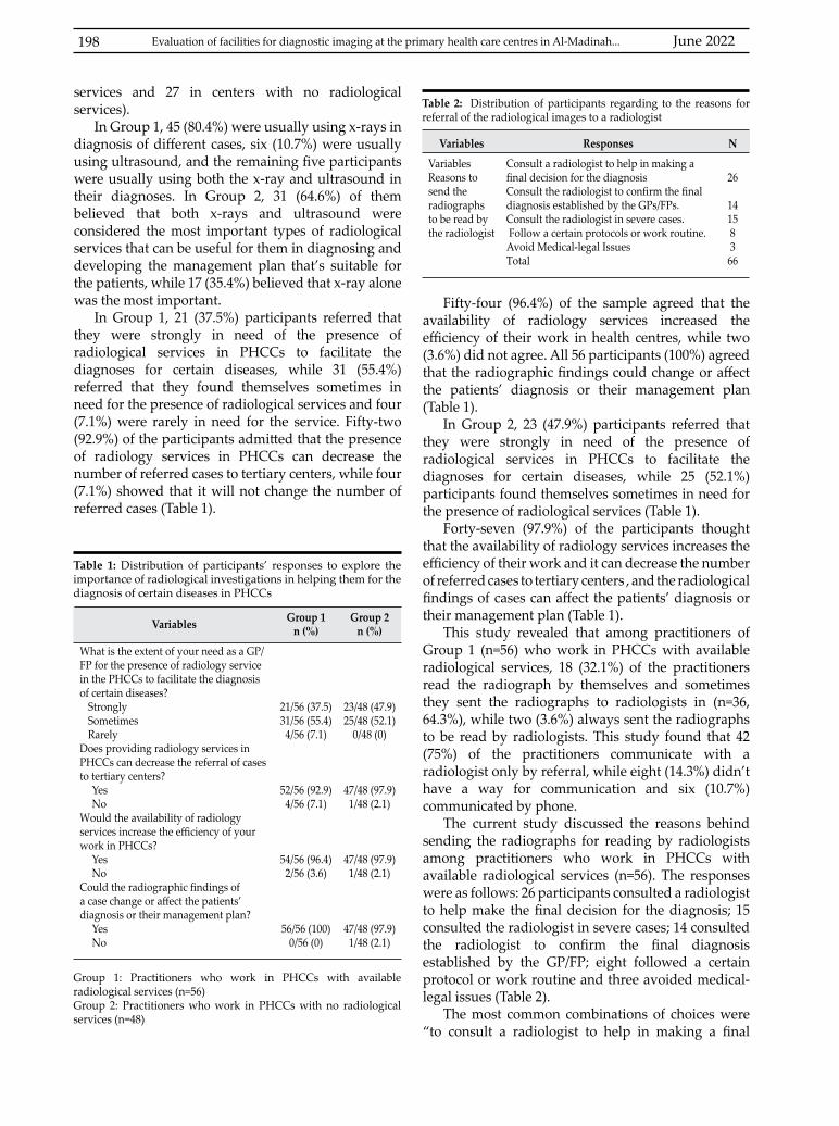
June 2022198
services and 27 in centers with no radiological services).
In Group 1, 45 (80.4%) were usually using x-rays in diagnosis of different cases, six (10.7%) were usually using ultrasound, and the remaining five participants were usually using both the x-ray and ultrasound in their diagnoses. In Group 2, 31 (64.6%) of them believed that both x-rays and ultrasound were considered the most important types of radiological services that can be useful for them in diagnosing and developing the management plan that’s suitable for the patients, while 17 (35.4%) believed that x-ray alone was the most important.
In Group 1, 21 (37.5%) participants referred that they were strongly in need of the presence of radiological services in PHCCs to facilitate the diagnoses for certain diseases, while 31 (55.4%) referred that they found themselves sometimes in need for the presence of radiological services and four (7.1%) were rarely in need for the service. Fifty-two (92.9%) of the participants admitted that the presence of radiology services in PHCCs can decrease the number of referred cases to tertiary centers, while four (7.1%) showed that it will not change the number of referred cases (Table 1).
Variables Group 1n (%)
Group 2n (%)
What is the extent of your need as a GP/FP for the presence of radiology service in the PHCCs to facilitate the diagnosis of certain diseases?
StronglySometimesRarely
Does providing radiology services in PHCCs can decrease the referral of cases to tertiary centers?
YesNo
Would the availability of radiology services increase the efficiency of your work in PHCCs?
YesNo
Could the radiographic findings of a case change or affect the patients’ diagnosis or their management plan?
YesNo
21/56 (37.5)31/56 (55.4)
4/56 (7.1)
52/56 (92.9)4/56 (7.1)
54/56 (96.4)2/56 (3.6)
56/56 (100)0/56 (0)
23/48 (47.9)25/48 (52.1)
0/48 (0)
47/48 (97.9)1/48 (2.1)
47/48 (97.9)1/48 (2.1)
47/48 (97.9)1/48 (2.1)
Table 1: Distribution of participants’ responses to explore the importance of radiological investigations in helping them for the diagnosis of certain diseases in PHCCs
Group 1: Practitioners who work in PHCCs with available radiological services (n=56)Group 2: Practitioners who work in PHCCs with no radiological services (n=48)
Variables Responses N
VariablesReasons to send the radiographs to be read by the radiologist
Consult a radiologist to help in making a final decision for the diagnosisConsult the radiologist to confirm the final diagnosis established by the GPs/FPs.Consult the radiologist in severe cases. Follow a certain protocols or work routine.Avoid Medical-legal IssuesTotal
26
14158366
Table 2: Distribution of participants regarding to the reasons for referral of the radiological images to a radiologist
Fifty-four (96.4%) of the sample agreed that the availability of radiology services increased the efficiency of their work in health centres, while two (3.6%) did not agree. All 56 participants (100%) agreed that the radiographic findings could change or affect the patients’ diagnosis or their management plan (Table 1).
In Group 2, 23 (47.9%) participants referred that they were strongly in need of the presence of radiological services in PHCCs to facilitate the diagnoses for certain diseases, while 25 (52.1%) participants found themselves sometimes in need for the presence of radiological services (Table 1).
Forty-seven (97.9%) of the participants thought that the availability of radiology services increases the efficiency of their work and it can decrease the number of referred cases to tertiary centers , and the radiological findings of cases can affect the patients’ diagnosis or their management plan (Table 1).
This study revealed that among practitioners of Group 1 (n=56) who work in PHCCs with available radiological services, 18 (32.1%) of the practitioners read the radiograph by themselves and sometimes they sent the radiographs to radiologists in (n=36, 64.3%), while two (3.6%) always sent the radiographs to be read by radiologists. This study found that 42 (75%) of the practitioners communicate with a radiologist only by referral, while eight (14.3%) didn’t have a way for communication and six (10.7%) communicated by phone.
The current study discussed the reasons behind sending the radiographs for reading by radiologists among practitioners who work in PHCCs with available radiological services (n=56). The responses were as follows: 26 participants consulted a radiologist to help make the final decision for the diagnosis; 15 consulted the radiologist in severe cases; 14 consulted the radiologist to confirm the final diagnosis established by the GP/FP; eight followed a certain protocol or work routine and three avoided medical-legal issues (Table 2).
The most common combinations of choices were “to consult a radiologist to help in making a final
Evaluation of facilities for diagnostic imaging at the primary health care centres in Al-Madinah...
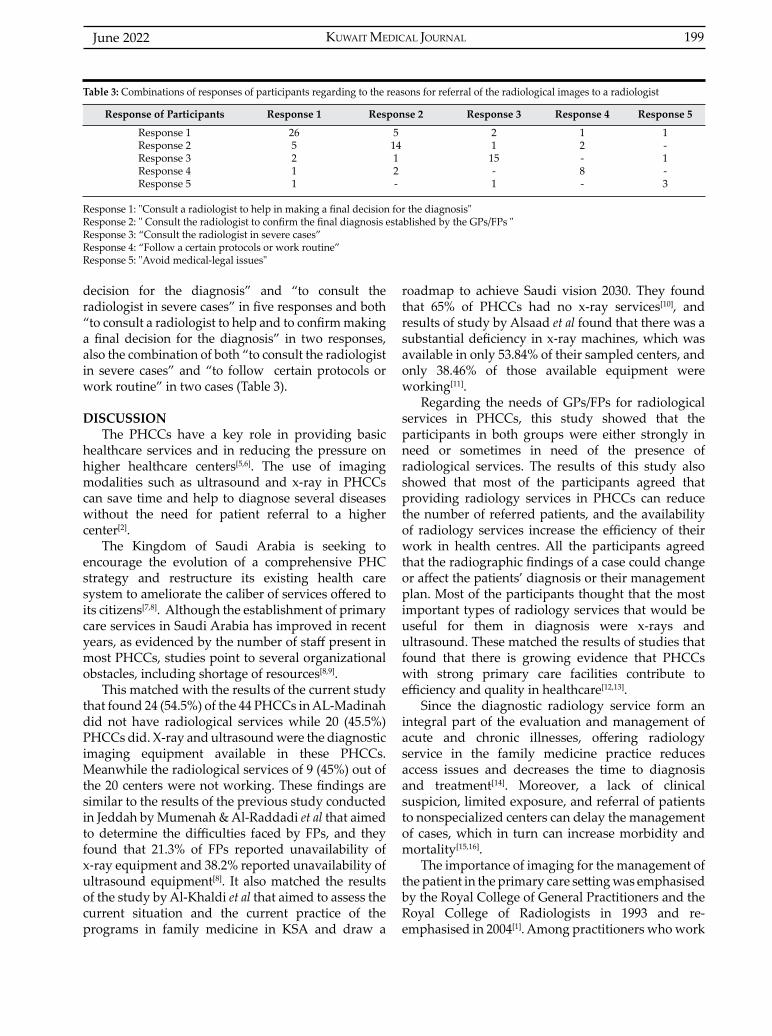
KUWAIT MEDICAL JOURNAL 199June 2022
decision for the diagnosis” and “to consult the radiologist in severe cases” in five responses and both “to consult a radiologist to help and to confirm making a final decision for the diagnosis” in two responses, also the combination of both “to consult the radiologist in severe cases” and “to follow certain protocols or work routine” in two cases (Table 3).
DISCUSSIONThe PHCCs have a key role in providing basic
healthcare services and in reducing the pressure on higher healthcare centers[5,6]. The use of imaging modalities such as ultrasound and x-ray in PHCCs can save time and help to diagnose several diseases without the need for patient referral to a higher center[2].
The Kingdom of Saudi Arabia is seeking to encourage the evolution of a comprehensive PHC strategy and restructure its existing health care system to ameliorate the caliber of services offered to its citizens[7,8]. Although the establishment of primary care services in Saudi Arabia has improved in recent years, as evidenced by the number of staff present in most PHCCs, studies point to several organizational obstacles, including shortage of resources[8,9].
This matched with the results of the current study that found 24 (54.5%) of the 44 PHCCs in AL-Madinah did not have radiological services while 20 (45.5%) PHCCs did. X-ray and ultrasound were the diagnostic imaging equipment available in these PHCCs. Meanwhile the radiological services of 9 (45%) out of the 20 centers were not working. These findings are similar to the results of the previous study conducted in Jeddah by Mumenah & Al-Raddadi et al that aimed to determine the difficulties faced by FPs, and they found that 21.3% of FPs reported unavailability of x‑ray equipment and 38.2% reported unavailability of ultrasound equipment[8]. It also matched the results of the study by Al-Khaldi et al that aimed to assess the current situation and the current practice of the programs in family medicine in KSA and draw a
roadmap to achieve Saudi vision 2030. They found that 65% of PHCCs had no x-ray services[10], and results of study by Alsaad et al found that there was a substantial deficiency in x‑ray machines, which was available in only 53.84% of their sampled centers, and only 38.46% of those available equipment were working[11].
Regarding the needs of GPs/FPs for radiological services in PHCCs, this study showed that the participants in both groups were either strongly in need or sometimes in need of the presence of radiological services. The results of this study also showed that most of the participants agreed that providing radiology services in PHCCs can reduce the number of referred patients, and the availability of radiology services increase the efficiency of their work in health centres. All the participants agreed that the radiographic findings of a case could change or affect the patients’ diagnosis or their management plan. Most of the participants thought that the most important types of radiology services that would be useful for them in diagnosis were x-rays and ultrasound. These matched the results of studies that found that there is growing evidence that PHCCs with strong primary care facilities contribute to efficiency and quality in healthcare[12,13].
Since the diagnostic radiology service form an integral part of the evaluation and management of acute and chronic illnesses, offering radiology service in the family medicine practice reduces access issues and decreases the time to diagnosis and treatment[14]. Moreover, a lack of clinical suspicion, limited exposure, and referral of patients to nonspecialized centers can delay the management of cases, which in turn can increase morbidity and mortality[15,16].
The importance of imaging for the management of the patient in the primary care setting was emphasised by the Royal College of General Practitioners and the Royal College of Radiologists in 1993 and re-emphasised in 2004[1]. Among practitioners who work
Response of Participants Response 1 Response 2 Response 3 Response 4 Response 5
Response 1Response 2Response 3Response 4Response 5
265211
51412-
2115-1
12-8-
1-1-3
Response 1: "Consult a radiologist to help in making a final decision for the diagnosis"Response 2: " Consult the radiologist to confirm the final diagnosis established by the GPs/FPs "Response 3: “Consult the radiologist in severe cases”Response 4: “Follow a certain protocols or work routine” Response 5: "Avoid medical-legal issues"
Table 3: Combinations of responses of participants regarding to the reasons for referral of the radiological images to a radiologist
June 2022200
in PHCCs with available radiological services (Group 1), this study revealed that 18/56 (32.1%) of the sample read the radiograph by themselves and 36/56 (64.3%) sent the radiographs to radiologists. This study also found that 75% of the practitioners communicate with a radiologist only by referral. These findings were supported by the results in a cross-sectional study that was conducted by Al-Yami et al. The authors concluded their findings as the physicians in many PHCCs need practice in reading normal X-ray films to avoid unnecessary referral of patients to specialized medical centres[16].
Sometimes GPs and FPs in PHCC refer patients directly to imaging departments for image-guided procedures. So, good collaboration between them and radiologists is needed and challenging at the same time. It serves both clinical disciplines and their patients; GP/FP currently aiming to refine its targets and strategy and radiology where a multiplicity collaboration has been key to the future of the discipline[1]. Referrals from primary to secondary care reflect this key role of GPs/FPs. The results of this study have shown that the most common reasons among the sampled practitioners to send the radiographs to be read by the radiologist were; “Consult a radiologist to help in making a final decision for the diagnosis”, “Consult the radiologist in severe cases” and “Consult the radiologist to confirm the final diagnosis established by the family doctor”, respectively.
The results of this study were supported by the findings in a cross-sectional study by Tandjung et al in Swiss primary care. They found that the most frequent referral recipients were radiologists (21.14%) and the most frequent aim of referrals was advice concerning diagnosis or therapy (46.3%)[17]. Grieve et al, in their study, mentioned that for these imaging examinations to be used effectively, the GPs/FPs should communicate formally with the radiologist outlining the clinical history and potential diagnosis for the radiologist to justify the investigation of choice[18].
CONCLUSIONThis study revealed that a higher percentage of
the PHCCs in Al Madinah did not have radiological services. Meanwhile, a high percent of available radiological services were not working yet. The most common available imaging equipment were x-rays and ultrasound. Providing radiology services in PHCCs increased the efficiency of the work in PHCCs and can reduce the referring of patients. Radiological findings of cases could affect the patients’ diagnosis and their management plan.
ACKNOWLEDGMENTConflicts of interest: There are no conflicts of interest.Authors’ contributions: All authors were involved in the design of the study. Mujahed A Turjoman, Moayad A Karbouji, Abdullah M Al Ahmadi and Rizq A Badawi did the data collection. All authors contributed to the data analysis and interpretation. Moustafa E Radwan, Tareef Sahal Daqqaq, Mujahed A Turjoman and Moayad A Karbouji drafted final version of the manuscript on which the other authors gave inputs and approved the final version.
REFERENCES
1. The Executive Council of the European Society of Radiology (ESR) and the Council of the World Organization of National Colleges, Academies and Academic Associations of General Practitioners/Family Physicians/ European regional branch (WONCA Europe). Radiology and primary care in Europe. Insights Imaging 2010; 1(2):46-52.
2. European Society of Radiology 2009. The future role of radiology in healthcare. Insights Imaging 2010; 1(1):2-11.
3. Chen F, Yang M, Li Q, Pan J, Li X, Meng Q. Does providing more services increase the primary hospitals’ revenue? An assessment of national essential medicine policy based on 2,675 counties in China. PloS One 2018; 13(1):e0190855.
4. Cherryman G. Imaging in primary care. Br J Gen Pract 2006; 56(529):563-564.
5. Al-Mousa N. Patients satisfaction in primary health care services in primary care setting Dhahran, Saudi Arabia. International Journal of Medical and Health Sciences 2017; 3(11):69-77.
6. Senitan M, Alhaiti AH, Gillespie J. Patient satisfaction and experience of primary care in Saudi Arabia: a systematic review. Int J Qual Health Care 2018; 30(10):751-759.
7. Sharaf E, Madan N, Sharaf A. Physician job satisfaction in primary care. Bahrain Medical Bulletin 2008; 30(2):30-32.
8. Mumenah SH, Al-Raddadi RM. Difficulties faced by family physicians in primary health care centers in Jeddah, Saudi Arabia. J Family Community Med 2015; 22(3):145-151.
9. Al-Ahmadi H, Roland M. Quality of primary health care in Saudi Arabia: a comprehensive review. Int J Qual Health Care 2005; 17(4):331-346.
10. Al-Khaldi YM, Al-Ghamdi EA, Al-Mogbil TI, Al-Khashan HI. Family medicine practice in Saudi Arabia: The current situation and Proposed Strategic Directions Plan 2020. J Family Community Med 2017; 24(3):156-163.
11. Alsaad SSM, Abu-Grain SHS, El-Kheir DYM. Preparedness of Dammam primary health care centers to deal with emergency cases. J Family Community Med 2017; 24(3):181-188.
Evaluation of facilities for diagnostic imaging at the primary health care centres in Al-Madinah...

KUWAIT MEDICAL JOURNAL 201June 2022
12. Kringos DS, Boerma W, van der Zee J, Groenewegen P. Europe’s strong primary care systems are linked to better population health but also to higher health spending. Health Aff 2013; 32(4):686-694.
13. Starfield B, Shi L, Macinko J. Contribution of primary care to health systems and health. Milbank Q 2005; 83(3):457-502.
14. American Academy of Family Physicians. AAFP Member Census, Table 13: Clinical Procedures Performed by Physicians at their Practice. Retrieved from http://www.aafp.org/about/the-aafp/family-medicine-facts/table-13.html., June 30, 2015.
15. Perez MR. Referral criteria and clinical decision support: radiological protection aspects for justification. Ann ICRP 2015; 44(1 Suppl):276-87.
16. Alyami A, Alshomrani Y, Suqaty R, Futtiny S, Alnaqib F, Albarakati M, et al. Perspectives of primary health care physicians on diagnosing and referring patients with apparent osteolytic lesions on plain X-ray films: a cross-sectional study. Adv Med Educ Pract 2016; 7:145-51.
17. Tandjung R, Hanhart A, Bartschi F, Keller R, Steinhauer A, Rosemann T, et al. Referral rates in Swiss primary care with a special emphasis on reasons for encounter. Swiss Med Wkly 2015; 145:w14244.
18. Grieve FM, Plumb AA, Khan SH. Radiology reporting: a general practitioner's perspective. Br J Radiol 2010; 83(985):17-22.
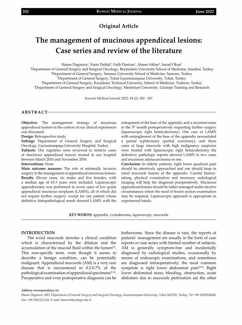
KUWAIT MEDICAL JOURNAL June 2022202
Original Article
Kuwait Medical Journal 2022; 54 (2): 202 - 207
The management of mucinous appendiceal lesions: Case series and review of the literature
ABSTRACT
KEY WORDS: appendix, cystadenoma, laparoscopy, mucocele
Hasan Dagmura1, Emin Daldal2, Fatih Dasiran3, Ahmet Akbas4, Ismail Okan5
1Department of General Surgery and Surgical Oncology, Bezmialem University School of Medicine, Istanbul, Turkey2Department of General Surgery, Samsun University School of Medicine, Samsun, Turkey
3Department of General Surgery, Tokat Gaziosmanpasa University, Tokat, Turkey4Department of General Surgery, Karadeniz Technical Universiy, School of Medicine, Trabzon, Turkey
5Department of General Surgery and Surgical Oncology, Medeniyet University, Göztepe Training and Research
Objective: The management strategy of mucinous appendiceal tumors in the context of our clinical experiences was discussed. Design: Retrospective studySettings: Department of General Surgery and Surgical Oncology, Gaziosmanpaşa University Hospital, TurkeySubjects: Our registries were reviewed to retrieve cases of mucinous appendiceal tumors treated in our hospital between March 2010 and November 2018.Interventions: NoneMain outcome measure: The role of minimally invasive surgery in the management of appendiceal mucinous lesions.Results: Eleven cases, six males and five females, with a median age of 65.3 years were included. Laparoscopic appendectomy was performed in seven cases of low grade appendiceal mucinous neoplasm (LAMN), all of which did not require further surgery, except for one patient whose definitive histopathological result showed LAMN with the
entrapment of the base of the appendix and a recurrent mass in the 5th month postoperatively requesting further surgery (laparoscopic right hemicolectomy). One case of LAMN with entanglement of the base of the appendix necessitated a partial typhlectomy (partial cecectomy), and three cases of large mucocele with high malignancy suspicion were treated with laparoscopic right hemicolectomy the definitive; pathologic reports showed LAMN in two cases and mucinous adenocarcinoma in one.Conclusion: In elderly patients, right lower quadrant pain should be attentively approached and one should keep in mind mucocele lesions of the appendix. Careful history-taking, physical examination and necessary radiological imaging will help the diagnosis preoperatively. Mucinous appendiceal lesions should be rather managed under elective circumstances where the need of frozen section examination may be required. Laparoscopic approach is appropriate in experienced hands.
Address correspondence to:Hasan Dagmura, MD, Department of General Surgery and Surgical Oncology, Gaziosmanpasa University, Tokat (60250), Turkey. Tel: +90 5418954688; Fax: +90 3562122142; E-mail: [email protected]
INTRODUCTIONThe word mucocele denotes a clinical condition
which is characterized by the dilation and the accumulation of the mucoid fluid within the lumen[1]. This non-specific term, even though it seems to describe a benign condition, can be potentially malignant. Appendiceal mucocele (AM) is a very rare disease that is encountered in 0.2-0.7% of the pathological examination of appendiceal specimens[2-4]. Preoperative and even postoperative diagnosis can be
bothersome. Since the disease is rare, the reports of patients’ management are usually in the form of case reports or case series with limited number of subjects. AM is generally symptom-free and incidentally diagnosed by radiological studies, occasionally by means of endoscopic examinations, and sometimes are diagnosed intraoperatively; the most common symptom is right lower abdominal pain[5,6]. Right lower abdominal mass, bleeding, obstruction, acute abdomen due to mucocele perforation are the other
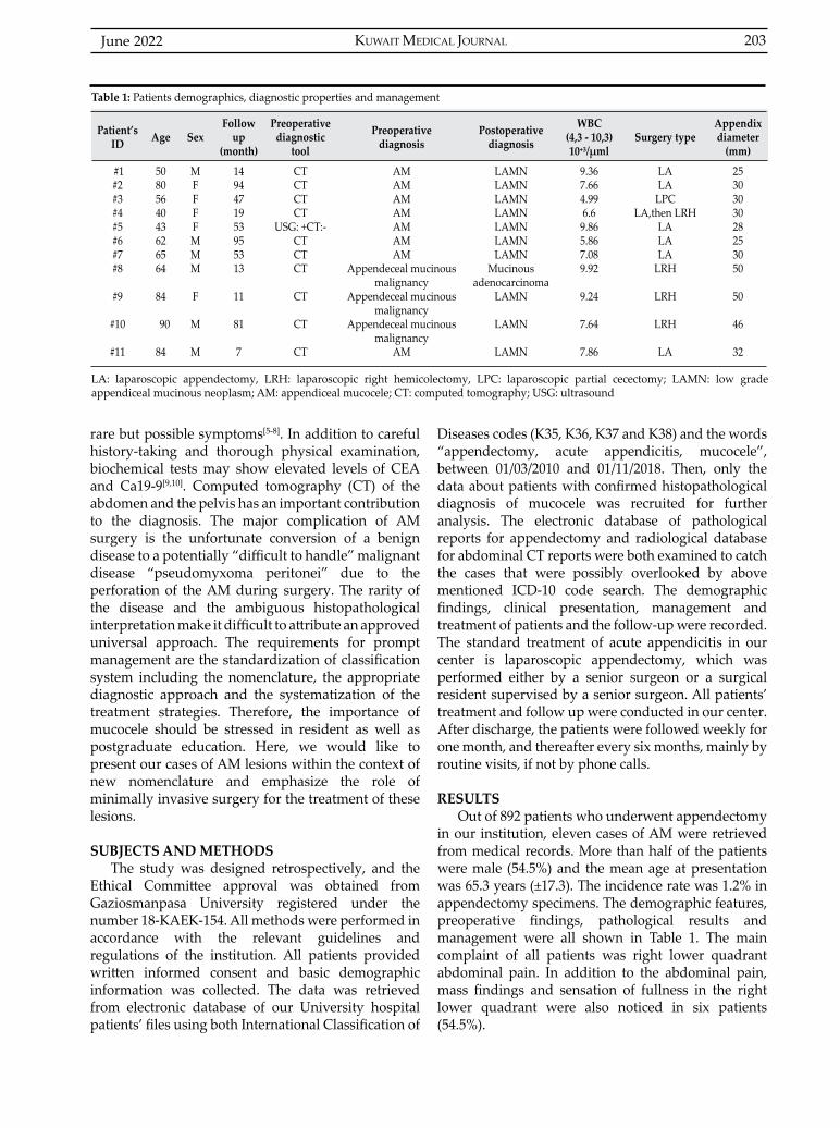
KUWAIT MEDICAL JOURNAL 203June 2022
rare but possible symptoms[5-8]. In addition to careful history-taking and thorough physical examination, biochemical tests may show elevated levels of CEA and Ca19-9[9,10]. Computed tomography (CT) of the abdomen and the pelvis has an important contribution to the diagnosis. The major complication of AM surgery is the unfortunate conversion of a benign disease to a potentially “difficult to handle” malignant disease “pseudomyxoma peritonei” due to the perforation of the AM during surgery. The rarity of the disease and the ambiguous histopathological interpretation make it difficult to attribute an approved universal approach. The requirements for prompt management are the standardization of classification system including the nomenclature, the appropriate diagnostic approach and the systematization of the treatment strategies. Therefore, the importance of mucocele should be stressed in resident as well as postgraduate education. Here, we would like to present our cases of AM lesions within the context of new nomenclature and emphasize the role of minimally invasive surgery for the treatment of these lesions.
SUBJECTS AND METHODSThe study was designed retrospectively, and the
Ethical Committee approval was obtained from Gaziosmanpasa University registered under the number 18-KAEK-154. All methods were performed in accordance with the relevant guidelines and regulations of the institution. All patients provided written informed consent and basic demographic information was collected. The data was retrieved from electronic database of our University hospital patients’ files using both International Classification of
Diseases codes (K35, K36, K37 and K38) and the words “appendectomy, acute appendicitis, mucocele”, between 01/03/2010 and 01/11/2018. Then, only the data about patients with confirmed histopathological diagnosis of mucocele was recruited for further analysis. The electronic database of pathological reports for appendectomy and radiological database for abdominal CT reports were both examined to catch the cases that were possibly overlooked by above mentioned ICD-10 code search. The demographic findings, clinical presentation, management and treatment of patients and the follow-up were recorded. The standard treatment of acute appendicitis in our center is laparoscopic appendectomy, which was performed either by a senior surgeon or a surgical resident supervised by a senior surgeon. All patients’ treatment and follow up were conducted in our center. After discharge, the patients were followed weekly for one month, and thereafter every six months, mainly by routine visits, if not by phone calls.
RESULTSOut of 892 patients who underwent appendectomy
in our institution, eleven cases of AM were retrieved from medical records. More than half of the patients were male (54.5%) and the mean age at presentation was 65.3 years (±17.3). The incidence rate was 1.2% in appendectomy specimens. The demographic features, preoperative findings, pathological results and management were all shown in Table 1. The main complaint of all patients was right lower quadrant abdominal pain. In addition to the abdominal pain, mass findings and sensation of fullness in the right lower quadrant were also noticed in six patients (54.5%).
Patient’s ID Age Sex
Follow up
(month)
Preoperative diagnostic
tool
Preoperativediagnosis
Postoperative diagnosis
WBC(4,3 - 10,3)10⁺³/µml
Surgery typeAppendix diameter
(mm)
#1#2#3#4#5#6#7#8
#9 #10
#11
5080564043626564
84
90
84
MFFFFMMM
F
M
M
1494471953955313
11
81
7
CTCTCTCT
USG: +CT:-CTCT CT
CT
CT
CT
AMAMAMAMAMAMAM
Appendeceal mucinousmalignancy
Appendeceal mucinousmalignancy
Appendeceal mucinousmalignancy
AM
LAMNLAMNLAMNLAMNLAMNLAMNLAMN
Mucinousadenocarcinoma
LAMN
LAMN
LAMN
9.367.664.996.69.865.867.089.92
9.24
7.64
7.86
LALA
LPCLA,then LRH
LALALA
LRH
LRH
LRH
LA
2530303028253050
50
46
32
LA: laparoscopic appendectomy, LRH: laparoscopic right hemicolectomy, LPC: laparoscopic partial cecectomy; LAMN: low grade appendiceal mucinous neoplasm; AM: appendiceal mucocele; CT: computed tomography; USG: ultrasound
Table 1: Patients demographics, diagnostic properties and management
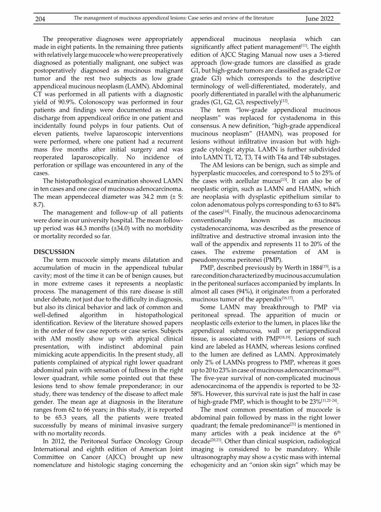
June 2022204
The preoperative diagnoses were appropriately made in eight patients. In the remaining three patients with relatively large mucocele who were preoperatively diagnosed as potentially malignant, one subject was postoperatively diagnosed as mucinous malignant tumor and the rest two subjects as low grade appendiceal mucinous neoplasm (LAMN). Abdominal CT was performed in all patients with a diagnostic yield of 90.9%. Colonoscopy was performed in four patients and findings were documented as mucus discharge from appendiceal orifice in one patient and incidentally found polyps in four patients. Out of eleven patients, twelve laparoscopic interventions were performed, where one patient had a recurrent mass five months after initial surgery and was reoperated laparoscopically. No incidence of perforation or spillage was encountered in any of the cases.
The histopathological examination showed LAMN in ten cases and one case of mucinous adenocarcinoma. The mean appendeceal diameter was 34.2 mm (± S: 8.7).
The management and follow-up of all patients were done in our university hospital. The mean follow-up period was 44.3 months (±34.0) with no morbidity or mortality recorded so far.
DISCUSSIONThe term mucocele simply means dilatation and
accumulation of mucin in the appendiceal tubular cavity; most of the time it can be of benign causes, but in more extreme cases it represents a neoplastic process. The management of this rare disease is still under debate, not just due to the difficulty in diagnosis, but also its clinical behavior and lack of common and well-defined algorithm in histopathological identification. Review of the literature showed papers in the order of few case reports or case series. Subjects with AM mostly show up with atypical clinical presentation, with indistinct abdominal pain mimicking acute appendicitis. In the present study, all patients complained of atypical right lower quadrant abdominal pain with sensation of fullness in the right lower quadrant, while some pointed out that these lesions tend to show female preponderance; in our study, there was tendency of the disease to affect male gender. The mean age at diagnosis in the literature ranges from 62 to 66 years; in this study, it is reported to be 65.3 years, all the patients were treated successfully by means of minimal invasive surgery with no mortality records.
In 2012, the Peritoneal Surface Oncology Group International and eighth edition of American Joint Committee on Cancer (AJCC) brought up new nomenclature and histologic staging concerning the
appendiceal mucinous neoplasia which can significantly affect patient management[11]. The eighth edition of AJCC Staging Manual now uses a 3-tiered approach (low-grade tumors are classified as grade G1, but high-grade tumors are classified as grade G2 or grade G3) which corresponds to the descriptive terminology of well-differentiated, moderately, and poorly differentiated in parallel with the alphanumeric grades (G1, G2, G3, respectively)[12].
The term “low-grade appendiceal mucinous neoplasm” was replaced for cystadenoma in this consensus. A new definition, “high-grade appendiceal mucinous neoplasm” (HAMN), was proposed for lesions without infiltrative invasion but with high-grade cytologic atypia. LAMN is further subdivided into LAMN T1, T2, T3, T4 with T4a and T4b substages.
The AM lesions can be benign, such as simple and hyperplastic mucoceles, and correspond to 5 to 25% of the cases with acellular mucus[13]. It can also be of neoplastic origin, such as LAMN and HAMN, which are neoplasia with dysplastic epithelium similar to colon adenomatous polyps corresponding to 63 to 84% of the cases[14]. Finally, the mucinous adenocarcinoma conventionally known as mucinous cystadenocarcinoma, was described as the presence of infiltrative and destructive stromal invasion into the wall of the appendix and represents 11 to 20% of the cases. The extreme presentation of AM is pseudomyxoma peritonei (PMP).
PMP, described previously by Werth in 1884[15], is a rare condition characterized by mucinous accumulation in the peritoneal surfaces accompanied by implants. In almost all cases (94%), it originates from a perforated mucinous tumor of the appendix[16,17].
Some LAMN may breakthrough to PMP via peritoneal spread. The apparition of mucin or neoplastic cells exterior to the lumen, in places like the appendiceal submucosa, wall or periappendiceal tissue, is associated with PMP[18,19]. Lesions of such kind are labeled as HAMN, whereas lesions confined to the lumen are defined as LAMN. Approximately only 2% of LAMNs progress to PMP, whereas it goes up to 20 to 23% in case of mucinous adenocarcinomas[20]. The five-year survival of non-complicated mucinous adenocarcinoma of the appendix is reported to be 32-58%. However, this survival rate is just the half in case of high-grade PMP, which is thought to be 23%[11,21-24].
The most common presentation of mucocele is abdominal pain followed by mass in the right lower quadrant; the female predominance[21] is mentioned in many articles with a peak incidence at the 6th decade[20,21]. Other than clinical suspicion, radiological imaging is considered to be mandatory. While ultrasonography may show a cystic mass with internal echogenicity and an “onion skin sign” which may be
The management of mucinous appendiceal lesions: Case series and review of the literature
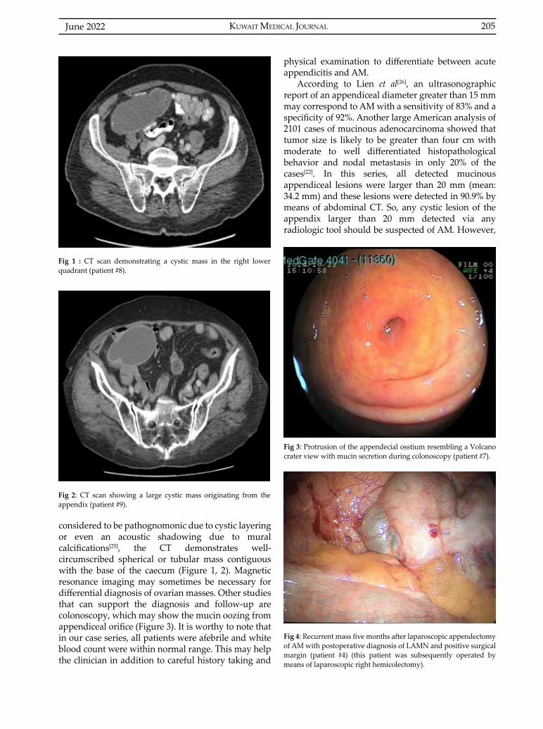
KUWAIT MEDICAL JOURNAL 205June 2022
considered to be pathognomonic due to cystic layering or even an acoustic shadowing due to mural calcifications[25], the CT demonstrates well-circumscribed spherical or tubular mass contiguous with the base of the caecum (Figure 1, 2). Magnetic resonance imaging may sometimes be necessary for differential diagnosis of ovarian masses. Other studies that can support the diagnosis and follow-up are colonoscopy, which may show the mucin oozing from appendiceal orifice (Figure 3). It is worthy to note that in our case series, all patients were afebrile and white blood count were within normal range. This may help the clinician in addition to careful history taking and
Fig 1 : CT scan demonstrating a cystic mass in the right lower quadrant (patient #8).
Fig 3: Protrusion of the appendecial osstium resembling a Volcano crater view with mucin secretion during colonoscopy (patient #7).
Fig 2: CT scan showing a large cystic mass originating from the appendix (patient #9).
Fig 4: Recurrent mass five months after laparoscopic appendectomy of AM with postoperative diagnosis of LAMN and positive surgical margin (patient #4) (this patient was subsequently operated by means of laparoscopic right hemicolectomy).
physical examination to differentiate between acute appendicitis and AM.
According to Lien et al[26], an ultrasonographic report of an appendiceal diameter greater than 15 mm may correspond to AM with a sensitivity of 83% and a specificity of 92%. Another large American analysis of 2101 cases of mucinous adenocarcinoma showed that tumor size is likely to be greater than four cm with moderate to well differentiated histopathological behavior and nodal metastasis in only 20% of the cases[22]. In this series, all detected mucinous appendiceal lesions were larger than 20 mm (mean: 34.2 mm) and these lesions were detected in 90.9% by means of abdominal CT. So, any cystic lesion of the appendix larger than 20 mm detected via any radiologic tool should be suspected of AM. However,
June 2022206
neoplastic mucinous appendiceal lesions tend to have a diameter greater than two centimeters.
The treatment of AM is purely surgical. The intraoperative finding of these lesions is of great importance, so detailed description of the lesion is mandatory. It should include the location of the tumor (tip vs. body vs. base), the distance of tumor from proximal resection margin, the presence or absence of mucin deposits on the serosal surface or in the wall of the appendix, or elsewhere in the abdomen, the presence or absence of adhesions to other structures, and the presence or absence of perforation.
For benign non-neoplastic lesions, laparoscopic appendectomy suffices, while laparoscopic appendectomy with lymphadenectomy, including all the fat from the mesoappendix, is indicated for LAMN and HAMN, and when the appendicular base is not compromised by the dilation and with no peritoneal involvement.
In some cases of LAMN and HAMN where there is involvement of the base or extraappendiceal involvement respectively, a more extended resection such as partial cecectomy is advised; in cases where the latter is not achievable, right hemicolectomy would be more appropriate in combination with medical treatment.
In this case series out of 11 cases of appendeceal cystic lesions, ten patients (90.9%) were diagnosed as LAMN, even though the histopathological diagnoses were the same. The treatment approach varied from laparoscopic appendectomy to laparoscopic cecectomy and even laparoscopic right hemicolectomy.
Laparoscopy is definitely not without drawbacks; rupture and spillage of a mucin containing epithelial cells lesion may lead to catastrophic changes in the management of such patients and even an upstaging of the tumor, which may affect survival negatively. In our study, one patient was reoperated because of the positivity of the surgical margin during her first operation and had laparoscopic right hemicolectomy due to recurrent mass in the ceacum originating from the base of the appendectomy (Figure 4). All patients were managed laparoscopically, six (54.5%) of which had laparoscopic appendectomy, one (9.1%) patient had laparoscopic partial cecectomy, another patient had appendectomy then reoperated due to recurrent mass and had laparoscopic right hemicloctomy, three (27.3%) patients had laparoscopic right hemicolectomy due to large potentially malignant appendeceal mucocle, where one of which showed mucinous adenocarcinoma of the appendix on histopatological examination and the other two subjects showed LAMN.
It was particularly noteworthy that there is a liaison between AM and colonic polyps and hence colonic
malignancies, which was previously confirmed by other studies[11,27]. Therefore, we recommend surveillance colonoscopy in patients with a diagnosis of AM. Even though there is no strong evidence showing the transformation of LAMN to mucinous adenocarcinoma, most pathologists consider the adenoma-adenocarcinoma sequence is comparable with the colonic polyp–adenocarcinoma sequence[28].
The relatively small number of patients in this study limited the ability to conclude a universal approach concerning diagnosis, treatment and follow up strategies. The low rate of recurrence can be linked to the short follow-up period; the absence of any spillage or rupture during laparoscopic surgery can be attributed again to the low sample size. However, this study may be considered as a leading point to conduct relatively larger studies with longer follow-up period and in a prospective manner.
CONCLUSIONAppendiceal mass should not be taken for granted,
especially for patients in the 6th decade with indistinct pain. It was particularly noteworthy that an appendiceal diameter larger than 2 cm suggests a mucinous lesion of the appendix. So, these patients should be approached with prudence and prompt preoperative investigation with CT, magnetic resonance imaging or even colonoscopy. Once the decision for exploration is taken under elective circumstances, the laparoscopic approach would be the most appropriate in experienced hands and the availability of frozen examination should be ensured to help the intraoperative decision making. Laparoscopic approach would be appropriate for patients with mucocele that can be removed without rupture.
ACKNOWLEDGMENTConflict of interests: NoneSource of funding: NoneCompeting interests: NoneData availability statement: The data sets generated during the current study are available from the corresponding author upon request.
Author contributions: All authors contributed equally to this study.
REFERENCES
1. Kumar V, Abbas AK, Fausto N, Robbins SL, Cotran RS. Robbins and Cotran pathologic basis of disease. 4th ed. Philadelphia: Elsevier Saunders; 2005.
2. Caracappa D, Gulla N, Gentile D, Listorti C, Boselli C, Cirocchi R, et al. Appendiceal mucocoele. A case report and literature review. Ann Ital Chir 2011; 82(3):239-245.
The management of mucinous appendiceal lesions: Case series and review of the literature
KUWAIT MEDICAL JOURNAL 207June 2022
3. Yilmaz M, Akbulut S, Kutluturk K, Sahin N, Arabaci E, Ara C, et al. Unusual histopathological findings in appendectomy specimens from patients with suspected acute appendicitis. World J Gastroenterol 2013; 19(25):4015-4022.
4. Ruiz-Tovar J, Teruel DG, Castineiras VM, Dehesa AS, Quindos PL, Molina EM. Mucocele of the appendix. World J Surg 2007; 31(3):542-548.
5. Dixit A, Robertson JH, Mudan SS, Akle C. Appendiceal mucocoeles and pseudomyxoma peritonei. World J Gastroenterol 2007; 13(16):2381-2384.
6. Caliskan K, Yildirim S, Bal N, Nursal TZ, Akdur AC, Moray G. Mucinous cystadenoma of the appendix: a rare cause of acute abdomen. Ulus Travma Acil Cerrahi Derg 2008; 14(4):303-307.
7. Kelemouridou E, Mogrampi SAL, Tsavis G, Verroiotou M, Rallis T, Fardellas I. Mucinous cystadenoma of the appendix. A diagnostic dilemma? Chirurgia (Bucur) 2011; 106(2):251-254.
8. Chong SJ, Chan MY. Mucinous cystadenoma of the appendix-an unusual cause of intestinal obstruction. Annals of the Academy of Medicine, Singapore 2001; 30(2):206-207.
9. McFarlane MEC, Plummer JM, Bonadie K. Mucinous cystadenoma of the appendix presenting with an elevated carcinoembryonic antigen (CEA): report of two cases and review of the literature. Int J Surg Case Rep 2013; 4(10):886-888.
10. Carmignani CP, Hampton R, Sugarbaker CE, Chang D, Sugarbaker PH. Utility of CEA and CA 19-9 tumor markers in diagnosis and prognostic assessment of mucinous epithelial cancers of the appendix. J Surg Oncol 2004; 87(4):162-166.
11. Carr NJ, Cecil TD, Mohamed F, Sobin LH, Sugarbaker PH, Gonzalez-Moreno S, et al. A consensus for classification and pathologic reporting of pseudomyxoma peritonei and associated appendiceal neoplasia: the results of the Peritoneal Surface Oncology Group International (PSOGI) modified Delphi process. Am J Surg Pathol 2016; 40(1):14-26.
12. Overman MJ, Asare EA, Compton CC, Appendix carcinoma. In: Amin M, ed. AJCC Cancer Staging Manual, 8th ed. Chicago, IL: Springer; 2017:237-250.
13. Higa E, Rosai J, Pizzimbono CA, Wise L. Mucosal hyperplasia, mucinous cystadenoma, and mucinous cystadenocarcinoma of the appendyx. A re-evalutation of appendiceal mucocele. Cancer 1973; 32(6):1525-41.
14. Yamane YD, Yamane H, Castro Júnior PC, Marsilac A, Mesquita RB, Paulo FL. Mucolele da apêndice-relato de caso e revisão da literatura. Rev bras Coloproct 2005; 25(3):256-60.
15. Werth R. Pseudomyxoma peritonei. Arch Gynakol 1884; 24:100.
16. Ramaswamy V. Pathology of mucinous appendiceal tumors and pseudomyxoma peritonei. Indian J Surg Oncol 2016; 7(2):258-267.
17. Misdraji J. Mucinous epithelial neoplasms of the appendix and pseudomyxoma peritonei. Mod Pathol 2015; 28 Suppl 1:S67-S79.
18. Misdraji J, Yantiss RK, Graeme-Cook FM, Balis UJ, Young RH. Appendiceal mucinous neoplasms: a clinicopathologic analysis of 107 cases. Am J Surg Pathol 2003; 27(8):1089-1103.
19. McDonald JR, O’Dwyer ST, Rout S, Chakrabarty B, Sikand K, Fulford PE, et al. Classification of and cytoreductive surgery for low-grade appendiceal mucinous neoplasms. Br J Surg 2012; 99(7):987-992.
20. van den Heuvel MG, Lemmens VE, Verhoeven RH, de Hingh IH. The incidence of mucinous appendiceal malignancies: a population-based study. Int J Colorectal Dis 2013; 28(9):1307-1310.
21. Benedix F, Reimer A, Gastinger I, Mroczkowski P, Lippert H, Kube R. Primary appendiceal carcinoma-epidemiology, surgery and survival: results of a German multi-center study. Eur J Surg Oncol 2010; 36(8):763-771.
22. Turaga KK, Pappas SG, Gamblin TC. Importance of histologic subtype in the staging of appendiceal tumors. Ann Surg Oncol 2012; 19(5):1379-1385.
23. McCusker ME, Cote TR, Clegg LX, Sobin LH. Primary malignant neoplasms of the appendix: a population-based study from the surveillance, epidemiology and end-results program. Cancer 2002; 94(12):3307-3312.
24. Ito H, Osteen RT, Bleday R, Zinner MJ, Ashley SW, Whang EE. Appendiceal adenocarcinoma: long-term outcomes after surgical therapy. Dis Colon Rectum 2004; 47(4):474-480.
25. Caspi B, Cassif E, Auslender R, Herman A, Hagay Z, Appelman Z. The onion skin sign: a specific sonographic marker of appendiceal mucocele. J Ultrasound Med 2004; 23(1):117-121.
26. Lien WC, Huang SP, Chi CL, Liu KL, Lin MT, Lai TI, et al. Appendiceal outer diameter as an indicator for differentiating appendiceal mucocele from appendicitis. Am J Emerg Med 2006; 24(7):801-5.
27. Fujiwara T, Hizuta A, Iwagaki H, Matsuno T, Hamada M, Tanaka N, et al. Appendiceal mucocele with concomitant colonic cancer: report of two cases. Dis Colon Rectum 1996; 39(2):232-236.
28. Kabbani W, Houlihan PS, Luthra R, Hamilton SR, Rashid A. Mucinous and nonmucinous appendiceal adenocarcinomas: different clinicopathological features but similar genetic alterations. Mod Pathol 2002; 15(6):599-605.

KUWAIT MEDICAL JOURNAL June 2022208
Original Article
Kuwait Medical Journal 2022; 54 (2): 208 - 214
The effects of ‘Adequacy of Anesthesia’ monitorization in general anesthesia on hemodynamics,
recovery, and the cost of anesthetic drugs
ABSTRACT
KEY WORDS: adequacy of anesthesia, cost of anesthesia, depth of anesthesia, general anesthesia, recovery
Yilmaz Resul1, Topal Ahmet2, Arican Sule2, Hacibeyoglu Gulcin2, Turk Seyda3
1Department of Anesthesiology and Reanimation, Zile State Hospital, Zile, Tokat, Turkey2Department of Anesthesiology and Reanimation, Necmettin Erbakan University, Meram Faculty of Medicine, Meram,
Konya, Turkey3Department of Anesthesiology and Reanimation, Beyhekim State Hospital, Selçuklu, Konya, Turkey
Objective: Adequacy of anaesthesia provides information about electrical activity of the brain with entropy, and analgesic response of the body with surgical pleth index (SPI). In this study, we aimed to evaluate the effects of anesthetic management performed using adequacy of anaesthesia monitorization on intraoperative hemodynamics, postoperative recovery and the cost of anesthetic drugs.Design: Prospective, randomized and controlled studySetting: Department of Anesthesiology and Reanimation, Necmettin Erbakan University, Meram School of Medicine, Konya, TurkeySubjects: A total of 120 patients scheduled for thyroidectomy operation under general anesthesia were included in the study. Intervention: Patients divided into two equal groups as Group Adequacy of Anesthesia (Group AoA, n=60) and
Group Control (Group C, n=60). For the induction, Group C was maintained with 1-2 mg/kg propofol and 1 μg/kg remifentanil injection, while based on the entropy and SPI values during the induction, Group AoA was injected with propofol and remifentanil so as to provide state entropy and SPI values of 50±10. Main outcome measures: Hemodynamic data, times and recovery data were recorded and statistically analyzed.Results: Extubation and recovery times were significantly shorter in Group AoA (P=0.29, P<0,01, P<<0.01; respectively). The cost of anesthesia calculated based on the amount of drugs used was significantly lower in group AoA (P<0.01).Conclusion: In this study where we compared the use of adequacy of anesthesia with conventional anesthetic depth monitorization methods, we found that the use of adequacy of anesthesia provided positive contributions to the cost of anesthetic drugs and post-anesthetic recovery.
Address correspondence to:Yilmaz Resul, Department of Anesthesiology and Reanimation, Zile State Hospital, Zile, Tokat, Turkey. Tel: +90 3562173228; E-mail: [email protected]
INTRODUCTIONThe term of anesthetic depth has largely changed
with the advancement in technology, use of novel pharmacological agents, and modernization of anesthetic monitorizations[1].
General anesthesia has three components including hypnosis, analgesia and immobility[2,3]. Today, among these three components of anesthesia, hypnosis is
monitored with entropy, analgesia with surgical pleth index (SPI) and immobility with neuromuscular transition (NMT). Adequacy of anesthesia (AoA) is an anesthetic monitorization software created by combining entropy and SPI.
AoA, a monitorization method used in general anesthesia practice, is helpful in evaluation of the individual responses of patients in administration of

KUWAIT MEDICAL JOURNAL 209June 2022
intravenous hypnotics, opioids and inhalational anesthetics agents. AoA provides information about electrical activity of the brain with entropy, and analgesic response of the body with SPI. In addition, it shows muscle relaxation adequacy with NMT, providing monitorization of hypnosis, analgesia and immobility that are the three components of general anesthesia. AoA is used in the evaluation of anesthetic depth, providing a combination of these non-invasive methods (Figure 1).
AoA aims to prevent both overuse of drugs by applying anesthesia deeper than needed, and wakefulness and awareness that occur due to superficial anesthesia applications during general anesthesia[4].
In this study, we aimed to evaluate the effects of anesthetic management performed on intraoperative hemodynamics, postoperative recovery and cost of anesthetic drugs using AoA monitorization.
SUBJECTS AND METHODSThis study was conducted after receiving approval
from the ethics committee (Ref. No:10643207-511.06-E.24117) in the operating room of our hospital between February 2018 and May 2018 in compliance with the Declaration of Helsinki. A total of 120 patients scheduled for thyroidectomy under general anesthesia with a physical status of American Society of Anesthesiology (ASA) I-II according to the classification by the ASA, who were aged between 18 and 65 years, and gave informed consent were included in the study.
Patients with disrupted orientation and cooperation, severe psychiatric disorders, pregnant or
Fig. 1: Adequacy of Anesthesia chart images from the Aisys CS2 – GE Healthcare, CARESCAPE-B650 monitor.
breastfeeding women, emergency cases, patients with a history of cardiac dysrhythmia and heart failure, those with chronic smoking, alcohol or substance abuse, patients who needed intraoperative inotropic drugs, and those who rejected to volunteer were excluded from the study. Patients’ demographic data were recorded. Patients were randomized with the sealed envelope method and divided into two equal groups as adequacy of anesthesia group (Group AoA, n=60) and control group (Group C, n=60).
As the standard monitorization, electrocardiography, non-invasive blood pressure, pulse oximetry, NMT, end-tidal carbon dioxide, and temperature were followed up. Patients in Group AoA were additionally monitored with entropy and SPI for AoA monitorization (Aisys CS2 - GE Healthcare, CARESCAPE-B650-Finland). Recording times of the data are given in Table 1.
Recording times
T0T1T2T3T4T5T6T7T8T9T10T11T12
Baseline1 min after induction1 min after extubationCase position for surgerySurgery incision10 min after surgery incision20 min after surgery incision30 min after surgery incision40 min after surgery incision50 min after surgery incisionEnd of surgeryTermination of anesthesiaExtubation
Table 1: Recording times of data
For the induction, Group C was injected with 1-2 mg/kg propofol (bolus) and 1 μg/kg remifentanil (infusion), and Group AoA was injected with propofol and remifentanil so as to provide state entropy and SPI values of 50±10. Rocuronium 0.6 mg/kg was administered for muscle relaxation, 2 mg/kg tramadol for postoperative analgesia, and 0.1 mg/kg ondansetron as antiemetic. Data of the 1st minute following anesthetic induction were recorded. All patients were intubated when the ratio of train-of-four reached to zero, data were recorded post-intubation and after the patient was given the necessary position for thyroid surgery. Data were monitored during the surgical incision, and then with ten-minute intervals.
For the maintenance of anesthesia, both groups were administered intravenous (iv) 0.25 μg/kg/min remifentanil infusion and 1 MAC sevoflurane as the inhalational agent. In Group C, maintenance doses of sevoflurane and remifentanil were given based on

June 2022210
clinical follow up, pulse, blood pressure, pupil reflex, and tear monitoring as in the conventional anesthetic applications; in Group AoA, the anesthesia was maintained by providing optimum levels (state entropy: 50±10, SPI: 50±10) of adequacy of anesthesia chart values. During general anesthesia, if the patient’s heart rate dropped down under 40/min, it was considered as bradycardic, and 0.5mg atropine (iv) was applied to patients. If patient’s systolic blood pressure was lower than 90 mmHg, it was considered as hypotensive and 10 mg ephedrine (iv) was applied to patients. When surgical skin suturing was completed, inhalational sevoflurane and remifentanil infusion were terminated. Fresh gas flow was raised to 10 L/min. Patients were awakened.
Anesthesia time was determined as the time from the initiation of induction to extubation, and extubation time was defined as the time from termination of anesthesia maintenance to extubation. The amount of anesthetic drugs, anesthesia time and extubation time were recorded.
Heart rate, pulse oximetry, non-invasive arterial blood pressure and fast-track recovery scores were monitored in patients taken to the postoperative recovery unit after wakening, and the patient with a fast-track score ≥12, providing none of the criteria were zero point, were referred to the ward (Table 2). The time between the extubation and referral to the ward was determined as the recovery time.
Parameters Description of the patient Score
Level of consciousness
Physical activity
Hemodynamic stability
Respiratory stability
Oxygen saturation status
Postoperative pain assessment
Postoperative emetic symptoms
Total score (max)
Score awake and orientedArousable with minimal stimulationResponsive only to tactile stimulationAble to move all extremities on commandSome weakness in movement of extremitiesUnable to voluntarily move extremitiesBlood pressure 15% of baseline MAP valueBlood pressure 15%–30% of baseline MAP valueBlood pressure 30% below baseline MAP valueAble to breathe deeplyTachypnea with good coughDyspneic with weak coughMaintains value, 90% on room airRequires supplemental oxygen (nasal prongs)Saturation, 90% with supplemental oxygenNone or mild discomfortModerate to severe pain controlled with IV analgesicsPersistent severe painNone or mild nausea with no active vomitingTransient vomiting or retchingPersistent moderate to severe nausea and vomiting
21021021021021021021014
MAP: mean arterial pressure
Table 2: Fast-track recovery point scoring table
Statistical analysisData obtained were analyzed using SPSS v. 20
(Statistical Package for Social Sciences, Inc., Chicago, IL). The continuous variables are expressed as mean ± SD or number (%). The categorical variables are given as number and percentage. Normality of the data was tested using Kolmogorov-Smirnov test. Mann-Whitney U test was used in analysis of the continuous parameters (age, weight, etc.). Chi-square test was used in the comparisons of paired groups and in the evaluation of the categorical variables. P <0.05 values were considered statistically significant.
RESULTSThis study was conducted with a total of 120
patients aged between 20 and 65 years, and no statistically significant difference was found between the groups in terms of the demographic data and ASA scores (P >0.05, Table 3).
No statistically significant difference was found between the groups in terms of anesthesia times.
Demography Group C (n=60)
Group AoA (n=60) P-value
Age Weight (kg)ASA score (I/II)Sex (M/F)
46.20±11.5577.18±14.50
43/1714/46
44.48±11.4276.63±11.31
47/1319/41
0.470.730.260.21
Table 3: Distribution of the demographics by groups
ASA: American Society of Anesthesiology
The effects of ‘Adequacy of Anesthesia’ monitorization in general anesthesia on hemodynamics, recovery...

KUWAIT MEDICAL JOURNAL 211June 2022
Extubation and recovery times were significantly shorter in Group AoA (P <0.01) (Table 4).
Hemodynamic monitoring was performed based on heart rate, and systolic blood pressure and diastolic blood pressures were measured with a non-invasive method. There was no statistically significant difference between the groups in hemodynamic parameters (P >0.05) (Figure 2, 3).
Fig. 2: Mean heart rate values of the groups
Fig. 3: Mean systolic and diastolic blood pressure values of the groups
Times Group C Group AoA P-value
Anesthesia time (min)Extubation time(min)Recovery time(min)
90.66±19.5112.56±2.8432.33±8.10
86.33±19.656.58±2.14
17.83±7.38
0.29<0.01<0.01
Table 4: Mean times calculated according to the groups
Anesthetic drugs Group C (n=60) Group AoA (n=60) P-value
Remifentanil (μg)Sevoflurane (ml)Propofol (mg)
1273.33±335.7221.56±5.49142.83±29.9
932.66±192.1116.18±3.80
129.50±22.43
<0.01<0.01<0.01
Table 5: Mean amounts of the drugs used for maintenance of anesthesia
Fig. 4: Mean total cost of the drugs used for anesthesia per one patient
The cost of anesthesia calculated based on the total amount of anesthetic drugs used was significantly lower in Group AoA (P <0.01; Tables 5, 6; Figure 4).
No significant difference was found between the groups in neuromuscular block drugs. The mean entropy and SPI values observed in Group AoA are given in Figure 5.
DISCUSSIONIn this study, in which the use of AoA was
compared with conventional anesthetic depth monitorization methods, we found that the use of AoA provided positive contributions to the amount of drugs consumed, cost of drugs and post-anesthetic recovery.
Anesthetic depth is determined with the stress response and cognitive changes in a patient under neuromuscular block. According to the traditional
Anesthetic drugs ($) Group C(n=60)
Group AoA (n=60) P-value
PropofolRemifentanilSevoflurane Total
0.52±0.111.54±0.415.66±1.448.75±1.77
0.47±0.081.13±0.234.25±1.006.76±1.17
0.04<0.010.01<0.01
Table 6: Mean costs of the drugs used for maintenance of anesthesia

June 2022212
practices, heart rate and blood pressure are guiding in evaluation of the stress response. Eyelash, cornea and conjunctiva reflexes, and the reflexes such as pupillary diameter and light reaction as well as indirect monitorization are used for determination of cognitive changes. However, it has been concluded that these clinical findings show a poor correlation with anesthetic depth[5]. Today, many anesthetists adjust doses of pharmacological agents that they use, by measuring hemodynamic responses and minimal alveolar concentrations of inspired and expired inhaled anesthetic. Unnecessary deep anesthesia may cause hypotension and late recovery, while insufficient anesthesia may lead to complications such as awareness[6]. Although monitorization of anesthetic depth has been shown to decrease the amount of anesthetic drugs used, effectively provide hemodynamic stabilization, and decrease times of wakening and extubation; the benefits that this method would provide in numerous issues including environmental pollution and costs are still underwork[7-9].
Methods based on electroencephalogram findings such as narcotrend, bispectral index (BIS) and entropy are used in the monitorization of anesthetic depth. AoA modul is a current system developed by combining entropy, and SPI which is used for the control of pain and stress response in general anesthesia, and it evaluates anesthetic depth and analgesia together. AoA is open to research in terms of the patient control under general anesthesia, post-anesthetic recovery, consumption and costs of anesthetic drugs.
When designing this study, we planned to apply AoA on thyroidectomy cases in order to prefer a surgery with sufficient time to also evaluate its effects on maintenance anesthesia period, and considering lower intraoperative external impacts. AoA charts were used for monitorization in Group AoA. Anesthetic target in this monitor was defined so as to provide entropy (40-60) values, providing surgical anesthetic depth, and SPI (50±10) values enough to provide adequate analgesia[9,10]. End-tidal inhaler anesthetic concentration was used to adjust entropy values, while the doses of remifentanil infusion were adjusted to achieve SPI target.
Slavlov et al[11] examined mobility of patients, hemodynamics and BIS values, and observed increased blood pressure in mobilized patients, while they found no correlation between BIS and hemodynamic data. In a study with pediatric patients, effect of BIS monitorization alone on hemodynamics was not sufficient[12]. In another study in which BIS monitorization was used to monitor anesthetic depth for cranial surgery, monitorization of anesthetic
depth had no contribution in the control of hemodynamics[13].
In the present study, we found that AoA monitorization showed similarity with the other monitorization methods, and obtained similar results in the hemodynamic evaluation with the conventional methods. However, systolic arterial pressure was under 90 mmHg in nine patients in Group C, while this finding was observed only in three patients in Group AoA. Significant difference between the groups in terms of the amount of drugs used suggests that hemodynamics was also influenced. In our study, although hypotension was more common in Group C, the difference did not reach statistical significance, and we think that this result was due to the hypotension and bradycardia protocol used.
The relationship between monitorization of anesthetic depth and its effects on drug consumption is a commonly studied subject in anesthesia applications. Drug consumption values were similar in the anesthesia applications performed based on BIS and entropy[14]. In a study with patients who underwent laparoscopic sleeve gastrectomy surgery, the use of BIS was reported to decrease the consumption of desflurane[15]. In a study with elderly Asian population, the use of BIS was shown to provide a reduction in isoflurane consumption by 40%[16]. In a study by Banister et al[17], a deeper anesthesia was achieved in the control group which was given sevoflurane and N2O, and the authors reported reductions by 25-40% in sevoflurane consumption in BIS group[17]. In a study examining effects of anesthesia application using SPI, it was reported that fentanyl consumption was decreased, while sevoflurane consumption was similar[18]. In our study, we used sevoflurane as the anesthetic agents for anesthesia maintenance, and provided analgesia with remifentanil infusion. Looking to the amount of drugs used during the maintenance per patient, the mean amount of remifentanil infusion was found as 1273.33 μg in Group C and 932.66 μg in Group AoA, whereas sevoflurane consumptions were found as 21.56 mL in Group C, and 16.18 mL in Group AoA. Therefore, the use of AoA decreased remifentanil consumption by mean 340.67 μg (26.75%) and sevoflurane consumption by mean 5.38 mL (24.95%). We concluded that the use of drugs for maintenance was significantly lower in Group AoA.
Effects of the monitorization of anesthetic depth on the cost of anesthesia remains controversial. In a review based on 22 studies controlling anesthetic depth with BIS, entropy, and narcotrend, although awareness issue was left disputable, monitorization of
The effects of ‘Adequacy of Anesthesia’ monitorization in general anesthesia on hemodynamics, recovery...
KUWAIT MEDICAL JOURNAL 213June 2022
anesthetic depth has been reported to have drug cost reducing effects[19]. In a study investigating effectiveness of SPI on opioid consumption, it was underlined that combined use of SPI and entropy was effective in dose adjustment and decreased both the amount of opioids used and hypnotic levels[4].
Similarly, in our study, we found that reductions in the amount of sevoflurane and remifentanil used significantly decreased the costs. In the calculations of total drug cost, cost per patients was found as 8.75 USD in Group C, and 6.76 USD in Group AoA with a 1.99 USD (22.75%) contribution per patient.
In a study by Punjasawadwong et al[20] examining 36 studies, BIS monitorization was found to shorten length of stay in postoperative recovery unit by 6.75 minutes[21]. In addition, it was reported that BIS was not effective on time to discharge. Chhabra et al[21]
reported that effects of the use of spectral entropy on weakening time has moderate reliable evidence, while Shepherd et al reported shortened recovery time[19]. In a study examining 170 patients who underwent SPI guided day case anesthesia, remifentanil and propofol infusion doses were decreased and recovery times were shortened[4].
AoA, which we found to have positive reflections on consumption and costs of drugs, also has effects facilitating post-anesthetic recovery. In the present study, no statistically significant difference was found between total anesthesia times. The mean extubation time was found as 6.58 minutes in Group AoA and 12.56 minutes in Group C, while the mean recovery time was found as 17.83 minutes in Group AoA and 32.33 minutes in Group C. Our results indicate that patients monitored with AoA experienced a more rapid recovery process.
CONCLUSIONThis study has some limitations. We could not
demonstrate effective results in hemodynamics with significant decrease in drug consumption, which we found. Also, at postoperative patient visit time, none of our patients presented similar information to intraoperative awareness, but we did not perform any test postoperatively.
Further controlled studies are needed to compare AoA and the other monitorization methods for anesthetic depth to evaluate hemodynamics, drug costs and recovery.
In conclusion, the use of AoA for the management of general anesthesia decreases the amount of drug, thus cost of anesthetic drugs, providing a more rapid recovery process in patients. We believe that development of targeted anesthetic management systems will provide a more controllable and more economical anesthetic management.
ACKNOWLEDGMENTSThe authors acknowledge Necmettin Erbakan
University Scientific Research Projects Coordination Unit (Project no:161218025) for the funding support.
Conflict of Interest: NoneAuthors contributions: Topal Ahmet and Turk
Seyda designed the study; Yilmaz Resul and Turk Seyda collected the data; Arican Sule and Hacibeyoglu Gulcin did statistical analysis; Yilmaz Resul, Topal Ahmet and Hacibeyoglu Gulcin prepared the manuscript.
REFERENCES
1. Stanski DR: Monitoring depth of anesthesia. In: Miller RD (eds), Anesthesia. New York: Churchill Livingstone Inc; 2000; 1087-116.
2. Chang JJ, Syafiie S, Kamil R, Lim TA. Automation of anaesthesia: a review on multivariable control. J Clin Monit Comput 2015; 29(2):231-9.
3. Butterworth D, Mackey DC, Wasnick JD. Nonkardiyovasküler monitorizasyon, Klinik anesteziyoloji. Ankara, Güneş Tıp Kitabevleri 2015; 123-142.
4. Bergmann I, Göhner A, Crozier TA, Hesjedal B, Wiese CH, Popov AF, et al. Surgical pleth index-guided remifentanil administration reduces remifentanil and propofol consumption and shortens recovery times in outpatient anaesthesia. Br J Anaesth 2013; 110(4):622-8.
5. Cooper HS, Epstein RH. Clinical utility of the bispectral index (BIS): Shortening the interval from end of surgery to extubation. Anesthesiology 1997; 87(3A):A437.
6. Mahla ME. Electroencephalogram in the OR. Seminars in Anesthesia 1997; 16:3-13.
7. Johansen JW, Sebel PS. Development and clinical application of electroenceohalographic bispectrum monitoring. Anesthesiology 2000; 93(5):1336-44.
8. Struys M, Versichelen L, Byttebier G, Mortier E, Moerman A, Rolly G. Clinical usefulness of the bispectral index dor titrating propofol target effect-site concentration. Aneaesthesia 1998; 53(1):4-12.
9. White PF, Tang J, Romero GF, Wender RH, Naruse R, Sloninsky A, et al. A comparision of state and response entropy versus bispectral index values during the perioperative period. Anesth Analg 2006; 102(1):160-7.
10. Struys MM, Vanpeteghem C, Huiku M, Uutela K, Blyaert NB, Mortier EP. Changes in a surgical stress index in response to standardized pain stimuli during propofol–remifentanil infusion. Br J Anaesth 2007; 99(3):359-67.
11. Slavov V, Motamed C, Massou N, Rebufat Y, Duvaldestin P. Systolic blood pressure, not BIS, is associated with movement during laryngoscopy and intubation. Can J Anesth 2002; 49:918-21.
12. İşçimen R, Korfalı G, Yavaşcaoğlu B. Pediyatrik Olgularda Bispektral İndeks Monitorizasyonun Hemodinami, Derlenme ve Kullanım Maliyeti Üzerine Etkileri. Uludağ Üniversitesi Tıp Fakültesi Dergisi 2008; 34(3):115-121.
June 2022214
13. Karaca I, Akcıl FE, Dilmen OK, Koksal GM, Tunalı Y. The effect of BIS usage on anaesthetic agent consumption, haemodynamics and recovery time in supratentorial mass surgery. Turk J Anaesthesiol Reanim 2014; 42(3):117-22.
14. Aimé I, Verroust N, Masson-Lefoll C, Taylor G, Laloë PA, Liu N, et al. Does monitoring bispectral index or spectral entropy reduce sevoflurane use? Anesth Analg 2006; 103(6):1469-77.
15. Ibrahim TH, Yousef GT, Hasan AM, Eldesuky HI. Effect of bispectral index monitoring on desflurane consumption and recovery time in morbidly obese patients undergoing laparoscopic sleeve gastrectomy. Anesth Essays Res 2013; 7(1):89-93.
16. Shafiq F, Naqvi HI, Ahmed A. Effects of bispectral index monitoring on isoflurane consumption and recovery profiles for anesthesia in an elderly asian population. J Anaesthesiol Clin Pharmacol 2012; 28(3):348-352.
17. Banister CF, Brosius KK, Sigl JC, Meyer BJ, Sebel PS. The effect of Bispectral Index Monitoring on anesthetic use and recovery in children anesthetized with sevoflurane in nitrous oxide. Anesth Analg 2001; 92(4):877-81.
18. Park JH, Lim BG, Kim H, Lee IO, Kong MH, Kim NS. Comparison of surgical pleth ındex–guided analgesia with conventional analgesia practices in children: A randomized controlled trial. Anesthesiology 2015; 122(6):1280-7.
19. Shepherd J, Jones J, Frampton GK, Bryant J, Baxter L, Cooper K. Clinical effectiveness and cost-effectiveness of depth of anaesthesia monitoring (E-Entropy, Bispectral Index and Narcotrend): a systematic review and economic evaluation. Health Technol Assess 2013; 17(34):1-264.
20. Punjasawadwong Y, Phongchiewboon A, Bunchungmongkol N. Bispectral index for improving anaesthetic delivery and postoperative recovery. Cochrane Database Syst Rev 2014; 2014(6):CD003843.
21. Chhabra A, Subramaniam R, Srivastava A, Prabhakar H, Kalaivani M, Paranjape S. Spectral entropy monitoring for adults and children undergoing general anaesthesia. Cochrane Database Syst Rev 2016; 3(3):CD010135.
The effects of ‘Adequacy of Anesthesia’ monitorization in general anesthesia on hemodynamics, recovery...
KUWAIT MEDICAL JOURNAL 215June 2022
Original Article
Kuwait Medical Journal 2022; 54 (2): 215 - 220
End-tidal carbon dioxide levels under surgical drapes during local eye surgery: Retrospective study
ABSTRACT
KEY WORDS: ambient air quality monitoring, carbondioxide accumulation, end-tidal carbondioxide, eye surgery, surgery drapes
Ilknur Suidiye Yorulmaz1, Ali Umit Esbah1, Onur Ozlu2, Kuddusi Teberik3, Muhammet Uzeyir Sozer4, Murat Kaya3
1Department of Anesthesiology and Intensive Care, Düzce University, Faculty of Medicine, Düzce, Turkey2Department of Anesthesiology and Intensive Care, TOBB Economy and Technology University, Faculty of Medicine,
Ankara, Turkey3Department of Ophthalmology, Düzce University, Faculty of Medicine, Düzce, Turkey
4Department of Anesthesiology and Intensive Care, Pamukkale University, Faculty of Medicine, Denizli, Turkey
Objectives: To investigate the end tidal carbon dioxide pressure values in order to determine carbon dioxide accumulation under surgical drapes and it’s hemodynamic effects based on anesthetic and surgical records in eye surgeries under local anesthesiaDesign: Retrospective studySetting: Department of Anesthesiology and Reanimation, Düzce Medical Faculty, Düzce, TurkeySubject: The data were collected from anesthetic records of patients (n=42) who were followed with noninvasive capnography in the operating room at Düzce University Hospital during the period of January 2016 to December 2016. Systolic, diastolic and mean arterial pressure, operation time, heart rate, ST segment analysis, ETCO2 pressure, pulse oximetry values were recorded. Time periods were determined as: after the anesthesia and before drape closure (baseline level), at 10 t h, 15th, 20th, 45th of the surgery and 5
minutes after drape removal.Intervention: Non-interventionalMain outcome measure: C a rbon dioxide accumulation under drapes and it’s hemodynamic effects in eye surgeries under local anesthesia.Results: The comparisons were made with basal status and time periods statistically. No differences were found between mean arterial pressures, heart rates, arrhythmias and pulse oximetry values of pati ents between time periods. We observed significant di fferences for ETCO2 levels between basal and the other ti me periods, except when the drapes were removed (P=0.001). Conclusion: Routine mo nitorisation of ETCO2 with noninvasive capnograph yprovides early detection of CO2 accumulation and CO2 rich air breathing during ophthalmic surgery.
Address correspondence to:Ilknur Suidiye Yorulmaz, Ph.D, Department of Anesthesiology, Faculty of Medicine, University of Düzce, Düzce, Turkey. Tel: +90 3805421390; Fax: +90 3805421387; E-mail: [email protected]; Orcid Id: https://Orcid.Org/0000-0002-1441-6360
INTRODUCTIONThe patients who undergo ophthalmic operations
with spontaneous breathing may be exposed to exhaled carbon dioxide accumulation because of covering with ophthalmic drapes. This elevated carbondioxide level may aggravate the haemodynamic and cardiac changes of the patients. These changes also cause significant changes in cerebral blood volume and intracranial pressure[1].
Cerebral blood flow (CBF) is controlled by arterial blood pressure, arterial carbon dioxide (CO2), arterial O2 and brain activity, and is largely constant in the
awake state. Although small changes in arterial CO2 are particularly potent to change CBF (1 mmHg variation in arterial CO2 changes CBF by 3-4%), the coupling mechanism is incompletely understood[2-3]. Elevated CO2 blood levels have a depressant effect on the central nervous system and can lead to coma in adults. Less is known about the effect of CO2 on the neurological function of infants[4].
The aim of this retrospective study is to determine the accumulation of CO2 under cover and to investigate its side effects based on anesthetic records in eye surgeries under local anesthesia.
June 2022216
SUBJECTS AND METHODSAfter approval from the Düzce Faculty of
Medicine Noninvasive Researches Ethics Committee (02.01.2017), data of all patients who underwent eye surgery in the operating room at Düzce University Faculty of Medicine Hospital during the period of 01 January 2016 to 31 December 2016 was collected. Of the 586 patients recorded, 79 had sterile surgical cotton fabric drapes, 501 had sterile plastic drape and six had no records. Patients whose anesthetic forms were followed with capnograph (Capnostream 20 p, Oridion, Israel) + sterile 3M plastic drape (n=42 patients) by the anesthesiology department were included in the study. All other patients were excluded. It was observed that 42 patient records met the retrospective study criteria. Demographic and clinical data were abstracted from anesthetic and clinical records.
When the cases were evaluated in terms of CO2 accumulation, it was observed that most of the patients were not followed up with carbon dioxide. It was also found that two types of sterile drapes (sterile cotton fabric surgical drape and 3M sterile drape) were used during these operations. It was determined that the CO2 measurements were done either by attaching the end of the capnograph sample line near the mouth of the patients or using the capnostream capnography device (n=42). For this reason, only the capnostream capnographic device and the data of the patients using plastic 3M sterile drape were evaluated in order to obtain the correct data. Capnostream capnograph records were taken from memory USB device.
Age, weight, height of the patients, operation type, duration of operation, types of local anesthetic used, noninvasive systolic, diastolic and mean arterial pressure, heart rates and ST segment analysis (Datex Ohmeda monitor, GE HealthCare, Finland) were recorded. Systolic, diastolic and mean arterial pressure measurements, end tidal carbon dioxide levels (EtCO2) and pulse oximeter values were extracted from anesthetic forms retrospectively.
Arrhythmia recordings were evaluated as (yes / no) over time periods. Surgical field was covered with reusable cotton fabric drape or disposable surgical adhesive plastic drape (SteriDrape, 3M). We included only the records of patients who were covered with the same kind of drapes (SteriDrape, 3M). We excluded records with other types of drape usage or without drape usage to provide homogenization and clarity of the results.
Data were collected from the anesthetic forms at the baseline, before closing drape, at 10th, 15th, 20th, 45th minutes after covering and 5 minutes after drape removal. Records with pulmonary disease, diabetes mellitus, sedative usage and alcohol usage were excluded from the study.
The comparisons were made with patients basal status and time periods statistically.
Statistical analysisDescriptive statistics (mean, standard deviation,
median, minimum, maximum, percent) of all variables included in the study were calculated. The normality assumption for quantitative variables was examined by the Shapiro Wilk test. Variables that did not provide a normality assumption were subjected to Box-Cox transformation. Repeated Measures ANOVA (post hoc Fisher’s LSD test) was used to compare time-dependent quantitative variables. The Cochran Q test was used to compare categorical variables measured at different periods. SPSS 22 program was used for statistical evaluations and P <0.05 was considered as statistically significant.
RESULTSBetween January and December 2016, the cases who
had undergone local eye operation were examined from the Düzce University Hospital records. A total of 586 patients who had undergone local eye surgery at the specified time interval were identified. Of these patients, 79 had sterile surgical cotton fabric drapes, 501 had sterile plastic drape and six had no records.
Of the 107 patients with end-tidal CO2 measurements, monitoring was performed by placing the tip of the capnograph sample line by the patient’s mouth (n=65) or using a noninvasive capnography device (n=42).
The 42 records that used a noninvasive capnography device provided search criterias as follows: cataract surgery was performed via phacoemulsification in 36 of the 42 records (86%), and extracapsular cataract extraction (14%) was applied to the remaining six records. Thirty-nine (93%) of the records were given topical and three (7%) were subjected to retrobulbar anesthesia. The mean duration of operation was 30.45±14.48 (range: 10-60 minutes). It was observed that the duration of the operation was under 20 minutes in ten patients. Only one patient had under 10 minute duration time. The duration of the operation was equal to or longer than 20 minutes in approximately ¾ of the 42. Twenty-five of the 42 patients were female and 17 of them were male.
We deducted that 2 l/min oxygen delivery was performed in all 42 patients from digital consumption data records of our hospital.
Systolic arterial pressure (SAP) values at different periods were found to change significantly with time (P=0.006). SAP values were fo und to decrease from basal levels on SAP 10th minu te (P=0.016), SAP 15th minute (P=0.025), SAP 20th minute (P=0.031), SAP 30th minute (P=0.004) and SAP after drapes were removed
End-tidal carbon dioxide levels under surgical drapes during local eye surgery: Retrospective study
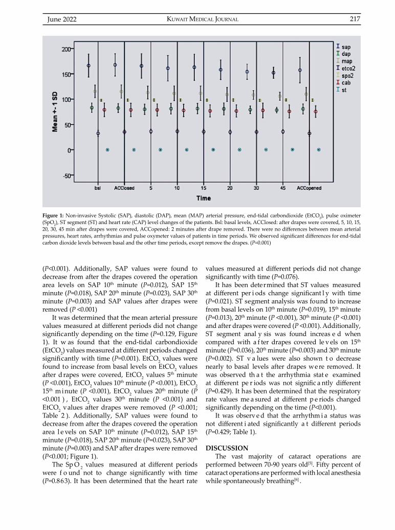
KUWAIT MEDICAL JOURNAL 217June 2022
(P<0.001). Additionally, SAP values were found to decrease from after the drapes covered the operation area levels on SAP 10th minute (P=0.012), SAP 15th minute (P=0.018), SAP 20th minute (P=0.023), SAP 30th minute (P=0.003) and SAP values after drapes were removed (P <0.001)
It was determined that the mean arterial pressure values measured at different periods did not change significantly depending on the time (P=0.129, Figure 1). It w as found that the end-tidal carbondioxide (EtCO2) values measured at different periods changed significantly with time (P=0.001). EtCO2 values were found to increase from basal levels on EtCO2 values after drapes were covered, EtCO2 values 5th minute (P <0.001), EtCO2 values 10th minute (P <0.001), EtCO2 15th m i nute (P <0.001), EtCO2 values 20th minute (P <0.001 ) , EtCO2 values 30th minute (P <0.001) and EtCO2 values after drapes were removed (P <0.001; Table 2 ). Additionally, SAP values were found to decrease from after the drapes covered the operation area l e vels on SAP 10th minute (P=0.012), SAP 15th minute (P=0.018), SAP 20th minute (P=0.023), SAP 30th minute (P=0.003) and SAP after drapes were removed (P<0.001; Figure 1).
The Sp O 2 values measured at different periods were f o und not to change significantly with time (P=0.863). It has been determined that the heart rate
values measured at different periods did not change significantly with time (P=0.076).
It has been determined that ST values measured at different per i ods change significant l y with time (P=0.021). ST segment analysis was found to increase from basal levels on 10th minute (P=0.019), 15th minute (P=0.013), 20th minute (P <0.001), 30th minute (P <0.001) and after drapes were covered (P <0.001). Additionally, ST segment anal y sis was found increas e d when compared with a f ter drapes covered le v els on 15th minute (P=0.036), 20th minute (P=0.003) and 30th minute (P=0.002). ST v a lues were also shown t o decrease nearly to basal levels after drapes we re removed. It was observed th a t the arrhythmia stat e examined at different pe r iods was not signific a ntly different (P=0.429). It has been determined that the respiratory rate values me a sured at different p e riods changed significantly depending on the time (P<0.001).
It was observ e d that the arrhythm i a status was not different i ated significantly a t different periods (P=0.429; Table 1).
DISCUSSIONThe vast majority of cataract operations are
performed between 70-90 years old[5]. Fifty percent of cataract operations are performed with local anesthesia while spontaneously breathing[6] .
Figure 1: Non-invasive Systolic (SAP), diastolic (DAP), mean (MAP) arterial pressure, end-tidal carbondioxide (EtCO2), pulse oximeter (SpO2), ST segment (ST) and heart rate (CAP) level changes of the patients. Bsl: basal levels, ACClosed: after drapes were covered, 5, 10, 15, 20, 30, 45 min after drapes were covered, ACCopened: 2 minutes after drape removed. There were no differences between mean arterial pressures, heart rates, arrhythmias and pulse oxymeter values of patients in time periods. We observed significant differences for end-tidal carbon dioxide levels between basal and the other time periods, except remove the drapes. (P=0.001)
June 2022218
In this age group, the incidence of one or more comorbidities such as diabetes, hypertension, coronary artery disease, heart failure or chronic renal failure is high[5]. The medical records of these high-risk patients should be reviewed before operation to ascertain intraoperative monitoring methods for the early diagnosis and prevention of hemodynamic and cardiovascular complications that may occur during the operation. Recommendations state that all
Time period n Mean (mmHg) Standard Deviation (mmHg) Minimum (mmHg) Maximum (mmHg)
BasalBefore drape closure5th minute10th minute15th minute20th minute30th minute45th minuteAfter drape removal
42424242352517642
32.3835.1736.2136.4835.5734.9235.1835.8332.36
5.854.664.164.334.704.134.113.765.62
172828292626282921
494746474743463944
Table 2: End tidal carbondioxide records in time periods
Distribution of arrhythmia n Percent
Basal YesNoTotal
Before drape closureYesNoTotal
5th minute YesNoTotal
10th minute YesNoTotal
15th minute YesNoTotal
20th minute YesNoTotal
30th minute YesNoTotal
45th minute YesNoTotal
After drapes removed YesNoTotal
43842
43842
43842
43842
43236
22325
01717
066
53742
9.590.5100.0
9.590.5100.0
9.590.5100.0
9.590.5100.0
11.188.9100.0
8.092.0100.0
0.0100.0100.0
0.0100.0100.0
11.988.1100.0
Table 1: Distribution of arrhythmia status at different periodspatients undergoing major eye surgery under local anesthesia should have careful monitoring including pulse oximetry, electrocardiogram, noninvasive blood pressure and EtCO2 measurement[7].
Several studies have shown that hypercapnia leads to coronary vasodilatation and has cardiac depresant effects[8]. In addition, it was demonstrated that administration of a gas mixture containing 20% CO2 caused marked depression of myocardial contractility in cats[8-12]. Abnormal ST-T segment changes were identical to those of ischemia. The patient whose coronary blood flow does not increase adequately with an increase in cardiac work demand is under risk of impared contractility of the heart, hypotension, arrhythmias and death[13].
Conventional facial masks make the surgical procedure difficult because they are so close to the operating area that they are used to provide oxygenation in eye surgery[14].
Various methods have been developed to detect CO2 accumulation under drapes[14-19]. In studies where nasal sampling line was connected to Datex Capnomac Ultima monitor, EtCO2 accumulation was not detected[14]. In another study, EtCO2 was measured with the help of an anesthesia monitor device (Cardiocap Datex, Helsinki, Finland) which has a special apparatus placed between the drapes and the faces of the patients. In this study, transcutaneous partial carbon dioxide was also measured.
Also in this study, it is stated that this increase in the EtCO2 values cannot be measured by routine monitoring methods and that the applied oxygen supplement does not prevent CO2 accumulation[15].
Microstream technology was used on Capnostream 20p capnograph for con t inuous view of ventilation adequacy on intubated a nd non-intubated patients from neonate to adult.
These devices are used for eye surgery in our clinic and all of the EtCO2 recordings in our surveys have been observed to be ob tained by means of a nasal cannula extending to t he mouth and measuring the pulse oximetry and EtC O2 measurement allowing both oxygen delivery. The four parameters measured
End-tidal carbon dioxide levels under surgical drapes during local eye surgery: Retrospective study
KUWAIT MEDICAL JOURNAL 219June 2022
by this device, EtCO2, oxygen saturation, respiratory rate and pulse rate, are integrated to give one number called integrated pulmonary index[19]. It has been observed that the records of the integrated pulmonary index are not available in our records.
In our records, there was no statistically significant change in noninvasive, mean, diastolic arterial blood pressure and heart rate as a function of time compared to baseline levels of the patients, but noninvasive systolic arterial pressure increased statistically significantly during operation compared to baseline values.
A meaningful and statistically significant increase that was observed in the EtCO2 levels indicates the accumulation of carbon dioxide under the cover. However, no new arrhythmia patterns was observed in any of the patients in our records. When the ST segment changes were examined in our records, it was found that there was an increase in the ST segment in the positive direction, that is, statistically and clinically significant ST elevations were observed.
In this sense, we think that the effects on the cardiovascular system can be evaluated more clearly by examining the serial electrocardiogram records to be taken in future prospective studies together with the increase of the underlying EtCO2 levels.
The Joint Working Party on Anesthesia in Ophthalmic Surgery by the Royal College of Ophthalmologists report in February 2012 recommended monitoring methods include: communication, clinical observation, pulse oximetry, electrocardiogram and non-invasive blood pressure. EtCO2 measurements are not included in this schema yet. Our aim in this study is to investigate the accumulation of carbon dioxide under drapes in eye surgery under local anesthesia and to raise awareness of the potential hazards of this accumulation. At the same time, we think that EtCO2 monitoring must be included in the necessary and basic monitoring techniques in these kind of operations.
CONCLUSIONEnd-tidal carbon dioxide levels should be
monitored in patients in whom sedation is performed, the airway is not secured and the anesthesiologist is difficult to reach, in the patient group who can accumulate carbon dioxide under the cover and cannot tolerate hypercarbia.
ACKNOWLEDGMENTDeclaration of interest: The authors declare no
conflict of interests. Funding: This study was not funded.All author’s contributed equally in the manuscript.
REFERENCES
1. Dinsmore M, Han JS, Fisher JA, Chan VWS, Venkatraghavan L. Effects of acute controlled changes in end-tidal carbon dioxide on the diameter of the optic nerve sheath: a transorbital ultrasonographic study in healthy volunteers. Anaesthesia 2017; 72(5):618-623.
2. Allen JG, MacNaughton P, Satish U, Santanam S, Vallarino J, Spengler JD. Associations of cognitive function scores with carbon dioxide, ventilation, and volatile organic compound exposures in office workers: A controlled exposure study of green and conventional office environments. Environ Health Perspect 2016; 124(6):805-812.
3. Howarth C, Sutherland B, Choi HB, Martin C, Lind BL, Khennouf L, et al. A critical role for astrocytes in hypercapnic vasodilation in the brain. J Neurosci 2017; 37(9):2403-2414.
4. Weeke LC, Dix LM, Groenendaal F, Lemmers PM, Dijkman KP, Andriessen P, et al. Severe hypercapnia causes reversible depression of aEEG background activity in neonates: an observational study. Arch Dis Child Fetal Neonatal Ed 2017; 102(5):F383-F388.
5. Fisher SJ, Cunningham RD. The medical profile of cataract patients. Clin Geriatr Med 1985; 1(2):339-344.
6. Courtney P. The National Cataract Surgery Survey: I. Method and descriptive features. Eye 1992; 6(Pt 5):487-492.
7. Kumar CM, Eke T, Dodds C, Deane JS, El-Hindy N, Johnston RL, et al. Local anaesthesia for ophthalmic surgery--new guidelines from the Royal College of Anaesthetists and the Royal College of Ophthalmologists. Eye (Lond) 2012; 26(6):897-898.
8. Jerusalem E, Starling EH. On the significance of carbon dioxide for the heart beat. J Physiol 1910; 40(4):279-294.
9. Smith HW. The actions of acids on turtle heart muscle with reference to the penetration of anions. Am J Physiol 1926; 76:411-447.
10. McElroy WT Jr, Gerdes AJ, Brown EB Jr. Effects of CO2, bicarbonate and pH on the performance of isolated perfused guinea pig hearts. Am J Physiol 1958; 195(2):412-416.
11. Monroe RG, French G, Whittenberger JL. Effects of hypocapnia and hypercapnia on myocardial contractility. Am J Physiol 1960; 199:1121-1124.
12. Foëx P, Fordham RM. Intrinsic myocardial recovery from the negative inotropic effects of acute hypercapnia. Cardiovasc Res 1972; 6(3):257-262.
13. Crystal GJ. Carbon dioxide and the heart: Physiology and clinical ımplications. Anesth Analg 2015; 121(3):610-623.
14. Risdall JE, Geraghty IF. Oxygenation of patients undergoing ophthalmic surgery under local anaesthesia. Anaesthesia 1997; 52(5):492-495.
15. Schlager A, Luger TJ. Oxygen application by nasal probe prevents hypoxia but not rebreathing of carbon dioxide in patients undergoing eye surgery under local anaesthesia. Br J Ophthalmol 2000; 84(4):399-402.

June 2022220
16. Ramanathan S, Capan L, Chalon J, Rand PB, Klein GS, Turndorf H. Minienvironmental control under the drapes during operations on the eyes of conscious patients. Anesthesiology 1978; 48(4):286-288.
17. Zeitlin GL, Hobin K, Platt J, Woitkoski N. Accumulation of carbon dioxide during eye surgery. J Clin Anesth 1989; 1(4):262-267.
18. Schlager A, Lorenz IH, Luger TJ. Transcutaneous CO2/O2 and CO2/air suction in patients undergoing cataract surgery with retrobulbar anaesthesia. Anaesthesia 1998; 53:1212-1218.
19. Kadam VR, Danesh M. Postoperative capnostream monitoring in patients with obstructive sleep apnoea symptoms- Case series. Sleep Sci 2016; 9(3):142-146.
End-tidal carbon dioxide levels under surgical drapes during local eye surgery: Retrospective study
KUWAIT MEDICAL JOURNAL 221June 2022
Original Article
Kuwait Medical Journal 2022; 54 (2): 221 - 226
Efficacy of lamotrigine for seizures, dysexecutive symptoms, anger and sadness rumination in patients with epilepsy
ABSTRACT
KEY WORDS: antiepileptic drugs, cognition, emotion
Amara Gul, Saima MehreenDepartment of Applied Psychology, The Islamia University of Bahawalpur, Pakistan
Objectives: To compare epilepsy patients and healthy individuals on dysexecutive symptoms, sadness and anger rumination and to assess efficacy of lamotrigine on seizure frequency, dysexecutive symptoms, sadness and anger rumination in patients with epilepsyDesign: Comparative cross-sectional research studySetting: Department of Neurology at Sheikh Zayed Hospital, Rahim Yar Khan and Nishter Hospital, Multan, PakistanSubjects: Forty patients with epilepsy and forty healthy individuals participated in the study from 1st August 2017 until 29th March 2018.Intervention: Healthy individuals had single whereas patients had two testing sessions (baseline vs. post- six months lamotrigine therapy as part of treatment). Patients completed Rumination on sadness scale, Anger Rumination Scale and Dysexecutive symptoms questionnaire during baseline and post-lamotrigine therapy. On contrary, healthy
individuals completed these measures in baseline session only. Main outcome measure: Rumination on Sadness Scale, Anger Rumination Scale and Dysexecutive symptoms questionnaire Results: Baseline scores showed that patients with epilepsy had dysexecutive symptoms (22.17±1.43 vs. 8.47±1.99), rumination on sadness (40.12±2.82 vs. 11.10±2.55), and anger (66.17±6.17 vs. 19.60±3.39) in contrast with healthy individuals. Post-treatment performance showed significant reduction in seizure frequency (7.37±1.58 vs. 1.95±0.78), dysexecutive symptoms (22.17±1.43 vs. 17.57±2.01), sadness (40.12±2.82 vs. 23.50±3.35) and anger (66.17±6.17 vs. 41.15±4.88) rumination in adults with epilepsy. Conclusion: Lamotrigine therapy is effective in reducing seizures, dysexecutive symptoms, anger and sadness rumination in adults with epilepsy.
Address correspondence to:Amara Gul, Assistant Professor, Department of Applied Psychology. The Islamia University of Bahawalpur. Pakistan. Tel: +92 (062) 9255065; E-mail: [email protected]
INTRODUCTIONEpilepsy is a noncommunicable neurological
condition which is diagnosed in 2.4 million people each year around the world. Among this number, 80 percent of people live in low- and middle-income countries. Epilepsy is characterized by recurrent seizures, cognitive and emotional disorders[1]. A large disparity occurs in treatment and care of epilepsy patients in the world, with treatment gap of 75% in low income and 50% in middle income countries[2]. The situation is equally alarming in Pakistan, where the number of people diagnosed with epilepsy has crossed 2 million and management of epilepsy is poor due to several reasons, for instance lack of awareness
and belief in supernatural phenomenons[3]. Though treatment of epilepsy is available in Pakistan, unfortunately only 25% of the affected population get treatment[4]. Studies with Pakistani population have suggested that patients with epilepsy are at high risk of developing psychological distress, anxiety, depression and social problems in marriage and daily activities[5]. It was also found that non-compliance of medication was associated with poor clinical result in Pakistani patients with epilepsy[6]. Another study demonstrated that patients with epilepsy had weaker prefrontal cortical functioning and emotion recognition as compared with healthy individuals[7].
June 2022222
There are many treatment options available for epilepsy. Lamotrigine (6-(2,3-Dichlorophenyl)-1,2,4-triazine-3,5-diamine), a phenyltriazine is a popular antiepileptic drug of unique characteristics[8]. Lamotrigine is associated with improved cognitive status and quality of life in epileptic patients[9]. Lamotrigine exerts antiseizure activity by preventing presynaptic release of excitatory neurotransmitters (glutamate) through blocking activated channels of sodium and stabilizing neuronal membrane[10]. In recent years, its’ effectiveness as a mood stabilizer has been explored. In vivo studies have demonstrated that lamotrigine down regulates responses of adenyl cyclase in brain mediated by cortical serotonin (5-HT1A)-receptor[11]. Studies examining behavioral and cognitive functioning of individuals with epilepsy have indicated presence of impaired inhibitory control and emotional intelligence in these patients[12].
To our knowledge, effect of lamotrigine on dysexecutive symptoms, sadness and anger rumination in adults with epilepsy has not been examined in existing literature. Specifically, with reference to Pakistani subjects, no study has yet explored effectiveness of lamotrigine on clinical and social cognition parameters such as seizure frequency, dysexecutive symptoms and emotional rumination. Therefore, the current study was designed to examine efficacy of lamotrigine on these parameters. It was hypothesized that (i) patients with epilepsy would show dysexecutive symptoms in contrast with healthy adults; (ii) patients with epilepsy would show higher sadness and anger rumination contrary with healthy adults; and (iii) lamotrigine would be beneficial in reducing seizures, dysexecutive symptoms, sadness and anger rumination in patients with epilepsy. The objective of this study was to compare patients and healthy individuals on dysexecutive symptoms, sadness and anger rumination and assessment of efficacy of lamotrigine on seizure frequency, dysexecutive symptoms, sadness and anger rumination in patients with epilepsy.
SUBJECTS AND METHODS This study has a comparative cross-sectional
research design and was conducted at the Department of Neurology Nishter Hospital, Multan, and Sheikh Zayed Hospital, Rahim Yar Khan, Pakistan from 1st August 2017 until 29th March 2018. Forty newly diagnosed patients with partial epilepsy were included having no history of treatment with lamotrigine and neurological disease excluding epilepsy. Their age range was 20-45 years and were prescribed lamotrigine as monotherapy for at least
six months with dose range of 50-200 mg per day. Patients with epilepsy were excluded if they were prescribed any antiepileptic drug except lamotrigine and were using any other medication at the time of diagnosis. Forty healthy counterparts took part in the study from the local community with age range of 20-45 years. They were included if they had no history or present symptoms of any neurological disease and psychiatric disorder. Healthy individuals were excluded if they were using any medication and had cognitive complaints. The study was ethically approved by The Islamia University of Bahawalpur, Pakistan and was conducted according to the declaration of Helsinki.
InstrumentsA revised version of Dysexecutive Questionnaire
(DEX)[13] was used to assess dysexecutive symptoms, inhibition and social regulation. The questionnaire contains 15 items which are scored on Likert scale (0 = never – 4 = very often), for example “I am lethargic or unenthusiastic about things”. Average completion time is lesser than five minutes. The questionnaire has good psychometric properties. Rumination on Sadness Scale (RSS)[14] is a thirteen self-report item scale with five response categories, high score shows greater sadness rumination. RSS has good reliability, convergent and discriminant validity. Anger Rumination Scale (ARS)[15] has 19 statements to be responded on four response categories. High score reflects greater anger rumination. The scale has good reliability, convergent and discriminant validity. All participants gave written informed consent. Testing of DEX, RSS and ARS were completely randomized across participants. In the baseline session, participants were asked to complete DEX, RSS and ARS. The follow-up session was conducted post six months of lamotrigine therapy. In follow-up session, patients were tested on DEX, RSS and ARS.
Statistical analysis
Data on demographic and clinical characteristics were analyzed through descriptive statistics. Scores pertaining to patients and healthy individuals were compared on ANOVA with Factor 6 (dysexecutive symptoms, inhibition, social regulation, total DEX scores, RSS, ARS) as within subjects x Group 2 (healthy individuals vs. patients with epilepsy) as between subjects group. Baseline and post- lamotrigine therapy scores of patients on DEX, RSS and ARS through repeated measures ANOVA with testing session as with-in subject factor 2 (pre vs. post- Lamotrigine treatment). Seizure frequency on baseline and post-treatment was compared between patients and healthy individuals through t-test.
Efficacy of lamotrigine for seizures, dysexecutive symptoms, anger and sadness rumination in patients ...
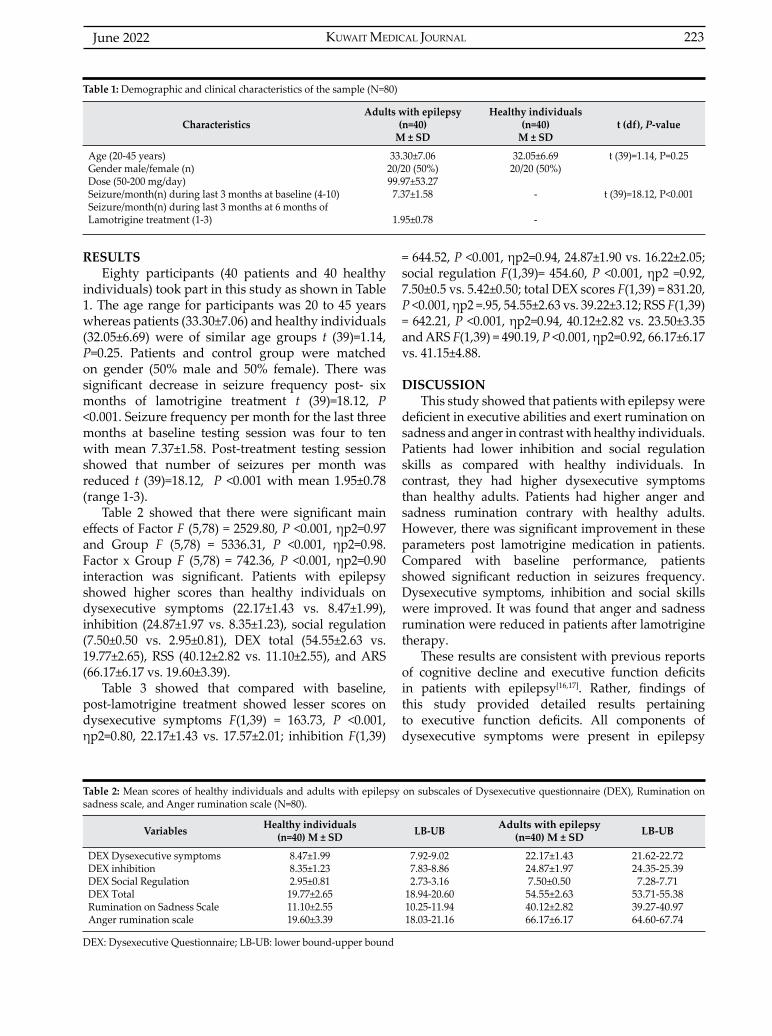
KUWAIT MEDICAL JOURNAL 223June 2022
RESULTS Eighty participants (40 patients and 40 healthy
individuals) took part in this study as shown in Table 1. The age range for participants was 20 to 45 years whereas patients (33.30±7.06) and healthy individuals (32.05±6.69) were of similar age groups t (39)=1.14, P=0.25. Patients and control group were matched on gender (50% male and 50% female). There was significant decrease in seizure frequency post- six months of lamotrigine treatment t (39)=18.12, P <0.001. Seizure frequency per month for the last three months at baseline testing session was four to ten with mean 7.37±1.58. Post-treatment testing session showed that number of seizures per month was reduced t (39)=18.12, P <0.001 with mean 1.95±0.78 (range 1-3).
Table 2 showed that there were significant main effects of Factor F (5,78) = 2529.80, P <0.001, ηp2=0.97 and Group F (5,78) = 5336.31, P <0.001, ηp2=0.98. Factor x Group F (5,78) = 742.36, P <0.001, ηp2=0.90 interaction was significant. Patients with epilepsy showed higher scores than healthy individuals on dysexecutive symptoms (22.17±1.43 vs. 8.47±1.99), inhibition (24.87±1.97 vs. 8.35±1.23), social regulation (7.50±0.50 vs. 2.95±0.81), DEX total (54.55±2.63 vs. 19.77±2.65), RSS (40.12±2.82 vs. 11.10±2.55), and ARS (66.17±6.17 vs. 19.60±3.39).
Table 3 showed that compared with baseline, post-lamotrigine treatment showed lesser scores on dysexecutive symptoms F(1,39) = 163.73, P <0.001, ηp2=0.80, 22.17±1.43 vs. 17.57±2.01; inhibition F(1,39)
= 644.52, P <0.001, ηp2=0.94, 24.87±1.90 vs. 16.22±2.05; social regulation F(1,39)= 454.60, P <0.001, ηp2 =0.92, 7.50±0.5 vs. 5.42±0.50; total DEX scores F(1,39) = 831.20, P <0.001, ηp2 =.95, 54.55±2.63 vs. 39.22±3.12; RSS F(1,39) = 642.21, P <0.001, ηp2=0.94, 40.12±2.82 vs. 23.50±3.35 and ARS F(1,39) = 490.19, P <0.001, ηp2=0.92, 66.17±6.17 vs. 41.15±4.88.
DISCUSSIONThis study showed that patients with epilepsy were
deficient in executive abilities and exert rumination on sadness and anger in contrast with healthy individuals. Patients had lower inhibition and social regulation skills as compared with healthy individuals. In contrast, they had higher dysexecutive symptoms than healthy adults. Patients had higher anger and sadness rumination contrary with healthy adults. However, there was significant improvement in these parameters post lamotrigine medication in patients. Compared with baseline performance, patients showed significant reduction in seizures frequency. Dysexecutive symptoms, inhibition and social skills were improved. It was found that anger and sadness rumination were reduced in patients after lamotrigine therapy.
These results are consistent with previous reports of cognitive decline and executive function deficits in patients with epilepsy[16,17]. Rather, findings of this study provided detailed results pertaining to executive function deficits. All components of dysexecutive symptoms were present in epilepsy
CharacteristicsAdults with epilepsy
(n=40) M ± SD
Healthy individuals (n=40)
M ± SDt (df), P-value
Age (20-45 years)Gender male/female (n)Dose (50-200 mg/day)Seizure/month(n) during last 3 months at baseline (4-10) Seizure/month(n) during last 3 months at 6 months of Lamotrigine treatment (1-3)
33.30±7.0620/20 (50%)99.97±53.277.37±1.58
1.95±0.78
32.05±6.6920/20 (50%)
-
-
t (39)=1.14, P=0.25
t (39)=18.12, P<0.001
Table 1: Demographic and clinical characteristics of the sample (N=80)
Variables Healthy individuals (n=40) M ± SD LB-UB Adults with epilepsy
(n=40) M ± SD LB-UB
DEX Dysexecutive symptomsDEX inhibitionDEX Social RegulationDEX Total Rumination on Sadness ScaleAnger rumination scale
8.47±1.998.35±1.232.95±0.8119.77±2.6511.10±2.5519.60±3.39
7.92-9.027.83-8.862.73-3.16
18.94-20.6010.25-11.9418.03-21.16
22.17±1.4324.87±1.977.50±0.5054.55±2.6340.12±2.8266.17±6.17
21.62-22.7224.35-25.397.28-7.71
53.71-55.3839.27-40.9764.60-67.74
DEX: Dysexecutive Questionnaire; LB-UB: lower bound-upper bound
Table 2: Mean scores of healthy individuals and adults with epilepsy on subscales of Dysexecutive questionnaire (DEX), Rumination on sadness scale, and Anger rumination scale (N=80).
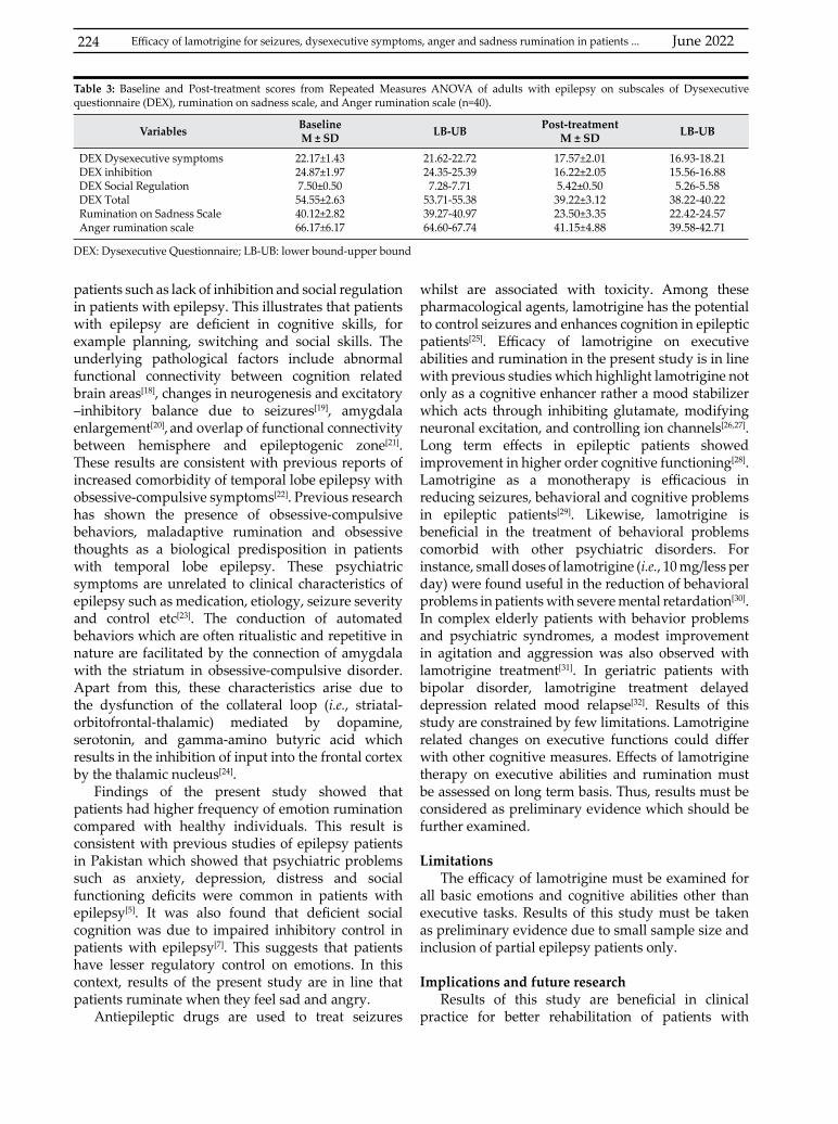
June 2022224
patients such as lack of inhibition and social regulation in patients with epilepsy. This illustrates that patients with epilepsy are deficient in cognitive skills, for example planning, switching and social skills. The underlying pathological factors include abnormal functional connectivity between cognition related brain areas[18], changes in neurogenesis and excitatory –inhibitory balance due to seizures[19], amygdala enlargement[20], and overlap of functional connectivity between hemisphere and epileptogenic zone[21]. These results are consistent with previous reports of increased comorbidity of temporal lobe epilepsy with obsessive-compulsive symptoms[22]. Previous research has shown the presence of obsessive-compulsive behaviors, maladaptive rumination and obsessive thoughts as a biological predisposition in patients with temporal lobe epilepsy. These psychiatric symptoms are unrelated to clinical characteristics of epilepsy such as medication, etiology, seizure severity and control etc[23]. The conduction of automated behaviors which are often ritualistic and repetitive in nature are facilitated by the connection of amygdala with the striatum in obsessive-compulsive disorder. Apart from this, these characteristics arise due to the dysfunction of the collateral loop (i.e., striatal-orbitofrontal-thalamic) mediated by dopamine, serotonin, and gamma-amino butyric acid which results in the inhibition of input into the frontal cortex by the thalamic nucleus[24].
Findings of the present study showed that patients had higher frequency of emotion rumination compared with healthy individuals. This result is consistent with previous studies of epilepsy patients in Pakistan which showed that psychiatric problems such as anxiety, depression, distress and social functioning deficits were common in patients with epilepsy[5]. It was also found that deficient social cognition was due to impaired inhibitory control in patients with epilepsy[7]. This suggests that patients have lesser regulatory control on emotions. In this context, results of the present study are in line that patients ruminate when they feel sad and angry.
Antiepileptic drugs are used to treat seizures
whilst are associated with toxicity. Among these pharmacological agents, lamotrigine has the potential to control seizures and enhances cognition in epileptic patients[25]. Efficacy of lamotrigine on executive abilities and rumination in the present study is in line with previous studies which highlight lamotrigine not only as a cognitive enhancer rather a mood stabilizer which acts through inhibiting glutamate, modifying neuronal excitation, and controlling ion channels[26,27]. Long term effects in epileptic patients showed improvement in higher order cognitive functioning[28]. Lamotrigine as a monotherapy is efficacious in reducing seizures, behavioral and cognitive problems in epileptic patients[29]. Likewise, lamotrigine is beneficial in the treatment of behavioral problems comorbid with other psychiatric disorders. For instance, small doses of lamotrigine (i.e., 10 mg/less per day) were found useful in the reduction of behavioral problems in patients with severe mental retardation[30]. In complex elderly patients with behavior problems and psychiatric syndromes, a modest improvement in agitation and aggression was also observed with lamotrigine treatment[31]. In geriatric patients with bipolar disorder, lamotrigine treatment delayed depression related mood relapse[32]. Results of this study are constrained by few limitations. Lamotrigine related changes on executive functions could differ with other cognitive measures. Effects of lamotrigine therapy on executive abilities and rumination must be assessed on long term basis. Thus, results must be considered as preliminary evidence which should be further examined.
Limitations The efficacy of lamotrigine must be examined for
all basic emotions and cognitive abilities other than executive tasks. Results of this study must be taken as preliminary evidence due to small sample size and inclusion of partial epilepsy patients only.
Implications and future researchResults of this study are beneficial in clinical
practice for better rehabilitation of patients with
Variables BaselineM ± SD LB-UB Post-treatment
M ± SD LB-UB
DEX Dysexecutive symptomsDEX inhibitionDEX Social RegulationDEX Total Rumination on Sadness ScaleAnger rumination scale
22.17±1.4324.87±1.977.50±0.5054.55±2.6340.12±2.8266.17±6.17
21.62-22.7224.35-25.397.28-7.71
53.71-55.3839.27-40.9764.60-67.74
17.57±2.0116.22±2.055.42±0.5039.22±3.1223.50±3.3541.15±4.88
16.93-18.2115.56-16.885.26-5.58
38.22-40.2222.42-24.5739.58-42.71
DEX: Dysexecutive Questionnaire; LB-UB: lower bound-upper bound
Table 3: Baseline and Post-treatment scores from Repeated Measures ANOVA of adults with epilepsy on subscales of Dysexecutive questionnaire (DEX), rumination on sadness scale, and Anger rumination scale (n=40).
Efficacy of lamotrigine for seizures, dysexecutive symptoms, anger and sadness rumination in patients ...
KUWAIT MEDICAL JOURNAL 225June 2022
epilepsy. Lamotrigine therapy could be administered to treat cognitive and emotion deficits. Future research must examine whether lamotrigine would be beneficial in the treatment of behavioral disorders.
CONCLUSION
Lamotrigine is efficacious in reduction of dysexecutive symptoms, seizure frequency, sadness and anger rumination in patients with epilepsy.
ACKNOWLEDGMENTAuthors gratefully acknowledge cooperation of
participants in the study.Conflict of Interest: No conflict of interest. Disclosure of grants or other funding: No grants or funding to be disclosed.Author’s contribution: Amara Gul: manuscript writing, conception of idea, data collection, data analysis, interpretation of results. Saima Mehreen: data collection, literature review, data analysis.
REFERENCES
1. World Health Organization. Epilepsy. 2019. Website: [https://www.who.int/news-room/fact-sheets/detail/epilepsy]. Accessed on 02 Jan 2019.
2. Meyer AC, Dua T, Ma J, Saxena S, Birbeck G. Global disparities in the epilepsy treatment gap: a systematic review. Bull World Health Organ 2010; 88(4):260-6.
3. Dawn. Over two million people suffer from epilepsy in Pakistan. 2015. Website: [https://www.dawn.com/news/1171867]. Accessed on 04 April 2018.
4. The News. Epilepsy is curable and treatment is affordable in Pakistan: neurologists. 2015. Website: [https://www.thenews.com.pk/print/52422-epilepsy-is-curable-and-treatment-is-affordable-in-pakistan-neurologists]. Accessed on 05 Dec 2018.
5. Sahar NU. Assessment of psychological distress in epilepsy: perspective from Pakistan. Epilepsy Res Treat 2012; 2012:171725.
6. Ullah S, Ali N, Khan A, Ali S, Nazish HR. The epidemiological characteristics of epilepsy in the province of Khyber Pakhtunkhwa, Pakistan. Front Neurol 2018; 9:845.
7. Gul A, Ahmad H. The relationship between dispositional empathy and prefrontal cortical functioning in patients with frontal lobe epilepsy. Pak J Med Sci 2017; 33(1):200-204.
8. Beattie K, Phadke G, Novakovic J. Lamotrigine. Profiles of drug substances, excipients and related methodology. Elsevier 2012; 37:245-285.
9. Aldenkamp AP, Baker GA. A systematic review of the effects of lamotrigine on cognitive function and quality of life. Epilepsy Behav 2001; 2(2):85-91.
10. Wright P, O’Neill MF. Psychopharmacology. Core Psychiatry. 3rd ed. Elsevier 2012; 585-615.
11. Lopez-Figueroa AL, Norton CS, Lopez-Figueroa MO, Armellini-Dodel D, Burke S, Akil H, et al. Serotonin
5-HT1A, 5-HT1B and 5-HT2A, receptor mRNA expression in subjects with major depression, bipolar disorder, and schizophrenia. Biol Psychiatry 2004; 55(3):225-33.
12. Gul A, Hussain I. The relationship between emotional intelligence and task switching in temporal lobe epilepsy. Neurosciences 2016; 21(1):64-68.
13. Shaw S, Oei TP, Sawang S. Psychometric validation of the Dysexecutive Questionnaire (DEX). Psychol Assess 2015; 27(1):138-47.
14. Conway M, Csank PA, Holm SL, Blake CK. On assessing individual differences in rumination on sadness. J Pers Assess 2000; 75(3):404-25.
15. Sukhodolsky GD, Golub A, Cromwell NE. Development and validation of the anger rumination scale. Personality and Individual Differences 2001; 31(5):689-700.
16. Gul A, Mehreen S. Levetiracetam efficacy on frontal lobe dysfunctions and anger rumination in patients with epilepsy. Epilepsy Behav 2018; 85:28-31.
17. Agah E, Asgari-Rad N, Ahmadi M, Tafakhori A, Aghamollaii V. Evaluating executive function in patients with temporal lobe epilepsy using the frontal assessment battery. Epilepsy Res 2017; 133:22-27.
18. Tailby C, Kowalczyk MA, Jackson GD. Cognitive impairment in epilepsy: the role of reduced network flexibility. Ann Clin Transl Neurol 2018; 5(1):29-40.
19. Holmes GL. Effect of seizures on the developing brain and cognition. Semin Pediatr Neurol 2016; 23(2):120-126.
20. Tamune H, Taniguchi G, Morita S, Kumakura Y, Kondo S, Kasai K. Emotional stimuli-provoked seizures potentially misdiagnosed as psychogenic non-epileptic attacks: A case of temporal lobe epilepsy with amygdala enlargement. Epilepsy Behav Case Rep 2017; 9:37-41.
21. Alba-Ferrara L, Kochen S, Hausmann M. Emotional prosody processing in epilepsy: Some insights on brain reorganization. Front Hum Neurosci 2018; 12:92.
22. Kaplan PW. Epilepsy and obsessive-compulsive disorder. Dialogues Clin Neurosci 2010; 12(2):241-248.
23. Alheid GF, Heimer L. New perspectives in basal forebrain organization of special relevance for neuropsychiatric disorders: the striatopallidal, amygdaloid and corticopetal components of substantia innominata. Neuroscience 1988;27(1):1-39.
24. Modell JG, Mountz JM, Curtis GC, Greden JF. Neurophysiologic dysfunction in basal ganglia/limbic striatal and thalamocortical circuits as a pathogenetic mechanism of obsessive-compulsive disorder. J Neuropsychiatry Clin Neurosci 1989; 1(1):27-36.
25. Eddy CM, Rickards HE, Cavanna AE. The cognitive impact of antiepileptic drugs. Ther Adv Neurol Disord 2011; 4(6):385-407.
26. Hahn CG, Gyulai L, Baldassano CF, Lenox RH. The current understanding of lamotrigine as a mood stabilizer. J Clin Psychiatry 2004; 65(6):791-804.
27. Khan A, Ginsberg LD, Asnis GM, Goodwin FK, Davis KH, Krishnan AA, et al. Effect of lamotrigine on cognitive complaints in patients with bipolar I disorder. J Clin Psychiatry 2004; 65(11):1483-90.

June 2022226
28. Lee SA, Lee HW, Heo K, Shin DJ, Song HK, Kim OJ, et al. Cognitive and behavioral effects of lamotrigine and carbamazepine monotherapy in patients with newly diagnosed or untreated partial epilepsy. Seizures 2011; 20(1):49-54.
29. Eun SH, Eun BL, Lee JS, Hwang YS, Kim KJ, Lee YM, et al. Effects of lamotrigine on cognition and behavior compared to carbamazepine as monotherapy for children with partial epilepsy. Brain Dev 2012; 34(10):818-23.
30. Kubagawa T, Furusho J, Isozaki Y. [Study of lamotrigine efficacy on behavior disorders affecting patients with severe mental retardation]. No To Hattatsu 2015; 47(4):289-92. Article in Japanese.
31. Aulakh JS, Hawkins JW, Athwal HS, Sheikh HS, Yesavage J, Tinklenberg JR. Tolerability and effectiveness of lamotrigine in complex elderly patients. J Geriatr Psychiatry Neurol 2005; 18(1):8-11.
32. Sajatovic M, Ramsay E, Nanry K, Thompson T. Lamotrigine therapy in elderly patients with epilepsy, bipolar disorder or dementia. Int J Geriatr Psychiatry 2007; 22(10):945-50.
Efficacy of lamotrigine for seizures, dysexecutive symptoms, anger and sadness rumination in patients ...
KUWAIT MEDICAL JOURNAL 227June 2022
Original Article
Kuwait Medical Journal 2022; 54 (2): 227 - 232
Arthroscopic assisted percutaneous figure of eight tension band wiring of patellar fractures
ABSTRACT
KEY WORDS: arthroscopic assisted reduction, cerclage wiring, patella fracture, tension band
Cumhur Deniz Davulcu1, Mehmet Can Unlu2, Yusuf Pirincci3, Taha Demir3, Aybars Kivrak4, Mahmut Kursat Ozsahin2
1Department of Orthopaedics and Traumatology, Izmir Ataturk Training and Research Hospital, Katip Celebi University, Izmir, Turkey
2Department of Orthopaedics and Traumatology, Istanbul University Cerrahpasa Faculty of Medicine, Istanbul, Turkey3Istanbul Medicine Hospital, Department of Orthopaedics and Traumatology, Istanbul, Turkey
4Department of Orthopaedics, Mus State Hospital, Mus, Turkey
Objective: Different techniques have been reported involving combinations of K-wires, screws and cerclage wiring for the surgical treatment of patellar fractures in the case of disruption in the extension mechanism or articular incongruence of 2 mm or more. The aim of this study was to describe a new and easy arthroscopic assisted reduction and percutaneous tension band wiring technique for displaced fractures of the patella that combines the advantages of minimally invasive surgery and stable internal fixation.Design: Retrospective studySetting: Department of Orthopaedics and Traumatology, Istanbul University Cerrahpasa Faculty of Medicine, Istanbul, TurkeySubjects: Seventeen patients were treated with arthroscopic assisted reductionIntervention: Arthroscopic assisted reduction and
percutaneous tension band wiring technique for transvers patella fractureMain outcome measures: Patients evaluated according to evidence of radiological union and articular incongruence, range of motion and activity levelResults: The mean follow-up period was 18 months and the mean patient age was 41 years. Nonunion, articular incongruence or reduction loss were not detected in any case. Conclusion: The described technique is minimally invasive and cosmetically pleasing, permits ideal image of the reduction and stability of the fracture, allows concomitant intraarticular pathology to be exposed, and facilitates early rehabilitation. It should be considered as an alternate method of treatment in displaced transverse patella fractures with an altered joint surface.
Address correspondence to:Dr. Cumhur Deniz Davulcu, Department of Orthopaedics and Traumatology, Katip Celebi University, Ataturk Training and Research Hospital, Izmir, Turkey. Tel: +90 5413808106; E-mail: [email protected]
INTRODUCTIONPatella fractures account for 1% of all fractures[1].
Surgery is indicated if there is disruption of extensor mechanism or articular incongruence of 2 mm and more[2,3]. Several techniques involving combinations of K-wires, screws and cerclage wiring are reported for surgical treatment of patella fractures[1,4-12].
Conventionally, the treatment of a displaced patella requires a wide incision to expose fracture fragments and joint surface[1,5]. Such incisions may lead to
adhesions, prolonged disability for work and cosmetically unpleasant scar tissue[5,13]. On the other hand, percutaneous surgical techniques preserve the vascular supply of patellar fragments, which may improve fracture consolidation[6]. With the use of arthroscopy, it is possible to evaluate the articular surface reduction without performing an arthrotomy, making it possible to perform a closed reduction and a percutaneous treatment of the fracture[5,7]. We describe a new and easy arthroscopic assisted reduction and
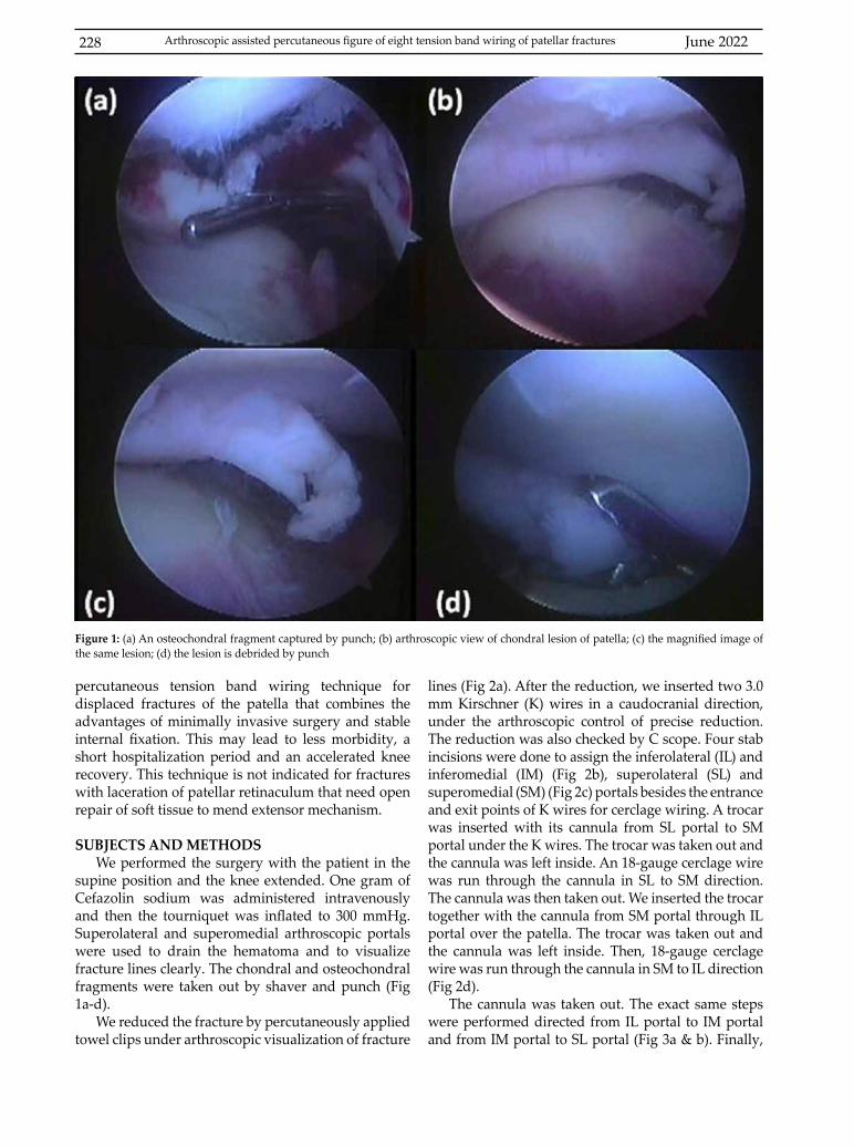
June 2022228 Arthroscopic assisted percutaneous figure of eight tension band wiring of patellar fractures
percutaneous tension band wiring technique for displaced fractures of the patella that combines the advantages of minimally invasive surgery and stable internal fixation. This may lead to less morbidity, a short hospitalization period and an accelerated knee recovery. This technique is not indicated for fractures with laceration of patellar retinaculum that need open repair of soft tissue to mend extensor mechanism.
SUBJECTS AND METHODSWe performed the surgery with the patient in the
supine position and the knee extended. One gram of Cefazolin sodium was administered intravenously and then the tourniquet was inflated to 300 mmHg. Superolateral and superomedial arthroscopic portals were used to drain the hematoma and to visualize fracture lines clearly. The chondral and osteochondral fragments were taken out by shaver and punch (Fig 1a-d).
We reduced the fracture by percutaneously applied towel clips under arthroscopic visualization of fracture
lines (Fig 2a). After the reduction, we inserted two 3.0 mm Kirschner (K) wires in a caudocranial direction, under the arthroscopic control of precise reduction. The reduction was also checked by C scope. Four stab incisions were done to assign the inferolateral (IL) and inferomedial (IM) (Fig 2b), superolateral (SL) and superomedial (SM) (Fig 2c) portals besides the entrance and exit points of K wires for cerclage wiring. A trocar was inserted with its cannula from SL portal to SM portal under the K wires. The trocar was taken out and the cannula was left inside. An 18-gauge cerclage wire was run through the cannula in SL to SM direction. The cannula was then taken out. We inserted the trocar together with the cannula from SM portal through IL portal over the patella. The trocar was taken out and the cannula was left inside. Then, 18-gauge cerclage wire was run through the cannula in SM to IL direction (Fig 2d).
The cannula was taken out. The exact same steps were performed directed from IL portal to IM portal and from IM portal to SL portal (Fig 3a & b). Finally,
Figure 1: (a) An osteochondral fragment captured by punch; (b) arthroscopic view of chondral lesion of patella; (c) the magnified image of the same lesion; (d) the lesion is debrided by punch

KUWAIT MEDICAL JOURNAL 229June 2022
near the SL portal, both wires are tensed and secured to each other with a single knot (Fig 3c).
C scope was used to check figure of eight tension band wiring fixation (Fig 4a & b). Reduction was controlled by arthroscope during the operation.
Stab incisions were closed. The operation time ranged from 40 minutes to 64 minutes with an average
of 51 minutes. In all patients, quadriceps setting exercises were encouraged immediately after the operation. An immobilizer brace was applied for two days. Patients were permitted to walk with crutches without weight bearing on the third day. Range of motion exercises were begun on the third day. Partial weight bearing was allowed three weeks
Figure 2: (a) Percutaneous fracture reduction under arthroscopic control; (b) inferolateral and inferomedial portals for cerclage wiring; (c) superolateral and superomedial portals for cerclage wiring; (d) cerclage wire applied through the cannula in superomedial to inferolateral direction
Figure 3: (a) Cerclage wire applied through the cannula in inferolateral to inferomedial direction; (b) cerclage wire applied through the cannula in inferomedial to superolateral direction; (c) wires are knotted near the superolateral portal
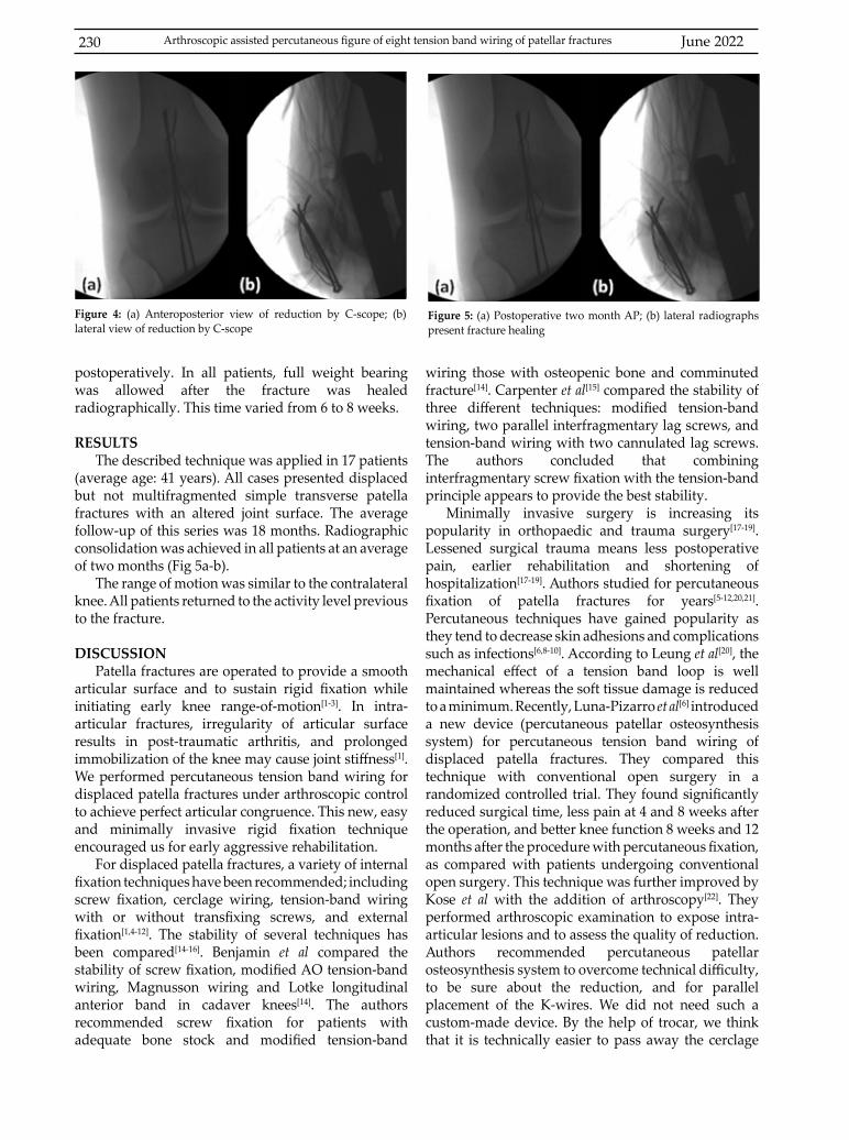
June 2022230
postoperatively. In all patients, full weight bearing was allowed after the fracture was healed radiographically. This time varied from 6 to 8 weeks.
RESULTSThe described technique was applied in 17 patients
(average age: 41 years). All cases presented displaced but not multifragmented simple transverse patella fractures with an altered joint surface. The average follow-up of this series was 18 months. Radiographic consolidation was achieved in all patients at an average of two months (Fig 5a-b).
The range of motion was similar to the contralateral knee. All patients returned to the activity level previous to the fracture.
DISCUSSIONPatella fractures are operated to provide a smooth
articular surface and to sustain rigid fixation while initiating early knee range-of-motion[1-3]. In intra-articular fractures, irregularity of articular surface results in post-traumatic arthritis, and prolonged immobilization of the knee may cause joint stiffness[1]. We performed percutaneous tension band wiring for displaced patella fractures under arthroscopic control to achieve perfect articular congruence. This new, easy and minimally invasive rigid fixation technique encouraged us for early aggressive rehabilitation.
For displaced patella fractures, a variety of internal fixation techniques have been recommended; including screw fixation, cerclage wiring, tension-band wiring with or without transfixing screws, and external fixation[1,4-12]. The stability of several techniques has been compared[14-16]. Benjamin et al compared the stability of screw fixation, modified AO tension-band wiring, Magnusson wiring and Lotke longitudinal anterior band in cadaver knees[14]. The authors recommended screw fixation for patients with adequate bone stock and modified tension-band
wiring those with osteopenic bone and comminuted fracture[14]. Carpenter et al[15] compared the stability of three different techniques: modified tension-band wiring, two parallel interfragmentary lag screws, and tension-band wiring with two cannulated lag screws. The authors concluded that combining interfragmentary screw fixation with the tension-band principle appears to provide the best stability.
Minimally invasive surgery is increasing its popularity in orthopaedic and trauma surgery[17-19]. Lessened surgical trauma means less postoperative pain, earlier rehabilitation and shortening of hospitalization[17-19]. Authors studied for percutaneous fixation of patella fractures for years[5-12,20,21]. Percutaneous techniques have gained popularity as they tend to decrease skin adhesions and complications such as infections[6,8-10]. According to Leung et al[20], the mechanical effect of a tension band loop is well maintained whereas the soft tissue damage is reduced to a minimum. Recently, Luna-Pizarro et al[6] introduced a new device (percutaneous patellar osteosynthesis system) for percutaneous tension band wiring of displaced patella fractures. They compared this technique with conventional open surgery in a randomized controlled trial. They found significantly reduced surgical time, less pain at 4 and 8 weeks after the operation, and better knee function 8 weeks and 12 months after the procedure with percutaneous fixation, as compared with patients undergoing conventional open surgery. This technique was further improved by Kose et al with the addition of arthroscopy[22]. They performed arthroscopic examination to expose intra-articular lesions and to assess the quality of reduction. Authors recommended percutaneous patellar osteosynthesis system to overcome technical difficulty, to be sure about the reduction, and for parallel placement of the K-wires. We did not need such a custom-made device. By the help of trocar, we think that it is technically easier to pass away the cerclage
Figure 4: (a) Anteroposterior view of reduction by C-scope; (b) lateral view of reduction by C-scope
Figure 5: (a) Postoperative two month AP; (b) lateral radiographs present fracture healing
Arthroscopic assisted percutaneous figure of eight tension band wiring of patellar fractures
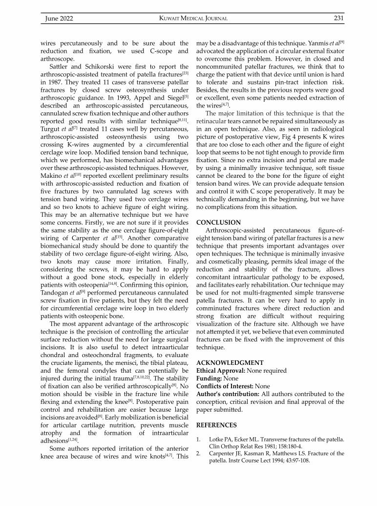
KUWAIT MEDICAL JOURNAL 231June 2022
wires percutaneously and to be sure about the reduction and fixation, we used C-scope and arthroscope.
Sattler and Schikorski were first to report the arthroscopic-assisted treatment of patella fractures[23] in 1987. They treated 11 cases of transverse patellar fractures by closed screw osteosynthesis under arthroscopic guidance. In 1993, Appel and Siegel[5] described an arthroscopic-assisted percutaneous, cannulated screw fixation technique and other authors reported good results with similar technique[8,11]. Turgut et al[7] treated 11 cases well by percutaneous, arthroscopic-assisted osteosynthesis using two crossing K-wires augmented by a circumferential cerclage wire loop. Modified tension band technique, which we performed, has biomechanical advantages over these arthroscopic-assisted techniques. However, Makino et al[10] reported excellent preliminary results with arthroscopic-assisted reduction and fixation of five fractures by two cannulated lag screws with tension band wiring. They used two cerclage wires and so two knots to achieve figure of eight wiring. This may be an alternative technique but we have some concerns. Firstly, we are not sure if it provides the same stability as the one cerclage figure-of-eight wiring of Carpenter et al[15]. Another comparative biomechanical study should be done to quantify the stability of two cerclage figure-of-eight wiring. Also, two knots may cause more irritation. Finally, considering the screws, it may be hard to apply without a good bone stock, especially in elderly patients with osteopenia[14,8]. Confirming this opinion, Tandogan et al[8] performed percutaneous cannulated screw fixation in five patients, but they felt the need for circumferential cerclage wire loop in two elderly patients with osteopenic bone.
The most apparent advantage of the arthroscopic technique is the precision of controlling the articular surface reduction without the need for large surgical incisions. It is also useful to detect intraarticular chondral and osteochondral fragments, to evaluate the cruciate ligaments, the menisci, the tibial plateau, and the femoral condyles that can potentially be injured during the initial trauma[7,8,10,22]. The stability of fixation can also be verified arthroscopically[8]. No motion should be visible in the fracture line while flexing and extending the knee[8]. Postoperative pain control and rehabilitation are easier because large incisions are avoided[8]. Early mobilization is beneficial for articular cartilage nutrition, prevents muscle atrophy and the formation of intraarticular adhesions[1,24].
Some authors reported irritation of the anterior knee area because of wires and wire knots[4,7]. This
may be a disadvantage of this technique. Yanmis et al[9] advocated the application of a circular external fixator to overcome this problem. However, in closed and noncommunited patellar fractures, we think that to charge the patient with that device until union is hard to tolerate and sustains pin-tract infection risk. Besides, the results in the previous reports were good or excellent, even some patients needed extraction of the wires[4,7].
The major limitation of this technique is that the retinacular tears cannot be repaired simultaneously as in an open technique. Also, as seen in radiological picture of postoperative view, Fig 4 presents K wires that are too close to each other and the figure of eight loop that seems to be not tight enough to provide firm fixation. Since no extra incision and portal are made by using a minimally invasive technique, soft tissue cannot be cleared to the bone for the figure of eight tension band wires. We can provide adequate tension and control it with C scope peroperatively. It may be technically demanding in the beginning, but we have no complications from this situation.
CONCLUSIONArthroscopic-assisted percutaneous figure-of-
eight tension band wiring of patellar fractures is a new technique that presents important advantages over open techniques. The technique is minimally invasive and cosmetically pleasing, permits ideal image of the reduction and stability of the fracture, allows concomitant intraarticular pathology to be exposed, and facilitates early rehabilitation. Our technique may be used for not multi-fragmented simple transverse patella fractures. It can be very hard to apply in comminuted fractures where direct reduction and strong fixation are difficult without requiring visualization of the fracture site. Although we have not attempted it yet, we believe that even comminuted fractures can be fixed with the improvement of this technique.
ACKNOWLEDGMENTEthical Approval: None requiredFunding: None Conflicts of Interest: NoneAuthor’s contribution: All authors contributed to the conception, critical revision and final approval of the paper submitted.
REFERENCES
1. Lotke PA, Ecker ML. Transverse fractures of the patella. Clin Orthop Relat Res 1981; 158:180-4.
2. Carpenter JE, Kasman R, Matthews LS. Fracture of the patella. Instr Course Lect 1994; 43:97-108.
June 2022232
3. Kaylor KL. Injuries to the patella and extensor mechanism. In: Levine AM, Rosemont IL, eds. Orthopedic Knowledge Update. Chicago: American Academy of Orthopedic Surgeons; 1996.p.153-8.
4. Hung LK, Chan KM, Chow YN, Leung PC. Fractured patella: operative treatment using the tension band principle. Injury 1985; 16(5)6:343-7.
5. Appel MH, Siegel H. Treatment of transverse fractures of the patella by arthroscopic percutaneous pinning. Arthroscopy 1993; 9(1):119-121.
6. Luna-Pizarro D, Amato D, Arellano F, Hernández A, López-Rojas P. Comparison of a technique using a new percutaneous osteosynthesis device with conventional open surgery for displaced patella fractures in a randomized controlled trial. J Orthop Trauma 2006; 20(8):529-35.
7. Turgut A, Gunal I, Acar S, Seber S, Gokturk E. Arthroscopic-assisted percutaneous stabilization of patellar fractures. Clin Orthop Relat Res 2001; 389:57-61.
8. Tandogan RN, Demirors H, Tuncay CI, Cesur N, Hersekli M. Arthroscopic-assisted percutaneous screw fixation of select patellar fractures. Arthroscopy 2002; 18(2):156-62.
9. Yanmis I, Oguz E, Atesalp AS, Ozkan H, Kurklu M, Demiralp B, et al. Application of circular external fixator under arthroscopic control in comminuted patella fractures: technique and early results. J Trauma 2006; 60(3):659-63.
10. Makino A, Aponte-Tinao L, Muscolo DL, Puigdevall M, Costa-Paz M. Arthroscopic-assisted surgical technique for treating patella fractures. Arthroscopy 2002; 18(6):671-5.
11. El-Sayed AMM, Ragab RK. Arthroscopic-assisted reduction and stabilization of transverse fractures of patella. Knee 2009; 16(1)54-7.
12. Biyani A, Mathur NC, Sharma JC. Percutaneous tension band wiring for minimally displaced fractures of the patella. Int Orthop 1990; 14(3):281-3.
13. Smith ST, Cramer KE, Kargas DE, Watson JT, Moed BR. Early complications in the operative treatment of patella fractures. J Orthop Trauma 1997; 11(3):183-7.
14. Benjamin J, Bried J, Dohm M, McMurthy M. Biomechanical evaluation of various forms of fixation of transverse patellar fractures. J Orthop Trauma 1987; 1(3):219-222.
15. Carpenter JE, Kasman RA, Patel N, Lee ML, Goldstein SA. Biomechanical evaluation of current patella fracture fixation techniques. J Orthop Trauma 1997; 11(5):351-6.
16. Weber MJ, Janecki CJ, McLeod P, Nelson CL, Thompson JA. Efficacy of various forms of fixation of transverse fractures of the patella. J Bone Joint Surg Am 1980; 62(2):215-20.
17. Yeung SH. Minimally invasive surgery in orthopaedics. Small is beautiful? Hong Kong Med J 2008; 14(4):303-7.
18. Seekamp A, Lehmann U, Pizanis A, Pohlemann T. New aspects for minimally invasive interventions in orthopaedic trauma surgery. Der Chirurg 2003; 74(4):301-9.
19. Browner BD, Alberta FG, Mastella DJ. A new era in orthopedic trauma care. Surg Clin North Am 1999; 79(6):1431-48.
20. Leung PC, Mak KH, Lee SY. Percutaneous tension band wiring: a new method of internal fixation for mildly displaced patella fracture. J Trauma 1983; 23(1):62-4.
21. Ma YZ, Zhang YF, Qu KF, Yeh YC. Treatment of fractures of the patella with percutaneous suture. Clin Orthop Relat Res 1984; 191:235-41.
22. Kose KC, Kuru I, Maralcan G, Altinel L. Comparison of a technique using a new percutaneous osteosynthesis device with conventional open surgery for displaced patella fractures. J Orthop Trauma 2007; 21(1):77-8; author reply 78.
23. Sattler RW, Schikorski MM. Transverse patellar fracture-reposition and stabilization from its arthroscopic viewpoint. Zentralbl Chir 1987; 112(23):1515-9.
24. Salter RB. Continuous passive motion (CPM): a biological concept for the healing and regeneration of articular cartilage, ligaments and tendons: from origination to research to clinical applications. Baltimore: Williams & Wilkins; 1993.
Arthroscopic assisted percutaneous figure of eight tension band wiring of patellar fractures
KUWAIT MEDICAL JOURNAL 233June 2022
Original Article
Kuwait Medical Journal 2022; 54 (2): 233 - 243
Computed tomography findings of organizing pneumonia
ABSTRACT
KEY WORDS: computed tomography, cryptogenic organizing pneumonia, organizing pneumonia
Yeliz Dadali1, Sercan Ozkacmaz1, Yurdanur Erdogan2, Havva Akmaz Unlu3, Funda Demirag4, Ilke Bursali5
1Department of Radiology, Faculty Of Medicine, Ahi Evran University, Kirsehir, Turkey 2Department of Chest Diseases, Ataturk Chest Disease and Chest Surgery Research and Training Hospital, Turkey 3Department of Radiology, Ankara Childrens Hematology Oncology Training and Research Hospital, Ankara, Turkey
4Department of Pathology, Ataturk Chest Disease and Chest Surgery Research and Training Hospital, Turkey 5Department of Radiology, Ataturk Chest Disease and Chest Surgery Research and Training Hospital, Turkey
Objective: In this study, we aimed to present computed tomography (CT) findings of 100 patients with histopathologically confirmed organizing pneumonia.Design: Retrospective study Setting: Ankara Atatürk Chest Diseases And Chest Surgery Training And Surgery Hospital, Ankara, TurkeySubjects: One hundred histopathologically confirmed organizing pneumonia patients between 2009 and 2013 admitted to our clinic.Intervention: A chest CT scan and the histopathological examination of the lung lesions of the patients were performed. Main outcome measure: The frequency and the types of CT findings of the patients with proven organizing pneumonia were examined.
Results: Among 100 patients, 73 were male and 27 were female, and the mean age of the patients was 60±11 (range:19-90) years. Pulmonary consolidation was detected in 87 patients and ground-glass opacity was seen in 76 patients. Multiple nodules were seen in 20 patients while a solitary nodule was seen in 15 patients. Acinar nodular pattern was detected in 29 patients, micronodular pattern in 28 patients, bronchocentric pattern in 33 patients, perilobular pattern in 14 patients, progressive fibrotic pattern in 8 patients and reversed halo sign in 12 patients. Lymph node enlargement was seen in 80 patients.Conclusion: Computed tomography is very important for the diagnosis of organizing pneumonia which has various radiological findings.
Address correspondence to:Sercan Ozkacmaz MD, Department of Radiology, Kirsehir Ahi Evran University Faculty of Medicine, Kirsehir, Turkey. Tel: 0532 3276412; E-mail: [email protected]
INTRODUCTIONOrganizing pneumonia (OP) is a nonspecific
response to pulmonary damage. 50% of the cases of OP are idiopathic and are termed as cryptogenic organizing pneumonia (COP), which is a well described clinical and pathological condition[1,2].
In the histopathological evaluation of OP, the discrimination of alveolar structure is difficult. The most prominent finding of granulation is the polyps, which are also known as Masson bodies (Fig. 1). They are interstitial infiltrates located in the granulation tissue which protrude into alveoli and bronchi[3].
Computed tomography (CT) is a very important diagnostic method for the examination of diffuse
parenchymal disease, identify the type of lesion, follow-up and determine the optimal biopsy site[4]. While CT findings of OP are various, the most characteristic features are subpleural patchy consolidation sites and usually accompanying ground-glass opacities[5].
In this study, we aimed to analyze the CT findings of OP in a relatively larger case series.
SUBJECTS AND METHODSCT findings of a total of 100 patients (73 males and
27 females) with histopathologically proven OP (Fig. 1) who were admitted to a tertiary Chest Disease Training Hospital, were retrospectively screened and
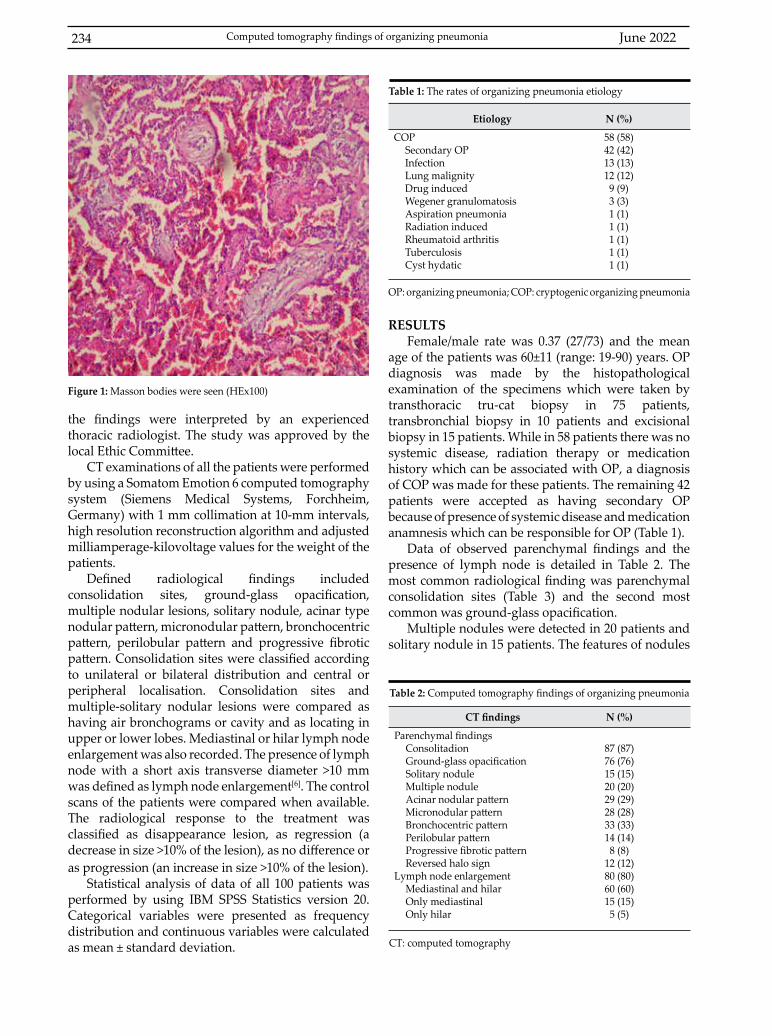
June 2022234
the findings were interpreted by an experienced thoracic radiologist. The study was approved by the local Ethic Committee.
CT examinations of all the patients were performed by using a Somatom Emotion 6 computed tomography system (Siemens Medical Systems, Forchheim, Germany) with 1 mm collimation at 10-mm intervals, high resolution reconstruction algorithm and adjusted milliamperage-kilovoltage values for the weight of the patients.
Defined radiological findings included consolidation sites, ground-glass opacification, multiple nodular lesions, solitary nodule, acinar type nodular pattern, micronodular pattern, bronchocentric pattern, perilobular pattern and progressive fibrotic pattern. Consolidation sites were classified according to unilateral or bilateral distribution and central or peripheral localisation. Consolidation sites and multiple-solitary nodular lesions were compared as having air bronchograms or cavity and as locating in upper or lower lobes. Mediastinal or hilar lymph node enlargement was also recorded. The presence of lymph node with a short axis transverse diameter >10 mm was defined as lymph node enlargement[6]. The control scans of the patients were compared when available. The radiological response to the treatment was classified as disappearance lesion, as regression (a decrease in size >10% of the lesion), as no difference or as progression (an increase in size >10% of the lesion).
Statistical analysis of data of all 100 patients was performed by using IBM SPSS Statistics version 20. Categorical variables were presented as frequency distribution and continuous variables were calculated as mean ± standard deviation.
Figure 1: Masson bodies were seen (HEx100)
RESULTSFemale/male rate was 0.37 (27/73) and the mean
age of the patients was 60±11 (range: 19-90) years. OP diagnosis was made by the histopathological examination of the specimens which were taken by transthoracic tru-cat biopsy in 75 patients, transbronchial biopsy in 10 patients and excisional biopsy in 15 patients. While in 58 patients there was no systemic disease, radiation therapy or medication history which can be associated with OP, a diagnosis of COP was made for these patients. The remaining 42 patients were accepted as having secondary OP because of presence of systemic disease and medication anamnesis which can be responsible for OP (Table 1).
Data of observed parenchymal findings and the presence of lymph node is detailed in Table 2. The most common radiological finding was parenchymal consolidation sites (Table 3) and the second most common was ground-glass opacification.
Multiple nodules were detected in 20 patients and solitary nodule in 15 patients. The features of nodules
Etiology N (%)
COPSecondary OPInfectionLung malignityDrug inducedWegener granulomatosisAspiration pneumoniaRadiation inducedRheumatoid arthritisTuberculosisCyst hydatic
58 (58)42 (42)13 (13)12 (12)9 (9)3 (3)1 (1)1 (1)1 (1)1 (1)1 (1)
Table 1: The rates of organizing pneumonia etiology
OP: organizing pneumonia; COP: cryptogenic organizing pneumonia
CT findings N (%)
Parenchymal findingsConsolitadionGround-glass opacificationSolitary noduleMultiple noduleAcinar nodular patternMicronodular patternBronchocentric patternPerilobular patternProgressive fibrotic patternReversed halo sign
Lymph node enlargementMediastinal and hilarOnly mediastinalOnly hilar
87 (87)76 (76)15 (15)20 (20)29 (29)28 (28)33 (33)14 (14)8 (8)
12 (12)80 (80)60 (60)15 (15)5 (5)
Table 2: Computed tomography findings of organizing pneumonia
CT: computed tomography
Computed tomography findings of organizing pneumonia
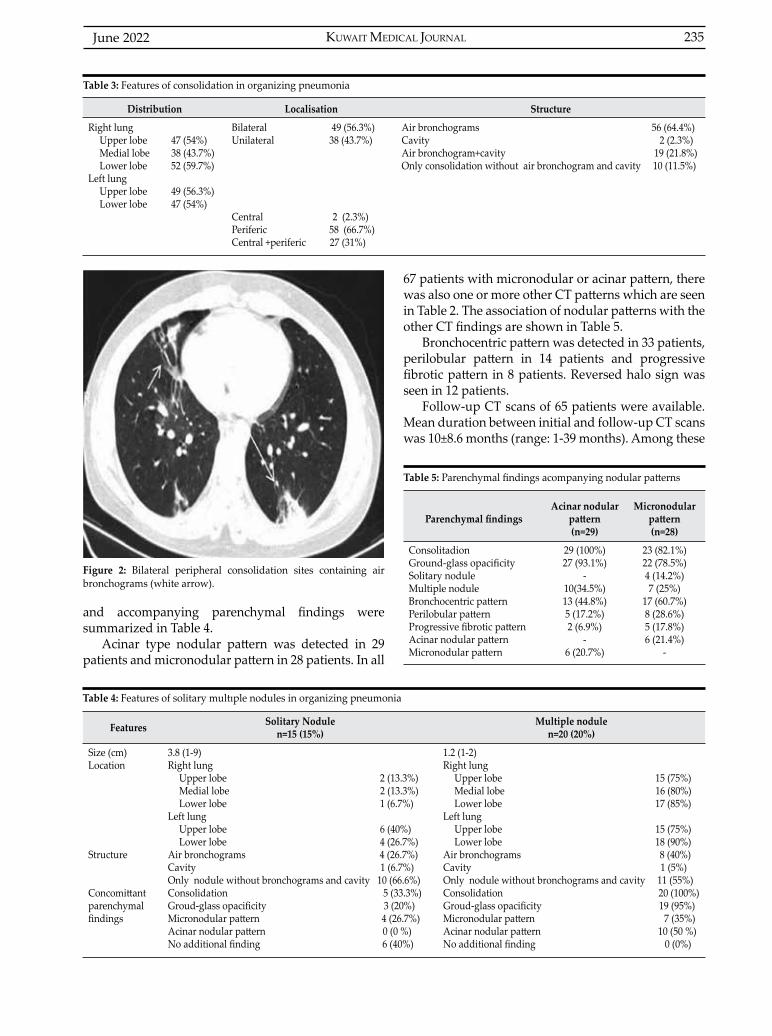
KUWAIT MEDICAL JOURNAL 235June 2022
Distribution Localisation Structure
Right lungUpper lobe 47 (54%) Medial lobe 38 (43.7%)Lower lobe 52 (59.7%)
Left lungUpper lobe 49 (56.3%)Lower lobe 47 (54%)
Bilateral 49 (56.3%) Unilateral 38 (43.7%)
Central 2 (2.3%)Periferic 58 (66.7%)Central +periferic 27 (31%)
Air bronchograms 56 (64.4%)Cavity 2 (2.3%)Air bronchogram+cavity 19 (21.8%)Only consolidation without air bronchogram and cavity 10 (11.5%)
Table 3: Features of consolidation in organizing pneumonia
Features Solitary Nodulen=15 (15%)
Multiple nodulen=20 (20%)
Size (cm)Location
Structure
Concomittant parenchymal findings
3.8 (1-9)Right lung
Upper lobe 2 (13.3%) Medial lobe 2 (13.3%)Lower lobe 1 (6.7%)
Left lungUpper lobe 6 (40%) Lower lobe 4 (26.7%)
Air bronchograms 4 (26.7%) Cavity 1 (6.7%) Only nodule without bronchograms and cavity 10 (66.6%)Consolidation 5 (33.3%)Groud-glass opacificity 3 (20%)Micronodular pattern 4 (26.7%)Acinar nodular pattern 0 (0 %)No additional finding 6 (40%)
1.2 (1-2)Right lung
Upper lobe 15 (75%) Medial lobe 16 (80%)Lower lobe 17 (85%)
Left lungUpper lobe 15 (75%) Lower lobe 18 (90%)
Air bronchograms 8 (40%) Cavity 1 (5%) Only nodule without bronchograms and cavity 11 (55%) Consolidation 20 (100%)Groud-glass opacificity 19 (95%)Micronodular pattern 7 (35%)Acinar nodular pattern 10 (50 %)No additional finding 0 (0%)
Table 4: Features of solitary multıple nodules in organizing pneumonia
Figure 2: Bilateral peripheral consolidation sites containing air bronchograms (white arrow).
and accompanying parenchymal findings were summarized in Table 4.
Acinar type nodular pattern was detected in 29 patients and micronodular pattern in 28 patients. In all
67 patients with micronodular or acinar pattern, there was also one or more other CT patterns which are seen in Table 2. The association of nodular patterns with the other CT findings are shown in Table 5.
Bronchocentric pattern was detected in 33 patients, perilobular pattern in 14 patients and progressive fibrotic pattern in 8 patients. Reversed halo sign was seen in 12 patients.
Follow-up CT scans of 65 patients were available. Mean duration between initial and follow-up CT scans was 10±8.6 months (range: 1-39 months). Among these
Parenchymal findingsAcinar nodular
pattern(n=29)
Micronodular pattern(n=28)
ConsolitadionGround-glass opacificitySolitary noduleMultiple noduleBronchocentric patternPerilobular patternProgressive fibrotic patternAcinar nodular patternMicronodular pattern
29 (100%)27 (93.1%)
-10(34.5%)13 (44.8%)5 (17.2%)2 (6.9%)
-6 (20.7%)
23 (82.1%)22 (78.5%)4 (14.2%)7 (25%)
17 (60.7%)8 (28.6%)5 (17.8%)6 (21.4%)
-
Table 5: Parenchymal findings acompanying nodular patterns
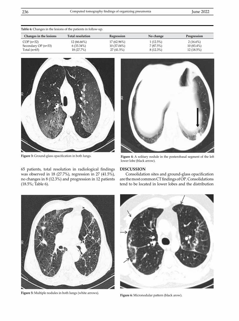
June 2022236
65 patients, total resolution in radiological findings was observed in 18 (27.7%), regression in 27 (41.5%), no changes in 8 (12.3%) and progression in 12 patients (18.5%; Table 6).
DISCUSSIONConsolidation sites and ground-glass opacification
are the most common CT findings of OP. Consolidations tend to be located in lower lobes and the distribution
Figure 3: Ground-glass opacification in both lungs. Figure 4: A solitary nodule in the posterobasal segment of the left lower lobe (black arrow).
Changes in the lesions Total resolution Regression No change Progression
COP (n=32)Secondary OP (n=33)Total (n=65)
12 (66.66%)6 (33.34%)18 (27.7%)
17 (62.96%) 10 (37.04%) 27 (41.5%)
1 (12.5%) 7 (87.5%) 8 (12.3%)
2 (16.6%) 10 (83.4%) 12 (18.5%)
Table 6: Changes in the lesions of the patients in follow-up.
Figure 6: Micronodular pattern (black arow).Figure 5: Multiple nodules in both lungs (white arrows).
Computed tomography findings of organizing pneumonia

KUWAIT MEDICAL JOURNAL 237June 2022
of lesions is usually bilateral and periferic[7,8]. While consolidation is detected in 87 (87%) of our patients, among them 49 (56.3%) have bilateral lesions. Consolidation was located in periferic parenchymal areas in 58 patients (66.7%) (Fig 2). Although all the lobes were involved by consolidation with similar rates, these lesions were found mostly in right lower lobe (Table 3). Air bronchograms and bronchial dilation can be seen in consolidation sites[5]. Among the 87 patients with consolidation, 75 (86.2%) were in
Figure 7: Acinar nodular pattern (white arrow).
air bronchograms and in 21 (24.1%) patients, cavitation was detected in consolidation (Table 3).
Consolidation areas are usually seen with ground-glass opacification. Ground-glass opacification frequency in the patients with OP was reported as 60%-86% in various studies[7,9-11]. We found a frequency of 76% (76/100 patients), which is consistent with previous studies (Fig. 3).
Solitary focal mass or nodule can be detected in OP. These lesions usually locate in upper lobes and can contain air bronchograms or cavities. Since radiological findings are sometimes not adequate to discriminate these lesions from malignant lesions, biopsy or surgical excision of the lesion is required[8]. In this study, solitary nodule was detected in 15 patients (Fig. 4). Mean diameter of the lesion was 3.8 cm (range: 1-9 cm). Solitary nodules are most commonly located in left upper lobe.
The diagnosis is difficult when a patient presents with multiple nodules with a diameter >1 cm because differential diagnosis includes various and complex conditions such as metastasis, lymphoma, infections, Wegener’s granulomatosis and Kaposi sarcoma[8,12]. Air bronchograms and cavity formations may be present within the nodules. Akira et al searched 59 patients with histopathologically proven COP and they found multiple nodules in 20% of their patients[12]. We also detected multiple nodules with a diameter of >1 cm in 20 patients (20%) in our study. Mean diameter of these nodules was 1.2 cm (range: 1-2 cm). While the nodules were located in each lobe with similar rates, the lower lobes were mostly involved (Fig. 5). The
Figure 8: Bronchocentric pattern (white arrow).
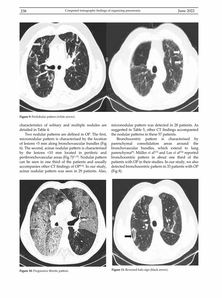
June 2022238
characteristics of solitary and multiple nodules are detailed in Table 4.
Two nodular patterns are defined in OP. The first, micronodular pattern is characterised by the location of lesions <5 mm along bronchovascular bundles (Fig 6). The second, acinar nodular pattern is characterised by the lesions <10 mm located in periferic and peribronchovascular areas (Fig 7)[1,13]. Nodular pattern can be seen in one third of the patients and usually accompanies other CT findings of OP[14]. In our study, acinar nodular pattern was seen in 29 patients. Also,
micronodular pattern was detected in 28 patients. As suggested in Table 5, other CT findings accompanied the nodular patterns in these 57 patients.
Bronchocentric pattern is characterised by parenchymal consolidation areas around the bronchovascular bundles, which extend to lung parenchyma[8]. Müller et al[13] and Lee et al[14] reported bronchocentric pattern in about one third of the patients with OP in their studies. In our study, we also detected bronchocentric pattern in 33 patients with OP (Fig 8).
Figure 9: Perilobular pattern (white arrow).
Figure 10: Progressive fibrotic pattern. Figure 11: Reversed halo sign (black arrow).
Computed tomography findings of organizing pneumonia
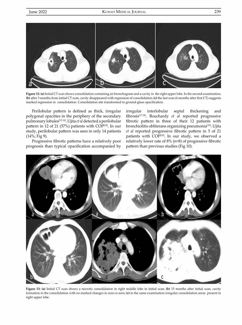
KUWAIT MEDICAL JOURNAL 239June 2022
Perilobular pattern is defined as thick, irregular polygonal opacities in the periphery of the secondary pulmonary lobules[15,16]. Ujita et al detected a perilobular pattern in 12 of 21 (57%) patients with COP[10]. In our study, perilobular pattern was seen in only 14 patients (14%; Fig 9).
Progressive fibrotic patterns have a relatively poor prognosis than typical opacification accompanied by
irregular interlobular septal thickening and fibrosis[17,18]. Bouchardy et al reported progressive fibrotic pattern in three of their 12 patients with bronchiolitis obliterans organizing pneumonia[19]. Ujita et al reported progressive fibrotic pattern in 5 of 21 patients with COP[10]. In our study, we observed a relatively lower rate of 8% (n=8) of progressive fibrotic pattern than previous studies (Fig 10).
Figure 12: (a) Initial CT scan shows consolidation containing air bronchogram and a cavity in the right upper lobe. In the second examination; (b) after 3 months from initial CT scan, cavity disappeared with regression of consolidation; (c) the last scan (6 months after first CT) suggests marked regression in consolidation. Consolidation site transformed to ground-glass opacification.
Figure 13: (a) Initial CT scan shows a necrotic consolidation in right middle lobe in initial scan; (b) 15 months after initial scan, cavity formation in the consolidation with no marked changes in sizes is seen; (c) in the same examination irregular consolidation areas present in right upper lobe.
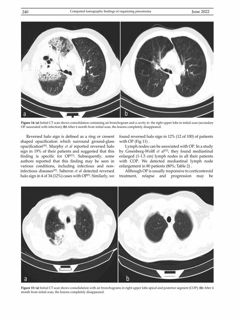
June 2022240
Reversed halo sign is defined as a ring or cresent shaped opacification which surround ground-glass opacification[20]. Murphy et al reported reversed halo sign in 19% of their patients and suggested that this finding is specific for OP[21]. Subsequently, some authors reported that this finding may be seen in various conditions, including infectious and non-infectious diseases[20]. Saberon et al detected reversed halo sign in 4 of 34 (12%) cases with OP[9]. Similarly, we
found reversed halo sign in 12% (12 of 100) of patients with OP (Fig 11) .
Lymph nodes can be associated with OP. In a study by Greenberg-Wolff et al[22], they found mediastinal enlarged (1-1.5 cm) lymph nodes in all their patients with COP. We detected mediastinal lymph node enlargement in 80 patients (80%; Table 2) .
Although OP is usually responsive to corticosteroid treatment, relapse and progression may be
Figure 14: (a) Initial CT scan shows consolidation containing air bronchogram and a cavity in the right upper lobe in initial scan (secondary OP associated with infection); (b) After 6 month from initial scan, the lesions completely disappeared.
Figure 15: (a) Initial CT scan shows consolidation with air bronchograms in right upper lobe apical and posterior segment (COP); (b) After 4 month from initial scan, the lesions completely disappeared.
Computed tomography findings of organizing pneumonia
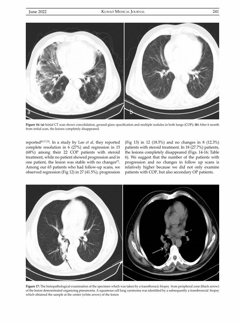
KUWAIT MEDICAL JOURNAL 241June 2022
reported[8,17,23]. In a study by Lee et al, they reported complete resolution in 6 (27%) and regression in 15 (68%) among their 22 COP patients with steroid treatment, while no patient showed progression and in one patient, the lesion was stable with no changes[7]. Among our 65 patients who had follow-up scans, we observed regression (Fig 12) in 27 (41.5%), progression
(Fig 13) in 12 (18.5%) and no changes in 8 (12.3%) patients with steroid treatment. In 18 (27.7%) patients, the lesions completely disappeared (Figs. 14-16; Table 6). We suggest that the number of the patients with progression and no changes in follow up scans is relatively higher because we did not only examine patients with COP, but also secondary OP patients.
Figure 16: (a) Initial CT scan shows concolidation, ground-glass opacification and multiple nodules in both lungs (COP); (b) After 6 month from initial scan, the lesions completely disappeared.
Figure 17: The histopathological examination of the specimen which was taken by a transthoracic biopsy from peripheral zone (black arrow) of the lesion demonstrated organizing pneumonia. A squamous cell lung carsinoma was identified by a subsequently a transbroncial biopsy which obtained the sample at the center (white arrow) of the lesion
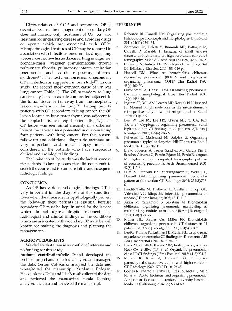
June 2022242
Differentiation of COP and secondary OP is essential because the management of secondary OP does not include only treatment of OP, but also treatment of underlying disease and avoiding drugs or agents which are associated with OP[24]. Histopathological features of OP may be reported in association with infectious pneumonia, drugs, lung abscess, connective tissue diseases, lung malignities, bronchiectasis, Wegener granulomatosis, chronic pulmonary fibrosis, pulmonary infarct, aspiration pneumonia and adult respiratory distress syndrome[25]. The most common reason of secondary OP is infection as suggested in our study[25]. In our study, the second most common cause of OP was lung cancer (Table 1). The OP secondary to lung cancer may be seen as a lesion located adjacent to the tumor tissue or far away from the neoplastic lesion anywhere in the lung[26]. Among our 12 patients with OP secondary to lung cancer, the OP lesion located in lung parenchyma was adjacent to the neoplastic tissue in eight patients (Fig 17). The OP lesion was seen simultaneously in a different lobe of the cancer tissue presented in our remaining four patients with lung cancer. For this reason, follow-up and additional diagnostic methods are very important, and repeat biopsy must be considered in the patients who have suspicious clinical and radiological findings.
The limitation of the study was the lack of some of the patients’ follow-up scans that did not permit to search the course and to compare initial and susequent radiologic findings.
CONCLUSIONAs OP has various radiological findings, CT is
very important for the diagnosis of this condition. Even when the disease is histopathologically proven, the follow-up these patients is essential because secondary OP must be kept in mind for the lesions which do not regress despite treatment. The radiological and clinical findings of the conditions which are associated with secondary OP must be well known for making the diagnosis and planning the management.
ACKNOWLEDGMENTSWe declare that there is no conflict of interests and
no funding for this study.Authors’ contribution:Yeliz Dadali developed the protocol/project and collected, analysed and managed the data; Sercan Ozkacmaz analysed the data and wrote/edited the manuscript; Yurdanur Erdogan, Havva Akmaz Unlu and Ilke Bursali collected the data and reviewed the manuscript; Funda Demirag analysed the data and reviewed the manuscript.
REFERENCES
1. Roberton BJ, Hansell DM. Organizing pneumonia: a kaleidoscope of concepts and morphologies. Eur Radiol 2011; 21(11):2244-54.
2. Zompatori M, Poletti V, Rimondi MR, Battaglia M, Carvelli P, Maraldi F. Imaging of small airways disease, with emphasis on high resolution computed tomography. Monaldi Arch Chest Dis 1997; 52(3):242-8.
3. Corrin B, Nicholson AG. Pathology of the Lungs. 3rd Ed. Edinburg: Elsevier; 2011. 308-310 p.
4. Hansell DM. What are bronchiolitis obliterans organizing pneumonia (BOOP) and cryptogenic organizing pneumonia (COP)? Clin Radiol 1992; 45(6):369-70.
5. Oikonomou A, Hansell DM. Organizing pneumonia: the many morphological faces. Eur Radiol 2002; 12(6):1486-96.
6. Ingram CE, Belli AM, Lewars MD, Reznek RH, Husband JE. Normal lymph node size in the mediastinum: a retrospective study in two patient groups. Clin Radiol 1989; 40(1):35-9.
7. Lee JW, Lee KS, Lee HY, Chung MP, Yi CA, Kim TS, et al. Cryptogenic organizing pneumonia: serial high-resolution CT findings in 22 patients. AJR Am J Roentgenol 2010; 195(4):916-22.
8. Polverosi R, Maffesanti M, Dalpiaz G. Organizing pneumonia: typical and atypical HRCT patterns. Radiol Med 2006; 111(2):202-12.
9. Bravo Soberón A, Torres Sánchez MI, García Río F, Sánchez Almaraz C, Parrón Pajares M, Pardo Rodríguez M. High-resolution computed tomography patterns of organizing pneumonia. Arch Bronconeumol 2006; 42(8):413-6.
10. Ujita M, Renzoni EA, Veeraraghavan S, Wells AU, Hansell DM. Organizing pneumonia: perilobular pattern at thin-section CT. Radiology 2004; 232(3):757-61.
11. Pandit-Bhalla M, Diethelm L, Ovella T, Sloop GD, Valentine VG. Idiopathic interstitial pneumonias: an update. J Thorac Imaging 2003; 18(1):1-13.
12. Akira M, Yamamoto S, Sakatani M. Bronchiolitis obliterans organizing pneumonia manifesting as multiple large nodules or masses. AJR Am J Roentgenol 1998; 170(2):291-5.
13. Müller NL, Staples CA, Miller RR. Bronchiolitis obliterans organizing pneumonia: CT features in 14 patients. AJR Am J Roentgenol 1990; 154(5):983-7.
14. Lee KS, Kullnig P, Hartman TE, Müller NL. Cryptogenic organizing pneumonia: CT findings in 43 patients. AJR Am J Roentgenol 1994; 162(3):543-6.
15. Faria IM, Zanetti G, Barreto MM, Rodrigues RS, Araujo-Neto CA, e Silva JLP, et al. Organizing pneumonia: chest HRCT findings. J Bras Pneumol 2015; 41(3):231-7.
16. Murata K, Khan A, Herman PG. Pulmonary parenchymal disease: evaluation with high-resolution CT. Radiology 1989; 170(3 Pt 1):629-35.
17. Gomes R, Padrao E, Dabo H, Pires FS, Mota P, Melo N, et al. Acute fibrinous and organizing pneumonia: A report of 13 cases in a tertiary university hospital. Medicine (Baltimore) 2016; 95(27):e4073.
Computed tomography findings of organizing pneumonia
KUWAIT MEDICAL JOURNAL 243June 2022
18. Cottin V, Cordier JF. Cryptogenic organizing pneumonia. Semin Respir Crit Care Med 2012; 33(5):462-75.
19. Bouchardy LM, Kuhlman JE, Ball WC, Hruban RH, Askin FB, Siegelman SS. CT findings in bronchiolitis obliterans organizing pneumonia (BOOP) with radiographic, clinical and histological correlation. J Comput Assist Tomogr 1993; 17(3):352-7.
20. Marchiori E, Zanetti G, Escuissato DL, Souza Jr AS, Meirelles GS, Fagundes J, et al. Reversed halo sign: high-resolution CT scan findings in 79 patients. Chest 2012; 141(5):1260-6.
21. Murphy JM, Schnyder P, Verschakelen J, Leuenberger P, Flower CD. Linear opacities on HRCT in bronchiolitis obliterans organising pneumonia. Eur Radiol 1999; 9(9):1813-7.
22. Greenberg-Wolff I, Konen E, Ben Dov I, Simansky D, Perelman M, Rozenman J. Cryptogenic organizing
pneumonia: variety of radiologic findings. Isr Med Assoc J 2005; 7(9):568-70.
23. Petitpierre N, Beigelman C, Letovanec I, Lazor R. [Cryptogenic organizing pneumonia]. Rev Mal Respir 17-703:(8)33 ;2016. Article in French.
24. Drakopanagiotakis F, Paschalaki K, Abu-Hijleh M, Aswad B, Karagianidis N, Kastanakis E, et al. Cryptogenic and secondary organizing pneumonia: clinical presentation, radiographic findings, treatment response, and prognosis. Chest 2011; 139(4):893-900.
25. Cordier JF. Organising pneumonia. Thorax 2000; 55(4):318-28.
26. Sánchez RA, Poce RM, Doménech AB, de Rota Avecilla AF, Bermúdez JLF. [Bronchiolitis obliterans organizing pneumonia and bronchogenic carcinoma coexisting in different parts of the lungs]. Arch Bronconeumol 2004; 40(3):141-3. Article in Spanish.
KUWAIT MEDICAL JOURNAL June 2022244
Original Article
Kuwait Medical Journal 2022; 54 (2): 244 - 248
Influence of neonatal care advancement on mortality and the incidence of bronchopulmonary
dysplasia in very low birth weight (VLBW) Saudi infants: two period’s comparison
ABSTRACT
KEY WORDS: advancement in neonatal care, bronchopulmonary dysplasia, mortality, very low birth weight infants
Badr Hasan SobaihDepartment of Pediatrics, King Saud University, College of Medicine, Riyadh, KSA
Objectives: To examine the influence of advanced neonatal care on mortality and incidence of bronchopulmonary dysplasia (BPD) in very low birth weight infantsDesign: Retrospective observational studySetting: Neonatal intensive care unit at King Khalid University Hospital, Riyadh, Kingdom of Saudi ArabiaSubjects: All infants admitted to our unit with birth weight below 1500 grams in two periods (1999-2007 and 2011-2018).Interventions: Mortality and BPD incidence rates were compared between the two groups.
Main outcome measures: Mortality and development of BPD.Results: Infants born during the later time period were 2.8 times more likely to die compared with the first period. Higher birth weight was associated with decreased odds of infant death (OR=0.997, P <0.001). Incidence of BPD was lower in the later period (P <0.05). Conclusions: Advancement in neonatal care in our institute led to decreased incidence of BPD. Mortality rate increased in later period, which might be a result of changes in unit admission policy.
Address correspondence to:Badr Hasan Sobaih, MBBS, ABP, Department of Pediatrics, King Saud University, College of Medicine, Riyadh, KSA. Tel: 0505453580; E-mail: [email protected]; [email protected]
INTRODUCTIONPremature delivery carries a great risk of neonatal
mortality and increases the risk of morbidities and future neurodevelopmental delay. Neonatal care, especially for very low birth weight (VLBW) infants, is ever-changing over the past years. In many countries worldwide, advancement in neonatal, as well as antenatal care, led to increased survival in neonate, and VLBW newborns in particular[1-4]. Bronchopulmonary dysplasia (BPD) remains a major challenge in the care of premature infants, despite improvement in neonatal care over the years. The increased survival of VLBW infants proportionally
increased the number of surviving sick babies who require advanced care to manage the evolving morbidities in such infants. BPD is one of the most studied topics in the field of neonatology, and most of the research and advancement in neonatal care is directed over the past decades on preventing or minimizing the development of BPD. An estimated 15 million babies are born premature worldwide. Premature infants are at a higher risk of death in comparison with full term babies due to complications of prematurity, mainly neonatal respiratory distress syndrome (NRDS) and BPD. The use of antenatal steroids has been globally adopted as a standard
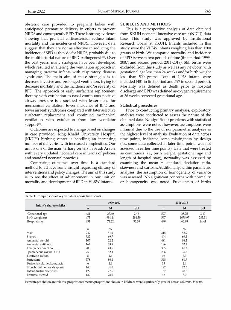
KUWAIT MEDICAL JOURNAL 245June 2022
obstetric care provided to pregnant ladies with anticipated premature delivery in efforts to prevent NRDS and consequently BPD. There is strong evidence showing that prenatal corticosteroids reduce infant mortality and the incidence of NRDS. However, data suggest that they are not as effective in reducing the incidence of BPD as they do for NRDS, probably due to the multifactorial nature of BPD pathogenesis[5]. Over the past years, many strategies have been developed which resulted in altering the ventilation approach in managing preterm infants with respiratory distress syndrome. The main aim of these strategies is to decrease invasive and prolonged ventilation, trying to decrease mortality and the incidence and/or severity of BPD. The approach of early surfactant replacement therapy with extubation to nasal continuous positive airway pressure is associated with lesser need for mechanical ventilation, lower incidence of BPD and fewer air leak syndromes compared with later selective surfactant replacement and continued mechanical ventilation with extubation from low ventilator support[6].
Outcomes are expected to change based on changes in care provided. King Khalid University Hospital (KKUH) birthing center is handling an increasing number of deliveries with increased complexities. Our unit is one of the main tertiary centers in Saudi Arabia with every updated neonatal care in terms of policies and standard neonatal practices.
Comparing outcomes over time is a standard method to achieve some insight regarding efficacy of interventions and policy changes. The aim of this study is to see the effect of advancement in our unit on mortality and development of BPD in VLBW infants.
SUBJECTS AND METHODSThis is a retrospective analysis of data obtained
from KKUH neonatal intensive care unit (NICU) data base. This study was approved by Institutional Research Board at KKUH. Infants included in this study were the VLBW infants weighing less than 1500 grams at birth. We compared mortality and incidence of BPD between two periods of time (first period: 1999-2007, and second period: 2011-2018). Still births were excluded from this study as well as any newborn with gestational age less than 24 weeks and/or birth weight less than 500 grams. Total of 1,078 infants were included (481 in first period and 597 in second period). Mortality was defined as death prior to hospital discharge and BPD was defined as oxygen requirement at 36 weeks corrected gestational age.
Statistical proceduresPrior to conducting primary analyses, exploratory
analyses were conducted to assess the nature of the obtained data. No significant problems with statistical assumptions were noted; however, assumptions were minimal due to the use of nonparametric analyses as the highest level of analysis. Evaluation of data across time points, indicated some missingness by design (i.e., some data collected in later time points was not assessed in earlier time points). Data that were treated as continuous (i.e., birth weight, gestational age and length of hospital stay), normality was assessed by examining the mean ± standard deviation ratio, skewness and kurtosis. Additionally, within parametric analyses, the assumption of homogeneity of variance was assessed. No significant concerns with normality or homogeneity was noted. Frequencies of births
Infant’s characteristics1999-2007 2011-2018
n M SD n M SD
Gestational ageBirth weight (g)Hospital stay
MaleBookedAntenatal steroidAntenatal antibioticEmergency c-sectionSpontaneous vaginal birthElective c-sectionSurfactantPeriventricular leukomalaciaBronchopulmonary dysplasiaPatent ductus arteriosusPostnatal steroid
481475451
n24933210516220925021
3786
145129132
27.60991.4471.32
%51.969.722.233.843.552.14.480.41.331.027.628.0
2.46284.5953.30
597597490
n315404481186355206193481312215742
28.751078.97
66.98
%52.969.286.232.161.235.53.361.92.422.328.58.0
3.10283.3184.41
Percentages shown are relative proportions; means/proportions shown in boldface were significantly greater across columns, P <0.05.
Table 1: Comparisons of key variables across time points
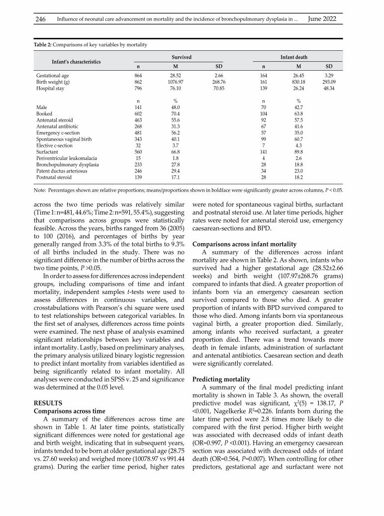
June 2022246 Influence of neonatal care advancement on mortality and the incidence of bronchopulmonary dysplasia in ...
across the two time periods was relatively similar (Time 1: n=481, 44.6%; Time 2: n=591, 55.4%), suggesting that comparisons across groups were statistically feasible. Across the years, births ranged from 36 (2005) to 100 (2016), and percentages of births by year generally ranged from 3.3% of the total births to 9.3% of all births included in the study. There was no significant difference in the number of births across the two time points, P >0.05.
In order to assess for differences across independent groups, including comparisons of time and infant mortality, independent samples t-tests were used to assess differences in continuous variables, and crosstabulations with Pearson’s chi square were used to test relationships between categorical variables. In the first set of analyses, differences across time points were examined. The next phase of analysis examined significant relationships between key variables and infant mortality. Lastly, based on preliminary analyses, the primary analysis utilized binary logistic regression to predict infant mortality from variables identified as being significantly related to infant mortality. All analyses were conducted in SPSS v. 25 and significance was determined at the 0.05 level.
RESULTSComparisons across time
A summary of the differences across time are shown in Table 1. At later time points, statistically significant differences were noted for gestational age and birth weight, indicating that in subsequent years, infants tended to be born at older gestational age (28.75 vs. 27.60 weeks) and weighed more (10078.97 vs 991.44 grams). During the earlier time period, higher rates
were noted for spontaneous vaginal births, surfactant and postnatal steroid use. At later time periods, higher rates were noted for antenatal steroid use, emergency caesarean-sections and BPD.
Comparisons across infant mortality
A summary of the differences across infant mortality are shown in Table 2. As shown, infants who survived had a higher gestational age (28.52±2.66 weeks) and birth weight (107.97±268.76 grams) compared to infants that died. A greater proportion of infants born via an emergency caesarean section survived compared to those who died. A greater proportion of infants with BPD survived compared to those who died. Among infants born via spontaneous vaginal birth, a greater proportion died. Similarly, among infants who received surfactant, a greater proportion died. There was a trend towards more death in female infants, administration of surfactant and antenatal antibiotics. Caesarean section and death were significantly correlated.
Predicting mortalityA summary of the final model predicting infant
mortality is shown in Table 3. As shown, the overall predictive model was significant, χ2(5) = 138.17, P <0.001, Nagelkerke R2=0.226. Infants born during the later time period were 2.8 times more likely to die compared with the first period. Higher birth weight was associated with decreased odds of infant death (OR=0.997, P <0.001). Having an emergency caesarean section was associated with decreased odds of infant death (OR=0.564, P=0.007). When controlling for other predictors, gestational age and surfactant were not
Infant’s characteristicsSurvived Infant death
n M SD n M SD
Gestational ageBirth weight (g)Hospital stay
MaleBookedAntenatal steroidAntenatal antibioticEmergency c-sectionSpontaneous vaginal birthElective c-sectionSurfactantPeriventricular leukomalaciaBronchopulmonary dysplasiaPatent ductus arteriosusPostnatal steroid
864862796
n14160246326848134332
56015
233246139
28.521076.97
76.10
%48.070.455.631.356.240.13.766.81.827.829.417.1
2.66268.7670.85
164161139
n70104926757997
1414283428
26.45830.1826.24
%42.763.857.541.635.060.74.389.82.618.823.018.2
3.29293.0948.34
Note: Percentages shown are relative proportions; means/proportions shown in boldface were significantly greater across columns, P < 0.05.
Table 2: Comparisons of key variables by mortality
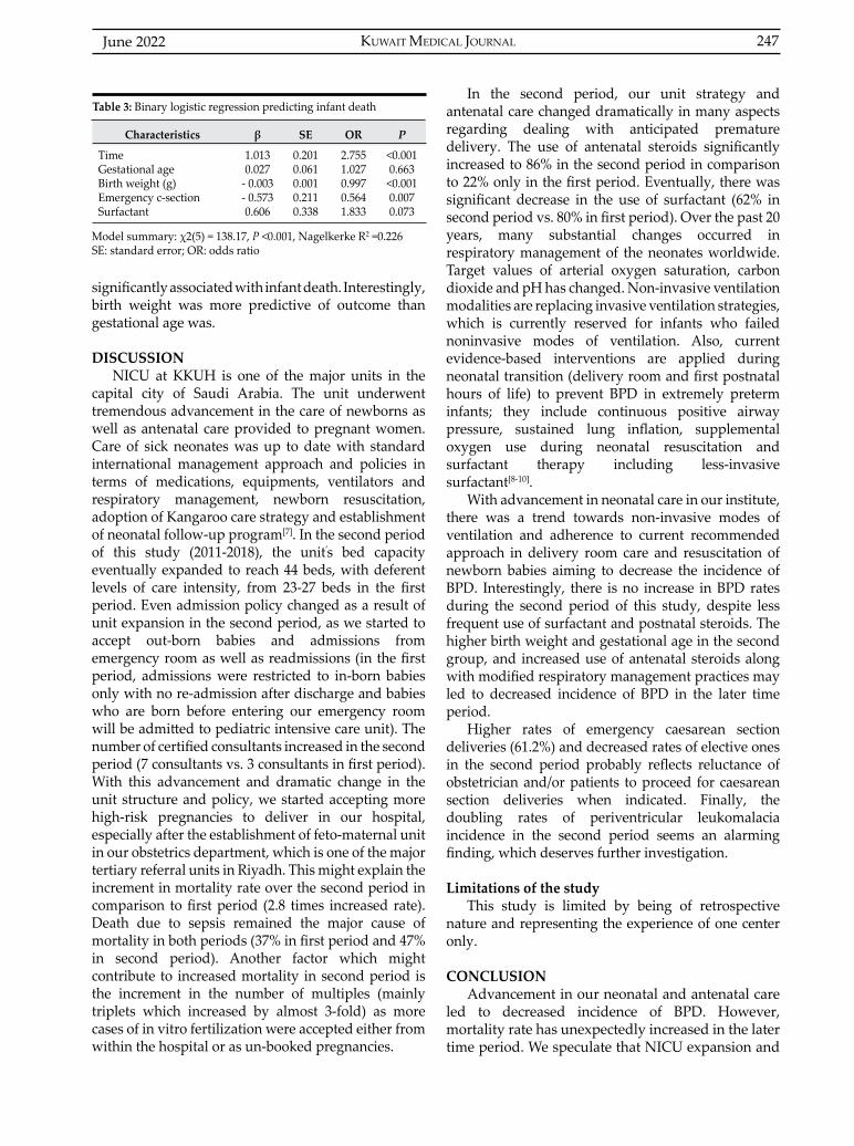
KUWAIT MEDICAL JOURNAL 247June 2022
significantly associated with infant death. Interestingly, birth weight was more predictive of outcome than gestational age was.
DISCUSSIONNICU at KKUH is one of the major units in the
capital city of Saudi Arabia. The unit underwent tremendous advancement in the care of newborns as well as antenatal care provided to pregnant women. Care of sick neonates was up to date with standard international management approach and policies in terms of medications, equipments, ventilators and respiratory management, newborn resuscitation, adoption of Kangaroo care strategy and establishment of neonatal follow-up program[7]. In the second period of this study (2011-2018), the unit’s bed capacity eventually expanded to reach 44 beds, with deferent levels of care intensity, from 23-27 beds in the first period. Even admission policy changed as a result of unit expansion in the second period, as we started to accept out-born babies and admissions from emergency room as well as readmissions (in the first period, admissions were restricted to in-born babies only with no re-admission after discharge and babies who are born before entering our emergency room will be admitted to pediatric intensive care unit). The number of certified consultants increased in the second period (7 consultants vs. 3 consultants in first period). With this advancement and dramatic change in the unit structure and policy, we started accepting more high-risk pregnancies to deliver in our hospital, especially after the establishment of feto-maternal unit in our obstetrics department, which is one of the major tertiary referral units in Riyadh. This might explain the increment in mortality rate over the second period in comparison to first period (2.8 times increased rate). Death due to sepsis remained the major cause of mortality in both periods (37% in first period and 47% in second period). Another factor which might contribute to increased mortality in second period is the increment in the number of multiples (mainly triplets which increased by almost 3-fold) as more cases of in vitro fertilization were accepted either from within the hospital or as un-booked pregnancies.
Characteristics β SE OR P
TimeGestational ageBirth weight (g)Emergency c-sectionSurfactant
1.0130.027
- 0.003- 0.5730.606
0.2010.0610.0010.2110.338
2.7551.0270.9970.5641.833
<0.0010.663<0.0010.0070.073
Table 3: Binary logistic regression predicting infant death
Model summary: χ2(5) = 138.17, P <0.001, Nagelkerke R2 =0.226SE: standard error; OR: odds ratio
In the second period, our unit strategy and antenatal care changed dramatically in many aspects regarding dealing with anticipated premature delivery. The use of antenatal steroids significantly increased to 86% in the second period in comparison to 22% only in the first period. Eventually, there was significant decrease in the use of surfactant (62% in second period vs. 80% in first period). Over the past 20 years, many substantial changes occurred in respiratory management of the neonates worldwide. Target values of arterial oxygen saturation, carbon dioxide and pH has changed. Non-invasive ventilation modalities are replacing invasive ventilation strategies, which is currently reserved for infants who failed noninvasive modes of ventilation. Also, current evidence-based interventions are applied during neonatal transition (delivery room and first postnatal hours of life) to prevent BPD in extremely preterm infants; they include continuous positive airway pressure, sustained lung inflation, supplemental oxygen use during neonatal resuscitation and surfactant therapy including less-invasive surfactant[8-10].
With advancement in neonatal care in our institute, there was a trend towards non-invasive modes of ventilation and adherence to current recommended approach in delivery room care and resuscitation of newborn babies aiming to decrease the incidence of BPD. Interestingly, there is no increase in BPD rates during the second period of this study, despite less frequent use of surfactant and postnatal steroids. The higher birth weight and gestational age in the second group, and increased use of antenatal steroids along with modified respiratory management practices may led to decreased incidence of BPD in the later time period.
Higher rates of emergency caesarean section deliveries (61.2%) and decreased rates of elective ones in the second period probably reflects reluctance of obstetrician and/or patients to proceed for caesarean section deliveries when indicated. Finally, the doubling rates of periventricular leukomalacia incidence in the second period seems an alarming finding, which deserves further investigation.
Limitations of the studyThis study is limited by being of retrospective
nature and representing the experience of one center only.
CONCLUSION Advancement in our neonatal and antenatal care
led to decreased incidence of BPD. However, mortality rate has unexpectedly increased in the later time period. We speculate that NICU expansion and
June 2022248
changing our unit admission policy, which permitted accepting out-born babies, pediatrics emergency room admissions and re-admission of discharged infants, as well as the establishment of feto-maternal unit, dealing with high risk pregnancies and accepting referred cases with congenital anomalies and potentially lethal conditions to be followed and delivered in our center, played a major role in increased mortality in later period, despite advancement in neonatal care.
ACKNOWLEDGMENTThe author would like to thank Dr. Amull Faris and
Dr. Rozina Banoo, who are working in neonatal follow-up program of King Khalid University Hospital, for their contribution in collecting all data.Conflict of interest: None
REFERENCES
1. Fanaroff AA, Stoll BJ, Wright LL, Carlo WA, Ehrenkranz RA, Stark AR, et al. Trends in neonatal morbidity and mortality for very low birthweight infants. Am J Obstet Gynecol 2007; 196(2):147.e1–8.
2. Itabashi K, Horiuchi T, Kusuda S, Kabe K, Itani Y, Nakamura T, et al. Mortality rates for extremely low birth weight infants born in Japan in 2005. Pediatrics 2009; 123(2):445-50.
3. Hack M. Survival and neurodevelopmental outcomes of preterm infants. J Pediatr Gastroenterol Nutr 2007; 45 Suppl 3:S141-2.
4. Goldani MZ, Barbieri MA, Silva AAM, Bettiol H. Trends in prenatal care use and low birth weight in southeast Brazil. Am J Public Health 2004; 94(8):1366-71.
5. Roberts D, Brown J, Medley N, Dalziel SR. Antenatal corticosteroids for accelerating fetal lung maturation for women at risk of preterm birth. Cochrane Database Syst Rev 2017; 3(3):CD004454.
6. Stevens TP, Harrington EW, Blennow M, Soll RF. Early surfactant administration with brief ventilation vs. selective surfactant and continued mechanical ventilation for preterm infants with or at risk for respiratory distress syndrome. Cochrane Database Syst Rev 2007; 2007(4):CD003063.
7. Sobaih BH. Neonatal follow-up program: where do we stand? Sudan J Paediatr 2012; 12(1):21-6.
8. Abreu-Pereiraa S, Pinto-Lopes R, Flor-de-Lima F, Rocina G, Guimaraes H. Ventilatory practices in extremely low birth weight infants in a level III neonatal intensive care unit. Pulmonology 2018; 24(6):337-44.
9. Gerull R, Manser H, Küster H, Arenz T, Arenz S, Nelle M. Less invasive ventilation in extremely low birth weight infants from 1997 to 2011: survey versus evidence. Eur J Pediatr 2015; 174(9):1189-96.
10. Foglia EE, Jensen EA, Kirpalani H. Delivery room interventions to prevent bronchopulmonary dysplasia in extremely preterm infants. J Perinatol 2017; 37(11):1171-79.
Influence of neonatal care advancement on mortality and the incidence of bronchopulmonary dysplasia in ...
KUWAIT MEDICAL JOURNAL 249June 2022
Address correspondence to:Dr. Hala Elsharaky, Alorf Hospital, 1st floor, Jahra, 01025 Kuwait. Tel: 60625693; E-mail: [email protected]
Kuwait Medical Journal 2022; 54 (2): 249 - 253
Cesarean scar pregnancy: Review and case reports
ABSTRACT
Mansour B Alorf1,2, Hala F Elsharaky2, Muntaha H Al-Salem2,3
1Fellowship of the Royal Colleges of Surgeons (FRCS), Canada2Department of Obstetrics and Gynecology, Alorf Hospital, Kuwait
3Arab Board of Obstetrics and Gynecology
Cesarean scar pregnancy (CSP) is one of the rarest forms of ectopic pregnancy with devastating complications. It occurs due to the implantation of gestational sac into the myometrial defect of previous cesarean scar, the niche. We are presenting a series of three case reports of CSP who were
diagnosed and treated in Alorf hospital over a two-year duration. Painless vaginal bleeding during the first trimester was the most common symptom. Methods of diagnosis and mode of treatment were described and discussed in relation to various medical literatures.
KEY WORDS: cesarean scar pregnancy, ectopic pregnancy, magnetic resonance imaging, methotrexate
Case Report
INTRODUCTIONCesarean scar pregnancy (CSP) is one rare form of
ectopic pregnancies[1]. The overall prevalence of ectopic pregnancy is approximately 2% relative to all pregnancies. About 97% of cases is in the fallopian tubes[2]. CSP is a condition in which the gestational sac is implanted within the previous hysterotomy scar[3,4]. The estimated incidence ranges from 1/1008 to 1/2500 of all pregnancies[5] and 72% of the patients had more than two cesarean sections[6].
CSP develops following injury to the endometrium and myometrium because of cesarean section, uterine curettage, myomectomy, hysterotomy, hysteroscopy or manual removal of the placenta[6]. It is thought to occur due to the implantation of the blastocyst in the fibrous scar tissue within the wedge- shaped myometrial defect ‘niche’ in the anterior wall of lower uterine segment following cesarean section[6,7]. This increases maternal morbidity and mortality due to placenta previa, placenta accreta, uterine rupture and hemorrhage[8].
Three patients of CSPs who were diagnosed and treated in our hospital are reported in detail.
CASE REPORTCase 1
A 28-year-old female patient was G6 P5+0 with a history of five recurrent previous cesarean sections.
The last three cesarean deliveries were one year apart, and the last cesarean delivery was on April 19, 2018.
Six months following the last cesarean section, she was misdiagnosed as having incomplete miscarriage that was treated conservatively without surgical intervention. She continued complaining of irregular uterine bleeding for one-month duration. Transvaginal ultrasound revealed irregular shaped cavity with hetero-echoic mass about 5×5 cm at the uterine isthmus. βhCG was 489.8 IU/L. Pelvic magnetic resonance imaging (MRI) showed irregular shaped cystic collection at lower uterine segment and complete myometrial discontinuity, intact serosa and there was no evidence of pelvic collection (Fig 1). The patient was diagnosed as having ectopic disturbed CSP. The hemoglobin level was 8.4 gm/dl and the patient was hemodynamically stable. Exploratory laparotomy was decided. Two units of cross matched packed red blood cells were transfused preoperatively.
After taking informed consent, transverse laparotomy was done. There was no intrabdominal bleeding. The uterus was bulky, myometrium was dehiscent at the site of previous scar with formation of hematoma size about 5×5 cm. With removal of hematoma, cystic tissue was found under the hematoma at the dehiscent myometrium (Fig 2A - 2D). The cystic tissue was excised (Fig 2E), sent to the pathology lab, and suction curettage for the uterine
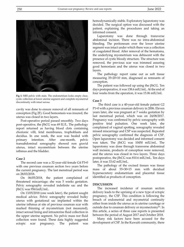
June 2022250
cavity was done to ensure removal of all remnants of conception (Fig 2F). Good hemostasis was insured, the uterus was closed in two layers.
Post-operative period passed smoothly. Two days post-operative, the βhCG was 69 IU/L. The pathology report returned as having blood clots containing chorionic villi, fetal membranes, trophoblasts and decidua. In one week, the scar was healed with primary intention. After one-month, pelvic transabdominal sonography showed non gravid uterus, intact myometrium between the uterine isthmus and the bladder.
Case 2The second case was a 32-year-old female G4 P3+0
with one previous cesarean section two years before the current pregnancy. The last menstrual period was on 28/03/2018.
On 06/05/2018, the patient complained of threatened miscarriage; she was 5 weeks gestation. Pelvic sonography revealed indefinite sac and the βhCG was 9561mIU/mL.
On 13/05/2018 (one week later), the patient sought medical advice. Pelvic sonography revealed bulky uterus with gestational sac implanted within the uterine isthmus at site of previous cesarean scar with marked thinning of myometrium (not measured), intact serosal lining and intrauterine fluid collection in the upper uterine segment. No pelvic mass nor fluid collection were found. These data highly suggested ectopic scar pregnancy. The patient was
hemodynamically stable. Exploratory laparotomy was decided. The surgical option was discussed with the patient, explaining the procedures and taking an informed consent.
Laparotomy was done through transverse abdominal incision. There was no intra-abdominal bleeding. The peritoneum over the lower uterine segment was intact under which there was a collection of coagulated blood. After removal of the hematoma, the underlying myometrium was dehiscent with the presence of cystic bloody structure. The structure was removed, the previous scar was trimmed assuring good hemostasis and the uterus was closed in two layers.
The pathology report came out as soft tissue measuring 35×20×10 mm, diagnosed as remnants of conception.
The patient was followed up using the βhCG. Ten days postoperative, it was 138.6 mIU/mL. At the end of four weeks from the operation, it was 15.06 mIU/mL
Case 3 The third case is a 40-year-old female patient G2
P1+0 with a previous cesarean delivery in 2006. Eleven years later, she was pregnant at 7 weeks gestation by last menstrual period, which was on 24/08/2017. Pregnancy was confirmed by pelvic sonography with positive fetal pulsation. One week later, she complained of vaginal spotting, sonography revealed missed miscarriage and CSP was suspected. Repeated pelvic sonography confirmed the diagnosis of CSP. Open laparotomy was decided and informed consent was taken. The βhCG was 10450 mIU/mL. The laparotomy was done through transverse abdominal wall incision, products of conception were removed, and the uterus was closed in two layers. Three days postoperative, the βhCG was 810.6 mIU/mL. Ten days later, it was 33.62 mIU/mL.
The pathology of the excised tissues was tissue mass of about 55×30×15 mm with decidual hypersecretory endometrium and placental tissue identified as products of conception.
DISCUSSIONThe increased incidence of cesarean section
delivery leads to the uprising of a new type of ectopic pregnancy, the CSP. This condition is following the breach of endometrial and myometrial continuity either from inside the uterus as in uterine curettage or outside due to cesarean delivery or myomectomy[6]. In this article, a series of three case reports is presented between the period of August 2017 and October 2018.
Many risk factors have been accused for the development of CSP. In the Kuwaiti community, there
Fig 1: MRI pelvic with stain. The endometrium looks empty clear, cystic collection at lower uterine segment and complete myometrial discontinuity with intact serosa.
Cesarean scar pregnancy: Review and case reports
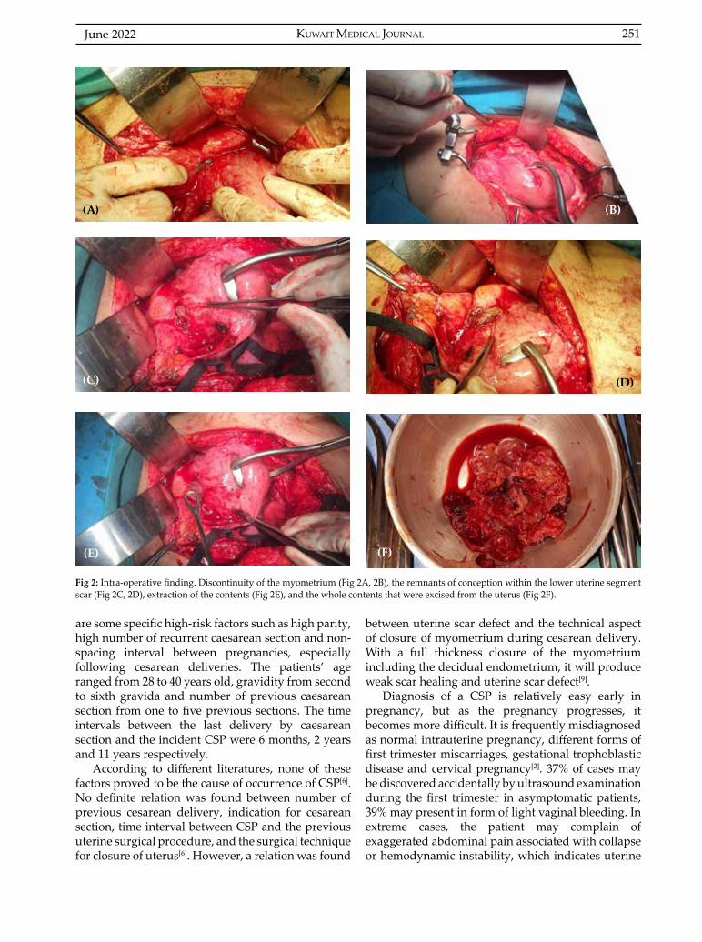
KUWAIT MEDICAL JOURNAL 251June 2022
are some specific high-risk factors such as high parity, high number of recurrent caesarean section and non-spacing interval between pregnancies, especially following cesarean deliveries. The patients’ age ranged from 28 to 40 years old, gravidity from second to sixth gravida and number of previous caesarean section from one to five previous sections. The time intervals between the last delivery by caesarean section and the incident CSP were 6 months, 2 years and 11 years respectively.
According to different literatures, none of these factors proved to be the cause of occurrence of CSP[6]. No definite relation was found between number of previous cesarean delivery, indication for cesarean section, time interval between CSP and the previous uterine surgical procedure, and the surgical technique for closure of uterus[6]. However, a relation was found
between uterine scar defect and the technical aspect of closure of myometrium during cesarean delivery. With a full thickness closure of the myometrium including the decidual endometrium, it will produce weak scar healing and uterine scar defect[9].
Diagnosis of a CSP is relatively easy early in pregnancy, but as the pregnancy progresses, it becomes more difficult. It is frequently misdiagnosed as normal intrauterine pregnancy, different forms of first trimester miscarriages, gestational trophoblastic disease and cervical pregnancy[2]. 37% of cases may be discovered accidentally by ultrasound examination during the first trimester in asymptomatic patients, 39% may present in form of light vaginal bleeding. In extreme cases, the patient may complain of exaggerated abdominal pain associated with collapse or hemodynamic instability, which indicates uterine
Fig 2: Intra-operative finding. Discontinuity of the myometrium (Fig 2A, 2B), the remnants of conception within the lower uterine segment scar (Fig 2C, 2D), extraction of the contents (Fig 2E), and the whole contents that were excised from the uterus (Fig 2F).
(A)
(D)
(F)(E)
(B)
(C)
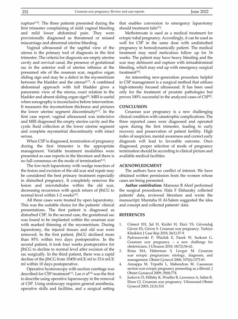
June 2022252
rupture[3,6]. The three patients presented during the first trimester complaining of mild vaginal bleeding and mild lower abdominal pain. They were provisionally diagnosed as threatened or missed miscarriage and abnormal uterine bleeding.
Vaginal ultrasound of the sagittal view of the uterus is the primary tool of diagnosis in the first trimester. The criteria for diagnosis are empty uterine cavity and cervical canal, the presence of gestational sac in the anterior wall of uterine isthmus at the presumed site of the cesarean scar, negative organ sliding sign and may be a defect in the myometrium between the bladder and the uterus[6,7]. A combined abdominal approach with full bladder gives a panoramic view of the uterus, exact relation to the bladder and absent sliding organ sign[6]. MRI is useful when sonography is inconclusive before intervention. It measures the myometrium thickness and pictures the lower uterine segment discontinuity[6,7]. In the first case report, vaginal ultrasound was indecisive and MRI diagnosed the empty uterine cavity and the cystic fluid collection at the lower uterine segment and complete myometrial discontinuity with intact serosa.
When CSP is diagnosed, termination of pregnancy during the first trimester is the appropriate management. Variable treatment modalities were presented as case reports in the literature and there is no full consensus on the mode of termination[6,1].
The low-tech laparotomy with wedge resection of the lesion and excision of the old scar and repair may be considered the best primary treatment especially in disturbed pregnancy. It completely removes the lesion and microtubules within the old scar, decreasing recurrence with quick return of βhCG to normal level within 1-2 weeks[10].
All three cases were treated by open laparotomy. This was the suitable choice for the patients’ clinical presentations. The first patient is diagnosed as disturbed CSP. In the second case, the gestational sac was found to be implanted within the cesarean scar with marked thinning of the myometrium. During laparotomy, the injured tissues and old scar were removed. In the first patient, βhCG declined more than 85% within two days postoperative. In the second patient, it took four weeks postoperative for βhCG to decline to normal level after excision of the sac surgically. In the third patient, there was a rapid decline of the βhCG from 10450 mUI/ ml to 33.6 mUI/ ml within 10 days postoperative.
Operative hysteroscopy with suction curettage was described for CSP treatment[11]. Lee et al[12] was the first to describe using operative laparoscopy in the removal of CSP. Using endoscopy requires general anesthesia, operative skills and facilities, and a surgical setting
that enables conversion to emergency laparotomy should treatment fails[13].
Methotrexate is used as a medical treatment for ectopic tubal pregnancy. Accordingly, it can be used as well for CSP in the same dose with undisturbed pregnancy in hemodynamically patient. The medical treatment may need meticulous follow up for 16 weeks. The patient may have heavy bleeding and the scar may dehiscent and rupture with intraabdominal bleeding, which may end up with emergency surgical treatment[14].
An interesting new-generation procedure helpful in CSP management is a surgical method that utilizes high-intensity focused ultrasound. It has been used only for the treatment of prostate pathologies but proves 100% successful in the analyzed cases of CSP[2].
CONCLUSIONCesarean scar pregnancy is a new challenging
clinical condition with catastrophic complications. The three reported cases were diagnosed and operated upon during the first trimester, leading to early recovery and preservation of patient fertility. High index of suspicion, mental awareness and correct early diagnosis will lead to favorable outcome. Once diagnosed, proper selection of mode of pregnancy termination should be according to clinical picture and available medical facilities.
ACKNOWLEDGMENTThe authors have no conflict of interest. We have
obtained written permission from the women whose cases are being presented.
Author contribution: Mansour B Alorf performed the surgical procedures; Hala F Elsharaky collected patients’ data, reviewed literature and wrote the manuscript; Muntaha H Al-Salem suggested the idea and concept and collected patients’ data.
REFERENCES 1. Cömert EH, Şal H, Kizilet H, Ekici YS, Güvendağ
Güven ES, Güven S. Cesarean scar pregnancy. Turkiye Klinikleri J Case Rep 2018; 26(1):37-9.
2. Pędraszewski P, Wlaźlak E, Panek W, Surkont G. Cesarean scar pregnancy – a new challenge for obstetricians. J Ultrason 2018; 18(72):56-62.
3. Rotas MA, Haberman S, Levgur M. Cesarean scar ectopic pregnancies: etiology, diagnosis, and management. Obstet Gynecol 2006; 107(6):1373-81.
4. Annappa M, Tripathi L, Mahendran M. Caesarean section scar ectopic pregnancy presenting as a fibroid. J Obstet Gynaecol 2009; 29(8):774.
5. Jurkovic D, Hillaby K, Woelfer B, Lawrence A, Salim R, Elson CJ. Cesarean scar pregnancy. Ultrasound Obstet Gynecol 2003; 21(3):310.
Cesarean scar pregnancy: Review and case reports
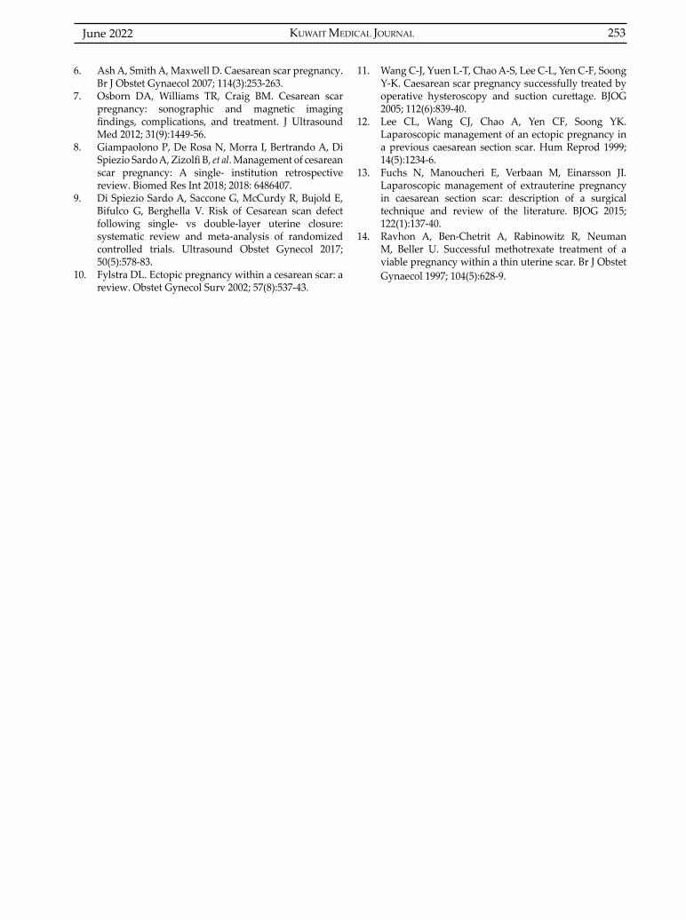
KUWAIT MEDICAL JOURNAL 253June 2022
6. Ash A, Smith A, Maxwell D. Caesarean scar pregnancy. Br J Obstet Gynaecol 2007; 114(3):253-263.
7. Osborn DA, Williams TR, Craig BM. Cesarean scar pregnancy: sonographic and magnetic imaging findings, complications, and treatment. J Ultrasound Med 2012; 31(9):1449-56.
8. Giampaolono P, De Rosa N, Morra I, Bertrando A, Di Spiezio Sardo A, Zizolfi B, et al. Management of cesarean scar pregnancy: A single- institution retrospective review. Biomed Res Int 2018; 2018: 6486407.
9. Di Spiezio Sardo A, Saccone G, McCurdy R, Bujold E, Bifulco G, Berghella V. Risk of Cesarean scan defect following single- vs double-layer uterine closure: systematic review and meta-analysis of randomized controlled trials. Ultrasound Obstet Gynecol 2017; 50(5):578-83.
10. Fylstra DL. Ectopic pregnancy within a cesarean scar: a review. Obstet Gynecol Surv 2002; 57(8):537-43.
11. Wang C-J, Yuen L-T, Chao A-S, Lee C-L, Yen C-F, Soong Y-K. Caesarean scar pregnancy successfully treated by operative hysteroscopy and suction curettage. BJOG 2005; 112(6):839-40.
12. Lee CL, Wang CJ, Chao A, Yen CF, Soong YK. Laparoscopic management of an ectopic pregnancy in a previous caesarean section scar. Hum Reprod 1999; 14(5):1234-6.
13. Fuchs N, Manoucheri E, Verbaan M, Einarsson JI. Laparoscopic management of extrauterine pregnancy in caesarean section scar: description of a surgical technique and review of the literature. BJOG 2015; 122(1):137-40.
14. Ravhon A, Ben-Chetrit A, Rabinowitz R, Neuman M, Beller U. Successful methotrexate treatment of a viable pregnancy within a thin uterine scar. Br J Obstet Gynaecol 1997; 104(5):628-9.
KUWAIT MEDICAL JOURNAL June 2022254
Address correspondence to:Behiye Deniz Kosovali, Ministry of Health Ankara City Hospital, University Neighbourhood Bilkent Street No 1, Cankaya, Ankara, Turkey. Tel: +90 3125526000; E-mail: [email protected]
Kuwait Medical Journal 2022; 54 (2): 254 - 257
Successful management of patient with fulminant myocarditis related refractory ventricular fibrillation by
veno-arterial extracorporeal membrane oxygenation
ABSTRACT
Asiye Yavuz1, Behiye Deniz Kosovali2, Mustafa Kemal Bayar1
1Department of Anesthesiology and Reanimation, Division of Intensive Care Unit, Ankara University, School of Medicine, Ankara, Turkey
2Department of Intensive Care Unit, Internal Disease, Ministry of Health Ankara City Hospital, Ankara, Turkey
Myocarditis could be a life-threatening clinical condition producing severe symptoms such as refractory arrythmia, cardiogenic shock or cardiac arrest. A 47-year-old female with a medical history of mild mitral stenosis was admitted to a hospital with flu-like symptoms followed by ventricular fibrilation (VF), and multiple organ dysfunction syndrome developed. Bedside transthoracic echocardiogram assessment revealed global left ventricle systolic dysfunction, thrombus in left atrium. Due to myocarditis
related persistent VF and cardiogenic shock, the patient was consulted to cardiovascular surgery department in order to perform peripheral venoarterial extracorporeal membrane oxygen application. Cardiovascular surgery team decided to perform mitral valve replacement for mitral stenosis and thrombectomy for left atrial mass. After surgery, the patient was discharged 15 days later without any neurologic deficit. VF could be persistent, unresponsive to medical treatment and defibrillation, and this is a life-threatening condition.
KEY WORDS: intensive care unit, malignant arrhythmia, myocarditis, veno-arterial extracorporeal membrane oxygenation, ventricular fibrilation,
Case Report
INTRODUCTIONThe incidence of myocarditis is not known evenly,
given that most patients aren’t admitted to the hospital and healed without noticing. Myocarditis could be a life-threatening clinical condition while severe symptoms occur such as refractory arrythmia, cardiogenic shock or cardiac arrest[1].
In this case report, we aim to draw attention to fulminant myocarditis related ventricular tachiarrythmia and how physicians can manage hard and severe clinical conditions.
CASE REPORTA 47-year-old female, with mild mitral stenosis in
her medical history, was admitted to a hospital with flu-like symptoms and she was discharged on medical
treatment, but her symptoms increased. She was then admitted to the hospital again. Arrythmia was detected in her physical examination and she was referred to a cardiologist. On her electrocardiography (ECG), high ventricular response atrial fibrillation was detected and left ventricular ejection fraction (LVEF) was 45%. Left ventricular (LV) wall motion abnormality, left atrial dilatation, advanced mitral stenosis, first or second degree mitral insufficiency, first degree tricuspid insufficiency, and first or second degree aortic insufficieny were determined on her transthoracic echocardiographical image (TTE) by the cardiologist. Hence, the patient was treated with enoxaparine sodium, warfarin sodium and metaprolol medication at the hospital. Two days after her discharge, owing to chest pain and tachycardia, the patient was re-admitted
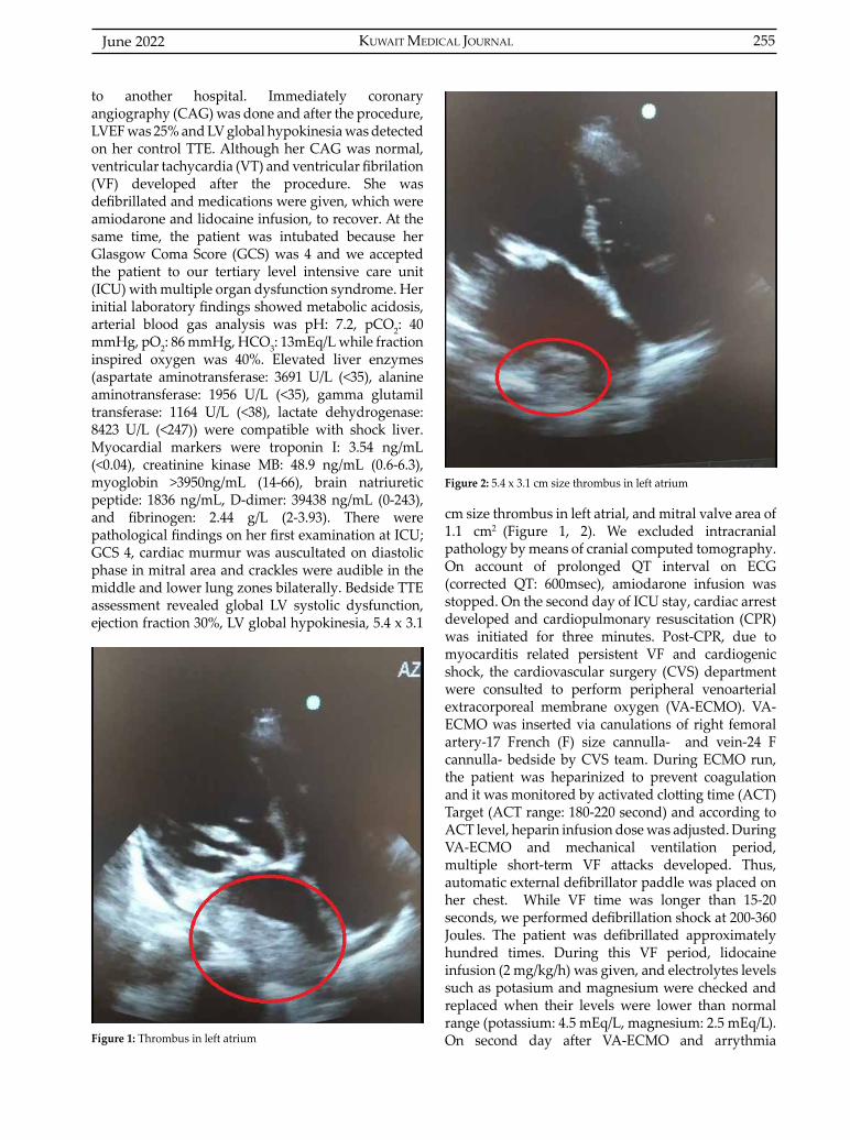
KUWAIT MEDICAL JOURNAL 255June 2022
to another hospital. Immediately coronary angiography (CAG) was done and after the procedure, LVEF was 25% and LV global hypokinesia was detected on her control TTE. Although her CAG was normal, ventricular tachycardia (VT) and ventricular fibrilation (VF) developed after the procedure. She was defibrillated and medications were given, which were amiodarone and lidocaine infusion, to recover. At the same time, the patient was intubated because her Glasgow Coma Score (GCS) was 4 and we accepted the patient to our tertiary level intensive care unit (ICU) with multiple organ dysfunction syndrome. Her initial laboratory findings showed metabolic acidosis, arterial blood gas analysis was pH: 7.2, pCO2: 40 mmHg, pO2: 86 mmHg, HCO3: 13mEq/L while fraction inspired oxygen was 40%. Elevated liver enzymes (aspartate aminotransferase: 3691 U/L (<35), alanine aminotransferase: 1956 U/L (<35), gamma glutamil transferase: 1164 U/L (<38), lactate dehydrogenase: 8423 U/L (<247)) were compatible with shock liver. Myocardial markers were troponin I: 3.54 ng/mL (<0.04), creatinine kinase MB: 48.9 ng/mL (0.6-6.3), myoglobin >3950ng/mL (14-66), brain natriuretic peptide: 1836 ng/mL, D-dimer: 39438 ng/mL (0-243), and fibrinogen: 2.44 g/L (2-3.93). There were pathological findings on her first examination at ICU; GCS 4, cardiac murmur was auscultated on diastolic phase in mitral area and crackles were audible in the middle and lower lung zones bilaterally. Bedside TTE assessment revealed global LV systolic dysfunction, ejection fraction 30%, LV global hypokinesia, 5.4 x 3.1
Fígure 1: Thrombus in left atrium
Figure 2: 5.4 x 3.1 cm size thrombus in left atrium
cm size thrombus in left atrial, and mitral valve area of 1.1 cm2 (Figure 1, 2). We excluded intracranial pathology by means of cranial computed tomography. On account of prolonged QT interval on ECG (corrected QT: 600msec), amiodarone infusion was stopped. On the second day of ICU stay, cardiac arrest developed and cardiopulmonary resuscitation (CPR) was initiated for three minutes. Post-CPR, due to myocarditis related persistent VF and cardiogenic shock, the cardiovascular surgery (CVS) department were consulted to perform peripheral venoarterial extracorporeal membrane oxygen (VA-ECMO). VA-ECMO was inserted via canulations of right femoral artery-17 French (F) size cannulla- and vein-24 F cannulla- bedside by CVS team. During ECMO run, the patient was heparinized to prevent coagulation and it was monitored by activated clotting time (ACT) Target (ACT range: 180-220 second) and according to ACT level, heparin infusion dose was adjusted. During VA-ECMO and mechanical ventilation period, multiple short-term VF attacks developed. Thus, automatic external defibrillator paddle was placed on her chest. While VF time was longer than 15-20 seconds, we performed defibrillation shock at 200-360 Joules. The patient was defibrillated approximately hundred times. During this VF period, lidocaine infusion (2 mg/kg/h) was given, and electrolytes levels such as potasium and magnesium were checked and replaced when their levels were lower than normal range (potassium: 4.5 mEq/L, magnesium: 2.5 mEq/L). On second day after VA-ECMO and arrythmia
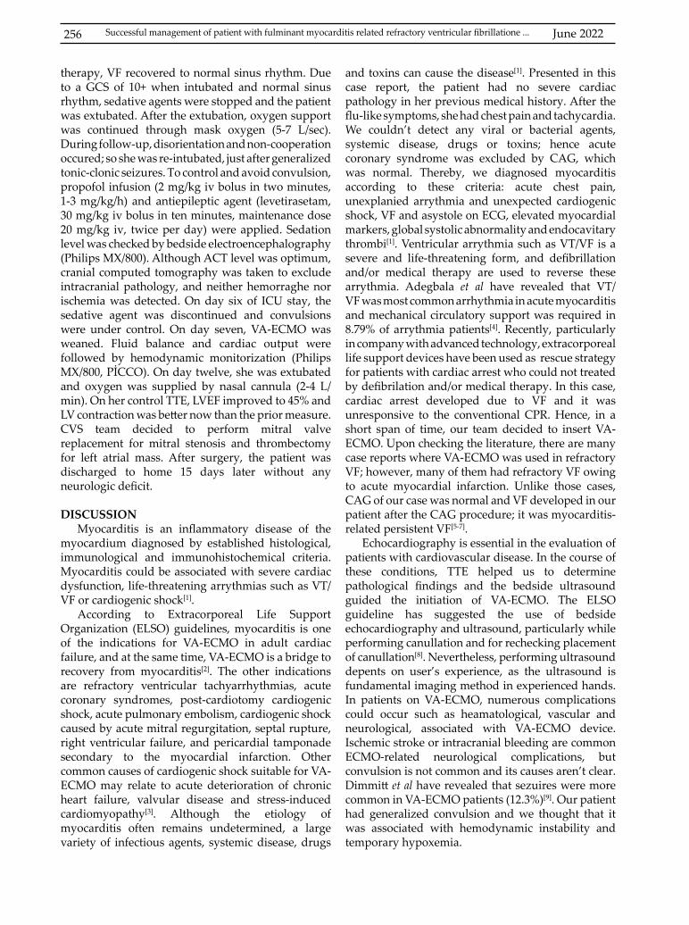
June 2022256
therapy, VF recovered to normal sinus rhythm. Due to a GCS of 10+ when intubated and normal sinus rhythm, sedative agents were stopped and the patient was extubated. After the extubation, oxygen support was continued through mask oxygen (5-7 L/sec). During follow-up, disorientation and non-cooperation occured; so she was re-intubated, just after generalized tonic-clonic seizures. To control and avoid convulsion, propofol infusion (2 mg/kg iv bolus in two minutes, 1-3 mg/kg/h) and antiepileptic agent (levetirasetam, 30 mg/kg iv bolus in ten minutes, maintenance dose 20 mg/kg iv, twice per day) were applied. Sedation level was checked by bedside electroencephalography (Philips MX/800). Although ACT level was optimum, cranial computed tomography was taken to exclude intracranial pathology, and neither hemorraghe nor ischemia was detected. On day six of ICU stay, the sedative agent was discontinued and convulsions were under control. On day seven, VA-ECMO was weaned. Fluid balance and cardiac output were followed by hemodynamic monitorization (Philips MX/800, PİCCO). On day twelve, she was extubated and oxygen was supplied by nasal cannula (2-4 L/min). On her control TTE, LVEF improved to 45% and LV contraction was better now than the prior measure. CVS team decided to perform mitral valve replacement for mitral stenosis and thrombectomy for left atrial mass. After surgery, the patient was discharged to home 15 days later without any neurologic deficit.
DISCUSSIONMyocarditis is an inflammatory disease of the
myocardium diagnosed by established histological, immunological and immunohistochemical criteria. Myocarditis could be associated with severe cardiac dysfunction, life-threatening arrythmias such as VT/VF or cardiogenic shock[1].
According to Extracorporeal Life Support Organization (ELSO) guidelines, myocarditis is one of the indications for VA-ECMO in adult cardiac failure, and at the same time, VA-ECMO is a bridge to recovery from myocarditis[2]. The other indications are refractory ventricular tachyarrhythmias, acute coronary syndromes, post-cardiotomy cardiogenic shock, acute pulmonary embolism, cardiogenic shock caused by acute mitral regurgitation, septal rupture, right ventricular failure, and pericardial tamponade secondary to the myocardial infarction. Other common causes of cardiogenic shock suitable for VA-ECMO may relate to acute deterioration of chronic heart failure, valvular disease and stress-induced cardiomyopathy[3]. Although the etiology of myocarditis often remains undetermined, a large variety of infectious agents, systemic disease, drugs
and toxins can cause the disease[1]. Presented in this case report, the patient had no severe cardiac pathology in her previous medical history. After the flu-like symptoms, she had chest pain and tachycardia. We couldn’t detect any viral or bacterial agents, systemic disease, drugs or toxins; hence acute coronary syndrome was excluded by CAG, which was normal. Thereby, we diagnosed myocarditis according to these criteria: acute chest pain, unexplanied arrythmia and unexpected cardiogenic shock, VF and asystole on ECG, elevated myocardial markers, global systolic abnormality and endocavitary thrombi[1]. Ventricular arrythmia such as VT/VF is a severe and life-threatening form, and defibrillation and/or medical therapy are used to reverse these arrythmia. Adegbala et al have revealed that VT/VF was most common arrhythmia in acute myocarditis and mechanical circulatory support was required in 8.79% of arrythmia patients[4]. Recently, particularly in company with advanced technology, extracorporeal life support devices have been used as rescue strategy for patients with cardiac arrest who could not treated by defibrilation and/or medical therapy. In this case, cardiac arrest developed due to VF and it was unresponsive to the conventional CPR. Hence, in a short span of time, our team decided to insert VA-ECMO. Upon checking the literature, there are many case reports where VA-ECMO was used in refractory VF; however, many of them had refractory VF owing to acute myocardial infarction. Unlike those cases, CAG of our case was normal and VF developed in our patient after the CAG procedure; it was myocarditis-related persistent VF[5-7].
Echocardiography is essential in the evaluation of patients with cardiovascular disease. In the course of these conditions, TTE helped us to determine pathological findings and the bedside ultrasound guided the initiation of VA-ECMO. The ELSO guideline has suggested the use of bedside echocardiography and ultrasound, particularly while performing canullation and for rechecking placement of canullation[8]. Nevertheless, performing ultrasound depents on user’s experience, as the ultrasound is fundamental imaging method in experienced hands. In patients on VA-ECMO, numerous complications could occur such as heamatological, vascular and neurological, associated with VA-ECMO device. Ischemic stroke or intracranial bleeding are common ECMO-related neurological complications, but convulsion is not common and its causes aren’t clear. Dimmitt et al have revealed that sezuires were more common in VA-ECMO patients (12.3%)[9]. Our patient had generalized convulsion and we thought that it was associated with hemodynamic instability and temporary hypoxemia.
Successful management of patient with fulminant myocarditis related refractory ventricular fibrillatione ...
KUWAIT MEDICAL JOURNAL 257June 2022
CONCLUSIONIn this case report, we would like to highlight a few
important points. First, bedside ultrasound plays a key role in ICU, either to diagnose cardiac pathology or as a guide to invasive procedures and for the follow up of the patients’ condition fastly and quickly. Secondly, VF could be persistent, unresponsive to medical treatment and defibrillation, leading to a life-threatening condition. However, physicans should not give up treatment and should think to apply VA-ECMO as a rescue therapy for both myocarditis-related severe arryhtymia and cardiogenic shock.
ACKNOWLEDGMENTPatient Consent: Written patient consent was
obtain from the patient.Conflict of Interest: None Financial Disclosure: None
REFERENCES
1. Caforio ALP, Pankuweit S, Arbustini E, Basso C, Gimeno-Blanes J, Felix SB, et al. Current state of knowledge on aetiology, diagnosis, management,and theraphy ofmyocarditis: a position statement of Euopean Society of Cardiology Working Groupon Myocardial and Pericardial Disease. Eur Heart J 2013; 34(33):2636-48.
2. Extracorporeal Life Support Organization (ELSO) Guidelines for Adult Cardiac Failure https://www.elso.org/Portals/0/IGD /Archive/FileManager/e 7 6 e f 7 8 e a b c u s e r s s h y e r d o c u m e n t s e l s o guidelinesforadultcardiacfailure1.3. May2015 pdf
3. Meuwese CL, Ramjankhan FZ, Braithwaite SA, de Jonge N, de Jong M, Buijsrogge MP, et al. Extracorporeal life support in cardiogenic shock: indications and management in current practice. Neth Heart J 2018; 26(2):58-66.
4. Adegbala O, Olagoke O, Akintoye E, Adejumo AC, Oluwole A, Jara C, et al. Predictors, burden, and the ımpact of arrhythmia on patients admitted for acute myocarditis. Am J Cardiol 2019; 123(1):139-44.
5. Fux T, Svenarud P, Grinnemo KH. Extracorporeal membrane oxygenation as a rescue of intractable ventricular fibrillation and bridge to heart transplantation. Eur J Heart Fail 2010; 12:301-4.
6. Golian M, Freed D, Jassal DS, Ravandi A. Successful cardiac resuscitation with extracorporeal membrane oxygenation in the setting of persistent ventricular fibrillation: a case report. BMC Research Notes 2014; 782.
7. Chung JW, Chang WH, Hyon MS. Extracorporeal life support after prolonged resuscitation for in-hospital cardiac arrest due to refractory ventricular fibrillation: Two cases resulting in a full recovery. Korean Circ J 2012; 42:423-26.
8. Ultrasound Guidance for Extra-corporeal Membrane Oxygenation General Guidelines. Extracorporeal Life Support Organisation, 2015 (Accessed May 2015, h t t p s : / / w w w . e l s o . o r g / P o r t a l s / 0 / F i l e s / e l s o _Ultrasoundguideance_ecmogeneral_guidelines_May2015.pdf)
9. Dimmitt RA, Moss RL, Rhine WD, Benitz WE, Henry MC, Vanmeurs KP. Venoarterial versus venovenous extracorporeal membrane oxygenation in congenital diaphragmatic hernia: The extracorporeal life support organization registry, 1990-1999. Journal of Pediatric Surgery 2001; 36(8):1199-204.
KUWAIT MEDICAL JOURNAL June 2022258
Address correspondence to:Dr. Aarti Chadha, MD, FRCPath, Clinical Microbiologist, Department of Microbiology, Mubarak Al Kabeer Hospital, Jabriya, Kuwait. E-mail: [email protected]
Kuwait Medical Journal 2022; 54 (2): 258 - 261
First case of bloodstream infection caused by Clostridium saccharolyticumin in the Middle East
ABSTRACT
Aarti Chadha, Wafaa Jamal, Vincent O RotimiDepartment of Microbiology, Mubarak Al Kabeer Hospital, Jabriya, Kuwait
In this report, we describe a rare case of bacteremia caused by an unusual Clostridium species, Clostridium saccharolyticum, an environmental anaerobic spore-forming gram-variable
bacillus. The clinical significance in humans remains poorly known. This is the first documented report of human infection by this organism in the Middle East.
KEY WORDS: clostridium, emerging pathogen, environmental flora
Case Report
INTRODUCTION Clostridium species are spore-forming, almost
always Gram-positive bacilli, though a few gram variable anaerobic bacilli have been described. They are ubiquitous in the environment and some species are part of the human gut microbiota. The most common Clostridium species in the intestinal microbiota are those from cluster IV such as Clostridium leptum and from cluster XIVa (Clostridium coccoides, Clostridium celerecrescens, Clostridium sphenoides, Clostridiumclostridioforme)[1]. In this communication, we report a rare case of severe bacteremia caused by C. saccharolyticum in our hospital, representing the first of such reported infection in this region.
CASE REPORT A 45-year-old Indian female presented to the
emergency room of Mubarak Al Kabeer hospital, Jabriya, Kuwait on September 21, 2019 with a four-day history of vomiting, constipation, abdominal pain mainly in the left iliac fossa and left flank. There was a history of laparotomy done six years back for a ruptured ovarian cyst as well as closure of an atrial septal defect done four years back in India. On examination, she was afebrile and vitally stable, with tenderness over the left lower quadrant of the abdomen. A computed tomography scan was done for
the patient, which suggested adhesive small bowel obstruction. She was admitted for emergency laparotomy and lysis of the adhesions. The surgery was complicated by the development of multiple enterotomies which were closed with vicryl. Intraoperative findings included multiple bands looping around the small and large intestines and a dilated proximal bowel. Post-operatively, she was shifted to the intensive care unit for observation and started on intravenous piperacillin-tazobactam 4.5g eight-hourly. The following day, she became hypotensive with severe tachypnea and complained of severe abdominal pain at the site of the wound. In addition, she had not passed any flatus or feces. Blood gas analysis showed metabolic acidosis with anion gap of 40 IU/L. A central internal jugular line was fixed for nor-epinephrine 1 μg/kg/min infusion and her antibiotic therapy was changed to meropenem 1g eight-hourly. At this time, a repeat computed tomography scan showed free intraperitoneal air and fluid levels suggestive of perforation of the bowel, bilateral pleural effusion and underlying collapse of the lung bases. These findings prompted an immediate second laparotomy. Intra-operatively, there were two perforations at the site of previously stitched enterotomies which were leaking air along with turbid fluid in the pelvis. These perforations were closed with
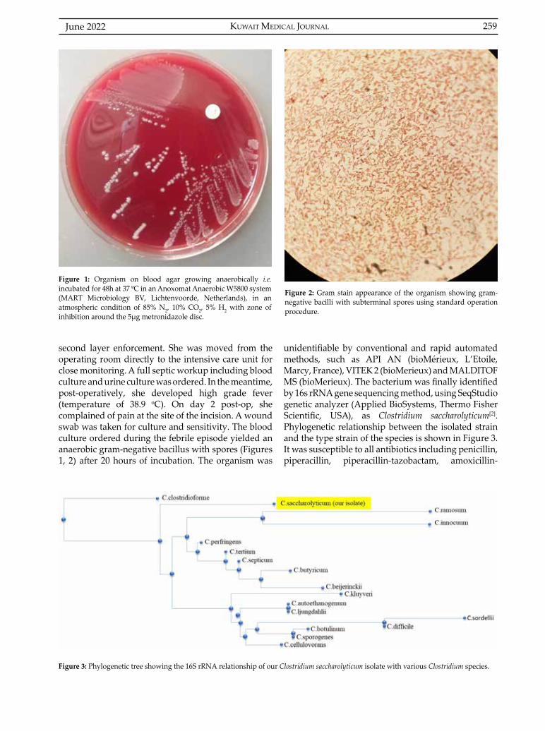
KUWAIT MEDICAL JOURNAL 259June 2022
second layer enforcement. She was moved from the operating room directly to the intensive care unit for close monitoring. A full septic workup including blood culture and urine culture was ordered. In the meantime, post-operatively, she developed high grade fever (temperature of 38.9 oC). On day 2 post-op, she complained of pain at the site of the incision. A wound swab was taken for culture and sensitivity. The blood culture ordered during the febrile episode yielded an anaerobic gram-negative bacillus with spores (Figures 1, 2) after 20 hours of incubation. The organism was
unidentifiable by conventional and rapid automated methods, such as API AN (bioMérieux, L’Etoile, Marcy, France), VITEK 2 (bioMerieux) and MALDITOF MS (bioMerieux). The bacterium was finally identified by 16s rRNA gene sequencing method, using SeqStudio genetic analyzer (Applied BioSystems, Thermo Fisher Scientific, USA), as Clostridium saccharolyticum[2]. Phylogenetic relationship between the isolated strain and the type strain of the species is shown in Figure 3. It was susceptible to all antibiotics including penicillin, piperacillin, piperacillin-tazobactam, amoxicillin-
Figure 1: Organism on blood agar growing anaerobically i.e. incubated for 48h at 37 ºC in an Anoxomat Anaerobic W5800 system (MART Microbiology BV, Lichtenvoorde, Netherlands), in an atmospheric condition of 85% N2, 10% CO2, 5% H2 with zone of inhibition around the 5μg metronidazole disc.
Figure 2: Gram stain appearance of the organism showing gram-negative bacilli with subterminal spores using standard operation procedure.
Figure 3: Phylogenetic tree showing the 16S rRNA relationship of our Clostridium saccharolyticum isolate with various Clostridium species.
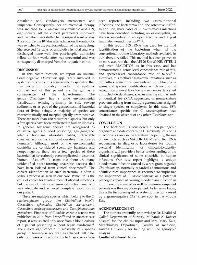
June 2022260
clavulanic acid, clindamycin, meropenem and imipenem. Consequently, her antimicrobial therapy was switched to IV amoxicillin-clavulanic acid 1.2g eight-hourly. All the clinical parameters improved, and the patient was shifted to the surgical ward on day 3 post-op. On the 10th day after admission, the antibiotic was switched to the oral formulation of the same drug. She received 18 days of antibiotics in total and was discharged home well. Her post-hospital admission follow-up four weeks after was uneventful and was consequently discharged from the outpatient clinic.
DISCUSSIONIn this communication, we report an unusual
Gram-negative Clostridium spp. rarely involved in systemic infections. It is conceivable to speculate that this bacterium probably invaded the systemic compartment of this patient via the gut as a consequence of the laparotomies. The genus Clostridium has a wide environmental distribution, existing primarily in soil, sewage sediments or as part of the gastrointestinal bacterial flora of living beings. A majority of species are characteristically and morphologically gram-positive. There are more than 160 recognized species, but only a few species have been implicated in human illnesses. In general, toxigenic species are recognized as causative agents of food poisoning, gas gangrene, tetanus, botulism, ulcerative colitis, intractable diarrhea, septicemia, and postoperative infections in humans[3]. Although most of the environmental clostridia are considered seemingly harmless and nonpathogenic, there are a few species of these bacteria that have already been implicated in a serious human infection[4]. It seems that there are many unidentified spore-forming anaerobic bacteria that have been isolated from clinical specimens[5]. The correct identification of such bacterium is often a tedious process as seen in our case. Penicillin is the drug of choice for treating most clostridial infections, but the use of high dose amoxicillin-clavulanic acid was adequate and achieved complete resolution in our patient.
There are multiple species which belong to the C. saccharolyticum group like Clostridium indolis, Clostridium sphenoides, Clostridium celerecrescens, Clostridium methoxybenzovorans and Desulfotomaculum guttoideum. First case of C. indolis chronic osteitis was published in 2016 from France[5] and in another case report, it was isolated only once from a blood culture of a patient presenting without sepsis syndrome[6]. The clinical significance of C. saccharolyticum species group in humans is not well established. Till date, only four cases of infections due to C. sphenoides have
been reported, including two gastro-intestinal infections, one bacteremia and one osteomyelitis[7–10]. In addition, three cases of C. celerecrescens infections have been described including an osteomyelitis, an abscess secondary to an open fracture and a post traumatic wound infection[11,12].
In this report, 16S rRNA was used for the final identification of the bacterium when all the conventional routine laboratory methods available in our laboratory failed. This method has been proven to be more accurate than the API 20 E or 20 NE, VITEK-2 and even MALDITOF as in this case, and has demonstrated a genus-level concordance rate of 96% and species-level concordance rate of 87.5%[2,13]. However, this method has its own limitations, such as difficulties sometimes encountered in obtaining a genus and species identification, which include the recognition of novel taxa, too few sequences deposited in nucleotide databases, species sharing similar and/or identical 16S rRNA sequences, or nomenclature problems arising from multiple genomovars assigned to single species or complexes. In this case, 88% concordance specific for C. saccharolyticum was obtained in the absence of any other Clostridium spp.
CONCLUSIONThe bacterium is considered a non-pathogenic
organism and data concerning C. saccharolyticum or its infections is scarce in the literature. Hopefully, the use of new tools, such as MALDI-TOF MS and 16S rRNA sequencing, in diagnostic laboratories for routine bacterial identification of difficult-to-identify organisms will provide a better understanding of the clinical significance of some clostridia in human infections. Our case report highlights a unique bloodstream infection caused by a rare gram-negative Clostridium sp. normally regarded as innocuous and of little clinical importance. It is pertinent to emphasize the importance of C. saccharolyticum as a potential pathogen capable of causing bloodstream infection in immuno-compromized as well as immuno-competent patients was the case of our patient. As far as we know, this is the first case report of systemic infection caused by a gram-negative Clostridium spp. in the Middle East.
ACKNOWLEDGMENTThe authors gratefully acknowledge Dr. Khalid Al
Qallaf, Department of Surgery, Mubarak Al Kabeer hospital for the clinical input and Mrs. Mary Kala, Microbiology Department, Faculty of medicine, Kuwait University for helping with the genotypic analysis.Conflict of interest: None
First case of bloodstream infection caused by Clostridium saccharolyticumin in the Middle East
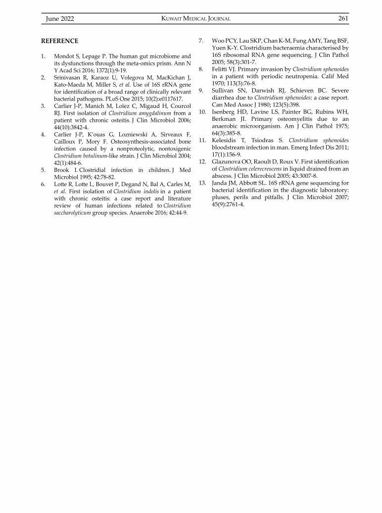
KUWAIT MEDICAL JOURNAL 261June 2022
REFERENCE
1. Mondot S, Lepage P. The human gut microbiome and its dysfunctions through the meta-omics prism. Ann N Y Acad Sci 2016; 1372(1):9-19.
2. Srinivasan R, Karaoz U, Volegova M, MacKichan J, Kato-Maeda M, Miller S, et al. Use of 16S rRNA gene for identification of a broad range of clinically relevant bacterial pathogens. PLoS One 2015; 10(2):e0117617.
3. Carlier J-P, Manich M, Loïez C, Migaud H, Courcol RJ. First isolation of Clostridium amygdalinum from a patient with chronic osteitis. J Clin Microbiol 2006; 44(10):3842-4.
4. Carlier J-P, K’ouas G, Lozniewski A, Sirveaux F, Cailloux P, Mory F. Osteosynthesis-associated bone infection caused by a nonproteolytic, nontoxigenic Clostridium botulinum-like strain. J Clin Microbiol 2004; 42(1):484-6.
5. Brook I. Clostridial infection in children. J Med Microbiol 1995; 42:78-82.
6. Lotte R, Lotte L, Bouvet P, Degand N, Bal A, Carles M, et al. First isolation of Clostridium indolis in a patient with chronic osteitis: a case report and literature review of human infections related to Clostridium saccharolyticum group species. Anaerobe 2016; 42:44-9.
7. Woo PCY, Lau SKP, Chan K-M, Fung AMY, Tang BSF, Yuen K-Y. Clostridium bacteraemia characterised by 16S ribosomal RNA gene sequencing. J Clin Pathol 2005; 58(3):301-7.
8. Felitti VJ. Primary invasion by Clostridium sphenoides in a patient with periodic neutropenia. Calif Med 1970; 113(3):76-8.
9. Sullivan SN, Darwish RJ, Schieven BC. Severe diarrhea due to Clostridium sphenoides: a case report. Can Med Assoc J 1980; 123(5):398.
10. Isenberg HD, Lavine LS, Painter BG, Rubins WH, Berkman JI. Primary osteomyelitis due to an anaerobic microorganism. Am J Clin Pathol 1975; 64(3):385-8.
11. Kelesidis T, Tsiodras S. Clostridium sphenoides bloodstream infection in man. Emerg Infect Dis 2011; 17(1):156-9.
12. Glazunova OO, Raoult D, Roux V. First identification of Clostridium celerecrescens in liquid drained from an abscess. J Clin Microbiol 2005; 43:3007-8.
13. Janda JM, Abbott SL. 16S rRNA gene sequencing for bacterial identification in the diagnostic laboratory: pluses, perils and pitfalls. J Clin Microbiol 2007; 45(9):2761-4.
KUWAIT MEDICAL JOURNAL June 2022262
Address correspondence to:Abdulkadir Yasir Bahar, MD, Department of Pathology, Kahramanmaraş Sütçü İmam University School of Medicine, Kahramanmaraş, Turkey. Tel: +90 5313819711; E-mail: [email protected]
Kuwait Medical Journal 2022; 54 (2): 262 - 268
Adenomatoid tumor of the adrenal gland synchronous with clear cell papillary renal cell
carcinoma: Report of an unusual case
ABSTRACT
Abdulkadir Yasir Bahar1, Murat Sahin2
1Department of Pathology, Kahramanmaraş Sütçü İmam University School of Medicine, Turkey 2Department of Internal Medicine, Kahramanmaraş Sütçü İmam University School of Medicine, Endocrinology Subdivision,
Turkey
Adenomatoid tumors are benign neoplasms with a mesothelial origin that may be seen especially in the genital system and in many organs, but rarely in the adrenal gland.
A male aged 30 years presented to the gastroenterology clinic complaining of abdominal pain and diarrhea. In abdominal computed tomography, a well-circumscribed hypodense (+50 HU) lesion was observed in the right adrenal gland with a size of 37x25x15 mm. In the upper pole of the right kidney, a well-defined solid mass with a size of 19x15x14 mm was observed that showed contrast involvement in the contrast-enhanced series and a 33 HU
density in the contrast-unenhanced series. Right partial nephrectomy and right adrenalectomy were applied to differentiate between non- functional adrenal mass and metastatic carcinoma. The renal tumor was diagnosed as clear cell papillary renal cell carcinoma (CCPRCC) and the adrenal mass was diagnosed as an adenomatoid tumor.
CCPRCC synchronous with adrenal adenomatoid tumor is unusual. Reviewing the literature for adenomatoid tumors, we report a case with an adenomatoid adrenal tumor accompanied by a CCPRCC. To the best of our knowledge, this is the first study in English literature reporting this kind of co-occurrence.
KEY WORDS: adenomatoid tumor, adrenal gland, clear cell papillary renal cell carcinoma
Case Report
INTRODUCTIONAdenomatoid tumors (AT) are benign neoplasms
with a mesothelial origin mostly seen in the genital system and in many organs, including the adrenal gland[1-3].
While some cases are reported by abdominal pain and hypertension, most of the patients are detected incidentally as asymptomatic conditions during routine imaging tests.
Clear cell papillary renal cell carcinoma (CCPRCC) is a relatively uncommon renal neoplasm characterized by an indolent biological behavior, described recently in the World Health Organization 2016 renal tumor classification.
In this study, summarizing the literature for AT, we report a case with an adenomatoid adrenal tumor accompanied by a CCPRCC.
CASE REPORTClinical history and radiological findings
A male patient aged 30 years presented to the gastroenterology clinic complaining of abdominal pain for a long time and diarrhea for the last week. The patient had no history of illness or enduring medicine usage. After the clinical evaluation, a contrast-enhanced abdominal computed tomography was ordered with the preliminary diagnosis of chronic pancreatitis. In computed tomography, a
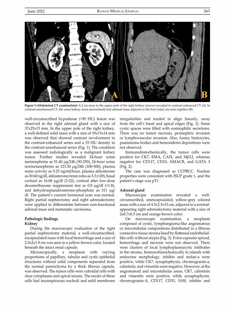
KUWAIT MEDICAL JOURNAL 263June 2022
well-circumscribed hypodense (+50 HU) lesion was observed in the right adrenal gland with a size of 37x25x15 mm. In the upper pole of the right kidney, a well-defined solid mass with a size of 19x15x14 mm was observed that showed contrast involvement in the contrast-enhanced series and a 33 HU density in the contrast-unenhanced series (Fig. 1). The condition was assessed radiologically as a malignant kidney tumor. Further studies revealed 24-hour urine metanephrine as 51.40 μg/24h (50-250), 24-hour urine normetanephrine as 123.30 μg/24h (100-500), plasma renin activity as 9.25 ng/ml/hour, plasma aldosterone as 30.60 ng/dl, aldosterone/renin ratio as 3.3 (<20), basal cortisol as 16.68 μg/dl (3-22), cortisol after low-dose dexamethasone suppression test as 0.8 μg/dl (<1.8), and dehydroepiandrosterone-phosphate as 311 μg/dl. The patient’s current hormonal scan was normal. Right partial nephrectomy and right adrenalectomy were applied to differentiate between non-functional adrenal mass and metastatic carcinoma.
Pathologic findingsKidney
During the macroscopic evaluation of the right partial nephrectomy material, a well-circumscribed, encapsulated mass with focal hemorrhage and a size of 2.5x2x1.9 cm was seen in a yellow-brown color, located beneath the intact renal capsule.
Microscopically, a neoplasm with varying proportions of papillary, tubular and cystic epithelial structures without solid components separated from the normal parenchyma by a thick fibrous capsule, was observed. The tumor cells were cuboidal cells with clear cytoplasms and apical snouts. The nuclei of these cells had inconspicuous nucleoli and mild membrane
irregularities and tended to align linearly, away from the cell’s basal and apical edges (Fig. 2). Some cystic spaces were filled with eosinophilic secretions. There was no tumor necrosis, perinephric invasion or lymphovascular invasion. Also, foamy histiocytes, psammoma bodies and hemosiderin depositions were not observed.
Immunohistochemically, the tumor cells were positive for CK7, EMA, CAIX, and 34β12, whereas negative for CD117, CD10, AMACR, and GATA 3 (Fig. 2).
The case was diagnosed as CCPRCC. Nuclear properties were consistent with ISUP grade 1, and the patient’s stage was pT1.
Adrenal glandMacroscopic examination revealed a well-
circumscribed, unencapsulated, yellow-grey colored mass with a size of 4.5x2.5x15 cm, adjacent to a normal-appearing right adrenalectomy material with a size of 2x0.7x0.5 cm and orange-brown color.
On microscopic examination, a neoplasm composed of cystic, lymphangioma-like angiomatous or microtubular compositions distributed in a fibrous connective tissue stroma lined by flattened endothelial-like cells without atypia (Fig. 3). Extra-capsular spread, hemorrhage and necrosis were not observed. There were clusters of focal lymphoplasmacytic infiltrates in the stroma. Immunohistochemically in islands with endocrine morphology, inhibin and melan-a were positive, while CK7, synaptophysin, chromogranin-a, calretinin, and vimentin were negative. However, at the angiomatoid and microtubular areas, CK7, calretinin and vimentin were positive, while synaptophysin, chromogranin-A, CD117, CD31, S100, inhibin and
Figure 1 (Abdominal CT examination): A 2 cm mass in the upper pole of the right kidney (arrow) revealed in contrast-enhanced CT (A). In contrast-unenhanced CT, the same kidney mass (arrowhead) and adrenal mass adjacent to the liver (star) are seen together (B).
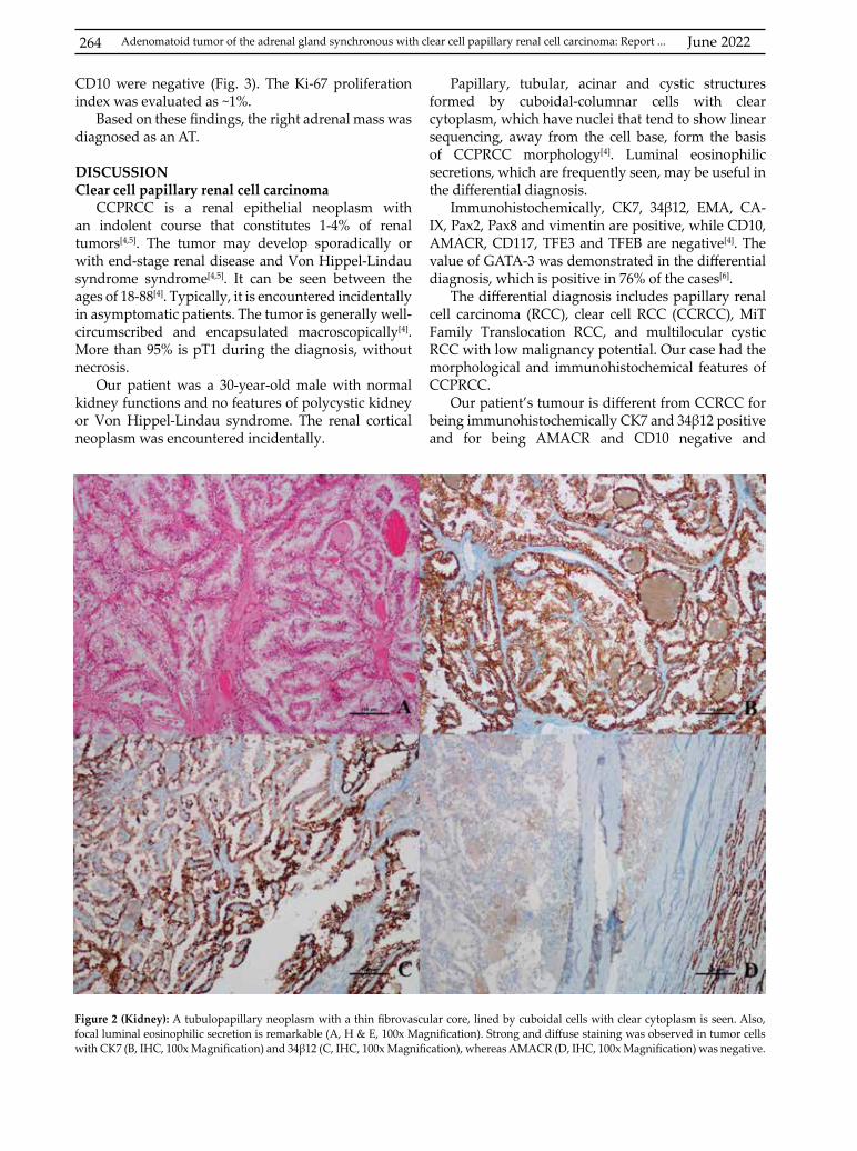
June 2022264 Adenomatoid tumor of the adrenal gland synchronous with clear cell papillary renal cell carcinoma: Report ...
Figure 2 (Kidney): A tubulopapillary neoplasm with a thin fibrovascular core, lined by cuboidal cells with clear cytoplasm is seen. Also, focal luminal eosinophilic secretion is remarkable (A, H & E, 100x Magnification). Strong and diffuse staining was observed in tumor cells with CK7 (B, IHC, 100x Magnification) and 34β12 (C, IHC, 100x Magnification), whereas AMACR (D, IHC, 100x Magnification) was negative.
CD10 were negative (Fig. 3). The Ki-67 proliferation index was evaluated as ~1%.
Based on these findings, the right adrenal mass was diagnosed as an AT.
DISCUSSIONClear cell papillary renal cell carcinoma
CCPRCC is a renal epithelial neoplasm with an indolent course that constitutes 1-4% of renal tumors[4,5]. The tumor may develop sporadically or with end-stage renal disease and Von Hippel-Lindau syndrome syndrome[4,5]. It can be seen between the ages of 18-88[4]. Typically, it is encountered incidentally in asymptomatic patients. The tumor is generally well-circumscribed and encapsulated macroscopically[4]. More than 95% is pT1 during the diagnosis, without necrosis.
Our patient was a 30-year-old male with normal kidney functions and no features of polycystic kidney or Von Hippel-Lindau syndrome. The renal cortical neoplasm was encountered incidentally.
Papillary, tubular, acinar and cystic structures formed by cuboidal-columnar cells with clear cytoplasm, which have nuclei that tend to show linear sequencing, away from the cell base, form the basis of CCPRCC morphology[4]. Luminal eosinophilic secretions, which are frequently seen, may be useful in the differential diagnosis.
Immunohistochemically, CK7, 34β12, EMA, CA-IX, Pax2, Pax8 and vimentin are positive, while CD10, AMACR, CD117, TFE3 and TFEB are negative[4]. The value of GATA-3 was demonstrated in the differential diagnosis, which is positive in 76% of the cases[6].
The differential diagnosis includes papillary renal cell carcinoma (RCC), clear cell RCC (CCRCC), MiT Family Translocation RCC, and multilocular cystic RCC with low malignancy potential. Our case had the morphological and immunohistochemical features of CCPRCC.
Our patient’s tumour is different from CCRCC for being immunohistochemically CK7 and 34β12 positive and for being AMACR and CD10 negative and
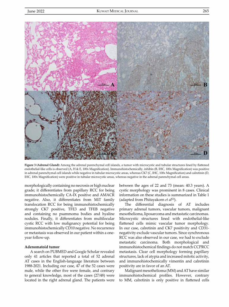
KUWAIT MEDICAL JOURNAL 265June 2022
morphologically containing no necrosis or high nuclear grade; it differentiates from papillary RCC for being immunohistochemically CA-IX positive and AMACR negative. Also, it differentiates from MiT family translocation RCC for being immunohistochemically strongly CK7 positive, TFE3 and TFEB negative and containing no psammoma bodies and hyaline nodules. Finally, it differentiates from multilocular cystic RCC with low malignancy potential for being immunohistochemically CD10 negative. No recurrence or metastasis was observed in our patient within a one-year follow-up.
Adenomatoid tumorA search on PUBMED and Google Scholar revealed
only 41 articles that reported a total of 52 adrenal AT cases in the English-language literature between 1988-2021. Including our case, 47 of the 52 cases were male, while the other five were female, and contrary to general knowledge, most of the cases (27/48) were located in the right adrenal gland. The patients were
between the ages of 22 and 73 (mean: 40.3 years). A cystic morphology was prominent in 8 cases. Clinical information on these studies is summarized in Table 1 (adapted from Phitayakorn et al[2]).
The differential diagnosis of AT includes primary adrenal tumors, vascular tumors, malignant mesothelioma, liposarcoma and metastatic carcinomas. Microcystic structures lined with endothelial-like flattened cells mimic vascular tumor morphology. In our case, calretinin and CK7 positivity and CD31-negativity exclude vascular tumors. Since synchronous RCC was also observed in our case, we had to exclude metastatic carcinoma. Both morphological and immunohistochemical findings do not match CCPRCC metastasis. Clear cell morphology forming papillary structures, lack of atypia and increased mitotic activity, and immunohistochemically vimentin and calretinin positivity are in favor of an AT.
Malignant mesothelioma (MM) and AT have similar immunohistochemical profiles. However, contrary to MM, calretinin is only positive in flattened cells
Figure 3 (Adrenal Gland): Among the adrenal parenchymal cell islands, a tumor with microcystic and tubular structures lined by flattened endothelial-like cells is observed (A, H & E, 100x Magnification). Immunohistochemically, inhibin (B, IHC, 100x Magnification) was positive in adrenal parenchymal cell islands while negative in tubular microcystic areas, whereas CK7 (C, IHC, 100x Magnification) and calretinin (D, IHC, 100x Magnification) were positive in tubular microcystic areas, whereas negative in the adrenal parenchymal cell areas.
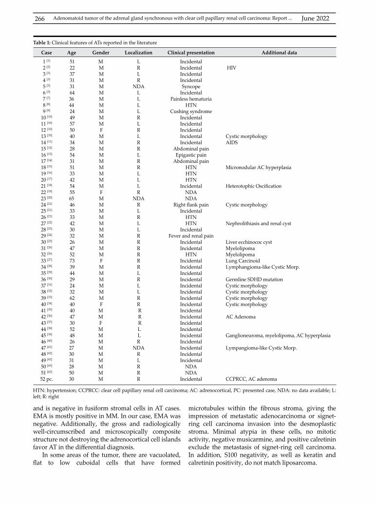
June 2022266
Case Age Gender Localization Clinical presentation Additional data
1 [1]
2 [2]
3 [3]
4 [3]
5 [3]
6 [3]
7 [7]
8 [8]
9 [9]
10 [10]
11 [10]
12 [10]
13 [10]
14 [11]
15 [12]
16 [13]
17 [14]
18 [15]
19 [16]
20 [17]
21 [18]
22 [19]
23 [20]
24 [21]
25 [21]
26 [21]
27 [22]
28 [23]
29 [24]
30 [25]
31 [26]
32 [26]
33 [27]
34 [28]
35 [29]
36 [30]
37 [31]
38 [32]
39 [33]
40 [34]
41 [35]
42 [36]
43 [37]
44 [38]
45 [39]
46 [40]
47 [41]
48 [42]
49 [42]
50 [43]
51 [43]
52 pc.
51 22 37 31 31 643644 24 49 57 50 40 34 28 54 31 51 33 42 54 5565 46 33 33 42 30 32 26 47 52 73 39 44 29 24 32 62 40 40 47 30 52 48 26273031285030
M M M M M M M M M M M F M M M M M M M M M F M M M M M M M M M M F M M M M M M FMMFMMMMMMMMM
LRLR
NDALLLLRLRLRRLRRLLLR
NDARLRLLRRRRRRLRLLRR R R R L LR
NDARLRRR
IncidentalIncidentalIncidentalIncidentalSyncope
IncidentalPainless hematuria
HTNCushing syndrome
IncidentalIncidentalIncidentalIncidentalIncidental
Abdominal painEpigastic pain
Abdominal painHTNHTNHTN
IncidentalNDANDA
Right flank painIncidental
HTNHTN
IncidentalFever and renal pain
IncidentalIncidental
HTNIncidentalIncidentalIncidentalIncidentalIncidentalIncidentalIncidentalIncidentalIncidentalIncidentalIncidentalIncidentalIncidentalIncidentalIncidentalIncidentalIncidental
NDANDA
Incidental
HIV Cystic morphologyAIDS Micronodular AC hyperplasia Heterotophic Oscification Cystic morphology Nephrolithiasis and renal cyst Liver ecchinococ cyst MyelolipomaMyelolipomaLung CarcinoidLymphangioma-like Cystic Morp. Germline SDHD mutationCystic morphologyCystic morphologyCystic morphologyCystic morphology AC Adenoma Ganglioneuroma, myelolipoma, AC hyperplasia Lympangioma-like Cystic Morp.
CCPRCC, AC adenoma
HTN: hypertension; CCPRCC: clear cell papillary renal cell carcinoma; AC: adrenocortical, PC: presented case, NDA: no data available; L: left; R: right
Table 1: Clinical features of ATs reported in the literature
and is negative in fusiform stromal cells in AT cases. EMA is mostly positive in MM. In our case, EMA was negative. Additionally, the gross and radiologically well-circumscribed and microscopically composite structure not destroying the adrenocortical cell islands favor AT in the differential diagnosis.
In some areas of the tumor, there are vacuolated, flat to low cuboidal cells that have formed
microtubules within the fibrous stroma, giving the impression of metastatic adenocarcinoma or signet-ring cell carcinoma invasion into the desmoplastic stroma. Minimal atypia in these cells, no mitotic activity, negative musicarmine, and positive calretinin exclude the metastasis of signet-ring cell carcinoma. In addition, S100 negativity, as well as keratin and calretinin positivity, do not match liposarcoma.
Adenomatoid tumor of the adrenal gland synchronous with clear cell papillary renal cell carcinoma: Report ...
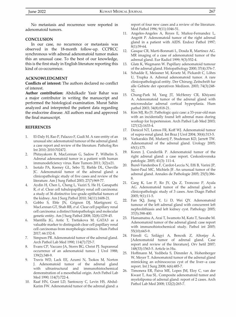
KUWAIT MEDICAL JOURNAL 267June 2022
No metastasis and recurrence were reported in adenomatoid tumors.
CONCLUSIONIn our case, no recurrence or metastasis was
observed in the 18-month follow-up. CCPRCC synchronous with adrenal adenomatoid tumor makes this an unusual case. To the best of our knowledge, this is the first study in English literature reporting this kind of co-occurrence.
ACKNOWLEDGMENTConflicts of interest: The authors declared no conflict of interest.Author contribution: Abdulkadir Yasir Bahar was a major contributor in writing the manuscript and performed the histological examination. Murat Sahin analyzed and interpreted the patient data regarding the endocrine disease. All authors read and approved the final manuscript.
REFERENCES
1. El-Daly H, Rao P, Palazzo F, Gudi M. A rare entity of an unusual site: adenomatoid tumour of the adrenal gland: a case report and review of the literature. Patholog Res Int 2010; 2010:702472.
2. Phitayakorn R, MacLennan G, Sadow P, Wilhelm S. Adrenal adenomatoid tumor in a patient with human immunodeficiency virus. Rare Tumors 2011; 3(2):e21.
3. Isotalo PA, Keeney GL, Sebo TJ, Riehle DL, Cheville JC. Adenomatoid tumor of the adrenal gland: a clinicopathologic study of five cases and review of the literature. Am J Surg Pathol 2003; 27(7):969-77.
4. Aydin H, Chen L, Cheng L, Vaziri S, He H, Ganapathi R, et al. Clear cell tubulopapillary renal cell carcinoma: a study of 36 distinctive low-grade epithelial tumors of the kidney. Am J Surg Pathol 2010; 34(11):1608-21.
5. Gobbo S, Eble JN, Grignon DJ, Martignoni G, MacLennan GT, Shah RB, et al. Clear cell papillary renal cell carcinoma: a distinct histopathologic and molecular genetic entity. Am J Surg Pathol 2008; 32(8):1239-45.
6. Mantilla JG, Antic T, Tretiakova M. GATA3 as a valuable marker to distinguish clear cell papillary renal cell carcinomas from morphologic mimics. Hum Pathol 2017; 66:152-8.
7. Simpson PR. Adenomatoid tumor of the adrenal gland. Arch Pathol Lab Med 1990; 114(7):725-7.
8. Evans CP, Vaccaro JA, Storrs BG, Christ PJ. Suprarenal occurrence of an adenomatoid tumor. J Urol 1988; 139(2):348-9.
9. Travis WD, Lack EE, Azumi N, Tsokos M, Norton J. Adenomatoid tumor of the adrenal gland with ultrastructural and immunohistochemical demonstration of a mesothelial origin. Arch Pathol Lab Med 1990; 114(7):722-4.
10. Raaf HN, Grant LD, Santoscoy C, Levin HS, Abdul-Karim FW. Adenomatoid tumor of the adrenal gland: a
report of four new cases and a review of the literature. Mod Pathol 1996; 9(11):1046-51.
11. Angeles-Angeles A, Reyes E, Muñoz-Fernandez L, Angritt P. Adenomatoid tumor of the right adrenal gland in a patient with AIDS. Endocr Pathol 1997; 8(1):59-64.
12. Gasque CR, Marti-Bonmati L, Dosda R, Martinez AG. MR imaging of a case of adenomatoid tumor of the adrenal gland. Eur Radiol 1999; 9(3):552-4.
13. Glatz K, Wegmann W. Papillary adenomatoid tumour of the adrenal gland. Histopathology 2000; 37(4):376-7.
14. Schadde E, Meissner M, Kroetz M, Pickardt C, Löhrs U, Trupka A. Adrenal adenomatoid tumor. A rare clinicopathological entity. Der Chirurg; Zeitschrift fur alle Gebiete der operativen Medizen. 2003; 74(3):248-52.
15. Chung-Park M, Yang JT, McHenry CR, Khiyami A. Adenomatoid tumor of the adrenal gland with micronodular adrenal cortical hyperplasia. Hum pathol 2003; 34(8):818-21.
16. Kim MJ, Ro JY. Pathologic quiz case: a 33-year-old man with an incidentally found left adrenal mass during workup for hypertension. Arch Pathol Lab Med 2003; 127(12):1633-4.
17. Denicol NT, Lemos FR, Koff WJ. Adenomatoid tumor of supra-renal gland. Int Braz J Urol 2004; 30(4):313-5.
18. Varkarakis IM, Mufarrij P, Studeman KD, Jarrett TW. Adenomatoid of the adrenal gland. Urology 2005; 65(1):175.
19. Koren J, Cunderlik P. Adenomatoid tumor of the right adrenal gland: a case report. Ceskoslovenska patologie. 2005; 41(3): 111-4.
20. Burel-Vandenbos F, Cardot-Leccia N, Effi B, Varini JP, Saint-Paul MC, Michiels JF. An unusual tumor of the adrenal gland. Annales de Pathologie 2005; 25(5):386-8.
21. Garg K, Lee P, Ro JY, Qu Z, Troncoso P, Ayala AG. Adenomatoid tumor of the adrenal gland: a clinicopathologic study of 3 cases. Ann Diagn Pathol 2005; 9(1):11-5.
22. Fan SQ, Jiang Y, Li D, Wei QY. Adenomatoid tumour of the left adrenal gland with concurrent left nephrolithiasis and left kidney cyst. Pathology 2005; 37(5):398-400.
23. Hamamatsu A, Arai T, Iwamoto M, Kato T, Sawabe M. Adenomatoid tumor of the adrenal gland: case report with immunohistochemical study. Pathol int 2005; 55(10):665-9.
24. Füredi G, Szilágyi A, Bencsik Z, Altorjay Á. [Adenomatoid tumor of adrenal gland. Case report and review of the literature]. Orv hetil 2007; 148(33):1563-5. Article in Hu.
25. Hoffmann M, Yedibela S, Dimmler A, Hohenberger W, Meyer T. Adenomatoid tumor of the adrenal gland mimicking an echinococcus cyst of the liver--a case report. Int J Surg 2008; 6(6):485-7.
26. Timonera ER, Paiva ME, Lopes JM, Eloy C, van der Kwast T, Asa SL. Composite adenomatoid tumor and myelolipoma of adrenal gland: report of 2 cases. Arch Pathol Lab Med 2008; 132(2):265-7.
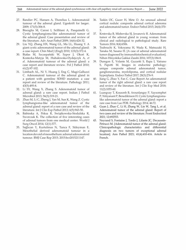
June 2022268
27. Bandier PC, Hansen A, Thorelius L. Adenomatoid tumour of the adrenal gland. Ugeskrift for laeger. 2009; 171(5):306-8.
28. Bisceglia M, Carosi I, Scillitani A, Pasquinelli G. Cystic lymphangioma-like adenomatoid tumor of the adrenal gland: Case presentation and review of the literature. Adv Anat Pathol 2009; 16(6):424-32.
29. Liu YQ, Zhang HX, Wang GL, Ma LL, Huang Y. A giant cystic adenomatoid tumor of the adrenal gland: a case report. Chin Med J (Engl) 2010; 123(3):372-4.
30. Białas M, Szczepański W, Szpor J, Okoń K, Kostecka-Matyja M, Hubalewska-Dydejczyk A, et al. Adenomatoid tumour of the adrenal gland: a case report and literature review. Pol J Pathol 2010; 61(2):97-102.
31. Limbach AL, Ni Y, Huang J, Eng C, Magi-Galluzzi C. Adenomatoid tumour of the adrenal gland in a patient with germline SDHD mutation: a case report and review of the literature. Pathology 2011; 43(5):495-8.
32. Li SY, Wang X, Zhang S. Adenomatoid tumor of adrenal gland: a rare case report. Indian J Pathol Microbiol 2013; 56(3):319-21.
33. Zhao M, Li C, Zheng J, Yan M, Sun K, Wang Z. Cystic lymphangioma-like adenomatoid tumor of the adrenal gland: report of a rare case and review of the literature. Int J Clin Exp Pathol 2013; 6(5):943-50.
34. Babinska A, Peksa R, Swiątkowska-Stodulska R, Sworczak K. The collection of five interesting cases of adrenal tumors from one medical center. World J Surg Oncol 2014; 12(1):377.
35. Sağlıcan Y, Kurtulmus N, Tunca F, Süleyman E. Mesothelial derived adenomatoid tumour in a location devoid of mesothelium: adrenal adenomatoid tumour. BMJ Case Rep 2015; 2015:bcr2015211147.
36. Taskin OC, Gucer H, Mete O. An unusual adrenal cortical nodule: composite adrenal cortical adenoma and adenomatoid tumor. Endocr Pathol 2015; 26(4):370-3.
37. Krstevska B, Mishevska SJ, Jovanovic R. Adenomatoid tumor of the adrenal gland in young woman: from clinical and radiological to pathological study. Rare Tumors 2016; 8(4):6506.
38. Tsubouchi K, Yokoyama H, Wada K, Matsuzaki H, Tanaka M, Sasano H. [A case of adrenal adenomatoid tumor diagnosed by immunohistochemical evaluation]. Nihon Hinyokika Gakkai Zasshi 2016; 107(3):184-8.
39. Duregon E, Volante M, Guzzetti S, Rapa I, Vatrano S, Papotti M. Images in endocrine pathology: unique composite adrenal adenomatoid tumor, ganglioneuroma, myelolipoma, and cortical nodular hyperplasia. Endocr Pathol 2017; 28(3):276-9.
40. Jiang G, Zhao Y, Fan C. Case Report An adenomatoid tumor of the right adrenal gland: a rare case report and review of the literature. Int J Clin Exp Med 2018; 11(2):1055-60.
41. Luangxay T, Kousonh B, Arounlangsy P, Xaysomphet P, Nitiyanant P, Benediktsson H. Cystic lymphangioma-like adenomatoid tumor of the adrenal gland: report a rare case from Lao PDR. Pathology 2014; 46:72.
42. Guan J, Zhao C, Li H, Zhang W, Lin W, Tang L, et al. Adenomatoid tumor of the adrenal gland: Report of two cases and review of the literature. Front Endocrinol 2021; 12:692553.
43. Neyrand S, Fontaine J, Tordo J, Lifante JC, Decaussin-Petrucci M. [Adenomatoid tumor of the adrenal gland: Clinicopathologic characteristics and differential diagnosis on two tumors of exceptional adrenal location]. Ann Pathol 2021; 41(4):410-416. Article in French.
Adenomatoid tumor of the adrenal gland synchronous with clear cell papillary renal cell carcinoma: Report ...
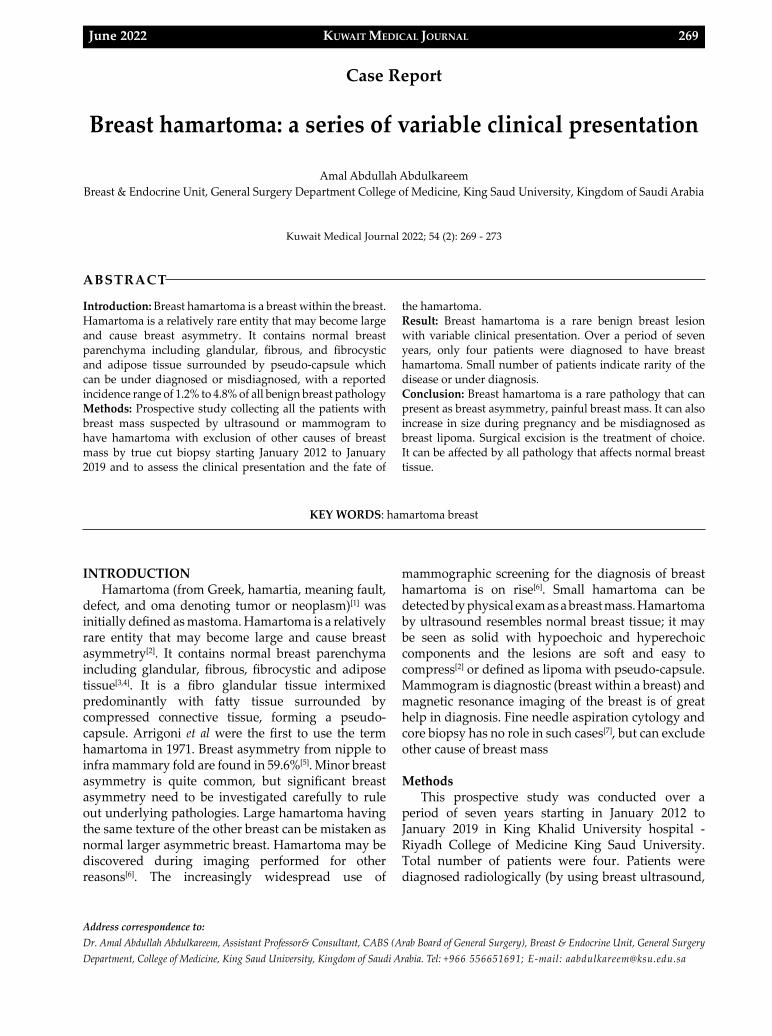
KUWAIT MEDICAL JOURNAL 269June 2022
Address correspondence to:Dr. Amal Abdullah Abdulkareem, Assistant Professor& Consultant, CABS (Arab Board of General Surgery), Breast & Endocrine Unit, General Surgery Department, College of Medicine, King Saud University, Kingdom of Saudi Arabia. Tel: +966 556651691; E-mail: [email protected]
Kuwait Medical Journal 2022; 54 (2): 269 - 273
Breast hamartoma: a series of variable clinical presentation
ABSTRACT
Amal Abdullah Abdulkareem Breast & Endocrine Unit, General Surgery Department College of Medicine, King Saud University, Kingdom of Saudi Arabia
Introduction: Breast hamartoma is a breast within the breast. Hamartoma is a relatively rare entity that may become large and cause breast asymmetry. It contains normal breast parenchyma including glandular, fibrous, and fibrocystic and adipose tissue surrounded by pseudo-capsule which can be under diagnosed or misdiagnosed, with a reported incidence range of 1.2% to 4.8% of all benign breast pathologyMethods: Prospective study collecting all the patients with breast mass suspected by ultrasound or mammogram to have hamartoma with exclusion of other causes of breast mass by true cut biopsy starting January 2012 to January 2019 and to assess the clinical presentation and the fate of
the hamartoma.Result: Breast hamartoma is a rare benign breast lesion with variable clinical presentation. Over a period of seven years, only four patients were diagnosed to have breast hamartoma. Small number of patients indicate rarity of the disease or under diagnosis.Conclusion: Breast hamartoma is a rare pathology that can present as breast asymmetry, painful breast mass. It can also increase in size during pregnancy and be misdiagnosed as breast lipoma. Surgical excision is the treatment of choice. It can be affected by all pathology that affects normal breast tissue.
KEY WORDS: hamartoma breast
Case Report
INTRODUCTIONHamartoma (from Greek, hamartia, meaning fault,
defect, and oma denoting tumor or neoplasm)[1] was initially defined as mastoma. Hamartoma is a relatively rare entity that may become large and cause breast asymmetry[2]. It contains normal breast parenchyma including glandular, fibrous, fibrocystic and adipose tissue[3,4]. It is a fibro glandular tissue intermixed predominantly with fatty tissue surrounded by compressed connective tissue, forming a pseudo-capsule. Arrigoni et al were the first to use the term hamartoma in 1971. Breast asymmetry from nipple to infra mammary fold are found in 59.6%[5]. Minor breast asymmetry is quite common, but significant breast asymmetry need to be investigated carefully to rule out underlying pathologies. Large hamartoma having the same texture of the other breast can be mistaken as normal larger asymmetric breast. Hamartoma may be discovered during imaging performed for other reasons[6]. The increasingly widespread use of
mammographic screening for the diagnosis of breast hamartoma is on rise[6]. Small hamartoma can be detected by physical exam as a breast mass. Hamartoma by ultrasound resembles normal breast tissue; it may be seen as solid with hypoechoic and hyperechoic components and the lesions are soft and easy to compress[2] or defined as lipoma with pseudo-capsule. Mammogram is diagnostic (breast within a breast) and magnetic resonance imaging of the breast is of great help in diagnosis. Fine needle aspiration cytology and core biopsy has no role in such cases[7], but can exclude other cause of breast mass
MethodsThis prospective study was conducted over a
period of seven years starting in January 2012 to January 2019 in King Khalid University hospital - Riyadh College of Medicine King Saud University. Total number of patients were four. Patients were diagnosed radiologically (by using breast ultrasound,
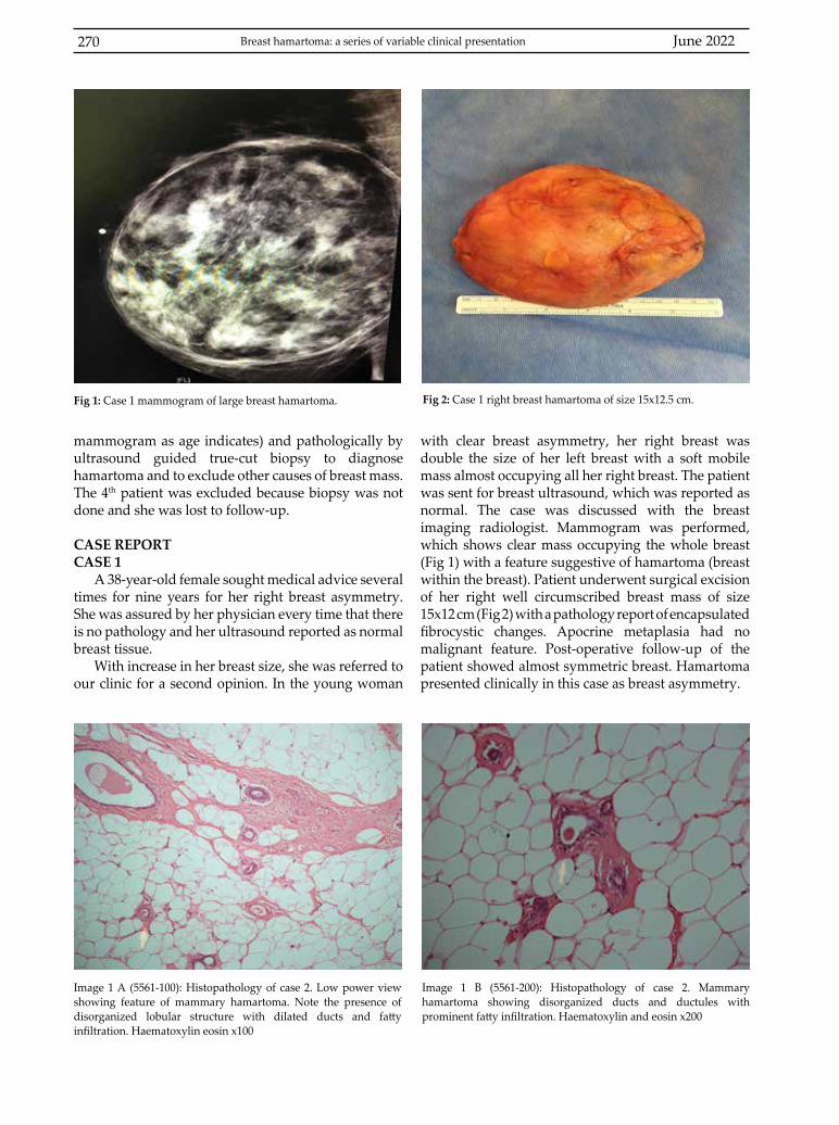
June 2022270
mammogram as age indicates) and pathologically by ultrasound guided true-cut biopsy to diagnose hamartoma and to exclude other causes of breast mass. The 4th patient was excluded because biopsy was not done and she was lost to follow-up.
CASE REPORTCASE 1
A 38-year-old female sought medical advice several times for nine years for her right breast asymmetry. She was assured by her physician every time that there is no pathology and her ultrasound reported as normal breast tissue.
With increase in her breast size, she was referred to our clinic for a second opinion. In the young woman
with clear breast asymmetry, her right breast was double the size of her left breast with a soft mobile mass almost occupying all her right breast. The patient was sent for breast ultrasound, which was reported as normal. The case was discussed with the breast imaging radiologist. Mammogram was performed, which shows clear mass occupying the whole breast (Fig 1) with a feature suggestive of hamartoma (breast within the breast). Patient underwent surgical excision of her right well circumscribed breast mass of size 15x12 cm (Fig 2) with a pathology report of encapsulated fibrocystic changes. Apocrine metaplasia had no malignant feature. Post-operative follow-up of the patient showed almost symmetric breast. Hamartoma presented clinically in this case as breast asymmetry.
Image 1 A (5561-100): Histopathology of case 2. Low power view showing feature of mammary hamartoma. Note the presence of disorganized lobular structure with dilated ducts and fatty infiltration. Haematoxylin eosin x100
Fig 1: Case 1 mammogram of large breast hamartoma. Fig 2: Case 1 right breast hamartoma of size 15x12.5 cm.
Image 1 B (5561-200): Histopathology of case 2. Mammary hamartoma showing disorganized ducts and ductules with prominent fatty infiltration. Haematoxylin and eosin x200
Breast hamartoma: a series of variable clinical presentation
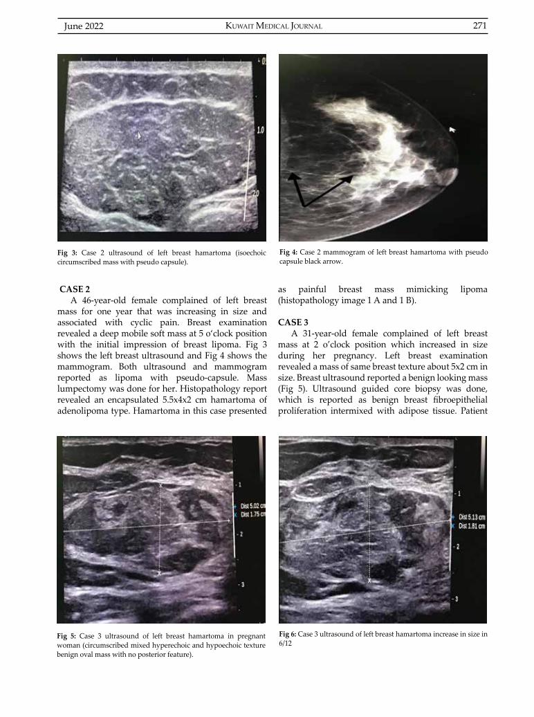
KUWAIT MEDICAL JOURNAL 271June 2022
CASE 2A 46-year-old female complained of left breast
mass for one year that was increasing in size and associated with cyclic pain. Breast examination revealed a deep mobile soft mass at 5 o‘clock position with the initial impression of breast lipoma. Fig 3 shows the left breast ultrasound and Fig 4 shows the mammogram. Both ultrasound and mammogram reported as lipoma with pseudo-capsule. Mass lumpectomy was done for her. Histopathology report revealed an encapsulated 5.5x4x2 cm hamartoma of adenolipoma type. Hamartoma in this case presented
as painful breast mass mimicking lipoma (histopathology image 1 A and 1 B).
CASE 3A 31-year-old female complained of left breast
mass at 2 o’clock position which increased in size during her pregnancy. Left breast examination revealed a mass of same breast texture about 5x2 cm in size. Breast ultrasound reported a benign looking mass (Fig 5). Ultrasound guided core biopsy was done, which is reported as benign breast fibroepithelial proliferation intermixed with adipose tissue. Patient
Fig 3: Case 2 ultrasound of left breast hamartoma (isoechoic circumscribed mass with pseudo capsule).
Fig 5: Case 3 ultrasound of left breast hamartoma in pregnant woman (circumscribed mixed hyperechoic and hypoechoic texture benign oval mass with no posterior feature).
Fig 4: Case 2 mammogram of left breast hamartoma with pseudo capsule black arrow.
Fig 6: Case 3 ultrasound of left breast hamartoma increase in size in 6/12

June 2022272
was followed clinically by breast ultrasound with slight increase in the size of her breast mass (Fig 6). Hamartoma increased in size during pregnancy, which may indicate hormonal influence to hamartoma as the rest of her breast tissue.
CASE 4A 33-year-old patient was diagnosed with deep
vein thrombosis, pulmonary embolism and cerebro vascular accidents with no neurological deficit. She was diagnosed to have protein c deficiency on anticoagulant medication and was found to have right breast mass. Her ultrasound reported a hamartoma 3x2 cm in size (Fig 7). Biopsy was not done and she was lost to follow-up. She was excluded from the study.
Fig 7: Case 4 ultrasound of right breast hamartoma (oval circumscribed heterogeneous mass with parallel oriented, no posterior features)
Case No. Age Duration of complain Presenting symptom Size of hamartoma
1 2 3 4
38 y46 y31 y33 y
9 years One year3 years
7 months
Right breast asymmetryPainful breast massMass increasing in size during pregnancyBreast mass
15x12.5 cm5.5 x4 cm5.8x3cm3x2 cm
Table 1: Duration of complaint, size of hamartoma and the clinical presentation
RESULTHamartoma is a breast within the breast which
represents benign proliferation of fibrous, glandular, and fatty tissue (fibro-adeno-lipoma) surrounded by a thin capsule of connective tissue. All components are found in normal breast tissue and this is why the lesions are considered hamartomatous. It is a rare
benign breast lesion that is detected in the breast during screening of breast asymmetry or discovered pathologically in combination with other breast pathologies. Large hamartoma can be considered as one of the differential diagnosis of breast asymmetry, especially for young women where mammogram is not frequently ordered as in case 1. It can be suspected if the patient complains of significant breast asymmetry with no clearly evident breast mass by physical exam. It can mimic lipoma in the breast as in case 2 or can present as a slow growing mass as in case 3. It can be affected by all pathologies that affect the breast tissue. Hamartomas can increase in size if not removed surgically. Ultrasound can diagnose small, localized hamartoma as a well-circumscribed lesion resembling normal breast tissue often mixed with both hyperechoic and hypoechoic components which are soft and easy to compress. The margins are often difficult to delineate. Mammogram is diagnostic (breast within a breast). Clinical correlation with radiological imaging and histopathology will lead to early diagnosis and treatment of breast hamartoma. Table I indicates the duration of complaint and the size of hamartoma as well the clinical presentation .
DISCUSSIONBreast hamartoma is a relatively rare entity and
some authors report 1.2% to 4.8% incidence of all benign breast masses. It may become large and cause breast asymmetry[8], which is a common complaint among young woman. Physical examination and clinical evaluation are assuring, but significant breast asymmetry or presence of mass indicates full clinical, radiological and pathological evaluation. Hamartoma doesn’t possess specific diagnostic histological feature. The role of fine needle aspiration cytology and core needle biopsy in making the diagnosis is limited and requires clinical and radiological correlation to avoid under diagnosis[4,7]. Case 1 hamartoma presented as breast asymmetry undiagnosed for nine years. The pathogenesis of breast hamartoma is still poorly understood[4]. Our patient falls histologically into the encapsulated fibrocystic changes type, which can be missed histologically and sonographically as normal breast. Both ultrasound and histopathology will be variable and nonspecific[4,9].
Breast hamartoma: a series of variable clinical presentation
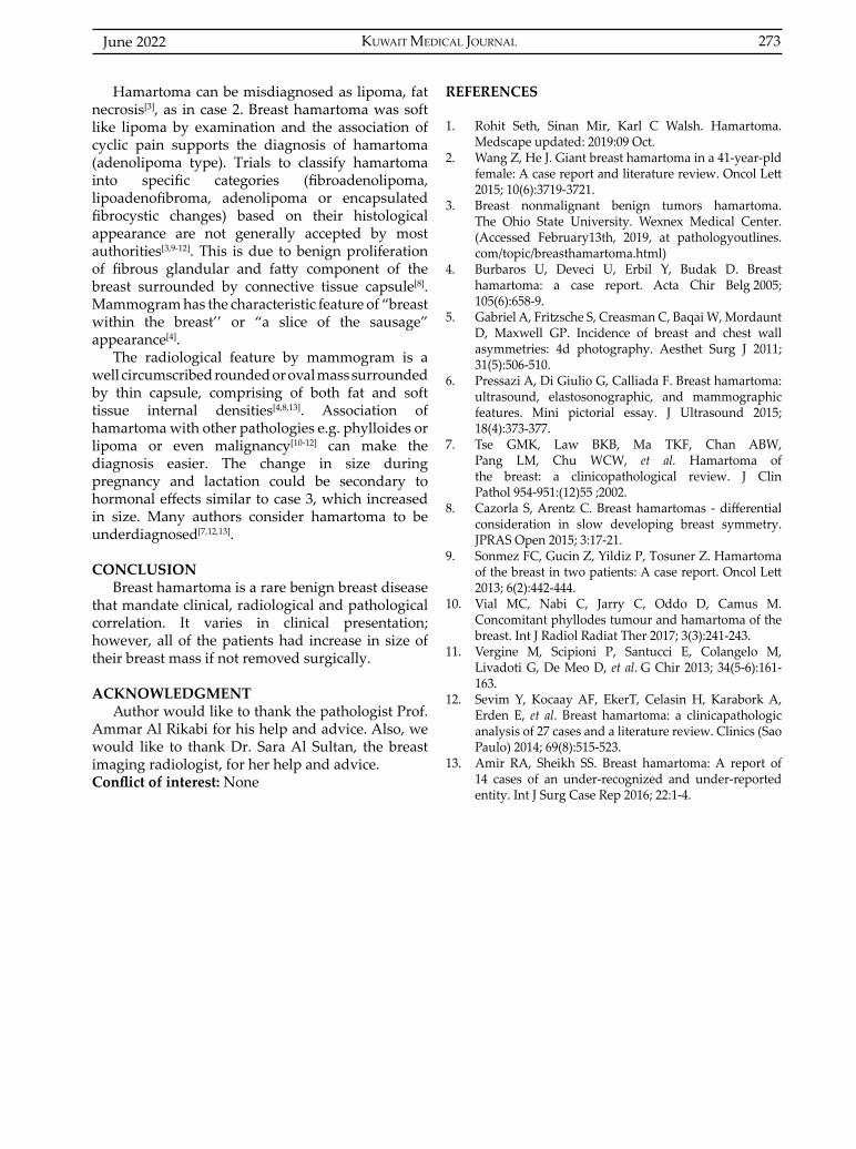
KUWAIT MEDICAL JOURNAL 273June 2022
Hamartoma can be misdiagnosed as lipoma, fat necrosis[3], as in case 2. Breast hamartoma was soft like lipoma by examination and the association of cyclic pain supports the diagnosis of hamartoma (adenolipoma type). Trials to classify hamartoma into specific categories (fibroadenolipoma, lipoadenofibroma, adenolipoma or encapsulated fibrocystic changes) based on their histological appearance are not generally accepted by most authorities[3,9-12]. This is due to benign proliferation of fibrous glandular and fatty component of the breast surrounded by connective tissue capsule[8]. Mammogram has the characteristic feature of “breast within the breast’’ or “a slice of the sausage” appearance[4].
The radiological feature by mammogram is a well circumscribed rounded or oval mass surrounded by thin capsule, comprising of both fat and soft tissue internal densities[4,8,13]. Association of hamartoma with other pathologies e.g. phylloides or lipoma or even malignancy[10-12] can make the diagnosis easier. The change in size during pregnancy and lactation could be secondary to hormonal effects similar to case 3, which increased in size. Many authors consider hamartoma to be underdiagnosed[7,12,13].
CONCLUSIONBreast hamartoma is a rare benign breast disease
that mandate clinical, radiological and pathological correlation. It varies in clinical presentation; however, all of the patients had increase in size of their breast mass if not removed surgically.
ACKNOWLEDGMENTAuthor would like to thank the pathologist Prof.
Ammar Al Rikabi for his help and advice. Also, we would like to thank Dr. Sara Al Sultan, the breast imaging radiologist, for her help and advice.Conflict of interest: None
REFERENCES
1. Rohit Seth, Sinan Mir, Karl C Walsh. Hamartoma. Medscape updated: 2019:09 Oct.
2. Wang Z, He J. Giant breast hamartoma in a 41-year-pld female: A case report and literature review. Oncol Lett 2015; 10(6):3719-3721.
3. Breast nonmalignant benign tumors hamartoma. The Ohio State University. Wexnex Medical Center. (Accessed February13th, 2019, at pathologyoutlines.com/topic/breasthamartoma.html)
4. Burbaros U, Deveci U, Erbil Y, Budak D. Breast hamartoma: a case report. Acta Chir Belg 2005; 105(6):658-9.
5. Gabriel A, Fritzsche S, Creasman C, Baqai W, Mordaunt D, Maxwell GP. Incidence of breast and chest wall asymmetries: 4d photography. Aesthet Surg J 2011; 31(5):506-510.
6. Pressazi A, Di Giulio G, Calliada F. Breast hamartoma: ultrasound, elastosonographic, and mammographic features. Mini pictorial essay. J Ultrasound 2015; 18(4):373-377.
7. Tse GMK, Law BKB, Ma TKF, Chan ABW, Pang LM, Chu WCW, et al. Hamartoma of the breast: a clinicopathological review. J Clin Pathol 954-951:(12)55 ;2002.
8. Cazorla S, Arentz C. Breast hamartomas - differential consideration in slow developing breast symmetry. JPRAS Open 2015; 3:17-21.
9. Sonmez FC, Gucin Z, Yildiz P, Tosuner Z. Hamartoma of the breast in two patients: A case report. Oncol Lett 2013; 6(2):442-444.
10. Vial MC, Nabi C, Jarry C, Oddo D, Camus M. Concomitant phyllodes tumour and hamartoma of the breast. Int J Radiol Radiat Ther 2017; 3(3):241-243.
11. Vergine M, Scipioni P, Santucci E, Colangelo M, Livadoti G, De Meo D, et al. G Chir 2013; 34(5-6):161-163.
12. Sevim Y, Kocaay AF, EkerT, Celasin H, Karabork A, Erden E, et al. Breast hamartoma: a clinicapathologic analysis of 27 cases and a literature review. Clinics (Sao Paulo) 2014; 69(8):515-523.
13. Amir RA, Sheikh SS. Breast hamartoma: A report of 14 cases of an under-recognized and under-reported entity. Int J Surg Case Rep 2016; 22:1-4.
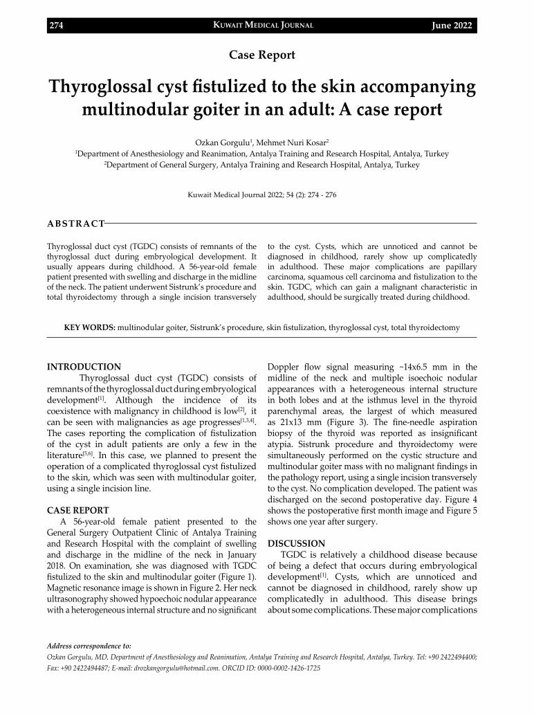
KUWAIT MEDICAL JOURNAL June 2022274
Address correspondence to:Ozkan Gorgulu, MD, Department of Anesthesiology and Reanimation, Antalya Training and Research Hospital, Antalya, Turkey. Tel: +90 2422494400; Fax: +90 2422494487; E-mail: [email protected]. ORCID ID: 0000-0002-1426-1725
Kuwait Medical Journal 2022; 54 (2): 274 - 276
Thyroglossal cyst fistulized to the skin accompanying multinodular goiter in an adult: A case report
ABSTRACT
Ozkan Gorgulu1, Mehmet Nuri Kosar2 1Department of Anesthesiology and Reanimation, Antalya Training and Research Hospital, Antalya, Turkey
2Department of General Surgery, Antalya Training and Research Hospital, Antalya, Turkey
Thyroglossal duct cyst (TGDC) consists of remnants of the thyroglossal duct during embryological development. It usually appears during childhood. A 56-year-old female patient presented with swelling and discharge in the midline of the neck. The patient underwent Sistrunk’s procedure and total thyroidectomy through a single incision transversely
to the cyst. Cysts, which are unnoticed and cannot be diagnosed in childhood, rarely show up complicatedly in adulthood. These major complications are papillary carcinoma, squamous cell carcinoma and fistulization to the skin. TGDC, which can gain a malignant characteristic in adulthood, should be surgically treated during childhood.
KEY WORDS: multinodular goiter, Sistrunk’s procedure, skin fistulization, thyroglossal cyst, total thyroidectomy
Case Report
INTRODUCTIONThyroglossal duct cyst (TGDC) consists of
remnants of the thyroglossal duct during embryological development[1]. Although the incidence of its coexistence with malignancy in childhood is low[2], it can be seen with malignancies as age progresses[1,3,4]. The cases reporting the complication of fistulization of the cyst in adult patients are only a few in the literature[5,6]. In this case, we planned to present the operation of a complicated thyroglossal cyst fistulized to the skin, which was seen with multinodular goiter, using a single incision line.
CASE REPORTA 56-year-old female patient presented to the
General Surgery Outpatient Clinic of Antalya Training and Research Hospital with the complaint of swelling and discharge in the midline of the neck in January 2018. On examination, she was diagnosed with TGDC fistulized to the skin and multinodular goiter (Figure 1). Magnetic resonance image is shown in Figure 2. Her neck ultrasonography showed hypoechoic nodular appearance with a heterogeneous internal structure and no significant
Doppler flow signal measuring ~14x6.5 mm in the midline of the neck and multiple isoechoic nodular appearances with a heterogeneous internal structure in both lobes and at the isthmus level in the thyroid parenchymal areas, the largest of which measured as 21x13 mm (Figure 3). The fine-needle aspiration biopsy of the thyroid was reported as insignificant atypia. Sistrunk procedure and thyroidectomy were simultaneously performed on the cystic structure and multinodular goiter mass with no malignant findings in the pathology report, using a single incision transversely to the cyst. No complication developed. The patient was discharged on the second postoperative day. Figure 4 shows the postoperative first month image and Figure 5 shows one year after surgery.
DISCUSSIONTGDC is relatively a childhood disease because
of being a defect that occurs during embryological development[1]. Cysts, which are unnoticed and cannot be diagnosed in childhood, rarely show up complicatedly in adulthood. This disease brings about some complications. These major complications
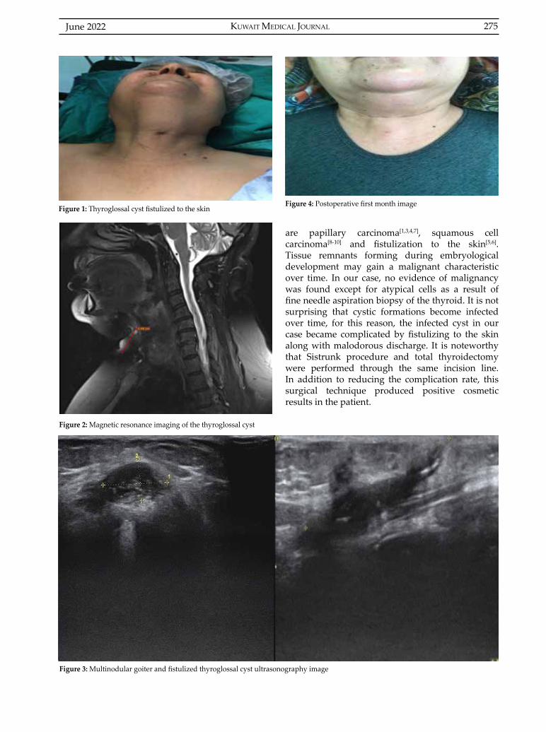
KUWAIT MEDICAL JOURNAL 275June 2022
are papillary carcinoma[1,3,4,7], squamous cell carcinoma[8-10] and fistulization to the skin[5,6]. Tissue remnants forming during embryological development may gain a malignant characteristic over time. In our case, no evidence of malignancy was found except for atypical cells as a result of fine needle aspiration biopsy of the thyroid. It is not surprising that cystic formations become infected over time, for this reason, the infected cyst in our case became complicated by fistulizing to the skin along with malodorous discharge. It is noteworthy that Sistrunk procedure and total thyroidectomy were performed through the same incision line. In addition to reducing the complication rate, this surgical technique produced positive cosmetic results in the patient.
Figure 1: Thyroglossal cyst fistulized to the skin
Figure 2: Magnetic resonance imaging of the thyroglossal cyst
Figure 3: Multinodular goiter and fistulized thyroglossal cyst ultrasonography image
Figure 4: Postoperative first month image
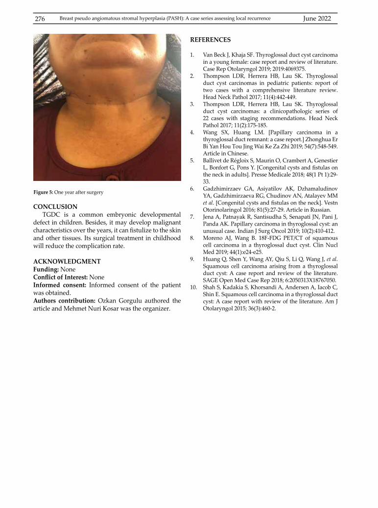
June 2022276 Breast pseudo angiomatous stromal hyperplasia (PASH): A case series assessing local recurrence
CONCLUSIONTGDC is a common embryonic developmental
defect in children. Besides, it may develop malignant characteristics over the years, it can fistulize to the skin and other tissues. Its surgical treatment in childhood will reduce the complication rate.
ACKNOWLEDGMENTFunding: None Conflict of Interest: NoneInformed consent: Informed consent of the patient was obtained.Authors contribution: Ozkan Gorgulu authored the article and Mehmet Nuri Kosar was the organizer.
Figure 5: One year after surgery
REFERENCES
1. Van Beck J, Khaja SF. Thyroglossal duct cyst carcinoma in a young female: case report and review of literature. Case Rep Otolaryngol 2019; 2019:4069375.
2. Thompson LDR, Herrera HB, Lau SK. Thyroglossal duct cyst carcinomas in pediatric patients: report of two cases with a comprehensive literature review. Head Neck Pathol 2017; 11(4):442-449.
3. Thompson LDR, Herrera HB, Lau SK. Thyroglossal duct cyst carcinomas: a clinicopathologic series of 22 cases with staging recommendations. Head Neck Pathol 2017; 11(2):175-185.
4. Wang SX, Huang LM. [Papillary carcinoma in a thyroglossal duct remnant: a case report.] Zhonghua Er Bi Yan Hou Tou Jing Wai Ke Za Zhi 2019; 54(7):548-549. Article in Chinese.
5. Ballivet de Régloix S, Maurin O, Crambert A, Genestier L, Bonfort G, Pons Y. [Congenital cysts and fistulas on the neck in adults]. Presse Medicale 2018; 48(1 Pt 1):29-33.
6. Gadzhimirzaev GA, Asiyatilov AK, Dzhamaludinov YA, Gadzhimirzaeva RG, Chudinov AN, Atalayev MM et al. [Congenital cysts and fistulas on the neck]. Vestn Otorinolaringol 2016; 81(5):27-29. Article in Russian.
7. Jena A, Patnayak R, Santisudha S, Senapati JN, Pani J, Panda AK. Papillary carcinoma in thyroglossal cyst: an unusual case. Indian J Surg Oncol 2019; 10(2):410-412.
8. Moreno AJ, Wang B. 18F-FDG PET/CT of squamous cell carcinoma in a thyroglossal duct cyst. Clin Nucl Med 2019; 44(1):e24-e25.
9. Huang Q, Shen Y, Wang AY, Qiu S, Li Q, Wang J, et al. Squamous cell carcinoma arising from a thyroglossal duct cyst: A case report and review of the literature. SAGE Open Med Case Rep 2018; 6:2050313X18767050.
10. Shah S, Kadakia S, Khorsandi A, Andersen A, Iacob C, Shin E. Squamous cell carcinoma in a thyroglossal duct cyst: A case report with review of the literature. Am J Otolaryngol 2015; 36(3):460-2.
KUWAIT MEDICAL JOURNAL 277June 2022
Address correspondence to:Dr. Zainab Al Lawati, Department of Hospital Affairs, Ministry of Health, Oman. Tel: +968 99201600; E-mail: [email protected]
Kuwait Medical Journal 2022; 54 (2): 277 - 282
Understanding the context of COVID-19 pandemic; lessons learnt for a long-term sustainable healthcare system
ABSTRACT
Zainab Al Lawati1, Alaa Al Lawati2
1Department of Hospital Affairs, Ministry of Health (MOH), Muscat, Oman2Ministry of Health, Muscat, Oman
Since COVID-19 invaded the globe, healthcare systems and economies have been crippled in various places. COVID-19 cases have been reported in 223 out of 232 countries, territories and regions named in the world, with a total count reaching 234,073,401 million, as of 30th September 2021. As of that day, the total number of cases in Oman reached 303,738 affecting 6% of the total population. The pandemic had overwhelming implications on the healthcare sector, and this will likely have a lasting impact on health ventures and delivery
models for years to come. This article will review the highlights of COVID-19
pandemic in the Sultanate of Oman, with particular attention to the impacts on the healthcare sector. The aim is to discuss the lessons learnt from managing the pandemic and use this experience for improving management of future healthcare quandaries. The objective is to educate health care workers and the public about the lessons learnt from handling the pandemic, to ensure sustainability and endurance of the healthcare sector.
KEY WORDS: corona virus, global pandemic, health services administration, Ministry of Health, World Health Organization
Short Communication
INTRODUCTIONIt has been a grim twenty-two months since
COVID-19 invaded the globe. The pandemic impacted multiple aspects of our lives. It severely challenged governments, health systems, economies and has devastated millions of lives and livelihoods all over the world. Out of 232 countries, territories and regions named in the world, COVID-19 cases have been reported in 223, with the total count reaching 234,073,401 million, as of September 30th 2021[1]. As of that day, the total number of cases in Oman reached 303,738, affecting 6% of the total population[1]. The World Health Organisation (WHO) reported a few surges in the number of cases, but since September 1st 2021, a global slowdown in the number of cases and death tolls has been noted. Despite that, the overwhelming implications of the pandemic on healthcare will likely have a lasting impact on health ventures and delivery models for years to come[2].
The WHO reported the first case of COVID-19
disease in Wuhan city, Hubei Province of China on December 31st 2019. This was followed up by a series of declarations, initially declaring its highest level of alarm and labelling the situation as a public health emergency of international concern. This was followed up by the identification of the causative agent, then labelling the situation a global pandemic. In March 2020, an international trial named ‘The Solidarity trial’ was launched by the WHO and its partners. The aim of this trial was to generate robust information about the disease from around the globe, in an attempt to identify the most effective treatment option for it[3]. Following detection of the first case of the virus in Oman, His Majesty issued royal orders to stipulate the formation of the Supreme Committee to study scopes for a mechanism to handle developments resulting from COVID-19 pandemic. As this was the first time to report ailments in the human race from this strain of novel agents, it incurred the need to take hasty measures. Following rapid assessments of the
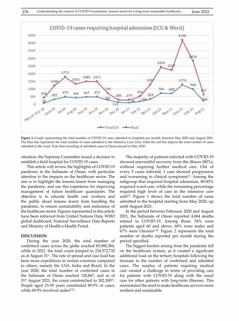
June 2022278
situation, the Supreme Committee issued a decision to establish a field hospital for COVID-19 cases.
This article will review the highlights of COVID-19 pandemic in the Sultanate of Oman, with particular attention to the impacts on the healthcare sector. The aim is to highlight the lessons learnt from managing the pandemic, and use this experience for improving management of future healthcare quandaries. The objective is to educate health care workers and the public about lessons learnt from handling the pandemic, to ensure sustainability and endurance of the healthcare sector. Figures represented in this article have been retrieved from United Nations Data, WHO global dashboard, National Surveillance Data Reports and Ministry of Health e-Health Portal.
DISCUSSION During the year 2020, the total number of
confirmed cases across the globe reached 83,900,384, while in 2021, the total count jumped to 218,572,732 as of August 31st. The rate of spread and case load has been more expeditious in certain countries compared to others, namely the USA, India and Brazil. In the year 2020, the total number of confirmed cases in the Sultanate of Oman reached 128,867, and as of 31st August 2021, the count outstretched to 302,300[1]. People aged 15-59 years constituted 89.9% of cases, while 69.9% involved males[3,4].
The majority of patients infected with COVID-19 showed uneventful recovery from the illness (80%), without requiring further medical care. Out of every 5 cases infected, 1 case showed progression and worsening in clinical symptoms[1]. Among the subgroup that required hospital admission, 80-85% required ward care, while the remaining percentage required high level of care in the intensive care unit[1]. Figure 1 shows the total number of cases admitted to the hospital starting from May 2020, up until August 2021.
In the period between February 2020 and August 2021, the Sultanate of Oman reported 4,064 deaths related to COVID-19. Among those, 54% were patients aged 60 and above, 68% were males and 67% were Omanis[1,4]. Figure 2 represents the total number of deaths reported per month during the period specified.
The biggest burden arising from the pandemic fell on the healthcare system, as it created a significant additional load on the tertiary hospitals following the increase in the number of confirmed and admitted cases. The surplus of patients requiring medical care created a challenge in terms of providing care for patients with COVID-19 along with the usual care for other patients with long-term illnesses. This necessitated the need to make healthcare services more resilient and sustainable.
Figure 1: Graph representing the total number of COVID-19 cases admitted to hospitals per month, between May 2020 and August 2021. The blue line represents the total number of cases admitted to the Intensive Care Unit, while the red line depicts the total number of cases admitted to the ward. Note that recording of admitted cases in Oman ensued in May 2020.
Understanding the context of COVID-19 pandemic; lessons learnt for a long-term sustainable healthcare...
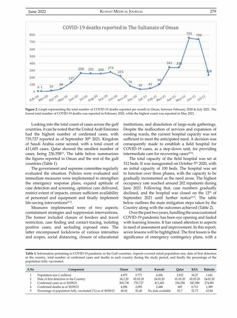
KUWAIT MEDICAL JOURNAL 279June 2022
(A) (B)
Looking into the total count of cases across the gulf countries, it can be noted that the United Arab Emirates had the highest number of confirmed cases, with 735,727 reported as of September 30th 2021. Kingdom of Saudi Arabia came second, with a total count of 411,605 cases. Qatar showed the smallest number of cases, being 236,558[1]. The table below summarizes the figures reported in Oman and the rest of the gulf countries (Table 1).
The government and supreme committee regularly evaluated the situation. Policies were evaluated and immediate measures were implemented to strengthen the emergency response plans, expand aptitude of case detection and screening, optimize care delivered, restrict extent of impacts, ensure sufficient availability of personnel and equipment and finally implement life-saving interventions[4,5].
Measures implemented were of two aspects; containment strategies and suppression interventions. The former included closure of borders and travel restriction, case finding and contact-tracing, isolating positive cases, and secluding exposed ones. The latter encompassed lockdowns of various intensities and scopes, social distancing, closure of educational
institutions, and dissolution of large-scale gatherings. Despite the reallocation of services and expansion of existing wards, the current hospital capacity was not sufficient to meet the anticipated need. A decision was consequently made to establish a field hospital for COVID-19 cases, as a step-down unit, for providing intermediate care for recovering cases[4-6].
The total capacity of the field hospital was set at 312 beds. It was inaugurated on October 5th 2020, with an initial capacity of 100 beds. The hospital was set to function over three phases, with the capacity to be gradually incremented as the need arose. The highest occupancy rate reached around 202 inpatients during June 2021. Following that, case numbers gradually declined, and the hospital was closed on the 12th of September 2021 until further notice[4,7]. The table below outlines the main mitigation steps taken by the Country along with the outcomes achieved (Table 2).
Over the past two years, handling the unaccustomed COVID-19 pandemic has been eye opening and fueled with learning lessons. It has raised attention to aspects in need of assessment and improvement. In this report, seven lessons will be highlighted. The first lesson is the significance of emergency contingency plans, with a
S.No Component Oman UAE Kuwait Qatar KSA Bahrain
12345
Population size (/ million)Date of first detection in the CountryConfirmed cases as of 30/09/21Confirmed deaths as of 30/09/21Percentage of population fully vaccinated (%) as of 30/09/21
4.97524.2.20303,738
4,09640.62
9.77102.02.20735,727
2,09582.40
4.20624.02.20411,605
2,448No data available
2.83201.03.20236,558
60581.7
34.2702.03.20547,090
8,71352.73
1.64124.02.20274,981
1,38963.84
Table 1: Information pertaining to COVID-19 pandemic in the Gulf countries. Aspects covered entail population size, date of first detection in the country, total number of confirmed cases and deaths in each country during the study period, and finally the percentage of the population fully vaccinated.
Figure 2: Graph representing the total number of COVID-19 deaths reported per month in Oman, between February 2020 & July 2021. The lowest total number of COVID-19 deaths was reported in February 2020, while the highest count was reported in May 2021.
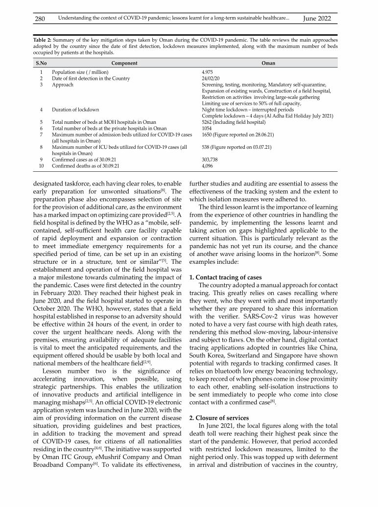
June 2022280
(A) (B)
designated taskforce, each having clear roles, to enable early preparation for unwonted situations[8]. The preparation phase also encompasses selection of site for the provision of additional care, as the environment has a marked impact on optimizing care provided[2,5]. A field hospital is defined by the WHO as a “mobile, self-contained, self-sufficient health care facility capable of rapid deployment and expansion or contraction to meet immediate emergency requirements for a specified period of time, can be set up in an existing structure or in a structure, tent or similar”[5]. The establishment and operation of the field hospital was a major milestone towards culminating the impact of the pandemic. Cases were first detected in the country in February 2020. They reached their highest peak in June 2020, and the field hospital started to operate in October 2020. The WHO, however, states that a field hospital established in response to an adversity should be effective within 24 hours of the event, in order to cover the urgent healthcare needs. Along with the premises, ensuring availability of adequate facilities is vital to meet the anticipated requirements, and the equipment offered should be usable by both local and national members of the healthcare field[2,9].
Lesson number two is the significance of accelerating innovation, when possible, using strategic partnerships. This enables the utilization of innovative products and artificial intelligence in managing mishaps[2,5]. An official COVID-19 electronic application system was launched in June 2020, with the aim of providing information on the current disease situation, providing guidelines and best practices, in addition to tracking the movement and spread of COVID-19 cases, for citizens of all nationalities residing in the country[4,6]. The initiative was supported by Oman ITC Group, eMushrif Company and Oman Broadband Company[6]. To validate its effectiveness,
further studies and auditing are essential to assess the effectiveness of the tracking system and the extent to which isolation measures were adhered to.
The third lesson learnt is the importance of learning from the experience of other countries in handling the pandemic, by implementing the lessons learnt and taking action on gaps highlighted applicable to the current situation. This is particularly relevant as the pandemic has not yet run its course, and the chance of another wave arising looms in the horizon[8]. Some examples include:
1. Contact tracing of cases
The country adopted a manual approach for contact tracing. This greatly relies on cases recalling where they went, who they went with and most importantly whether they are prepared to share this information with the verifier. SARS-Cov-2 virus was however noted to have a very fast course with high death rates, rendering this method slow-moving, labour-intensive and subject to flaws. On the other hand, digital contact tracing applications adopted in countries like China, South Korea, Switzerland and Singapore have shown potential with regards to tracking confirmed cases. It relies on bluetooth low energy beaconing technology, to keep record of when phones come in close proximity to each other, enabling self-isolation instructions to be sent immediately to people who come into close contact with a confirmed case[8].
2. Closure of servicesIn June 2021, the local figures along with the total
death toll were reaching their highest peak since the start of the pandemic. However, that period accorded with restricted lockdown measures, limited to the night period only. This was topped up with deferment in arrival and distribution of vaccines in the country,
S.No Component Oman
123
4
567
8
910
Population size ( / million)Date of first detection in the CountryApproach
Duration of lockdown
Total number of beds at MOH hospitals in OmanTotal number of beds at the private hospitals in OmanMaximum number of admission beds utilized for COVID-19 cases (all hospitals in Oman) Maximum number of ICU beds utilized for COVID-19 cases (all hospitals in Oman)Confirmed cases as of 30.09.21 Confirmed deaths as of 30.09.21
4.975 24/02/20Screening, testing, monitoring, Mandatory self-quarantine,Expansion of existing wards, Construction of a field hospital,Restriction on activities involving large-scale gatheringLimiting use of services to 50% of full capacity,Night time lockdown – interrupted periods Complete lockdown – 4 days (Al Adha Eid Holiday July 2021)5262 (Including field hospital)10541650 (Figure reported on 28.06.21)
538 (Figure reported on 03.07.21)
303,7384,096
Table 2: Summary of the key mitigation steps taken by Oman during the COVID-19 pandemic. The table reviews the main approaches adopted by the country since the date of first detection, lockdown measures implemented, along with the maximum number of beds occupied by patients at the hospitals.
Understanding the context of COVID-19 pandemic; lessons learnt for a long-term sustainable healthcare...
KUWAIT MEDICAL JOURNAL 281June 2022
rendering Oman with the second highest overall death rates among the gulf countries. The global experience has shown that insufficient or delayed enactment of lockdown measures impacted the efficiency of hindering the spread of infection, the former seen in USA and India and the latter in the United Kingdom, Russia and France[8,9].
Lesson number four is the importance of instigating coping strategies. The preceding period witnessed a marked rise in implementing telemedicine in the delivery of care. This service decreased the need for space required and the burden of commuting on patients, especially the ones following up on a regular basis for chronic conditions[2].
The fifth lesson learnt is the significance of effective communication and information management, as employee engagement is the driver of productivity. This encompasses both the clinical staff and the community. Implementation of non-pharmaceutical interventions have shown to be of valuable impact in controlling the disease spread, and ensuring reliable and smooth access to updated information about the situation will ultimately generate public cooperation in implementing the steps agreed on[2,5].
The sixth lesson learnt is the significant impact agile leadership has in times of crisis. It is the main driver of productivity and among the main influencing parameters in employee retention, both of which are vital during onerous periods. Four agile leadership skills that were vital before, during and will continue to be important after this pandemic are: humility (eliminating the need to know everything), adaptability (willing to revise own opinion, open to new ideas), visionary (having a clear long-term plan, despite short-term qualms) and remaining engaged during difficult periods[10].
The final lesson to be raised in this report is the need to think of creative solutions and utilise talents accordingly. Right from the start of the pandemic, shortage of emergency supplies, including ventilation devices and personal protective equipment, surfaced in multiple regions across the globe. Despite the lesson learnt, there appears to be no straight forward solution for this issue; the current pandemic mainly affected the respiratory system, and the next pandemic could target a different system, rendering storage of respiratory equipment in vain. Moreover, storing supplies in large quantities is neither feasible financially nor logistics wise, necessitating the need for alternative answers to future pandemics[9].
The experience Oman has witnessed with regards to responding to this pandemic has emphasized some of the strategies that should be accentuated in the sector of healthcare management, along with the role of technology and telemedicine in healthcare, which
in turn will strengthen and integrate the system, and enable rapid reorganization of services, ensuring that it remains robust, sustainable and innovative during onerous periods[9].
LIMITATIONSThis review is limited by the internal validity and
accuracy of figures reported, as the reporting of cases may have been controlled or filtered in the regions reported.
CONCLUSIONThis retrospective review provided a concise
summary of COVID-19 pandemic in Oman. It has highlighted the main events involved, steps taken to mitigate it, and the impact it has had on the healthcare system, highlighting areas in need of attention and improvement. The lessons learned include preparing contingency plans, incorporating artificial intelligence, learning from the experience of other countries, implementing innovative strategies, communicating effectively, maintaining agile leadership and implementing creative solutions.
A significant proportion of the information reported was obtained from news agency reports. Such an article is therefore vital to corroborate and document the information stated. The urgent steps taken during COVID-19 pandemic to make the health care system innovative and robust should not be forgotten once the storm passes, and instead should be implemented within the health care system. The battle against this virus will continue to exist, but with global solidarity and national unity, the light at the end of the tunnel will continue to brighten, bringing hopes for a nearby liberty and resumption to normality.
ACKNOWLEDGMENT The authors would like to thank Mr. A Al Qasmi
(MOH, DGSMC, Quality Department) for his assistance in validating local figures. Author Statement: The authors contributed equally in the conception, drafting and approval of the final version of this manuscript. Both authors are accountable for all aspects of the work, and information has been checked for accuracy in the best possible and comprehensive manner.
DISCLOSURE The authors declare no conflicts of interest. This
research received no specific grant from any funding agency in the public, commercial or not-for-profit sectors. The figures represented in this report were based on information from global dashboards, national surveillance reports and e-Health portals. The research department at the Ministry of Health was consulted,
June 2022282
and an approval certificate was deemed unnecessary in view of the data being retrieved from openly available resources.
REFERENCES
1. COVID-19 Coronavirus Pandemic: Worldometer, 2021. (Accessed June 15, 2021 at https://www.worldometers.info/coronavirus/#countries)
2. Butler SM. After COVID-19: Thinking differently about running the health care system. JAMA 2020; 323(24):2450-2451.
3. World Health Organisation’s COVID-19 response: WHO Timeline, 2021. (Accessed September 29, 2021 at https://www.who.int/emergencies/diseases/novel-coronavirus-2019/interactive-timeline#!)
4. Ministry of Health (MOH) e-notification system: Tarassud plus, 2021. (Accessed September 15, 2021 at https://tarassud.moh.gov.om/#/login)
5. Norton I, von Schreeb J, Aitken P, Herard P, Lajolo C. Classification and minimum standards for foreign medical teams in sudden onset disasters, Global Health Cluster, World Health Organisation (WHO) publications, Switzerland, 2021. (Accessed October 12, 2021 at https://cdn.who.int/media/docs/default-source/documents/publications/classification-and-minimum-
standards-for-foreign-medical-teams-in-suddent-onset-disasters65829584-c349-4f98-b828-f2ffff4fe089.pdf?sfvrsn=43a8b2f1_1)
6. Oman launches technological surveillance system to track COVID-19: WHO Newsroom, 2020. (Accessed October 3, 2021 at https://www.who.int/news-room/feature-stories/detail/oman-launches-technological-surveillance-system-to-track-covid-19)
7. WHO collaboration in Oman’s response to COVID-19: World Health Organisation, 2021. (Accessed September 15, 2021 at http://www.emro.who.int/omn/oman-news/who-collaboration-on-omans-response-to-covid-19.html)
8. Lee ACK, English P, Pankhania B, Morling JR. Where England’s pandemic response to COVID-19 went wrong. Public Health 2021; 192:45–48.
9. Stenseth N, Dharmarajan G, Li R, Shi Z-L, Yang R, Gao GF. Lessons learnt from the COVID-19 pandemic, Frontiers in Public Health: Infectious diseases – surveillance, prevention & treatment, 2021. (Accessed October 18, 2021 at https://www.frontiersin.org/articles/10.3389/fpubh.2021.694705/full)
10. Yokoi T. 4 agile leadership competencies to outlast Covid-19: Forbes, 2020. (Accessed October 21, 2021 at https://www.forbes.com/sites/tomokoyokoi/2020/06/02/4-agile-leadership-competencies-to-outlast-covid-19/?sh=42b502f96e0c)
Understanding the context of COVID-19 pandemic; lessons learnt for a long-term sustainable healthcare...
KUWAIT MEDICAL JOURNAL 283June 2022
Address correspondence to:Hyok Ju Ri, MD, Room 505, Building B (Hospitalization Building) of the Second Hospital of Dalian Medical University, Shahe District, Dalian City, Liaoning Province, in China. Tel: (+86)18842643985; E-mail: [email protected]; ORCiD: 0000-0002-5172-7029
Kuwait Medical Journal 2022; 54 (2): 283 - 286
How to understand the “Stealth” Omicron subvariant and prepare for its challenge
ABSTRACT
HyokJu Ri1,2, GunHyok Kim1,3, HyonSu Jo1,2, CholSik Ri1,2
1Department of Hernia and Colorectal Surgery, The Second Hospital of Dalian Medical University, Dalian, 116023, People’sRepublic of China
2Department of Colorectal Surgery, The Hospital of Pyongyang Medical College, Pyongyang, 999093, D.P.R of Korea3Department of Pathophysiology, The University of Hamhung Medical College in D.P.R of Korea
BA.2, nicknamed “stealth omicron”, is one of three known subvariants of Omicron. BA.2 differs from BA.1 (the original Omicron variant) in some mutations, including in the spike protein. This mutation makes it more difficult to identify as Omicron subvariant on several tests, and BA.2 is more infectious and more likely to infect vaccinated people compared to BA.1. BA.2 has been detected in at least 40
countries and in all continents except Antarctica, and the World Health Organization is continuing to monitor its spread, while BA.2 is beginning to replace the original Omicron strain in many countries. This commentary provides an overview of current knowledge and unknowns about this new variant and summarizes the important study findings for the purpose of informing experts.
KEY WORDS: coronavirus, COVID-19, SARS-CoV-2, stealth Omicron variant
Short Communication
INTRODUCTIONThe first case of an unidentified form of viral
pneumonia were reported in Wuhan city, Hubei province in China, in December 2019[1]. This virus infection of pneumonia was identified as the novel coronavirus (SARS-COV-2) by the scientific research institution in China, and later it was named “COVID-19” by the World Health Organization (WHO)[2].
This unknown virus gradually spread over the whole world. The mortality rate of COVID-19 is lower than other coronaviruses such as severe acute respiratory syndrome and Middle East respiratory syndrome[3]. However, COVID-19 is highly transmissible and rapidly spread, as it can be transmitted by respiratory droplets and contact and several unknown ways[4].
Though several vaccines have been developed to treat and prevent COVID-19 infections, the world has not ended its struggle with these viruses.
At this time, the new variant was discovered in South Africa and threatened the world concerning
new pandemic coming from this variant[5]. WHO first reported the new strain on November 24, 2021, after a case was reported in South Africa in November, later designated it as one of “variants of concern” which showed a marked degree of mutation (Fig 1), with 20I (Alpha, V1) or B.1.1.7, 20H (Beta, V2) or B.1.351, 20I (Gamma, V3) or P.1, 21A (Delta).
Following the discovery of the SARS-CoV-2 Omicron variant (B.1.1.529), the global COVID-19 outbreak has resurfaced after appearing to be relentlessly spreading over the past two years. This new Omicron variant became a major dominant virus in COVID-19 pandemic everywhere (Fig 2)[6].
Cases of the Omicron variant are on the decline worldwide, but a different version of Omicron is now gaining traction. This is called as “stealth” variant, officially known as BA.2, armed with even higher transmission potential, and possibly a greater ability to evade the immune response, than the original Omicron, leading experts to fear it could further prolong the COVID-19 pandemic[7].
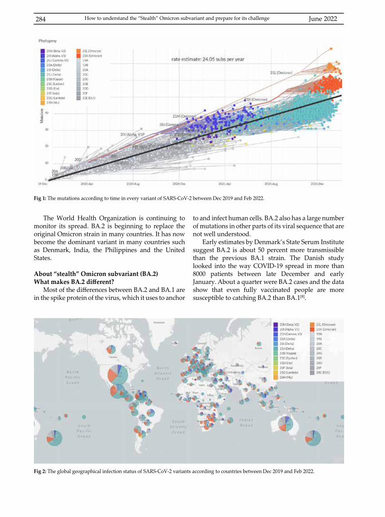
June 2022284
The World Health Organization is continuing to monitor its spread. BA.2 is beginning to replace the original Omicron strain in many countries. It has now become the dominant variant in many countries such as Denmark, India, the Philippines and the United States.
About “stealth” Omicron subvariant (BA.2)What makes BA.2 different?
Most of the differences between BA.2 and BA.1 are in the spike protein of the virus, which it uses to anchor
to and infect human cells. BA.2 also has a large number of mutations in other parts of its viral sequence that are not well understood.
Early estimates by Denmark’s State Serum Institute suggest BA.2 is about 50 percent more transmissible than the previous BA.1 strain. The Danish study looked into the way COVID-19 spread in more than 8000 patients between late December and early January. About a quarter were BA.2 cases and the data show that even fully vaccinated people are more susceptible to catching BA.2 than BA.1[8].
Fig 1: The mutations according to time in every variant of SARS-CoV-2 between Dec 2019 and Feb 2022.
Fig 2: The global geographical infection status of SARS-CoV-2 variants according to countries between Dec 2019 and Feb 2022.
How to understand the “Stealth” Omicron subvariant and prepare for its challenge
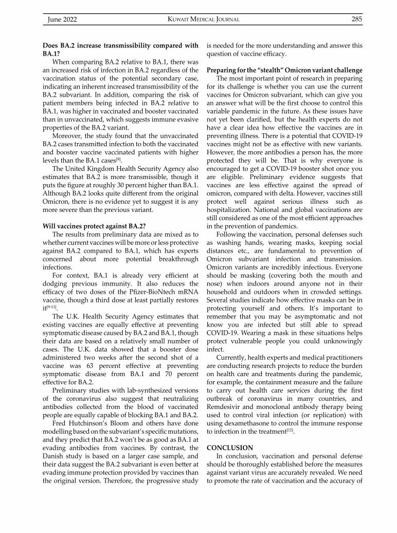
KUWAIT MEDICAL JOURNAL 285June 2022
Does BA.2 increase transmissibility compared with BA.1?
When comparing BA.2 relative to BA.1, there was an increased risk of infection in BA.2 regardless of the vaccination status of the potential secondary case, indicating an inherent increased transmissibility of the BA.2 subvariant. In addition, comparing the risk of patient members being infected in BA.2 relative to BA.1, was higher in vaccinated and booster vaccinated than in unvaccinated, which suggests immune evasive properties of the BA.2 variant.
Moreover, the study found that the unvaccinated BA.2 cases transmitted infection to both the vaccinated and booster vaccine vaccinated patients with higher levels than the BA.1 cases[8].
The United Kingdom Health Security Agency also estimates that BA.2 is more transmissible, though it puts the figure at roughly 30 percent higher than BA.1. Although BA.2 looks quite different from the original Omicron, there is no evidence yet to suggest it is any more severe than the previous variant.
Will vaccines protect against BA.2?The results from preliminary data are mixed as to
whether current vaccines will be more or less protective against BA.2 compared to BA.1, which has experts concerned about more potential breakthrough infections.
For context, BA.1 is already very efficient at dodging previous immunity. It also reduces the efficacy of two doses of the Pfizer-BioNtech mRNA vaccine, though a third dose at least partially restores it[9-11].
The U.K. Health Security Agency estimates that existing vaccines are equally effective at preventing symptomatic disease caused by BA.2 and BA.1, though their data are based on a relatively small number of cases. The U.K. data showed that a booster dose administered two weeks after the second shot of a vaccine was 63 percent effective at preventing symptomatic disease from BA.1 and 70 percent effective for BA.2.
Preliminary studies with lab-synthesized versions of the coronavirus also suggest that neutralizing antibodies collected from the blood of vaccinated people are equally capable of blocking BA.1 and BA.2.
Fred Hutchinson’s Bloom and others have done modelling based on the subvariant’s specific mutations, and they predict that BA.2 won’t be as good as BA.1 at evading antibodies from vaccines. By contrast, the Danish study is based on a larger case sample, and their data suggest the BA.2 subvariant is even better at evading immune protection provided by vaccines than the original version. Therefore, the progressive study
is needed for the more understanding and answer this question of vaccine efficacy.
Preparing for the “stealth” Omicron variant challengeThe most important point of research in preparing
for its challenge is whether you can use the current vaccines for Omicron subvariant, which can give you an answer what will be the first choose to control this variable pandemic in the future. As these issues have not yet been clarified, but the health experts do not have a clear idea how effective the vaccines are in preventing illness. There is a potential that COVID-19 vaccines might not be as effective with new variants. However, the more antibodies a person has, the more protected they will be. That is why everyone is encouraged to get a COVID-19 booster shot once you are eligible. Preliminary evidence suggests that vaccines are less effective against the spread of omicron, compared with delta. However, vaccines still protect well against serious illness such as hospitalization. National and global vaccinations are still considered as one of the most efficient approaches in the prevention of pandemics.
Following the vaccination, personal defenses such as washing hands, wearing masks, keeping social distances etc., are fundamental to prevention of Omicron subvariant infection and transmission. Omicron variants are incredibly infectious. Everyone should be masking (covering both the mouth and nose) when indoors around anyone not in their household and outdoors when in crowded settings. Several studies indicate how effective masks can be in protecting yourself and others. It’s important to remember that you may be asymptomatic and not know you are infected but still able to spread COVID-19. Wearing a mask in these situations helps protect vulnerable people you could unknowingly infect.
Currently, health experts and medical practitioners are conducting research projects to reduce the burden on health care and treatments during the pandemic, for example, the containment measure and the failure to carry out health care services during the first outbreak of coronavirus in many countries, and Remdesivir and monoclonal antibody therapy being used to control viral infection (or replication) with using dexamethasone to control the immune response to infection in the treatment[12].
CONCLUSIONIn conclusion, vaccination and personal defense
should be thoroughly established before the measures against variant virus are accurately revealed. We need to promote the rate of vaccination and the accuracy of
June 2022286
the variant virus test to find the infected persons and treat them in time. There is an urgent need to explore the virology and biology of Omicron subvariants, define clinical phenomes and therapies, monitor dynamics of genetic changes, and protect people from this subvariant. It is up to every country to make common efforts and compliance with all health requirements.
ACKNOWLEDGMENTEthics: Not applicable.Conflict of interest: All authors have substantially contributed to conducting the underlying research and drafting this manuscript. To the best of our knowledge, none of the authors have a conflict of interest, financial, or otherwise.
REFERENCES
1. Rachel JB, Gururaj N, Smitha T, Daniel TD, Harishini BS, Rosaian AS. Innovative diagnostic approach and investigation trends in COVID19-A systematic review. J Oral Maxillofac Pathol 2020: 24(3):421-436.
2. Cucinotta D, Vanelli M. WHO declares COVID-19 a pandemic. Acta Biomed 2020: 91(1):157-160.
3. Fani M, Teimoori A, Ghafari S. Comparison of the COVID-2019 (SARS-CoV-2) pathogenesis with SARS-CoV and MERS-CoV infections. Future Virol 2020.
4. Fuentes-Aspe R, Huaiquilaf-Jorquera S, Oliveros MJ, Soto A. Characteristics of the coronavirus disease 2019: A review of emerging literature. Medwave 2021: 21(5):e8206.
5. Daria S, Bhuiyan MA, Islam R. Detection of highly muted coronavirus variant Omicron (B.1.1.529) is triggering the alarm for South Asian countries: Associated risk factors and preventive actions. J Med Virol 2021; 94(4):1267-1268.
6. Kandeel M, Mohamed MEM, El-Lateef HMA, Venugopala KN, El-Beltagi HS. Omicron variant genome evolution and phylogenetics. J Med Virol 2022; 94(4):1627-1632.
7. Greaney AJ, Starr TN, Bloom JD. An antibody-escape calculator for mutations to the SARS-CoV-2 receptor-binding domain. bioRxiv. Preprint. 2021 Dec 7.
8. Lyngse FP, Kirkeby CT, Denwood M, Christiansen LE, Molbak K, Moller CH, et al. Transmission of SARS-CoV-2 Omicron VOC subvariants BA.1 and BA.2: Evidence from Danish households. medRxiv, 2022: p. 2022:01.28.22270044.
9. Doria-Rose NA, Shen X, Schmidt SD, O’Dell S, McDanal C, Feng W, et al. Booster of mRNA-1273 strengthens SARS-CoV-2 Omicron neutralization. medRxiv 2021: 2021:12.15.21267805.
10. Willett BJ, Grove J, MacLean OA, Wilkie C, Logan N, De Lorenzo G, et al. The hyper-transmissible SARS-CoV-2 Omicron variant exhibits significant antigenic change, vaccine escape and a switch in cell entry mechanism. medRxiv, 2022: p. 2022:01.03.21268111.
11. Collie S, Moultrie H, Bekker LG, Gray G. Effectiveness of BNT162b2 Vaccine against Omicron Variant in South Africa. N Engl J Med 2022; 386(5):494-496.
12. Mohiuddin M, Kasahara K. Investigating the aggressiveness of the COVID-19 Omicron variant and suggestions for possible treatment options. Respir Med 2022; 191:106716.
How to understand the “Stealth” Omicron subvariant and prepare for its challenge
KUWAIT MEDICAL JOURNAL 287June 2022
Selected Abstracts of Articles Published Elsewhere by Authors in Kuwait
Kuwait Medical Journal 2022; 54 (2): 287 - 290
Protein/Creatinine ratio versus 24-hours urine protein in preeclampsia
Ibrahim A Abdelazim 1 2, Osama O Amer 3, Svetlana Shikanova 4, Bakyt Karimova 4
1Department of Obstetrics, and Gynaecology, Ain Shams University, Cairo, Egypt. [email protected] Hospital, Kuwait Oil Company (KOC), Ahmadi, Kuwait. [email protected].
3Ghamra Military Hospital, Cairo, Egypt.4West Kazakhstan Marat Ospanov Medical University, Aktobe, Kazakhstan.
Ginekol Pol. 2022 Feb 14. doi: 10.5603/GP.a2021.0233. Online ahead of print.
OBJECTIVESProteinuria is one of the diagnostic criteria of preeclampsia (PE). Measurement of 24-hour urine protein is the gold standard method for detection of proteinuria in PE. The 24-hour urine sampling is time-consuming, and inconvenient. To evaluate the accuracy of protein/creatinine (P/C) ratio in detection of significant proteinuria ( > 1 g/24-hours urine) in PE.
MATERIAL AND METHODSOne hundred and ten (110) preeclamptic women were included in this study and admitted for blood pressure monitoring, 24-hour urine collection, fetal well-being assessment and spot urine sample for measuring of P/C ratio. After thorough history and clinical examination, routine antenatal investigations were done for the women included in the study according to the hospital protocol, and to excluded
Sodium–glucose cotransporter-2 inhibitors induced euglycemic diabetic ketoacidosis: Two case reports
and a review of the literature
Zouheir Ibrahim Bitar1, Ossama Sajeh Maadarani1, Fawaz Alabdali2, Ahmed Teama3, Walid Elsawah3, Mohammed Jaber Mohsen4, Mahmoud Mostafa Elzoueiry4
1Critical Care Unit, Ahmadi Hospital, Kuwait Oil Company, Fahaheel, Kuwait2Head of Endocrinology and Diabetic Unit, Ahmadi Hospital, Kuwait Oil Company, Fahaheel, Kuwait
3Emergency Physician, Emergency Department, Ahmadi Hospital, Kuwait Oil Company, Fahaheel, Kuwait4Internal Medicine Department, Ahmadi Hospital, Kuwait Oil Company, Fahaheel, Kuwait
Clin Case Rep. 2022;10:e05440. https://doi.org/10.1002/ccr3.5440
If not detected early, euglycemic diabetic ketoacidosis can be a serious adverse effect of sodium–glucose cotransporter-2 (SGLT2) inhibitors. Unfortunately, euglycemic diabetic ketoacidosis is underreported in recent trials and missed because of normal blood sugar levels and nonspecific symptoms on presentation. We present two patients with type 2 diabetes mellitus who developed dapagliflozin-associated euglycemic diabetic ketoacidosis followed by hyperglycemic ketoacidosis. The second patient had euglycemic ketoacidosis twice despite instructions to stop using the medication dapagliflozin.
Selected Abstracts of Articles Published Elsewhere by Authors in Kuwait June 2022288
The Pediatric COVID-19 Registry in Kuwait (PCR-Q8): Methodology and Results of Pilot Phase
Sarah Qabazard, Dalia Al-Abdulrazzaq, Hessa Al-Kandari, Mariam Ayed, Ayed Alanezi, Nufoud Al-Shammari, Zaid Alharbash, Muna Al-Khabbaz, Khaled Kalander, Saadoun Bin-Hassan, Abdulla
Alfraij, Mohammad Alghounaim, Khaled Alsaeid, Hashem Al-Hashemi
Med Princ Pract. 2022 May 20. doi: 10.1159/000524756. Online ahead of print.
OBJECTIVEEstablishing a pediatric COVID-19 registry in Kuwait (PCR-Q8) deemed imperative during the pandemic to study children infected with severe acute respiratory syndrome-related coronavirus 2 (SARS-CoV-2) focusing on mode of presentation, therapeutic interventions, disease severity and early outcomes. This manuscript describes the rapid establishment of the PCR-Q8 registry showcasing an infrastructure of the development process and presents the results of the pilot phase.
SUBJECT AND METHODSThe registry was developed and implemented using the general key steps from Gliklich et al. resource titled “Registries for Evaluating Patient Outcomes: A User’s Guide” as a guide for best practice, experience from a previously established pediatric diabetes registry in Kuwait and several other COVID-19 registries developed globally. During the pilot phase, a convenience sample of 120 children were included, of those 66 (55%) were male.
RESULTSExperience and expertise from other COVID-19 registries, guidance provided by the World Health Organization (WHO) and effective collaboration and cooperation between the stakeholders, study group and data enterers during these challenging times were critical for the development and implementation of the registry. Our results were similar to international reports where most children presented with mild disease (69.2%), majority (70.2%) had normal chest X-ray, and the most common symptom at presentation was fever (77%).
CONCLUSIONWe anticipate the PCR-Q8 development to be a steppingstone for more in-depth investigation of SARS-CoV-2 infection in children in Kuwait and further other registry establishments.
pre-existing chronic renal disease. Twenty-four-hour urine collection started on the morning following hospital admission. Spot urine samples were obtained shortly before the 24-hour urine collection for measuring of P/C ratio. Collected data statistically analyzed to evaluate the accuracy of P/C ratio in detection of significant proteinuria in PE.
RESULTSThe P/C ratio of 1.35 ± 2.54 had 94.4% sensitivity, 94.9% specificity, 97.1% positive predictive value (PPV), 90.2% negative predictive value (NPV), and 94.5% overall accuracy in detection of significant proteinuria ( > 1 g/24-hour urine) in PE.
CONCLUSIONS The P/C ratio of 1.35 ± 2.54 had 94.4% sensitivity, 94.9% specificity, 97.1% PPV, 90.2% NPV, and 94.5% overall accuracy in detection of significant proteinuria ( > 1 g/24-hour urine) in PE. This study suggests the use of P/C ratio as an alternative to 24-hour urine protein to detect significant proteinuria in PE.
KUWAIT MEDICAL JOURNAL 289June 2022
Estimated incidence, prevalence, mortality, and registration of childhood cancer (ages 0-14 years) in the WHO Eastern
Mediterranean region: an analysis of GLOBOCAN 2020 data
Ibtihal Fadhil 1, Ranin Soliman 2, Sawsan Jaffar 3, Sawsan Al Madhi 3, Raya Saab 4, Asim Belgaumi 5, Alaa Elhaddad 61Eastern Mediterranean NCD Alliance, Kuwait City, Kuwait. Electronic address: [email protected].
2Department of Continuing Education, University of Oxford, Oxford, UK; Health Economics and Value Unit, Children’s Cancer Hospital Egypt 57357, Cairo, Egypt.
3Friends of Cancer Patients, Sharjah, United Arab Emirates.4Children’s Cancer Institute, Department of Paediatrics and Adolescent Medicine, American University of Beirut
Medical Center, Beirut, Lebanon.5Department of Oncology, Aga Khan University, Karachi, Pakistan.
6Paediatric Oncology Department, Children’s Cancer Hospital Egypt 57357, Cairo, Egypt; Paediatric Oncology Department, National Cancer Institute, Cairo University, Cairo, Egypt.
Lancet Child Adolesc Health. 2022 May 20;S2352-4642(22)00122-5. doi: 10.1016/S2352-4642(22)00122-5. Online ahead of print.
BackgroundThere is little evidence about childhood cancer burden in the WHO Eastern Mediterranean region (EMR). We aimed to provide an estimate of childhood cancer burden in the EMR, examine the connection between age-standardised mortality rate and level of income (gross domestic product [GDP] per capita), and reflect on the current status of childhood cancer registration in the EMR.
MethodsUsing the GLOBOCAN 2020 data from the Cancer Surveillance Unit of the International Agency for Research on Cancer, we extracted data for childhood cancer (at ages 14-0 years) incidence, prevalence, and mortality for 22 countries in the EMR, the EMR as a whole, and other WHO regions, and categorised by main cancer types. Childhood cancers were classified according to the 10th revision of the International Classification of Diseases. We also searched MEDLINE, Google Scholar, and the grey literature between May 17 and Aug 2, 2021, for English-language articles and reports about the status of childhood cancer registration in the EMR. We further examined the connection between age-standardised mortality rate and GDP per capita for the 22 countries in the EMR.
FindingsThe total estimated number of incident childhood cancer cases in the EMR was 847 23 in 2020, with an age-standardised incidence rate of 10·1 per 100 000 children at risk, ranging from 7·3 per 100 000 children at risk in Pakistan to 13·8 per 100 000 children at risk in Iran. The estimated number of incident cases was 7451 (age-standardised incidence rate 3·10 per 100 000 children at risk) for leukaemia, 3006 (1·30 per 100 000 children at risk) for brain and CNS tumours, 2222 (0·92 per 100 000 children at risk) for non-Hodgkin lymphoma, 1569 (0·67 per 100 000 children at risk) for kidney cancers, and 1420 (0·58 per 100 000 children at risk) for Hodgkin lymphoma. In 2020, the number of total estimated childhood cancer deaths in the EMR was 10 535, with an age-standardised mortality rate of 4·4 (per 100 000 children at risk, ranging from 0·8 per 100 000 children at risk in Qatar to 7·2 per 100 000 children at risk in Somalia. A negative correlation was found between countries’ GDP per capita (income level) and mortality rates (r=-0·77, p<0·0001). The scarcity of data and quality of cancer registries in EMR countries prevented further analysis.
INTERPRETATIONGiven the variable quality and coverage of cancer registries in EMR countries, these findings are likely to be underestimates. Nevertheless, these data, especially the high mortality rates, reflect a need for effective national childhood cancer plans in line with the WHO Global Initiative for Childhood Cancer to improve survival.
Selected Abstracts of Articles Published Elsewhere by Authors in Kuwait June 2022290
Increased expression of advanced glycation endproducts in the gingival crevicular fluid compromises periodontal
status in cigarette-smokers and waterpipe users
Dena Ali 1, Fatemah AlAhmari 2, Toshinari Mikami 3 4, Jagan Kumar Baskaradoss 51Department of General Dental Practice, Kuwait University, P. O. Box 24923, 13110, Safat, Kuwait. [email protected].
2Department of Periodontics and Community Dentistry, College of Dentistry, King Saud University, Riyadh, Saudi Arabia.3Pax Creation Medical Lab, Morioka, Japan.
4Department of Oral Pathology, Oral Lab Central College of Stomatology, China Medical University, Shenyang, China.5Department of Developmental and Preventive Sciences, Kuwait University, Kuwait City, Kuwait.
BMC Oral Health. 2022 May 25;22(1):206. doi: 10.1186/s12903-022-02240-z.
BACKGROUNDThe aim was to assess the association between levels of advanced glycation endproducts (AGEs) in the gingival crevicular fluid (GCF) and periodontal parameters among cigarette-smokers and waterpipe-users.
METHODSSelf-reported cigarette-smokers; waterpipe-users and never-smokers were included. Demographic data was recorded using a questionnaire. Periodontal parameters (plaque index [PI], gingival index [GI], clinical attachment loss [AL], probing depth [PD], and marginal bone loss [MBL]) were assessed in all groups. The GCF samples were collected using standard techniques and assessed for AGEs levels using enzyme-linked immunosorbent assay. Sample-size estimation was done and group-comparisons were done. Correlation between levels of GCF AGEs levels and periodontal parameters was assessed using a logistic regression model. Level of significance was set at P < 0.01.
RESULTSEighty-two individuals (28 cigarette-smokers, 28 waterpipe-users and 26 never-smokers) were included. There was no difference in mean ages of all patients. Cigarette-smokers had a smoking history of ± 5.1 0.2 pack years and waterpipe-users were using waterpipe for 0.6 ± 4.4 years. There was no statistically significant difference in PI, GI, clinical AL, PD and MBL in all groups. Levels of AGEs were significantly higher among cigarette-smokers (P < 0.001) and waterpipe-users (P < 0.001) than never-smokers. There was no significant correlation between levels of GCF AGEs levels and periodontal parameters in all groups.
CONCLUSIONClinical periodontal status of individuals with a short history of cigarette-smoking and waterpipe-usage may appear similar to never-smokers. On a molecular level, cigarette-smoking and waterpipe-users express raised levels of AGEs than never-smokers that sirens about the ongoing yet latent periodontal inflammatory process.

KUWAIT MEDICAL JOURNAL 291June 2022
International Conference on Nutrition & Health (ICNH)Jun 01, 2022Canada, MontrealEmail: [email protected] Website: http://asar.org.in/Conference/25265/ICNH/
International Conference on Medical and Health Sciences (ICMHS)Jun 02, 2022Singapore, SingaporeConference End Date :2022-06-02Event Website: http://academicsconference.com/Conference/18704/ICMHS/
International Conference on Healthcare and Clinical Gerontology (ICHCG)Jun 02, 2022United Arab Emirates, Dubai Email: [email protected] Website: http://sciencefora.org/Conference/10837/ICHCG/
International Conference on Medical and Biological Engineering (ICMBE)Jun 03, 2022United Kingdom, EdinburghEmail: [email protected] Event Website: http://technoarete.com/Conference/6869/ICMBE/
International Conference on Healthcare and Clinical Gerontology (ICHCG)Jun 04, 2022Korea (South), Busan Email: [email protected] Website: http://sciencefora.org/Conference/10945/ICHCG/
International Conference on Medical and Health Sciences (ICMHS)Jun 05, 2022Turkey, IstanbulEmail: [email protected]
International Conference on Cell and Tissue Science (ICCTS)Jun 05, 2022Netherlands, RotterdamEmail: [email protected] Website: http://conferencefora.org/Conference/31841/ICCTS/
International Conference on Healthcare and Clinical Gerontology (ICHCG)Jun 06, 2022Australia, AdelaideEmail: [email protected] Website: http://sciencefora.org/Conference/11101/ICHCG/
International Conference on Cell and Tissue Science (ICCTS)Jun 07, 2022Germany, StuttgartEmail: [email protected] Website: http://conferencefora.org/Conference/31889/ICCTS/
International Conference on Medical and Health Sciences (ICMHS)Jun 07, 2022United Kingdom, OxfordEmail: [email protected] Website: http://scienceplus.us/Conference/20557/ICMHS/
International Conference on Recent Advances in Medical, Medicine and Health Sciences (ICRAMMHS)Jun 09, 2022Singapore, SingaporeEmail: [email protected] Website: http://wrfer.org/Conference/18542/ICRAMMHS/
International Conference on Recent Advances in Medical, Medicine and Health Sciences (ICRAMMHS)Jun 12, 2022Qatar, DohaEmail: [email protected] Website: http://wrfer.org/Conference/18654/ICRAMMHS/
Forthcoming Conferences and MeetingsCompiled and edited by
Vineetha Elizabeth Mammen
Kuwait Medical Journal 2022; 54 (2): 291 - 297

Forthcoming Conferences and Meetings June 2022292
International Conference on Nursing Ethics and Medical Ethics (ICNEME)Jun 17, 2022United States, New YorkEmail: [email protected] Website: http://wrfase.org/Conference2022/6/USA/ICNEME/
1317 th International Conference on Medical and Health Sciences (ICMHS)Jun 19, 2022Italy, FlorenceEmail: [email protected] Website: http://iserd.co/Conference2022/Italy/6/ICMHS/
1318 th International Conference on Medical, Biological and Pharmaceutical Sciences (ICMBPS)Jun 19, 2022United Kingdom, EdinburghEmail: [email protected] Website: http://iastem.org/Conference2022/UK/4/ICMBPS/
1312 th International Conference on Recent Advances in Medical and Health SciencesJun 19, 2022Malaysia, PutrajayaEmail: [email protected] Website: http://academicsworld.org/Conference2022/Malaysia/8/ICRAMHS/
World Conference on Pharma Industry and Medical Devices (WCPIMD)Jun 19, 2022India, Hyderabad, TelanganaEmail: [email protected] Website: http://iferp.org/Conference/6973/world-conference-on-pharma-industry-and-medical-devices/
International Conference on Medical and Health Sciences (ICMHS)Jun 20, 2022United Kingdom, CambridgeEmail: [email protected] Website: http://scienceplus.us/Conference/20565/ICMHS/
1297 th International Conference on Science, Health and Medicine (ICSHM)Jun 21, 2022Czech Republic, PragueEmail: [email protected] Website: http://iser.co/Conference2022/CzechRepublic/1/ICSHM/
1319th International Conference on Medical, Biological and Pharmaceutical Sciences (ICMBPS)Jun 21, 2022Turkey, AntalyaEmail: [email protected] Website: http://iastem.org/Conference2022/Turkey/2/ICMBPS/
International Conference on Medical and Health Sciences (ICMHS)Jun 25, 2022United Arab Emirates, Dubai Email: [email protected] Website: http://academicsconference.com/Conference/19736/ICMHS/
International Conference on Medical, Medicine and Health Sciences (ICMMH)Jun 26, 2022Greece, CreteEmail: [email protected] Website: http://iierd.com/Conference/1693/ICMMH/
International Conference on Medical, Pharmaceutical and Health Sciences (ICMPH)Jun 27, 2022Canada, MontrealEmail: [email protected] Website: http://gsrd.co/Conference2022/6/Canada/2/ICMPH/
International Conference on Medical and Biological Engineering (ICMBE)Jun 28, 2022Indonesia, BaliEmail: [email protected] Website: http://technoarete.com/Conference/6996/ICMBE/
1312th International Conference on Medical and Biosciences (ICMBS)Jun 28, 2022United States, Philadelphia Email: [email protected] Website: http://researchworld.org/Conference2022/USA/9/ICMBS/
1324th International Conference on Medical, Biological and Pharmaceutical Sciences (ICMBPS)Jun 29, 2022Italy, MilanEmail: [email protected] Website: http://iastem.org/Conference2022/Italy/4/ICMBPS/

KUWAIT MEDICAL JOURNAL 293June 2022
1320th International Conference on Recent Advances in Medical and Health Sciences (ICRAMHS)Jul 01, 2022United Arab Emirates, Dubai Email: [email protected] Website: http://academicsworld.org/Conference2022/UAE/5/ICRAMHS/
World Disability & Rehabilitation Conference (WDRC)Jul 01, 2022Canada, MontrealEmail: [email protected] Website: http://asar.org.in/Conference/25275/WDRC/
1303rd International Conference on Science, Health and Medicine (ICSHM)Jul 02, 2022Germany, BerlinEmail: [email protected] Website: http://iser.co/Conference2022/Germany/3/ICSHM/
1353rd International Conference on Recent Advances in Medical Science (ICRAMS)Jul 02, 2022United Arab Emirates, Abu DhabiEmail: [email protected] Website: http://theiier.org/Conference2022/UAE/3/ICRAMS/
International Conference on Medical, Pharmaceutical and Health Sciences (ICMPH)Jul 03, 2022Germany, BerlinEmail: [email protected] Website: http://gsrd.co/Conference2022/7/Germany/ICMPH/
International Conference on Medical Health Science, Pharmacology & Bio Technology (ICMPB)Jul 03, 2022India, Hyderabad, TelanganaEmail: [email protected] Website: http://issrd.org/Conference/14869/international-conference-on-medical-health-science-pharmacology--bio-technology/
International Conference on Medical and Health Sciences (ICMHS)Jul 04, 2022United Kingdom, LondonEmail: [email protected] Website: http://scienceplus.us/Conference/21458/ICMHS/
1316th International Conference on Medical and Biosciences (ICMBS)Jul 05, 2022Sweden, StockholmEmail: [email protected] Website: http://researchworld.org/Conference2022/Sweden/1/ICMBS/
International Conference on Obesity and Chronic Diseases (ICOCD)Jul 07, 2022United Arab Emirates, Dubai Email: [email protected] Website: http://iared.org/Conference/168/ICOCD/
International Conference on Medical Ethics and Professionalism (ICMEP)Jul 08, 2022New Zealand, ChristchurchEmail: [email protected] Website: http://sciencefora.org/Conference/13545/ICMEP/
International Conference on Healthcare and Clinical Gerontology (ICHCG)Jul 09, 2022Japan, KitakyushuEmail: [email protected] Website: http://sciencefora.org/Conference/13560/ICHCG/
World Disability & Rehabilitation ConferenceJul 14, 2022India, Pune, MaharashtraEmail: [email protected] Website: http://asar.org.in/Conference/31234/WDRC/
International Conference on Recent Advances in Medical Science (ICRAMS)Jul 15, 2022United States, MassachusettsEmail: [email protected] Website: http://theiier.org/Conference2022/US/62/ICRAMS/
International Conference on Cell and Tissue Science (ICCTS)Jul 15, 2022South Africa, DurbanEmail: [email protected] Website: http://conferencefora.org/Conference/32664/ICCTS/

Forthcoming Conferences and Meetings June 2022294
International Conference on Medical and Health SciencesJul 17, 2022United States, BostonEmail: [email protected] Website: http://academicsconference.com/Conference/23291/ICMHS/
International Conference on Advances in Health and Medical Science (ICAHMS)Jul 19, 2022Singapore, SingaporeEmail: [email protected] Website: http://saard.org/Conference2022/7/Singapore/2/ICAHMS/
International Conference on Medical Ethics and Professionalism (ICMEP)Jul 20, 2022Malaysia, Kuala LumpurEmail: [email protected] Website: http://sciencefora.org/Conference/13700/ICMEP/
1187 th International Conference on Pharma and Food (ICPAF)Jul 21, 2022Turkey, AntalyaEmail: [email protected] Website: http://academicsera.com/Conference2022/Turkey/3/ICPAF/
International Conference on Medical Health Science, Pharmacology & Bio Technology (ICMPB)Jul 24, 2022Italy, RomeEmail: [email protected] Website: http://issrd.org/Conference/14206/international-conference-on-medical-health-science-pharmacology-&-bio-technology/
1368 th International Conference on Recent Advances in Medical Science (ICRAMS)Jul 25, 2022South Africa, JohannesburgEmail: [email protected] Website: http://theiier.org/Conference2022/SouthAfrica/2/ICRAMS/
1369 th International Conference on Recent Advances in Medical Science (ICRAMS)Jul 27, 2022Japan, KyotoEmail: [email protected] Website: http://theiier.org/Conference2022/Japan/7/ICRAMS/
1321st International Conference on Science, Health and Medicine (ICSHM)Aug 01, 2022Ireland, DublinEmail: [email protected] Website: http://iser.co/Conference2022/Ireland/1/ICSHM/
1344 th International Conference on Medical, Biological and Pharmaceutical Sciences (ICMBPS)Aug 02, 2022Germany, BerlinEmail: [email protected] Website: http://iastem.org/Conference2022/Germany/4/ICMBPS/
International Conference on Recent Advances in Medical, Medicine and Health Sciences (ICRAMMHS)Aug 04, 2022Switzerland, GenevaEmail: [email protected] Website: http://wrfer.org/Conference/21005/ICRAMMHS/
1158 th International Conference on Medical & Health Science (ICMHS)Aug 08, 2022Australia, BrisbaneEmail: [email protected] Website: http://researchfora.com/Conference2022/Australia/6/ICMHS/
1199 th International Conference on Sports Nutrition and Supplements (ICSNS)Aug 10, 2022Bahrain, ManamaEmail: [email protected] Website: http://academicsera.com/Conference2022/Bahrain/1/ICSNS/
1350 th International Conference on Medical and Health Sciences (ICMHS)Aug 11, 2022Spain, BarcelonaEmail: [email protected] Website: http://iserd.co/Conference2022/Spain/4/ICMHS/
1366 th International Conferences on Medical and Health Science (ICMHS)Aug 12, 2022France, ParisEmail: [email protected] Website: http://theires.org/Conference2022/France/3/ICMHS/

KUWAIT MEDICAL JOURNAL 295June 2022
International Conference on Recent Advances in Medical, Medicine and Health Sciences (ICRAMMHS)Aug 17, 2022United States, CambridgeEmail: [email protected] Website: http://wrfer.org/Conference/21127/ICRAMMHS/
1204 th International Conference on Pharma and Food (ICPAF)Aug 18, 2022United States, Los AngelesEmail: [email protected] Website: http://academicsera.com/Conference2022/USA/11/ICPAF/
1351st International Conference on Recent Advances in Medical and Health Sciences (ICRAMHS)Aug 21, 2022Turkey, AntalyaEmail: [email protected] Website: http://academicsworld.org/Conference2022/Turkey/3/ICRAMHS/
International Conference on Oncolytic Virus Therapeutics (ICOVT)Aug 21, 2022United Kingdom, LondonEmail: [email protected] Website: http://www.conferenceonline.net/Conference/311/ICOVT/
International Conference on Nutrition & Health (ICNH)Aug 22, 2022Malaysia, Kuala LumpurEmail: [email protected] Website: http://www.conferenceonline.net/Conference/315/ICNH/
1349 th International Conference on Medical and Biosciences (ICMBS)Aug 23, 2022Luxembourg, Luxembourg CityEmail: [email protected] Website: http://researchworld.org/Conference2022/Luxembourg/1/ICMBS/
International Conference on Medical Health Science, Pharmacology & Bio Technology (ICMPB)Aug 25, 2022Japan, TokyoEmail: [email protected] Website: http://issrd.org/Conference/14283/international-conference-on-medical-health-science-pharmacology-&-bio-technology/
International Conference on Medical, Medicine and Health Sciences (ICMMH)Aug 27, 2022United Arab Emirates, Dubai Email: [email protected] Website: http://iierd.com/Conference/1857/ICMMH/
International Virtual Conference on COVID-19 and its Effect (IVCCE)Aug 29, 2022Russian Federation, MoscowEmail: [email protected] Website: http://www.conferenceonline.net/Conference/310/IVCCE/
International Conference on Medical and Health Sciences (ICMHS)Aug 30, 2022Canada, MontrealEmail: [email protected] Website: http://academicsconference.com/Conference/22587/ICMHS/
1362nd International Conference on Medical, Biological and Pharmaceutical Sciences (ICMBPS)Sep 01, 2022Ireland, DublinEmail: [email protected] Website: http://iastem.org/Conference2022/Ireland/1/ICMBPS/
International Conference on Medical Health Science, Pharmacology & Bio Technology (ICMPB)Sep 02, 2022Canada, OttawaEmail: [email protected] Website: http://issrd.org/Conference/14297/international-conference-on-medical-health-science-pharmacology-&-bio-technology/
1213 th International Conference on Pharma and Food (ICPAF)Sep 04, 2022Germany, FrankfurtEmail: [email protected] Website: http://academicsera.com/Conference2022/Germany/5/ICPAF/
International Conference on Recent advancement in Medical Education, Nursing, and Health Sciences (ICRAMNH)Sep 07, 2022Japan, KobeEmail: [email protected] Website: http://irfconference.org/Conference/14585/ICRAMNH/

Forthcoming Conferences and Meetings June 2022296
International Conference on Cell and Tissue Science (ICCTS)Sep 09, 2022United States, San JoseEmail: [email protected] Website: http://conferencefora.org/Conference/33607/ICCTS/
1369 th International Conference on Medical, Biological and Pharmaceutical Sciences (ICMBPS)Sep 12, 2022Morocco, MarrakeshEmail: [email protected] Website: http://iastem.org/Conference2022/Morocco/2/ICMBPS/
International Conference on Medical, Medicine and Health Sciences (ICMMH)Sep 15, 2022Australia, SydneyEmail: [email protected] Website: http://iierd.com/Conference/1901/ICMMH/
1367 th International Conference on Recent Advances in Medical and Health Sciences (ICRAMHS)Sep 16, 2022Switzerland, ZurichEmail: [email protected] Website: http://academicsworld.org/Conference2022/Switzerland/1/ICRAMHS/
International Conference on Cardiology and Diabetes (ICCD)Sep 16, 2022United States, New YorkEmail: [email protected] Website: http://iared.org/Conference/214/ICCD/
International Research Conference on COVID-19 and its Impact on Mental Health (IRCCIMH)Sep 16, 2022India, Ernakulam, KeralaEmail: [email protected] Website: http://researchconferences.in/Conference/2669/international-research-conference-on-covid-19-and-its-impact-on-mental-health/
1267 th International Conference on Food Microbiology and Food Safety (ICFMFS)Sep 19, 2022United Kingdom, EdinburghEmail: [email protected] Website: http://theires.org/Conference2022/UK/6/ICFMFS/
World Disability & Rehabilitation Conference (WDRC)Sep 20, 2022New Zealand, AucklandEmail: [email protected] Website: http://asar.org.in/Conference/29412/WDRC/
1404 th International Conference on Recent Advances in Medical Science (ICRAMS)Sep 21, 2022Italy, VeniceEmail: [email protected] Website: http://theiier.org/Conference2022/Italy/5/ICRAMS/
International Conference on Science, Health and Medicine (ICSHM)Sep 24, 2022Canada, CalgaryEmail: [email protected] Website: http://iser.co/Conference2022/Canada/43/ICSHM/
International Conference on Medical Ethics and Professionalism (ICMEP)Sep 24, 2022Hong Kong, Hong KongEmail: [email protected] Website: http://sciencefora.org/Conference/13328/ICMEP/
1369 th International Conference on Medical and Biosciences (ICMBS)Sep 25, 2022France, ParisEmail: [email protected] Website: http://researchworld.org/Conference2022/France/3/ICMBS/
International Conference on Oncolytic Virus Therapeutics (ICOVT)Sep 27, 2022United States, New YorkEmail: [email protected] Website: http://www.conferenceonline.net/Conference/392/ICOVT/
International Video Conference on Healthcare (IVCH)Sep 28, 2022Turkey, IstanbulEmail: [email protected] Website: http://www.conferenceonline.net/Conference/407/IVCH/
KUWAIT MEDICAL JOURNAL 297June 2022
1230 th International Conference on Sports Nutrition and Supplements (ICSNS)Oct 01, 2022Ireland, DublinEmail: [email protected] Website: http://academicsera.com/Conference2022/Ireland/2/ICSNS/
International Conference on Medical and Health Sciences (ICMHS)Oct 02, 2022United Kingdom, GlasgowEmail: [email protected] Website: http://scienceplus.us/Conference/20811/ICMHS/
International Conference on Medical, Pharmaceutical and Health Sciences (ICMPH)Oct 05, 2022Japan, TokyoEmail: [email protected] Website: http://gsrd.co/Conference2022/10/Japan/1/ICMPH/
International Conference on Medical, Pharmaceutical and Health Sciences (ICMPH)Oct 09, 2022Qatar, DohaEmail: [email protected] Website: http://gsrd.co/Conference2022/10/Qatar/ICMPH/
International Conference on Recent Advances in Medical, Medicine and Health Sciences (ICRAMMHS)Oct 10, 2022Australia, George TownEmail: [email protected] Website: http://wrfer.org/Conference/21666/ICRAMMHS/
1281st International Conference on Food Microbiology and Food Safety (ICFMFS)Oct 12, 2022Oman, MuscatEmail: [email protected] Website: http://theires.org/Conference2022/Oman/2/ICFMFS/
International Conference on Medical, Medicine and Health Sciences (ICMMH)Oct 13, 2022Japan, TokyoEmail: [email protected] Website: http://iierd.com/Conference/1976/ICMMH/
International Conference on Medical, Pharmaceutical and Health Sciences (ICMPH)Oct 17, 2022Switzerland, BernEmail: [email protected] Website: http://gsrd.co/Conference2022/10/Switzerland/ICMPH/
1420 th International Conference on Recent Advances in Medical Science (ICRAMS)Oct 18, 2022United Kingdom, LondonEmail: [email protected] Website: http://theiier.org/Conference2022/UK/7/ICRAMS/
2nd Edition of International Vaccines CongressOct 19, 2022United States, Orlando, Florida Email: [email protected] Website: https://vaccinescongress.com/
International Conference on Medical, Pharmaceutical and Health Sciences (ICMPH)Oct 20, 2022Korea (South), SeoulEmail: [email protected] Website: http://gsrd.co/Conference2022/10/SouthKorea/ICMPH/
1386 th International Conference on Medical and Biosciences (ICMBS)Oct 21, 2022Italy, VeniceEmail: [email protected] Website: http://researchworld.org/Conference2022/Italy/8/ICMBS/
International Conference on Recent Advances in Medical, Medicine and Health Sciences (ICRAMMHS)Oct 22, 2022Hong Kong, Hong KongEmail: [email protected] Website: http://wrfer.org/Conference/21178/ICRAMMHS/
1388 th International Conference on Medical and Biosciences (ICMBS)Oct 24, 2022Australia, MelbourneEmail: [email protected] Website: http://researchworld.org/Conference2022/Australia/7/ICMBS/
KUWAIT MEDICAL JOURNAL June 2022298
1. DRACUNCULIASIS (GUINEA-WORM DISEASE)
KEY FACTS· Dracunculiasis is a crippling parasitic disease on
the verge of eradication, with 27 human cases reported in 2020.
· From the time infection occurs, it takes between 10–14 months for the transmission cycle to complete. About this time, a mature female worm emerges from the body.
· The parasite is transmitted mostly when people drink stagnant water contaminated with parasite-infected water fleas.
· Dracunculiasis was endemic in 20 countries in the mid-1980s.Dracunculiasis is rarely fatal, but infected people
become non-functional for weeks and months. It affects people in rural, deprived, and isolated communities who depend mainly on open stagnant surface water sources such as ponds for drinking water.
Scope of the problem
During the mid-1980s an estimated 3.5 million cases of dracunculiasis occurred in 20 countries worldwide, 17 countries of which were in Africa and the 3 others in Asia. The number of reported cases fell to fewer than 10 000 cases for the first time in 2007, dropping further to 542 cases (2012). Over the past eight years, human cases have stayed at double digits (54 in 2019 and 27 human cases in 2020). These human cases were reported from four countries: Angola (1), Chad (12), Ethiopia (11), Mali (1), South Sudan (1) and Cameroon (1) - likely imported from Chad.
Transmission, life-cycle and incubation periodAbout a year after infection, a painful blister forms
– 90% of the time on the lower leg – and one or more worms emerge accompanied by a burning sensation. To soothe the burning pain, patients often immerse the infected part of the body in water. The worm(s) then releases thousands of larvae (baby worms) into the water. These larvae reach the infective stage after being ingested by tiny crustaceans or copepods, also called water fleas.
People swallow the infected water fleas when drinking contaminated water. The water fleas are killed in the stomach, but the infective larvae are liberated. They then penetrate the wall of the intestine and migrate through the body. The fertilized female worm (which measures 60–100 cm long) migrates under the skin tissues until it reaches its exit point, usually at the lower limbs, forming a blister or swelling from which it eventually emerges. The worm takes 10–14 months to emerge after infection.
Prevention
There is no vaccine to prevent the disease, nor is there any medication to treat patients. Prevention is possible, however, and successful implementation of preventive strategies have driven the disease to the verge of eradication. Prevention strategies include:· heightening surveillance to detect every human
case and infected animal within 24 hours of worm emergence,
· preventing transmission from each worm by treatment, and regular cleaning and bandaging of affected areas of skin until the worm is completely expelled from the body;
· preventing contamination of drinking-water by preventing infected people and infected animals
WHO-Facts Sheet1. Dracunculiasis (guinea-worm disease)
2. Hypertension3. Monkeypox4. Oral health
5. Rehabilitation
Compiled and edited byVineetha E Mammen
Kuwait Medical Journal 2022; 54 (2): 298 - 309
Address correspondence to: Office of the Spokesperson, WHO, Geneva. Tel.: (+41 22) 791 2599; Fax (+41 22) 791 4858; Email: [email protected]; Web site: http://www.who.int/
KUWAIT MEDICAL JOURNAL 299June 2022
(dogs and cats) with emerging worms from wading into water;
· ensuring wider access to improved drinking-water supplies to prevent infection;
· filtering water from open water bodies before drinking;
· implementing vector control by using the larvicide temephos; and
· promoting health education and behavioural change.
The road to eradicationIn May 1981, the Interagency Steering Committee
for Cooperative Action for the International Drinking Water Supply and Sanitation Decade (1981–1990) proposed the elimination of dracunculiasis as an indicator of success of the Decade. In the same year, WHO’s decision-making body, the World Health Assembly, adopted resolution WHA 34.25, recognizing that the International Drinking Water Supply and Sanitation Decade presented an opportunity to eliminate dracunculiasis. This led to WHO and the United States Centers for Disease Control and Prevention formulating the strategy and technical guidelines for an eradication campaign.
In 1986, The Carter Center joined the battle against the disease and, in partnership with WHO and UNICEF, has since been at the forefront of eradication activities. To give it a final push, in 2011 the World Health Assembly called on all Member States where dracunculiasis is endemic to expedite the interruption of transmission and enforce nationwide surveillance to ensure eradication of dracunculiasis.
Country certification
To be declared free of dracunculiasis, a country is required to have reported zero instances of transmission and maintained active surveillance for at least 3 consecutive years.
After this period, an international certification team visits the country to assess the adequacy of the surveillance system and to review records of investigations regarding rumoured cases or infected animals and subsequent actions taken.
Indicators such as access to improved drinking-water sources in infected areas are examined and assessments are conducted in villages to confirm the absence of transmission. Risks of reintroduction of the disease are also assessed. Finally, a report is submitted to the International Commission for the Certification of Dracunculiasis Eradication (ICCDE) for review.
Since 1995, the ICCDE has met 15 times and on its recommendation, WHO has certified 199 countries, territories, and areas (belonging to 187 Member States) as free of dracunculiasis.
Kenya, a formerly endemic country, was the last to attain this status in February 2018.
Ongoing surveillance
WHO recommends active surveillance in a country and/or area that has recently interrupted guinea-worm disease transmission to be maintained for a minimum of 3 consecutive years. Ongoing surveillance is essential to ensure that no human cases and infected animals have been missed and to prevent reoccurrence of the disease.
As the incubation period of the worm takes 10–14 months, a single missed emerged worm could delay eradication by a year or more. Evidence of re-emergence was brought to light in Ethiopia (2008) after the national eradication programme claimed interruption of transmission, and more recently in Chad (2010) where transmission re-occurred after the country reported zero cases for almost 10 years.
A country reporting zero cases over a period of 14 consecutive months is believed to have interrupted transmission. It is then classified as being in the pre-certification stage for at least 3 years since the last indigenous case, during which intense surveillance activities must be continued. Even after certification, surveillance should be maintained until global eradication is declared.
Challenges
Finding and containing the last remaining cases and infected animals are the most difficult and expensive stages of the eradication process, as these usually occur in remote, often inaccessible, rural areas.
Insecurity, with the resulting lack of access to disease-endemic areas, is a major constraint, especially in countries where cases and animal infections are still occurring.
Dracunculus medinensis infection in dogs continues to pose a challenge to the global eradication campaign particularly in Chad, Ethiopia and Mali. The phenomenon was noted in Chad in 2012, and since then several dogs with emerging worms, genetically identical to those emerging in humans, continue to be detected in the same at-risk area. In 2020, Chad reported 1508 infected dogs and 63 infected cats; Ethiopia reported three infected dogs, four infected baboons, and eight infected cats. Mali reported infections in nine dogs.
Transmission in animals can be interrupted through enhanced surveillance to detect all infected animals and to contain them (tethering of infected animals and pro-active tethering), provision of health education for community members and animal owners, and implementation of vigorous and comprehensive vector control interventions.
WHO-Facts Sheet June 2022300
WHO responseIn response to dracunculiasis, WHO advocates
for eradication, provides technical guidance, coordinates eradication activities, enforces surveillance in dracunculiasis-free areas and monitors and reports on progress achieved.
WHO is the only organization mandated to certify countries as free of the disease transmission following recommendations made by the ICCDE. The ICCDE currently comprises 9 public health experts. The Commission meets as and when necessary to evaluate the status of transmission in countries applying for certification of dracunculiasis eradication and to recommend whether a country should be certified as free of dracunculiasis’ transmission.
2. HYPERTENSION
KEY FACTS· Hypertension - or elevated blood pressure - is
a serious medical condition that significantly increases the risks of heart, brain, kidney and other diseases.
• An estimated 1.28 billion adults aged 30-79 years worldwide have hypertension, most (two-thirds) living in low- and middle-income countries
• An estimated 46% of adults with hypertension are unaware that they have the condition.
• Less than half of adults (42%) with hypertension are diagnosed and treated.
• Approximately 1 in 5 adults (21%) with hypertension have it under control.
• Hypertension is a major cause of premature death worldwide.
• One of the global targets for noncommunicable diseases is to reduce the prevalence of hypertension by 33% between 2010 and 2030.
What is hypertension?Blood pressure is the force exerted by circulating
blood against the walls of the body’s arteries, the major blood vessels in the body. Hypertension is when blood pressure is too high. Blood pressure is written as two numbers. The first (systolic) number represents the pressure in blood vessels when the heart contracts or beats. The second (diastolic) number represents the pressure in the vessels when the heart rests between beats.
Hypertension is diagnosed if, when it is measured on two different days, the systolic blood pressure readings on both days is ≥140 mmHg and/or the diastolic blood pressure readings on both days is ≥90 mmHg.
What are the risk factors for hypertension?Modifiable risk factors include unhealthy diets
(excessive salt consumption, a diet high in saturated fat and trans fats, low intake of fruits and vegetables), physical inactivity, consumption of tobacco and alcohol, and being overweight or obese.
Non-modifiable risk factors include a family history of hypertension, age over 65 years and co-existing diseases such as diabetes or kidney disease.
What are common symptoms of hypertension?Hypertension is called a “silent killer”. Most
people with hypertension are unaware of the problem because it may have no warning signs or symptoms. For this reason, it is essential that blood pressure is measured regularly.
When symptoms do occur, they can include early morning headaches, nosebleeds, irregular heart rhythms, vision changes, and buzzing in the ears. Severe hypertension can cause fatigue, nausea, vomiting, confusion, anxiety, chest pain, and muscle tremors.
The only way to detect hypertension is to have a health professional measure blood pressure. Having blood pressure measured is quick and painless. Although individuals can measure their own blood pressure using automated devices, an evaluation by a health professional is important for assessment of risk and associated conditions.
What are the complications of uncontrolled hypertension?
Among other complications, hypertension can cause serious damage to the heart. Excessive pressure can harden arteries, decreasing the flow of blood and oxygen to the heart. This elevated pressure and reduced blood flow can cause:· Chest pain, also called angina.· Heart attack, which occurs when the blood
supply to the heart is blocked and heart muscle cells die from lack of oxygen. The longer the blood flow is blocked, the greater the damage to the heart.
· Heart failure, which occurs when the heart cannot pump enough blood and oxygen to other vital body organs.
· Irregular heart beat which can lead to a sudden death.Hypertension can also burst or block arteries
that supply blood and oxygen to the brain, causing a stroke. In addition, hypertension can cause kidney damage, leading to kidney failure.
KUWAIT MEDICAL JOURNAL 301June 2022
Why is hypertension an important issue in low- and middle-income countries?
The prevalence of hypertension varies across regions and country income groups. The WHO African Region has the highest prevalence of hypertension (27%) while the WHO Region of the Americas has the lowest prevalence of hypertension (18%).
The number of adults with hypertension increased from 594 million in 1975 to 1.13 billion in 2015, with the increase seen largely in low- and middle-income countries. This increase is due mainly to a rise in hypertension risk factors in those populations.
How can the burden of hypertension be reduced?Reducing hypertension prevents heart attack,
stroke, and kidney damage, as well as other health problems.
Prevention· Reducing salt intake (to less than 5g daily).· Eating more fruit and vegetables.· Being physically active on a regular basis.· Avoiding use of tobacco.· Reducing alcohol consumption.· Limiting the intake of foods high in saturated fats.· Eliminating/reducing trans fats in diet.
Management· Reducing and managing stress.· Regularly checking blood pressure.· Treating high blood pressure.· Managing other medical conditions.
What is the WHO response?The World Health Organization (WHO) is
supporting countries to reduce hypertension as a public health problem.
In 2021, the WHO released a new guideline for on the pharmacological treatment of hypertension in adults. The publication provides evidence-based recommendations for the initiation of treatment of hypertension, and recommended intervals for follow-up. The document also includes target blood pressure to be achieved for control, and information on who, in the health-care system, can initiate treatment.
To support governments in strengthening the prevention and control of cardiovascular disease, WHO and the United States Centers for Disease Control and Prevention (U.S. CDC) launched the Global Hearts Initiative in September 2016, which includes the HEARTS technical package. The six modules of the HEARTS technical package (Healthy-lifestyle counselling, Evidence-based treatment protocols, Access to essential medicines and
technology, Risk-based management, Team-based care, and Systems for monitoring) provide a strategic approach to improve cardiovascular health in countries across the world.
In September 2017, WHO began a partnership with Resolve to Save Lives, an initiative of Vital Strategies, to support national governments to implement the Global Hearts Initiative. Other partners contributing to the Global Hearts Initiative are: the CDC Foundation, the Global Health Advocacy Incubator, the Johns Hopkins Bloomberg School of Public Health, the Pan American Health Organization (PAHO) and the U.S. CDC. Since implementation of the programme in 2017 in 18 low- and middle-income countries, 3 million people have been put on protocol-based hypertension treatment through person-centred models of care. These programmes demonstrate the feasibility and effectiveness of standardized hypertension control programmes.
3. MONKEYPOX
Key facts· Vaccines used during the smallpox eradication
programme also provided protection against monkeypox. Newer vaccines have been developed of which one has been approved for prevention of monkeypox
· Monkeypox is caused by monkeypox virus, a member of the Orthopoxvirus genus in the family Poxviridae.
· Monkeypox is usually a self-limited disease with the symptoms lasting from 2 to 4 weeks. Severe cases can occur. In recent times, the case fatality ratio has been around 3–6%.
· Monkeypox is transmitted to humans through close contact with an infected person or animal, or with material contaminated with the virus.
· Monkeypox virus is transmitted from one person to another by close contact with lesions, body fluids, respiratory droplets and contaminated materials such as bedding.
· Monkeypox is a viral zoonotic disease that occurs primarily in tropical rainforest areas of central and west Africa and is occasionally exported to other regions.
· An antiviral agent developed for the treatment of smallpox has also been licensed for the treatment of monkeypox.
· The clinical presentation of monkeypox resembles that of smallpox, a related orthopoxvirus infection which was declared eradicated worldwide in 1980. Monkeypox is less contagious than smallpox and causes less severe illness.
WHO-Facts Sheet June 2022302
· Monkeypox typically presents clinically with fever, rash and swollen lymph nodes and may lead to a range of medical complications.
IntroductionMonkeypox is a viral zoonosis (a virus transmitted
to humans from animals) with symptoms similar to those seen in the past in smallpox patients, although it is clinically less severe. With the eradication of smallpox in 1980 and subsequent cessation of smallpox vaccination, monkeypox has emerged as the most important orthopoxvirus for public health. Monkeypox primarily occurs in central and west Africa, often in proximity to tropical rainforests, and has been increasingly appearing in urban areas. Animal hosts include a range of rodents and non-human primates.
The pathogen
Monkeypox virus is an enveloped double-stranded DNA virus that belongs to the Orthopoxvirus genus of the Poxviridae family. There are two distinct genetic clades of the monkeypox virus: the central African (Congo Basin) clade and the west African clade. The Congo Basin clade has historically caused more severe disease and was thought to be more transmissible. The geographical division between the two clades has so far been in Cameroon, the only country where both virus clades have been found.
Natural host of monkeypox virus
Various animal species have been identified as susceptible to monkeypox virus. This includes rope squirrels, tree squirrels, Gambian pouched rats, dormice, non-human primates and other species. Uncertainty remains on the natural history of monkeypox virus and further studies are needed to identify the exact reservoir(s) and how virus circulation is maintained in nature.
Outbreaks
Human monkeypox was first identified in humans in 1970 in the Democratic Republic of the Congo in a 9-year-old boy in a region where smallpox had been eliminated in 1968. Since then, most cases have been reported from rural, rainforest regions of the Congo Basin, particularly in the Democratic Republic of the Congo and human cases have increasingly been reported from across central and west Africa.
Since 1970, human cases of monkeypox have been reported in 11 African countries: Benin, Cameroon, the Central African Republic, the Democratic Republic of the Congo, Gabon, Cote d’Ivoire, Liberia, Nigeria, the Republic of the Congo, Sierra Leone and South Sudan. The true burden of monkeypox is not known. For example, in 1996–97, an outbreak was reported in the
Democratic Republic of the Congo with a lower case fatality ratio and a higher attack rate than usual. A concurrent outbreak of chickenpox (caused by the varicella virus, which is not an orthopoxvirus) and monkeypox was found, which could explain real or apparent changes in transmission dynamics in this case. Since 2017, Nigeria has experienced a large outbreak, with over 500 suspected cases and over 200 confirmed cases and a case fatality ratio of approximately 3%. Cases continue to be reported until today.
Monkeypox is a disease of global public health importance as it not only affects countries in west and central Africa, but the rest of the world. In 2003, the first monkeypox outbreak outside of Africa was in the United States of America and was linked to contact with infected pet prairie dogs. These pets had been housed with Gambian pouched rats and dormice that had been imported into the country from Ghana. This outbreak led to over 70 cases of monkeypox in the U.S. Monkeypox has also been reported in travelers from Nigeria to Israel in September 2018, to the United Kingdom in September 2018, December 2019, May 2021 and May 2022, to Singapore in May 2019, and to the United States of America in July and November 2021. In May 2022, multiple cases of monkeypox were identified in several non-endemic countries. Studies are currently underway to further understand the epidemiology, sources of infection, and transmission patterns.
Transmission
Animal-to-human (zoonotic) transmission can occur from direct contact with the blood, bodily fluids, or cutaneous or mucosal lesions of infected animals. In Africa, evidence of monkeypox virus infection has been found in many animals including rope squirrels, tree squirrels, Gambian poached rats, dormice, different species of monkeys and others. The natural reservoir of monkeypox has not yet been identified, though rodents are the most likely. Eating inadequately cooked meat and other animal products of infected animals is a possible risk factor. People living in or near forested areas may have indirect or low-level exposure to infected animals.
Human-to-human transmission can result from close contact with respiratory secretions, skin lesions of an infected person or recently contaminated objects. Transmission via droplet respiratory particles usually requires prolonged face-to-face contact, which puts health workers, household members and other close contacts of active cases at greater risk. However, the longest documented chain of transmission in a community has risen in recent years from 6 to 9 successive person-to-person infections. This may
KUWAIT MEDICAL JOURNAL 303June 2022
reflect declining immunity in all communities due to cessation of smallpox vaccination. Transmission can also occur via the placenta from mother to fetus (which can lead to congenital monkeypox) or during close contact during and after birth. While close physical contact is a well-known risk factor for transmission, it is unclear at this time if monkeypox can be transmitted specifically through sexual transmission routes. Studies are needed to better understand this risk.
Signs and symptoms
The incubation period (interval from infection to onset of symptoms) of monkeypox is usually from 6 to 13 days but can range from 5 to 21 days.
The infection can be divided into two periods:· the invasion period (lasts between 0–5 days)
characterized by fever, intense headache, lymphadenopathy (swelling of the lymph nodes), back pain, myalgia (muscle aches) and intense asthenia (lack of energy). Lymphadenopathy is a distinctive feature of monkeypox compared to other diseases that may initially appear similar (chickenpox, measles, smallpox)
· the skin eruption usually begins within 1–3 days of appearance of fever. The rash tends to be more concentrated on the face and extremities rather than on the trunk. It affects the face (in 95% of cases), and palms of the hands and soles of the feet (in 75% of cases). Also affected are oral mucous membranes (in 70% of cases), genitalia (30%), and conjunctivae (20%), as well as the cornea. The rash evolves sequentially from macules (lesions with a flat base) to papules (slightly raised firm lesions), vesicles (lesions filled with clear fluid), pustules (lesions filled with yellowish fluid), and crusts which dry up and fall off. The number of lesions varies from a few to several thousand. In severe cases, lesions can coalesce until large sections of skin slough off.Monkeypox is usually a self-limited disease with
the symptoms lasting from 2 to 4 weeks. Severe cases occur more commonly among children and are related to the extent of virus exposure, patient health status and nature of complications. Underlying immune deficiencies may lead to worse outcomes. Although vaccination against smallpox was protective in the past, today persons younger than 40 to 50 years of age (depending on the country) may be more susceptible to monkeypox due to cessation of smallpox vaccination campaigns globally after eradication of the disease. Complications of monkeypox can include secondary infections, bronchopneumonia, sepsis, encephalitis, and infection of the cornea with ensuing loss of vision. The extent to which asymptomatic infection may occur is unknown.
The case fatality ratio of monkeypox has historically ranged from 0 to 11 % in the general population and has been higher among young children. In recent times, the case fatality ratio has been around 3–6%.
Diagnosis
The clinical differential diagnosis that must be considered includes other rash illnesses, such as chickenpox, measles, bacterial skin infections, scabies, syphilis, and medication-associated allergies. Lymphadenopathy during the prodromal stage of illness can be a clinical feature to distinguish monkeypox from chickenpox or smallpox.
If monkeypox is suspected, health workers should collect an appropriate sample and have it transported safely to a laboratory with appropriate capability. Confirmation of monkeypox depends on the type and quality of the specimen and the type of laboratory test. Thus, specimens should be packaged and shipped in accordance with national and international requirements. Polymerase chain reaction (PCR) is the preferred laboratory test given its accuracy and sensitivity. For this, optimal diagnostic samples for monkeypox are from skin lesions – the roof or fluid from vesicles and pustules, and dry crusts. Where feasible, biopsy is an option. Lesion samples must be stored in a dry, sterile tube (no viral transport media) and kept cold. PCR blood tests are usually inconclusive because of the short duration of viremia relative to the timing of specimen collection after symptoms begin and should not be routinely collected from patients.
As orthopoxviruses are serologically cross-reactive, antigen and antibody detection methods do not provide monkeypox-specific confirmation. Serology and antigen detection methods are therefore not recommended for diagnosis or case investigation where resources are limited. Additionally, recent or remote vaccination with a vaccinia-based vaccine (e.g. anyone vaccinated before smallpox eradication, or more recently vaccinated due to higher risk such as orthopoxvirus laboratory personnel) might lead to false positive results.
In order to interpret test results, it is critical that patient information be provided with the specimens including: a) date of onset of fever, b) date of onset of rash, c) date of specimen collection, d) current status of the individual (stage of rash), and e) age.
Therapeutics
Clinical care for monkeypox should be fully optimized to alleviate symptoms, manage complications and prevent long-term sequelae. Patients should be offered fluids and food to maintain adequate nutritional status. Secondary bacterial infections should be treated as indicated. An antiviral
WHO-Facts Sheet June 2022304
agent known as tecovirimat that was developed for smallpox was licensed by the European Medical Association (EMA) for monkeypox in 2022 based on data in animal and human studies. It is not yet widely available.
If used for patient care, tecovirimat should ideally be monitored in a clinical research context with prospective data collection.
Vaccination
Vaccination against smallpox was demonstrated through several observational studies to be about 85% effective in preventing monkeypox. Thus, prior smallpox vaccination may result in milder illness. Evidence of prior vaccination against smallpox can usually be found as a scar on the upper arm. At the present time, the original (first-generation) smallpox vaccines are no longer available to the general public. Some laboratory personnel or health workers may have received a more recent smallpox vaccine to protect them in the event of exposure to orthopoxviruses in the workplace. A still newer vaccine based on a modified attenuated vaccinia virus (Ankara strain) was approved for the prevention of monkeypox in 2019. This is a two-dose vaccine for which availability remains limited. Smallpox and monkeypox vaccines are developed in formulations based on the vaccinia virus due to cross-protection afforded for the immune response to orthopoxviruses.
PreventionRaising awareness of risk factors and educating
people about the measures they can take to reduce exposure to the virus is the main prevention strategy for monkeypox. Scientific studies are now underway to assess the feasibility and appropriateness of vaccination for the prevention and control of monkeypox. Some countries have, or are developing, policies to offer vaccine to persons who may be at risk such as laboratory personnel, rapid response teams and health workers.
Reducing the risk of human-to-human transmission
Surveillance and rapid identification of new cases is critical for outbreak containment. During human monkeypox outbreaks, close contact with infected persons is the most significant risk factor for monkeypox virus infection. Health workers and household members are at a greater risk of infection. Health workers caring for patients with suspected or confirmed monkeypox virus infection, or handling specimens from them, should implement standard infection control precautions. If possible, persons previously vaccinated against smallpox should be selected to care for the patient.
Samples taken from people and animals with suspected monkeypox virus infection should be handled by trained staff working in suitably equipped laboratories. Patient specimens must be safely prepared for transport with triple packaging in accordance with WHO guidance for transport of infectious substances.
The identification in May 2022 of clusters of monkeypox cases in several non-endemic countries with no direct travel links to an endemic area is atypical. Further investigations are underway to determine the likely source of infection and limit further onward spread. As the source of this outbreak is being investigated, it is important to look at all possible modes of transmission in order to safeguard public health.
Reducing the risk of zoonotic transmission
Over time, most human infections have resulted from a primary, animal-to-human transmission. Unprotected contact with wild animals, especially those that are sick or dead, including their meat, blood and other parts must be avoided. Additionally, all foods containing animal meat or parts must be thoroughly cooked before eating.
Preventing monkeypox through restrictions on animal trade
Some countries have put in place regulations restricting importation of rodents and non-human primates. Captive animals that are potentially infected with monkeypox should be isolated from other animals and placed into immediate quarantine. Any animals that might have come into contact with an infected animal should be quarantined, handled with standard precautions and observed for monkeypox symptoms for 30 days.
How monkeypox relates to smallpox
The clinical presentation of monkeypox resembles that of smallpox, a related orthopoxvirus infection which has been eradicated. Smallpox was more easily transmitted and more often fatal as about 30% of patients died. The last case of naturally acquired smallpox occurred in 1977, and in 1980 smallpox was declared to have been eradicated worldwide after a global campaign of vaccination and containment. It has been 40 or more years since all countries ceased routine smallpox vaccination with vaccinia-based vaccines. As vaccination also protected against monkeypox in west and central Africa, unvaccinated populations are now also more susceptible to monkeypox virus infection.
Whereas smallpox no longer occurs naturally, the global health sector remains vigilant in the event it
KUWAIT MEDICAL JOURNAL 305June 2022
could reappear through natural mechanisms, laboratory accident or deliberate release. To ensure global preparedness in the event of reemergence of smallpox, newer vaccines, diagnostics and antiviral agents are being developed. These may also now prove useful for prevention and control of monkeypox.
4. ORAL HEALTH
KEY FACTS· Oral diseases, while largely preventable, pose a
major health burden for many countries and affect people throughout their lifetime, causing pain, discomfort, disfigurement and even death.
· It is estimated that oral diseases affect nearly 3.5 billion people.
· Untreated dental caries (tooth decay) in permanent teeth is the most common health condition according to the Global Burden of Disease 2019.
· Treatment for oral health conditions is expensive and usually not part of universal health coverage (UHC).
· Most low- and middle-income countries are unable to provide services to prevent and treat oral health conditions.
· Oral diseases are caused by a range of modifiable risk factors, including sugar consumption, tobacco use, alcohol use and poor hygiene, and their underlying social and commercial determinants.
Oral health conditionsMost oral health conditions are largely preventable
and can be treated in their early stages. The majority of cases are dental caries (tooth decay), periodontal diseases, oral cancers, oro-dental trauma, cleft lip and palate, and noma (severe gangrenous disease starting in the mouth mostly affecting children).
The Global Burden of Disease Study 2019 estimated that oral diseases affect close to 3.5 billion people worldwide, with caries of permanent teeth being the most common condition1. Globally, it is estimated that 2 billion people suffer from caries of permanent teeth1 and 520 million children suffer from caries of primary teeth1.
In most low- and middle-income countries, the prevalence of oral diseases continues to increase with growing urbanization and changes in living conditions. This is primarily due to inadequate exposure to fluoride (in the water supply and oral hygiene products such as toothpaste), availability and affordability of food with high sugar content and poor access to oral health care services in the community. Marketing of food and beverages high in sugar, as well as tobacco and alcohol, have led to a growing consumption of
products that contribute to oral health conditions and other noncommunicable diseases.
Dental caries (tooth decay)Dental caries result when plaque forms on the
surface of a tooth and converts the free sugars (all sugars added to foods by the manufacturer, cook, or consumer, plus sugars naturally present in honey, syrups, and fruit juices) contained in foods and drinks into acids that destroy the tooth over time. A continued high intake of free sugars, inadequate exposure to fluoride and a lack of removal of plaque by toothbrushing can lead to caries, pain and sometimes tooth loss and infection.
Periodontal (gum) diseasePeriodontal disease affects the tissues that both
surround and support the tooth. The disease is characterized by bleeding or swollen gums (gingivitis), pain and sometimes bad breath. In its more severe form, the gum can come away from the tooth and supporting bone, causing teeth to become loose and sometimes fall out. Severe periodontal diseases are estimated to affect around 14% of the global adult population, representing more than one billion cases worldwide1. The main causes of periodontal disease are poor oral hygiene and tobacco use.
Oral cancerOral cancer includes cancers of the lip, other parts
of the mouth and the oropharynx. The global incidence of cancers of the lip and oral cavity is estimated at 4 cases per 100 000 people. However, there is wide variation across the globe, from 0 to around 22 cases per 100 000 people2. Oral cancer is more common in men and in older people, and it varies strongly by socio-economic condition.
Tobacco, alcohol and areca nut (betel quid) use are among the leading causes of oral cancer3. In North America and Europe, human papillomavirus infections are responsible for a growing percentage of oral cancers among young people4.
Oro-dental traumaOro-dental trauma results from injury to the teeth,
mouth and oral cavity. Around 20% of people suffer from trauma to teeth at some point in their life5. Oro-dental trauma can be caused by oral factors such as lack of alignment of teeth and environmental factors (such as unsafe playgrounds, risk-taking behaviour, road accidents and violence). Treatment is costly and lengthy and sometimes can even lead to tooth loss, resulting in complications for facial and psychological development and quality of life.
WHO-Facts Sheet June 2022306
NomaNoma is a severe gangrenous disease of the mouth
and the face. It mostly affects children aged 2–6 years suffering from malnutrition, affected by infectious disease, living in extreme poverty with poor oral hygiene or with weakened immune systems.
Noma is mostly found in sub-Saharan Africa, although cases have also been reported in Latin America and Asia6. Noma starts as a soft tissue lesion (a sore) of the gums, inside the mouth. The initial gum lesion then develops into an acute necrotizing gingivitis that progresses rapidly, destroying the soft tissues and further progressing to involve the hard tissues and skin of the face.
According to latest estimates (from 1998) there are 140 000 new cases of noma annually. Without treatment, noma is fatal in 90% of cases7. Survivors suffer from severe facial disfigurement, have difficulty speaking and eating, endure social stigma, and require complex surgery and rehabilitation. Where noma is detected at an early stage, its progression can be rapidly halted through basic hygiene, antibiotics and improved nutrition.
Cleft lip and palateOrofacial clefts, the most common craniofacial
birth defects, have a global prevalence of between 1 in 1000-1500 births with wide variation in different studies and populations8,9. Genetic predisposition is a major cause. However, poor maternal nutrition, tobacco consumption, alcohol and obesity during pregnancy also play a role10. In low-income settings, there is a high mortality rate in the neonatal period. If lip and palate clefts are properly treated by surgery, complete rehabilitation is possible.
Noncommunicable diseases and common risk factorsMost oral diseases and conditions share modifiable
risk factors such as tobacco use, alcohol consumption and an unhealthy diet high in free sugars that are common to the 4 leading noncommunicable diseases (cardiovascular disease, cancer, chronic respiratory disease and diabetes).
In addition, diabetes has been linked in a reciprocal way with the development and progression of periodontal disease11. There is also a causal link between the high consumption of sugar and diabetes, obesity and dental caries.
Oral health inequalitiesOral diseases disproportionally affect the poor and
socially disadvantaged members of society. There is a very strong and consistent association between socioeconomic status (income, occupation and educational level) and the prevalence and severity of
oral diseases12. This association exists from early childhood to older age and across populations in high-, middle- and low-income countries.
PreventionThe burden of oral diseases and other
noncommunicable diseases can be reduced through public health interventions by addressing common risk factors.
These include:· promoting a well-balanced diet low in free sugars
and high in fruit and vegetables, and favouring water as the main drink;
· stopping use of all forms of tobacco, including chewing of areca nuts;
· reducing alcohol consumption; and· encouraging use of protective equipment when
doing sports and travelling on bicycles and motorcycles (to reduce the risk of facial injuries).Adequate exposure to fluoride is an essential factor
in the prevention of dental caries.An optimal level of fluoride can be obtained from
different sources such as fluoridated drinking water, salt, milk and toothpaste. Twice-daily tooth brushing with fluoride-containing toothpaste (1000 to 1500 ppm) should be encouraged13.
Access to oral health servicesUnequal distribution of oral health professionals
and a lack of appropriate health facilities to meet population needs in most countries means that access to primary oral health services is often low. Out-of-pocket costs for oral health care can be major barriers to accessing care. Paying for necessary oral health care is among the leading reasons for catastrophic health expenditures, resulting in an increased risk of impoverishment and economic hardship14,15.
WHO responseThe World Health Assembly approved a Resolution
on oral health in 2021 at the 74th World Health Assembly. The Resolution recommends a shift from the traditional curative approach towards a preventive approach that includes promotion of oral health within the family, schools and workplaces, and includes timely, comprehensive and inclusive care within the primary health-care system. The Resolution affirms that oral health should be firmly embedded within the noncommunicable disease agenda and that oral health-care interventions should be included in universal health coverage programs.
The World Health Assembly delegates asked WHO: to develop a draft global strategy on tackling oral diseases for consideration by WHO governing bodies in 2022; and by 2023: to translate the global
KUWAIT MEDICAL JOURNAL 307June 2022
strategy into an action plan for oral health; to develop “best buy” interventions on oral health; and to explore the inclusion of noma within the roadmap for neglected tropical diseases 2021-2030. WHO was asked to report back on progress and results until 2031 as part of the consolidated report on noncommunicable diseases.
REFERENCES
1. Global Burden of Disease Collaborative Network. Global Burden of Disease Study 2019 (GBD 2019). Seattle: Institute of Health Metrics and Evaluation (IHME); 2020. Available from http://ghdx.healthdata.org/gbd-results-tool.
2. The Global Cancer Observatory (Globocan). 2020. Lip, oral cavity. Available from https://gco.iarc.fr/today/data/factsheets/cancers/1-Lip-oral-cavity-fact-sheet.pdf
3. Mehrtash H, Duncan K, Parascandola M, et al. Defining a global research and policy agenda for betel quid and areca nut. Lancet Oncol. 2017;18(12):e767-e775.
4. Mehanna H, Beech T, Nicholson T, et al. Prevalence of human papillomavirus in oropharyngeal and nonoropharyngeal head and neck cancer--systematic review and meta-analysis of trends by time and region. Head Neck. 2013;35(5):747-755.
5. Petti S, Glendor U, Andersson L. World traumatic dental injury prevalence and incidence, a meta-analysis - One billion living people have had traumatic dental injuries. Dent Traumatol. 2018.
6. Rickart, A. J., Rodgers, W., Mizen, K., Merrick, G., Wilson, P., Nishikawa, H., & Dunaway, D. J. (2020). Facing Africa: Describing Noma in Ethiopia. The American journal of tropical medicine and hygiene, 103(2), 613–618. https://doi.org/10.4269/ajtmh.20-0019
7. World Health Organization, Regional Office for Africa , 2017. Information Brochure for Early Detection and Management of Noma. Available at: https://apps.who.int/iris/handle/10665/254579.
8. Birth defects surveillance. A manual for programme managers. Geneva: World Health Organization; 2020.
9. Salari N, Darvishi N, Heydari M, Bokaee S, Darvishi F, Mohammadi M. Global prevalence of cleft palate, cleft lip and cleft palate and lip: A comprehensive systematic review and meta-analysis. J Stomatol Oral Maxillofac Surg. 2021;S2468-7855(21)00118X. doi:10.1016/j.jormas.2021.05.008.
10. Mossey PA, Little J, Munger RG, Dixon MJ, Shaw WC. Cleft lip and palate. Lancet. 2009;374(9703):1773-1785.
11. Wu, Cz., Yuan, Yh., Liu, Hh. et al. Epidemiologic relationship between periodontitis and type 2 diabetes mellitus. BMC Oral Health 20, 204 (2020). https://doi.org/10.1186/s12903-020-01180-w
12. Marco A Peres and Al. Oral diseases: a global public health challenge. Lancet. 2019 https://doi.org/10.1016/S0140-6736(19)31146-8
13. Walsh, T, et al. Fluoride toothpastes of different concentrations for preventing dental caries. Cochrane Database Syst Rev 2019; 3(3):Cd007868. doi:10.1002/14651858.CD007868.pub3.
14. Thomson S, Cylus J, Evetovits T. Can people afford to
pay for heatlh care? New evidence on financial protection in Europe. Kopenhagen: WHO Regional Office for Europe (WHO EURO); 2019
15. Bernabé, E., Masood, M. & Vujicic, M. The impact of out-of-pocket payments for dental care on household finances in low and middle income countries. BMC Public Health 17, 109 (2017). https://doi.org/10.1186/s12889-017-4042-0
5. REHABILITATION
KEY FACTS· Rehabilitation is an essential part of universal
health coverage along with promotion of good health, prevention of disease, treatment and palliative care.
· Rehabilitation helps a child, adult or older person to be as independent as possible in everyday activities and enables participation in education, work, recreation and meaningful life roles such as taking care of family.
· Globally, an estimated 2.4 billion people are currently living with a health condition that benefits from rehabilitation.
· The need for rehabilitation worldwide is predicted to increase due to changes in the health and characteristics of the population. For example, people are living longer, but with more chronic disease and disability.
· Currently, the need for rehabilitation is largely unmet. In some low- and middle-income countries, more than 50% of people do not receive the rehabilitation services they require. Rehabilitation services are also amongst the health services most severely disrupted by the COVID-19 pandemic.
What is rehabilitation?Rehabilitation is defined as “a set of interventions
designed to optimize functioning and reduce disability in individuals with health conditions in interaction with their environment”.
Put simply, rehabilitation helps a child, adult or older person to be as independent as possible in everyday activities and enables participation in education, work, recreation and meaningful life roles such as taking care of family. It does so by addressing underlying conditions (such as pain) and improving the way an individual functions in everyday life, supporting them to overcome difficulties with thinking, seeing, hearing, communicating, eating or moving around.
Anybody may need rehabilitation at some point in their lives, following an injury, surgery, disease or illness, or because their functioning has declined with age.
WHO-Facts Sheet June 2022308
Some examples of rehabilitation include:· Exercises to improve a person’s speech, language
and communication after a brain injury.· Modifying an older person’s home environment to
improve their safety and independence at home and to reduce their risk of falls.
· Exercise training and education on healthy living for a person with a heart disease.
· Making, fitting and educating an individual to use a prosthesis after a leg amputation.
· Positioning and splinting techniques to assist with skin healing, reduce swelling, and to regain movement after burn surgery.
· Prescribing medicine to reduce muscle stiffness for a child with cerebral palsy.
· Psychological support for a person with depression.· Training in the use of a white cane, for a person
with vision loss.Rehabilitation is highly person-centered, meaning
that the interventions and approach selected for each individual depends on their goals and preferences. Rehabilitation can be provided in many different settings, from inpatient or outpatient hospital settings, to private clinics, or community settings such as an individual’s home.
The rehabilitation workforce is made up of different health workers, including but not limited to physiotherapists, occupational therapists, speech and language therapists and audiologists, orthotists and prosthetists, clinical psychologists, physical medicine and rehabilitation doctors, and rehabilitation nurses.
The benefits of rehabilitationRehabilitation can reduce the impact of a broad
range of health conditions, including diseases (acute or chronic), illnesses or injuries. It can also complement other health interventions, such as medical and surgical interventions, helping to achieve the best outcome possible. For example, rehabilitation can help to reduce, manage or prevent complications associated with many health conditions, such as spinal cord injury, stroke, or a fracture.
Rehabilitation helps to minimize or slow down the disabling effects of chronic health conditions, such as cardiovascular disease, cancer and diabetes by equipping people with self-management strategies and the assistive products they require, or by addressing pain or other complications.
Rehabilitation is an investment, with cost benefits for both the individuals and society. It can help to avoid costly hospitalization, reduce hospital length of stay, and prevent re-admissions. Rehabilitation also enables individuals to participate in education and
gainful employment, remain independent at home, and minimize the need for financial or caregiver support.
Rehabilitation is an important part of universal health coverage and is a key strategy for achieving Sustainable Development Goal 3 – “Ensure healthy lives and promote well-being for all at all ages”.
Misconceptions about rehabilitationRehabilitation is not only for people with long-term
or physical impairments. Rather, rehabilitation is a core health service for anyone with an acute or chronic health condition, impairment or injury that limits functioning, and as such should be available for anyone who needs it.
Rehabilitation is not a luxury health service that is available only for those who can afford it. Nor is it an optional service to try only when other interventions to prevent or cure a health condition fail.
For the full extent of the social, economic and health benefits of rehabilitation to be realized, timely, high quality and affordable rehabilitation interventions should be available to all. In many cases, this means starting rehabilitation as soon as a health condition is noted and continuing to deliver rehabilitation alongside other health interventions.
Unmet global need for rehabilitationGlobally, about 2.4 billion people are currently
living with a health condition that benefits from rehabilitation. With changes taking place in the health and characteristics of the population worldwide, this estimated need for rehabilitation is only going to increase in the coming years.
People are living longer, with the number of people over 60 years of age predicted to double by 2050, and more people are living with chronic diseases such as diabetes, stroke and cancer. At the same time, the ongoing incidence of injury (such as a burn) and child developmental conditions (such as cerebral palsy) persist. These health conditions can impact an individual’s functioning and are linked to increased levels of disability, for which rehabilitation can be beneficial.
In many parts of the world, this increasing need for rehabilitation is going largely unmet. More than half of people living in some low- and middle-income countries who require rehabilitation services do not receive them. Rehabilitation services are consistently amongst the health services most severely disrupted by the COVID-19 pandemic.
Global rehabilitation needs continue to be unmet due to multiple factors, including:· Lack of prioritization, funding, policies and plans
KUWAIT MEDICAL JOURNAL 309June 2022
for rehabilitation at a national level.· Lack of available rehabilitation services outside
urban areas, and long waiting times.· High out-of-pocket expenses and non-existent or
inadequate means of funding.· Lack of trained rehabilitation professionals, with
less than 10 skilled practitioners per 1 million population in many low- and middle-income settings.
· Lack of resources, including assistive technology, equipment and consumables.
· The need for more research and data on rehabilitation.
· Ineffective and under-utilized referral pathways to rehabilitation.
Rehabilitation in emergenciesNatural hazards such as earthquakes or disease
outbreaks and human induced hazards including conflict, terrorism or industrial accidents can generate overwhelming rehabilitation needs as a result of injury or illness. They also simultaneously disrupt existing services and have the greatest impact on the most vulnerable populations and the weakest health systems.
While the important role of rehabilitation in emergencies is recognized in clinical and humanitarian guidelines, it is rarely considered as part of health system preparedness and early response. The result is that pre-existing limitations in rehabilitation services
are magnified, health service delivery is less efficient, and people directly affected are at risk of increased impairment and disability.
WHO responseFor rehabilitation to reach its full potential, efforts
should be directed towards strengthening the health system as a whole and making rehabilitation part of health care at all levels of the health system, and as part of universal health coverage.
In 2017, WHO launched the Rehabilitation 2030 initiative, which emphasizes the need for health system strengthening, and calls for all stakeholders worldwide to come together to work on different priority areas, including: improving leadership and governance; developing a strong multidisciplinary rehabilitation workforce; expanding financing for rehabilitation; and improving data collection and research on rehabilitation.
WHO is responding to the identified challenges and promoting health system strengthening for rehabilitation through:· Providing technical support and building capacity
at country level· Increasing leadership, prioritization and resource
mobilization· Developing norms, standards and technical
guidance· Shaping the research agenda and monitoring
progress


