Embed Size (px)
Citation preview
2001, 75(1):311. DOI: 10.1128/JVI.75.1.311-322.2001. J. Virol.
Furman, Allison R. Jilbert and William S. MasonCarol E. Aldrich, Darren S. Miller, Samuel Litwin, Phillip A. Yuao Zhu, Toshiki Yamamoto, John Cullen, Jeffry Saputelli, SynthesisLiver during Inhibition of Viral DNA Kinetics of Hepadnavirus Loss from the
http://jvi.asm.org/content/75/1/311Updated information and services can be found at:
These include:
REFERENCEShttp://jvi.asm.org/content/75/1/311#ref-list-1at:
This article cites 45 articles, 24 of which can be accessed free
CONTENT ALERTS more»articles cite this article),
Receive: RSS Feeds, eTOCs, free email alerts (when new
http://journals.asm.org/site/misc/reprints.xhtmlInformation about commercial reprint orders: http://journals.asm.org/site/subscriptions/To subscribe to to another ASM Journal go to:
on June 11, 2014 by guesthttp://jvi.asm
.org/D
ownloaded from
on June 11, 2014 by guest
http://jvi.asm.org/
Dow
nloaded from
JOURNAL OF VIROLOGY,0022-538X/01/$04.0010 DOI: 10.1128/JVI.75.1.311–322.2001
Jan. 2001, p. 311–322 Vol. 75, No. 1
Copyright © 2001, American Society for Microbiology. All Rights Reserved.
Kinetics of Hepadnavirus Loss from the Liver during Inhibitionof Viral DNA Synthesis
YUAO ZHU,1* TOSHIKI YAMAMOTO,1 JOHN CULLEN,2 JEFFRY SAPUTELLI,1 CAROL E. ALDRICH,1
DARREN S. MILLER,3 SAMUEL LITWIN,1 PHILLIP A. FURMAN,4 ALLISON R. JILBERT,3
AND WILLIAM S. MASON1
Fox Chase Cancer Center, Philadelphia, Pennsylvania 191111; Department of Microbiology, Parasitology, andPathology, North Carolina State University, Raleigh, North Carolina 270662; Triangle Pharmaceuticals, Inc., Durham,
North Carolina 277074; and Infectious Diseases Laboratories, Institute of Medical and Veterinary Science, andDepartment of Microbiology and Immunology, University of Adelaide, Adelaide, SA 5000, Australia3
Received 17 April 2000/Accepted 2 October 2000
Hepadnaviruses replicate by reverse transcription, which takes place in the cytoplasm of the infectedhepatocyte. Viral RNAs, including the pregenome, are transcribed from a covalently closed circular (ccc) viralDNA that is found in the nucleus. Inhibitors of the viral reverse transcriptase can block new DNA synthesisbut have no direct effect on the up to 50 or more copies of cccDNA that maintain the infected state. Thus, duringantiviral therapy, the rates of loss of cccDNA, infected hepatocytes (1 or more molecules of cccDNA), andreplicating DNAs may be quite different. In the present study, we asked how these losses compared whenwoodchucks chronically infected with woodchuck hepatitis virus were treated with L-FMAU [1-(2-fluoro-5-methyl-b-L-arabinofuranosyl) uracil], an inhibitor of viral DNA synthesis. Viremia was suppressed for at least8 months, after which drug-resistant virus began replicating to high titers. In addition, replicating viral DNAswere virtually absent from the liver after 6 weeks of treatment. In contrast, cccDNA declined more slowly,consistent with a half-life of ;33 to 50 days. The loss of cccDNA was comparable to that expected from theestimated death rate of hepatocytes in these woodchucks, suggesting that death of infected cells was one of themajor routes for elimination of cccDNA. However, the decline in the actual number of infected hepatocyteslagged behind the decline in cccDNA, so that the average cccDNA copy number in infected cells dropped duringthe early phase of therapy. This observation was consistent with the possibility that some fraction of cccDNAwas distributed to daughter cells in those infected hepatocytes that passed through mitosis.
Lamivudine is a potent inhibitor of the hepatitis B virus(HBV) DNA polymerase and can quickly reduce liver injury inHBV carriers (34), apparently by suppressing virus replication.However, the majority of carriers are not cured by lamivudine,and drug-resistant virus emerges in most, often in associationwith an increase in virus titers towards pretreatment levels (3,5, 9, 18, 25, 33, 34, 38). Difficulty in completely eliminatingHBV stems directly from the mechanism by which this virusreproduces. When a hepadnavirus infects a cell, the incomingviral genome matures into a single covalently closed circularDNA (cccDNA). This cccDNA, located in the nucleus, servesas the template for the transcription of the larger-than-unit-length pregenomic RNA and of the subviral RNA species (8).A virus-encoded reverse transcriptase converts the pregenomicRNA into a partially double-stranded DNA genome in a seriesof reactions that take place inside virus nucleocapsids (36, 41),which are found in the cytoplasm of the infected cell. The virusnucleocapsids are subsequently enveloped and, after process-ing of the envelope glycoproteins (7, 30), are released from thecell as mature virions. In a pathway that is negatively regulatedby the viral envelope proteins, a fraction of the virus nucleo-capsids are transported to the cell nucleus to produce addi-tional copies of cccDNA (37). Estimates of cccDNA copy num-ber range from 5 to 50 or more per hepatocyte (21, 22, 31).
Because the infected state of a hepatocyte is defined by thepresence of cccDNA, its stability is important in any consider-ation of antiviral therapies employing inhibitors of viral DNAsynthesis. Cell culture studies with primary hepatocytes, whichdo not divide, indicated a high degree of cccDNA stability((32); however, see reference 12). This stability may be a majorreason why infections are harder to eliminate by polymeraseinhibitors in “healthy” carriers with a slower rate of hepatocytedeath and compensatory regeneration than in individuals withactive hepatitis. However, it is not known whether cccDNA islost during mitosis, as proposed, for example, for the Epstein-Barr virus (EBV) plasmid in the absence of EBNA1 (27), orwhether it is distributed to daughter cells along with hostchromosomes. Loss during mitosis would lead to a rate ofcccDNA decline, in the absence of viral DNA synthesis, thatwould equal approximately twice the rate of infected-celldeath. That is, cccDNA would be lost through cell death as wellas through division of an infected cell that divided to replacethe cell that died. In contrast, retention of cccDNA throughthe mitotic event would lead to a rate of loss equal to the rateof infected-cell death. However, in the latter case, because ofthe initially high cccDNA copy number, the fraction of infectedhepatocytes would not begin to decline detectably until theaverage cccDNA copy in the liver dropped to ;1 to 2 (pro-vided that all cccDNA molecules have the potential to betranscriptionally active). Thus, a long period of treatment, incomparison to the average life time of the infected hepatocyte,would be needed in order to facilitate complete cccDNA loss
* Corresponding author. Mailing address: Fox Chase Cancer Cen-ter, 7701 Burholme Ave., Philadelphia, PA 19111. Phone: (215) 728-2402. Fax: (215) 728-3105. E-mail: [email protected].
311
on June 11, 2014 by guesthttp://jvi.asm
.org/D
ownloaded from
from an infected liver. Current estimates place the half-life ofinfected hepatocytes in HBV carriers at between 2 weeks andmany months, depending on the severity of hepatitis and, thus,the extent of hepatocyte destruction by the immune system(34).
The present study was carried out in order to determine howcccDNA levels changed in the hepatocyte population duringantiviral therapy. For our experiments, we used woodchuckschronically infected with woodchuck hepatitis virus (WHV).As an antiviral agent, we employed L-FMAU [1-(2-fluoro-5-methyl-b-L-arabinofuranosyl) uracil] (11, 34a), which is highlyeffective against WHV replication in vivo.
We found that L-FMAU produced up to 95 to 99% loss ofcccDNA after 30 weeks. The average half-life of the cccDNAover the course of the in vivo experiments that would producethe loss observed after 30 weeks was ;33 to 50 days, similar tothe half-life of hepatocytes estimated from proliferating-cellnuclear antigen (PCNA) staining indices of liver biopsy spec-imens collected during therapy. Immunoperoxidase and in situhybridization of liver tissue sections revealed that the frac-tional loss of infected hepatocytes was ;5 to 10-fold less thanthe loss of cccDNA, consistent with the hypothesis thatcccDNA is not lost but distributed to daughter hepatocytesduring mitosis. Evidence in support of this hypothesis was alsorecently obtained using primary cultures of woodchuck hepa-tocytes infected with WHV and induced to proliferate by ad-dition of epidermal growth factor (13).
After about 36 weeks of L-FMAU administration, virus ti-ters in the serum, which had dropped below the limits ofdetection of our assays (;106 per ml), began to rise. Prior tothis, mutant WHVs were detected with a distribution of nu-cleoside changes in the active site of the DNA polymerase,some of which were identical to those found after long-termlamivudine therapy (29, 45). The delayed spread of some ofthese mutants into apparently uninfected hepatocytes sug-gested that they may have a slower in vivo growth rate in thepresence of L-FMAU than wild-type WHV in the absence ofdrug.
MATERIALS AND METHODS
Woodchucks. Adult uninfected and WHV-infected woodchucks (Marmotamonax), trapped in New York state and in Delaware, respectively, were pur-chased from Northeastern Wildlife (South Plymouth, N.Y.) and housed in thelaboratory animal facility of the Fox Chase Cancer Center (FCCC). Experimentswith woodchucks were reviewed and approved by the FCCC Institutional AnimalCare and Use Committee. L-FMAU (10 mg per kg of body weight) was admin-istered orally once a day (between 6 and 10 a.m.) in Dyets Woodchuck ControlDiet (Dyets, Inc., Bethlehem, Pa.) at a concentration of 10 mg/ml. A total of 11chronically infected woodchucks were used in this study. Four (343, 344, 345, and346) had not been previously treated. Another seven, used to evaluate crossresistance between lamivudine and L-FMAU, had received a 14-month dose oflamivudine (44), which had ended 5 months prior to the treatment with eitherL-FMAU or placebo (Dyets Woodchuck Control Diet without L-FMAU). Thesera of this latter group contained WHV genetic variants characteristic of theresistance to lamivudine that had developed in these woodchucks. Serum col-lection and liver biopsy during the course of these studies were carried out aspreviously described (22).
L-FMAU treatment of woodchuck primary hepatocytes. Primary woodchuckhepatocyte cultures were prepared and maintained at 37°C on 60-mm tissuecultures dishes coated with rat tail collagen in a serum-free L15 medium sup-plemented with insulin, hydrocortisone, and phosphonoacetic acid, as previouslydescribed (2, 32). Culture fluids (3 ml) were changed daily. WHV infection ofhepatocytes was established by adding 50 ml of serum (ca. 108 virions) from achronically infected woodchuck at 2 days postseeding. Starting 4 days after WHV
infection, L-FMAU was present in culture medium at a 10 mM concentration.Hepatocyte monolayers were harvested at 4, 8, 16, 24, 32, and 40 days after WHVinfection and stored at 280°C for subsequent extraction of viral nucleic acids.
Analysis of WHV DNA. Total DNA and cccDNA isolations from primaryhepatocyte cultures and subsequent analyses were performed as described pre-viously (32). Isolation of total DNA and cccDNA from woodchuck liver biopsyspecimens was also carried out following a previously described procedure (21).Briefly, approximately 0.05 to 0.1 g of liver tissue was disrupted with a loose-fitting Dounce homogenizer in 1.5 ml of 10 mM Tris-HCl (pH 7.5)–10 mMEDTA. The number of nuclei in each homogenate was determined followingstaining of aliquots with ethidium bromide and counting in a hemacytometerunder fluorescent illumination. Each homogenate was divided into two aliquots,one for extraction of non-protein-bound cccDNA, and the other for total DNAisolation (21, 37). Either 10 mg of total DNA or the cccDNA extracted from 106
liver cells was subjected to electrophoresis through 1.5% agarose gels. (Thegenome of the woodchuck was assumed to weigh 5 pg per diploid cell.) Knownamounts of full-length, cloned viral DNA were electrophoresed as a control. TheDNAs were then transferred to a nitrocellulose filter (Schleicher & Schuell,Keene, N.H.) following partial depurination and fragmentation with alkali, sim-ilar to the procedure described by Wahl et al. (40), and hybridized with a32P-labeled WHV DNA probe representing the complete genome. Radioactivesignals were quantified using a Fuji phosphoimager (Fuji Corporation, Tokyo,Japan). The amount of viral DNA present in the liver samples was estimated bycomparison to the hybridization control and, for the purpose of copy numberestimations, is presented as equivalents of full-length, double-stranded WHVDNA.
Serum virus titers. To measure WHV titers in serum, 50 ml of serum wascentrifuged through a 4-ml, 10 to 20% sucrose step gradient containing 150 mMNaCl and 20 mM Tris-HCl (pH 7.5) for 3 h at 50,000 rpm in a Beckman SW60rotor. The virus pellet was then resuspended in 50 ml of 100 mM NaCl–10 mMTris-HCl (pH 7.5)–10 mM EDTA–0.1% sodium dodecyl sulfate (SDS)–2 mg ofpronase per ml and incubated at 37°C for 1 h. The samples were then subjectedto 1.5% agarose gel electrophoresis, transferred to nitrocellulose membranes,hybridized with 32P-labeled WHV probe, and quantified as described above.
Detection of WHV core antigen- and nucleic acid-positive hepatocytes, PCNA-positive hepatocytes, and infiltrates of CD3-positive cells in liver tissue sections.Biopsy specimens for immunoperoxidase assays and in situ hybridizations werefixed in a 3:1 mixture of ethanol and glacial acetic acid for 20 min at 4°C and thenovernight at 4°C in 100% ethanol, followed by dehydration and embedding inparaffin wax. Immunoperoxidase staining for WHV core antigen, PCNA, andCD3-positive leukocytes and in situ hybridization for detection of WHV nucleicacids were carried out as previously described (17, 29).
Genotyping of WHV DNA. Direct sequencing of PCR products and of clonedPCR products, as well as determination of restriction site polymorphisms, wasused for genotyping (45). Virions were collected from 50 ml of woodchuck serumby centrifugation, and viral DNA was released by digestion with a mixture of SDSand pronase, as described above. The pronase-treated mixture was then ex-tracted twice with phenol-chloroform-isoamyl alcohol (25:24:1), and viral DNAwas precipitated by the addition of 2 volumes of ethanol. cccDNA for genotypingwas extracted from liver tissue as described above, subsequently purified using analkaline extraction protocol (42), and digested with EcoRI. PCR amplification ofa region spanning the active site of the viral DNA polymerase was carried out asdescribed (45). The primers were 59-AGATTGGTGGTGCACTTCTCTCAGG-39 (WHV nucleotides 385 to 408) and 59-CCACGGAATTGTCAGTGCCCAACC-39 (nucleotides 1474 to 1451), using the numbering system of Kodama et al.(23). The PCR products were purified using a QIAquick kit (Qiagen Inc., Hilden,Germany) according to the manufacturer’s instructions, and both strands weresequenced. Selected products were also cloned prior to sequence analysis.
Transfection of HepG2 cells. For transfection, we used plasmids containingWHV DNA in which transcription of the viral pregenome is directed by acytomegalovirus immediate-early promoter. The wild-type construct and variantsof the wild type containing type I, II, or III mutations of the polymerase activesite were reported previously (45). Twenty hours before transfection, HepG2cells were seeded at 3 3 106 cells per 6-cm tissue culture dish in F-12 minimalessential medium-containing 10% fetal calf serum. L-FMAU at the indicatedconcentrations was added at this time and maintained at the same concentrationin the medium thereafter. Transfection was carried out 1 to 2 days postseedingusing a CaPO4 coprecipitation protocol (39). At 4 days posttransfection, the cellswere harvested, and WHV core DNA was extracted (45). One quarter of eachsample was subjected to Southern blot analysis, as described above.
SDH assay. Sorbital dehydrogenase (SDH) in serum was determined by Ani-lytics, Inc., Gaithersburg, Md. Concentrations are expressed in internationalunits per liter.
312 ZHU ET AL. J. VIROL.
on June 11, 2014 by guesthttp://jvi.asm
.org/D
ownloaded from
Histopathology. Histopathology was done on formalin-fixed liver sectionsstained with hematoxylin and eosin. Liver injury was graded on a subjective scale.Inflammation was a major determinant, with the other factors, hepatocyte ne-crosis, vacuolization, biliary hyperplasia, Kupffer cell activation, and variation inhepatocyte nuclear size, influencing the degree of injury. Scoring was as follows:0, no evidence of liver injury; 6, scant numbers of inflammatory cells in portaltracts with minimal inflammation in the liver parenchyma; 1, mild accumulationsof lymphocytes in portal areas and focal accumulations in the parenchyma, withindividual hepatocyte necrosis, Kupffer cell aggregates, and variation in hepato-cyte nuclear size also present; 2, moderate inflammation of portal tracts with sitesof extension into the terminal distributing vasculature; and 3, moderate to ex-tensive inflammatory infiltrate extending from the portal tract into adjacentparenchyma or portal inflammation accompanied by moderate to extensive pa-renchymal inflammation.
Computational model of redistribution and loss of cccDNA during cell divi-sion when viral DNA synthesis is inhibited. For the purpose of computation, theliver was divided into compartments, each containing cells with a particularnumber of cccDNA copies. The model was initialized to contain a particulardistribution of copies; for example, a truncated Poisson distribution with a meanof 30 copies and truncation limits of 20 and 40 copies can be specified. In thetruncated Poisson, 100% of the cccDNA is initially present at a copy number of20 to 40. Liver size was held constant by adjusting the cell replication rate tocompensate for the cell killing rate. Each replicating cell binomially and sym-metrically distributes its cccDNA to its two daughters. The sequence of events is:cells are killed, cells divide to repopulate the liver, and cccDNA is redistributedamong replicated daughters. In the model, a specified fraction of cccDNA mayalso be lost during this cell division. In that case, this fraction is removed priorto redistribution of the remaining cccDNA copies. At completion, the programsupplies the resulting distribution of cccDNA copies. Other program outputsincluded graphs of the fraction of cells infected over time, fraction of cccDNAleft over time, total cell deaths over time, and the daily cell death rate. (Scriptsfor the MacIntosh are available upon request from S. Litwin [email protected].)
RESULTS
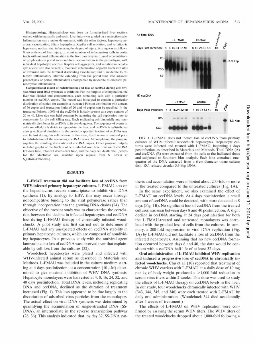
L-FMAU treatment did not facilitate loss of cccDNA fromWHV-infected primary hepatocyte cultures. L-FMAU acts onthe hepadnavirus reverse transcriptase to inhibit viral DNAsynthesis (1). By analogy to EBV, this may occur throughnoncompetitive binding to the viral polymerase rather thanthrough incorporation into the growing DNA chains (24). Theobjective of the present study was to characterize the correla-tion between the decline in infected hepatocytes and cccDNAloss during L-FMAU therapy of chronically infected wood-chucks. A pilot study was first carried out to determine ifL-FMAU had any unexpected effects on cccDNA stability inprimary hepatocyte cultures, which are composed of nondivid-ing hepatocytes. In a previous study with the antiviral agentlamivudine, no loss of cccDNA was observed over that explain-able by cell loss from the cultures (32).
Woodchuck hepatocytes were plated and infected withWHV-infected animal serum as described in Materials andMethods. L-FMAU was included in the culture medium start-ing at 4 days postinfection, at a concentration (10 mM) deter-mined to give maximal inhibition of WHV DNA synthesis.Hepatocyte monolayers were harvested at 4, 8, 16, 24, 32, and40 days postinfection. Total DNA levels, including replicatingDNA and cccDNA, declined as the duration of treatmentincreased (Fig. 1). This loss appeared to be due largely to thedissociation of adsorbed virus particles from the monolayers.The actual effect on viral DNA synthesis was determined byquantifying the accumulation of single-stranded DNA (SS-DNA), an intermediate in the reverse transcription pathway(28, 36). This analysis indicated that, by day 32, SS-DNA syn-
thesis and accumulation were inhibited about 200-fold or morein the treated compared to the untreated cultures (Fig. 1A).
In the same experiment, we also examined the effect ofL-FMAU on cccDNA levels. At 4 days postinfection, a smallamount of cccDNA could be detected, with more detected at 8days (Fig. 1B). No significant loss of cccDNA from the treatedcultures was seen between days 8 and 40 postinfection. A slightdecline in cccDNA starting at 24 days postinfection for boththe L-FMAU-treated and untreated monolayers was corre-lated with the gradual loss of cells from the cultures. In sum-mary, a 200-fold suppression in viral DNA replication (Fig.1A) by L-FMAU did not facilitate a loss of cccDNA from theinfected hepatocytes. Assuming that no new cccDNA forma-tion occurred between days 8 and 40, the data would be con-sistent with a cccDNA half-life of at least 32 days.
Oral administration of L-FMAU inhibited WHV replicationand induced a progressive loss of cccDNA in chronically in-fected woodchucks. Chu et al. (10) reported that treatment ofchronic WHV carriers with L-FMAU at a daily dose of 10 mgper kg of body weight produced a .1,000-fold reduction inserum virus titers within 2 weeks. This dose was used to studythe effects of L-FMAU therapy on cccDNA levels in the liver.In our study, four woodchucks chronically infected with WHV(343, 344, 345, and 346) were each treated with L-FMAU bydaily oral administration. (Woodchuck 344 died accidentallyafter 4 weeks of treatment.)
The effects of L-FMAU on WHV replication were con-firmed by assaying the serum WHV titers. The WHV titers ofthe treated woodchucks dropped about 1,000-fold following 4
FIG. 1. L-FMAU does not induce loss of cccDNA from primarycultures of WHV-infected woodchuck hepatocytes. Hepatocyte cul-tures were infected and treated with L-FMAU, beginning 4 dayspostinfection, as described in Materials and Methods. Total DNA (A)and cccDNA (B) were extracted from the cells at the indicated timesand subjected to Southern blot analysis. Each lane contained one-quarter of the DNA extracted from a 6-cm-diameter tissue culturedish. RC, relaxed circular 3.3-kbp DNA.
VOL. 75, 2001 MAINTENANCE OF HEPADNAVIRUS cccDNA 313
on June 11, 2014 by guesthttp://jvi.asm
.org/D
ownloaded from
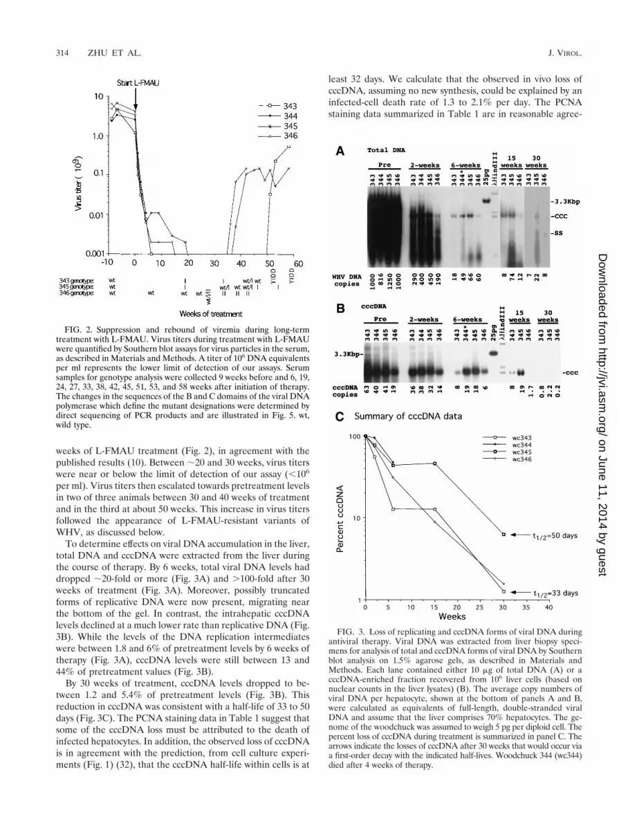
weeks of L-FMAU treatment (Fig. 2), in agreement with thepublished results (10). Between ;20 and 30 weeks, virus titerswere near or below the limit of detection of our assay (,106
per ml). Virus titers then escalated towards pretreatment levelsin two of three animals between 30 and 40 weeks of treatmentand in the third at about 50 weeks. This increase in virus titersfollowed the appearance of L-FMAU-resistant variants ofWHV, as discussed below.
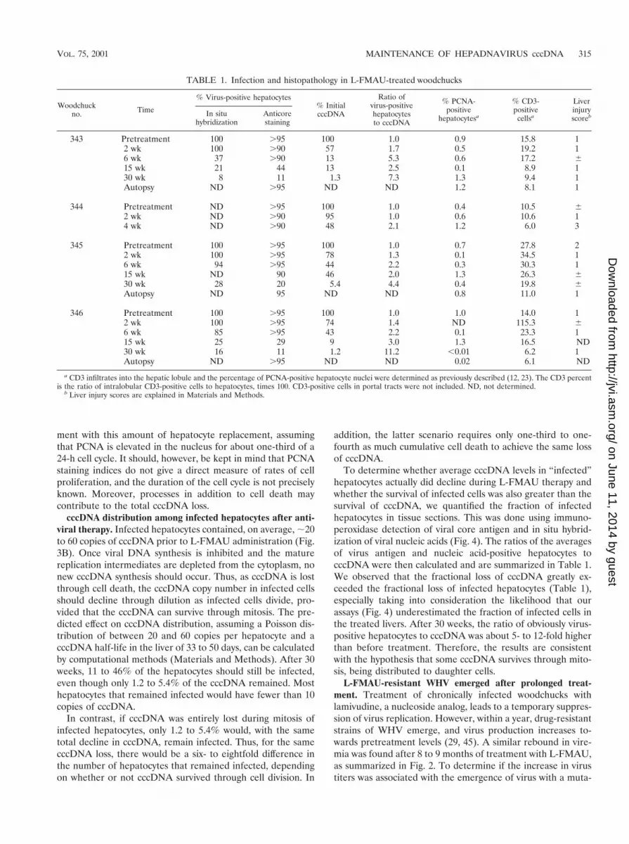
To determine effects on viral DNA accumulation in the liver,total DNA and cccDNA were extracted from the liver duringthe course of therapy. By 6 weeks, total viral DNA levels haddropped ;20-fold or more (Fig. 3A) and .100-fold after 30weeks of treatment (Fig. 3A). Moreover, possibly truncatedforms of replicative DNA were now present, migrating nearthe bottom of the gel. In contrast, the intrahepatic cccDNAlevels declined at a much lower rate than replicative DNA (Fig.3B). While the levels of the DNA replication intermediateswere between 1.8 and 6% of pretreatment levels by 6 weeks oftherapy (Fig. 3A), cccDNA levels were still between 13 and44% of pretreatment values (Fig. 3B).
By 30 weeks of treatment, cccDNA levels dropped to be-tween 1.2 and 5.4% of pretreatment levels (Fig. 3B). Thisreduction in cccDNA was consistent with a half-life of 33 to 50days (Fig. 3C). The PCNA staining data in Table 1 suggest thatsome of the cccDNA loss must be attributed to the death ofinfected hepatocytes. In addition, the observed loss of cccDNAis in agreement with the prediction, from cell culture experi-ments (Fig. 1) (32), that the cccDNA half-life within cells is at
least 32 days. We calculate that the observed in vivo loss ofcccDNA, assuming no new synthesis, could be explained by aninfected-cell death rate of 1.3 to 2.1% per day. The PCNAstaining data summarized in Table 1 are in reasonable agree-
FIG. 2. Suppression and rebound of viremia during long-termtreatment with L-FMAU. Virus titers during treatment with L-FMAUwere quantified by Southern blot assays for virus particles in the serum,as described in Materials and Methods. A titer of 106 DNA equivalentsper ml represents the lower limit of detection of our assays. Serumsamples for genotype analysis were collected 9 weeks before and 6, 19,24, 27, 33, 38, 42, 45, 51, 53, and 58 weeks after initiation of therapy.The changes in the sequences of the B and C domains of the viral DNApolymerase which define the mutant designations were determined bydirect sequencing of PCR products and are illustrated in Fig. 5. wt,wild type.
FIG. 3. Loss of replicating and cccDNA forms of viral DNA duringantiviral therapy. Viral DNA was extracted from liver biopsy speci-mens for analysis of total and cccDNA forms of viral DNA by Southernblot analysis on 1.5% agarose gels, as described in Materials andMethods. Each lane contained either 10 mg of total DNA (A) or acccDNA-enriched fraction recovered from 106 liver cells (based onnuclear counts in the liver lysates) (B). The average copy numbers ofviral DNA per hepatocyte, shown at the bottom of panels A and B,were calculated as equivalents of full-length, double-stranded viralDNA and assume that the liver comprises 70% hepatocytes. The ge-nome of the woodchuck was assumed to weigh 5 pg per diploid cell. Thepercent loss of cccDNA during treatment is summarized in panel C. Thearrows indicate the losses of cccDNA after 30 weeks that would occur viaa first-order decay with the indicated half-lives. Woodchuck 344 (wc344)died after 4 weeks of therapy.
314 ZHU ET AL. J. VIROL.
on June 11, 2014 by guesthttp://jvi.asm
.org/D
ownloaded from
ment with this amount of hepatocyte replacement, assumingthat PCNA is elevated in the nucleus for about one-third of a24-h cell cycle. It should, however, be kept in mind that PCNAstaining indices do not give a direct measure of rates of cellproliferation, and the duration of the cell cycle is not preciselyknown. Moreover, processes in addition to cell death maycontribute to the total cccDNA loss.
cccDNA distribution among infected hepatocytes after anti-viral therapy. Infected hepatocytes contained, on average, ;20to 60 copies of cccDNA prior to L-FMAU administration (Fig.3B). Once viral DNA synthesis is inhibited and the maturereplication intermediates are depleted from the cytoplasm, nonew cccDNA synthesis should occur. Thus, as cccDNA is lostthrough cell death, the cccDNA copy number in infected cellsshould decline through dilution as infected cells divide, pro-vided that the cccDNA can survive through mitosis. The pre-dicted effect on cccDNA distribution, assuming a Poisson dis-tribution of between 20 and 60 copies per hepatocyte and acccDNA half-life in the liver of 33 to 50 days, can be calculatedby computational methods (Materials and Methods). After 30weeks, 11 to 46% of the hepatocytes should still be infected,even though only 1.2 to 5.4% of the cccDNA remained. Mosthepatocytes that remained infected would have fewer than 10copies of cccDNA.
In contrast, if cccDNA was entirely lost during mitosis ofinfected hepatocytes, only 1.2 to 5.4% would, with the sametotal decline in cccDNA, remain infected. Thus, for the samecccDNA loss, there would be a six- to eightfold difference inthe number of hepatocytes that remained infected, dependingon whether or not cccDNA survived through cell division. In
addition, the latter scenario requires only one-third to one-fourth as much cumulative cell death to achieve the same lossof cccDNA.
To determine whether average cccDNA levels in “infected”hepatocytes actually did decline during L-FMAU therapy andwhether the survival of infected cells was also greater than thesurvival of cccDNA, we quantified the fraction of infectedhepatocytes in tissue sections. This was done using immuno-peroxidase detection of viral core antigen and in situ hybrid-ization of viral nucleic acids (Fig. 4). The ratios of the averagesof virus antigen and nucleic acid-positive hepatocytes tocccDNA were then calculated and are summarized in Table 1.We observed that the fractional loss of cccDNA greatly ex-ceeded the fractional loss of infected hepatocytes (Table 1),especially taking into consideration the likelihood that ourassays (Fig. 4) underestimated the fraction of infected cells inthe treated livers. After 30 weeks, the ratio of obviously virus-positive hepatocytes to cccDNA was about 5- to 12-fold higherthan before treatment. Therefore, the results are consistentwith the hypothesis that some cccDNA survives through mito-sis, being distributed to daughter cells.
L-FMAU-resistant WHV emerged after prolonged treat-ment. Treatment of chronically infected woodchucks withlamivudine, a nucleoside analog, leads to a temporary suppres-sion of virus replication. However, within a year, drug-resistantstrains of WHV emerge, and virus production increases to-wards pretreatment levels (29, 45). A similar rebound in vire-mia was found after 8 to 9 months of treatment with L-FMAU,as summarized in Fig. 2. To determine if the increase in virustiters was associated with the emergence of virus with a muta-
TABLE 1. Infection and histopathology in L-FMAU-treated woodchucks
Woodchuckno. Time
% Virus-positive hepatocytes% InitialcccDNA
Ratio ofvirus-positivehepatocytesto cccDNA
% PCNA-positive
hepatocytesa
% CD3-positive
cellsa
LiverinjuryscorebIn situ
hybridizationAnticorestaining
343 Pretreatment 100 .95 100 1.0 0.9 15.8 12 wk 100 .90 57 1.7 0.5 19.2 16 wk 37 .90 13 5.3 0.6 17.2 615 wk 21 44 13 2.5 0.1 8.9 130 wk 8 11 1.3 7.3 1.3 9.4 1Autopsy ND .95 ND ND 1.2 8.1 1
344 Pretreatment ND .95 100 1.0 0.4 10.5 62 wk ND .90 95 1.0 0.6 10.6 14 wk ND .90 48 2.1 1.2 6.0 3
345 Pretreatment 100 .95 100 1.0 0.7 27.8 22 wk 100 .95 78 1.3 0.1 34.5 16 wk 94 .95 44 2.2 0.3 30.3 115 wk ND 90 46 2.0 1.3 26.3 630 wk 28 20 5.4 4.4 0.4 19.8 6Autopsy ND 95 ND ND 0.8 11.0 1
346 Pretreatment 100 .95 100 1.0 1.0 14.0 12 wk 100 .95 74 1.4 ND 115.3 66 wk 85 .95 43 2.2 0.1 23.3 115 wk 25 29 9 3.0 1.3 16.5 ND30 wk 16 11 1.2 11.2 ,0.01 6.2 1Autopsy ND .95 ND ND 0.02 6.1 ND
a CD3 infiltrates into the hepatic lobule and the percentage of PCNA-positive hepatocyte nuclei were determined as previously described (12, 23). The CD3 percentis the ratio of intralobular CD3-positive cells to hepatocytes, times 100. CD3-positive cells in portal tracts were not included. ND, not determined.
b Liver injury scores are explained in Materials and Methods.
VOL. 75, 2001 MAINTENANCE OF HEPADNAVIRUS cccDNA 315
on June 11, 2014 by guesthttp://jvi.asm
.org/D
ownloaded from
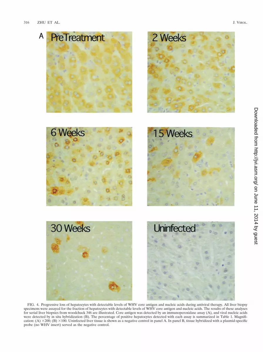
FIG. 4. Progressive loss of hepatocytes with detectable levels of WHV core antigen and nucleic acids during antiviral therapy. All liver biopsyspecimens were assayed for the fraction of hepatocytes with detectable levels of WHV core antigen and nucleic acids. The results of these analysesfor serial liver biopsies from woodchuck 346 are illustrated. Core antigen was detected by an immunoperoxidase assay (A), and viral nucleic acidswere detected by in situ hybridization (B). The percentage of positive hepatocytes detected with each assay is summarized in Table 1. Magnifi-cation: (A) 3200; (B) 3100. Uninfected liver tissue is shown as a negative control in panel A. In panel B, tissue hybridized with a plasmid-specificprobe (no WHV insert) served as the negative control.
316 ZHU ET AL. J. VIROL.
on June 11, 2014 by guesthttp://jvi.asm
.org/D
ownloaded from

FIG. 4—Continued.
VOL. 75, 2001 MAINTENANCE OF HEPADNAVIRUS cccDNA 317
on June 11, 2014 by guesthttp://jvi.asm
.org/D
ownloaded from
tion(s) in the active sites of the polymerase, direct sequencingof PCR products spanning this region was carried out.
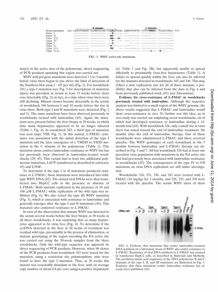
WHV with pol gene mutations were detected 1.5 to 3 monthsbefore virus titers began to rise above the limit of detection ofthe Southern blot assay (;106 per ml) (Fig. 2). For woodchuck345, a type I mutation (see Fig. 5 for descriptions of mutationtypes) was prevalent in serum at least 15 weeks before titersrose detectably (Fig. 2); in fact, at a time when virus titers werestill declining. Mutant viruses became detectable in the serumof woodchuck 346 between 6 and 10 weeks before the rise invirus titers. Both type I and II mutations were detected (Fig. 2and 5). The same mutations have been observed previously inwoodchucks treated with lamivudine (45). Again, the muta-tions were present before the liver biopsy at 30 weeks, at whichtime many hepatocytes appeared to be no longer infected(Table 1, Fig. 4). In woodchuck 343, a third type of mutationwas seen (type VIII; Fig. 5). In this animal, L-FMAU resis-tance was associated with the initial detection of the type Imutation and the later emergence of a YMDD to YIDD mu-tation in the C domain of the polymerase (Table 2). Thismutation alone confers lamivudine resistance to WHV but hasnot previously been observed in lamivudine-treated wood-chucks (29, 45). This variant had at least two additional poly-merase mutations, L467F (numbered as described in reference45) and L594F.
To determine if the type I or II mutations produced resis-tance to L-FMAU, these mutations were introduced into wild-type WHV DNA (45). The cloned viral DNAs were then trans-fected into HepG2 cells in the presence or absence ofL-FMAU. Both mutants replicated in the presence of 10 and100 mM L-FMAU, while replication of the wild type was in-hibited (Fig. 6). We also tested the type III WHV mutation(Fig. 5), which is associated with resistance to lamivudine andgenerally emerges after the type I and II mutations (45). Thismutation also conferred resistance to L-FMAU.
In view of the observation that mutant WHV was detected inthe serum several weeks before the liver biopsy at 30 weeks inall three woodchucks, it was surprising that so many hepato-cytes appeared to be virus free (Fig. 4). To determine if thecccDNA detected in the liver at 30 weeks of treatment wasresidual wild type, presumably in the process of elimination, ormutant, genotyping of the region encoding the Pol active sitewas carried out using the 30-week samples from the threewoodchucks. Only the wild-type sequence was apparent bydirect sequencing of PCR products. However, when 96 clonesof the PCR products from woodchuck 343 were tested for themutation, using a restriction site polymorphism, nine werefound to have the type I mutation. Thus, at 30 weeks thismutant was reasonably abundant in the liver, with an averagecopy number of about 0.8 per core antigen-positive hepatocyte
(cf. Table 1 and Fig. 3B), but apparently unable to spreadefficiently to presumably virus-free hepatocytes (Table 1). Afailure to spread quickly within the liver can also be inferredfor the mutants detected in woodchucks 345 and 346. This mayreflect a slow replication rate for all of these mutants, a pos-sibility that also can be inferred from the data in Fig. 6 andfrom previously published work (45) (see Discussion).
Evidence for cross-resistance of L-FMAU in woodchuckspreviously treated with lamivudine. Although the sequenceanalysis was limited to a small region of the WHV genome, theabove results suggested that L-FMAU and lamivudine wouldshow cross-resistance in vivo. To further test this idea, an invivo study was carried out employing seven woodchucks, six ofwhich had developed resistance to lamivudine during a 14-month trial (44). With woodchuck 326, only a small rise in virustiters was noted toward the end of lamivudine treatment. Sixmonths after the end of lamivudine therapy, four of thesewoodchucks were administered L-FMAU and three receivedplacebo. The WHV genotypes of each woodchuck in the 5months between lamivudine and L-FMAU therapy are de-scribed in Fig. 5 and 7. All but woodchuck 326 had at least oneprevalent virus population with mutations in the Pol active sitethat had previously been associated with lamivudine resistancein woodchucks (45). The consequences of the type IV to VIImutations on virus DNA replication are still under investiga-tion.
Woodchucks 326, 331, 336, and 342 were treated with L-FMAU (10 mg/kg) for 2 months, and 328, 335, and 338 weretreated with the placebo. The serum WHV titers of these
FIG. 5. WHV active-site mutations.
FIG. 6. Evidence that mutations that confer lamivudine-resistantDNA synthesis on a laboratory strain of WHV also confer resistance toL-FMAU. Resistance of viral DNA synthesis to L-FMAU was assayedin transfected HepG2 cells, as described in Materials and Methods.The predicted amino acid sequences of the DNA polymerase B and Cdomains of the type I, II, and III mutations are illustrated in Fig. 5.Evidence that these mutations confer lamivudine resistance has al-ready been published (45).
318 ZHU ET AL. J. VIROL.
on June 11, 2014 by guesthttp://jvi.asm
.org/D
ownloaded from
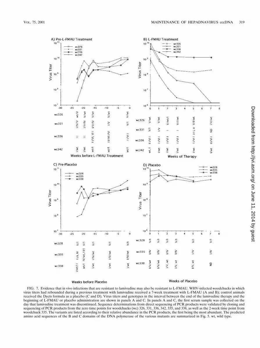
FIG. 7. Evidence that in vivo infections that are resistant to lamivudine may also be resistant to L-FMAU. WHV-infected woodchucks in whichvirus titers had rebounded during a previous treatment with lamivudine received a 7-week treatment with L-FMAU (A and B); control animalsreceived the Dyets formula as a placebo (C and D). Virus titers and genotypes in the interval between the end of the lamivudine therapy and thebeginning of L-FMAU or placebo administration are shown in panels A and C. In panels A and C, the first serum sample was collected on theday that lamivudine treatment was discontinued. Sequence determinations from direct sequencing of PCR products were validated by cloning andsequencing of PCR products from the zero time points for woodchucks (wc) 326, 331, 336, 342, 335, and 338, as well as the 2-week time point fromwoodchuck 335. The variants are listed according to their relative abundance in the PCR products, the first being the most abundant. The predictedamino acid sequences of the B and C domains of the DNA polymerase of the various mutants are summarized in Fig. 5. wt, wild type.
VOL. 75, 2001 MAINTENANCE OF HEPADNAVIRUS cccDNA 319
on June 11, 2014 by guesthttp://jvi.asm
.org/D
ownloaded from
woodchucks were monitored during the course of treatmentand are summarized in Fig. 7. Titers of virus in the woodchuckstreated with placebo stayed relatively unchanged (Fig. 7C andD). During L-FMAU treatment, the serum titers in woodchuck336, harboring a prevalent type II mutant, also remained rel-atively constant (Fig. 7A and B). Virus titers decreased about25-fold in woodchuck 342, with a mixture of wild-type and typeI mutant virus. In contrast, the decline in virus titers in the twowoodchucks harboring a mixture of wild-type and type V mu-tant virus (no. 326) or type V and type I mutants (no. 331)decreased as rapidly as in woodchucks harboring the wild typeas the predominant species (Fig. 2). Residual virus detected inwoodchuck 326 after 5 weeks showed a shift towards a wild-type/type I mutant combination, while that in woodchuck 331showed a shift to a type II variant. Why the type I variant incombination with the wild-type virus appeared to producegreater resistance to L-FMAU (in 342) than in combinationwith the type V variant (in 331) is unclear. While the type Ivariant is unable to make the S envelope protein due to a stopcodon introduced in the overlapping S gene by the mutation, itis not apparent why this would make any difference in such ashort-term experiment. The results might be explainable if thepolymerase functioned as a homodimeric protein, with resis-tance occurring through complementation of wild-type andtype I mutant subunits.
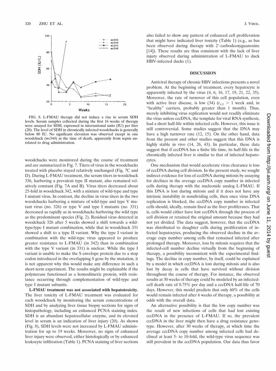
L-FMAU treatment was not associated with hepatotoxicity.The liver toxicity of L-FMAU treatment was evaluated foreach woodchuck by monitoring the serum concentrations ofSDH and by analyzing liver tissue biopsy sections for signs ofhistopathology, including an enhanced PCNA staining index.SDH is an abundant hepatocellular enzyme, and its elevatedlevel in serum is an indication of liver injury (20). As shown(Fig. 8), SDH levels were not increased by L-FMAU adminis-tration for up to 19 weeks. Moreover, no signs of enhancedliver injury were observed, either histologically or by enhancedleukocyte infiltration (Table 1). PCNA staining of liver sections
also failed to show any pattern of enhanced cell proliferationthat might have indicated liver toxicity (Table 1) (e.g., as hasbeen observed during therapy with 29-carbodeoxyguanosine[14]). These results are thus consistent with the lack of liverinjury observed during administration of L-FMAU to duckHBV-infected ducks (1).
DISCUSSION
Antiviral therapy of chronic HBV infections presents a novelproblem. At the beginning of treatment, every hepatocyte isapparently infected by the virus (4, 6, 16, 17, 19, 21, 22, 35).Moreover, the rate of turnover of this cell population, evenwith active liver disease, is low (34) (t1/2 . 1 week and, in“healthy” carriers, probably greater than 1 month). Thus,merely inhibiting virus replication would not readily eliminatethe virus unless cccDNA, the template for viral RNA synthesis,had a short half-life within infected cells. However, this issue isstill controversial. Some studies suggest that the DNA mayhave a high turnover rate (12, 15). On the other hand, datafrom the present and other studies suggest that this DNA ishighly stable in vivo (14, 26, 43). In particular, these datasuggest that if cccDNA has a finite life time, its half-life in thechronically infected liver is similar to that of infected hepato-cytes.
One mechanism that would accelerate virus clearance is lossof cccDNA during cell division. In the present study, we soughtindirect evidence for loss of cccDNA during mitosis by assayingfor declines in the average cccDNA copy number in infectedcells during therapy with the nucleoside analog L-FMAU. Ifthis DNA is lost during mitosis and if it does not have anyintrinsic instability in nondividing cells, then once virus DNAreplication is blocked, the cccDNA copy number in infectedcells should, ideally, remain fixed as the liver proliferates. Thatis, cells would either have lost cccDNA through the process ofcell division or retained the original amount because they hadnot yet divided. The data suggest, however, that the cccDNAwas distributed to daughter cells during proliferation of in-fected hepatocytes, producing the observed decline in the av-erage copy number among cells that remained infected afterprolonged therapy. Moreover, loss by mitosis requires that theinfected-cell number decline virtually from the beginning oftherapy, a possibility inconsistent with the experimental find-ings. The decline in copy number, by itself, could be explainedby a model in which cccDNA is lost during mitosis and is alsolost by decay in cells that have survived without divisionthroughout the course of therapy. For instance, the observedresults at 30 weeks of therapy could be modeled by an infected-cell death rate of 0.75% per day and a cccDNA half-life of 70days. However, this model predicts that only 60% of the cellswould remain infected after 6 weeks of therapy, a possibility atodds with the overall data.
An alternative possibility is that the low copy number wasthe result of new infections of cells that had lost existingcccDNA in the presence of L-FMAU. If so, the prevalentcccDNA in the liver might then have a drug resistance geno-type. However, after 30 weeks of therapy, at which time theaverage cccDNA copy number among infected cells had de-clined at least 5- to 10-fold, the wild-type virus sequence wasstill prevalent in the cccDNA population. Our data thus favor
FIG. 8. L-FMAU therapy did not induce a rise in serum SDHlevels. Serum samples collected during the first 16 weeks of therapywere assayed for SDH, expressed in international units (IU) per liter(20). The level of SDH in chronically infected woodchucks is generallybelow 80 IU. No significant elevation was observed except in onewoodchuck (wc344) at the time of death, apparently from sepsis un-related to drug administration.
320 ZHU ET AL. J. VIROL.
on June 11, 2014 by guesthttp://jvi.asm
.org/D
ownloaded from
but do not prove the hypothesis that cccDNA survives throughmitosis and is distributed to each daughter cell, resulting in adecline in cccDNA copy number per cell. Data from a recentstudy (13) of WHV cccDNA survival in primary hepatocytecultures that were induced to undergo limited proliferation byaddition of epidermal growth factor were also consistent withthis possibility.
Examination of the data in Fig. 2 and in a previous study(45) revealed an unexpected result. The type I mutation wassometimes detectable as a prevalent species in serum virus atearly times in therapy, when virus titers were still declining.Since the same mutation may be associated with the laterrebound of virus titers (Fig. 2) (45) and since the type I mu-tation confers L-FMAU resistance on a laboratory strain ofWHV (Fig. 6), the reason for the continued decline at earlytimes is not obvious. Several possibilities, not necessarily mu-tually exclusive, need to be considered. First, additional muta-tions outside the sequenced region of the polymerase maycontribute to mutant fitness. This was not evident in a previousstudy, in which the complete pol gene of selected type I mu-tants was sequenced (45). However, the possibility has notbeen ruled out. Second, the type I mutant may be a commonquasispecies that is generated as a result of errors during re-verse transcription of a pregenomic RNA that was transcribedfrom wild-type cccDNA. In that case, it would be expected thatvirus titers would continue to decline until a significant fractionof this mutant virus could be converted to cccDNA. Third, thetype I mutant may have a low replication rate, which, togetherwith the need for coinfection with a virus producing the viralenvelope proteins, may delay its spread to uninfected hepato-cytes.
ACKNOWLEDGMENTS
We are grateful to Christoph Seeger, John Taylor, and Jesse Sum-mers for helpful suggestions during this work and for a critical readingof the manuscript, to A. Cywinski and the DNA Sequencing Facility ofthe FCCC for sequence determinations, and to Wendy Foster (Uni-versity of Adelaide) for technical assistance. Oligonucleotides weresynthesized in the institutional DNA Synthesis Facility under the di-rection of T. Yeung.
This work was supported by USPHS grants AI-18641, 3P01-CA-4073711S1, and CA-06927 from the National Institutes of Health, byan appropriation from the Commonwealth of Pennsylvania, and by aproject grant from the National Health and Medical Research Councilof Australia (A.R.J.).
REFERENCES
1. Aguesse-Germon, S., S. H. Liu, M. Chevallier, C. Pichoud, C. Jamard, C.Borel, C. K. Chu, C. Trepo, Y. C. Cheng, and F. Zoulim. 1998. Inhibitoryeffect of 29-fluoro-5-methyl-beta-L-arabinofuranosyl-uracil on duck hepatitisB virus replication. Antimicrob. Agents Chemother. 42:369–376.
2. Aldrich, C. E., L. Coates, T. T. Wu, J. Newbold, B. C. Tennant, J. Summers,C. Seeger, and W. S. Mason. 1989. In vitro infection of woodchuck hepato-cytes with woodchuck hepatitis virus and ground squirrel hepatitis virus.Virology 172:247–252.
3. Allen, M. I., M. Deslauriers, C. W. Andrews, G. A. Tipples, K. A. Walters,D. L. Tyrrell, N. Brown, and L. D. Condreay. 1998. Identification and char-acterization of mutations in hepatitis B virus resistant to lamivudine. Lami-vudine Clinical Investigation Group. Hepatology 27:1670–1677.
4. Barker, L. F., F. V. Chisari, P. P. McGrath, D. W. Dalgard, R. L. Kirschstein,J. D. Almeida, T. S. Edgington, D. G. Sharp, and M. R. Peterson. 1973.Transmission of type B viral hepatitis to chimpanzees. J. Infect. Dis. 127:648–652.
5. Bartholomeusz, A., L. C. Groenen, and S. A. Locarnini. 1997. Clinical expe-rience with famciclovir against hepatitis B virus and development of resis-tance. Intervirology 40:337–342.
6. Berquist, K. R., J. M. Peterson, B. L. Murphy, J. W. Ebert, J. E. Maynard,
and R. H. Purcell. 1975. Hepatitis B antigens in serum and liver of chim-panzees acutely infected with hepatitis B virus. Infect. Immun. 12:602–605.
7. Block, T. M., X. Lu, A. Mehta, J. Park, B. S. Blumberg, and R. Dwek. 1998.Role of glycan processing in hepatitis B virus envelope protein trafficking.Adv. Exp. Med. Biol. 435:207–216.
8. Buscher, M., W. Reiser, H. Will, and H. Schaller. 1985. Transcripts and theputative RNA pregenome of duck hepatitis B virus: implications for reversetranscription. Cell 40:717–724.
9. Chayama, K., Y. Suzuki, M. Kobayashi, A. Tsubota, M. Hashimoto, Y.Miyano, H. Koike, I. Koida, Y. Arase, S. Saitoh, N. Murashima, K. Ikeda,and H. Kumada. 1998. Emergence and takeover of YMDD motif mutanthepatitis B virus during long-term lamivudine therapy and re-takeover bywild type after cessation of therapy. Hepatology 27:1711–1716.
10. Chu, C. K., J. Du, B. Tennant, J. Jacob, L. A. Graham, S. Peek, B. Korba,J. L. Gerin, J. W. Witcher, and F. D. Boudinot. 1999. Pharmacokinetic andpharmacodynamic studies of 1-(2-fluoro-5-methyl-b-L-arabinofuranosyl)uracil (L-FMAU) in the woodchuck model of hepatitis B virus (HBV)infection. Antivir. Res. 34:A52.
11. Chu, C. K., T. Ma, K. Shanmuganathan, C. Wang, Y. Xiang, S. B. Pai, G. Q.Yao, J. P. Sommadossi, and Y. C. Cheng. 1995. Use of 29-fluoro-5-methyl-b-L-arabinofuranosyluracil as a novel antiviral agent for hepatitis B virus andEpstein-Barr virus. Antimicrob. Agents Chemother. 39: 979–981.
12. Civitico, G. M., and S. A. Locarnini. 1994. The half-life of duck hepatitis Bvirus supercoiled DNA in congenitally infected primary hepatocyte cultures.Virology 203:81–89.
13. Dandri, M., M. R. Burda, H. Will, and J. Petersen. 2000. Increased hepa-tocyte turnover and inhibition of woodchuck hepatitis B virus replication byadefovir in vitro do not lead to reduction of the closed circular DNA.Hepatology 32:139–146.
14. Fourel, I., J. M. Cullen, J. Saputelli, C. E. Aldrich, P. Schaffer, D. R. Averett,J. Pugh, and W. S. Mason. 1994. Evidence that hepatocyte turnover isrequired for rapid clearance of duck hepatitis B virus during antiviral therapyof chronically infected ducks. J. Virol. 68:8321–8330.
15. Genovesi, E. V., L. Lamb, I. Medina, D. Taylor, M. Seifer, S. Innaimo, R. J.Colonno, D. N. Standring, and J. M. Clark. 1998. Efficacy of the carbocyclic29-carbocyclic 29-deoxyguanosine nucleoside BMS-200475 in the woodchuckmodel of hepatitis B virus infection. Antimicrob. Agents Chemother. 42:3209–3217.
16. Guidotti, L. G., R. Rochford, J. Chung, M. Shapiro, R. Purcell, and F. V.Chisari. 1999. Viral clearance without destruction of infected cells duringacute HBV infection. Science 284:825–829.
17. Guo, J.-T., H. Zhou, C. Liu, C. Aldrich, J. Saputelli, T. Whitaker, M. L.Barrasa, W. S. Mason, and C. Seeger. 2000. Apoptosis and regeneration ofhepatocytes during recovery from transient hepadnavirus infections. J. Virol.74:1495–1505.
18. Honkoop, P., H. G. Niesters, R. A. de Man, A. D. Osterhaus, and S. W.Schalm. 1997. Lamivudine resistance in immunocompetent chronic hepatitisB: incidence and patterns. J. Hepatol. 26:1393–1395.
19. Hoofnagle, J. H., T. Michalak, A. Nowoslawski, R. J. Gerety, and L. F.Barker. 1978. Immunofluorescence microscopy in experimentally induced,type B hepatitis in the chimpanzee. Gastroenterology 74:182–187.
20. Hornbuckle, W. E., E. S. Graham, L. Roth, B. H. Baldwin, C. Wickenden,and B. C. Tennant. 1985. Laboratory assessment of hepatic injury in thewoodchuck (Marmota monax). Lab. Anim. Sci. 35:376–381.
21. Jilbert, A. R., T. T. Wu, J. M. England, P. M. Hall, N. Z. Carp, A. P.O’Connell, and W. S. Mason. 1992. Rapid resolution of duck hepatitis Bvirus infections occurs after massive hepatocellular involvement. J. Virol.66:1377–1388.
22. Kajino, K., A. R. Jilbert, J. Saputelli, C. E. Aldrich, J. Cullen, and W. S.Mason. 1994. Woodchuck hepatitis virus infections: very rapid recovery aftera prolonged viremia and infection of virtually every hepatocyte. J. Virol.68:5792–5803.
23. Kodama, K., N. Ogasawara, H. Yoshikawa, and S. Murakami. 1985. Nucle-otide sequence of a cloned woodchuck hepatitis virus genome: evolutionalrelationship between hepadnaviruses. J. Virol. 56:978–986.
24. Kukhanova, M., Z. Y. Lin, M. Yas’co, and Y. C. Cheng. 1998. Uniqueinhibitory effect of 1-(29-deoxy-29-fluoro-beta-L-arabinofuranosyl)-5-methyl-uracil 59-triphosphate on Epstein-Barr virus and human DNA polymerases.Biochem. Pharmacol. 55:1181–1187.
25. Ling, R., D. Mutimer, M. Ahmed, E. H. Boxall, E. Elias, G. M. Dusheiko, andT. J. Harrison. 1996. Selection of mutations in the hepatitis B virus poly-merase during therapy of transplant recipients with lamivudine. Hepatology24:711–713.
26. Luscombe, C., J. Pedersen, E. Uren, and S. Locarnini. 1996. Long-termganciclovir chemotherapy for congenital duck hepatitis B virus infection invivo: Effect on intrahepatic-viral DNA, RNA, and protein expression. Hepa-tology 24:766–773.
27. Mackey, D., and B. Sugden. 1999. The linking regions of EBNA1 are essen-tial for its support of replication and transcription. Mol. Cell. Biol. 19:3349–3359.
28. Mason, W. S., C. Aldrich, J. Summers, and J. M. Taylor. 1982. Asymmetricreplication of duck hepatitis B virus DNA in liver cells: free minus-strand
VOL. 75, 2001 MAINTENANCE OF HEPADNAVIRUS cccDNA 321
on June 11, 2014 by guesthttp://jvi.asm
.org/D
ownloaded from
DNA. Proc. Natl. Acad. Sci. USA 79:3997–4001.29. Mason, W. S., J. Cullen, G. Moraleda, J. Saputelli, C. E. Aldrich, D. S.
Miller, B. Tennant, L. Frick, D. Averett, L. D. Condreay, and A. R. Jilbert.1998. Lamivudine therapy of WHV-infected woodchucks. Virology 245:18–32.
30. Mehta, A., T. M. Block, and R. A. Dwek. 1998. The role of N-linked glyco-sylation in the secretion of hepatitis B virus. Adv. Exp. Med. Biol. 435:195–205.
31. Miller, R. H., and W. S. Robinson. 1984. Hepatitis B virus DNA forms innuclear and cytoplasmic fractions of infected human liver. Virology 137:390–399.
32. Moraleda, G., J. Saputelli, C. E. Aldrich, D. Averett, L. Condreay, and W. S.Mason. 1997. Lack of effect of antiviral therapy in nondividing hepatocytecultures on the closed circular DNA of woodchuck hepatitis virus. J. Virol.71:9392–9399.
33. Niesters, H. G., P. Honkoop, E. B. Haagsma, R. A. de Man, S. W. Schalm,and A. D. Osterhaus. 1998. Identification of more than one mutation in thehepatitis B virus polymerase gene arising during prolonged lamivudine treat-ment. J. Infect. Dis. 177:1382–1385.
34. Nowak, M. A., S. Bonhoeffer, A. M. Hill, R. Boehme, H. C. Thomas, and H.McDade. 1996. Viral dynamics in hepatitis B virus infection. Proc. Natl.Acad. Sci. USA 93:4398–4402.
34a.Peek, S. F., P. J. Cote, J. R. Jacob, I. A. Toshkov, W. E. Hornbuckle, B. H.Baldwin, F. V. Wells, C. K. Chu, J. L. Gerin, B. C. Tennant, and B. E. Korba.Antiviral activity of clevudine [L-FMAU, 1-(2-fluoro-5-methyl-b-L-arabino-furanosyl) uracil] against WHV replication and gene expression in chroni-cally infected woodchucks (M. monax). Hepatology, in press.
35. Ponzetto, A., P. J. Cote, E. C. Ford, R. H. Purcell, and J. L. Gerin. 1984. Coreantigen and antibody in woodchucks after infection with woodchuck hepa-titis virus. J. Virol. 52:70–76.
36. Summers, J., and W. S. Mason. 1982. Replication of the genome of a
hepatitis B-like virus by reverse transcription of an RNA intermediate. Cell29:403–415.
37. Summers, J., P. M. Smith, and A. L. Horwich. 1990. Hepadnavirus envelopeproteins regulate covalently closed circular DNA amplification. J. Virol.64:2819–2824.
38. Tipples, G. A., M. M. Ma, K. P. Fischer, V. G. Bain, N. M. Kneteman, andD. L. J. Tyrrell. 1996. Mutation in HBV DNA-dependent DNA polymeraseconfers resistance to lamivudine in vivo. Hepatology 24:714–717.
39. van der Eb, A. J., and F. L. Graham. 1980. Assay of transforming activity oftumor virus DNA. Methods Enzymol. 65:826–839.
40. Wahl, G. M., M. Stern, and G. R. Stark. 1979. Efficient transfer of largeDNA fragments from agarose gels to diazobenzyloxymethyl paper and rapidhybridization by using dextran sulfate. Proc. Natl. Acad. Sci. USA 76:3683–3687.
41. Wei, Y., and D. Ganem. 1996. Relationship between viral DNA synthesis andvirion envelopment in hepatitis B viruses. J. Virol. 70:6455–6458.
42. Yang, W., W. S. Mason, and J. Summers. 1996. Covalently closed circularviral DNA formed from two types of linear DNA in woodchuck hepatitisvirus-infected liver. J. Virol. 70:4567–4575.
43. Zhang, Y.-Y., and J. Summers. 2000. Low dynamic state of viral competitionin a chronic avian hepadnavirus infection. J. Virol. 74:5257–5265.
44. Zhou, T., J.-T. Guo, F. A. Nunes, K. L. Molnar-Kimber, J. M. Wilson, C. E.Aldrich, J. Saputelli, S. Litwin, L. Condreay, C. Seeger, and W. S. Mason.2000. Combination therapy with lamivudine and adenovirus causes transientsuppression of chronic woodchuck hepatitis virus infections. J. Virol. 74:11754–11763.
45. Zhou, T., J. Saputelli, C. E. Aldrich, M. Deslauriers, L. D. Condreay, andW. S. Mason. 1999. Emergence of drug-resistant populations of woodchuckhepatitis virus in woodchucks treated with the antiviral nucleoside lamivu-dine. Antimicrob. Agents Chemother. 43:1947–1954.
322 ZHU ET AL. J. VIROL.
on June 11, 2014 by guesthttp://jvi.asm
.org/D
ownloaded from


