Embed Size (px)
Citation preview
Jahresbericht 2016 der SCS Foundation Stiftung der Schweizerischen Chemischen Gesellschaft
Bern, 04. August 2017 Dr. Alain De Mesmaeker David Spichiger Präsident Geschäftsführer

1
Bilder, Text und Gestaltung: David Spichiger Zwecks Lesefreundlichkeit wird die geschlechtliche Doppelformulierung weg-gelassen. Alle männlichen Formulierungen gelten sinngemäss auch für weib-liche Personen. SCS Foundation
c/o Schweizerische Chemische Gesellschaft Haus der Akademien Laupenstrasse 7, Postfach 3001 Bern T: +41 31 306 92 92 [email protected] www.scs-foundation.ch

2
Inhaltsverzeichnis
2 2016 in Zahlen
3 Die SCS Foundation
5 Aktivitäten allgemeiner Fonds
8 Aktivitäten Alfred Werner Fonds
10 Partner und Gönner
Anhänge
i. Revisionsbericht
ii. Bilanz und Erfolgsrechnung
iii. CHIMIA Bericht: Review Werner Scholar-
ship winners 2015-2017
iv. CHIMIA Bericht: Review SCS Fall
Meeting 2016, inkl. Gewinner der ‚Best
Oral & Best Poster Presentation Awards’
v. CHIMIA Issue 4/2016: Laureates Junior
Prizes SCS Fall Meeting 2016
2016 in Zahlen
3 Anzahl unterstützte Projekte
10 Anzahl unterstützende Firmen
8 Anzahl Partner-Universitäten
247'750 Direkte Projekt-Vergabungen1
316’750 Summe der Sponsoreneinnahmen1
+81'109 Bilanzerfolg1
50'000 Stiftungskapital1
111’489 Kapital Allgemeiner Fonds (Dez 16)1
1'219’283 Kapital Werner Fonds (Dez 16)1
1 Angaben in CHF

3
Die SCS Foundation
Die SCS Foundation (SCS Stiftung, SCS Fon-
dation) ist eine Stiftung gemäss Art. 80-89
ZGB, welche im Handelsregister des Kantons
Bern eingetragen ist (CHE-114.458.707) und
unter Aufsicht der Eidgenössischen Stiftungs-
aufsicht (Eidg. Departement des Innern, EDI),
in Bern, agiert.
Die Stiftung wurde im Jahr 2008 gegründet
und per 01.01.2014 mit der 'Stiftung für Sti-
pendien auf dem Gebiete der Chemie (Werner
Stiftung)' fusioniert.
Ziel und Zweck der Stiftung
Zweck der Stiftung ist die Förderung und Un-
terstützung der Naturwissenschaften, im Be-
sonderen in den Bereichen Chemie und Bio-
chemie. Dazu gehört die Unterstützung von
Wissenschaft, Forschung, Lehre, Ausbildung
und Früherziehung durch Vorhaben, Mass-
nahmen, Projekte und Werke aller Art. Der
Stiftungszweck wird insbesondere verwirklicht
durch entsprechende Finanzierungen, Aus-
schüttungen, Unterstützungsbeiträge, Vergabe
von Stipendien, Preisverleihungen und durch
alle weiteren Aktivitäten, die der Erreichung
des Stiftungszweckes dienen. Die Stiftung
verfolgt öffentliche bzw. gemeinnützige Zwe-
cke und ist politisch und konfessionell neutral.
Sie verfolgt keine kommerziellen Zwecke und
erstrebt keinen Gewinn.
Organe der Stiftung
Stiftungsrat
Dem Stiftungsrat obliegen die strategische
Führung der Stiftung und die Kontrolle über die
Erfüllung des Stiftungszwecks.
Die Mitglieder des Stiftungsrats sind auch zu-
ständig für die Äufnung der Fonds.
Mitglieder des Stiftungsrats mit Stimmrecht
§ Dr. Alain De Mesmaeker, Präsident (2015
Präsident elect, ab 2016 Präsident)
§ Prof. Peter Chen, Vizepräsident (seit 2011)
§ Dr. Hans Peter Lüthi, Mitglied und Quästor
(seit 2011)
§ David Spichiger, Geschäftsführer (seit 2011)
§ Prof. Richard Ernst, Mitglied (2008-2016)
§ Prof. Titus Jenny, Mitglied (seit 2015)
§ Dr. Reto Naef, Mitglied (seit 2015)
§ Prof. Lothar Helm, Mitglied (seit 2015)
§ Prof. Richard Ernst, Mitglied (2008-2016)
Neu im Stiftungsrat per Januar 2017
§ Prof. Ulrich W. Suter, ETH Zurich / SATW
§ Dr. Gerardo M. Ramos Tombo, Syngenta
§ Prof. Beat Ernst, Universität Basel
4
Geschäftsstelle
Die Geschäftsführung der Stiftung wird von
David Spichiger wahrgenommen, welcher
auch die Geschäftsstelle der Schweizerischen
Chemischen Gesellschaft (SCG) führt. Die
Geschäftsstelle ist insbesondere für die Berei-
che operative Finanzen, Kommunikation und
Administration zuständig und agiert als
Schnittstelle zur Eidgenössischen Stiftungs-
aufsicht des EDI.
Anlageausschuss
Der Anlageausschuss definiert die Anlagestra-
tegie und legt diese dem Stiftungsrat zur Ge-
nehmigung vor. Mitglieder des Anlageaus-
schusses sind:
§ Dr. Hans Peter Lüthi (als Quästor ex-officio),
ETH Zürich
§ David Spichiger, Geschäftsführer SCS Foun-
dation
§ Simon Wyss, Privatbank von Graffenried,
Bern (seit Oktober 2014)
Vergabeausschuss SCS Fonds
Der Vergabeausschuss des SCS Funds defi-
niert die Mittelvergabe aus dem allgemeinen
Fonds der Stiftung. Die Mitglieder werden
durch den Präsidenten und den Quästor der
Stiftung sowie den Präsidenten der sechs Divi-
sionen der Schweizerischen Chemischen Ge-
sellschaft repräsentiert.
Vergabeausschuss Alfred Werner Fonds
Der Vergabeausschuss des Alfred Werner
Fonds definiert die Mittelvergabe aus dem
gleichnamigen Fonds und spricht Stipendien
des Master's Student Scholarships Programm.
Die Mitglieder werden durch die Donatorenfir-
men sowie die Vertreter der Partneruniversitä-
ten repräsentiert.
Rechnung
Das Rechnungswesen inklusive Jahresab-
schluss wird durch die Firma TREUA Baum-
gartner, Belp, wahrgenommen.
Revision
Als Revisor amtet Peter Baumgartner von der
Firma REVITREU Baumgartner, Gerzensee.

5
Aktivitäten
Allgemeiner Fonds
Best Oral Presentation Award of the SCS Fall Meeting 2016 at the University of Zürich
On the occasion of the SCS Fall Meeting 2016,
that took place on September 15 at the
University of Zürich, Metrohm sponsored again
the Best Oral Presentation Award. Dr. Markus
Steinke, Vice President Marketing at Metrohm,
handed the awards over to the winners (see
picture).
The prize is awarded for the two best
presentations of each parallel session. The
main criteria are the scientific quality and
originality of the research, plus the quality of
the presentation.
Prizes for Winners
- cash contribution of CHF 500.00
- travel voucher of CHF 1'000.00 towards the
participation in an international conference.
- invitation to present the research in the
laureates issue of CHIMIA, corresponding to
a monetary value of CHF 1200.00.
Prizes for Runners‘ up
- cash contribution of CHF 400.00
The Prize has been sponsored by the Metrohm
Foundation for the past 8 years.
Winners 2016
Analytical Sciences
1st Riedo Andreas, University of Bern 2nd Cuartero Maria, University of Geneva Analytical Sciences
1st Rahel Bucher, University of Zurich 2nd Lyndsey Hendriks, ETH Zurich Catalysis Science & Engineering
1st Waiz Karim, ETH Zurich/ Paul Scherrer Institute
2nd Vladimir Paunovic, ETH Zurich Computational Chemistry
1st Christopher Stein, ETH Zurich 2nd Clelia Spreafico, ETH Zurich Inorganic & Coordination Chemistry
1st Deven Paul Estes, ETH Zurich 2nd Suzanne Maria Jansze, EPF Lausanne Medicinal Chemistry & Chemical Biology
1st Olivia Paula Schmidt, University of Zurich Organic Chemistry
1st David Kossler, EPF Lausanne 2nd Dominik Lotter, University of Basel Physical Chemistry
1st Dominik Józef Kubicki, EPF Lausanne 2nd Maarten van Reijzen, EPF Lausanne Polymers, Colloids & Interfaces
1st Milad Radiom, University of Geneva 2nd Ralph Z. Lange, ETH Zurich

6
Best Poster Presentation Award of the SCS
Fall Meeting 2016 at the University of Zürich
On the occasion of the SCS Fall Meeting 2016,
that took place on September 15 at the
University of Zürich, DSM Nutritional Products
sponsored again the Best Poster Presentation
Award. Dr. Roman Imhof from DSM handed
the awards over to the winners (see picture).
The prizes were given for the best posters of
each parallel session. The main criteria are the
scientific quality and originality of the research,
plus the quality of the presentation.
Prize for Winners
- cash contribution of CHF 500.00
- travel voucher of CHF 750.00 towards the
participation in an international conference.
- invitation to present the research in the
laureates issue of CHIMIA, corresponding to
a monetary value of CHF 1200.00.
Runners‘ up Prize
- cash contribution of CHF 300.00
The Prize has been sponsored by DSM
Nutritional Products for more than 10 years
now.
Winners 2016
Analytical Sciences
1st Liviana Klein, ETH Zurich 2nd Benjamin Spenger, EMPA Elena-Diana Burghelea, HES-SO Catalysis Science & Engineering
1st Vincent Lebrun, University of Basel 2nd Aswin Gopakumar, EPF Lausanne Athanasia Tsoukalou, ETH Zurich Computational Chemistry
1st Gregor Nils Simm, ETH Zurich 2nd Akshaya Kumar Das, University of Basel Inorganic & Coordination Chemistry
1st Marta Falcone, EPF Lausanne 2nd Vivian Marina Merk, ETH Zurich Thibaud Rossel, Gymnase français de
Bienne Medicinal Chemistry & Chemical Biology
1st Fabian Brockmeyer, Northeastern Univ. Marc Heitz, University of Bern 2nd Fahimeh Moradi-Afrapoli, Uni Basel Isabel P. Kerschgens, University of Zurich Organic Chemistry
1st Franck le Vaillant, EPF Lausanne 2nd Santiago Lascano, University of Geneva Yun-Suk Jang, EPF Lausanne Physical Chemistry
1st Marine Eva Fedora Bouduban, EPFL 2nd Maximilian Beyer, ETH Zurich Joseph Samuel Beckwith, Uni Genève Polymers, Colloids & Interfaces
1st Fabian Deuber, ZHAW 2nd Yee Song Ko, EPF Lausanne G. Nedelcu, ETH Zurich

7
Aktivitäten
Alfred Werner Fonds
Das Alfred Werner Master's Student Scholar-
ship Programm unterstützt Master Studenten
der Studienrichtungen Chemie und Biochemie
an Schweizer Universitäten oder Eidgenössi-
schen Technischen Hochschulen.
Der Fonds unterstützt jährlich 8-10 bestaus-
gewiesene ausländische Studenten mit einem
Beitrag von CHF 25’000, um ihnen ein Mas-
ters-Studium in der Schweiz zu ermöglichen.
Studienzeit 2014-2016
Im Jahr 2014 wurden aufgrund der Reorgani-
sation und der Integration des Werner Fonds
in die SCS Foundation keine Stipendien ge-
sprochen.
Studienzeit 2015-2017
Im Jahr 2015 hat der Werner Fonds folgende
Studenten mit einem Stipendium unterstützt,
die das Masterstudium im Herbst 2015 begon-
nen haben:
Ms. Cassandra Oji-Okora Ogadimma: EPF Lausanne BSc at Lomonosov Moscow State University of fine chemical technologies, Russia
Mr. Loren Ban: ETH Zurich BSc at University of Zagreb, Croatia
Mr. Riccardo Tarchini: ETH Zurich BSc at Politecnico di Torino, Italy
Ms. Sarah Folliet: University of Geneva MSc in chemistry, Department of Organic Chemistry, University of Geneva, Switzerland
Mr. Lluc Farrera: University of Geneva BSc at University of Barcelona, Spain
Ms. Yuting Feng: University of Geneva BSc at McGill University, Montréal, QC, Canada
Mr. Ali Tuna: University of Zurich BSc at Istanbul University, Turkey
Studienzeit 2016-2018
Mr. Jean Behaghel de Bueren: EPFL BSc at Univ. de Louvain‐la-Neuve, Belgium
Ms. Zahra Pourmand Tehrani: EPFL BSc at Amirkabir University of Technology, Tehran, Iran
Ms. Dora Harangozo: ETH Zurich BSc at Josip Juraj Strossmayer University of Osjek, Croatia
Ms. Viktoriia Morad: ETH Zurich BSc at Ivan Franko National University of Lviv, Ukraine
Mr. Zlatko Jončev: University of Basel BSc at University of Belgrade, Serbia
Mr. Heorhii Humeniuk: University of Geneva BSc at National University Kiev, Ukraine
Mr. Benjamin Planterose: Univ. Geneva BSc at University of Seville, Spain
Mr. Haitham Kandeel: EPF Lausanne (2016-2017), BSc at The American University in Cairo, Egypt

8
Visit at Lonza in Visp, April 20, 2016
On behalf of the SCS Foundation Werner
Fund, Dr. Christoph Täschler, Principal
Scientist, Process Development at Lonza,
organized a networking event and a a visit of
the Lonza Visp facilites to give the students
exposure to a chemical production site and to
stimulate their interaction with the industrial
partners of the program. All seven Werner
scholarship holders as well as Christian
Chapuis, Firmenich SA, Geneva, and member
of the Werner Fund Committee, and David
Spichiger, Director of the SCS Foundation,
followed the invitation and spent an exciting
day in Visp.
A short introduction to the SCS Foundation by
David Spichiger, followed by short
presentations of the MSc. thesis projects by
the students, opened the day. Dr. Täschler
then continued the program by presenting the
wide range of activities and services Lonza
provides worldwide. After a delicious lunch, the
group visited three different sites of the plant
and got an idea of how new research findings
are transformed into market products. It was
very impressive to look at chemical processes
outside an academic laboratory and to get an
appreciation of the scaling-up processed
involved to produce not only milligrams, but
tons of a substance.
We thank Dr. Täschler for hosting the first
event of this kind and, given its success, we
will continue these visits to chemical plants in
the coming years.
9
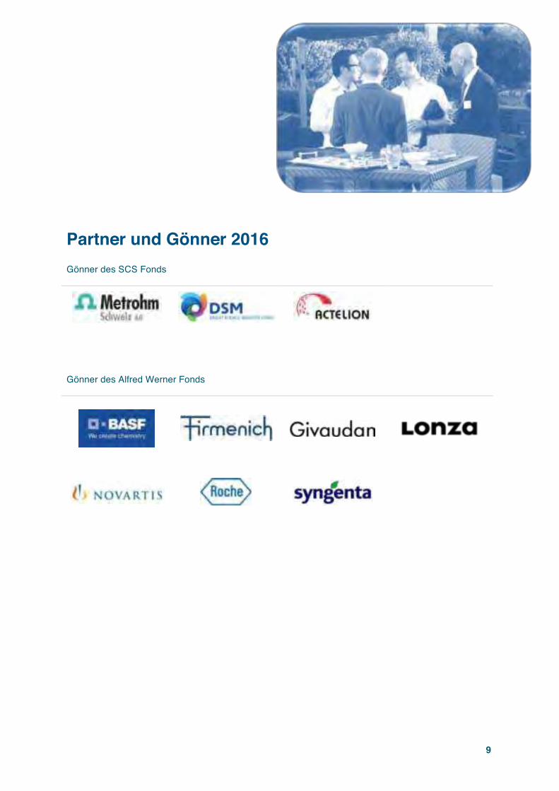
Partner und Gönner 2016
Gönner des SCS Fonds
Gönner des Alfred Werner Fonds


Columns CHIMIA 2017, 71, No. 9 A000
ALFRED WERNER FUND, MASTER’S STUDENT SCHOLARSHIPS
The Alfred Werner Fund of the SCS
Foundation was established in 2014 and
continues the initiatives and projects of
the former foundation "Stiftung für Sti-
pendien auf dem Gebiete der Chemie",
also known as “Werner Stiftung”. The
SCS Foundation is very proud to provide
this program in collaboration with the
Swiss chemical and pharmaceutical indus-
try.
https://scs-foundation.ch
Alfred Werner Master's Student Scholarships The program invites scholarship applications to carry out Master degree
studies in Chemistry or Biochemistry at a Swiss University or Federal
Institute of Technology.
The Foundation offers 8 to 10 scholarships of CHF 25'000 each as a
one-time contribution to the cost of the Master study program. This
opportunity targets students from foreign countries in the top 10% of
their undergraduate programs. The goal of the program is to bring in
young talents to Swiss Universities or FIT or to keep them after the
BSc studies in Switzerland.
Partner Universities / Federal Institutes of Technology
The program is supported by
Winners of the Scholarships 2017-19/20
The committee of the Werner Fund is proud to announce the ten win-
ners of the next term that starts in Fall 2017 and Spring 2018 respec-
tively:
§ Mrs Alena Budinská
BSc at Institute of Chemical Technology, Prague, Czech Republic
Support for her MSc studies at ETH Zurich
§ Mrs Jessica Caldwell
BA at Indiana University Purdue, University Columbus, USA
Support for her MSc studies at University of Fribourg
§ Mr Durbis J. Castillo-Pazos
BSc at Universidad Autónoma del Estado de Mexico (UAEM),
Mexico
Support for his MSc studies at University of Zürich
§ Mr Ihor Cherniukh
BSc at Ivan Franko National University of Lviv, Ukraine
Support for his MSc studies at ETH Zurich
§ Mr Brett M. Garabedian
BSc at University of California, Berkeley, USA
Support for hgis MSc studies at University of Basel
§ Mr Kevin Maik Jablonka
BSc at Technische Universität, München, Germany
Support for his MSc studies at EPF Lausanne
§ Mrs Asma Mansouri
BSc at University Houari Boumediene, Alger, Algeria
Support for her MSc studies at University of Geneva
§ Mr Dragan Miladinov
BSc at University of Novi Sad, Serbia
Support for his MSc studies at University of Basel
§ Mr Luka Milosevic
BSc at University of Belgrade, Serbia
Support for his MSc studies at EPF Lausanne
§ Mrs Kleni Mulliri
BSc and MSc at University of Tirana, Albanien
Support for her MSc studies at University of Bern
Summaries of the Master Thesis from Students of the Scholarship term 2015-17
Photoelectron spectroscopy of water clusters at 26.35eV
Loren Ban1 1ETH Zürich, Laboratory of Physical Chemistry, Vladimir-Prelog-Weg 2, CH-8093 Zürich, Switzerland. E-mail: [email protected]
Keywords: water � photoelectron spectroscopy � electron scattering � anisotropy parameter
Loren Ban
Nationality: Croatian
Bachelor at University of Zagreb, Croatia
Master at ETH Zürich
Master thesis supervisors:
Prof. Dr. Ruth Signorell
Thomas Gartmann
Studies on liquid water have been extensive throughout many
scientific disciplines due to its omnipresence in nature. However,
certain questions about water-based systems still remain unan-
swered. Namely, the molecular picture of electron scattering in
liquid water is far from being completely understood. Detailed
knowledge about the electron scattering cross-sections (SCS) would
be necessary in order to explain the behavior of the secondary elec-
trons (SEs) generated following ionization by high-energy radiation
(cosmic-rays, X-rays, fast-moving charged particles). The SEs
generally have initial energies below 30 eV and are referred to as
the low-energy electrons (LEEs) in the literature. It was shown by
Sanche and coworkers that the LEEs play a significant role in DNA
damage and ozone depletion [1,2,3]. Since the efficiency of these
processes depends on the energetics of the LEEs, determination of
CHIMIA Report/Company News CHIMIA 2017, 71, No. 9 A000
the electron SCS could offer a possibility to improve radiotherapy
and understand atmospheric processes.
The high energy scattering can be well described by dielectric
models and good agreement with experiments is obtained [4].
However, no accurate SCS of water are available for electrons with
energies below 50 eV. Both experiments and theory face problems
in the low energy regime where the electron scatters off molecular
degrees of freedom (inter- and intramolecular vibrations and elec-
tronic). Therefore, a different approach is required. Combining a
detailed scattering model with photoelectron studies of water clus-
ters formed in a molecular beam is a promising step towards under-
standing electron SCS below 50 eV.
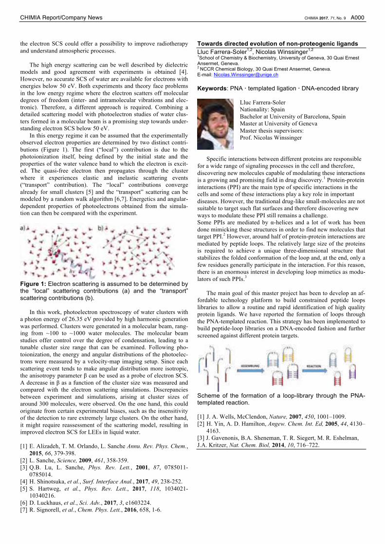
In this energy regime it can be assumed that the experimentally
observed electron properties are determined by two distinct contri-
butions (Figure 1). The first (“local”) contribution is due to the
photoionization itself, being defined by the initial state and the
properties of the water valence band to which the electron is excit-
ed. The quasi-free electron then propagates through the cluster
where it experiences elastic and inelastic scattering events
(“transport” contribution). The “local” contributions converge
already for small clusters [5] and the “transport” scattering can be
modeled by a random walk algorithm [6,7]. Energetics and angular-
dependent properties of photoelectrons obtained from the simula-
tion can then be compared with the experiment.
Figure 1: Electron scattering is assumed to be determined by the “local” scattering contributions (a) and the “transport” scattering contributions (b).
In this work, photoelectron spectroscopy of water clusters with
a photon energy of 26.35 eV provided by high harmonic generation
was performed. Clusters were generated in a molecular beam, rang-
ing from ~100 to ~1000 water molecules. The molecular beam
studies offer control over the degree of condensation, leading to a
tunable cluster size range that can be examined. Following pho-
toionization, the energy and angular distributions of the photoelec-
trons were measured by a velocity-map imaging setup. Since each
scattering event tends to make angular distribution more isotropic,
the anisotropy parameter β can be used as a probe of electron SCS.
A decrease in β as a function of the cluster size was measured and
compared with the electron scattering simulations. Discrepancies
between experiment and simulations, arising at cluster sizes of
around 300 molecules, were observed. On the one hand, this could
originate from certain experimental biases, such as the insensitivity
of the detection to rare extremely large clusters. On the other hand,
it might require reassessment of the scattering model, resulting in
improved electron SCS for LEEs in liquid water.
[1] E. Alizadeh, T. M. Orlando, L. Sanche Annu. Rev. Phys. Chem.,
2015, 66, 379-398.
[2] L. Sanche, Science, 2009, 461, 358-359.
[3] Q.B. Lu, L. Sanche, Phys. Rev. Lett., 2001, 87, 0785011-
0785014.
[4] H. Shinotsuka, et al., Surf. Interface Anal., 2017, 49, 238-252.
[5] S. Hartweg, et al., Phys. Rev. Lett., 2017, 118, 1034021-
10340216.
[6] D. Luckhaus, et al., Sci. Adv., 2017, 3, e1603224.
[7] R. Signorell, et al., Chem. Phys. Lett., 2016, 658, 1-6.
Towards directed evolution of non-proteogenic ligands
Lluc Farrera-Soler1,2, Nicolas Winssinger1,2 1School of Chemistry & Biochemistry, University of Geneva, 30 Quai Ernest Ansermet, Geneva. 2 NCCR Chemical Biology, 30 Quai Ernest Ansermet, Geneva.
E-mail: [email protected]
Keywords: PNA � templated ligation � DNA-encoded library
Lluc Farrera-Soler
Nationality: Spain
Bachelor at University of Barcelona, Spain
Master at University of Geneva
Master thesis supervisors:
Prof. Nicolas Winssinger
Specific interactions between different proteins are responsible
for a wide range of signaling processes in the cell and therefore,
discovering new molecules capable of modulating these interactions
is a growing and promising field in drug discovery.1 Protein-protein
interactions (PPI) are the main type of specific interactions in the
cells and some of these interactions play a key role in important
diseases. However, the traditional drug-like small-molecules are not
suitable to target such flat surfaces and therefore discovering new
ways to modulate these PPI still remains a challenge.
Some PPIs are mediated by α-helices and a lot of work has been
done mimicking these structures in order to find new molecules that
target PPI.2 However, around half of protein-protein interactions are
mediated by peptide loops. The relatively large size of the proteins
is required to achieve a unique three-dimensional structure that
stabilizes the folded conformation of the loop and, at the end, only a
few residues generally participate in the interaction. For this reason,
there is an enormous interest in developing loop mimetics as modu-
lators of such PPIs.3
The main goal of this master project has been to develop an af-
fordable technology platform to build constrained peptide loops
libraries to allow a routine and rapid identification of high quality
protein ligands. We have reported the formation of loops through
the PNA-templated reaction. This strategy has been implemented to
build peptide-loop libraries on a DNA-encoded fashion and further
screened against different protein targets.
Scheme of the formation of a loop-library through the PNA-templated reaction.
[1] J. A. Wells, McClendon, Nature, 2007, 450, 1001–1009.
[2] H. Yin, A. D. Hamilton, Angew. Chem. Int. Ed, 2005, 44, 4130–
4163.
[3] J. Gavenonis, B.A. Sheneman, T. R. Siegert, M. R. Eshelman,
J.A. Kritzer, Nat. Chem. Biol, 2014, 10, 716–722.

CHIMIA Report/Company News CHIMIA 2017, 71, No. 9 A000
1.00E+00
1.00E+01
1.00E+02
1.00E+03
1.00E+04
1.00E+05
1.00E+06
0 10 20 30 40 50 60
ConcentrationofinfectiveM
S2(PFU
/ml)
Time(min)
Maghemite
Nano- Maghemite
Hematite
Nano- Hematite
Magnetite
Nanomagnetite
Fe(II)(Homogenous)
Fe(III)(Homogenous)
Polyene Cyclization for the Synthesis of Fragrance In-
gredients
Yuting Feng Uniersity of Geneva, 30 Quai Ernest Ansermet, Geneva, Mail: [email protected]
Yuting Feng
Nationality: China
Bachelor at McGill University (Canada)
Master at University of Geneva
Master thesis supervisors:
Dr. Fridtjof Schröder, Givaudan
Prof. Clément Mazet, University of Geneva
Based on previous research of polyene cyclizations both in aca-
demia and in the industry, the direct cyclization of polyenes by
Lewis or Brønsted acid or photochemically was investigated with
polycylic fragrance ingredients or their precursors as target. By-
products were separated and elucidated to obtain a mechanistic
understanding of the related reactions. Fine-tuning of the interaction
between acid catalysts and solvents via PCA and DOE improved
yields and product selectivities as well as sustainability, e.g. atom-
efficiency, safety and robustness of the processes.
Future plan After graduation, Yuting will pursue a PhD at her Alma Mater
McGill University, focusing on organic synthesis and medicinal
chemistry. She will develop her research and communication skills
for a successful future career as a research scientist.
Expanding the scope of nickel-catalyzed cross-coupling reactions
Sarah Folliet 5 route des vallées 74100 Annemasse. France [email protected]
Sarah Folliet
Nationality: France
Bachelor at University of Geneva
Master at University of Geneva
Master thesis supervisor:
Prof. Clément Mazet
Nickel-catalyzed cross-couplings have been the subject of a
growing interest in contemporary organic chemistry. My work
focused on the development of a nickel-catalyzed cross-coupling to
access a broad range of valuable olefinic motifs that are difficult to
access by conventional methods. Investigations are ongoing and
results will be reported in due course.
Future plans
After my Master, I will move to United Kingdom to pursue doctoral
studies in organic chemistry at the University of Cambridge. I am
looking forward to starting in order to acquire more knowledge in
terms of chemistry and to be daily involved in scientific research.
Enhancing solar disinfection of viruses by the homoge-
nous and heterogenous photo-Fenton processes in drinking water
Oji-Okoro Ogadimma Cassandra1, Stefanos Giannakis1, Ce-sar Pulgarin1 1SB, ISIC, Group of Advanced Oxidation Processes (GPAO), École Polytech-nique Fédérale de Lausanne (EPFL), Station 6, CH-1015 Lausanne, Switzer-land
Oji-Okoro Ogadimma Cassandra
Nationality: Nigeria
Bachelor at Moscow State University of Fine
Chemical Technologies
Master at EPF Lausanne
Master thesis Supervisor: Prof. César Pulgarín and
Dr. Stefanos Giannakis
Solar-assisted advanced oxidation processes have the advantage
of using a free, easily accessible energy source and therefore pre-
sent an ecological point-of-use alternative to conventional water
treatment methods, for remote settlements around the equator. They
involve the generation of so called “reactive oxygen species” (ROS)
which are responsible for the degradation of organic materials.
Combined with catalysts available in nature, as iron, and the photo-
generated H2O2, forming the photo-Fenton process, solar-based
treatment has a greater chance of success in developing countries.
The photo-Fenton process has been proved efficient in the deg-
radation of pollutants in water and improvement of microbial quali-
ty [1,2]. The classical Fenton process is initiated by the reaction of
ferrous ion with H2O2 generating ferric ion and oxidative hydroxyl
radicals followed by the slower conversion of ferric ion back to
ferrous ion [3].
The aim of this work was to analyze the efficiency of photo-
Fenton processes catalyzed by ferrous and ferric ion on the inacti-
vation of viruses using MS2 bacteriophage as a viral model in the
context of drinking water, and compare with the homogeneous one.
As a result of the limited solubility of free iron at neutral pH, the
efficiency of iron(hydr)oxides as an alternative iron source was
assessed to extend the application of the process in natural aquatic
systems. Iron oxides undergo reduction in the presence of light
producing ferrous ion, the catalyst of the Fenton reaction and hy-
droxyl radical, the predominant reactive species [4].
Figure: Inactivation of MS2 virus by homogenous and heterogenous photo-Fenton (Solar irradiance 900W/m2.Concentration of iron, iron oxides, H2O2 = 1mg/L)
The efficiency of the process is improved by the adsorption of
virus particles on the surface of the oxides enhancing the produc-
tion of reactive species near the virus particle rather than in the bulk
solution. The impact of grain size of the iron oxides and presence of
natural organic matter which could compete with virus for ROS
were also assessed.
CHIMIA Report/Company News CHIMIA 2017, 71, No. 9 A000
Conclusions
• Homogenous and heterogenous photo-Fenton processes carried
out with low concentrations of reagents achieved rapid and
complete inactivation of MS2 virus
• Photolysis of Fe-organo complexes improved the homogenous
photo-Fenton process in the presence of NOM while the effi-
ciency of the process was reduced with oxides due to competi-
tion between NOM and virus particles for sorption sites.
• Heterogenous photo-Fenton was more efficient with ~5µm
oxides than with nano-oxides due to a higher amount of oxida-
tive action per area.
References [1] Giannakis, S., Polo López, M.I., Spuhler, D., Sánchez Pérez, J.A., Fernán-
dez Ibáñez, P. and Pulgarin, C. (2016) Solar disinfection is an augmentable,
in situ-generated photo-Fenton reaction—Part 1: A review of the mecha-
nisms and the fundamental aspects of the process. Applied Catalysis B: En-
vironmental 199, 199-223.
[2] Giannakis, S., Polo López, M.I., Spuhler, D., Sánchez Pérez, J.A., Fernán-
dez Ibáñez, P. and Pulgarin, C. (2016) Solar disinfection is an augmentable,
in situ-generated photo-Fenton reaction—Part 2: A review of the applica-
tions for drinking water and wastewater disinfection. Applied Catalysis B:
Environmental 198, 431-446.
[3] Pignatello, J. J., Oliveros, E., & MacKay, A. (2006). Advanced oxidation
processes for organic contaminant destruction based on the Fenton reaction
and related chemistry. Critical reviews in environmental science and tech-
nology, 36(1), 1-84.
[4] Ruales-Lonfat, C., et al. (2015). Iron oxides semiconductors are efficients
for solar water disinfection: A comparison with photo-Fenton processes at
neutral pH. Applied Catalysis B: Environmental 166, 497-508.
Validation of an experimental protocol for crystal size
and shape control
Riccardo Tarchini1 1ETH Zürich, Vladimir-Prelog-Weg 2, CH-8093 Zürich, Switzerland. E-mail: [email protected];
Keywords: Industrial crystallization; Design of Experiments; Cristal size and shape control.
Riccardo Tarchini
Nationality: Italy;
Bachelor at Politecnico di Torino;
Master at ETH Zurich;
Master thesis supervisor:
Prof. Marco Mazzotti;
Serving as both a separation and a purification stage at the same
time, crystallization is a widely employed unit operation in the
chemical industry. Among crystals main properties of interest, their
size and shape greatly affects their processability and the perfor-
mance of the final product.
Despite its widespread application in many processes, crystalli-
zation is nevertheless an intrinsically difficult process to control,
due to the high number of physical phenomena that undergo in
solution and affect crystals formation and growth.
A process comprising repeated cycles of crystallization (cool-
ing), milling and dissolution (heating) stages had been developed
previously in order to control crystal size and shape distribution.
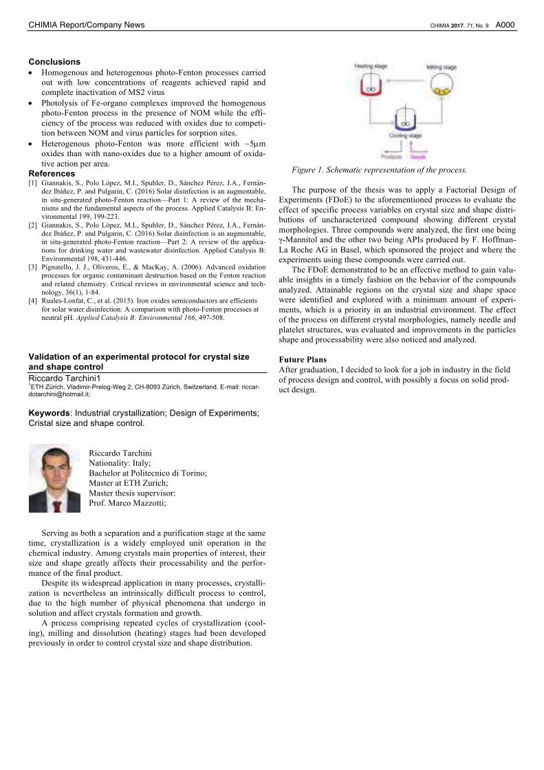
Figure 1. Schematic representation of the process.
The purpose of the thesis was to apply a Factorial Design of
Experiments (FDoE) to the aforementioned process to evaluate the
effect of specific process variables on crystal size and shape distri-
butions of uncharacterized compound showing different crystal
morphologies. Three compounds were analyzed, the first one being
γ-Mannitol and the other two being APIs produced by F. Hoffman-
La Roche AG in Basel, which sponsored the project and where the
experiments using these compounds were carried out.
The FDoE demonstrated to be an effective method to gain valu-
able insights in a timely fashion on the behavior of the compounds
analyzed. Attainable regions on the crystal size and shape space
were identified and explored with a minimum amount of experi-
ments, which is a priority in an industrial environment. The effect
of the process on different crystal morphologies, namely needle and
platelet structures, was evaluated and improvements in the particles
shape and processability were also noticed and analyzed.
Future Plans
After graduation, I decided to look for a job in industry in the field
of process design and control, with possibly a focus on solid prod-
uct design.

A744 CHIMIA 2016, 70, No. 10 Community news
Community Newswww.scg.ch
www.chemanager-online.com
For the first time, a session on ChemicalEducation was offered in the form of asymposium and workshop entitled “Fu-ture of Chemical Education” attracting140 teachers from secondary school onupwards. The event, embedded in theFall Meeting, marked the successfulstart of the initiative to implement a newSCS Division of Chemical Education.
The four lectures and the four parallel workshops provided newideas, new scientific topics as well as best practice in theoretical,practical and experimental chemical education.
Visit the website http://scg.ch/fallmeeting/2016 to get moreinformation about the program, the organizers, the sponsors andthe exhibitors. The site also allows to access all abstracts or tobrowse through the picture galleries.
Awards for the best Oral and the best PosterPresentations
In collaboration with Metrohm AG and DSM NutritionalProducts, SCS offered again a very attractive and prestigiousaward program for the best oral and the best poster presentations.The winners obtained CHF 500 in cash plus a travel voucher toattend a conference. Overall, during the 20 minute award cere-mony more than CHF 40’000 change hand. This is probably themost higly renumerated award program in the field, and we arevery proud and happy to cooperate with our sponsoring partners.We wish to express our sincere gratitude to the Metrohm Foun-dation and to DSM Nutritional Products Ltd for their generoussupport.
Dr. Markus Steinke, Vice President Marketing at Metrohm,awarded the following winners for their excellent oral presenta-tions:
Analytical Sciences– Winner: Rahel Bucher, University of Zurich– Runner up: Lyndsey Hendriks, ETH ZurichCatalysis Science & Engineering– Winner: Waiz Karim, ETH Zurich/Paul Scherrer Institute– Runner up: Vladimir Paunovic, ETH ZurichComputational Chemistry– Winner: Christopher Stein, ETH Zurich– Runner up: Clelia Spreafico, ETH Zurich
SwiSS ChemiCal SoCiety NewS
A Brief Review of the SCS Fall Meeting 2016
On September 15, 2016, the traditionalSCS Fall Meeting took place at Univer-sity of Zurich Irchel Campus, hostedjointly with ETH Zurich. The meetingattracted as many as 1’100 participantsfrom academia, industry, and, for thefirst time ever, from education. Withclose to 600 scientific contributions, theFall Meeting again offered a fantastic
platform to the predominantly young scientists to meet peers aswell as experts and specialists to discuss the results of their re-search. The one-day event also offers the opportunity to widenthe scientific knowledge and to connect with researchers fromother fields of chemistry or chemical biology.
In each of the eight thematic sessions, talks were given byawards winners, invited speakers and a representative of the ses-sion sponsor. All other oral contributions were from PhD stu-dents and post-doctoral fellows. The plenary sessions covered thelecture of our new honorary member, Prof. E. Peter Kündig, theSandmeyer and Paracelsus Award Lectures, presented by a teamof Sika researchers and Michel Graetzel of the EPF Lausanne.
Contributions per Session Posters TalksAnalytical Sciences 54 11Catalysis Science & Engineering 62 11Computational Chemistry 33 11Inorganic & Coordination Chemistry 84 11Medicinal Chemistry & Chemical Biology 68 13Organic Chemistry 82 11Physical Chemistry 53 12Polymers, Colloids & Interfaces 51 11Plenary Session 0 3Total 487 93

Community news CHIMIA 2016, 70, No. 10 A745
Inorganic & Coordination Chemistry– Winner: Deven Paul Estes, ETH Zurich– Runner up: Suzanne Maria Jansze, EPF LausanneMedicinal Chemistry & Chemical Biology– Winner: Olivia Paula Schmidt, University of ZurichOrganic Chemistry– Winner: David Kossler, EPF Lausanne- Runner up: Dominik Lotter, University of BaselPhysical Chemistry– Winner: Dominik Józef Kubicki, EPF Lausanne– Runner up: Maarten van Reijzen, EPF LausannePolymers, Colloids & Interfaces– Winner: Milad Radiom, University of Geneva– Runner up: Ralph Z. Lange, ETH Zurich
For the ceremony of the Best Poster Presentation AwardDr. Roman Imhof of DSM joined the team and handed over theprices to the 24 winners.
Analytical Sciences– Winner: Liviana Klein, ETH Zurich– Runner up: Benjamin Spenger, EMPA
Elena-Diana Burghelea, HES-SOCatalysis Science & Engineering– Winner: Vincent Lebrun, University of Basel– Runner up: Aswin Gopakumar, EPF Lausanne
Athanasia Tsoukalou, ETH ZurichComputational Chemistry– Winner: Gregor Nils Simm, ETH Zurich– Runner up: Akshaya Kumar Das, University of BaselInorganic & Coordination Chemistry– Winner: Marta Falcone, EPF Lausanne– Runner up: Vivian Marina Merk, ETH Zurich
Thibaud Rossel, Gymnase français de BienneMedicinal Chemistry & Chemical Biology– Winner: Fabian Brockmeyer, Northeastern University
Marc Heitz, University of Bern– Runner up: Fahimeh Moradi-Afrapoli, University of Basel
Isabel P. Kerschgens, University of ZurichOrganic Chemistry– Winner: Franck le Vaillant, EPF Lausanne– Runner up: Santiago Lascano, University of Geneva
Yun-Suk Jang, EPF LausannePhysical Chemistry– Winner: Marine Eva Fedora Bouduban, EPF Lausanne– Runner up: Maximilian Beyer, ETH Zurich
Joseph Samuel Beckwith, University of GenevaPolymers, Colloids & Interfaces– Winner: Fabian Deuber, ZHAW– Runner up: Yee Song Ko, EPF Lausanne
G. Nedelcu, ETH Zurich
Wewould also like to thank the award juries for their excellentwork (see Fall Meeting Web site for their names and affiliation).
The prizewinners will present their award winning researchin the Laureates Issue of CHIMIA 4/2017.
ILMAC 2016 – A convincing industry meeting pointand specialist trade fair for process and laboratorytechnology
ILMAC, Switzerland’s most important specialist trade fairfor process and laboratory technology, came to an end on Friday,23 September 2016. In the course of the four-day event, more than12’000 professional visitors obtained information about productinnovations, technological applications and process solutions.The issue of “Industrie 4.0”, a topical one affecting the entire sec-tor, was the central subject dealt with at the ILMAC Forum andwas examined from both the theoretical and practical perspec-tives. The “Lunch & Learn” sessions, in particular, organisedby the Swiss Chemical Society aroused intense interest amongstthe audience. The 20th ILMAC, held from 20 to 23 September2016 at Messe Basel, has been an encouraging event. The issueof “Industrie 4.0” has definitely arrived in process and laboratorytechnology and will keep on exercising the specialists for a longtime to come. The sector is, however, displaying dynamism andinterest in facing up to the current challenges of automation anddigitisation. The 12’000 and more specialists from the pharma-ceutical, chemical, biotechnology, cosmetics, food and drinkssegments attended their “in-house trade fair” at the very heartof the life science cluster in the Basel region. On the final day ofthe trade fair, high-ranking representatives from government andthe chemical industry in the Upper Rhine region visited ILMACin person and were convinced by what they experienced there.
Mirroring the marketFor the first time, the stands of the exhibitors presenting pro-
cess and laboratory technology were interspersed. In that way,ILMAC reflected the trend of technological applications movingcloser to one another on the market and the holistic planning ofprocesses. That concept turned out to be successful, and the tradefair came in for praise on account of its clear arrangement andtop quality.
First-hand informationVisitors were able to use the ILMAC Forum, the Lunch &
Learn sessions, the LabTec 4.0 and the Cleanroom Control Fo-rum to benefit from the practical experience and valuable knowl-edge of successful business people and experts from research,development and education and to experience live demonstra-tions.

CHIMIA2016,VOLU
ME70,N
UMBER4/16,PA
GES233–312/CHIMIAD70(4)233–312(2016)
www.chimia.ch
ISSN 0009-4293 4/2016
Laureates: Junior Prizes,
SCS Fall Meeting 2015
CHIMIA2016,VOLU
ME70,N
UMBER4/16,PA
GES233–312/CHIMIAD70(4)233–312(2016)
Cover picture by Anne-Clémence Corminboeuf, Riccardo Petraglia and Laurent Vannay, Institute of Chemical Sciences and Engineering, EPFLPhotos by Patrick Favre, Institute of Chemical Sciences and Engineering, EPFL
laureates: Junior prizes of the sCs fall meeting 2015 CHIMIA 2016, 70, No. 4 233
Editorial
As a sign of the vitality and variety of chemical research being carried out in Switzerland, the SCSFall Meeting 2015, organized under the auspices of the SCS Division of Fundamental Research, gath-ered about 800 participants on the premises of the EPF Lausanne on September 4, 2015. The 507scientific contributions (oral and poster presentations) and 12 invited lectures attracted collaboratorsfrom both academia and chemical/pharmaceutical industry to present, discuss and share recentscientific achievements which were presented in eight parallel sessions and two plenary sessions.The excitement of the young researchers disclosing the most important results of their research waspalpable, and the social components of this large and lively annual event gave excellent opportunitiesto make new contacts and enlarge professional networks. What a sign of enthusiasm to see a fullauditorium for the final lecture and plenary session until 6.30 pm!
The SCS Fall Meeting was again a perfect platform to honor scientists from academic institu-tions and industries for their outstanding contributions to chemical research and development. Inparticular, young researchers were honored for the scientific quality of their contributions by theSCS-Metrohm Awards for Best Oral Communications and the SCS-DSM Best Poster Awards. Thisissue of CHIMIA contains articles by these laureates and will give you a flavor of the research beingdeveloped in Switzerland in a wide range of chemical fields.
I would like to warmly congratulate all prize winners, thank the authors for their contributions,Metrohm and DSM for the generous sponsorship of the awards and their support to young talentedchemists. My acknowledgements also go to the jury members for their excellent selections of laure-ates, from a considerable number of contributions.
Best Poster Award winner articles sponsored by DSM• Jasmin Krismer, ETH Zurich• Stefan Knecht, ETH Zurich• Christoph Heinz, EPF Lausanne• Lucas-Alexandre Stern, EPF Lausanne• Anaëlle Dumas, University Paris-Sud• Heiner Saßmannshausen, ETH Zurich
Best Oral Communication Award winner articles sponsored by Metrohm• Andreas Riedo, University of Bern• Mark Bispinghoff, ETH Zurich• Adrian Najer, University of Basel• Maximilian Moser, ETH Zurich• Franziska A. Balmer, University of Bern
Finally, I sincerely thank the organizing team of the Institute of Chemical Sciences and Engineer-ing (ISIC) at EPFL for their hard work and crucial contribution to the success of the SCS Fall Meeting2015. We are looking forward to the 2016 edition, which will be held at the University of Zurich onSeptember 15, 2016.
I hope that you will enjoy the content of this issue and I wish you all good reading.
Sandrine GerberChair of the SCS Fall Meeting 2015EPF Lausanne
Sandrine Gerber

234 CHIMIA 2016, 70, No. 4 Contents inhalt sommaire
Laureates: Junior Prizes of theSCS Fall Meeting 2015
editorial
233 S. Gerber
236 Single-cell MALDI Tandem Mass Spectrometry: Unambiguous Assignment of Small Biomoleculesfrom Single Chlamydomonas reinhardtii CellsJ. Krismer, R. F. Steinhoff, R. Zenobi*
240 Efficient Water Electrolysis Using Ni2P as a Bifunctional Catalyst:Unveiling the Oxygen Evolution Catalytic Properties of Ni2PL.-A. Stern, X. Hu*
244 New Approaches for ab initio Calculations of Molecules with Strong Electron CorrelationS. Knecht*, E. D. Hedegård, S. Keller, A. Kovyrshin, Y. Ma, A. Muolo, C. J. Stein, M. Reiher*
252 PLGA-PEG-supported Pd Nanoparticles as Efficient Catalystsfor Suzuki-Miyaura Coupling Reactions in WaterA. Dumas*, A. Peramo, D. Desmaële, P. Couvreur
258 Total Synthesis of Fijiolide AC. Heinz, N. Cramer*
263 Exotic Chemistry with Ultracold Rydberg AtomsH. Saßmannshausen, J. Deiglmayr, F. Merkt*
268 Laser Ablation/Ionisation Mass Spectrometry:Sensitive and Quantitative Chemical Depth Profiling of Solid MaterialsA. Riedo*, V. Grimaudo, P. Moreno-García, M. B. Neuland, M. Tulej, P. Broekmann, P. Wurz
274 Halogen Chemistry on Catalytic SurfacesM. Moser, J. Pérez-Ramírez*
279 PH3 as a Phosphorus Source for Phosphinidene–Carbene Adductsand Phosphinidene–Transition Metal Complexes.M. Bispinghoff*, H. Grützmacher
284 Do Hydrogen Bonds Influence Excitonic Splittings?F. A. Balmer, P. Ottiger, S. Leutwyler*
288 Giant Host Red Blood Cell Membrane Mimicking PolymersomesBind Parasite Proteins and Malaria ParasitesA. Najer, S. Thamboo, C. G. Palivan, H.-P. Beck, W. Meier*

236 CHIMIA 2016, 70, No. 4 laureates: Junior prizes of the sCs fall meeting 2015
doi:10.2533/chimia.2016.236 Chimia 70 (2016) 236–239 © Swiss Chemical Society
*Correspondence: Prof. Dr. R. ZenobiDepartment of Chemistry and Applied BiosciencesETH ZurichVladimir-Prelog-Weg 3, CH-8093 ZurichE-mail: [email protected]
Single-cell MALDI Tandem MassSpectrometry: Unambiguous Assignmentof Small Biomolecules from SingleChlamydomonas reinhardtii Cells
Jasmin Krismer§, Robert F. Steinhoff, and Renato Zenobi*
§SCS-DSM Award for best poster presentation in Analytical Sciences
Abstract: The analysis of compounds from single cells is a major challenge in analytical life science. Labelingstrategies, for instance fluorescence detection, are well established for measuring proteins with single cellsensitivity, but they mostly fail to detect small molecules. More recently mass spectrometry has entered the realmof single cell sensitivity and enables the label-free and highly parallelized detection of small biomolecules fromsingle cells. The assignment of signals detected in single cells, however, generally has to rely on measurementsin whole cell culture extracts. Isobaric structures, contaminations, higher noise levels and the high variability inthe abundance of peaks between single cells complicate the assignment of peaks in single-cell spectra. Tandemmass spectrometry would be very useful for compound identification via mass spectrometry directly in single-cell analyses. Here we present the first single cell tandem mass spectra collected using matrix-assisted laser-desorption/ionization. The spectra obtained allow the assignment of most compounds detected in the spectra.We also show that the fragmentation is not restricted to the most abundant peaks in the spectra, but over adynamic range of more than one order of magnitude.
Keywords: Collision-induced-dissociation · Matrix-assisted laser-desorption/ionization · Single-cell massspectrometry · Tandem mass spectrometry
Introduction
Individual cells are the smallest func-tioning unit of life and a key challenge inanalytical life science. The analytical sci-entist is challenged by the high sensitivityand selectivity required for the analysis ofsingle cells.[1] The biologist, on the otherhand, seeks to understand the individualcell within the biological context of itspopulation or tissue, which adds highthroughput to the list of challenges foranalytical methods to overcome. Thereare plenty of single-cell techniques inthe area of DNA,[2,3] RNA[4,5] and proteinanalysis[6,7] that have found their way intobiologists’ labs. These techniques benefitfrom the amplification tools adopted frommolecular biology and the high linear dy-namic range and sensitivity of fluorescentprobes.
However, few such tools exist for theanalysis of small molecules from singlecells,[8] despite the fact that there is a great
need for phenotypic and metabolic charac-terization of single cells, especially sincechanges in genomes and transcriptomesare often poor predictors of cellular pheno-types.[9]Mass spectrometry has made greatprogress with respect to sensitivity and res-olution in the past decades.Matrix-assistedlaser desorption/ionization (MALDI) massspectrometry[10] in particular can provideboth the high throughput and the sensitiv-ity needed for the analysis of large num-bers of single cells.[11]Analyzing complexand dynamic biological systems such ascells, however, requires not just detectionof masses but also the assignment of thesignals detected to functionally relate theseto biological processes.
Tandem mass spectrometry is an im-portant part of the methodological toolboxof a mass spectrometrist for several rea-sons.[12] First, different constitutional iso-mers can be attributed to a single isobaricsignal, which makes it possible to identifymolecules using characteristic fragments.Second, contamination from differentsources like solvents, the cell culture me-dium, cell manipulating devices or theMALDI-matrix can give rise to interfer-ences that show the same or highly similarmasses. This effect is especially importantin single-cell mass spectrometry because
the weight of single cells is in the rangeof 10–12–10–15 grams.[13,14] Third there isgreat variability in the relative abundanceof molecules between single cells, whichcan complicate the peak assignment.Unfortunately the MS/MS capabilities ofmass spectrometry approaches for singlecell analysis have very much lagged be-hind.
Recently we reported a method for thehigh-throughput analysis of single cellsfor Chlamydomonas reinhardtii, a greenfreshwater algae and a well-studied modelorganism in photosynthesis and biofuelresearch.[15] The method allowed the par-allel detection and relative quantitation ofmore than 20 assigned peaks from differ-ent lipid classes with high throughput. MS/MS capabilities would greatly enhance theexplanatory power based on higher confi-dence in peak assignment.
Here, we present the first ever MS/MS spectra collected from single cells us-ing MALDI as an ionization method. Thecells were analyzed using 2,5-dihydroxy-benzoic acid (DHB) as a matrix, in positiveion mode. The method allowed us to assignabout 15 compounds in our spectra belong-ing to different lipid and pigment classes.The assignments based on the single celltandem mass spectra are in good agree-

laureates: Junior prizes of the sCs fall meeting 2015 CHIMIA 2016, 70, No. 4 237
studied, since the species is both a modelorganism in photosynthesis and a can-didate for biofuel production. From anexperimental perspective one of the keyadvantages in using the algae as a modelfor optimizing and developing a singlecell method is that due to its autofluores-cent pigments in the form of chlorophyllsit is possible to visualize cell lysis, a keystep for successful single cell mass spec-trometry.[15] Two aspects are very impor-tant here: the first is to make sure that theactual measurement is taken on a singlecell. In the current procedure the cells areplaced on a microarray by spotting a cellculture at an optimized cell density. Thisprocedure leaves most spots occupied withsingle cells, which can be identified, basedon fluorescence scans (Fig. 1A). However,some spots remain empty while multiplecells occupy others. These spots are dis-carded for analysis. The second key step islysis and co-crystallization (Fig. 1B). Onlyspots that show well-lysed cells as shownin Fig. 1 are considered forMS/MS. Poorlylysed cells may still show signals in MSmode, but these are generally too low inintensity to perform MS/MS experiments.
Relying on the sample preparationprotocol for single-cell MALDI optimizedfor C. reinhardtii we were able to assign16 compounds to 14 mass spectral peaks(Table 1) by performing single-cell MS/MS measurements. The assignments arebacked up by literature. The most promi-nent signal in the single-cell spectra is thepigment chlorophyll a. However due to thepresence of DHB, which is an acid, the ex-traction of the lipids is carried out under
Single-cell MS/MSThe tandem mass spectra were record-
ed on a reflectronMALDI-TOF instrument(AB Sciex 5800, Toronto, CA). The spotsfor MS/MS were selected from the platesbased on the fluorescence scans. Spectrawere collected from spots showing singlecells in the fluorescence scan after quench-ing. To confirm the fact that the signal isproduced only in the presence of cells MS/MS were recorded for the same precursormass on spots showing no cells. In none ofthe cases did we detected fragments of cel-lular analytes in the absence of cells. TheMALDI parameters used were: a delayedextraction time of 300 ns, a laser intensityof 5500 a.u. and a laser repetition rate of1000Hz. In all cases spectrawere collectedwith and without metastable suppression.Precursors were isolated using a mass win-dow of ±1.5 Da, using the QuanTis timedion selector of the reflectron TOF systemoperated with a TIS offset of 1.5 mm.
Data AnalysisPeak picking was performed according
to a S/N ≥10 criterion. Spectra were cali-brated externally using chlorophyll a as areference. The spectra were smoothed us-ing the default settings in theData Explorersoftware (ABSciex, Toronto).
Results and Discussion
The tandemmass spectrometry methodwas optimized on a chlorophyll a standardand a lipid extract of C. reinhardtii. Thelipid composition of C. reinhardtii is well
ment with spectra obtained by analyzinglipid extracts, as well as the relevant mo-lecular biology literature.[16–18]
Experimental Section
Chemicals2,5-dihydroxybenzoic acid (DHB)
was purchased from Sigma-Aldrich.Chlorophyll a (from Anacystis nidulans),chloroform (>99.8%), acetone (puriss.p.a., ≥99.5%) and 2-propanol (puriss.,≥99.5%) were purchased from SigmaAldrich, Switzerland. Water (Optima® LC/MS grade) was purchased from Fisher-Chemicals. Hutners trace element solutionfor cell culture media preparation was ob-tained from the Chlamydomonas ResourceCenter (St. Paul, MN, USA)
MS/MS of Lipid ExtractsA lipid extract was performed with the
Bligh-Dyer method[19] using C. reinhardtiiwild-type strain CC-125 obtained fromthe Chlamydomonas Resource Center, StPaul, MN. The cells were cultured in tris-acetate-phosphate medium.[20] To aid theextraction the cells were sonicated for 30seconds. The chloroform phase was col-lected and stored at –20 °C. Then the chlo-roform was evaporated and the lipids solu-bilized in aqueous acetone (80 vol% ac-etone, 20 vol% water). The samples weremixed with DHB (10 mg/mL in 80% aque-ous acetone) on the MALDI target (384Opti-TOF 123 mm × 61 mm SS, ABSciex,Toronto) in a sandwich fashion (0.5 µLDHB – 0.5 µL lipid extract – 0.5 µLDHB).The MS/MS method used for extractswas identical to the one described below.
Sample Preparation for Single-cellMS/MS
Wild-type C. reinhardtii cells (strainCC125) were centrifuged (3500 × g for5 min) three times and resuspended inwater. The cells were placed on a micro-structured array for MALDI mass spec-trometry as reported previously.[15] Inshort, the array consists of 55 × 26 spots of300 µm diameter each spaced by 720 µmon a stainless steel plate. A layer of matrixwas added first, followed by deposition ofthe washed cells into the spots (Fig. 1A).Cellular metabolism was quenched im-mediately by immersing the entire arrayinto liquid nitrogen. Extraction of analytesand co-crystallization with the matrix isachieved by recurrent extraction adding5 × 5 nL of 10 mg/mL of DHB dissolved in80% aqueous acetone and letting the spotscrystallize after each step. Fluorescencescans at 630 nm excitation and 670 nmemission were collected of the blank slide,of the cells after quenching, and after ma-trix application.
2 cells (1 dividing)
no cell
well lysed1 cell 1 cell
poorly lysed
2 ce1 cell
microarray spot
DHB matrix layer
cells (1 dividing) 1 cell
lysed cell
extractive matrix layer
well lys
lylysed
exextrac
sed
cell
ctive mmatrixix laylayyyyyyyyyyyyyyyyyyyyyyyyyyyyyyyyyyyyyyyyyyyyyyyyyyerererererererererererererererererererererererererererer
1 cell1 cell well lysed
well lysed
A Cells on the chip prior to lysis B Extracted cells
cell
Fig. 1. Sample preparation procedure for single-cell MS/MS. Chlorophyll autofluorescence ismeasured at 630 nm excitation and 670 nm emission. Both of the images are scaled identi-cally for better visibility (0-3000 instead of 0-65000) and falsely colored. The diameter of a spotis 300 µm. Cells are around 10 µm but appear larger due to over-scaling and scattering. A: Theautofluorescent cells are imaged on the slide prior to lysis to determine the number of cells ineach spot. As shown in the graphical abstract below a layer of matrix is applied prior to apply-ing the cells. B: Scanning the slides after co-crystallization can monitor the success of cell lysis.Matrix autofluorescence is negligible. Only spots containing well-lysed cells should be used forsingle-cell MS/MS experiments.

238 CHIMIA 2016, 70, No. 4 laureates: Junior prizes of the sCs fall meeting 2015
lipid, which in turn contributes about 50%of the total thylakoid lipid, the relative in-tensity of the peak is relatively low at 18%.This can be attributed to a lower ioniza-tion efficiency due to the sodium adductformation.
The major extraplastidic lipid classesare the diacylglyceryl-trimethylhomoser-
previously reported by Vieler et al.[21] Inthe single-cell mass spectra the sodium ad-ducts are most prominent. The SC-MS/MSspectra of both MGDG (see Fig. 2B and2E) and DGDG lipids are characterized byneutral loss of the fatty acid side chains.[22]Despite the fact that MGDG (16:4/18:3)makes up for almost 80% of the MGDG
low pH conditions. This leads to a releaseof the Mg2+ ion from the porphyrin macro-cycle. Chlorophyll a is therefore detectedas pheophytin a ([M-Mg2+3H]+ = 871.57Da) using DHB as a matrix.[17,21] This isalso true for chlorophyll b ([M-Mg2+3H]+
= 885.55 Da). The single-cell MS/MS ofchlorophyll a is dominated by the signalat 593.28 Da due to the loss of the phytolchain (Fig. 2A and 2D). Chlorophyll b frag-mentation leads to the formation of 607.25Da fragment due to phytol loss. Since chlo-rophylls are absorbing the wavelength ofthe MALDI laser at 355 nm both CID aswell as in-source decay contribute to frag-mentation. An increase in laser intensitywas found to enhance fragmentation evenin the absence of CID gas, which supportsthis interpretation (data not shown).
Furthermore, the spectra show all of themost abundant lipids, which can be detect-ed in positive ionmode, both from the plas-tidic, i.e. of organellar origin and extraplas-tidic membrane systems. Plastidic mem-brane systems that include the thylakoidmembranes, in which the photosyntheticcomplexes are embedded, mainly consistof the galactolipids mono- and digalacto-syl-diacylglyceol (MGDG and DGDG)and sulfolipids in the form of sulfoqui-novosyl-diacylglycerol (SQDG).[18] Sincethe extraction protocol was optimized forchlorophyll, it is reasonable that the majormembrane constituents are co-extracted.MGDG and DGDG can be detected in theform of sodium ([M+Na+]+) or potassium([M+K+]+) adducts using MALDI-MS as
200
400
600
800
100
300
500
1000
1500
2000
2500
3000
800200 400 600
500
200
400
m/z
593.28
533.26
489.23
519.29
59.09
474.36
496.36
Intensity/a.u.
A
B
C
871.57
734.57
767.49
HN
NH N
N
O
OO
O
O
593.28
519.24
533.25
Chlorophyll a
[M-Mg2++3H+]+
871.57 Da
H+
D
MGDG (16:4/18:3)
[M+Na+]+
767.47 Da
Na+
489.25
519.29
E
O O
O
O
O
O
HO HO
HO
HO
N
O
HO
O
OO
O
O
+
DGTS (16:0/18:3)
[M+H+]+
734.59 Da
474.38
496.36
456.37
F
236.15
236.16 59.09
Fig. 2, Single-cell MS/MS and corresponding chemical structures of different compound classesdetected in single cells. A/D: Single-cell MS/MS of chlorophyll a the structure showing the frag-mentation explaining the most abundant signals in the MS/MS spectra. B/E: SC-MS/MS and thechemical structure of MGDG, a plastidic galactolipid. C/F: SC-MS/MS and chemical structureof a DGTS, an extraplastidic lipid. The highly stabilized charge leads to the detection of the lipidheadgroup even in single-cell spectra. The positions of the two fatty acids (sn1 or sn2) cannot bedetermined using MS/MS but are adapted from literature.[16]
Table 1. List of compounds assigned by SC-MS/MS in Chlamydomonas reinhardtii using DHB in positive ion mode. Relative intensities relate tosingle-cell MS spectra collected from the same population. Numbers in brackets relate to the fatty acid composition (carbon number: number ofdouble bonds) the position of the fatty acid or the position of the double bonds cannot be determined using single-cell MS/MS.
metabolite name rel.int.
species detected parent mass main fragment other fragments
Pigments Chlorophyll a 100 [M-Mg2++3H+]+ 871.57 593.28 533.25, 519.30
Chlorophyll b 34 [M-Mg2++3H+]+ 885.55 607.25 547.24, 533.29
Galactolipids MGDG (16:4/18:3) 18 [M+Na+]+ 767.47 489.24 519.28
DGDG (18:3/16:3) 19 [M+Na+]+ 931.53 653.27 681.28
DGDG (18:2/16:3) 15 [M+Na+]+ 933.55 653.28 683.45
DGDG (18:3/16:0) 9 [M+Na+]+ 937.58 659.31 681.22
DGDG (18:2/16:0) 9 [M+Na+]+ 939.6 659.3 683.24
Homoserine lipids DGTS (16:0/18:4) 51 [M+H+]+ 732.58 474.33 494.30, 236.16
DGTS (16:0/18:3) 49 [M+H+]+ 734.59 474.37 496.38, 236.16,474.37, 456.34
DGTS (16:0/18:2) 21 [M+H+]+ 736.61 474.36 498.33
DGTS (18:3/18:4) 19 [M+H+]+ 754.55 496.42 494.42
DGTS (18:3/18:3),DGTS (18:4/18:2)
20 [M+H+]+ 756.57 496.32 498.36, 494.29,236.12
DGTS (18:2/18:3) 26 [M+H+]+ 758.6 496.36 498.39
DGTS (18:2/18:2),DGTS (18:1/18:3)
18 [M+H+]+ 760.61 498.36 496.45, 500.38

laureates: Junior prizes of the sCs fall meeting 2015 CHIMIA 2016, 70, No. 4 239
would like to thank Dr. Jens Sobek from theFunctional Genomics Center Zurich for the as-sistance using the microarrayer for the samplepreparation and Prof. Dr. Julia Vorholt from theMicrobiology Institute at ETH Zurich for al-lowing us to use their sterile bench.
Received: January 12, 2016
[1] E. Strauss, Nat. Chem. Biol. 2010, 6, 873.[2] H. Li, U. B. Gyllensten, X. F. Cui, R. K. Saiki,
H. A. Erlich, Nature 1988, 335, 414.[3] R. S. Lasken, Curr. Opin. Microbiol. 2007, 10,
510.[4] F. Tang, C. Barbacioru, Y. Wang, E. Nordman,
C. Lee, N. Xu, X. Wang, J. Bodeau, B. B Tuch,A. Siddiqui, K. Lao, M. A. Surani, Nat. Meth.2009, 6, 377.
[5] S. Islam, U. Kjällquist, A. Moliner, P. Zajac,J.-B. Fan, P. Lönnerberg, S. Linnarsson, Nat.Protocols 2012, 7, 813.
[6] J. Lippincott-Schwartz, G. H. Patterson, Science2003, 300, 87.
[7] B. N. G. Giepmans, S. R. Adams, M. H.Ellisman, R.Y. Tsien, Science 2006, 312, 217.
[8] R. Zenobi, Science 2013, 342, 1243259.[9] M. D. Ritchie, E. R. Holzinger, R. Li, S. A.
Pendergrass, D. Kim, Nat. Rev. Genet. 2015,16, 85.
[10] M. Karas, D. Bachmann, U. Bahr, F.Hillenkamp, Int. J. Mass Spectrom. Ion Proc.1987, 78, 53.
[11] T. H. Ong, D. J. Kissick, E. T. Jansson, T. J.Comi, E. V. Romanova, S. S. Rubakhin, J. V.Sweedler, Anal. Chem. 2015, 87, 7036.
[12] F. McLafferty, Science 1981, 214, 280.[13] T. P. Burg, M. Godin, S. M. Knudsen, W. Shen,
G. Carlson, J. S. Foster, Nature 2007, 446,1066.
[14] G. Popescu, K. Park, M. Mir, R. Bashir, LabChip 2014, 14, 646.
[15] J. Krismer, J. Sobek, R. F. Steinhoff, S.n R.Fagerer, M. Pabst, R. Zenobi, Appl. Environ.Microbiol. 2015, 81, 5546.
[16] H. M. Nguyen, S. Cuiné, A. Beyly-Adriano, B.Légeret, E. Billon, Pa. Auroy, F. Beisson, G.Peltier,Y. Li-Beisson, Plant Physiol. 2013, 163,914.
[17] T. Suzuki, H. Midonoya, Y. Shioi, Anal.Biochem. 2009, 390, 57.
[18] W. R. Riekhof, C. Benning, ‘GlycerolipidBiosynthesis’, in ‘The ChlamydomonasSourcebook’ 2ndEd.,Chap. 2, Ed.E.H.H.B.S.B.Witman, 2009, Academic Press: London. pp.41-68.
[19] E. G. Bligh, W. J. Dyer, Can. J. Biochem.Physiol. 1959, 37, 911.
[20] D. S. Gorman, R.P. Levine, Proc. Nat. Acad.Sci. USA 1965, 54, 1665.
[21] A. Vieler, C. Wilhelm, R. Goss, R. Süss, J.Schiller, Chem. Phys. Lipids 2007, 150, 143.
[22] L. Fouillen, B. Colsch, R. Lessire, ‘The LipidWorld Concept of Plant Lipidomics’, in‘Advances in Botanical Research’, Chap. 7, Ed.R. Dominique, 2013, Academic Press. pp. 331-376.
tively. One explanation for the detection ofmultiple isobars in the case of 756.57 Daand 760.61 Da is the similar abundance ofthe two contributing DGTS species.[16]
Conclusions
Peak assignment in single-cell spec-tra is a challenge for the advancement ofsingle-cell mass spectrometry techniques.The certainty of the actual assignmentbased on fragment detection – despite alldatabase searches and high mass accuracy –is unsurpassed. The successful implemen-tation of MS/MS experiments on thesingle-cell level as shown here reflects theprogress made with respect to sensitivityand reproducibility, and adds an importantamendment to single-cell MALDI massspectrometry.
AcknowledgementWe would like to thank the physics work-
shop of ETH Zurich and Dr. Rolf Brönnimannfrom EMPA Dübendorf for the help in pro-ducing the microarrays for MALDI-MS. We
ines (DGTS) which functionally replacephosphatidylcholines in C. reinhardtii.[18]The DGTS lipids show a great variety offatty acid side chains in the single-cellspectra (Table 1, Fig. 2C and 2F). Onereason for this lipid class to be readily ob-served might be the high ionization effi-ciency of the highly polar lipid headgroup.The trimethylhomoserine-headgroup itselfas well as various fragments generated bythe loss of fatty acid side chains can alsobe detected in single-cell MS/MS spectra(Fig. 3). In contrast to the galactolipids theside chain fragmentation leads both to thefatty acid neutral loss and the cleavage ofthe acyl group with the latter being moreprominent in the single-cell MS/MS spec-tra. The relative abundance of the differentDGTS species agrees well with previouslyreported values.[16] A unique advantage ofapplying MS/MS is that fragmentation ofthe side chains allows the assignment ofmultiple isobaric species. We were ableto assign multiple isobars for the sig-nal at 756.57 Da (DGTS (18:2/18:2) andDGTS (18:1/18:3)) and 760.61 Da (DGTS(18:3/18:3) andDGTS (18:4/18:2)) respec-
59.09
236.16
474.32
496.36
MS/MS Precursor mass = 734.59 Da
m/z
MS/MS Precursor mass = 732.58 Da474.32
494.33
236.16
Intensity/a.u.
Δm/z = 238.25
-(16:0)
Δm/z = 258.26
-(18:4)
Δm/z = 260.27
-(18:3)
Δm/z = 238.23
-(16:0)
Fig. 3. Precursor ionsuppressed single-cell MS/MS from twoDGTS lipids. TheDGTS at 732.58 Da(upper spectrum)fragments to 474.32Da and 494.33 Dacorresponding to theacyl loss of the fattyacids 18:4 and 16:0respectively. DGTSat 734.59 Da (lowerspectrum) fragmentsto 474.32 Da and496.36 Da corre-sponding to the acylloss of 18:3 and 16:0,respectively.

240 CHIMIA 2016, 70, No. 4 laureates: Junior prizes of the sCs fall meeting 2015
doi:10.2533/chimia.2016.240 Chimia 70 (2016) 240–243 © Swiss Chemical Society
*Correspondence: Prof. Dr. X. HuLaboratory of Inorganic Synthesis and CatalysisInstitute of Chemical Sciences and EngineeringÉcole Polytechnique Fédérale de Lausanne (EPFL)ISIC-LSCI, BCH 3305CH-1015 LausanneE-mail: [email protected]
Efficient Water Electrolysis Using Ni2P
as a Bifunctional Catalyst: Unveiling theOxygen Evolution Catalytic Properties ofNi
2P
Lucas-Alexandre Stern§ and Xile Hu*
§SCS-DSM Award for best poster presentation in Catalysis Science and Engineering
Abstract: The excellent bifunctional catalytic activity of nickel phosphide (Ni2P) for water splitting is reported.Ni2P, an active hydrogen evolving catalyst, is shown to be highly active for oxygen evolution. Only 290 mVof overpotential is required to generate a current density of 10 mA cm–2 in 1 M KOH. Under oxygen evolvingconditions, Ni2P undergoes structural modification to form a Ni2P/NiOx core-shell assembly, the catalytic activespecies. Ni2P is applied on both electrodes of an alkaline electrolyser and a current density of 10 mA cm–2 isgenerated at 1.63 V.
Keywords: Electrochemistry · Janus catalyst · Ni2P · Oxygen evolution · Water splitting
1. Introduction
Global scale exploitation of renewableenergy resources, such as wind and solarenergy, demands efficient energy storagetechniques.Electrochemicalwater splittingis one of the most attractive methodfor energy storage.[1] Water splitting issubdivided in two half-reactions, namely,the hydrogen evolution reaction (HER,Eqn. (1)) and the oxygen evolution reaction(OER, Eqn. (2)).
2 H+ + 2 e– → H2
(1)
2 H2O→ 4 H+ + 4 e– + O
2(2)
Both reactions require electrocatalyststo proceed efficiently. State-of-the-artcatalysts rely on the use of scarce andexpensive noble metals, e.g. platinum.[2]While promising Earth-abundant water-
splitting catalysts have been developed,only few materials are able to catalyseboth HER and OER in the same media.Bifunctional catalysts made from Earth-abundantmetalswouldfacilitateproductionand implementationofelectrolyserdevices.The reported bifunctional systems includeCo,[2] Ni,[3] Cu,[4] NiFe LDH,[5] NiFeO
x,[6]
NiCo2S4,[7]Ni
5P4,[8]CoO
x,[9]CoP,[10]FeP.[11]
In this article, we show that Ni2P, an active
HER catalyst, is efficient for the oxygenevolution reaction in alkaline medium.Under OER conditions, the material formsa Ni
2P/NiO
xcore-shell heterostructure.
This assembly generates a current densityof 10 mA cm–2 at an overpotential ofonly 290 mV.[12] The Janus behaviour ofthe material permits the fabrication of anefficient alkaline electrolyser using Ni
2P as
catalyst for both the cathode and the anode.
2. Oxygen Evolution Activity of Ni2P
The Ni2P nanoparticles were prepared
via a solid-state thermal reactionpreviously reported by our group.[13]Briefly, a phosphorus source, NaH
2PO
2(0.66 g, for analysis, Acros), and a nickelsalt, NiCl
2.6H
2O (0.3 g, ReagentPlus®,
Aldrich), were ground together at ambientatmosphere and then placed in a quartzboat. This was then transferred into atubular furnace and heated at 250 °Cunder a constant flow of nitrogen. Theobtained powder was further ground andthe impurities were washed off from theproduct using copious amount of distilledwater.Theproductwas thendried inanovenat 50 °C for a few hours. X-ray diffractionof the obtained powder is shown in Fig.1b. The diffraction signal was compared
Fig. 1. a) TEM image of polycrystalline Ni2P. b) Powder X-ray diffraction pattern of the Ni2Pnanoparticles. Adapted by permission of The Royal Society of Chemistry from ref. [12].

laureates: Junior prizes of the sCs fall meeting 2015 CHIMIA 2016, 70, No. 4 241
Ni2P nanoparticles were characterized
prior and after catalysis by high-resolutionTEM, energy-dispersive X-ray elementmapping and X-ray photoelectronspectroscopy (XPS) (Fig. 3). The dataindicate that prior to catalysis crystalline
3. Ni2P/NiO
xCore-shell Structure:
The OER Catalytic Active Species
To identify the nature of the chemicalmodifications undergone by nickelphosphide during water oxidation, the
to the reference diffraction patternobtained from the International Center ofDiffraction Data. The overlapping signalsindicated the successful synthesis of purephase Ni
2P. The transmission electron
microscopy (TEM) image shows thatthe Ni
2P nanoparticles have an averaged
size of 50 nm and are coated with a thinamorphous layer (Fig. 1a).
To evaluate the catalytic activity ofNi2P
for the oxygen evolution reaction, linearsweep voltammetry scans were performedin 1MKOH.The catalytic activity of nickelphosphide was measured and compared tovarious nanomaterials including IrO
2, Ni,
NiOxand an electrodeposited Ni(OH)
2film
(Fig. 2a). IrO2(99.9% Ir, abcr) and NiO
x(99.8% trace metals basis, Aldrich) wereused as received, while Ni nanoparticlesand Ni(OH)
2film preparation has been
reported previously.[14] The loading ofIrO
2, Ni, NiO
xapplied on the glassy carbon
electrodewas identical toNi2P loading.Fig.
2a shows that Ni2P nanoparticles catalytic
activity is superior to that of the differentcatalysts evaluated. The overpotential togenerate a current density of 10 mA cm–2
is only 290 mV for Ni2P. To reach similar
current density, IrO2and Ni(OH)
2requires
an additional 40 mV of overpotentialcompared to Ni
2P. Ni and NiO
xdrive a
current density of 10 mA cm–2 at 365 mVof overpotential.
The electrochemical surface area(ESCA) of the materials was assessed andcompared. For this purpose, the double-layer capacitance of the materials wascalculated. Fig. 2b shows the ESCA ofthe nickel-based catalysts. The ESCA forthe different materials were expressed interms of surface averaged double layercapacitance. Ni
2P have higher surface
area for similar loading than other nickelmaterials: 176.9 µF cm–2, followed byNi(OH)
2137.2 µF cm–2. NiO
xand Ni
nanoparticles have smaller ESCA values68 and 37 µF cm–2 respectively. Thecorrelation determined between the ESCAof Ni, NiO
x, Ni(OH)
2and their respective
catalytic activity at the fixed overpotentialof 325 mV cannot be applied to Ni
2P.
This indicates that high surface area is notthe sole reason for the superior catalyticactivity of nickel phosphide.
The stability of the nickel phosphidenanoparticles was probed by galvanostaticexperiment. Fig. 2c shows that theoverpotential to generate 10 mA cm–2
increased only of 10 mV over the courseof 10 hours indicating high stability ofthe catalyst. The nickel phosphide oxygenevolution activity was also compared toseveral state-of-the-art materials (Table 1).It can be seen that Ni
2P is more active than
many Ni and Co-based oxides, and evenIrO
2in base.
Table 1. Comparison of the OER catalytic activity of state-of-the-art catalysts. Adapted bypermission of The Royal Society of Chemistry from ref. [12].
Material Loading [mg cm-2] η@ 10 mA cm-2 [mV] Ref.
Ni 0.14 377 This work
NiOx
0.14 364 This work
Ni(OH)2
0.14 331 This work
Ni2P 0.14 290 This work
NiOx
0.02 360 [15c]
α-Ni(OH)2
0.20 331 [15d]
β-Ni(OH)2
0.20 444 [15d]
NiCo2O
40.53 565 [15e]
NiFe-LDH 0.20 320 [16]
CoOx@CN 0.42 410 [9]
BSCF82 0.25 320 [17]
IrO2
- 320 [15b]
IrO2a 0.35 275 [18]
aCatalytic activity measured in acidic conditions
Fig. 2. a) Linearsweep voltammetric(LSV) curves of Ni2P,Ni nanoparticles,NiOx nanoparticles,electrodepositedNi(OH)2, and IrO2 in1 M KOH. b)Correlation of thecurrent density atan overpotential of325 mV, with theelectrochemicalcapacitanceof the differentnickel materials.c) Galvanostaticmeasurement on theNi2P nanoparticlesin 1 M KOH at aconstant currentdensity of 10 mA cm–2
over 10 h. Conditions:pretreated workingelectrode, pre-activated Ni2Pcatalysts, 5 mV s–1,0.14 mg cm–2.Adapted bypermission of TheRoyal Society ofChemistry fromref. [12].

242 CHIMIA 2016, 70, No. 4 laureates: Junior prizes of the sCs fall meeting 2015
reactions. The stability of the alkalineelectrolyser was tested via galvanostaticmeasurement. A constant current densityof 10 mA cm–2 was applied for 10 h.Fig. 4 inset shows that the overpotentialhardly increases during the course of theexperiment and indicates high stabilityof the fabricated electrolyser. To confirmthat all the current generated results fromwater splitting, the total pressure increasedue to hydrogen and oxygen evolution wasrecorded using a pressure sensor duringthe electrochemical measurement. Thepressure sensor data allows the amountof gas evolved to be calculated over thecourse of the water electrolysis. The
cathode and anode catalyst was fabricated.Thecatalystsupportusedfor thisdevicewasnickel foam.The loadingof catalyst appliedon the two Ni foam was 10 mg cm–2. Fig.4 shows the measured catalytic activity ofthe alkaline electrolyser in 1 M KOH. Theactivity is compared with bare Ni foamsas reference. To generate a current densityof 10 mA cm–2 the alkaline electrolyseremploying Ni
2P as catalyst only requires
400 mV of overpotential while bare Nifoams demands 560mVof overpotential toreach the same value of current density.[12]During water electrolysis, the cathode andthe anode were separated using a glassfrit membrane to avoid any undesirable
nanoparticles of Ni2P are embedded in an
amorphous matrix, which is oxygen-rich,contains phosphorus, and is free of anymetallic content (Fig. 3a–e). This layerlikely consists of residual P
2O
5as a side
product of the catalyst synthesis. Importantstructural change can be observed on thecatalytic material after oxygen evolutionfor 1 h. TheHRTEM image (Fig. 3g) showsthat after OER, the layer surrounding thenanoparticles is composed of ultrafineparticles of about 2–3 nm diameter. Closerinspection of the particles in the layerrevealed the presence of lattice registryindicative of crystalline materials. FastFourier transform (FFT) (Fig. 3g inset)allowed measurement of the crystal latticespacing of the fine particles. The distancebetween two crystal planes is characteristicof NiO
xmaterial. A crystal lattice spacing
of the material’s core was also determinedand it is characteristic of the (100) facetof Ni
2P. Elemental mapping images of the
catalyst afterOER (Fig. 3h–k) clearly showthe Ni
2P/NiO
xcore-shell heterostructure
that the material adopts under oxidativeconditions. Nickel is present in bothlayers of the material. Oxygen coatshomogeneously the particle surface.Phosphorus is present only in the core.The structure of the material was furthercorroborated by XPS measurements (Fig.3l).[19] Electrochemical measurements alsovalidate the formation of a Ni
2P/NiO
Xcore-
shell heterostructure during OER. Theoxidation peak observed is indicative of theNi2+/Ni3+ oxidation in many nickel oxidehydroxide and bimetallic NiFeO
xOER
catalysts.[13,15a,15d,20] The pre-activation ofthe catalyst is, thus, hypothesized to allowthe in situ formation of a stable Ni
2P/NiO
xcore-shell heterostructure with improvedOER catalytic properties.
The enhanced catalytic activity of theNi
2P/NiO
Xcore-shell structure compared
to other NiOxcatalysts for OER has
two possible origins. On one hand theunique assembly provides significantimprovement in electron conduction.[21]Indeed, the metallic Ni
2P core might
provide an efficient electron pathway tothe insulating NiO
xshell. On the other
hand, Ni2P allows the in situ formation
of ultrafine nanoparticles of NiOx, which
allows higher active site exposure.Synergistic effects, which warrant furtherinvestigation, may play a role as well.
4. Efficient and InexpensiveAlkaline Electrolyser FabricatedUsing Ni
2P as Catalyst
The hydrogen evolving capabilitiesof Ni
2P have been reported recently.[13,22]
Given the presented OER catalytic activity,an alkaline electrolyser using Ni
2P as both
Fig. 3. a) High-resolution TEM (HRTEM) image of the Ni2P nanoparticles prior to oxygenevolution catalysis. b–e) Corresponding energy dispersive X-ray (EDX) elemental maps of thesystem prior to catalysis (scale bar: 9 nm). b) Ni elemental mapping. c) Phosphorus elementalmapping. d) Oxygen elemental mapping. e) Combined elemental mapping. f) High-resolutiondepth-profiling XPS spectra of the Ni 2p area. As the profiling depth increases (arrow direction),the FWHM decreases, indicative of stronger metallic Ni content. g) HRTEM image of the Ni2Pnanoparticles after electrochemical pretreatment at 1.5 V vs. RHE for one hour. Inset (lower left):FFT of the framed area (middle). The spots observed on the FFT are indicative of registry orderand so of crystallinity. The lattice fringes spacing of the materials were determined using FFT.They correspond to the characteristic (100) facet of Ni2P and specific facets of nickel oxides/hydroxides species, NiOx. h–k) Corresponding EDX maps of the elements on the sample regionshown in (g) (scale bar: 20 nm). h) Nickel elemental mapping. i) Phosphorus elemental mapping.j) Oxygen elemental mapping. k) Combined elemental mapping of Ni, O, and P. l) High-resolutiondepth-profiling XPS spectra of the Ni 2p area after catalytic activity. As the profiling depthincreases (arrow direction), the large FWHM, that is indicative of nickel oxide species, decreases,indicative of stronger metallic Ni content. This confirms the presence of a surface oxide layeraround metallic nickel phosphide core. Adapted by permission of The Royal Society of Chemistryfrom ref. [12].

laureates: Junior prizes of the sCs fall meeting 2015 CHIMIA 2016, 70, No. 4 243
[1] N. S. Lewis, D. G. Nocera, Proc. Natl. Acad.Sci. USA 2006, 103, 15729.
[2] S. Cobo, J. Heidkamp, P.-A. Jacques, J. Fize,V. Fourmond, L. Guetaz, B. Jousselme, V.Ivanova, H. Dau, S. Palacin, M. Fontecave, V.Artero, Nat. Mater. 2012, 11, 802.
[3] C. He, X. Wu, Z. He, J. Phys. Chem. C 2014,118, 4578.
[4] X. Liu, H. Zheng, Z. Sun, A. Han, P. Du, ACSCatal. 2015, 5, 1530.
[5] J. Luo, J.-H. Im, M. T. Mayer, M. Schreier, M.K. Nazeeruddin, N.-G. Park, S. D. Tilley, H. J.Fan, M. Graetzel, Science 2014, 345, 1593.
[6] H. Wang, H.-W. Lee,Y. Deng, Z. Lu, P.-C. Hsu,Y. Liu, D. Lin, Y. Cui, Nat. Commun. 2015, 6,7261.
[7] D. Liu, Q. Lu, Y. Luo, X. Sun, A. M. Asiri,Nanoscale 2015, 7, 15122.
[8] M. Ledendecjer, S. K. Calderón, C. Papp, H.-P. Steinrück, M Antonietti, M. Shalom, Angw.Chem. Int. Ed. 2015, 54, 12361.
[9] H. Jin, J. Wang, D. Su, Z. Wei, Z. Pang, Y.Wang, J. Am. Chem. Soc. 2015, 137, 2688.
[10] a) N. Jiang, B. You, M. Sheng, Y. Sun, Angw.Chem. Int. Ed. 2015, 54, 6251; b) Y. Yang, H.Fei, G. Ruan, J. M. Tour, Adv. Mater. 2015, 27,3175; c) J. Chang, Y. Xiao, M. Xiao, J. Ge, C.Liu, W. Xing, ACS Catal. 2015, 5, 6874.
[11] Y. Yan, B. Y. Xia, X. Ge, Z. Liu, A. Fisher, X.Wang, Chem. Eur. J. 2015, 21, 18062.
[12] L.-A. Stern, L. Feng, F. Song, X. Hu, EnergyEnviron. Sci. 2015, 8, 2347.
[13] L. Feng, H. Vrubel, M. Bensimon, X. Hu, Phys.Chem.Chem. Phys. 2014, 16, 5917.
[14] a) Y. Chen, D.-L. Peng, D. Lin, X. Luo,nanotechnol. 2007, 18, 505703; b) D. A.Corrigan, R. M. Bendert, J. Electrochem. Soc.1989, 136, 723.
[15] a) L. Trotochaud, J. K. Ranney, K. N. Williams,S. W. Boettcher, J. Am. Chem. Soc. 2012, 134,17253; b) C. C. L. McCrory, S. H. Jung, J. C.Peters, T. F. Jaramillo, J. Am. Chem. Soc. 2013,135, 16977; c) K. Fominykh, J. M. Feckl, J.Sicklinger, M. Doeblinger, S. Boecklein, J.Ziegler, L. Peter, J. Rathousky, E.-W. Scheidt,T. Bein, D. Fattakhova-Rohlfing, Adv. Funct.Mater. 2014, 24, 3123; d) M. Gao, W. Sheng, Z.Zhuang, Q. Fang, S. Gu, J. Jiang,Y.Yan, J. Am.Chem. Soc. 2014, 136, 7077; e) H. Shi, G. Zhao,J. Phys. Chem. C 2014, 118, 25939.
[16] M. Gong,Y. G. Li, H. L.Wang,Y.Y. Liang, J. Z.Wu, J. G. Zhou, J. Wang, T. Regier, F.Wie, H. J.Dai, J. Am. Chem. Soc. 2013, 135, 8452.
[17] K. J. May, C. E. Carlton, K. A. Stoerzinger, M.Risch, J. Suntivich, Y.-L. Lee, A. Grimaud, Y.Shao-Horn, J. Phys. Chem. Lett. 2012, 3, 3264.
[18] L. Ouattara, S. Fierro, O. Frey, M. Koudelka,C. Comninellis, J. Appl. Electrochem. 2009, 39,1361.
[19] P. E. R. Blanchard, A. P. Grosvenor, R. G.Cavell, A. Mar, J. Mater. Chem. 2009, 19, 6015.
[20] a) R. L. Doyle, I. J. Godwin, M. P. Brandon, M.E. G. Lyons, Phys. Chem. Chem. Phys. 2013,15, 13737; b) L.-A. Stern, X. Hu, FaradayDiscuss. 2014, 176, 363.
[21] a) M. Gong, W. Zhou, M.-C. Tsai, J. Zhou, M.Guan, M.-C. Lin, B. Zhang,Y. Hu, D.-Y. Wang,J. Yang, S. J. Pennycook, B.-J. Hwang, H. Dai,Nat. Commun. 2014, 5, 4695; b) G. Elmaci, C.E. Frey, P. Kurz, B. Zumreoglu-Karan, Inorg.Chem. 2015, 54, 2734; c) L. Trotochaud, S. L.Young, J. K. Ranney, S. W. Boettcher, J. Am.Chem. Soc. 2014, 136, 6744.
[22] E. J. Popczun, J. R. McKone, C. G. Read, A.J. Biacchi, A. M. Wiltrout, N. S. Lewis, R. E.Schaak, J. Am. Chem. Soc. 2013, 135, 9267.
theoretical volume of gas evolved can thenbe calculated by determining the chargepassed over the course of the experiment.Comparison between experimental andtheoretical volume of gas determines then
Fig. 4. LSV to evaluate the alkaline electrolyser activity under gas separating conditions. The Ni2Psystem (loading 10 mg cm–2, activated anode) necessitates only 400 mV to generate 10 mA cm–2.The Ni foam support was evaluated under similar conditions (gas separation, activated anodebut without catalyst) and required 560 mV to generated 10 mA cm–2. A galvanostatic experimenton the Ni2P alkaline electrolyser with glass frit separation was performed (inset) and indicates thegood stability of the system over 10 hours of electrolysis at 10 mA cm-2. An optical photographillustrates the cell used for these experiments. Adapted by permission of The Royal Society ofChemistry from ref. [12].
Fig. 5. Faraday yield measurement of thealkaline electrolyser fabricated from Ni2Ploaded Ni foams (loading 10 mg cm–2). Thequantity of gas evolved was determined bya pressure sensor. The overlapping linesbetween the theoretical and experimentalvalues indicate a quantitative Faraday yield ofthe system, after an induction period of about250 s. During this induction period, the gasgenerated are dissolved in the solution to reachan equilibrium. After the induction period,the gas generated can be measured by thepressure sensor. The galvanostatic experimentwas performed over 1 h at a constant currentdensity of 10 mA cm–2. The anode wasactivated prior the galvanostatic experiment.The cathode was used as it is. Adapted bypermission of The Royal Society of Chemistryfrom ref. [12].
the Faradaic efficiency of the alkalineelectrolyser. Fig. 5 illustrates that theexperimental and theoretical amountof gas evolved overlaps indicative of aquantitative Faraday efficiency for thedevice. Ni
2P is thus an active and stable
bifunctional catalyst for water splitting inalkaline media.
5. Conclusion
In summary, Ni2P is an active catalyst
for OER in alkaline conditions. Thecatalyst is prepared via a simple solid-state reaction from abundant and cheapreagents. The catalytic active species is aNi
2P/NiO
xcore-shell heterostructure that
is formed in situ under OER conditions.The catalytic activity is superior toseveral state-of-the-art catalysts. TheJanus behaviour of the catalyst allowsthe fabrication of an efficient alkalineelectrolyser who generates 10 mA cm–2
at an overpotential of only 400 mV. Ni2P
shows promise of potential applications forfuture devices and highlights the potentialof Earth-abundant catalysts as viableelectrocatalysts for energy conversiondevices and fuel cell applications.
AcknowledgementThis work is supported by the EPFL. We
thank Dr. Ligang Feng and Dr. Fang Song atEPFL for assistance in the experiments.
Received: December 1, 2015

244 CHIMIA 2016, 70, No. 4 laureates: Junior prizes of the sCs fall meeting 2015
doi:10.2533/chimia.2016.244 Chimia 70 (2016) 244–251 © Swiss Chemical Society
*Correspondence: Dr. S. Knecht, Prof. Dr. M. ReiherE-mail: [email protected];[email protected] ZürichLaboratorium für Physikalische ChemieVladimir-Prelog-Weg 2,CH-8093 Zürich
New Approaches for ab initioCalculations of Molecules with StrongElectron Correlation
Stefan Knecht§*, Erik Donovan Hedegård, Sebastian Keller, Arseny Kovyrshin, Yingjin Ma, AndreaMuolo, Christopher J. Stein, and Markus Reiher*
§SCS-DSM Award for best poster presentation in Computational Chemistry
Abstract: Reliable quantum chemical methods for the description of molecules with dense-lying frontier orbitalsare needed in the context of many chemical compounds and reactions. Here, we review developments that ledto our new computational toolbox which implements the quantum chemical density matrix renormalization groupin a second-generation algorithm. We present an overview of the different components of this toolbox.
Keyword: ab initio quantum chemistry · DMRG · Matrix product operators · Strong electron correlation ·Tensor network states
1. Introduction
Computational modeling has undoubt-edly become an integral part of chemicalresearch.[1] For instance, understandinga (photo-)chemical process in atomisticdetail – including all elementary reactionsteps involved – calls for a reliable, butfeasible and preferably black-box com-putational approach that provides a suffi-ciently accurate approximation to the ex-act solution of the electronic Schrödingerequation. A typical example for a complexchemical process is a reaction catalyzed bya transition-metal complex.[2]Ametal cen-ter can directly activate a reagent throughbond formation and/or bond breaking oract as a photoacceptor for a subsequentenergy transfer to the reagent from anelectronically excited state. The commonchallenge is then to quantitatively describeopen shells that will emerge in such pro-cesses, which is far from trivial to meet.[3–7]The presence of open shells and/or activat-
ed bonds usually entails a multiconfigu-rational electronic structure where strongstatic electron correlation becomes sizable.Molecules with these features then exhibitmany dense-lying orbitals in the frontier-orbital region.
Although by construction a single-con-figuration ansatz, to date, density function-al theory (DFT)[6] is by far the most popu-lar approach to study photochemistry (seefor example ref. [8] and references therein)and transition-metal chemistry[4,9,10] be-cause of its low computational cost, oftenat reasonable accuracy, and because of itsfavorable scaling with the size of a mol-ecule. The standard approach to describehalf-filled shells in molecules with smallHOMO-LUMO gaps is to break a symme-try of the system within DFT, usually thetotal-spin symmetry.[11,12] Besides knownfundamental problems of single-configu-ration DFT in correctly describing strongstatic correlation[13] all spin symmetriescan be properly introduced in DFT leadingto spin-state dependent functionals,[14] butlittle work along these lines has been car-ried out so far.[15]
A multiconfigurational wave functionbased-method tailored to recover staticcorrelation is the complete active space(CAS) ansatz,[16] often combined with asimultaneous self-consistent field (SCF)optimization of the orbital basis. Naturally,CASSCF-type approaches have been ap-plied in theoretical photochemistry andtransition-metal chemistry.[17–19] The cen-tral idea of a CASSCF-type approach isthe selection of an active space of N elec-trons in L orbitals to yield a CAS(N,L),on which a full configuration interaction
(FCI) expansion of the wave function isconstructed,
(1)|ΨCAS-CI = 𝐶𝐶����…����,…,��
|𝑛𝑛�𝑛𝑛�…𝑛𝑛� ,
where |n1n2…n
L⟩ is an occupation num-
ber vector corresponding to an orthonor-mal basis state, e.g. a Slater determinant.This procedure, however, does not yield anexact wave function as the orbital basis isrestricted. Moreover, the underlying FCIexpansion still scales exponentially withrespect to the number of active electronsand orbitals so that the computational fea-sibility of traditional CASmethods reachesa limit at a CAS size of about CAS(18,18).[19]
Originally developed to study the phys-ics of spin chains, the density matrix re-normalization group (DMRG)[20,21] algo-rithm emerged as a viable alternative totraditional CAS methods. This is rootedin the fact that it is capable of iterativelyconverging to the exact solution in a givenactive orbital space with polynomial ratherthan exponential cost.[22] In DMRG, CASsare accessible with up to about 100 orbitalsexceeding by far the limits encountered bytraditional CAS methods. Many quantum-chemical DMRG implementations, with(DMRG-SCF) and without (DMRG-CI)orbital optimization, have been developedsince the late 1990s.
Here, we present an overview of ourrecent efforts to further develop methodsthat rely on the DMRG. They comprisefeatures (i) to locate for a given moleculethe minimum structure of its ground and
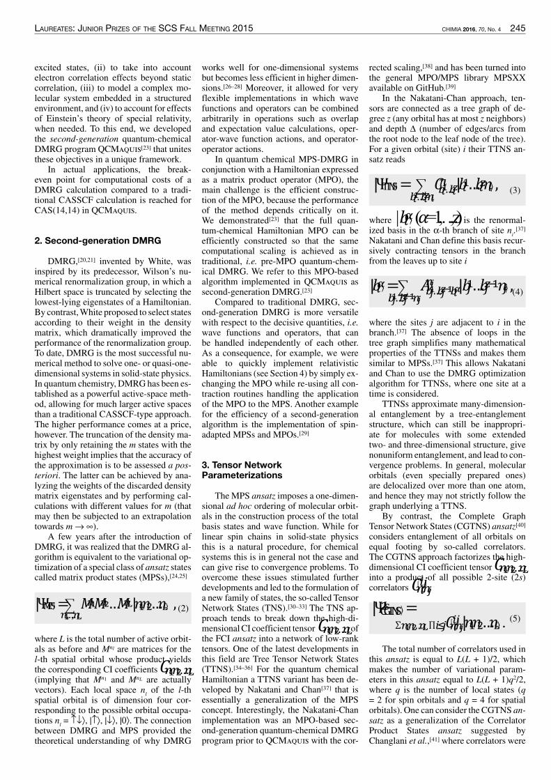
laureates: Junior prizes of the sCs fall meeting 2015 CHIMIA 2016, 70, No. 4 245
rected scaling,[38] and has been turned intothe general MPO/MPS library MPSXXavailable on GitHub.[39]
In the Nakatani-Chan approach, ten-sors are connected as a tree graph of de-gree z (any orbital has at most z neighbors)and depth ∆ (number of edges/arcs fromthe root node to the leaf node of the tree).For a given orbital (site) i their TTNS an-satz reads
(3)|ΨTTNS = 𝐶𝐶���…����� |𝑏𝑏��…𝑏𝑏��𝑛𝑛� ,
���…�����
where | 𝑏𝑏�� (𝛼𝛼 = 1,…,z) is the renormal-ized basis in the α-th branch of site n
i.[37]
Nakatani and Chan define this basis recur-sively contracting tensors in the branchfrom the leaves up to site i
(4)𝐴𝐴���…���������� |
���…�������𝑏𝑏��…𝑏𝑏���� 𝑛𝑛� ,|𝑏𝑏�� =
where the sites j are adjacent to i in thebranch.[37] The absence of loops in thetree graph simplifies many mathematicalproperties of the TTNSs and makes themsimilar to MPSs.[37] This allows Nakataniand Chan to use the DMRG optimizationalgorithm for TTNSs, where one site at atime is considered.
TTNSs approximate many-dimension-al entanglement by a tree-entanglementstructure, which can still be inappropri-ate for molecules with some extendedtwo- and three-dimensional structure, givenonuniform entanglement, and lead to con-vergence problems. In general, molecularorbitals (even specially prepared ones)are delocalized over more than one atom,and hence they may not strictly follow thegraph underlying a TTNS.
By contrast, the Complete GraphTensor Network States (CGTNS) ansatz[40]considers entanglement of all orbitals onequal footing by so-called correlators.The CGTNS approach factorizes the high-dimensional CI coefficient tensor 𝐶𝐶����…��into a product of all possible 2-site (2s)correlators 𝐶𝐶������ .
(5)𝐶𝐶������ |𝑛𝑛�𝑛𝑛�…𝑛𝑛��������…�� .
|������� =
The total number of correlators used inthis ansatz is equal to L(L + 1)/2, whichmakes the number of variational param-eters in this ansatz equal to L(L + 1)q2/2,where q is the number of local states (q= 2 for spin orbitals and q = 4 for spatialorbitals). One can consider the CGTNS an-satz as a generalization of the CorrelatorProduct States ansatz suggested byChanglani et al.,[41]where correlators were
works well for one-dimensional systemsbut becomes less efficient in higher dimen-sions.[26–28] Moreover, it allowed for veryflexible implementations in which wavefunctions and operators can be combinedarbitrarily in operations such as overlapand expectation value calculations, oper-ator-wave function actions, and operator-operator actions.
In quantum chemical MPS-DMRG inconjunction with a Hamiltonian expressedas a matrix product operator (MPO), themain challenge is the efficient construc-tion of the MPO, because the performanceof the method depends critically on it.We demonstrated[23] that the full quan-tum-chemical Hamiltonian MPO can beefficiently constructed so that the samecomputational scaling is achieved as intraditional, i.e. pre-MPO quantum-chem-ical DMRG. We refer to this MPO-basedalgorithm implemented in QCMaquis assecond-generation DMRG.[23]
Compared to traditional DMRG, sec-ond-generation DMRG is more versatilewith respect to the decisive quantities, i.e.wave functions and operators, that canbe handled independently of each other.As a consequence, for example, we wereable to quickly implement relativisticHamiltonians (see Section 4) by simply ex-changing the MPO while re-using all con-traction routines handling the applicationof the MPO to the MPS. Another examplefor the efficiency of a second-generationalgorithm is the implementation of spin-adapted MPSs and MPOs.[29]
3. Tensor NetworkParameterizations
The MPS ansatz imposes a one-dimen-sional ad hoc ordering of molecular orbit-als in the construction process of the totalbasis states and wave function. While forlinear spin chains in solid-state physicsthis is a natural procedure, for chemicalsystems this is in general not the case andcan give rise to convergence problems. Toovercome these issues stimulated furtherdevelopments and led to the formulation ofa new family of states, the so-called TensorNetwork States (TNS).[30–33] The TNS ap-proach tends to break down the high-di-mensionalCI coefficient tensor 𝐶𝐶����…�� ofthe FCI ansatz into a network of low-ranktensors. One of the latest developments inthis field are Tree Tensor Network States(TTNS).[34–36] For the quantum chemicalHamiltonian a TTNS variant has been de-veloped by Nakatani and Chan[37] that isessentially a generalization of the MPSconcept. Interestingly, the Nakatani-Chanimplementation was an MPO-based sec-ond-generation quantum-chemical DMRGprogram prior to QCMaquis with the cor-
excited states, (ii) to take into accountelectron correlation effects beyond staticcorrelation, (iii) to model a complex mo-lecular system embedded in a structuredenvironment, and (iv) to account for effectsof Einstein’s theory of special relativity,when needed. To this end, we developedthe second-generation quantum-chemicalDMRG program QCMaquis[23] that unitesthese objectives in a unique framework.
In actual applications, the break-even point for computational costs of aDMRG calculation compared to a tradi-tional CASSCF calculation is reached forCAS(14,14) in QCMaquis.
2. Second-generation DMRG
DMRG,[20,21] invented by White, wasinspired by its predecessor, Wilson’s nu-merical renormalization group, in which aHilbert space is truncated by selecting thelowest-lying eigenstates of a Hamiltonian.By contrast,White proposed to select statesaccording to their weight in the densitymatrix, which dramatically improved theperformance of the renormalization group.To date, DMRG is the most successful nu-merical method to solve one- or quasi-one-dimensional systems in solid-state physics.In quantum chemistry, DMRGhas been es-tablished as a powerful active-space meth-od, allowing for much larger active spacesthan a traditional CASSCF-type approach.The higher performance comes at a price,however. The truncation of the density ma-trix by only retaining the m states with thehighest weight implies that the accuracy ofthe approximation is to be assessed a pos-teriori. The latter can be achieved by ana-lyzing the weights of the discarded densitymatrix eigenstates and by performing cal-culations with different values for m (thatmay then be subjected to an extrapolationtowards m→∞).
A few years after the introduction ofDMRG, it was realized that the DMRG al-gorithm is equivalent to the variational op-timization of a special class of ansatz statescalled matrix product states (MPSs),[24,25]
(2)𝑀𝑀��𝑀𝑀����,…,��
… 𝑀𝑀�� |𝑛𝑛�𝑛𝑛�…𝑛𝑛� ,|ΨMPS =
where L is the total number of active orbit-als as before and Mnl are matrices for thel-th spatial orbital whose product yieldsthe corresponding CI coefficients 𝐶𝐶����…��(implying that Mn1 and MnL are actuallyvectors). Each local space n
lof the l-th
spatial orbital is of dimension four cor-responding to the possible orbital occupa-tions n
l= ↑↓⟩, |↑⟩, |↓⟩, |0⟩. The connection
between DMRG and MPS provided thetheoretical understanding of why DMRG

246 CHIMIA 2016, 70, No. 4 laureates: Junior prizes of the sCs fall meeting 2015
ber of sweeps, and the initialization proce-dure of the MPS in the first sweep, the so-called warm-up sweep. This increase in thenumber of control parameters is a threat tothe routine application of DMRG in stan-dard computational chemistry. As our aimis to make DMRG a valuable and reliabletool for computational chemistry, easy us-age as well as stable and fast convergenceare of paramount importance for a ‘black-box’ set-up of such calculations.
While the number of sweeps requiredfor convergence and the number of re-normalized states necessary can be easilycontrolled, the ordering of the orbitals onthe lattice and the initialization proce-dure need more sophisticated ideas. Bothproblems were addressed by Legeza andco-workers[60,61] by making use of entan-glement measures for the active orbitalsexpressed in terms of one- and two-orbitalvon Neumann entropies.[62–64] Especiallythe mutual information matrix I, which isa measure for the entanglement of pairs oforbitals, proved to be a valuable tool in theanalysis of MPS wave functions and multi-reference wave functions in general. Forfast convergence of a DMRG calculation,it is essential that highly entangled orbitalsare close to each other on the lattice. Thiswill be guaranteed if the orbitals are or-dered according to the Fiedler vector,[65,66]which is the eigenvector corresponding tothe second smallest eigenvalue of the graphLaplacian L
g, defined in this case as L
g=
D − I, where D is a diagonal matrix, Dii=
ΣjIij(i and j are labels for the orbitals on
the lattice, i.e. for the orbitals chosen to bein the CAS). The Fiedler vector minimizesthe cost measure[61]
(6)ω = 𝐼𝐼��|𝑖𝑖 − 𝑗𝑗|���
.
This Fiedler ordering significantlyimproves the convergence and is imple-mented in QCMaquis.
Convergence can further be improvedby a suitable guess of the environmentstates in the warm-up sweep. While an ob-vious choice is to start from an MPS thatcontains a reference determinant (such asthe Hartree-Fock determinant or the deter-minant with the largest weight in configu-ration-interaction language), it is possibleto improve on this by including the mostimportant determinants into the initialMPS. These most important determinantsare selected by varying the occupation onthose sites that have the highest one-orbitalentropies. If these determinants are furtherlimited to have a specific excitation levelwith respect to a reference determinant,this initialization procedure invented byLegeza[67,68] is referred to as CI-DEAS.Calculations starting from a CI-DEAS
(loosely speaking, the relativistic analogof two degenerate non-relativistic α- andβ-spin orbitals). Performing calculationsin a Kramers basis reduces the possiblenumber of two-electron integrals (ij|kl)arising from all combinations of unbarredand barred indices, 16 in total, to only sixsymmetric non-redundant integrals.[53]
Double groups are constructed fromthe direct product of point groups and thesub-group {E, E} where E represents a ro-tation through 2π and E a rotation through4π. Double groups are in general non-abe-lian, which gives rise to additional com-plications for symmetry multiplications inquantum chemistry programs. If time re-versal symmetry can be considered (e.g. inthe absence of an external magnetic field),it can be shown[50] that certain classes oftwo-electron integrals are real, complex, orcan be excluded a priori, because they areequal to zero. Finally, for systems of inter-est to chemists, the number of particles isconserved, which implies that the unitaryone-dimensional group U(1) can also beincluded along with all other symmetriesintroduced above. In our case, with DMRGas the post-Hartree-Fockmethod of choice,U1DG symmetry is employed to decreasethe number of many-particle states forsymmetry reasons. Characters and multi-plication tables for C
1, C
i, C
2, C
2h, C
64and
C32hdouble groups[54]were implemented in
QCMaquis.[51]The relativistic DMRG model in
QCMaquis supports double group sym-metries in order to assign to every site anirreducible representation correspondingto the spinor placed there. No assumptionsare made with respect to the spinor basiswhich can be either a Kramers-restricted orKramers-unrestricted basis. In addition, noformal distinction is made between barredand unbarred spinors but simplificationsdue to the selected symmetry may leadto an elimination of certain terms in theHamiltonian. Finally, no explicit referenceof two- or four-component quantities ismade inside QCMaquis and the only inputdata for the calculations are the pre-com-puted relativistic one- and two-electronintegrals from Molcas,[19] Dalton,[55]Molpro,[56] Dirac[57] or Bagel.[58]
5. Set-up, Parameter Dependence,and Convergence Acceleration
The ability of DMRG to handle activeorbital spaces that are much larger thanthose of conventional CASSCF approach-es comes with an additional set of mainlytechnical parameters that can affect con-vergence and accuracy.[59] Among theseparameters are the ordering of the orbitalsas sites on the one-dimensional lattice, thenumber of renormalized statesm, the num-
only used between nearest-neighbor sites.Higher accuracy can be achieved by invok-ing higher-order correlators (3-site correla-tors, 4-site correlators, and so forth).[40–42]While we continue to investigate such gen-eral decompositions of CI coefficients,[42]the advantage of the MPS ansatz is that itcan be efficiently optimized by the DMRGalgorithm.
4. Relativistic Hamiltonians andSymmetries
In 1928 C. G. Darwin wrote:[43] In a re-cent paper Dirac has brilliantly removedthe defects before existing in the mechan-ics of the electron, and has shown how thephenomena usually called the ‘spinningelectron’ fit into place in the complete the-ory. Since the non-relativistic Schrödingerequation was spin-free, it was clear at thattime that a new formalism was needed tocombine quantum theory with Einstein’stheory of special relativity. Since the 1970snumerous unusual features have been rec-ognized in heavy-element chemistry andspectroscopy that can only be explainedconsidering a relativistic quantum descrip-tion of electrons.[44–47] The liquid state ofmercury under ambient condition[48] andthe lead battery in cars[49] are prominentexamples for which these so-called ‘rela-tivistic effects’ are in operation.
Today, relativistic electronic structuretheory is a mature and well-understoodfield.[47,50] Once a relativistic Hamiltonianis chosen, established electronic-structuremethods can be employed to approxi-mate the wave function. Our QCMaquisprogram package can handle the symme-try properties of the Dirac-Coulomb andDirac-Coulomb-Breit Hamiltonians aswell as of their two-component analogs.[51]Whereas the first such implementation intoa traditional DMRGprogram[52] could onlyhandle real double groups (DG), these lim-itations are overcome in QCMaquis.
In the molecular spinor basis sig-nificant computational savings can beachieved by adopting the symmetries thatare obeyed by two- and four-componentHamiltonians. In non-relativistic quantumchemistry, one only needs to treat spaceinversions and rotations because all othersymmetries can be generated by a succes-sive application of these two. In the rela-tivistic framework, time and space are tiedtogether to the space-time, and hence thetime reversal operator𝒦𝒦 is to be addressedand double point groups need to be takeninto account. The effect of 𝒦𝒦 on a wavefunction Ψ(t) yields the time-reversedwave function Ψ(–t). It can be shown[50]that the pair Ψ,𝒦𝒦Ψ = Ψ correspondsto a doubly-degenerate fermionic statefunction which is called a Kramers pair
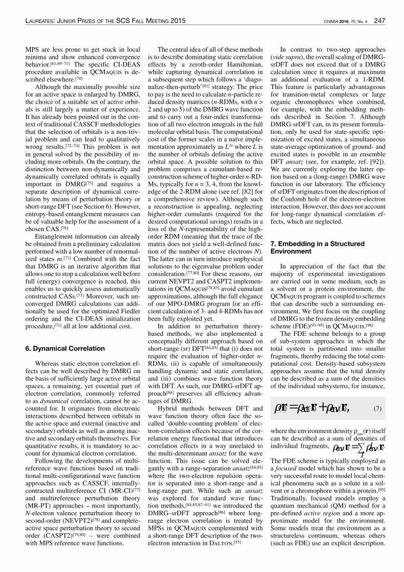
laureates: Junior prizes of the sCs fall meeting 2015 CHIMIA 2016, 70, No. 4 247
In contrast to two-step approaches(vide supra), the overall scaling of DMRG-srDFT does not exceed that of a DMRGcalculation since it requires at maximuman additional evaluation of a 1-RDM.This feature is particularly advantageousfor transition-metal complexes or largeorganic chromophores when combined,for example, with the embedding meth-ods described in Section 7. AlthoughDMRG-srDFT can, in its present formula-tion, only be used for state-specific opti-mization of excited states, a simultaneousstate-average optimization of ground- andexcited states is possible in an ensembleDFT ansatz (see, for example, ref. [92]).We are currently exploring the latter op-tion based on a (long-range) DMRG wavefunction in our laboratory. The efficiencyof srDFT originates from the description ofthe Coulomb hole of the electron-electroninteraction. However, this does not accountfor long-range dynamical correlation ef-fects, which are neglected.
7. Embedding in a StructuredEnvironment
In appreciation of the fact that themajority of experimental investigationsare carried out in some medium, such asa solvent or a protein environment, theQCMaquis program is coupled to schemesthat can describe such a surrounding en-vironment. We first focus on the couplingof DMRG to the frozen density embeddingscheme (FDE)[93–95] in QCMaquis.[96]
The FDE scheme belongs to a groupof sub-system approaches in which thetotal system is partitioned into smallerfragments, thereby reducing the total com-putational cost. Density-based subsystemapproaches assume that the total densitycan be described as a sum of the densitiesof the individual subsystems, for instance,
(7)𝜌𝜌 𝐫𝐫 = 𝜌𝜌��� 𝐫𝐫 + 𝜌𝜌��� 𝐫𝐫 ,
where the environment density ρenv(r) itself
can be described as a sum of densities ofindividual fragments, 𝜌𝜌��� 𝐫𝐫 = 𝜌𝜌����
�𝐫𝐫 .
The FDE scheme is typically employed asa focused model which has shown to be avery successful route to model local chem-ical phenomena such as a solute in a sol-vent or a chromophore within a protein.[95]Traditionally, focused models employ aquantum mechanical (QM) method for apre-defined active region and a more ap-proximate model for the environment.Some models treat the environment as astructureless continuum, whereas others(such as FDE) use an explicit description.
The central idea of all of these methodsis to describe dominating static correlationeffects by a zeroth-order Hamiltonian,while capturing dynamical correlation ina subsequent step which follows a ‘diago-nalize-then-perturb’[81] strategy. The priceto pay is the need to calculate n-particle re-duced density matrices (n-RDMs, with n >2 and up to 5) of the DMRGwave functionand to carry out a four-index transforma-tion of all two-electron integrals in the fullmolecular orbital basis. The computationalcost of the former scales in a naïve imple-mentation approximately as L2n where L isthe number of orbitals defining the activeorbital space. A possible solution to thisproblem comprises a cumulant-based re-construction schemeof higher-order n-RD-Ms, typically for n = 3, 4, from the knowl-edge of the 2-RDM alone (see ref. [82] fora comprehensive review). Although sucha reconstruction is appealing, neglectinghigher-order cumulants (required for thedesired computational savings) results in aloss of the N-representability of the high-order RDM (meaning that the trace of thematrix does not yield a well-defined func-tion of the number of active electrons N).The latter can in turn introduce unphysicalsolutions to the eigenvalue problem underconsideration.[77,80] For these reasons, ourcurrent NEVPT2 and CASPT2 implemen-tations in QCMaquis[78,83] avoid cumulantapproximations, although the full eleganceof our MPO-DMRG program for an effi-cient calculation of 3- and 4-RDMs has notbeen fully exploited yet.
In addition to perturbation theory-based methods, we also implemented aconceptually different approach based onshort-range (sr) DFT[84,85] that (i) does notrequire the evaluation of higher-order n-RDMs, (ii) is capable of simultaneouslyhandling dynamic and static correlation,and (iii) combines wave function theorywith DFT. As such, our DMRG-srDFT ap-proach[86] preserves all efficiency advan-tages of DMRG.
Hybrid methods between DFT andwave function theory often face the so-called ‘double-counting problem’ of elec-tron-correlation effects because of the cor-relation energy functional that introducescorrelation effects in a way unrelated tothe multi-determinant ansatz for the wavefunction. This issue can be solved ele-gantly with a range-separation ansatz[84,85]where the two-electron repulsion opera-tor is separated into a short-range and along-range part. While such an ansatzwas explored for standard wave func-tion methods,[84,85,87–91] we introduced theDMRG–srDFT approach[86] where long-range electron correlation is treated byMPSs in QCMaquis complemented witha short-range DFT description of the two-electron interaction in Dalton.[55]
MPS are less prone to get stuck in localminima and show enhanced convergencebehavior.[61,69–71] The specific CI-DEASprocedure available in QCMaquis is de-scribed elsewhere.[70]
Although the maximally possible sizefor an active space is enlarged by DMRG,the choice of a suitable set of active orbit-als is still largely a matter of experience.It has already been pointed out in the con-text of traditional CASSCF methodologiesthat the selection of orbitals is a non-triv-ial problem and can lead to qualitativelywrong results.[72–74] This problem is notin general solved by the possibility of in-cluding more orbitals. On the contrary, thedistinction between non-dynamically anddynamically correlated orbitals is equallyimportant in DMRG[75] and requires aseparate description of dynamical corre-lation by means of perturbation theory orshort-range DFT (see Section 6). However,entropy-based entanglement measures canbe of valuable help for the assessment of achosen CAS.[76]
Entanglement information can alreadybe obtained from a preliminary calculationperformed with a low number of renormal-ized states m.[71] Combined with the factthat DMRG is an iterative algorithm thatallows one to stop a calculation well beforefull (energy) convergence is reached, thisenables us to quickly assess automaticallyconstructed CASs.[71] Moreover, such un-converged DMRG calculations can addi-tionally be used for the optimized Fiedlerordering and the CI-DEAS initializationprocedure,[71] all at low additional cost.
6. Dynamical Correlation
Whereas static electron correlation ef-fects can be well described by DMRG onthe basis of sufficiently large active orbitalspaces, a remaining, yet essential part ofelectron correlation, commonly referredto as dynamical correlation, cannot be ac-counted for. It originates from electronicinteractions described between orbitals inthe active space and external (inactive andsecondary) orbitals as well as among inac-tive and secondary orbitals themselves. Forquantitative results, it is mandatory to ac-count for dynamical electron correlation.
Following the developments of multi-reference wave functions based on tradi-tional multi-configurational wave functionapproaches such as CASSCF, internally-contracted multireference CI (MR-CI)[77]and multireference perturbation theory(MR-PT) approaches – most importantly,N-electron valence perturbation theory tosecond-order (NEVPT2)[78] and complete-active space perturbation theory to secondorder (CASPT2)[79,80] – were combinedwith MPS reference wave functions.

248 CHIMIA 2016, 70, No. 4 laureates: Junior prizes of the sCs fall meeting 2015
An example of the former is a polariz-able continuum model,[97] while the mostrenowned explicit model is probably thequantum mechanics/molecular mechan-ics (QM/MM) coupling scheme.[98,99] FDEstrives forhigheraccuracy thanQM/MMbymoving beyond a purely classical descrip-tion for the environment and by also allow-ing for a polarization of the environment.
The FDE approach was originally de-vised within DFT and then ρ
env(r) is ob-
tained from Kohn-Sham DFT calculationsof the individual fragments constitutingthe environment. This density is used toconstruct an effective embedding opera-tor which enters the Kohn-Sham equationsof the active region, thereby including theeffect of the surrounding environment.Originally, the environment density waskept frozen, which can be a severe approxi-mation in cases of large mutual polariza-tion of active region and environment. Tohandle such cases, the two regions can beallowed to polarize each other by iterative-ly exchanging the role of active and en-vironment subsystems until convergence,known as ‘freeze-and-thaw’ cycles.[100]The DMRG-FDE implementation buildsupon an extension of the original DFT-in-DFT based scheme in order to treat the ac-tive region with a wave function method.[101,102] The DMRG-FDE electronic energyreads[96]
(8),𝐸𝐸tot = 𝐸𝐸actDMRG + 𝐸𝐸���KS-DFT + 𝐸𝐸intOF-DFT
where the first term is
(9)𝐸𝐸actDMRG = ΨMPS 𝐻𝐻MPO Ψ��� .
In practice, this term is evaluated asthe pseudo-energy
,(10)
ℇactDMRG = ΨMPS|𝐻𝐻MPO +𝜐𝜐embact 𝜌𝜌act, 𝜌𝜌env 𝒓𝒓� |ΨMPS����
which can be optimized in an MPO-basedformalism in a way that ensures that theembedding potential is obtained self-con-sistently.[96]The sum in Eqn. (10) runs overthe active electrons N and the MPS ob-tained from Eqn. (10) yields the energy inEqn. (9) 𝐸𝐸envKS-DFT in Eqn. (8) is the environ-ment energy evaluated within DFT, while𝐸𝐸int������ is the interaction between activeand environment subsystems. 𝐸𝐸int������ al-so contains a so-called non-additive energycorrection, arising from the exchange-cor-relation functional and the kinetic energyoperator. This non-additive kinetic energyis most efficiently evaluated by orbital-free
DFT (OF-DFT), which requires accurateorbital-free kinetic energy functionals (see,e.g. ref. [95] and references cited therein).
Several other embedding schemes alsobuild on a divide-and-conquer approach.Many of these schemes have complemen-tary strengths and weaknesses. In additionto the FDE scheme, we have very recentlycombined[103] QCMaquis with a polariz-able embedding (PE) scheme[104] that wasshown[105] to yield promising results incombination with multireference methods.In PE, environment fragment densities arerepresented by a classical multipole expan-sion with atom-centered multipoles and(anisotropic) polarizabilitites.
8. Structure Optimization
A major area of research in computa-tional chemistry encompasses the studyand prediction of (photo-)chemical reac-tion mechanisms, in which the determina-tion of stable intermediates, the locationof transition states and the exploration ofexcited state reaction pathways are crucialtasks that necessitate access to a reliablepotential energy surface (PES). Such sta-tionary states can be determined by cal-culating the first (‘gradient’) and second(‘Hessian’) derivatives of the electronicenergy with respect to all nuclear dis-placements at a fixed reference geometry,which makes a fast and computationallystable evaluation of gradients and Hessianelements an essential feature of ab initiomethods.
Since the majority of structure optimi-zation algorithms follow a gradient-onlydriven optimization scheme (with approxi-mate Hessian evaluations) to find extremalpoints on the PES, we focus here on thederivative of a DMRG state |Ψ
MPS⟩ in its or-
bital-optimized form from a DMRG-SCFcalculation. In this case, the Hellmann-Feynman theorem holds such that nocoupled-perturbed equation for the orbitalrelaxation part needs to be solved.[106,107]An analytic energy gradient is readily ob-tained by taking the first derivative of theelectronic energy EDMRG-SCF with respectto the nuclear displacement vector R at agiven reference geometry (0),
(11)
����������
�𝐑𝐑 0=
ΨMPS��MPO�R 0
ΨMPS ,
where 𝛨𝛨��� is the electronic Hamiltonianin MPO format. It can then be shown[70]that the gradient evaluation in Eqn. (11)reduces to a simple evaluation of 1- and2-RDMs that are to be contracted with thederivatives of the one- and two-electron in-tegrals in full analogy to CASSCF.[106]
Although a majority of chemical re-actions takes place on a single Born-Oppenheimer PES (adiabatic processes),in particular photo-chemical processesproceed through one or several conicalintersections of two PES along a reactionpathway. Since the Born-Oppenheimer ap-proximation breaks down in the vicinity ofa conical intersection, non-adiabatic tran-sitions (driven by non-zero non-adiabaticcoupling elements between the intersect-ing states) are possible. A computationallysound description of such a case is there-fore best achieved in a state-averaged wavefunction optimization approach which al-lows one to treat a number of (near-degen-erate) electronic states simultaneously onequal footing.
In contrast to the state-specific case,where the wave function (and therefore theenergy) is fully variational, this is no lon-ger the case for the energy of a given targetstate in a state-averaged space of all statesunder consideration. The gradient evalua-tion for a target state requires therefore theuse of the Lagrange technique,[108,109] inwhich the wave function of the target stateis further relaxed with respect to all varia-tional parameters (orbital rotations andCI coefficient changes) in order to obtaina now fully variational (‘state-specific’)wave function. With the latter at hand, thegradient of the target state in a state-aver-age wave function optimization can thenbe evaluated according to Eqn. (11).[70]
Within QCMaquis, a target state caneasily be tracked during the structure op-timization by using the MPS of the pre-ceding step as a starting guess for thecurrent step.[23,70] This procedure ensuresa maximum overlap between both states.Additionally, each state is successivelycalculated[23] which prevents state-flippingor state-crossing in case of near degenera-cies. The latter task is less trivial within theframework of traditional DMRG, becausein this case all states are calculated simul-taneously. If state-flipping or crossing oc-curs, it will be more difficult to track thetarget root requiring particular tools suchas the maximum overlap technique.[107]
9. The Singlet-Triplet Gapof Methylene
As an example, we present results forthe singlet-triplet (S-T) gap of methylene,CH
2.AlthoughCH
2is a small molecule and
clearlynota typical target forDMRG-basedapproaches, it is a benchmark molecule fornew theoretical methods.[110] Here, we se-lect small active spaces to demonstrate thatDMRG-SCF (with NEVPT2) yields thesame results as traditional CASSCF-typeapproaches. Methylene has, in accordancewith Walsh’s rules, a bent C
2vequilibrium

laureates: Junior prizes of the sCs fall meeting 2015 CHIMIA 2016, 70, No. 4 249
perturbation theory that in turn leads to anoverstabilization of the triplet 𝑋𝑋3B1 groundstate. Finally, note that the DFT S-T gapsare off by almost a factor of two.
10. Conclusions
A reliable computational explorationof complex chemical reactions requiressophisticated ab initio approaches thatare capable of accurately describing anelectronic structure dominated by strongstatic correlation. The quantum-chemicalDMRG algorithm iteratively convergesto the exact solution of the electronicSchrödinger equation within a given com-plete active (orbital) space. Unlike tradi-tional CASSCF-based approaches, whichsuffer from an exponential scaling of thecomputational cost with respect to an in-crease in the number of active orbitals andelectrons, DMRGwith its polynomial scal-ing emerged as a new option to explore thespectroscopy and chemical reactivity ofmolecular complexes with active spacescomprising up to 100 orbitals. Differentpossibilities are available to take into ac-count dynamical correlation effects basedon a DMRG wave function. Structure op-timizations in ground and electronicallyexcited states are possible. Also an em-bedding in a structured environment canbe efficiently modeled. Finally, relativisticDMRG models allow us to account forspin-orbit coupling and other ’relativisticeffects’ in a rigorous way. All these build-
plemented in the Turbomole 6.5 programpackage[120]). All results are compiled inTable 1 for the S-T splitting in methylenealong with previous theoretical results andthe experimental reference value.
As expected, the deviations of ourS-T splittings from the experimental ref-erence decrease with an increasing ac-tive orbital space (with the exception ofCAS(6,12) where clearly a poor referenceyields accidentally a seemingly accurateDMRG-SCF/NEVPT2 result). Moreover,our calculated S-T splittings agree wellwith previous theoretical results obtainedby various methods. Somewhat surpris-ing is the excellent performance of thesingle-reference CCSDT model[123] forthe multi-configurational character of thesinglet 𝑎𝑎1A1 state that is best described ina two-configuration model.[110] However,our CASSCF calculation with a CAS(6,20)yields a distribution of 91% of configura-tion 2𝑎𝑎�� 1𝑏𝑏�� 3𝑎𝑎�� and 3% of 2𝑎𝑎�� 1𝑏𝑏�� 1𝑏𝑏��.Interestingly,CASPT2 (NEVPT2)does notimprove on the CASSCF (DMRG-SCF)S-T splitting (again with the CAS(6,12)result for DMRG-SCF/NEVPT2 as anexception), but yields a result that devi-ates more from experiment. This obser-vation was already made for CASPT2 inref. [121]. In addition, we also observe asimilar trend for DMRG-SCF/NEVPT2.A possible explanation might be a differ-ential dynamical-correlation effect whereless dynamical correlation is recovered forthe singlet 𝑎𝑎1A1 state than for the tripletstate by either variant of multi-reference
structure (see ref. [111] for a qualitativestudy on the angle dependence of the S-Tgap). The HOMO of the singlet 𝑎𝑎1A1 stateis doubly occupied and of symmetry a
1.
In the triplet 𝑋𝑋3B1 state this electron pairbecomes unpaired with one electron nowresiding in an orbital of symmetry b
1that
corresponds to the LUMO in the singletstate.
Unlike its heavier valence-isoelectron-ic homologs silylene (SiH
2) and germyl-
ene (GeH2), which feature ground states
of singlet spin symmetry,[112,113]methylenehas a triplet degenerate ground state withthe lowest-energy singlet state lying about9.2 kcal/mol (9.0 kcal/mol including zero-point vibrational corrections) higher in en-ergy.[114] A qualitative explanation for thisobservation could be based on the magni-tude of the HOMO-LUMO gap which in-creases from CH
2to SiH
2to GeH
2, but as
discussed in detail in ref. [115] other elec-tronic and steric effects need to be takeninto account to arrive at a quantitative un-derstanding.
For the results presented in this work,we employed a cc-pVTZ basis set.[116]The equilibrium structures of the triplet𝑋𝑋3B1 ground state and the first excitedsinglet 𝑎𝑎1A1 state correspond to those de-termined by Sherrill and co-workers withan FCI/TZ2P approach.[117]Adapting a C
2vstructure, the HCH angle and C–H bondlengths are 133.29° (101.89°) and 1.0775Å (1.1089 Å), respectively, in the triplet(singlet) state.
We carried out a series of state-specificCASSCF, CASSCF/CASPT2, DMRG-SCF, and DMRG-SCF/NEVPT2 calcu-lations with increasing size of the activeorbital space to study the S-T splitting inmethylene. The CASSCF and CASSCF/CASPT2 calculations were performedwith a developers’ version of the Molcas8[19] software package with its default ze-roth-order Hamiltonian for CASPT2. TheDMRG-SCF and DMRG-SCF/NEVPT2calculations were carried out withQCMaquis and our local NEVPT2 imple-mentation.[78c] For DMRG-SCF/NEVPT2we report only data for the so-called par-tially contracted approach as the results forthe strongly contracted approach are simi-lar. The number of renormalized DMRGblock states m was set to m = 1024 whichis sufficient to reach CASSCF accuracy forthose active orbital spaces where a com-parison with traditional CASSCF data waspossible. Our CAS(6,6) comprises threeorbitals in symmetry a1, one in b1, andtwo in b2, while the CAS(6,12) comprisessix orbitals in a
1, two in b
1, and four in b
2and CAS(6,20) comprises eight orbitals ina1, four in b
1, six in b
2, and two in a
2. For
comparison, we also performed single-point DFT calculations with the PBE[118]
and PBE0[119] density functionals (as im-
Table 1: Calculated adiabatic singlet-triplet gap, E(a1A1)–E(X3B1) sin kcal/mol, for methylene.
Method singlet-triplet gap
CAS(6,6) CAS(6,12) CAS(6,20)
CASSCF 10.53 5.71 9.93
CASSCF/CASPT2 11.87 10.56 10.26
DMRG-SCF 10.53 5.71 9.93
DMRG-SCF/NEVPT2 11.71 9.13 10.17
PBE 16.03
PBE0 17.72
previous work
CAS-BCCC4 (ref. [121]) 9.60
MR–CISD+Q (ref. [121]) 9.68
FCIa (ref. [117]) 11.14
MR(6,12)MP2b (ref. [122]) 9.9
CCSDT (ref. [123]) 9.0c
Exp. (ref. [114]) 8.99d/9.37d
aTZ2P basis set; one core and one virtual orbital frozen; bcc-pVTZ basis set; equilibrium structurestaken from ref. [110]; cEquilibrium structures optimized with CCSD(T)/6- 311++G(2d,2p);extrapolation to the complete basis set limit from CCSDT/cc-pVTZ and CCSDT/cc-pVQZcalculations; dModified for direct comparison with the electronic energy difference; see also refs.[114], [123].

250 CHIMIA 2016, 70, No. 4 laureates: Junior prizes of the sCs fall meeting 2015
K. G. Dyall, S. Dubillard, U. Ekström, E.Eliav, T. Enevoldsen, E. Faßhauer, T. Fleig, O.Fossgaard, A. S. P. Gomes, T. Helgaker, J. K.Lærdahl, Y. S. Lee, J. Henriksson, M. Iliaš, Ch.R. Jacob, S. Knecht, S. Komorovský, O. Kullie,C. V. Larsen, H. S. Nataraj, P. Norman, G.Olejniczak, J. Olsen,Y. C. Park, J. K. Pedersen,M. Pernpointner, R. di Remigio, K. Ruud,P. Sałek, B. Schimmelpfennig, J. Sikkema,A. J. Thorvaldsen, J. Thyssen, J. van Stralen,S. Villau-me, O. Visser, T. Winther, and S.Yamamoto (see http://www.diracprogram.org).
[58] T. Shiozaki et al., BAGEL, BrilliantlyAdvanced General Electronic-structure Library.http://www.nubakery.org under the GNU PublicLicense, accessed December 16, 2015.
[59] S.Keller, M. Reiher, Chimia 2014, 68, 200.[60] Ö. Legeza, J. Sólyom, Phys. Rev. B 2003, 68,
195116[61] G. Barcza, Ö. Legeza, K.H. Marti, M. Reiher,
Phys. Rev. A 2011, 83, 012508.[62] Ö. Legeza, J. Sólyom, Phys. Rev. B 2004, 70,
205118.[63] Ö. Legeza, J. Sólyom, Phys. Rev. Lett. 2006, 96,
116401.[64] J. Rissler, R. M. Noack, S. R. White, Chem.
Phys. 2006, 323, 519.[65] M. Fiedler, Czech. Math. J. 1973, 23, 298.[66] M. Fiedler, Czech. Math. J. 1975, 25, 619.[67] Ö. Legeza, J. Sólyom, Lorentz Center, Leiden
University, TheNetherlands, 2004, InternationalWorkshop on Recent Progress and Prospects inDensity-Matrix Renormalization.
[68] Ö. Legeza, CECAM workshop for tensor net-work methods for quantum chemistry, ETHZurich, 2010.
[69] E. Fertitta, B. Paulus, G. Barcza, Ö. Legeza,Phys. Rev. B 2014, 90, 245129.
[70] Y. Ma, S. Keller, C. J. Stein, S. Knecht, R.Lindh, M. Reiher, 2016, in preparation.
[71] C. J. Stein, M. Reiher, J. Chem. TheoryComput., doi:10.1021/acs.jctc.6b00156.
[72] M.W. Schmidt, M. S. Gordon, Annu. Rev. Phys.Chem. 1998, 49, 233.
[73] P. Å. Malmqvist, K. Pierloot, A. R. M. Shahi, C.J. Cramer, L. Gagliardi, J. Chem. Phys. 2008,128, 204109.
[74] V.Veryazov, P.Å. Malmqvist, B. O. Roos, Int. J.Quantum Chem. 2011, 111, 3329.
[75] S. Keller, K. Boguslawski, T. Janowski, M.Reiher, P. Pulay, J. Chem. Phys. 2015, 142,244104.
[76] K. Boguslawski, P. Tecmer, Ö. Legeza, M.Reiher, J. Phys. Chem. Lett. 2012, 3, 3129.
[77] M. Saitow, Y. Kurashige, T. Yanai, J. Chem.Phys. 2013, 139, 044118.
[78] a) S. Sharma, G. K.-L. Chan, J. Chem. Phys.2014, 141, 111101; b) S. Guo, M. A. Watson,W. Hu, Q. Sun, G. K.-L. Chan, J. Chem. TheoryComput. doi:10.1021/acs.jctc.5b012225; c) S.Knecht, S. Keller, C. Angeli, M. Reiher, 2015,unpublished.
[79] Y. Kurashige, T. Yanai, J. Chem. Phys. 2011,135, 094104.
[80] Y. Kurashige, J. Chalupsky, T. N. Lan, T.Yanai,J. Chem. Phys. 2014, 141, 174111.
[81] I. Shavitt, Int. J. Mol. Sci. 2002, 3, 639.[82] F.E.Harris, Int. J.QuantumChem.2002,90, 105.[83] S. Knecht, S. Keller, A. Muolo, T. Shiozaki, M.
Reiher, 2015, unpublished.[84] A. Savin, ‘Recent Developments and
Applications of Modern Density FunctionalTheory’, Elsevier, Amsterdam, 1996, p. 327.
[85] A. Savin, H.-J. Flad, Int. J. Quantum Chem.1995, 56, 327.
[86] E. D. Hedegård, S. Knecht, J. S. Kielberg, H.J. Aa. Jensen, M. Reiher, J. Chem. Phys. 2015,142, 224108.
[87] E. Fromager, J. Toulouse, H. J. Aa. Jensen, J.Chem. Phys. 2007, 126, 074111.
[88] E. Goll, H.-J. Werner, H. Stoll, Z. Phys. Chem.2010, 224, 481.
Maeshima, Y. Akutsu, Nucl. Phys. B 2000, 575,504.
[31] T. Nishino, Y. Hieida, K. Okunishi, N.Maeshima, Y. Akutsu, A. Gendiar, Prog. Theor.Phys. 2001, 105, 409.
[32] A. Gendiar, T. Nishino, Phys. Rev. E 2002, 65,046702.
[33] A. Gendiar, N. Maeshima, T. Nishino, Prog.Theor. Phys. 2003, 110, 691.
[34] Y. Shi, L. Duan, G. Vidal, Phys. Rev. 2006, 74,022320.
[35] V. Murg, F. Verstraete, O. Legeza, R. M. Noack,Phys. Rev. B 2010, 82, 205105.
[36] G. Barcza, O. Legeza, K. H. Marti, M. Reiher,Phys. Rev. A 2011, 83, 012508.
[37] N. Nakatani, G. K.-L. Chan, J. Chem. Phys.2013, 138, 134113.
[38] S. Sharma, 2014, private communication.[39] G. K.-L. Chan, 2016, private communication.[40] K. H. Marti, B. Bauer, M. Reiher, M. Troyer, F.
Verstraete, New J. Phys. 2010, 12, 103008.[41] H. J. Changlani, J. M. Kinder, C. J. Umrigar, G.
K.-L. Chan, Phys. Rev. B 2009, 80, 245116.[42] A. Kovyrshin, M. Reiher, 2016, in preparation.[43] C. G. Darwin, Proc. Roy. Soc. London A 1928,
118, 654.[44] P. Pyykkö, Adv. Quantum Chem. 1978, 11, 353.[45] K. S. Pitzer, Acc. Chem. Res. 1979, 12, 271.[46] P. Pyykkö, Chem. Rev. 1988, 88, 563.[47] M. Reiher, A. Wolf, ‘Relativistic Quantum
Chemistry’, 2nd ed., Wiley-VCH: Weinheim,2015.
[48] F. Calvo, E. Pahl, M. Wormit, P. Schwerdtfeger,Angew. Chem. Int. Ed. 2013, 52, 7583.
[49] R. Ahuja, A. Blomqvist, P. Larsson, P. Pyykkö,P. Zaleski-Ejgierd, Phys. Rev. Lett. 2011, 106,018301.
[50] K. Faegri, K. G. Dyall, ‘Introduction toRelativistic Quantum Chemistry’, Oxford UP,NewYork, 2007.
[51] S. Battaglia, S. Keller, S. Knecht, A. Muolo, M.Reiher, 2016, in preparation.
[52] S. Knecht, Ö. Legeza, M. Reiher, J. Chem.Phys. 2014, 140, 041101.
[53] J. Thyssen, Dissertation, Department ofChemistry, University of Southern Denmark,2001.
[54] G. F. Koster, ‘Properties of the thirty-two pointgroups’, M.I.T. Press, 1963.
[55] K. Aidas, C. Angeli, K. L. Bak, V. Bakken, R.Bast, L. Boman, O. Christiansen, R. Cimiraglia,S. Coriani, P. Dahle, E. K. Dalskov, U. Ekström,T. Enevoldsen, J. J. Eriksen, P. Ettenhuber, B.Fernández, L. Ferrighi, H. Fliegl, L. Frediani,K. Hald, A. Halkier, C. Hättig, H. Heiberg,T. Helgaker, A. C. Hennum, H. Hettema, E.Hjertenæs, S. Høst, I.-M. Høyvik,M. F. Iozzi, B.Jansík,H. J.Aa. Jensen,D. Jonsson,P. Jørgensen,J. Kauczor, S. Kirpekar, T. Kjærgaard, W.Klopper, S. Knecht, R. Kobayashi, H. Koch, J.Kongsted, A. Krapp, K. Kristensen, A. Ligabue,O. B. Lutnæs, J. I. Melo, K. V. Mikkelsen, R. H.Myhre, C. Neiss, C. B. Nielsen, P. Norman, J.Olsen, J. M. H. Olsen, A. Osted, M. J. Packer,F. Pawlowski, T. B. Pedersen, P. F. Provasi, S.Reine, Z. Rinkevicius, T. A. Ruden, K. Ruud,V. V. Rybkin, P. Salek, C. C. M. Samson,A. S. de Merás, T. Saue, S. P. A. Sauer, B.Schimmelpfennig, K. Sneskov, A. H. Steindal,K. O. Sylvester-Hvid, P. R. Taylor, A. M. Teale,E. I. Tellgren, D. P. Tew, A. J. Thorvaldsen, L.Thøgersen, O. Vahtras, M. A. Watson, D. J.D. Wilson, M. Ziolkowski, H. Ågren, WIREsComput. Mol. Sci. 2013, 4, 269.
[56] H.-J. Werner, P. J. Knowles, G. Knizia, F. R.Manby, M. Schütz, et al., MOLPRO, version2010.1, a package of ab initio programs, www.molpro.com, 2010.
[57] DIRAC, a relativistic ab initio electronic struc-ture program, Release DIRAC14 (2014), writ-ten by T. Saue, L. Visscher, H. J. Aa. Jensen,and R. Bast. with contributions fromV. Bakken,
ing blocks complete our new computation-al toolbox QCMaquis that is available freeof charge from our webpage.[124]
AcknowledgmentsThis work was supported by ETH
Zurich (Research Grant ETH-34 12-2 andETH Fellowship FEL-27 14-1) and by theSchweizerischer Nationalfonds (Project No.200020 156598). CJS thanks the Fonds derChemischen Industrie for a Kékule PhD fel-lowship. EDH thanks theVillum foundation fora post-doctoral fellowship.
Received: December 31, 2015
[1] P. Pyykkö, J. F. Stanton, Chem. Rev. 2012, 112,1.
[2] E. Negishi, Angew. Chem. Int. Ed. 2011, 50,6738.
[3] M. Reiher, Chimia 2009, 63, 140.[4] C. J. Cramer, D. G. Truhlar, Phys. Chem. Chem.
Phys. 2009, 11, 10757.[5] K. Boguslawski, C. R. Jacob, M. Reiher, J.
Chem. Theory Comput. 2011, 7, 2740.[6] K. Burke, J. Chem. Phys. 2012, 136, 150901.[7] K. Boguslawski, K. H. Marti, Ö Legeza, M.
Reiher, J. Chem. Theory Comput. 2012, 8, 1970.[8] Y.-J. Liu, D. Roca-Sanjuan, R. Lindh,
Photochemistry 2012, 40, 42.[9] C. Garino, L. Salassa, Phil. Trans. Roy. Soc.
London A 2013, 371.[10] V. Barone, M. Biczysko, I. Carnimeo,
‘Computational Tools for Structure,Spectroscopy and Thermochemistry’, Ed. V.P. Ananikov, Wiley-VCH, 2014, chap. 10, pp.249–320.
[11] L. Noodleman, J. Chem. Phys. 1981, 74, 5737.[12] L. Noodleman, D. Post, E. Baerends, Chem.
Phys. 1982, 64, 159.[13] A. J. Cohen, P. Mori-Sánchez, W.Yang, Science
2008, 321, 792.[14] C. R. Jacob, M. Reiher, Int. J. Quantum Chem.
2012, 112, 3661.[15] K. Boguslawski, C. R Jacob, M. Reiher, J.
Chem. Phys. 2013, 138, 044111.[16] J. Olsen, Int. J. Quantum Chem. 2011, 111,
3267.[17] K. Pierloot, Mol. Phys. 2003, 101, 2083.[18] D. Roca-Sanjuan, F. Aquilante, R. Lindh,
WIREs 2012, 2, 585.[19] F. Aquilante, J. Autschbach, R. K. Carlson, L.
F. Chibotaru, M. G. Delcey, L. DeVico, I. Fdez.Galvn, N. Ferr, L. M. Frutos, L. Gagliardi,M. Garavelli, A. Giussani, C. E. Hoyer, G. LiManni, H. Lischka, D. Ma, P. Å. Malmqvist, T.Mller, A. Nenov, M. Olivucci, T. B. Ped- ersen,D. Peng, F. Plasser, B. Pritchard, M. Reiher,I. Rivalta, I. Schapiro, J. Segarra-Mart, M.Stenrup, D. G. Truhlar, L. Ungur, A. Valentini,S. Vancoillie, V. Veryazov, V. P. Vysotskiy, O.Weingart, F. Zapata, R. Lindh, J. Comput.Chem. 2016, in press.
[20] S. R. White, Phys. Rev. Lett. 1992, 69, 2863.[21] S. R. White, Phys. Rev. B 1993, 48, 10345.[22] U. Schollwöck, Ann. Phys. 2011, 326, 96.[23] S. Keller, M. Dolfi, M. Troyer, M. Reiher, J.
Chem. Phys. 2015, 143, 244118.[24] S. Östlund, S. Rommer, Phys. Rev. Lett. 1995,
75, 3537.[25] F. Verstraete, D. Porras, J. I. Cirac, Phys. Rev.
Lett. 2004, 93, 227205.[26] G. Vidal, J. Latorre, E. Rico, A. Kitaev, Phys.
Rev. Lett. 2003, 90, 227902.[27] T. Barthel, M.-C. Chung, U. Schollwöck, Phys.
Rev. A 2006, 74, 022329.[28] N. Schuch, M. M.Wolf, F.Verstraete, J. I. Cirac,
Phys. Rev. Lett. 2008, 100, 030504.[29] S. Keller, M. Reiher, 2016, arXiv:1602.01145.[30] T. Nishino, K. Okunishi, Y. Hieida, N.
laureates: Junior prizes of the sCs fall meeting 2015 CHIMIA 2016, 70, No. 4 251
[116] T. H. Dunning Jr., J. Chem. Phys. 1989, 90,1007.
[117] C. D. Sherrill, M. L. Leininger, T. J. V. Huis,H. F. S. III, J. Chem. Phys. 1998, 108, 1040.
[118] J. P. Perdew, K. Burke, M. Ernzerhof, Phys.Rev. Lett. 1996, 77, 3865.
[119] J. Perdew, M. Ernzerhof, K. Burke, J. Chem.Phys. 1996, 105, 9982.
[120] R. Ahlrichs, M. Bär, M. Häser, H. Horn, C.Kölmel, Chem. Phys. Lett. 1989, 162, 165.
[121] T. Fang, J. Shen, S. Li, in ‘Block corre-lated coupled cluster theory with a completeactive-space self-consistent-field referencefunction: the general formalism and applica-tions’, ‘Recent Progress in Coupled ClusterMethods’, Eds. P. Carsky, J. Paldus, J. Pittner,Springer Science+Business Media B.V., 2010,pp. 145–174.
[122] H. Nakano, T. Nakajima, T. Tsuneda, K.Hirao, ‘Recent advances in ab initio, densityfunctional theory, and relativistic electronicstructure theory’, in ‘Theory and Applicationsof Computational Chemistry: The First FortyYears’, Eds. C. Dykstra, G. Frenking, K. Kim,Elsevier B.V., 2005, pp. 507–557.
[123] A. Perera, J. R. W. Molt, V. F. Lotrich, R. J.Bartlett, Theor. Chem. Acc. 2014, 133, 1514.
[124] www.reiher.ethz.ch/software/maquis.html,2015.
[103] E. D. Hedegård, M. Reiher, 2016, in prepara-tion.
[104] J. M. Olsen, K. Aidas, J. Kongsted, J. Chem.Theory Comput. 2010, 6, 3721.
[105] E. D. Hedegård, J.M.H. Olsen, S. Knecht, J.Kongsted, H. J. Aa. Jensen, J. Chem. Phys.2015, 142, 114113.
[106] P. G. Szalay, T. Müller, G. Gidofalvi, H.Lischka, R. Shepard, Chem. Rev. 2012, 112,108.
[107] W. Hu, G. K.-L. Chan, J. Chem. TheoryComput. 2015, 11, 3000.
[108] J. Stålring, A. Bernhardsson, R. Lindh, Mol.Phys. 2001, 99, 103.
[109] M. G. Delcey, T. B. Pedersen, F. Aquilante, R.Lindh, J. Chem. Phys. 2015, 143, 044110.
[110] J. C. W. Bauschlicher, P. R. Taylor, J. Chem.Phys. 1986, 85, 6510.
[111] V. Staemmler, Theoret. Chim. Acta 1973, 31,49.
[112] A. Kasdan, E. Herbst, W. C. Lineberger, J.Chem. Phys. 1975, 62, 541.
[113] O. M. Nefedov, S. P. Kolensnikov, I. A. Ioffe,J. Organomet. Chem. Library 1977, 5, 181.
[114] P. Jensen, P. R. Bunker, J. Chem. Phys. 1988,89, 1327.
[115] Y. Apeloig, R. Pauncz, M. Karni, R. West, W.Steiner, D. Chapman, Organometallics 2003,22, 3250.
[89] E. Fromager, R. Cimiraglia, H. J. Aa. Jensen,Phys. Rev. A 2010, 81, 024502.
[90] E. Fromager, S. Knecht, H. J. Aa. Jensen, J.Chem. Phys. 2013, 138, 084101.
[91] E. Fromager, Mol. Phys. 2015, 113, 419.[92] B. Senjean, E. D. Hedegård, M. M. Alam, S.
Knecht, E. Fromager, Mol. Phys. 2016, http://dx.doi.org/10.1080/00268976.2015.1119902.
[93] P. Cortona, Phys. Rev. B 1991, 44, 8454.[94] T. A. Wesolowski, A. Warshel, J. Phys. Chem.
1993, 97, 8050.[95] C. R. Jacob, J. Neugebauer, WIREs Comput.
Mol. Sci. 2014, 4, 325.[96] T. Dresselhaus, J. Neugebauer, S. Knecht, S. F.
Keller,Y. Ma, M. Reiher, J. Chem. Phys. 2015,142, 044111.
[97] J. Tomasi, B. Mennucci, R. Cammi, Chem.Rev. 2005, 105, 2999.
[98] A.Warshel, M. Levitt, J. Mol. Biol. 1976, 103,227.
[99] H. M. Senn, W. Thiel, Angew. Chem. Int. Ed.2009, 48, 1198.
[100] T. A. Wesolowski, A. Warshel, Chem. Phys.Lett. 1996, 248, 71.
[101] N. Govind,Y. A. Wang, A. J. R. da Silva, E. A.Carter, Chem. Phys. Lett. 1998, 97, 129.
[102] N. Govind, Y. A. Wang, E. A. Carter, J. Chem.Phys. 1999, 110, 7677.

252 CHIMIA 2016, 70, No. 4 laureates: Junior prizes of the sCs fall meeting 2015
doi:10.2533/chimia.2016.252 Chimia 70 (2016) 252–257 © Swiss Chemical Society
*Correspondence: Dr. A. DumasInstitut Galien Paris-SudUMR 8612, CNRS, Univ. Paris-SudUniversité Paris‐SaclayFaculté de Pharmacie5 rue JB Clément92296 Châtenay-Malabry, FranceE-mail: [email protected]
PLGA-PEG-supported Pd Nanoparticlesas Efficient Catalysts for Suzuki-MiyauraCoupling Reactions in Water
Anaëlle Dumas§*, Arnaud Peramo, Didier Desmaële, and Patrick Couvreur
§SCS-DSM Award for best poster presentation in Chemical Biology
Abstract: Chemical transformations that can be performed selectively under physiological conditions are highlydesirable tools to track biomolecules and manipulate complex biological processes. Here, we report a newnanocatalyst consisting of small palladium nanoparticles stabilized on the surface of PLGA-PEG nanoparticlesthat show excellent catalytic activity for the modification of biological building blocks through Suzuki-Miyauracross-coupling reactions in water. Brominated or iodinated amino acids were coupled with aryl boronic acidsin phosphate buffer in good yields. Interestingly, up to 98% conversion into the coupled amino acid couldbe achieved in 2 h at 37 °C using the stable, water-soluble cyclic triolborate as organometallic partner in thepresence of only 1 mol% of palladium. These results pave the way for the modification of biomolecules incomplex biological systems such as the intracellular space.
Keywords: Amino acid chemistry · Palladium nanoparticles · Suzuki-Miyaura cross-coupling
Introduction
The tracking and manipulation of in-dividual cellular components provide un-precedented opportunities to understandcomplex cellular processes and may opennew avenues for the treatment of humandisease.[1] Among the functional toolsavailable for the manipulation of biolog-ical systems, ‘bioorthogonal reactions’offer versatile platforms for the selectiveand controlled modification of biomole-cules of interest.[1] These transformationsinvolve covalent bond formation betweennon-native chemical tags that react highlychemoselectively together under physio-logical conditions, while remaining inerttowards the myriad of other functionalgroups found in biological environments.The major difficulty resides in findingnon-perturbing chemical handles that reactrapidly, while generating no or non-toxicside products.[2] As a result, only a hand-
ful of reactions have been developed thatfulfil all of these criteria.[3] Among them,the Staudinger ligation (azide-phosphine),copper-catalysed or strain-promotedazide-alkyne cycloadditions and tetrazineligation approaches are the most widelyutilised bioorthogonal reactions to date.[4]While these transformations have beenhelpful addressing a number of funda-mental biological questions,[1b,5] toxicity,cross-reactivity with endogenous thiols orother biomolecules, and poor stability ofthe reagents in biological environments,complicate their application in a numberof settings.[6] Therefore, the design of trulybioorthogonal reactions, involving stable,selective and synthetically accessible re-action partners remains an area of activeinvestigation in chemical biology.[3,7]
Palladium-mediated cross-couplingreactions provide new possibilities forchemical reporter strategies.[8] The reac-tive groups associated with these process-es are rarely found in natural systems andcross-coupling reactions proceedwith highspecificity and broad functional group tol-erance. However, the poor stability underaerobic conditions of most palladium(0)complexes used as catalysts, and the hightemperatures required for activation, haverestricted these reactions to the convention-al organic synthesis of small molecules.In the last decade, efforts to develop wa-ter- and air-stable catalytic systems activewithin the narrow biologically-relevantpH and temperature windows, have ena-bled the use of palladium reactions in bio-
logical settings.[6d,9] Palladium-catalysedcross-coupling reactions involving proteinsubstrates suffered from low yields untilDavis and coworkers reported the 2-ami-no-4,6-dihydroxypyrimidine (ADHP)-based catalyst [Pd(OAc)
2(ADHP)
2] ena-
bling efficient Suzuki-Miyaura reactionof halogenated amino acids and peptideswith boronic acids.[10] This system provedits efficacy in the preparation of struc-turally-defined protein-conjugates withexcellent conversions at chemically-in-stalled or genetically-encoded aryl iodideson purified proteins and on the surfaceof bacteria.[10,11] In parallel, the analogueN,N’-dimethyl ADHP-palladium(ii) com-plex was reported by Li et al. to promotecopper-free Sonogashira cross-couplingreactions on alkyne-labelled proteins in-side bacterial cells.[12]
These results propelled palladium-me-diated reactions in the very demanding bio-orthogonal chemistry toolbox. However,these transformations still suffer fromdrawbacks as the non-specific interactionsof the homogenous palladium complexeswith nitrogen-, sulphur-, or oxygen-con-taining biological building blocks maycause cytotoxicity[12] and require the re-moval of the palladium catalyst after reac-tion using metal scavengers.[11] The use ofheterogeneous palladium catalysts couldovercome these problems by reducingnon-specific binding to biomolecules.[8]Palladium nanoparticles (Pd NPs) stabi-lised by organic polymers, or inorganicsupports have already demonstrated their

laureates: Junior prizes of the sCs fall meeting 2015 CHIMIA 2016, 70, No. 4 253
preparation (Fig. 1C). Macroscopic aggre-gation was, however, observed after sixdays of storage.
Catalytic Activity of Pd-PLGA-PEGNanoassemblies in Suzuki-MiyauraCross-coupling
The catalytic activity of the preparedassemblies was tested on model Suzuki-Miyaura reactions involving halogenatedphenylalanine and tyrosine analogues inthe presence of 1 mol% palladium in theform of Pd-PLGA-PEG nanoassembliesand in pH 8.0 phosphate buffer at 37 °C(Table 2). In these conditions, the reac-tion of N-Boc-4-iodo-l-phenylalanine (1)with phenyl boronic acid reached 42% and68% conversion after 2 and 18 h respec-
eter (Fig. 1D) positioned on the surface ofthe polymeric nanoparticles. Inductively-coupled plasma mass spectrometry (ICP-MS) quantified the presence of 4.8 atomsof palladium/polymer chain (Pd/polymermass ratio = 0.01). DLS analysis revealedthat palladium loading resulted in an in-crease in diameter going from 157.9 ± 7.8nm for PLGA-PEG NPs to 176.3 ± 6.8nm for Pd-PLGA-PEG nanoassemblies.Reduction of the negative surface charge(Zeta potential) from –14.32 ± 0.45 mV to–7.68 ± 5.71 mV was also observed uponpalladium loading (Table 1).
Despite their low absolute surfacecharge, the Pd-PLGA-PEG nanoassem-blies showed reasonable stability, with nosignificant aggregation at least 3 days after
activity in Suzuki-Miyaura coupling withsmall molecules. In this context, they pres-ent attractive alternatives to homogenouscatalysts owing to their facile separationfrom final products, possible reusabilityand cost-effectiveness as no elaborate li-gand is necessary for their stabilisation.[13]However, most reported Pd NPs-mediatedreactions require the use of organic co-sol-vents, surfactants, strong bases and/or hightemperatures, which are not compatiblewith biological applications.[14] In our con-tinued effort to develop efficient nanotech-nologies to promote biologically compati-ble reactions, we report here the elabora-tion of palladium nanoparticles (ca. 5.6 nmdiameter) stabilized on the surface of larg-er poly(d,l-lactide–co–glycolide)–block–poly(ethylene glycol) copolymer nano-particles (PLGA–PEG NPs, ca. 158 nmdiameter) as catalysts for the Suzuki-Miyaura cross-coupling reaction betweenhalogenated amino acids and aryl-boronreagents under physiologically-relevantconditions. The prepared palladium-PL-GA-PEG (Pd-PLGA-PEG) nanoassem-blies (176 nm mean diameter) were foundto be good catalysts for the coupling ofbrominated or iodinated amino acids witharyl boronic acids in phosphate buffer.Interestingly, the coupling with a stablewater-soluble cyclic triolborate was evenmore efficient, yielding up to 98% con-version in 2 h at 37 °C in the presence ofonly 1 mol% of palladium, even at slightlyacidic pH. The remarkable activity of thesepalladium-loaded nanoassemblies even inthe absence of added base, the stabilityand the water-solubility of the cyclic triol-borate[15] make these reaction conditions apotential tool for the modification of bio-logical systems through carbon–carbonbond formation.
Results and Discussion
Preparation of Palladium-loadedNanoassemblies
The palladium-loaded PLGA-PEG na-noparticles were assembled according to astraightforward two-step process. StablePLGA-PEG nanoparticles (157.9 nm ± 7.8nm hydrodynamic diameter, as revealedby dynamic light scattering (DLS) analy-sis) were prepared according to the emul-sion-evaporation method. These polymericnanoparticles were then incubated with anaqueous solution of K
2PdCl
4. The mixture
was stirred vigorously for 15 h at 25 °C.Excess palladium was then removed fromthe stable palladium-loaded nanoassem-blies by ultracentrifugation. Cryogenictransmission electron microscopy (cryo-TEM) images of the Pd-PLGA-PEG nano-assemblies (Fig. 1A and B) showed palla-dium nanoparticles of 5.6 ± 1.2 nm diam-
Fig. 1. Cryo-TEM images of (A) a representative sample and (B) a zoom of Pd-PLGA-PEG nano-assemblies. (C) Evolution of the mean diameter of Pd-PLGA-PEG nanoassemblies upon incuba-tion at 25 °C in water for several days after resuspension (PLGA-PEG concentration = 0.2 mg/mL), (D) Size distribution of the palladium nanoparticles on the PLGA-PEG (304 randomly chosensamples on cryo-TEM images, mean diameter: 5.59 nm ± 1.17nm)
Table 1. Physicochemical characterisation of PLGA-PEG NPs and Pd-PLGA-PEG nanoassemblies(NAs). Each value represents the average of more than six experiments ± standard deviation.
Hydrodynamicdiameter [nm]
Polydispersityindex
Zeta potential[mV]
PLGA-PEG NPs 157.9 ± 7.8 0.094 ± 0.016 –14.32 ± 0.45
Pd-PLGA-PEG NAs 176.3 ± 6.8 0.113 ± 0.039 –7.68 ± 5.71

254 CHIMIA 2016, 70, No. 4 laureates: Junior prizes of the sCs fall meeting 2015
to react in the Suzuki-Miyaura cross-cou-pling reaction even in the absence of addedbase. The phenyl cyclic triolborate deriva-tive (7) was prepared from phenyl boronicacid following a reported procedure.[15b]Interestingly, the reaction of this compoundwith N-Boc-4-iodo-l-phenylalanine (1) inthe presence of the catalytic Pd-PLGA-PEG nanoassemblies led to excellent con-version in 2 h in pH 8.0 phosphate buffer at37 °C (Table 3, entry 3). The reaction withthe less reactive 4-bromo-phenylalanineanalogue (2) led to 90% and 98% conver-sion in 2 and 18 h respectively (Table 3,entry 4). However, by analogy with the ob-servation made with the arylboronic acids,no reaction was observed with N-Boc-4-chloro-l-phenylalanine (3) (Table 3, entry5). Remarkably, although Suzuki-Miyauracross-couplings with aryl boronic acidsusually require the addition of an exoge-
6-methyl-2-phenyl-1,3,6,2-dioxazaboro-cane-4,8-dione (phenyl boronic MIDAesters) are known partners of the Suzuki-Miyaura cross-coupling reaction with theadvantage of being monomeric, crystallineand air- and moisture-stable reagents.[18]Although usually more efficient than thecorresponding boronic acids in some reac-tion settings,[15a] little or no reaction wasobserved with our system after 18 h (Table3, entry 1 & 2).
The air- and moisture-stable tetraco-ordinated boronate complex 7 was thentested. These reagents were reported asuseful reaction partners in Suzuki-Miyauracross-couplings as the quaternization ofthe boron centre with the anionic ligandenhanced significantly the nucleophilicityof the organic groups attached, facilitatingtransmetallation on the metal centre.[15]Asa result, such reagents have been reported
tively as determined by 1H-NMR (Table2, entry 1). Surprisingly, reaction withthe 4-bromo-analog (2) showed 43% and98% conversion efficiency after 2 and 18 hrespectively under the same conditions(Table 2, entry 2). Although aryl bromidesare usually less reactive than their iodinat-ed analogues in the Suzuki-Miyaura re-action,[16] the poor solubility of N-Boc-4-iodo-l-phenylalanine in phosphate buffermight explain the reversed reactivity pat-tern observed here. However, coupling ofthe chlorinated analogue (3) did not givethe desired product under the mild condi-tions used here (Table 2, entry 3). Whenapplied to N-Boc-3-iodo-l-tyrosine (4),our standard conditions yielded the ex-pected coupling product in 67% and 70%conversion after 2 and 18 h respectively(Table 2, entry 4).
Exploring the electronic effect of sub-stituents on the phenyl boronic acid re-vealed that, as expected, substitution onthe 3-position of the phenyl ring with anelectron-withdrawing nitro group, sig-nificantly slowed down the reaction rate,probably due to a decreased nucleophilic-ity of the boron reagent, disfavouring thetransmetallation step. In this case, no re-action was observed with N-Boc-4-bromo-l-phenylalanine (2) whereas, the couplingwith N-Boc-4-iodo-l-phenylalanine (1)afforded the corresponding adduct in 35%conversion in 2 h (Table 2, entry 5). Bycontrast, an electron-donating methoxygroup on the 4-position of the phenyl bo-ronic acid facilitated the reaction. Thus, asexpected from an increased nucleophilici-ty of the boron coupling partner, couplingof N-Boc-4-bromo-l-phenylalanine (2)with 4-methoxyphenylboronic acids wasfound more efficient than in the absence ofsubstituent, giving 62% conversion in 2 h(Table 2, entry 6).
Aryl boronic acids are the most wide-ly used coupling partners in the Suzuki-Miyaura cross-coupling reaction, never-theless they can suffer from a poor stabilityand give side reactions such as protode-boronation, di- and trimerization leadingto the formation of byproducts, reducingyields and demanding laborious purifica-tion processes.[17]Alternative organoboronreagents, such as pinacol esters, potassiumtrifluoroborates, or boronates, display im-proved physico-chemical properties pro-viding clear advantages over standard bo-ronic acids under specific Suzuki-Miyauracoupling conditions. To investigate theeffect of the boron species on the reactionoutcome, different water-soluble boroniccoupling partners were tested in a modelsystem involving the halogenated N-Boc-l-phenylalanine derivatives 1–3 withphenyl-boron reagents 5–7 in the pres-ence of 1 mol% of PLGA-PEG-supportedpalladium. Aryltrifluoroborate salts and
Table 2. Suzuki-Miyaura cross-coupling reactions between halogenated amino acid and phenylboronic acid derivatives catalyzed by Pd-PLGA-PEG nanoassemblies.
No R1-X R2 Time [h] Conversion [%]a
1 H 218
4268
2 H 218
4398
3 H 218
00
4 H 218
6770
5 3-NO2
X = Br 18X = I 2
035
6 4-OMe 2 62
1 equiv. R1-X, 3 equiv. R-B(OH)2, 1mol% Pd on PLGA-PEG nanoparticles. aThe conversion wasdetermined by 1H-NMR spectroscopy on the crude reaction mixture.

laureates: Junior prizes of the sCs fall meeting 2015 CHIMIA 2016, 70, No. 4 255
surfactants. Noteworthy is the remarkableefficiency of the coupling process whenusing cyclic triolborate reagents as boronpartners. Indeed, the reaction of N-Boc-4-iodo-l-phenylalanine (1) with a phenylcyclic triolborate derivative 7 led to 98%conversion in 2 h in the presence of 1mol%palladium in aqueous phosphate buffer at37 °C, near-physiological pH and on a syn-thetically relevant scale.
The Suzuki-Miyaura cross-couplingvariant reported here works even at slight-ly acidic pH. This will present a significantadvantage compared to the original versionof the reaction in environments where thepH cannot be controlled, such as in the in-tracellular space. In addition, the chelatingeffect of the polymer chains stabilisingthe metal may reduce non-specific inter-actions with surrounding biomolecules,and thus minimize toxicity as comparedto small-molecule-stabilized homogenouscatalysts. In this context, we believe thatthe present approach will contribute to thedesign of a catalytic system able to selec-tively achieve an irreversible carbon–car-bon bond formation in complex biologicalsystems.
Material and Methods
MaterialsUnless otherwise stated, all reagents
and solvents were purchased from Sigma-Aldrich. Potassium tetrachloropalladate(II) (K
2PdCl
4) 99.99% trace metal basis
(Sigma-Aldrich, Lot N° MKBL2083V)was used as a palladium source.
N -Boc-4- iodo-l-phenyla lanine ,N-Boc-4-bromo-l-phenylalanine andN-Boc-4-chloro-l-phenylalanine werepurchased fromAlfaAesar. N-Boc-3-iodo-l-tyrosine was prepared as reported else-where.[19] PLGA-PEG (poly(ethylene gly-col)-poly(dl-lactide-co-glycolide) 50:50Resomer® PEG type RGP d 50105 (MW50000 g/mol) (diblock, 10% PEG with5000 Dalton, Evonik IndustriesAG, Essen,Germany) was generously donated by thegroup of Prof. Elias Fattal, UniversitéParis-Saclay. Deionized MilliQ purifiedwater was used for all chemical reactionsand for nanoparticles synthesis. Proton nu-clear magnetic resonance (1H NMR) spec-tra were recorded on a Bruker AV500 (500MHz) or Bruker AV400 (400 MHz). Allchemical shifts are quoted on the δ scale inppm using residual solvent as the internalstandard. Ultracentrifugation was achievedusing Optima LE-80K UltracentrifugeBeckman Coulter apparatus.
PLGA-PEG Nanoparticle (PLGA-PEG NPs) Preparation
Plain PLGA-PEG nanoparticleswere prepared by emulsion-evaporation.
Pd-PLGA-PEG nanoassemblies (1mol%palladium) showed complete conversionafter 2 h at 37 °C in phosphate buffer. Thecoupled product was isolated by columnchromatography in 81% yield validatingthe usefulness of our process for synthet-ic transformations of polyfunctional, wa-ter-soluble substrates under mild condi-tions.
Conclusion and Perspectives
The Pd-PLGA-PEG nanoassembliesreported here showed excellent catalyticactivities for the coupling of halogenatedamino acid analogues via Suzuki-Miyaurareaction in water and mild conditions with-out the need for additional co-solvents or
nous base, the reaction between N-Boc-4-iodo-l-phenylalanine (1) and the triol-borate (7) catalysed by the palladium-as-semblies gave excellent conversions in 2 heven in neutral and slightly acidic buffers(97% conversion at pH 7.0 and 6.0) (Table3, entries 6 & 7). The remarkable activityof these palladium-loaded nanoassembliesin the absence of added bases representsa significant advantage over standardSuzuki-Miyaura coupling conditions forreactions involving base-sensitive reactionpartners or where little control over the pHis possible.
Interestingly, the process is not limit-ed to the micromolar scale but could bescaled to afford milligram quantities. Thereaction of 33 mg of N-Boc-4-iodo-l-phenylalanine (1) with 7 in the presence of
Table 3. Suzuki-Miyaura cross-coupling reactions between halogenated phenylalanine andvarious organoboron reagents catalysed by Pd-PLGA-PEG nanoassemblies
No. X Y2-B-Ph pH Time [h] Conversion [%]a
1 I 8.0 18 4
2 I 8.0 18 0
3 I 8.0 218
98100
4 Br 8.0 218
9098
5 Cl 8.0 18 0
6 I 7.0 2 97
7 I 6.0 2 97
1 equiv. R1-X, 3 equiv. Y2B-Ph, 1 mol% Pd on PLGA-PEG nanoparticles. aThe conversion wasdetermined by 1H-NMR spectroscopy on the crude reaction mixture.

256 CHIMIA 2016, 70, No. 4 laureates: Junior prizes of the sCs fall meeting 2015
Practically, PLGA-PEG (50 mg) was dis-solved into CH
2Cl
2(4 mL). The organic
phase was emulsified into 10 mL of 1.5%sodium cholate (w/v) aqueous solution us-ing a vortex for 1 min and then a vibratingmetallic tip at 180V, at 30% amplitude for1 min at 0 °C. The organic solvent wasevaporated by magnetic stirring for 4 h.The nanoparticles were recovered by ul-tracentrifugation (1 h, 27 440 g, 4 °C) andthe pellet was resuspended in MilliQ waterto form a stock suspension of 5 mg PLGA-PEG / mL.
Pd-loading on PLGA-PEGNanoparticles (Pd-PLGA-PEG NPs)
PLGA-PEGNPs stock suspension (200µL, 1 mg PLGA-PEG, 0.02 µmol) was di-luted with 990 µL MilliQ water. K
2PdCl
4stock solution (10 µL, 100 mM, 1 µmol)was added and the mixture was briefly vor-texed and then incubated on a thermostatedshaker (bioSan TS-100) at 25 °C and 800rpm for 15 h. The resulting nanoassemblieswere recovered by ultracentrifugation (1 h,24 696 g, 4 °C), the supernatant was re-moved and the pellet was resuspended in450 µL MilliQ water.
Size and Zeta PotentialThe hydrodynamic diameter (dH) and
polydispersity index (PDI) of the PLGA-PEG NPs and Pd-PLGA-PEG nanoassem-blies were measured by quasi elastic lightscattering, using a Zetasizer Nano ZS in-strument (Malvern, France). Suspensionswere diluted in water and filtered over a1 µm-pore glass filter. Measurements wereperformed in triplicate at 20 °C, at an angleof 173° to avoid multiple scattering. Thecounting time was set at 60 s. Zeta poten-tial measurements were carried out withthe same instrument, at 25 °C, in 1 mMNaCl. The average palladium nanoparticlesize and size distribution on the surface ofthe PLGA-PEG NPs were obtained fromrandomly chosen particles on Cryo-TEMimages using imageJ software.
Cryogenic Transmission ElectronMicroscopy (cryo-TEM)
For cryogenic transmission electronmicroscopy (cryo-TEM) analysis, 5 µL ofnanoassembly suspension at 5mg/mLweredeposited onto a Lacey Formvar/carbon300Mesh Copper grid (Ted Pella). Most ofthe drop was removed with a blotting filterpaper and the residual thin film remainingwas vitrified by plunging into a bath ofliquid ethane slush. The grids were storedunder liquid nitrogen temperature untiltransfer to a 626 Single Tilt CryotransferSystem (Gatan Inc.) and observed witha JEOL 2100HC microscope operated at200KV (JEOL Europe SAS). Images wererecorded under low dose conditions on aGatan Ultrascan 4k x 4k Digital (CCD)
Camera System Camera at a nominal mag-nification of 80x at a defocus level of 2 µm.
Determination of Pd EncapsulationEfficacy by ICP-MS
The association of Pd to PLGA-PEGnanoparticles (i.e. encapsulation efficacy)was determined by inductively coupledplasma-mass spectrometry (ICP-MS) byAntellis (Parc technologique du Canal, 3rue des satellites, 31400 Toulouse).
Potassium 4-methyl-1-phenyl-2,6,7-trioxa-1-borate-bicyclo[2.2.2]octane (7)
A mixture of phenylboronic acid(1 mmol, 121.9 mg, 1 equiv.) and1,1,1-tris(hydroxymethyl)ethane (1 mmol,120.1 mg, 1 equiv.) in toluene (2 mL) washeated under reflux with azeotropic distil-lation using a Dean-Stark apparatus for 4h. The solvent was removed under reducedpressure and the residue taken up into tolu-ene (2 mL) and solid KOH (0.9 mmol, 50.4mg, 0.9 equiv.) was added. The reactionmixture was heated at reflux with removalof the water by the Dean-Stark method for4 h. The solid that precipitated was filtered,washed with acetone and dried under re-duced pressure to afford the expected salt7 (219.6 mg, 90% yield). 1H NMR (300MHz, DMSO) δ 7.30 (d, J = 7.2 Hz, 2H),6.92 (m, 3H), 3.56 (s, 6H), 0.47 (s, 3H). 13CNMR (75 MHz, DMSO) δ 132.61, 125.86,124.31, 74.19, 16.76, (C-B signal was notdetected).
General Method for Suzuki–Miyaura Coupling on Small Scale
In an Eppendorf tube, the arylhalide(0.008 mmol, 1 equiv.) and the boronic ac-id (0.024 mmol, 3 equiv.) were suspendedin phosphate buffer pH 8.0 (200 mM, 50µL). The suspension of Pd-PLGA-PEGnanoassemblies (0.00008 mmol Pd, 0.01equiv., 450 µL) was added and the reactionwas mixed and shaken on a thermostat-ed shaker (bioSan TS-100) at 800rpm at37 °C.After 2/18h, the reaction was frozenin liquid nitrogen and the solution lyoph-ilised. The crude product was dissolved inMeOD and analysed by 1H-NMR.
Synthesis of N-Boc-4-phenyl-l-phenylalanine via Suzuki-Miyauracoupling involving cyclic triol-borate 7
N-Boc-4-iodo-l-phenylalanine (0.08mmol, 31.3 mg, 1 equiv.) and potassium4-methyl-1-phenyl-2,6,7-trioxa-1-bo-rate-bicyclo[2.2.2]octane (7) (0.240mmol,58.5 mg, 3 equiv.) were dissolved in phos-phate buffer (200 mM, 500 µL). ThenPd-PLGA-PEG nanoassemblies (0.0008mmol Pd, 0.01 equiv., 4500 µL) were add-ed and the reaction was stirred at 37 °C.After 2 h, the solution was acidified with a
citric acid solution 10% (5 mL). The aque-ous solution was extracted with EtOAc (5 x5 mL). The combined organic layers werethen washedwith brine (10mL), dried overMgSO
4,filtered and concentrated. The res-
idue was purified by chromatography onsilica gel column (100/0 to 95/5 DCM/MeOH) to provide the expected N-Boc-4-phenyl-l-phenylalanine (22.1 mg, 81%).1H NMR (500 MHz, MeOD) δ 7.47 (d, J =7.3 Hz, 2H), 7.40 (d, J = 7.7 Hz, 2H), 7.30(t, J = 7.6 Hz, 2H), 7.20 (d, J = 7.4 Hz, 3H),4.21 (m, 1H), 3.11 (dd, J = 13.6, 4.8 Hz,1H), 2.86 (dd, J = 13.6, 8.0 Hz, 1H), 1.24(s, 9H). MS (ESI+) m/z (%): 364.1 (100%)[M+Na]+.
AcknowledgementsThis work is supported by a public grant
overseen by the French National ResearchAgency (ANR) as part of the ‘Investissementsd’Avenir’ program (Labex NanoSaclay, ref-erence: ANR-10-LABX-0035). We thankJean-Francois Gallard (Institut de Chimiedes Substances Naturelles, CNRS-ICSNUPR 2301, Université Paris-Sud) andCamille Dejean (BioCIS, UMR-CNRS-8076,Université Paris-Sud) for technical assistanceon NMR measurements; The Service d’Ana-lyse des Médicaments et Métabolites (SAMM)and Stéphanie Nicolaÿ for technical assis-tance on mass spectrometry measurements;Ghislaine Frébourg (Service de MicroscopieElectronique de l’IFR de Biologie Intégrative,la FRM 2006, SESAME 2005 et le CNRS-INSB) and Julie Mougin (Institut Galien, UMRCNRS 8612, Université Paris-Sud) for techni-cal assistance on cryo-TEMmeasurements.ADthanks the Swiss National Science Foundationfor funding.
Received: January 15, 2016
[1] a) J. A. Prescher, C. R. Bertozzi, Nat. Chem.Biol. 2005, 1, 13; b) M. Grammel, H. C. Hang,Nat. Chem. Biol. 2013, 9, 475.
[2] a) E. M. Sletten, C. R. Bertozzi, Angew. Chem.Int. Ed. 2009, 48, 6974; b) K. Lang, J. W. Chin,Acs Chem. Biol. 2014, 9, 16.
[3] M. King,A.Wagner, Bioconjugate Chem. 2014,25, 825.
[4] a) E. M. Sletten, C. R. Bertozzi, Acc. Chem.Res. 2011, 44, 666; b) A. C. Knall, C. Slugovc,Chem. Soc. Rev. 2013, 42, 5131.
[5] a) L. I. Willems, W. A. Van der Linden, N. Li,K. Y. Li, N. Liu, S. Hoogendoorn, G. A. Vander Marel, B. I. Florea, H. S. Overkleeft, Acc.Chem. Res. 2011, 44, 718; b) Z. Q. Li, D. Y.Wang, L. Li, S. J. Pan, Z. K. Na, C.Y. J. Tan, S.Q. Yao, J. Am. Chem. Soc. 2014, 136, 9990; c)J. Li, S. Jia, P. R. Chen, Nat. Chem. Biol. 2014,10, 1003.
[6] a) R. Ekkebus, S. I. van Kasteren, Y. Kulathu,A. Scholten, I. Berlin, P. P. Geurink, A. de Jong,S. Goerdayal, J. Neefjes, A. J. R. Heck, D.Komander, H. Ovaa, J. Am. Chem. Soc. 2013,135, 2867; b) M. F. Debets, S. S. Van Berkel, J.Dommerholt, A. J. Dirks, F. P. J. T. Rutjes, F. L.Van Delft, Acc. Chem. Res. 2011, 44, 805; c) M.T. Taylor, M. L. Blackman, O. Dmitrenko, J. M.Fox, J. Am. Chem. Soc. 2011, 133, 9646; d) J.Li, P. R. Chen, ChemBioChem 2012, 13, 1728;e) H. E. Murrey, J. C. Judkins, C. W. A. Ende,T. E. Ballard, Y. Z. Fang, K. Riccardi, L. Di, E.

laureates: Junior prizes of the sCs fall meeting 2015 CHIMIA 2016, 70, No. 4 257
1929; g) H. Veisi, A. R. Faraji, S. Hemmati, A.Gil, Appl. Organomet. Chem. 2015, 29, 517.
[15] a) A. J. J. Lennox, G. C. Lloyd-Jones, Chem.Soc. Rev. 2014, 43, 412; b) Y. Yamamoto, M.Takizawa, X. Q.Yu, N. Miyaura, Angew. Chem.Int. Ed. 2008, 47, 928.
[16] J. K. Stille, K. S.Y. Lau, Acc. Chem. Res. 1977,10, 434.
[17] D. G. Hall, in ‘Boronic Acids’, Wiley-VCHVerlag GmbH & Co. KGaA, 2006, pp. 1–99.
[18] a) D. M. Knapp, E. P. Gillis, M. D. Burke,J. Am. Chem. Soc. 2009, 131, 6961; b) G. A.Molander, B. Canturk, Angew. Chem. Int. Ed.2009, 48, 9240.
[19] A. Dumas, C. D. Spicer, Z. H. Gao, T. Takehana,Y.Y.A. Lin, T.Yasukohchi, B. G. Davis, Angew.Chem. Int. Ed. 2013, 52, 3916.
[12] N. Li, R. K. V. Lim, S. Edwardraja, Q. Lin, J.Am. Chem. Soc. 2011, 133, 15316.
[13] a) M. Pérez-Lorenzo, J. Phys. Chem. Lett.2012, 3, 167; b) P. Taladriz-Blanco, P. Herves,J. Perez-Juste, Top. Catal. 2013, 56, 1154.
[14] a) B. J. Gallon, R. W. Kojima, R. B. Kaner, P.L. Diaconescu, Angew. Chem. Int. Ed. 2007,46, 7251; b) S. W. Kim, M. Kim, W. Y. Lee, T.Hyeon, J. Am. Chem. Soc. 2002, 124, 7642; c)Y. Li, X. M. Hong, D. M. Collard, M. A. El-Sayed, Org. Lett. 2000, 2, 2385; d) C. Liu, Q.J. Ni, P. P. Hu, J. S. Qiu, Org. Biomol. Chem.2011, 9, 1054; e) Y. Z. Piao, Y. J. Jang, M.Shokouhimehr, I. S. Lee, T. Hyeon, Small 2007,3, 255; f) A. Prastaro, P. Ceci, E. Chiancone,A. Boffi, R. Cirilli, M. Colone, G. Fabrizi, A.Stringaro, S. Cacchi, Green Chem. 2009, 11,
R. Guilmette, J. W. Schwartz, J. M. Fox, D. S.Johnson, J. Am. Chem. Soc. 2015, 137, 11461.
[7] H. W. Shih, D. N. Kamber, J. A. Prescher, Curr.Opin. Chem. Biol. 2014, 21, 103.
[8] S. V. Chankeshwara, E. Indrigo, M. Bradley,Curr. Opin. Chem. Biol. 2014, 21, 128.
[9] a) R. M. Yusop, A. Unciti-Broceta, E. M. V.Johansson, R. M. Sanchez-Martin, M. Bradley,Nat. Chem. 2011, 3, 239; b) K. Kodama, S.Fukuzawa, H. Nakayama, K. Sakamoto, T.Kigawa, T. Yabuki, N. Matsuda, M. Shirouzu,K. Takio, S. Yokoyama, K. Tachibana,ChemBioChem 2007, 8, 232.
[10] J. M. Chalker, C. S. C. Wood, B. G. Davis, J.Am. Chem. Soc. 2009, 131, 16346.
[11] C. D. Spicer, B. G. Davis, Chem. Commun.2011, 47, 1698.

258 CHIMIA 2016, 70, No. 4 laureates: Junior prizes of the sCs fall meeting 2015
doi:10.2533/chimia.2016.258 Chimia 70 (2016) 258–262 © Swiss Chemical Society
*Correspondence: Prof. Dr. N. CramerEcole Polytechnique Fédérale de LausanneEPFL SB ISIC LCSABCH 4305CH-1015 LausanneE-mail: [email protected]‡An original report on this work has been publishedpreviously: C. Heinz, N. Cramer, J. Am. Chem. Soc.2015, 137, 11278.
Total Synthesis of Fijiolide A‡
Christoph Heinz§ and Nicolai Cramer*
§SCS-DSM Award for best poster presentation in Organic Chemistry
Abstract: Fijiolide A is a secondary metabolite isolated from a marine-derived actinomycete of the genusNocardiopsis. It was found to significantly reduce the TNF-α induced activity of the transcription factor NFκB,which is considered a promising target for the treatment of cancer and inflammation-related diseases.Wedisclosean enantioselective synthesis of fijiolide A enabled by a fully intermolecular, yet regioselective cyclotrimerization ofthree unsymmetrical alkynes to construct its tetra-substituted arene core. An atropselective macroetherificationenables the assembly of the strained [2.6]paracyclophane motif. A late-stage glycosylation of the macrocyclicaglycone at its tertiary alcohol position allowed for the first total synthesis of fijiolide A.
Keywords: [2+2+2] Cycloaddition · Fijiolide · Glycosylation · Paracyclophane formation · Total synthesis
FijiolidesA (1) andBwere first isolatedin 2010 by Fenical and coworkers from amarine-derived actinomycete of the genusNocardiopsis, discovered in a sedimentsample of the Beqa Lagoon, Fiji.[1] Bothsecondary metabolites display inhibitoryactivity against TNF-α induced activationof nuclear factor kappa-light-chain-en-hancer of activated B cells (NFκB). Due toits pivotal role in immunological process-es, NFκB has been termed ‘central media-tor of the human immune response’.[2] Asa rapid acting primary transcription factor,it regulates the expression of nearly 500genes,[3] many of whom encode tumori-genesis and inflammation-relevant pro-teins including cyclooxygenase (COX-2),matrix metalloproteinase (MMP-9), induc-ible nitric oxide synthase (iNOS) or anti-apoptotic proteins bcl-2 and bcl-xl.[4]
The structures of fijiolides A and Bbear close resemblance to the Bergman-cyclization product of the enediyneC-1027(Scheme 1).[5,6] Consequently, a commonbiosynthetic origin from the 9-memberedenediyne precursor pre-fijiolide (2) hasbeen suggested.[7] Recently, a supportingpiece of evidence has been provided by Ohet al.who reported the isolation of fijiolidesA and B together with the cycloaromatizedC-1027 chromophore from an actinomy-cete strain, collected at the East Siberiancontinentalmargin.[8]In analogy to the spo-rolides and cyanosporasides,[9,10] the ex-
clusively mono-chlorinated cyclopenta[a]indene core of the fijiolides is believed toresult from a regioselective ionic trappingof the intermediate p-benzyne biradicalspecies 3.[11]
The inhibitory properties of fijiolide Awith respect to the TNF-α induced NFκBactivity renders 1 of pharmacological in-terest. Concomitant with an intriguing mo-lecular architecture, fijiolideA represents aformidable target for total synthesis. Froma synthetic point of view, 1 appears par-ticularly challenging due to its rotationallyrestricted [2.6]paracyclophane core givingrise to planar chirality and non-biaryl atro-pisomerism. The strained structural motifis embedded into the fijiolide A aglyconewhich is glycosylated at its cyclopenta-
dieneol moiety. A synthesis of 1 requiresthe challenging installation of an aminoribopyranose unit at a hindered tertiaryalcohol.
Retrosynthetically, fijiolide A was en-visioned to be accessed from precursor 4via a late-stage elimination of an allylic al-cohol to install the cyclopentadienyl motifand a glycosylation of the tertiary allylicalcohol with the known Schmidt donor 5(Scheme 2).[12]An atropselective assemblyof the rotationally restricted [2.6]paracy-clophane core could be realized by a cy-clization reaction forming the phenol etherbond rather than a more obvious macro-lactonization approach. An additionalstrategic fission of the six-atom paracyclo-phane bridge at the ester functionality dis-
O
Cl
fijiolide A (1): R=Acfijiolide B: R=H
O ClHO O
O
RHN
OH
OMe2N
OHMe
Me
OH
O
OCl
HO O
OH2N
O
OMe2N
OHMe
Me
OH
OO
HN
OMe
O
C-1027
O
O ClHO O
OAcHN
OH
OMe2N
OHMe
Me
OH pre-fijiolide (2)
1"H""Cl"
O
O ClHO O
O
AcHN
OH
OMe2N
OHMe
Me
OH 3
Scheme 1. Structures of fijiolide A and B and related enediyne natural products.

laureates: Junior prizes of the sCs fall meeting 2015 CHIMIA 2016, 70, No. 4 259
ner and allows for isolation of desired ar-ylboronic acid derivative 16 in 59% yield(67% brsm of 10) as a single regioisomer.
group adjacent to the ruthenium atom (19preferred over 20). Thus, insertion of thethird alkyne proceeds in a productive man-
assembles 4 into indanyl cyclopentenone 6and β-amino acid fragment 7. In turn, thecarbocyclic skeleton of 6 was envisagedto result from a Pauson-Khand reaction ofallene-yne 8. The de novo synthesis of ben-zene derivative 8 from alkynes 9, 10 and11 via an intermolecular [2+2+2] cycload-dition was considered to provide a higherstep-economy than more conventionalarene functionalizations.
Our synthesis of fijiolideA commencedwith the preparation of enantioenrichedpropargylic alcohol 10 viaWeinreb amide13 (Scheme 3).[13,14]Although commercial-ly available, 13 was prepared in two stepsand excellent yield by TBDPS-protectionof methyl glycolate, followed by treat-ment with N-methoxy-N-methylamidehydrochloride in the presence of iPrMgCl.In turn, treatment of 13 with lithium tri-methylsilylacetylide afforded ynone 14.Noteworthy, quenching the reaction withacetic acid turned out to be key for high pu-rity of 14, and thus for direct employmentof the crude product in the ensuing transferhydrogenation step. Other work-up proce-dures, e.g. the addition of aqueous ammo-nium chloride led to partial silyl cleavage.The terminal ynone, thus formed, proved tobe a strong poison of Noyori’s (Ts-Dpen)Ru(p-cymene) catalyst.[15] The acetic acidquench furnished a clean ynone 14 and al-lowed for the direct asymmetric reductionto propargyl alcohol 10 in 93% yield and96% ee. Diynylboronate 9was synthesizedfromTMS acetylene dimer 15 according toa modified literature procedure involvingisolation of the intermediate lithium triiso-propoxydiynylborate.[16]
With 9 and 10 in hand, realization ofthe envisioned [2+2+2] cycloaddition be-came the paramount synthetic endeavor(Scheme 4). Intermolecular cyclotrimer-izations of unsymmetrical alkynes allowfor the rapid construction of highly sub-stituted benzene derivatives. However, thedifficulty in attaining satisfactory levels ofchemo- and regioselectivity to produce asingle isomer strongly limits its applicationin organic synthesis.[17] In 2004,Yamamotoand coworkers reported a ruthenium-cata-lyzed regioselective cyclotrimerizationenabled by a dynamic temporary borontether between an alkynylboronate speciesand a propargylic alcohol component.[18]Accordingly, alkynes 9 and 10 were mixedin vacuo in order to remove the releasedisopropanol and shift the equilibrium to thetransesterified boronate 17. Upon additionof Cp*Ru(cod)Cl in DCE, the tethered1,6-diyne moiety of 17 acts as a competentsubstrate for oxidative cyclization, givingruthenacycle 18. Coordination of terminalalkyne 11 to the metal center occurs prefer-entially with an orientation avoiding stericrepulsion between its silylated propargylmoiety and the bulky, yet removable TMS
O
Cl
fijiolide A (1)
O ClHO O
O
AcHN
OH
OMe2N
OHMe
Me
OHHO
ClO ClHO O
OAcHN
OTBSHO
H
OOMe2N
OMe
Me
O CCl3
NH
(iPr)2Si OSi(iPr)2
NHAcCO2H
ClRO
RO
Cl OTBS
HOTsO
OH
O
glycosylation
elimination
cyclophane formation(SN2)
esterification
4
5
Cl OTBS
OTBS•
8 6
7
TMS
B(OiPr)2
TMS
OHOTBDPS
10
9
11
TMS
Pauson-Khand[2+2+2]
Scheme 2. Retrosynthetic analysis of fijiolide A.
B OHO
DCE, 23 °C, 36 h
TMS OTBDPS
TMS
TMS
59%
TMS
TMS
B(OiPr)2
OB
RuOTBDPS
iPrO
Cp*
Cl TMS
TMS
TMS
H
9 + 10
OHOTBDPS
TMS
OOTBDPS
TMS
BiPrO
TMS+ iPrOH
[2+2+2]
OB
RuOTBDPS
iPrO
Cp*
Cl TMS
TMS
H
OB
RuOTBDPS
iPrO
Cp*
Cl TMS
TMS
TMSpreferred
vs
13 mol%
18
(67% bsrm)
10
9
1611
17
11
16
20 19
Cycloaddition
Cp*Ru(cod)Cl1 mbar
Scheme 4. Intermolecular [2+2+2] cycloaddition to assemble the fijiolide arene core.
OHOTBDPS
TMS
OOTBDPS
TMS
OOTBDPSN
O
OOHMeO
DMAP, DMF, 99%1) TBDPSCl, imidazole,
iPrMgCl, THF, 98%
97%TMS Li,
12 2) Me(OMe)NH〈HCl 13
1493%, 96% ee10
THF,then AcOH
TMS
TMS
1) MeLi.LiBr, Et2O,then B(OiPr)3
2) HCl, Et2O56%
15
TMS
B(OiPr)2
9
RuHNNTs
(S,S-Ts-Dpen)Ru(p-cy)iPrOH
Scheme 3. Synthesis of propargyl alcohol 10 and diynylboronate 9.

260 CHIMIA 2016, 70, No. 4 laureates: Junior prizes of the sCs fall meeting 2015
Taking further advantage of the dy-namic boron substituent as a chlorine atomsynthon, oxidative halodeboronation of 16could be realized in the presence of NCS/CuCl.[19]Subsequent exposure of 21 to ex-cess TBAF resulted in global desilylationand triggered the anticipated isomeriza-tion of the propargyl group, generating therequired terminal allene.[20] Intermediatediol 22 was protected as its correspond-ing bis-TBS ether. Ensuing subjection to aMo(CO)
6mediated allenic Pauson-Khand
reaction afforded indenyl cyclopentenone23.[21]Due to its inherent base-sensitivity,dihydroxylation of 23 could not be per-formed under classical Sharpless con-ditions (AD-mix).[22] Consequently, amodified dihydroxylation procedure wasemployed proceeding with high chemose-lectivity for the more electron-rich indenyldouble bond. However, almost no facialselectivity could be attained. Selective ac-tivation of the secondary benzylic hydrox-yl group with TsCl (24) over the tertiaryalcohol, and cleavage of the primary TBSether under acidic conditions (CSA) pro-vided fragment 6 (Scheme 5). The sepa-ration of the desired diastereomer 6 wasconveniently realized at this stage by flashchromatography.
The synthesis of the second build-ing block, β-amino acid 7, proceededsmoothly starting from 5-chloroveratricacid (25) (Scheme 6). A decarboxylativeClaisen condensation afforded the corre-sponding β-keto ester,[23] which was fur-ther converted into enamine 26 by treat-ment with ammonium acetate. Subsequentenantioselective enamine reduction in thepresence of ((R)-DM-Segphos)RuOAc
2produced β-amino ester 27 in 98% ee.[24]Acetylation, followed by global demethyl-ation withAlBr
3/EtSH,[25] and TES protec-
tion of the intermediate catechol moietyfurnished β-amino acid 7 in a total of sixsteps.
Ester formation of building blocks 6and 7was performed using EDCI (Scheme7). The enone was diastereoselectively re-duced to the secondary alcohol 28 underLuche conditions.[26] Subsequent expo-sure of 28 to CsF in DMF at ambient tem-perature cleaved first both phenolic silylgroups. Heating the reaction mixture to100 °C then enabled an ensuing macrocy-clization via intramolecular nucleophilicdisplacement. Remarkably, the macro-etherification proceeded in an atrop- andregioselective manner, allowing for isola-tion of desired [2.6]paracyclophane 4 in60% yield. We attained this selectivity inthe paracyclophane formation step by us-ing an unprotected C(11) hydroxyl groupand an in situ deprotected C(22) phenolichydroxyl group. These groups are assumedto coordinate the cesium cation, resultingin preorganized intermediate 29. The fa-
Cl OTBS
HOTsO
OH
O
OTBS
OTBS
O Cl
OHTMSOTBDPS
ClTMS
B OHOTMS OTBDPS
TMSTMS
6 (45%)
OH
OH•
Cl2) TBSOTf, 2,6-lutidineCH2Cl2
1) TBAF, THF
70%TMS
1) OsO4, (DHQ)2PHAL, NMO2) THF/H2O, 71%2) TsCl, DMAP, pyridine, 92%
Cl OTBS
HOTsO
OTBS
O
24
Cl OTBS
HOTsO
OH
O
6b (43%)
+
3) Mo(CO)6, DMSO, PhMe
NCS, CuClH2O, MeCN
16 21
23 2252% (3 steps)
CSAMeOH/CH2Cl2
Scheme 5. Completion of carbocyclic fragment 6.
1)
MeOOMe
Cl
25
MeO2C CO2K
((R)-DM-Segphos)Ru(OAc)2,AcOH, H2, MeOH
2) NH4OAc, MeOH MeOOMe
Cl
26
NH2 OOMe
57% (3 steps)3) TESCl, Et3N, DMFTESO
OTES
Cl
7
AcHN OOH 2) AlBr3, EtSH, CH2Cl2
56%, 98% ee
OOH
MeOOMe
Cl
27
NH2 OOMe
>99% (2 steps)
MgCl2, THF
1) AcCl, Et3N, CH2Cl2
1,1'-carbonyldiimidazole
Scheme 6. Synthesis of β-amino acid 7.
CsF, 4Å-MSDMF, 100 °C
OTESOTES
ClNHAcO
OOTBSClHO
TsOHO
1) EDCI, DMAP
68% (2 steps)
2) NaBH4, CeCl3〈7H2O
HO
ClO ClHO O
OAcHN
OTBSHO
HHO
ClO Cl
CsO
O
O
AcHN
OTBSHO
HOTs
Cs
294 (60%)
templatinggroups
HO
ClO OTBSHO
H
OH
ClAcHN O
O
30 (8%)
28
76
+
+ 11
22
22
11
Scheme 7. Regio-and atropselective synthesis of the fijiolide [2.6]paracyclophane core.

laureates: Junior prizes of the sCs fall meeting 2015 CHIMIA 2016, 70, No. 4 261
encumbered tertiary alcohol to assemblethe entire carbon backbone. Our concisesynthesis of fijiolide A in only 18 steps inthe longest linear sequence allows now forthe synthesis of analogues and their bio-logical evaluation, which is a current sub-ject of research in our laboratory.
AcknowledgmentsChristoph Heinz is grateful to DSM and
the Swiss Chemical Society for being awardedwith the SCS-DSM best poster presentationaward. We thank Professor William Fenical forsharing the NMR FID data of fijiolide A andthe Ecole Polytechnique Fédérale de Lausannefor financial support.
Received: January 20, 2016
[1] S.-J. Nam, S. P. Gaudêncio, C. A. Kauffman,P. R. Jensen, T. P. Kondratyuk, L. E. Marler, J.M. Pezzuto, W. Fenical, J. Nat. Prod. 2010, 73,1080.
[2] H. L. Pahl, Oncogene 1999, 18, 6853.[3] S. C. Gupta, C. Sundaram, S. Reuter, B. B.
Aggarwal, Biochim. Biophys. Acta 2010, 1799,775.
[4] R. J. Anto, A. Mukhopadhyay, S. Shishodia,C. G. Gairola, B. B. Aggarwal, Carcinogenesis2002, 23, 1511.
[5] a) J. Hu, Y.-C. Xue, M.-Y. Xie, R. Zhang, T.Otani, Y. Minami, Y. Yamada, T. Marunaka,J. Antibiot. 1988, 41, 1575; For a synthesis ofthe C-1027 core, see b) M. Inoue, I. Ohashi, T.Kawaguchi, M. Hirama, Angew. Chem. Int. Ed.2008, 47, 1777.
[6] For a review on enediyne antibiotics, see: I. A.Maretina, B. A. Trofimov, Russ. Chem. Rev.2006, 75, 825.
[7] a) M. Jean, S. Tomasi, P. van de Weghe, Org.Biomol. Chem. 2012, 10, 7453; b) W. Liu, S.D. Christenson, S. Standage, B. Shen, Science2002, 297, 1170; c) J. Zhang, S. G. Van Lanen,J. Ju, W. Liu, P. C. Dorrestein, W. Li, N. L.Kelleher, B. Shen, Proc. Natl. Acad. Sci. USA2008, 105, 494.
[8] K. Moon, C.-H. Ahn, Y. Shin, T. Won, K. Ko,S. Lee, K.-B. Oh, J. Shin, S.-I. Nam, D.-C. Oh,Marine Drugs 2014, 12, 2526.
[9] G. O. Buchanan, P. G. Williams, R. H. Feling,C. A. Kauffman, P. R. Jensen, W. Fenical, Org.Lett. 2005, 7, 2731.
[10] D.-C. Oh, P. G. Williams, C. A. Kauffman, P. R.Jensen, W. Fenical, Org. Lett. 2006, 8, 1021.
[11] C. L. Perrin, B. L. Rodgers, J. M. O’Connor, J.Am. Chem. Soc. 2007, 129, 4795.
[12] I. Sato, Y. Akahori, T. Sasaki, T. Kikuchi, M.Hirama, Chem. Lett. 1999, 867; b) Y. Tamura,I. Sato, M. Hirama, Synlett 2010, 2156; c) M. F.Semmelhack, Y. Jiang, D. Ho, Org. Lett. 2001,3, 2403; d) K.-i. Iida, T. Ishii, M. Hirama, T.Otani, Y. Minami, K.-i. Yoshida, TetrahedronLett. 1993, 34, 4079.
[13] C. Heinz, N. Cramer, J. Am. Chem. Soc. 2015,137, 11278.
[14] a) S. Nahm, M. Weinreb, Tetrahedron Lett.1981, 22, 3816; b) H. Nemoto, M. Nagamochi,H. Ishibashi, K. Fukumoto, J. Org. Chem. 1994,59, 74.
[15] K. Matsumura, S. Hashiguchi, T. Ikariya, R.Noyori, J. Am. Chem. Soc. 1997, 119, 8738.
[16] A. M. McAnoy, S. Dua, D. Schröder, J. H.Bowie, H. Schwarz, J. Phys. Chem. A 2004,108, 2426.
[17] For reviews, see: a) P. R. Chopade, J. Louie,Adv. Synth. Catal. 2006, 348, 2307; b) D. L. J.Broere, E. Ruijter, Synthesis 2012, 44, 2639.
CH2Cl
2) predominantly led to silylation of
the tertiary alcohol. This result was attrib-uted to glycosyl donor 5, carrying a basicamino group. In order to suppress the par-asitic C(9)-OH silylation various solventsand Lewis acidic glycosylation promotorswere screened. TBSOTf in toluene wasfound to efficiently activate the glycosyldonor, whereas the undesired silylationreaction could be entirely suppressed. Theglycosylation proceeded with completeselectivity for the desired β-anomer andin a remarkable yield of 49% consideringthe complexity and steric hindrance of theglycosyl acceptor.
With the blocked C(9) hydroxyl group,installation of the cyclopentadiene motifwas re-investigated. Direct exposure ofthe glycosylation product to H
2O
2/Et
3N in
THF afforded a 1:1 mixture of the Griecoelimination product[28] and a compoundtentatively assigned as the seleno-Mislow-Evans [2,3]-sigmatropic rearranged al-lylic alcohol.[29] TES protection of the freephenolic hydroxyl group solved this issue.Thus, treatment of 32 with a biphasic mix-ture of H
2O
2/Et
3N and toluene in a sonica-
tor afforded the desired cyclopentadienein 67% yield. Finally, global desilylationwith HF·py proceeded smoothly and pro-vided targeted fijiolide A in 84% isolatedyield.
In summary, we disclose an enantio-and atropselective first total synthesisof fijiolide A. Our strategy encompassesa ruthenium-catalyzed cycloaddition ofthree unsymmetrical alkynes to constructthe heavily substituted arene core withcomplete regioselectivity. An atropselec-tive macroetherification gives access to thestrained paracyclophane framework and isfollowed by glycosylation of a sterically
vorable templating interactions in 29 arebelieved to facilitate nucleophilic substitu-tion of the tosylate by the proximate phe-nolate, resulting in formation of the desiredatrop- and regioisomer 4.
Having overcome the crucial macrocy-clization hurdle, our focus shifted towardsinstallation of the cyclopentadiene moietyby elimination of C(11)-OH.Of the numer-ous dehydration reagents examined, onlymethyltriphenoxyphosphonium iodide(MTPI)[27] allowed for isolation of the tar-geted unstable cyclopentadiene, althoughin unsatisfactory and non-reproducibleyields. In accordance with additionalmodel studies, the free cyclopentadienolmoiety was identified as the main cause forthe poor stability, particularly towards ac-id conditions. Therefore, glycosylation ofany cyclopentadienol containing precursorwith trichloroacetimidate 5 under commonSchmidt conditions (TMSOTf/BF
3·OEt
2)
was deemed inauspicious. Consequently,we built on a stepwise dehydration ap-proach comprising a selective Mitsunobu-type substitution of the secondary allylichydroxyl group by an o-nitrophenyl sel-enide to produce 31 (Scheme 8). From theoutset, late-stage glycosylation of the ste-rically encumbered tertiary C(9) hydroxylgroup was considered a key challenge andwas expected to necessitate a precedingprotection of the free phenol. However,silylation of 31 (TBSOTf, 2,6-lutidine,CH
2Cl
2) was found to occur selectively at
C(9)-OH without the expected silyl arylether being formed. The observed reluc-tance of the phenol to engage in the si-lylation reaction prompted us to employ 31directly as the glycosyl acceptor. Initial at-tempts on glycosylation with Schmidt do-nor 5 under standard conditions (TMSOTf/
O
Cl
fijiolide A (1)
O ClHO O
OAcHN
OHOMe2N
OHMe
Me
OH
1) 5, TBSOTf, PhMe, 49%2) TESOTf, 2,6-lutidineCH2Cl2, 71%
OMe2NOMe
Me
O(iPr)2Si O
Si(iPr)2
O
ClO ClO
OAcHN
OTBSSe
NO2TESO
1) H2O2, Et3N, PhMe))), 67%
2) HF.py, pyridineTHF, 84%
HO
ClO ClHO OO
AcHN
OTBSSe
NO2 31
o-NO2PhSeCN
HO
ClO ClHO OO
AcHN
OTBSHOH 93%
4
PBu3, THF, −78 °C
32
9
9
11
OOMe2NOMe
Me
O CCl3NH
(iPr)2Si OSi(iPr)25
Scheme 8. Completion of the synthesis.

262 CHIMIA 2016, 70, No. 4 laureates: Junior prizes of the sCs fall meeting 2015
[18] For seminal reports on ruthenium-catalyzed[2+2+2] cycloadditions, see: a) C. Ernst, O.Walter, E. Dinjus, J. Prakt. Chem. 1999, 341,801; b) Y. Yamamoto, R. Ogawa, K. Itoh,Chem. Commun. 2000, 549; c) Y. Yamamoto,T. Arakawa, R. Ogawa, K. Itoh, K. J. Am.Chem. Soc. 2003, 125, 12143; For [2+2+2]cycloadditions of temporarily boron-tetheredalkynes, see: d) Y. Yamamoto, J. Ishii, H.Nishiyama, K. Itoh, J. Am. Chem. Soc.2004, 126, 3712; e) Y. Yamamoto, J. Ishii,H. Nishiyama, K. Itoh, J. Am. Chem. Soc.2005, 127, 9625; f) Y. Yamamoto, J. Ishii, H.Nishiyama, K. Itoh, Tetrahedron 2005, 61,11501.
[19] H. Wu, J. Hynes, Org. Lett. 2010, 12, 1192.[20] K.-S. Masters, M. Wallesch, S. Bräse, J. Org.
Chem. 2011, 76, 9060.
[21] a) J. L. Kent, H. Wan, K. M. Brummond,Tetrahedron Lett. 1995, 36, 2407; b) J. L. Kent,H.Wan, K. M. Brummond, J. Org. Chem. 1998,63, 6535.
[22] a) E. N. Jacobsen, I. Marko, W. S. Mungall, G.Schroeder, K. B. Sharpless, J. Am. Chem. Soc.1988, 110, 1968; b) K. B. Sharpless,W.Amberg,Y. L. Bennani, G. A. Crispino, J. Hartung, K.S. Jeong, H. L. Kwong, K. Morikawa, Z. M.Wang, D. Xu, X. L. Zhang, J. Org. Chem. 1992,57, 2768; c) H. C. Kolb, M. S. VanNieuwenhze,K. B. Sharpless, Chem. Rev. 1994, 94, 2483.
[23] D. W. Brooks, L. D. L. Lu, S. Masamune,Angew. Chem. Int. Ed. 1979, 18, 72.
[24] K. Matsumura, X. Zhang, K. Hori, T.Murayama, T. Ohmiya, H. Shimizu, T. Saito, N.Sayo, Org. Process Res. Dev. 2011, 15, 1130.
[25] M. Node, K. Nishide, K. Fuji, E. Fujita, J. Org.Chem. 1980, 45, 4275.
[26] A. L. Gemal, J. L. Luche, J. Am. Chem. Soc.1981, 103, 5454.
[27] J.-R. Dormoy, B. Castro, in ‘Encyclopedia ofReagents for Organic Synthesis’, John Wiley &Sons, Ltd, 2001.
[28] a) M. Nishizawa, S. Gilman, P. A. Grieco, J.Org. Chem. 1976, 41, 1485; b) K. B. Sharpless,M. W. Young, R. F. Lauer, Tetrahedron Lett.1973, 14, 1979; c) H. J. Reich, S. Wollowitz,J. E. Trend, F. Chow, D. F. Wendelborn, J. Org.Chem. 1978, 43, 1697.
[29] a) H. J. Reich, J. Org. Chem. 1975, 40, 2570; b)D. A. Evans, G. C. Andrews, Acc. Chem. Res.1974, 7, 147; c) K. B. Sharpless, R. F. Lauer, J.Am. Chem. Soc. 1972, 95, 7154.

laureates: Junior prizes of the sCs fall meeting 2015 CHIMIA 2016, 70, No. 4 263doi:10.2533/chimia.2016.263 Chimia 70 (2016) 263–267 © Swiss Chemical Society
*Correspondence: Prof. Dr. F. MerktLaboratory of Physical ChemistryETH ZurichVladimir-Prelog-Weg 2CH-8093 ZürichE-mail: [email protected]
Exotic Chemistry with Ultracold RydbergAtoms
Heiner Saßmannshausen§, Johannes Deiglmayr, and Frédéric Merkt*
§SCS-DSM Award for best poster presentation in Physical Chemistry
Abstract: We review recent experiments carried out with dense (1012 cm–3) ultracold (T = 40 µK) samples ofCs atoms which have the goal to characterize, by high-resolution spectroscopy, the interactions between Csatoms, Cs+ ions and electrons that lead to the formation of metastable long-range molecules. The types ofmolecules observed in these experiments and the mechanisms leading to the aggregation of atoms in weaklybound molecules are very different from those encountered in warmer samples. In particular, we present resultson molecules with binding energies of less than 0.05 J/mol and discuss their properties in the context of a newcategory ofmolecular states arising from slow-electron–atomscattering and their relation to atomic andmolecularRydberg states. One of the astonishing aspects of these types of molecules is that they can still be treated in goodapproximation in the realm of the Born-Oppenheimer approximation despite a huge electronic-state density.Non-Born-Oppenheimer effects are revealed by the decay of the molecules into neutral and charged fragments.
Keywords: Dipole-dipole interactions · Long-range Rydberg molecules · Rydberg states ·Singlet and triplet electron-Cs scattering length · Ultracold Rydberg gases
1. Rydberg States and Long-rangeRydberg Molecules
Powerful methods to cool down atomicsamples to ultracold temperatures,[1–4] i.e.temperatures below 1 mK, offer chemiststhe opportunity to study atom/atom inter-actions andmolecule formation in a regimepreviously not accessible. This regime ischaracterized, in our experiments, by a typ-ical temperature of 40 µK corresponding toa thermal energy of h · 800 kHz or 0.3 mJ/mol and to an extremely low average ve-locity of 0.05 m/s in the case of Cs atoms.Such conditions enable the investigation offragile molecules bound by much less thanthe thermal energy at room temperature.Such molecules can be formed and studiedby high-resolution photoassociation spec-troscopy starting from ultracold atomicsamples. Using ultracold samples in spec-troscopic experiments has the additionaladvantages of negligible Doppler widths(200 kHz in the UV and for Cs atoms at40 µK) and long interaction times, result-ing in small energy uncertainties, relevant,
e.g. for the spectroscopic determinationof binding energies. These advantages arecurrently exploited in an increasing rangeof applications in atomic and molecularphysics and in chemistry focusing on thefundamental understanding of interatomicand intramolecular long-range interactions(see e.g. refs [5,6]) and precision measure-ments of atomic and molecular properties(see e.g. refs [7–10]).
Rydberg states of atoms and moleculesare at the heart of the research presented inthis contribution. These states consist of apositively charged ion core and an excitedelectron (the Rydberg electron) located ina large orbit of dimension given approxi-mately by the Bohr radius a
0n2, where n
is the principal quantum number. At suf-ficiently high values of n, the Rydbergelectron is insensitive to the structure ofthe positively charged ion core, and theRydberg level structure becomes simi-lar to that of the hydrogen atom. HighRydberg states of all atoms and moleculescan be described in good approximation byRydberg’s empirical formula[11,12]
(1)𝐸𝐸 𝑛𝑛 = 𝐸𝐸� 𝛼𝛼� − ����(����)�, (
where h, c, EI(α +), R
M, and δ
lare Planck’s
constant, the speed of light in vacuum, theenergy of the ionic state α + relative to the
Table 1. Scaling of selected properties of atoms in high Rydberg states of principal quantumnumber n. Numerical values correspond to the 33p3/2 state of Cs.
Property n scaling Cs (33p3/2)
Binding energy n–2 127 cm–1, 16 meV, 3.8 THz
Orbital radius n2 70 nm
Radiative lifetime n3 > 50 µs
Induced dipole moment n2 3000 Debye
Local electron density n–6
Polarizability n7
C6van der Waals coefficient n11

264 CHIMIA 2016, 70, No. 4 laureates: Junior prizes of the sCs fall meeting 2015
shifted almost exclusively by the quadraticStark effect of the 70p
3/2Rydberg state.
The externally applied electric-fieldstrengths are given on the y axis of Fig.1b. At the point of maximal frequency (i.e.minimal quadratic Stark shift) the strayelectric field in the direction of the exter-nally applied field is compensated.[17] Thispoint lies at the apex of the parabola pre-sented as a dashed black line. Measuringthe Stark effect of high Rydberg states byapplying electric fields in all three spatialdimensions enables us to reduce a back-ground electric stray field of ~0.3 V/cm tobelow 1 mV/cm. This level of compensa-tion is necessary to avoid asymmetric linebroadenings and shifts in precision mea-surements of Rydberg transitions and toachieve the afore-mentioned precision andaccuracy. A full account of this investiga-tion is given in ref. [10].
4. Pressure Shift, Fermi Pseudo-potential and Long-range RydbergMolecules
At the maximal atom density in ourMOT, the average distance between neigh-bouring atoms is on the order of 1 µm andthe probability that ground-state atoms arelocated within the large Rydberg orbit (di-ameter of about 140 nm at n = 33, see Table1) becomes significant. We can thereforestudy interactions between Rydberg andground-state atoms in detail by observ-ing how the Rydberg spectrum is affectedby the increased atom density, ultimatelyleading to the well-known pressure shiftof Rydberg transitions. This shift was firstmeasured by Amaldi and Segré in 1934[18]and interpreted by Fermi[19] as arising fromthe scattering of the Rydberg electron withatoms located within the electron orbit.
3. Precision Measurements ofRydberg States of Cs
When we carry out precision measure-ments of atomic properties we i) operatethe trap at low atom densities to avoidbroadening by interactions of the Rydbergatoms with ground-state atoms, ii) use lowlaser intensities to avoid AC Stark shiftsof the transitions, and, more importantly,to avoid van-der-Waals interactions be-tween Rydberg atoms, and iii) minimizethe temperature of the trapped sample toreduce Doppler broadening. Under theseconditions, we can record transitions fromthe 6s
1/2ground state to np Rydberg states
of Cs with a resolution (full width at halfmaximum) of 1 MHz as illustrated in Fig.1a. When the laser frequency is calibratedwith a frequency comb, the line centerscan be determined at an absolute accuracyof 60 kHz. Extrapolation of the measuredRydberg series using Rydberg’s formula(Eqn. (1)), taking into account the energydependence of the quantum defect up tothe term linear in energy, enabled us todetermine the first ionization energy of Cs(E
I= hc · 31406.4677325(14) cm−1) to ex-
tremeprecisionand toextract improvedval-ues of the quantumdefects of the pRydbergseries and their energy dependence.[10]Such a high precision and accuracy canonly be achieved by careful compensationof stray magnetic and electric fields. Weaccomplish this by monitoring and mini-mizing the frequency shifts of the micro-wave transition between the F = 3 and F =4 hyperfine components of the 6s
1/2ground
state and of the np3/2← 6s
1/2transitions us-
ing solenoids and electrodes designedfor this purpose. An exemplary measure-ment of the stray electric field along onespatial coordinate is presented in Fig. 1b.The measured 70p
3/2← 6s
1/2transition is
neutral ground state, the mass-dependentRydberg constant and the quantum defect,which, in any non-hydrogenic atom ormolecule, depends on the orbital angularmomentum quantum number l and devi-ates from zero in penetrating low-l states.
The scaling with n of the physicalproperties of Rydberg atoms and mole-cules most relevant to our studies are sum-marized in Table 1. This Table illustratesthat high Rydberg states have a large or-bital radius, are weakly bound, long lived,highly polarisable, and easily perturbed byground-state or Rydberg atoms in their vi-cinity. The physical properties of atoms ormolecules in high Rydberg states are pri-marily determined by their quantum stateand are thus universal.
The experiments summarized belowrely on the combination of ultracold sam-ples of Cs atoms prepared in a magneto-optical trap (MOT) and the unusual prop-erties of Rydberg states at high n values.These experiments have been described inmore detail in refs [10,13–16].
2. Experimental
The experiments were performed underultrahigh vacuum using ultracold samplesof Cs atoms released from aMOT at a den-sity of 1012 atoms/cm3 and a temperature of40 µK. All atoms were prepared in eitherthe F = 3 or the F = 4 hyperfine componentof the 6s
1/2ground state, from which they
were photoexcited to high np3/2
Rydbergstates in single-photon transitions in the ul-traviolet (UV) at a wavelength of ~319 nm.The UV radiation driving the transitions iseither obtained by doubling the frequencyof the continuous-wave output of a single-mode ring dye laser at ~639 nm resultingin a bandwidth of 1 MHz in the UV, or bypulse amplification of the 639 nm radiationin dye cells, followed by frequency dou-bling in a BBO crystal, resulting in 5 nslong UV pulses with a bandwidth of 140MHz at a repetition rate of 10 Hz. Thefrequency of the laser is calibrated with awavemeter and with a frequency comb.[10]The Rydberg atoms or molecules are de-tected by applying pulsed electric poten-tials to a pair of electrodes surrounding thephotoexciation region, causing pulsed fieldionisation and extraction of the ions to-wards a charged-particle detector throughan ion time-of-flight mass spectrometer.The resulting Cs+ and Cs2+ ions are de-tected in separate channels, which enablesus to detect molecular products resultingfrom the interaction of Cs Rydberg atomswith other Cs atoms located in their vicin-ity. Before photoexcitation, we turn off alloptical and magnetic trapping fields andcarefully compensate remaining electricand magnetic stray fields.
Fig. 1. a) Example of a high-resolution recording of the 42p1/2 ← 6s1/2 transition. b) Example forthe spectroscopic determination of the electric stray field along the lab-frame z axis. The figurecontains several spectra of the 70p3/2 ← 6s1/2 transition measured in various externally appliedelectric fields. The values of the applied electric-field strengths correspond to the vertical offset ofthe spectra. The line centers are marked by orange dots and the dashed line indicates the para-bolic curve fitted to the observed quadratic Stark shifts.

laureates: Junior prizes of the sCs fall meeting 2015 CHIMIA 2016, 70, No. 4 265
experimental determination of the sin-glet scattering length of electron-Cs col-lisions,[13] and later, following the samemodel, also for electron-Rb collisions.[29]The experimental observations confirmedthe calculations and also the linear scal-ing of A
0with α. Our contribution to the
studies of these rather unusual molecularstates came from the realization that thehyperfine interaction in the ground state ofCs (and of the lighter alkali-metal atoms)is larger than the interaction describedby Eqn. (2) for the Rydberg states withn in the range 25–40 studied experimen-tally (see discussion in refs [13,30]). Thehyperfine interaction of the ground-stateatom thus couples the electron spin to itsown nuclear spin, which effectively mixessinglet and triplet channels and gives riseto a second class of long-range molecularstates with mixed singlet-triplet scatteringcharacter and even weaker molecular bind-ing energies than the first class of purelytriplet scattering character.[13] An examplefor the resulting potential-energy curvesand vibrational eigenstates is given in Fig.3a for 33p
3/2.
In the Rydberg spectra, the molecularstates appear on the low-frequency sideof the atomic resonances, see Fig. 3b fora measurement in the vicinity of the 33p
3/2← 6s
1/2transition. The spectrum reveals
among several other lines two strong lines.The first one, marked with a thick blueassignment bar at the top of Fig. 3b, ap-pears at larger detuning from the atomictransition and arises from pure triplet
(4)�� 𝑘𝑘
� = − ���� +
��.
In this model, the only parameterneeded to describe the molecular states isthe zero-energy electron-atom scatteringlength A
0, for which calculations exist for
many different elements. The calculatedvalues of A
0for the rare-gas atoms and the
alkali-metal atoms are presented in Fig. 2as a function of the polarisability of theatom in its ground state. In the case ofthe rare-gas atoms with completely filledsubshells, only one scattering channel ex-ists. The alkali-metal atoms with a singlevalence electron in the ns atomic orbit (n= 2, 3, 4, 5, and 6 for Li, Na, K, Rb, andCs, respectively) exhibit a singlet (S = 0)and a triplet (S = 1) scattering channel de-pending on the relative orientation of thespins of the valence electron and the scat-tered (Rydberg) electron. Fig. 2 reveals asurprisingly simple, linear scaling of thescattering lengths with the polarisabilitiesα of the ground-state atoms. It also revealsa stronger binding interaction in the long-rangemolecules resulting from triplet scat-tering than for singlet scattering channels.This difference can be attributed to theexchange interaction between the Rydbergelectron and the valence electron of theground-state atom.
High-resolution photoassociation spec-troscopy of long-range Rydberg moleculesand a fit of their binding energies on thebasis of Eqn. (2) have yielded the first
This scattering can be treated using whatis now known as a Fermi-contact-typepseudo-potential
(2)𝑉𝑉 𝑅𝑅 = 2𝜋𝜋𝜋𝜋� Ψ(𝑅𝑅) �.
In this model, the interaction between theRydberg and the ground-state atoms is re-duced to the ‘local’ interaction between theelectron,with the electron density at the po-sition R of the ground-state atom relative tothe Rydberg ion core given by the square oftheRydberg electronwavefunction |Ψ(R)|2.In Eqn. (2), A
0is the zero-energy s-wave
scattering length of the electron-atom col-lision. Because the Rydberg electron has avery small kinetic energy at the distance ofhigh probability around the classical outerturning point, the model considers s-wavescattering only, and all higher partial wavesdo not contribute because the energy of thecollision is assumed to be lower than thecentrifugal barrier in the electron-atomscattering for l > 0. Fermi showed that Eqn.(2) adequately describes the pressure shiftsand broadenings of Rydberg lines arisingfrom the presence of many ground-stateatoms inside the Rydberg-electron orbit.[19]
In the limit of a singleground-state atomwithin the Rydberg-electron orbit and fora negative scattering length A
0, the Fermi
pseudo-potential leads to the existence ofa shallow oscillating molecular potentialV(R), proportional to the Rydberg elec-tron density.[20] Greene and coworkers[21]pointed out that V(R) can actually bind theRydberg atom to the ground-state atom,giving rise to long-range Rydberg mol-ecules with rovibrational levels that couldbe observed by high-resolution spectros-copy in ultracold gases. The size of theselong-range Rydberg molecules is given bythe size of the Rydberg-electron orbit andis enormous, about 50 nm at n =30. The ex-istence of these long-range molecules wasconfirmed experimentally in studies of ns
1/2(n = 32–36) Rydberg states of Rb in an ul-tracold sample by Pfau and coworkers.[22]The molecular potential can be refined byconsidering a linear energy dependence ofthe scattering length on the electron quasi-classical momentum k[23]
(3)𝐴𝐴 𝑘𝑘 = 𝐴𝐴� + �� 𝛼𝛼𝑘𝑘 𝑅𝑅 ,
where α is the polarisability of the ground-state atom. The kinetic energy k2/2 of theRydberg electron corresponds to the dif-ference between the total energy and theCoulomb potential energy using the rela-tion (in atomic units)
Fig. 2. Electron-atom scattering lengths A0 as a function of the polarisability α for different ele-ments and scattering channels. Values for the rare gases He, Ne, Ar, Kr and Xe are taken from ref[24] and are indicated by purple five-pointed stars. For the alkali-metal atoms Li, Na, K, Rb andCs, the singlet and triplet scattering lengths are presented in orange and black, respectively (datataken from refs [25–27]). The singlet and triplet electron-H scattering lengths (data taken fromref [28]) are indicated by open squares. The values determined from a fit to high-resolution long-range Rydberg photoassociation spectroscopy are shown in green (triplet) and magenta (singlet)for Rb[29] and Cs[13]. Solid lines represent linear fits to the data of the different groups of elements.

266 CHIMIA 2016, 70, No. 4 laureates: Junior prizes of the sCs fall meeting 2015
the formation of molecular resonances re-sulting from the interaction between twoCs Rydberg atoms. Because of the scalingof the van der Waals coefficients with n11
(see Table 1), the long-range interactionsbetween two ultracold atoms in high-nRydberg states can dominate the dynam-ics of the system. To treat the long-rangevan der Waals interaction theoretically,one usually expresses it as a multipole ex-pansion, the leading and often dominatingterm being the dipole-dipole interaction,which results formally in configuration in-teractions between near-degenerate levelsof the atom pair, such as
sis enormous and the Born-Oppenheimerapproximation would be expected to fail.The reason why this is not the case is that,although the electronic motion is slow, thenuclear motion is still much slower at thelow temperature of the atom sample andthe large distances where photoassociationinto these molecules takes place. A fullaccount of this investigation is given inref. [13].
5. Rydberg-atom DimersInteracting Through the van derWaals Interaction
A final example illustrating the unusualproperties ofRydberg atomsand their inter-actions is presented in Fig. 4, which shows
scattering. The second one, marked witha thick red assignment bar, is observed atsmaller detuning and results from mixedsinglet-triplet scattering. At the positionsof the molecular resonances, we actuallyphotoassociate two ultracold Cs atoms andform a long-range metastable Cs
2mol-
ecule. When we detect the molecules bypulsed field ionisation, we predominantlyobserve Cs
2+ ions (see Fig. 3b). The cal-
culated positions of the vibrational groundstates agree well with our experimentalresults over a wide range of n values (n =26–34), as illustrated by Fig. 3c.
For a chemist it may seem extraordi-nary that potential functions of the typerepresented in Fig. 3a can at all faith-fully describe molecules in an energy re-gion where the electronic-state density
Fig. 3. a) Born-Oppenheimer (BO) potential energy curves calculated for the long-range Cs2 Rydberg molecular states resulting from pure tripletscattering (blue line) and mixed singlet-triplet scattering located near the Cs 6s1/2 (F = 4) + Cs 33p3/2 dissociation asymptote. The wavefunctions ofthe vibrational states are indicated by thin, the binding energies of the vibrational ground states by thick lines. b) Experimental spectra recorded onthe low-frequency side of the atomic 33p3/2 ← 6s1/2 (F = 4) transition. The Cs+ and Cs2
+ ion signals are displayed as full and dashed lines, respec-tively. The spectra were measured with a 0.5 µs long 150 mW UV pulse, and a delay of 5 µs between photoexcitation and pulsed-field ionisation.The spectral positions of vibrational levels calculated on the basis of the BO potentials displayed on the left-hand side of the figure are indicated byvertical lines at the top of the panel. c) Comparison of the binding energies (solid symbols) of all measured long-range molecules in their vibrationalground states in the range of principal quantum numbers between 26 and 34 with the calculated values (open symbols).
a)
np312nP312tpn'(R) �E(n) h 2�v
• •
II R) n 1 /2(n 1) 1 /2
,' �Eint ( R)
I
I
b)
�·0.5
..ci 0.4
- 0.2
-6 -4
c)
0
-20
0.20 0.25 0.30 0.35 0.40 0.45
R(µm)
0.0 0.5
s(E)
Fig. 4. a) Simplified and schematic configuration-interaction diagram describing the interactions between two Rydberg atoms at long range. Laserexcitation is symbolized by blue arrows. The excitation of non-interacting atoms is only allowed to np states (thick arrows), and forbidden, e.g., tons1/2 states (dashed thin arrows). Ψ(R) are the molecular states resulting from the long-range interactions. b) Spectrum recorded in the vicinity of the24p3/2 ← 6s1/2 transition with a 5-ns-long pulsed UV laser. c) Calculated potential-energy curves of interacting Rydberg-atom-pair states. The colourcoding refers to the np character of the molecular states. The spectrum displayed on the right-hand side was simulated on the basis of the poten-tial-energy functions, as explained in detail in ref [16].
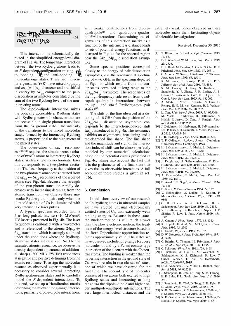
laureates: Junior prizes of the sCs fall meeting 2015 CHIMIA 2016, 70, No. 4 267
(5)𝑛𝑛𝑛�/� + 𝑛𝑛𝑛�/�
������������� ��������
𝑛𝑛𝑛�/� + (𝑛𝑛 + 1)𝑛�/�. (
This interaction is schematically de-picted in the simplified energy-level dia-gram of Fig. 4a. The long-range interactionbetween the two Rydberg atoms leads toan R-dependent configuration mixing andto ‘bonding’ Ψ��� and ‘anti-bonding’ Ψ��molecular eigenstates. These two molecu-lar eigenstates Ψ(R) have mixed np
3/2np
3/2and ns
1/2(n+1)s
1/2character and are shifted
in energy by ∆Eintcompared to the pair-
dissociation asymptotes constituted by thesum of the two Rydberg levels of the non-interacting atoms.
The dipole–dipole interaction mixesthe optically accessible p Rydberg stateswith Rydberg states of s character that arenot accessible in single-photon transitionsfrom the 6s ground state. The intensityof the transitions to the mixed molecularstates, formed by the interacting Rydbergatoms, is proportional to the p-character ofthe mixed states.
The observation of such resonanc-es[6,31–34] requires the simultaneous excita-tion of twoCs atoms to interactingRydbergstates. With a single monochromatic laserthis corresponds to a two-photon excita-tion. The laser frequency at the position ofthe two-photon resonances is detuned fromthe np
3/2← 6s
1/2resonances of the isolated
atoms (see Fig. 4a). Because the strengthof the two-photon transition rapidly de-creases with increasing detuning from theatomic transition, we observe these mo-lecular Rydberg-atom pairs only when theultracold sample of Cs is illuminated withvery intense UV laser pulses.
A typical spectrum recorded with a5 ns long pulsed, intense (~10 MW/cm2)UV laser is presented in Fig. 4b. The laserfrequency is calibrated with a wavemeterand is referenced to the atomic 24p
3/2←
6s1/2transition, which is strongly saturated
under the conditions where the Rydberg-atom-pair states are observed. Next to thesaturated atomic resonance, we observe thedensity-dependent appearance of addition-al, sharp (~300 MHz FWHM) resonancesat negative and positive detunings from theatomic resonance. To assign the molecularresonances observed experimentally it isnecessary to consider several interactingRydberg-atom-pair states and to carefullymodel the R-dependent interactions. Tothis end, we set up a Hamiltonian matrixdescribing the relevant long-range interac-tions, primarily dipole-dipole interactions,
with weaker contributions from dipole–quadrupole[15] and quadrupole–quadru-pole[16] interactions. Determining the ei-genvalues of this interaction matrix as afunction of the internuclear distance leadsto sets of potential-energy functions, as il-lustrated in Fig. 4c for the spectral regionnear the 24p
3/224p
3/2dissociation asymp-
tote.Some spectral positions correspond
closely to the energy of pair-dissociationasymptotes, e.g. the resonance at a detun-ing of ~ –6 GHz in the spectrum depictedin Fig. 4b, which results from molecu-lar states correlated at long range to the23s
1/224s
1/2asymptote. The resonances on
the high-frequency side result from quad-rupole-quadrupole interactions betweennp
3/2np
3/2and nfn´f Rydberg-atom pair
states.[16]The shift of the line observed at a de-
tuning of –6 GHz from the position of the23s
1/224s
1/2dissociation asymptote cor-
responds to the interaction-induced shift∆E
int, introduced in Fig. 4a. The resonance
exhibits an asymmetric broadening and ared-degraded line shape. The line shapeand the magnitude and sign of the interac-tion-induced shift can be almost perfectlymodeled by our numerical calculationsbased on the potential curves presented inFig. 4c, taking into account the fact thatit is the p-character of the final state thatgives rise to observable intensities. A fullaccount of these studies is given in ref.[16].
6. Conclusion
In this short overview of our researchon Cs Rydberg atoms in ultracold sampleswe have studied unusual electronicallyexcited states of Cs
2with extremely weak
binding energies. Because in these statesthe nuclear motion is still much slowerthan the slow electronic motion, the treat-ment of the energy-level structure based onthe Born-Oppenheimer approximation re-mains approximately valid. The states wehave observed include long-range Rydbergmolecules bound by a Fermi-contact-typeinteraction of the electron with the Cs neu-tral atoms. The binding is weaker than thehyperfine interaction in the ground state ofCs, which results in two classes of states,one of which we have observed for thefirst time. The second type of moleculesconsists of two atoms both excited to highRydberg states and interacting at longrange via the dipole–dipole and higher or-der multipole–multipole interactions. Thevery large internuclear distances and the
extremely weak bonds observed in thesemolecules make them fascinating objectsof scientific investigations.
Received: December 30, 2015
[1] T. Hänsch. A. Schawlow, Opt. Commun. 1975,13, 68.
[2] D. J. Wineland, W. M. Itano, Phys. Rev. A 1979,20, 1521.
[3] E. L. Raab, M. Prentiss, A. Cable, S. Chu, D. E.Pritchard, Phys. Rev. Lett. 1987, 59, 2631.
[4] C. Monroe, W. Swan, H. Robinson, C. Wieman,Phys. Rev. Lett. 1990, 65, 1571.
[5] K. M. Jones, E. Tiesinga, P. D. Lett, P. S.Julienne, Rev. Mod. Phys. 2006, 78, 483.
[6] S. M. Farooqi, D. Tong, S. Krishnan, J.Stanojevic, Y. P. Zhang, J. R. Ensher, A. S.Estrin, C. Boisseau, R. Côté, E. E. Eyler, P. L.Gould, Phys. Rev. Lett. 2003, 91, 183002.
[7] A. Marte, T. Volz, J. Schuster, S. Dürr, G.Rempe, E. G. M. van Kempen, B. J. Verhaar,Phys. Rev. Lett. 2002, 89, 283202.
[8] L. Carr, J. Ye, New J. Phys. 2009, 11, 055009.[9] M. Mack, F. Karlewski, H. Hattermann, S.
Höckh, F. Jessen, D. Cano, J. Fortágh, Phys.Rev. A 2011, 83, 052515.
[10] J. Deiglmayr, H. Herburger, H. Saßmannshau-sen, P. Jansen, H. Schmutz, F. Merkt, Phys. Rev.A 2016, 93, 013424.
[11] J. R. Rydberg, Z. Phys. Chem. 1890, 5, 227.[12] T. F. Gallagher, ‘Rydberg Atoms’, Cambridge
University Press, Cambridge, 1994.[13] H. Saßmannshausen, F. Merkt, J. Deiglmayr,
Phys. Rev. Lett. 2015, 114, 133201.[14] H. Saßmannshausen, F. Merkt, J. Deiglmayr,
Phys. Rev. A 2013, 87, 032519.[15] J. Deiglmayr, H. Saßmannshausen, P. Pillet,
F. Merkt, Phys. Rev. Lett. 2014, 113, 193001.[16] H. Saßmannshausen, F. Merkt, J. Deiglmayr,
Phys. Rev. A 2015, 92, 032505.[17] A. Osterwalder , F. Merkt, Phys. Rev. Lett.
1999, 82, 1831.[18] E. Amaldi, E. Segre, Il Nuovo Cimento 1934,
11, 145.[19] E. Fermi, Il Nuovo Cimento 1934, 11, 157.[20] V. Kokoouline, O. Dulieu, R. Kosloff, F.
Masnou-Seeuws, J. Chem. Phys. 1999, 110,9865.
[21] C. H. Greene, A. S. Dickinson, H. R.Sadeghpour, Phys. Rev. Lett. 2000, 85, 2458.
[22] V. Bendkowsky, B. Butscher, J Nipper, J. P.Shaffer, R. Löw, T. Pfau, Nature 2009, 458,1005.
[23] A. Omont, J. Phys. (Paris) 1977, 38, 1343.[24] K. Rupnik, U. Asaf, S.P. McGlynn, J. Chem.
Phys. 1990, 92, 2303.[25] E. Karule, Phys. Lett. 1965, 15, 137.[26] D.W. Norcross, J. Phys. B: At. Mol. Phys. 1971,
4, 1458.[27] C. Bahrim, U. Thumm, I. I. Fabrikant, J. Phys.
B: At. Mol. Opt. Phys. 2001, 34, L195.[28] C. Schwartz, Phys. Rev. 1961, 124, 1468.[29] F. Böttcher, A. Gaj, K. M. Westphal, M.
Schlagmüller, K. S. Kleinbach, R. Löw, T.Cubel Liebisch, T. Pfau, S. Hofferberth,arXiv:1510.01097, 2015.
[30] D. A. Anderson, S. A. Miller, G. Raithel, Phys.Rev. A 2014, 90, 062518.
[31] J. Stanojevic, R. Côté, D. Tong, S. M. Farooqi,E. E. Eyler, P. L. Gould, Eur. Phys. J. D 2006,40, 3.
[32] J. Stanojevic, R. Côté, D. Tong, E. E. Eyler, P.L. Gould, Phys. Rev. A, 2008, 78, 052709.
[33] K. R. Overstreet, A. Schwettmann, J. Tallant, J.P. Shaffer, Phys. Rev. A 2007, 76, 011403.
[34] K. R. Overstreet, A. Schwettmann, J. Tallant, D.Booth, J. P. Shaffer, Nat. Phys. 2009, 5, 581.

268 CHIMIA 2016, 70, No. 4 laureates: Junior prizes of the sCs fall meeting 2015
doi:10.2533/chimia.2016.268 Chimia 70 (2016) 268–273 © Swiss Chemical Society
*Correspondence: Dr. A. Riedoa
E-mail: [email protected] Institute, Space Research and PlanetarySciencesUniversity of Bern,Sidlerstrasse 5, CH-3012 Bern, SwitzerlandbDepartment of Chemistry and BiochemistryInterfacial Electrochemistry GroupUniversity of BernFreiestrasse 3, CH-3012 Bern, Switzerland
Laser Ablation/Ionisation MassSpectrometry: Sensitive and QuantitativeChemical Depth Profiling of SolidMaterials
Andreas Riedo§a*, Valentine Grimaudob, Pavel Moreno-Garcíab, Maike B. Neulanda, Marek Tuleja,Peter Broekmannb and Peter Wurza
§SCS-Metrohm Award for best oral presentation in Analytical Sciences
Abstract: Direct quantitative and sensitive chemical analysis of solid materials with high spatial resolution, bothin lateral and vertical direction is of high importance in various fields of analytical research, ranging from insitu space research to the semiconductor industry. Accurate knowledge of the chemical composition of solidmaterials allows a better understanding of physical and chemical processes that formed/altered the material andallows e.g. to further improve these processes. So far, state-of-the-art techniques such as SIMS, LA-ICP-MS orGD-MS have been applied for chemical analyses in these fields of research. In this report we review the currentmeasurement capability and the applicability of our Laser Ablation/Ionisation Mass Spectrometer (instrumentname LMS) for the chemical analysis of solids with high spatial resolution. The most recent chemical analysesconducted on various solid materials, including e.g. alloys, fossils and meteorites are discussed.
Keywords: Chemical depth profiling · Chemical mapping of heterogeneous materials ·High spatial resolution analysis · Laser ablation · Laser ionisation mass spectrometry
1. Introduction
Sensitive chemical analysis of solidmaterials with high spatial resolution in themicro- and nanometre regime is of consid-erable interest and importance in variousfields of academic and industrial research.These include e.g. the chemical analysisof layered solar panels,[1,2] the analysis ofspace-weathered samples,[3] the investiga-tions of contaminants embedded withinelectrodeposited Cu interconnects[4,5] andextensive chemical analyses of corrodedglass,[6] ancient celadon[7] or geologicalsamples,[8] amongmany others. In compar-ison to bulk analyses, measurement tech-niques with high spatial resolution enabledetailed chemical insights into the samplematerial and provide better understandingof physical and chemical processes thatmay have fashioned or modified the ana-lyte of interest.
Various sensitive and high-resolu-tion measurement techniques availablenowadays on the market, such as AugerElectron Spectroscopy (AES),[9,10] X-rayPhotoelectron Spectroscopy (XPS),[9,11]Secondary Ion Mass Spectrometry(SIMS)[5,9] and Secondary Neutron MassSpectroscopy (SNMS),[12]GlowDischargeTime-of-Flight Mass Spectrometry (GD-TOF-MS)[1,9] or LaserAblation InductivelyCoupled Plasma Mass Spectrometry (LA-ICP-MS)[2,6,9] are widely applied for theunderstanding of the above listed questionsconcerning the chemical composition ofsolid materials. Each analytical techniquediffers in its measurement capabilities andlimitations, including e.g. its detectionsensitivity (e.g. mass spectroscopic versusmass spectrometric), quantitative nature ofconducted measurements (e.g. SIMS ver-sus LA-ICP-MS), lateral profiling capa-bilities (e.g. SIMS versus GD-TOF-MS),vertical profiling capabilities (SIMS ver-sus LA-ICP-MS), measurement speed ac-quisition (e.g. XPS versus GD-TOF-MS),time for data interpretation (GD-TOF-MSversus AES), or limitations concerningthe suitable sample shape.[9,12] Therefore,typically a combination of diverse meas-urement techniques is required to providedetailed insights into the sample materialand hence, to answer the scientific ques-tions of interest.
With advances in vacuum technology,improved high speed electronics (e.g. dataacquisition system) and laser systems withsignificantly increased performance, LaserAblation/Ionisation Mass Spectrometry(LIMS) has proved, in the last decade, to bea truly alternative measurement techniquefor the sensitive and quantitative chemicalinvestigation of solid materials with highspatial resolution and figures of merit com-parable to state-of-the-art measurementtechniques, such as SIMS, LA-ICP-MS orGD-TOF-MS.[4,7,13–16]The implementationof ultrafast pulsed laser systems used asablation and ionisation source with pulsewidths within the femtosecond regime andpulse to pulse energy stability better thanthe %-level (e.g. CPA system, Clark-MXRInc., USA) has allowed the measurementperformance and capabilities of LIMS tobe significantly increased.[4,14,17] In com-parison to laser systems providing longerpulses the application of a femtosecondlaser ablation/ionisation source has vari-ous analytical advantages, including e.g.minimised elemental fractionation dueto reduced thermal heating of the samplematerial (heat affected zone, HAZ)[18] andthe lack of interaction between the ejectedplasma during the laser ablation processand the laser pulse.[19,20] Another impor-tant advantage concerns the improvedstoichiometry and ionisation efficiency of

laureates: Junior prizes of the sCs fall meeting 2015 CHIMIA 2016, 70, No. 4 269
In the current setup an ultrafast pulse lasersystem (λ = 775 nm, τ ~ 190 fs, repetitionrate ≤1 kHz, intensity ≤1 mJ/pulse, CPAsystem, Clark-MXR Inc., USA) is usedas ablation/ionisation source and is po-sitioned outside the vacuum chamber.[17]Laser pulses are guided towards the vacu-um chamber by dielectric mirrors, pass theentrance window of the vacuum chamberand are focused through the mass analys-er towards the sample surface using a lenssystem installed right on top of the massanalyser. The current lens system allowscircular laser ablation generating a craterdiameter in the range of about 10–15 µm.[4]Samples are positioned about 1 mm awayfrom the entrance of the LMS by using ahigh resolution xyz-micro translationalstage with a position accuracy of about2 µm. Only positively charged ions, gener-ated during the ablation/ionisation process,can enter the ion optical system of LMSwhere they are subsequently accelerated,focussed and confined in the field free drifttube. The ions are finally reflected by theion mirror towards the detector system bypassing the field free drift tube a secondtime. In Fig. 1 the scheme and principlesof operation of LMS are shown. Massseparation occurs along the ion trajectorybecause of the different velocity of dif-ferent mass ions, having the same energyand the ions arrive at the detector systemin sequence of their mass-to-charge ratio(m/z). A multi-readout multi-channel plate
and crater.[16] Depending on the applied la-ser irradiance the dimension of the craterin one direction is about 5 µm, and showsan increased dimension in the other axis.An integrated ion funnel and a differentialpumping system allow the ion source to beoperated at pressures up to about 0.3 mbar.This latter LIMS system can be used forelemental imaging with a lateral resolu-tion on the µm-scale but also for chemicaldepth profiling studies with a resolution ofabout 6 nm.[8]
The LIMS system developed by thegroup of Wurz (instrument name LMS)was originally designed for in situ inves-tigation of the chemical composition ofsolids on planetary objects, for a landerplatform on-board ESA’s BepiColombospace mission for the detailed explorationofMercury’s surface.[26]Unfortunately, thelander platform was cancelled later on dueto budgetary reasons but since its first de-sign in 2003 LMS has been continuouslyfurther developed.[4,13,17,32–42] Today, LMSrepresents the most powerful and versa-tile LIMS system ever designed for insitu space investigations and exhibits topperforming figures of merit. The systemhas the capability to conduct quantitativemeasurements with high detection sensi-tivity (10 ppb, atomic fraction) and witha dynamic range of about eight orders ofmagnitude.[17,37] It allows measurementsof element isotopes with high accuracy,[38]enables the elemental imaging of hetero-geneous materials with micrometre reso-lution,[23,32,35,36] and provides high verticaldepth profiling capabilities with sub-na-nometre resolution.[4,13] The measurementcapabilities of LMS for elemental imag-ing and depth profiling of solid materialsare presented in more detail in section 3,where most recent and current measure-ments conducted on different samplemate-rials, including the chemical mapping of anAllende meteorite sample, depth profilingof Si-supported Cu layers, chemical anal-ysis of micrometre fossils embedded in anargonite matrix, and capabilities towards3D elemental imaging of bronze-alloysamples are presented.
2. Experimental Setup
LMS is a reflectron-type time-of-flight(RTOF) mass spectrometer and was orig-inally designed for in situ measurementson planetary objects. The principles of op-eration and technical scheme of LMS arebriefly discussed in the following. Detailedinformation of the current setup can befound in recent publications.[4,17,26,37]
Since parameters such as size, powerconsumption and mass are highly limitedon a spacecraft the LMSmass analyser hasthe dimensions of only 160 mm × 60 mm.
the elements,[14,17] which together with theability of clean material ablation[19–22] andhigh pulse-to-pulse reproducibility enablea laser ablation in a highly controlled man-ner.[4,17]
So far, various LIMS systems havebeen designed and constructed specifical-ly to investigate different scientific ques-tions and for the application in variableenvironments, ranging from elementalmapping[8,23] to depth profiling analy-ses,[4,16] from laboratory[7,8,12,14–16,24,25] in-struments to instruments with the capabil-ity to operate on planetary objects.[17,26–31]However, and to our best current knowl-edge, only the designs from the researchgroups of Huang,[8,14,15,24] Cui et al.,[16] andWurz,[4,13,17,23,32] have reached the meas-urement capabilities and performancecomparable to the state-of-the-art meas-urement techniques for chemical analy-sis with high spatial resolution. All thesesystems consist of a TOF mass analyser,a sophisticated detector system and areequipped with a femtosecond laser abla-tion/ionisation source and differ only intheir technical realisation. In the followingthe LIMS systems from Cui et al.[16] andthe Huang group[8,14,15,24] are discussed on-ly briefly. Readers should consult the list-ed references for further information. Tointroduce the reader in more detail to theLIMS measurement technique, the perfor-mance and principles of operation of theLIMS system developed in our group arediscussed in more detail afterwards.
The LIMS system from the Huanggroup (MCP detector system in chevronconfiguration, dynamic range of about 6–7orders of magnitude, lateral resolution ofabout 40 µm) is based on an orthogonalion source design (the principal axis of thelaser ablation/ionisation ion source is or-thogonal to the principal ion optical axisof the mass spectrometer) and consists of areflectron-type time-of-flight (TOF) massspectrometer.[8,14,15,24] In comparison to theother LIMS systems, inert buffer gas suchas He or Ar is used for collisional coolingof energetic ions from the ablation processas well as charge reduction of multiplecharged ions produced during the laser ab-lation process.[15,33] The system has a de-tection sensitivity down to 30 ppb (g/g, Li)but due to contaminants within the buffergas sensitivities for a few elements, e.g. C,N or O, rises to few hundreds of ppm.[8,24]So far, the system has been used for chem-ical mapping of solids with high lateralresolution[8] and no depth profiling studieswere carried out, to the best of our knowl-edge. In the LIMS system from Cui et al.(MCP detector system in chevron config-uration), laser ablation is realised by thelaser hitting the sample at a 60° incidentangle, instead of perpendicular incidence,which results in an elliptical ablation spot
Fig. 1. Simplified schematic drawing of ourLMS instrument showing briefly its principle ofoperation.

270 CHIMIA 2016, 70, No. 4 laureates: Junior prizes of the sCs fall meeting 2015
gations of Allende, so far, were conductedseparately on specifically selected samplematerial, such as chondrules or CAIs (Ca-Al-rich inclusions) that were mechanicallyextracted by dedicated tools for the pur-pose of measurement.[e.g.43,44] The elemen-tal mapping using LMS, however, allowsthe chemical composition of Allende to bequantified without any dedicated samplepreparation. Furthermore, by knowing thechemical composition of many surface po-sitions on any geological sample, a miner-alogical bulk analysis of the sample can beprovided. Fig. 4 presents the mineralogicalanalysis resulting from the LMS measure-ments on Allende conducted with the na-nosecond laser system. The measurementsare in good agreement with the expectedelemental composition of the meteorite. Inthis campaign most measurements repre-sent mixtures of known minerals, whichcan be explained by the small size of min-eral grains and the comparably larger sizeof the laser spot. Therefore, the collectivityof the 138 measurement points is in goodagreement with the literature Allende bulkcomposition.[23]
Fig. 3. Spot-wise chemical investigation(13 × 63 single surface positions) of a sampleof Allende meteorite using LMS. a) Detail ofrectangular section from Fig. 2, element mapof b) Cr, c) S, and d) Fe.
the nanosecond laser system. In this meas-urement campaign 500 single waveformsper analysed location were acquired, eachan accumulation of 100 single laser shotmass spectra (50’000 single laser shotswere applied). Detailed information on themeasurements can be found in ref. [23].Subsequently, a dense raster of 63 × 13single surface positions (spacing of 50 µm)using the femtosecond laser system wasconducted (black rectangle in Fig. 2). Oneach surface position 30 waveforms wereacquired, each an accumulation of 2’000single laser shot mass spectra (60’000 sin-gle laser shots were applied), and analysed.
Fig. 2. Composite microscope image of anAllende meteorite sample. The surface areasurrounded by the black rectangle was inves-tigated spot-wise in detail by LMS using thefemtosecond laser system (see Fig. 3).
Fig. 3 presents the high resolution el-emental mapping (preliminary analysis)of the selected Allende surface area (blackframe) conducted using the femtosecondlaser system. In panel a) the investigatedsurface area (Fig. 2) is magnified and sev-eral inclusions are visible; the three mostdistinct inclusions are marked in black (A1and A2). Panels b) – d) illustrate the abun-dance distribution for Cr, S, and Fe, respec-tively.While within the areasA1 andA2 FeandS are less abundant, a higher abundanceof Cr was measured at the boundary of areaA1. In general, mass spectrometric investi-
(MCP) detector system in chevron config-uration (stack of 2 MCP plates) is used forthe detection of the incoming ions. Thespecial design of the detector system andthe multi-readout of signals allow meas-urements to be conducted with a high dy-namic range of about eight orders of mag-nitude and hence, allow signals frommajorto trace elements to be measured simulta-neously. Two high speedADC cards (8 bit,2 channels each, up to 4 GS/s) are used torecord the electrical signals generated bythe MCP detector system.
The laser system (initiation, energy/pulse, number of single laser shots), themicro translational stage and the acquisi-tion system are controlled fully remotelyby a custom-made software package. Theremote access to these units allows meas-urements with specific needs to be con-ducted, such as single spot measurementsor rastering over a complete sample sur-face stepwise or continuously, includingthe acquisition of single laser shot massspectra.[4,13,17,23,32,36]
3. Measurement Examples andDiscussions
The studies presented in the followingare a set of examples of what has beenachieved so farwith theLMS.Additionally,current research on a wide variety of mate-rials is being carried out to further demon-strate the capabilities and versatility ofLMS.
3.1 Two-dimensional ElementalImaging of an Allende MeteoriteFragment
Fig. 2 shows a composite microscopeimage of an Allende meteorite sample,a highly heterogeneous extra-terrestri-al material, composed of matrix materialand various chondrules and inclusions.[23]The Allende sample was selected for massspectrometric investigations using LMS todemonstrate the performance of the instru-ment for in situ chemical analysis of het-erogeneous samples on planetary objects,such as planets, moons, and asteroids. Themeasurements demonstrate the capabil-ity of LMS to provide chemical maps ofhighly heterogeneous materials. Chemicalanalyses were conducted using two differ-ent laser systems, a nanosecond laser sys-tem (λ = 266 nm, τ ~ 3 ns, pulse repetitionrate of 20 Hz, ablation crater of ~20 µm indiameter, ≤ 1GW/cm2)[23] and a femtosec-ond laser system (λ = 775 nm, τ ~ 190 fs,pulse repetition rate of 1 kHz, ablation cra-ter of ~12 µm in diameter, ~30 TW/cm2).Mass spectrometric measurements wereconducted on 138 single surface positionswith a horizontal and vertical spacing of600 µm and 426 µm respectively, using
Fig. 4. Mineralogicalanalysis of Allendeas a result of meas-urements conductedwith the nanosec-ond laser system.Literature mean (ingreen) from Strackeet al.[45] and McSweenand Huss.[46]
Reprinted fromNeuland et al.[23]
Copyright (2016),with permission fromElsevier.

laureates: Junior prizes of the sCs fall meeting 2015 CHIMIA 2016, 70, No. 4 271
a laser irradiance of about 5.5 TW/cm2wasapplied for this measurement campaign(laser ablation crater with a diameter ofabout 15 µm).
The current measurement performanceof LMS, including the used depth profilingmeasurement protocol, allowed the locali-sation of fossil veins even within the sam-
200 nm per waveform.[32] In this campaignthe depth resolution was electronicallylimited, by the accumulation of 2’000 sin-gle laser shot mass spectra to one wave-form. Accumulation of a reduced numberof mass spectra would increase the depthresolution. The femtosecond laser systemwas used as ablation/ionisation source and
3.2 Chemical Analysis with Sub-nanometre Vertical Resolution
Single laser shot mass spectrometricmeasurements were conducted on Cu sam-ples with defined thickness (typically 10µm) to investigate the correlation betweenlaser pulse energies (laser irradiances) andresultingmean ablation rate, that is the ratiobetween layer thickness and number of ap-plied single laser shots needed to perforatethe layer.[4] The Cu was electrochemicallydeposited on a Si-supported Cu seed layerunder galvanostatic conditions in the pres-ence of different plating additives (SPS,Imep, PEI, and PAG).[4] The femtosecondlaser system was used as ablation/ionisa-tion source and laser pulses were focussedonto the sample surface with a diameterof about 14 µm. For the first time quan-titative mass spectrometric depth profileswith a mean ablation rate at the sub-nano-metre regime were achieved by applyingthe LIMS measurement technique (Fig. 5).
Fig. 6 demonstrates the quantitativedepth profiling capability of LMS. Itshows a depth profile section of a mul-ti-layered Cu film (sample S4), where theabundances of the major contaminant C, Nand O, mainly originating from the electro-plating additives SPS and Imep, are mon-itored with sub-nanometre resolution.[4]These additives are applied to improve thecopper electrodeposition of interconnects.However, under specific electrochemicalconditions they can form quasi-periodicembedded contamination layers inside theCu film.[4,5,47–50] The sharp increase andsubsequent decrease of the C, N and O sig-nals are in phase and exhibit anti-correlat-ed intensity oscillations with the recordedCu signal, as was expected from previousdepth profiling experiments obtained withthe SIMS technique. However, in contrastto traditional SIMS measurements, theLMS measurements neither showed atten-uation nor broadening of these oscillationswith increasing material erosion, indicat-ing a smoother ablation procedure andnegligible roughening effects at the craterbottom.
3.3 Micrometre-size FossilsEmbedded in an Argonite Matrix
Fig. 7 shows microscope images of ageological sample with embedded fossilswithin an argonite matrix. Mass spectro-metric measurements were conductedwithin the square M1 (200 × 200 µm2),marked in panel a) and enlarged in panelb). The area of interest was investigatedspot-wise by a raster of 10 × 10 surface po-sitions with a spacing of 20 µm. From eachsingle surface position 20 waveforms wererecorded, each an accumulation of 2’000single laser shot mass spectra (40’000 sin-gle laser shot mass spectra in total), whichcorresponds to a depth resolution of about
Fig. 5. Correlationbetween mean abla-tion rate and appliedpulse energy (laserirradiance). For thefirst time quantitativemass spectromet-ric measurementswith sub-nanometerresolution per singlelaser shot were real-ized using the LIMSmeasurement tech-nique.[4] Reprintedwith permission fromGrimaudo et al.[4]Copyright (2016)American ChemicalSociety.
Fig. 6. Depth pro-filing section of theCu film (sample S4)showing the anti-cor-relation between themajor contaminantsC, N, and O andCu.[4] Reprinted withpermission fromGrimaudo et al.[4]
Copyright (2016)American ChemicalSociety.
Fig. 7. Microscope image of a geological sample containing fossils of mi-crometre dimensions. The area marked by a black square (M1) in panel a)is shown enlarged in b). The black veins correspond to fossils embeddedin the sample matrix. M1 was spot-wise investigated (100 single surfacepositions) using LMS. Image adapted with permission from Tulej et al.[32]
Copyright (2016) Mary Ann Liebert, Inc.

272 CHIMIA 2016, 70, No. 4 laureates: Junior prizes of the sCs fall meeting 2015
ple matrix where optical microscopes findtheir limitations. In Fig. 8 the signals frombio-relevant elements, B and S, are pre-sented for the different surface positions.For this analysis all waveforms from eachsingle surface position were accumulatedto build one single mass spectrum and spe-cial attention was devoted to the B and Ssignals. Surface positions 45 and 66 turnedout to contain bio-relevant elements (seeFig. 8), but no fossil veins were visible onthese positions in the microscope image.Furthermore, the depth profiling procedurewith LMS allowed these fossil signaturesto be localised in the matrix within thedepth profiling accuracy.[32]
3.4 Three-dimensional ElementalImaging of Bronze Alloys
Alloys are used in various fields ofresearch, ranging from applications with-in the microchip industry to technologiesfor energy conversion.[51–55] Most recentmeasurements using the LMS instrumentwere conducted on Cu-Pb-Sn alloys con-taining different abundances of these threemajor elements. Mass spectrometric meas-urements with a lateral and vertical resolu-tion within the micrometre and sub-micro-metre regime, respectively, were conduct-
ed to provide a 3D chemical record of theelemental distribution of Cu, Sn, and Pbwithin the alloys. For these measurementsthe femtosecond laser system was used asablation/ionisation source whereas laserpulses of a few TW/cm2 were focussedonto a surface area of about 10–15 µm indiameter. The alloy surfaces were investi-gated by a 4 × 4 single-spot raster and up to1’000 waveforms were recorded from eachsurface position, each an accumulation of200 single laser shot mass spectra.[56]
In Fig. 9 preliminary analyses of thechemical depth profile (the first 500 wave-forms, top of profile corresponds to thesample surface) are presented showingthe spatial distribution of the elements Pb,Sn and Cu at three different surface posi-tions.[56]While green indicates the expect-ed elemental abundances according to theempirical formula (Cu
80Sn
10Pb
10alloy),
blue and red show decreased or enrichedelemental abundances, respectively. Theaccumulation over all mass spectrometricinformation from one raster experimentresulted in mean elemental abundances ofabout (73 ± 11) wt%, (8 ± 4) wt% and (19± 10) wt%, respectively for Cu, Sn and Pb.The latest results illustrate the possibilityof applying LMS to investigate not only
the elemental distribution at the surface ofa material but also to get an insight intothe homogeneity in all three dimensionsgiving evidence about the quality of thealloying material.[56]
4. Conclusions
Chemical analysis with high spatialresolution in the micro- and nanometre re-gime is of high interest in various fields ofresearch, where detailed chemical insightswithin the solid material are necessary toe.g. further improve production process-es. So far, measurement techniques suchas SIMS, LA-ICP-MS and GD-MS havebeen used to conduct measurements withhigh spatial resolution. However, with theadvances in the last decades regarding la-ser systems, electronics, and vacuum tech-nology, LIMS has emerged as a truly alter-native direct measurement technique withmeasurement capabilities comparable tothe state-of-the art techniques. NowadaysLIMS instruments can conduct sensitive(ppb level) and quantitative mass spectro-metric analyses of solid materials with alateral resolution in the micrometre regimeand a vertical resolution at the sub-nano-metre level, both of considerable impor-tance to provide a 3D chemical insight intothe analyte of interest.
AcknowledgmentsThis work is supported by the Swiss
National Science Foundation.
Received: January 15, 2016
[1] J. Pisonero, N. Bordel, C. Gonzalez de Vega,B. Fernández, R. Pereiro, A. Sanz-Medel, Anal.Bioanal. Chem. 2013, 405, 5655.
[2] A. Gutiérrez-González, C. González-Gago, J.Pisonero, N. Tibbetts, A. Menéndez, M. Vélez,N. Bordel, J. Anal. At. Spectrom. 2015, 30, 191.
[3] T. Noguchi, T. Nakamura, M. Kimura, M. E.Zolensky, M. Tanaka, T. Hashimoto, M. Konno,A. Nakato, T. Ogami, A. Fujimura, M. Abe, T.Yada, T. Mukai, M. Ueno, T. Okada, K. Shirai,Y. Ishibashi, R. Okazaki, Science 2011, 333,1121.
[4] V. Grimaudo, P. Moreno-García, A. Riedo, M.B. Neuland, M. Tulej, P. Broekmann, P. Wurz,Anal. Chem. 2015, 87, 2037.
[5] N. T. M. Hai, D. Lechner, F. Stricker, J. Furrer,P. Broekmann, ChemElectroChem 2015, 2, 664.
[6] S. J. M. Van Malderen, J. T. Van Elteren, F.Vanhaecke, Anal. Chem. 2015, 87, 6125.
[7] M. He, Y. Xiao, S. Zhang, R. Liu, W. Hang, B.Huang, Appl. Surf. Sci. 2015, 351, 624.
[8] R. Huang, B. Zhang, D. Zou,W. Hang, J. He, B.Huang, Anal. Chem. 2011, 83, 1102.
[9] J. S. Becker, ‘Inorganic Mass Spectrometry- Principles and Applications’, John Wiley &Sons Ltd., England, 2007.
[10] J. Pantoja-Enríquez, E. Gómez-Barojas, R.Silva-González, U. Pal, Sol. Energy Mater. Sol.Cells 2007, 91, 1392.
[11] M. Duta, S. Mihaiu, C. Munteanu, M.Anastasescu, P. Osiceanu, A. Marin, S. Preda,M. Nicolescu, M. Modreanu, M. Zaharescu, M.Gartner, Appl. Surf. Sci. 2015, 344, 196.
Fig. 8. The accumula-tion of all mass spec-trometric measure-ments conducted onone surface positionallowed to identifyembedded fossilsveins by monitoringbio-relevant ele-ments, here B and S.Image adapted withpermission from Tulejet al.[32] Copyright(2016) Mary AnnLiebert, Inc.
Fig. 9. Preliminarydepth profiling stud-ies of bronze alloysare shown. By aver-aging all individualmass spectrometricmeasurements thenominal abundancesof Pb, Sn and Cuin wt% is derived.However, in certainapplications the spa-tial distribution of in-corporated elementsare of high interest.

laureates: Junior prizes of the sCs fall meeting 2015 CHIMIA 2016, 70, No. 4 273
matrix’, in ‘Chondrules and their origins(A85-26528 11-91)’, Houston, TX, Lunar andPlanetary Institute, 1983, pp. 145–161.
[45] A. Stracke, H. Palme, M. Gellissen, C. Münker,T. Kleine, K. Birbaum, D. Günther, B. Bourdon,J. Zipfel, Geochim. Cosmochim. Acta 2012, 85,114.
[46] H. Y. McSween Jr., G. R. Huss, ‘Cosmo-chemistry’, 2010, Cambridge University Press.
[47] N. T. M. Hai, K. W. Krämer, A. Fluegel, M.Arnold, D. Mayer, P. Broekmann, Electrochim.Acta 2012, 83, 367.
[48] N. T. M. Hai, J. Odermatt, V. Grimaudo, K.W.Krämer, A. Fluegel, M. Arnold, D. Mayer, P.Broekmann, J. Phys. Chem. C 2012, 116, 6913.
[49] K. Krischer, N. Mazouz, P. Grauel, Angew.Chem. Int. Ed. 2001, 40, 850.
[50] P. Strasser, M. Eiswirth, M. T. M. Koper, J.Electroanal. Chem. 1999, 478, 50.
[51] H.-A. Kuhn, I. Altenberger, A. Käufler,H. Hölzl, M. Fünfer, ‘Properties of HighPerformance Alloys for ElectromechanicalConnectors (CH3)’, in ‘Copper Alloys – EarlyApplications and Current Performance –Enhancing Processes’, Ed. L. Collini, InTech,2012.
[52] G. Zangari, Coatings 2015, 5, 195.[53] C. Gütz, M. Selt, M. Bänziger, C. Bucher,
C. Römelt, N. Hecken, F. Gallou, Tomás R.Galvão, S.R. Waldvogel, Chem. Eur. J. 2015,21, 13878.
[54] C. Edinger, S. R.Waldvogel, Eur. J. Org. Chem.2014, 2014, 5144.
[55] C. Edinger, V. Grimaudo, P. Broekmann, S. R.Waldvogel, ChemElectroChem 2014, 1, 1018.
[56] V. Grimaudo, P. Moreno-García, A. Riedo, M.Tulej, P. Broekmann, P. Wurz, Anal. Chem., tobe submitted, 2016.
[27] W. B. Brinckerhoff, G. G. Managadze, R. W.McEntire, A. F. Cheng, W. J. Green, Rev. Sci.Instr. 2000, 71, 536.
[28] W.B. Brinckerhoff, Planet. Space. Sci. 2005,53, 817.
[29] X. Li, W. B. Brinckerhoff, G.G. Managadze,D.E. Pugel, C.M. Corrigan, J.H. Doty, Int. J.Mass Spectrom. 2012, 323 63.
[30] W. B. Brinckerhoff, Appl. Phys. A, 2004, 79,953.
[31] G. G. Managadze, P. Wurz, R. Z. Sagdeev, A.E. Chumikov, M. Tuley, M. Yakovleva, N. G.Managadze, A. L. Bondarenko, Sol. Sys. Res.2010, 44, 376.
[32] M. Tulej, A. Neubeck, M. Ivarsson, A. Riedo,M. B. Neuland, S. Meyer, P. Wurz, Astrobiol.2015, 15, 669.
[33] Y. Lin, Q. Yu, R. Huang, W. Hang, J. He, B.Huang, Spectrochim. Acta Part B 2009, 64,1204.
[34] M. Tulej, M. Iakovleva, I. Leya, P. Wurz, Anal.Bional. Chem. 2011, 399, 2185.
[35] A. Neubeck, M. Tulej, M. Ivarsson, C. Broman,A. Riedo, S. McMahon, P. Wurz, S. Bengtsson,Int. J. Astrobiol. 2016, 15, 133.
[36] M. Tulej, A. Riedo, M. B. Neuland, S. Meyer,P. Wurz, N. Thomas, V. Grimaudo, P. Moreno-García, P. Broekmann, A. Neubeck, M.Ivarsson, Geostand. Geoanal. Res. 2014, 38,441.
[37] A. Riedo, A. Bieler, M. Neuland, M. Tulej, P.Wurz, J. Mass Spectrom. 2013, 48, 1.
[38] A. Riedo, S. Meyer, B. Heredia, M. B. Neuland,A. Bieler, M. Tulej, I. Leya, M. Iakovleva, K.Mezger, P.Wurz, Planet. Space Sci. 2013, 87, 1.
[39] M. Tulej, A. Riedo, M. Iakovleva, P. Wurz, Int.J. Spec. 2012, 2012, 234949-1.
[40] A. Bieler, K. Altwegg, L. Hofer, A. Jäckel, A.Riedo, T. Sémon, P. Wurz, J. Mass Spectrom.2011, 46, 1143.
[41] A. Riedo, P. Wahlström, J. A. Scheer, P. Wurz,M. Tulej, J. Appl. Phys. 2010, 108, 114915-1.
[42] P. Moreno-García, V. Grimaudo, A. Riedo, M.Tulej, P. Wurz, P. Broekmann, Rapid Commun.Mass Spectrom. 2016, 30, 1031.
[43] Y. Liu, C. Chai, Chem. Geol. 1997, 141, 105.[44] R. M. Housley, E. H. Cirlin, ‘On the alteration
of Allende chondrules and the formation of
[12] P. Le Coustumer, P. Chapon, A. Tempez, Y.Popov, G. Thompson, I. Molchan, N. Trigoulet,P. Skeldon, A. Licciardello, N. Tuccitto, I.Delfanti, K. Fuhrer, M. Gonin, J. Whitby,M. Hohl, C. Tanner, N. Bordel Garcia, L.Lobo Revilla, J. Pisonero, R. Pereiro, C.Gonzalez Gago, A. Sanz Medel, M. GanciuPetcu, A. Surmeian, C. Diplasu, A. Groza,N. Jakubowski, R. Dorka, S. Canulescu, J.Michler, P. Belenguer, T. Nelis, A. Zahri, P.Guillot, L. Thérese, A. Littner, R. Vaux, J.Malherbe, F. Huneau, F. Stevie, H. Francois-Saint-Cyr, ‘Analysis of thin and thick Films’, in‘Mass Spectrometry Handbook’, Ed. M.S. Lee,2012, John Wiley & Sons, Inc., Hoboken, NJ,USA.
[13] A. Riedo, V. Grimaudo, P. Moreno-García, M.B. Neuland, M. Tulej, P. Wurz, P. Broekmann,J. Anal. At. Spectrom. 2015, 30, 2371.
[14] B. Zhang, M. He, W. Hang, B. Huang, Anal.Chem. 2013, 85, 4507.
[15] Q. Yu, R. Huang, L. Li, L. Lin, W. Hang, J. He,B. Huang, Anal. Chem. 2009, 81, 4343.
[16] Y. Cui, J. F. Moore, S. Milasinovic, Y. Liu, R.J. Gordon, L. Hanley, Rev. Sci. Instr. 2012, 83,093702.
[17] A. Riedo, M. Neuland, S. Meyer, M. Tulej, P.Wurz, J. Anal. At. Spectrom. 2013, 28, 1256.
[18] R. E. Russo, X. Mao, J. J. Gonzales, J. Yoo,Spectroscopy 2013, 28, 24.
[19] R. Hergenröder, O. Samek, V. Hommes, MassSpectrom. Rev. 2006, 25, 551.
[20] X. Zeng, X. L. Mao, R. Greif, R. E. Russo,Appl. Phys. A: Mater. Sci. Process. 2005, 80,237.
[21] R. E. Russo, X. Mao, J. J. Gonazales, S. S. Mao,Anal. At. Spectrom. 2002, 17, 1072.
[22] D. J. Hwang, H. Jeon, C. P. Grigoropoulos, J.Yoo, R. E. Russo, App. Phys. Lett. 2007, 91,251118.
[23] M. B. Neuland, S. Meyer, K. Mezger, A. Riedo,M. Tulej, P.Wurz, Planet. Space Sci. 2014, 101,196.
[24] R. Huang, Q. Yu, L. Li, Y. Lin, W. Hang, J. He,B. Huang,Mass Spectrom. Rev. 2011, 30, 1256.
[25] W. Hang, J. Anal. At. Spectrom. 2005, 20, 301.[26] U. Rohner, J. A. Whitby, P. Wurz, Meas. Sci.
Technol. 2003, 14, 2159.

274 CHIMIA 2016, 70, No. 4 laureates: Junior prizes of the sCs fall meeting 2015
doi:10.2533/chimia.2016.274 Chimia 70 (2016) 274–278 © Swiss Chemical Society
*Correspondence: Prof. Dr. J. Pérez-RamírezE-mail: [email protected] for Chemical and BioengineeringDepartment of Chemical and Applied BiosciencesETH ZurichVladimir-Prelog-Weg 1CH-8093 Zurich
Halogen Chemistry on Catalytic Surfaces
Maximilian Moser§ and Javier Pérez-Ramírez*
§SCS-Metrohm Award for best oral presentation in Catalysis Science and Engineering
Abstract: Halogens are key building blocks for the manufacture of high-value products such as chemicals,plastics, and pharmaceuticals. The catalytic oxidation of HCl and HBr is an attractive route to recover chlorineand bromine in order to ensure the sustainability of the production processes. Very few materials withstandthe high corrosiveness and the strong exothermicity of the reactions and among them RuO2 and CeO2-basedcatalysts have been successfully applied in HCl oxidation. The search for efficient systems for HBr oxidationwas initiated by extrapolating the results of HCl oxidation based on the chemical similarity of these reactions.Interestingly, despite its inactivity in HCl oxidation, TiO2 was found to be an outstanding HBr oxidation catalyst,which highlighted that the latter reaction is more complex than previously assumed. Herein, we discuss theresults of recent comparative studies of HCl and HBr oxidation on both rutile-type (RuO2, IrO2, and TiO2) andceria-based catalysts using a combination of advanced experimental and theoretical methods to provide deepermolecular-level understanding of the reactions. This knowledge aids the design of the next-generation catalystsfor halogen recycling.
Keywords: Ceria-zirconia · Hydrogen halides · Oxidation · Prompt gamma activation analysis ·Rutile-type oxides.
Introduction
Halogens are widely applied as reac-tive intermediates in the synthesis of vari-ous commodities in the chemical, pharma-ceutical, and plastics industry.[1] However,about half of the halogens are reduced toundesired hydrogen halides and halidesalts. The copious formation of HCl orHBr by-products is a serious challenge,since options to deal with this waste streamsuch as marketing or neutralization are un-attractive.[1] The recovery of Cl
2and Br
2from HCl and HBr has been recognized asthe most appealing route in order to main-tain an economically feasible and environ-mentally friendly process design.Amongstthe available options, catalytic oxidationhas been shown to be the most waste- andcost-efficient method to achieve this.[1,2]
The study of HCl oxidation dates backto the 1860s, when Henry Deacon invent-
ed the process to recover chlorine for theLeblanc process.[1a,b] The corrosivenessand the high exothermicity of the reactionwas detrimental to the CuCl
2-based cata-
lyst, which suffered from volatilization ofits active phase. The Deacon process waseventually abandoned in the 1890s due tosubstitution of the Leblanc process by theSolvay process.[1a,b] Still, various compa-nies and researchers attempted throughoutthe 20th century to improve the Cu-basedcatalysts or to identify new systems.[1a,b]At the turn of the millennium, RuO
2-
based catalysts, developed independent-ly by Sumitomo Chemicals and BayerMaterialScience, were implemented asthe new generation of highly active andstable HCl oxidation catalysts for chlo-rine recycling within the manufacture ofpolyurethanes and polycarbonates.[1a] Inthe continuous search for more alterna-tive catalysts, other metal oxides such asCeO
2, U
3O
8, and CuCrO
2, have been dis-
covered.[3] In particular, ceria has receivedconsiderable attention, which pinnacled inthe formulation of a technical CeO
2/ZrO
2catalyst with stable performance in pilotstudies.[4]
The development of an analoguecatalytic HBr oxidation process wasfirst attempted in the 1930s by GeorgeHooker, who adopted the original Deaconconcept.[5] HBr oxidation has been inves-tigated by different companies in the pastdecades, but the implementation was in-hibited due to the difficulties of identify-ing stable catalysts and a safe process.[2b]Research in bromine recovery was revi-
talized when the increased production ofunconventional natural gas prompted thepotential use of Br
2to functionalize the in-
ert alkanes into alkyl bromides for furtherconversion into fuels and chemicals.[1c,2a]The search for a cost-efficient bromine re-covery process logically led to the investi-gation of HCl oxidation catalysts for HBroxidation. CeO
2-based and rutile-type (i.e.
RuO2and IrO
2) materials have been shown
to catalyze HBr oxidation, corroboratingthe anticipated chemical similarities ofboth reactions.[6] However, the unexpectedactivity of TiO
2in HBr oxidation, despite
its inactivity in HCl oxidation, underlinedthat the mechanism of HBr oxidation hasfeatures distinct from that of HCl oxida-tion.[7] To close this gap in understanding,dedicated studies are needed to rationalizethe relationship between the state of thesurface and the apparent performance overboth rutile-type and ceria-based catalystsin HBr oxidation.
In this article, we discuss the strat-egy for an integrated approach to halogenchemistry and present advanced methods,i.e. temporal analysis of products (TAP)and prompt gamma activation analysis(PGAA), as key techniques to experimen-tally analyze mechanistic aspects and thestate of the catalyst surface under workingconditions. We outline the interplay be-tween the surface chemistry and the per-formance in HCl and HBr oxidation overrutile-type and ceria-based catalysts. Wehighlight the necessity to individually op-timize the catalyst formulation for eitherHCl or HBr oxidation.

laureates: Junior prizes of the sCs fall meeting 2015 CHIMIA 2016, 70, No. 4 275
evolution barrier. With respect to the cata-lyst scale up, both metal oxides can be dis-persed in the form of small nano layers orclusters on TiO
2-rutile, which is due to lat-
tice matching (RuO2) or electronic effects
(IrO2). Owing to their robustness in HCl
oxidation, both catalysts were consideredpotential candidates for a stable oxidationof HBr. In fact, the comparison of their ac-tivities in HCl and HBr oxidation revealedthat they oxidize HBr at much lower tem-perature (Fig. 2a).[6] The activity of TiO
2for HBr oxidation[7] was not expected dueto the inactivity in HCl oxidation which ledto the commonly accepted theory that TiO
2cannot catalyze hydrogen halide oxidationdue to its inability to adsorb and dissociatemolecular oxygen.[16]
In-depth studies were undertaken to as-sess and compare the rutile-type catalystsin HCl and HBr oxidation. Despite its sta-bility in HCl oxidation, RuO
2brominates
in contrast to TiO2, which led to changes in
the crystallinity observed by HRTEM andsupported by ab initio thermodynamics,suggesting that the replacement of latticeoxygen is more exothermic in RuO
2than
TiO2, facilitating a strong bromination of
the surface and subsurface (Fig. 2b).[17]To shed light on the reaction mecha-
nism, the surface of TiO2was studied with
XPS, which evidenced the formation ofTi3+ sites during the HBr oxidation;[7] anindication of the formation of surface de-fects. Theoretical calculations on this de-fect formation ultimately proposed that thereplacement of the bridge oxygen with abromine atom is a self-doping mechanismthat eventually enables the adsorption ofmolecular oxygen due to the smaller ener-getic barrier to transfer the electrons fromthe bromide to the oxygen (Fig. 2c).[7] Incontrast to that, the energetic barrier of thechlorine doped surface is too large to en-able the activation of molecular oxygen onthe surface.
DFT indicated that the reaction mech-
make TAP a unique technology to bridgethe pressure and material gap betweenUHV and real-life catalytic conditions.[10]
PGAAhasbeen shown tobeadirect andreliable method to determine the elementalcomposition of the catalysts under reactionconditions.[4,8c,12] The working principle isbased on radiative neutron capture, or (n,γ )reaction upon irradiation of the samplewith cold neutrons (Fig. 1c). The nucleicapture the neutrons and promptly emittypically 2 to 4 gamma rays in a cascadebefore returning to the ground state. Thegamma radiation is characteristic, i.e. theenergy values of the gamma rays identifythe nuclide, and their intensities are pro-portional to the number of atoms presentin the analyzed volume. The sensitivity andthe detection limit of PGAA differ from el-ement to element, and depend on the par-tial gamma-ray production cross-section,the detector efficiency and the level of thebaseline at the analytical line. The dynamicrange of the method is limited by either thetoo low sensitivity, such as for oxygen, orby the too high counting rate that overloadsthe counting system, e.g. with iridium.[13]
Surface Chemistry andPerformance of Rutile-typeCatalysts for Halogen Production
RuO2-based materials are the current
state-of-the-art catalysts for HCl oxidationand have received widespread attentionamongst researchers, who aimed to under-stand the reaction mechanism and to lowerthe amount of precious ruthenium requiredin the catalyst composition.[14] BesidesRuO
2, also IrO
2was discovered to be ac-
tive in HCl oxidation.[15]Both metal oxidescomprise a dense rutile-type structure thatinhibits the penetration of chlorine atomsinto the subsurface layers. IrO
2operates,
however, at much higher temperature com-pared to RuO
2due to its larger chlorine
Integrated Approach to HalogenChemistry
Throughout the research on HCl andHBr oxidation, the study of the surfacechemistry under working conditions hasbeen hampered by the corrosiveness of thereactants. Mechanistic studies mostly em-phasized on ex situ characterization suchas X-ray diffraction (XRD), X-ray photo-electron spectroscopy (XPS), and high-res-olution transmission electron microscopy(HRTEM),aswellasmolecularmodeling.[8]Further, ultra-high vacuum (UHV) meth-ods, such as the high-resolution core-levelshift, enables the study of surface process-es under reaction atmosphere over singleRuO
2crystals.[9] However, to connect the
molecular-level studies on model surfacesto the apparent performance of real-lifecatalysts the pressure and materials gapshave to be closed.[10] For this purpose,an integrated approach was developedby combining different characterizationtechniques (Fig. 1a). The key methods ofthis strategy are theoretical studies usingdensity functional theory (DFT), transientmechanistic studies using temporal analy-sis of products (TAP),[11] and the quantifi-cation of the halogen uptake underworkingconditions with operando prompt gammaactivation analysis (PGAA).[4,8c,12]
TAP is a highly advanced techniqueto study mechanisms of experimentallydemanding reactions over industriallyrelevant catalysts.[10] Small amounts (106
molecules) of the reactants are pulsed overthe catalyst bed and are analyzed by quad-rupole mass spectroscopy (QMS) as shownin Fig. 1b. The reactants can be pulsedindividually, simultaneously, or in pump-probe fashion, i.e. with distinct time delaybetween individual reactant pulses. Keyadvantages of TAP comprise (i) a millisec-ond time resolution, (ii) the use of practicalcatalysts, (iii) excellent temperature con-trol, and (iv) safe operation. These aspects
Fig. 1. The synchronization of advanced techniques bridges the pressure and materials gap between molecular studies and catalytic evaluationunder working conditions (a). Transient mechanistic studies on real-life catalysts are conducted using TAP at millisecond time resolution (b). Theuptake of halogens on the catalysts under working conditions is quantified using PGAA (c).

276 CHIMIA 2016, 70, No. 4 laureates: Junior prizes of the sCs fall meeting 2015
tures and the formation of Ce-Zr mixedoxides.[4] The formation of mixed Ce-Zroxides positively affects the performancein HCl oxidation due to improved redoxproperties and higher thermal stability.[13]However, the impact of the catalyst’s mor-phology, i.e. how well Ce and Zr have tobe intermixed at an atomic or particle levelto achieve these properties, was unknown.
To study the impact of zirconia onthe performance of ceria, different ceria-zirconia mixed oxides were prepared ex-hibiting distinct morphologies that areeither homogeneous or non-homogeneousmixed Ce-Zr oxides as shown by energy-dispersive X-ray spectroscopy (EDS) map-ping (Fig. 3a). The catalytic performancein HCl oxidation of both materials is verycomparable.[13]However, in HBr oxidationdistinctions became evident as the catalyt-ic activity of non-homogeneous materialsdropped more strongly within 40 h com-pared to the homogeneous mixed oxide(Fig. 3b).
PGAA studies on the different mixedCe-Zr oxides indicated that CeO
2-based
catalysts are more prone to brominationthan chlorination (Fig. 3c,d), which isdetrimental to their stability. The chlorineuptake under different reaction conditions
idenced the completely different behaviorof the rutile materials under working con-ditions, which points to the need for furtheroptimization of the stability of RuO
2if an
application at low reaction temperatures isdesired. One way could be the formulationof mixed Ru-Ti oxides to decrease the sur-face bromination and inhibit the penetra-tion of bromine into the subsurface.
Stability and Activity of MixedCe-Zr Oxides in HCl and HBrOxidation
Similar stability issues, as seen forRuO
2in HBr oxidation, were already
known from the case of CeO2in HCl oxi-
dation.[3a] CeO2suffers from subsurface
chlorination under harsh reaction condi-tions as evidenced by operando PGAA,forming stable and inactive cerium chlo-ride.[12] In an attempt to stabilize the CeO
2different support materials were investigat-ed, which led to the discovery of the sup-ported CeO
2/ZrO
2systems.[4] It was shown
that the ZrO2support reduced the chlorine
uptake and increased the activity of CeO2
significantly, which was suggested to belinked to stabilization of CeO
2nanostruc-
anism follows the same path with bromineevolution as the most energy demandingstep (Fig. 2d).[7] The calculations werecorroboratedbyTAPstudies,whichshowedthat the bromine evolution from the surfaceis the slowest step. Furthermore, the timedelay between the HBr and the Br
2pulse
increases in the order: RuO2< TiO
2< IrO
2,
which is in agreement with the increase inthe calculated energy barriers (Fig. 2e)[17]This also supports that bromine evolutionis easier on TiO
2than IrO
2and explains
that the apparently similar activity of bothcatalysts in HBr oxidation results from acompensation effect between the surfacecoverage and the reaction kinetics.[7,17]
The quantitative determination of thehalogen uptake under working condi-tions using PGAA demonstrates that RuO
2suffers from extensive subsurface bromi-nation, at low O
2:HBr ratios and tempera-
tures, which agrees with the microscopicstudies and ab initio thermodynamics.TiO
2exhibits inherently low halogen cov-
erage (30–50%) at all conditions due toits unique defect-driven mechanism (Fig.2f).[17] In HCl oxidation, TiO
2is inactive
and the chlorination of the active RuO2is
limited to the surface. Correlating the halo-gen uptake to the apparent performance ev-
Fig. 2. Activity versus temperature for RuO2 (red), IrO2 (green) and TiO2 (blue) in HCl (dashed line) and HBr (solid lines) oxidation (a). HRTEMmicrographs of RuO2 (b) treated in HBr (top) and HCl (bottom). Illustration of the band gap of the semiconductor TiO2 (c). DFT calculation for thehalogen evolution on halogenated rutile-type surfaces (d). Normalized HBr and Br2 responses after simultaneous HBr and O2 pulsing for rutile-typecatalysts (e). Activity versus halogen coverage over TiO2 and RuO2 in HCl (open symbols) and HBr (solid symbols) oxidation at different oxygenpartial pressures (f).

laureates: Junior prizes of the sCs fall meeting 2015 CHIMIA 2016, 70, No. 4 277
four times higher than that of chlorine, dueto the stronger tendency for brominationthan chlorination. As the bromine contentis higher than that of chlorine, the optimumfor the HBr oxidation seems to have furthershifted towards a higher Zr content (Fig.3e), which underlines the distinctions inthe optimal design of Ce-Zr catalysts forapplication in either HCl or HBr oxidation.In this respect, the Ce:Zr ratio is the keyoptimization parameter to tune the stabilityof the CeO
2-based catalysts.
Conclusions
The study of catalytic hydrogen halideoxidation is experimentally challenging,which presents significant demands forthe mechanistic analysis and characteriza-tion of catalysts under working conditions.The combination of advanced techniquessuch as TAP and operando PGAA withtheoretical calculations comprise an opti-mal approach to understand halogen chem-istry on catalytic materials. This strategyenabled the rationalization of the excep-tional activity of TiO
2and the stability is-
sues of RuO2- and CeO
2-based catalysts in
HBr oxidation. The comparison betweenHBr and HCl oxidation indicated that thecatalyst design criteria applied in HCl oxi-dation cannot be directly extrapolated tothe recycling of Br
2. Thus, catalyst design
should be tailored for each reaction. Theinvestigation showed that the halogen cov-erage under working conditions is the bestparameter to assess the catalytic perfor-mance, which also highlights the signifi-cance of PGAA for this research. The sur-face halogenation can be tuned by optimiz-ing the reaction conditions or the catalystformulation which provides the foundationfor future work on developing more stableand active catalysts not only for halogenrecovery, but also for other halogen-basedreactions such as the oxyhalogenation ofhydrocarbons.
AcknowledgementsThe Swiss National Science Foundation
(SNF project number 200021-156107) and theEuropean Union’s 7th Framework Programme(NMI3-II grant number 283883) are thankedfor financial support. Dr. Detre Teschner andProf. Núria López are thanked for the long-standing and fruitful collaboration on the halo-gen field.
Received: December 18, 2016
[1] a) J. Pérez-Ramírez, C. Mondelli, T. Schmidt,O. F.-K. Schlüter, A. Wolf, L. Mleczko, T.Dreier, Energy Environ. Sci. 2011, 4, 4786;b) C. Mondelli, A. P. Amrute, M. Moser, T.Schmidt, J. Pérez-Ramírez, Chimia 2012, 9, 66;c) E. McFarland, Science 2012, 338, 340.
[2] a) G. A. Olah, B. Gupta, M. Farina, J. D.Felberg, W. M. Ip, A. Husain, R. Karpeles,
composition of the homogeneous ceria-zirconia solid solution on the catalytic per-formance, CeO
2-ZrO
2mixed oxides with
variable Zr content were tested in HCl andHBr oxidation.[13] The highest reaction rateover ceria-zirconia in both HBr and HCloxidation was achieved with Zr contentsbetween ca. 90 and 70 mol.% (Fig. 3e) atwhich point the halogen content reachesa minimum (Fig. 3f). For pure CeO
2(i.e.
100 mol.%), the bromine content is about
was comparable over both materials anddid not significantly change when reduc-ing oxygen partial pressures. The bromineuptake of the non-homogeneous samplewas up to 50% higher compared to the ho-mogeneous counterpart (Fig. 3c).[13] Thisindicates that the stability can be optimizedby the atomic-level intermixing of the Ceand Zr phases apart from tuning the reac-tion conditions.
In order to assess the influence of the
Fig. 3. EDS mapping of different mixed Ce-Zr oxides (a). Activity versus time on stream (b) andhalogen uptake at different oxygen partial pressures (c,d) for homogeneous (blue) and non-homogeneous (red) mixed Ce-Zr oxides. Activity (e) and halogen content (f) versus Ce content ofthe homogeneous catalysts in HCl (red) and HBr (blue) oxidation.

278 CHIMIA 2016, 70, No. 4 laureates: Junior prizes of the sCs fall meeting 2015
K. Lammertsma, A. K. Melhotra, N. J.Trivedi, J. Am. Chem. Soc. 1985, 107, 7097;b) P. F. Schubert, R. D. Beatty, S. Mahajan,‘Catalytic Bromine Recovery from HBrWaste’,Environmental Catalysis, American ChemicalSociety, Washington DC, 1994, p. 405.
[3] a) A. P. Amrute, C. Mondelli, M. Moser, G.Novell-Leruth, N. López, D. Rosenthal, R.Farra, M. E. Schuster, D. Teschner, T. Schmidt,J. Pérez-Ramírez, J. Catal. 2012, 286, 287;b) A. P. Amrute, F. Krumeich, C. Mondelli, J.Pérez-Ramírez, Chem. Sci. 2013, 4, 2209; c)A. P. Amrute, G. O. Larrazábal, C. Mondelli, J.Pérez-Ramírez, Angew. Chem., Int. Ed. 2013, 3,657.
[4] M.Moser, C.Mondelli, T. Schmidt, F. Girgsdies,M. E. Schuster, R. Farra, L. Szentmiklósi, D.Teschner, J. Pérez-Ramírez, Appl. Catal. B2013, 132–133, 123.
[5] G. W. Hooker, US Patent 2163877, 1939.[6] M. Moser, L. Rodríguez-García, A. P. Amrute,
J. Pérez-Ramírez, ChemCatChem 2013, 5,3520.
[7] M. Moser, I. Czekaj, N. López, J. Pérez-Ramírez, Angew. Chem., Int. Ed. 2014, 53,8628.
[8] a) N. López, J. Gómez-Segura, R. P. Marín,J. Pérez-Ramírez, J. Catal. 2008, 255, 29; b)D. Teschner, R. Farra, L.-D. Yao, R. Schlögl,H. Soerijanto, R. Schomäcker, T. Schmidt, L.Szentmiklósi, A. P. Amrute, C. Mondelli, J.Pérez-Ramírez, G. Novell-Leruth, N. López,J. Catal. 2012, 285, 273; c) D. Teschner, G.Novell-Leruth, R. Farra, A. Knop-Gericke,R. Schlögl, L. Szentmiklósi, M. G. Hevia, H.Soerijanto, R. Schomäcker, J. Pérez-Ramírez,N. López, Nat. Chem. 2012, 4, 739.
[9] a) D. Crihan, M. Knapp, S. Zweidinger, E.Lundgren, C. J. Weststrate, J. N. Andersen, A.P. Seitsonen, H. Over, Angew. Chem., Int. Ed.2008, 47, 2131; b) S. Zweidinger, D. Crihan,M. Knapp, J. P. Hofmann, A. P. Seitsonen, C.J. Weststrate, E. Lundgren, J. N. Andersen, H.Over, J. Phys. Chem. C 2008, 112, 9966.
[10] J. Pérez-Ramírez, E. V. Kondratenko, Catal.Today 2007, 121, 160.
[11] a) M. A. G. Hevia, A. P. Amrute, T. Schmidt,J. Pérez-Ramírez, J. Catal. 2010, 276, 141; b)A. P. Amrute, C. Mondelli, M. A. G. Hevia, J.Pérez-Ramírez, J. Phys. Chem. C 2011, 115,1056; c) A. P. Amrute, C. Mondelli, M. A. G.Hevia, J. Pérez-Ramírez, ACS Catal. 2011, 1,583.
[12] R. Farra, M. García-Melchor, M. Eichelbaum,M. Hashagen, W. Frandsen, J. Allan, F.Girgsdies, L. Szentmiklósi, N. López, D.Teschner, ACS Catal. 2013, 3, 2256.
[13] M. Moser, G. Vilé, S. Colussi, F. Krumeich,D. Teschner, L. Szentmiklósi, A. Trovarelli, J.Pérez-Ramírez, J. Catal. 2015, 331, 128.
[14] a) K. Seki, Catal. Surv. Asia 2010, 14, 168;b) C. Mondelli, A. P. Amrute, F. Krumeich, T.Schmidt, J. Pérez-Ramírez, ChemCatChem2011, 3, 657.
[15] M. Moser, C. Mondelli, A. P. Amrute, A.Tazawa, D. Teschner, M. Schuster, A. Klein-Hoffman, N. López, T. Schmidt, T. J. Pérez-Ramírez, ACS Catal. 2013, 3, 2813.
[16] A. Toftelund, I. C. Man, H.A. Hansen, F.Abild-Pedersen, T. Bligaard, J. Rossmeisl, F. Studt,ChemCatChem 2012, 4, 1856.
[17] M.Moser,V. Paunovic, Z. Guo, L. Szentmiklósi,M. G. Hevia, M. Higham, N. López, D.Teschner, J. Pérez-Ramírez, Chem. Sci. 2016,doi:10.1039/C5SC04247J.

laureates: Junior prizes of the sCs fall meeting 2015 CHIMIA 2016, 70, No. 4 279doi:10.2533/chimia.2016.279 Chimia 70 (2016) 279–283 © Swiss Chemical Society
*Correspondence: M. BispinghoffLaboratory of Inorganic ChemistryETH ZürichVladimir-Prelog-Weg 1CH-8093 ZürichE-mail: [email protected]
PH3as a Phosphorus Source for
Phosphinidene–Carbene Adductsand Phosphinidene–Transition MetalComplexes.
Mark Bispinghoff§* and Hansjörg Grützmacher
§SCS-Metrohm Award for best oral presentation in Inorganic and Coordination Chemistry
Abstract: The parent phosphinidene–carbene adduct NHC=PH is a versatile synthon in main group chemistryand can act as an electronically flexible ligand for transition metals. Previously, it could only be synthesized withsterically demanding N-aryl substituents. This paper describes simple methods for the synthesis of NHC=PHadducts with varying steric demand from elemental phosphorus or easily accessible phosphorus sources.Furthermore, the reactivity of NHCs towards PH3 was investigated. It was shown how a NHC inserts into the P–Hbond of PH3, which opens up a route to NHC=PH adducts from an imidazolium salt and PH3. The adducts wereused in the simple syntheses of bis(phosphinidene) mercury(ii) and group 6 pentacarbonyl parent phosphinidenecomplexes. Their electronic and structural properties were investigated to elucidate the influence of the NHC onthe phosphinidene and identify possible applications.
Keywords: Carbene insertion · N-Heterocyclic carbenes · Phosphine gas PH3 · Phosphinidene complexes ·Transition metal complexes
1. Introduction
Phosphinidenes are a highly reac-tive class of low-coordinate main groupcompounds, which easily oligomerize tocyclophosphanes.[1] Free phosphinideneshave eluded isolation so far. However,they can be stabilized in the presence ofLewis bases or in the coordination sphereof a metal.[2] Since their first preparation,N-heterocyclic carbenes (NHC) have beenused for the stabilization of numerousmaingroup species,[3] including substitutedphosphinidenes PR.[4] Recently, the NHC-stabilized parent phosphinidene NHC=PHhas been isolated using three different syn-thetic protocols (Scheme 1).[5–7]
The parent phosphinidene–carbene ad-duct DippNHC=PH 1a (DippNHC = bis(2,6-diisopropylphenyl)imidazol-2-ylidene)can be prepared from a phosphasilene andthe corresponding carbene by H–P trans-fer.[5] It is also accessible from sodiumphosphaethynolate, Na(OCP) or the sub-stituted heptaphosphide (Me
3Si)
3P7and
the corresponding imidazolium salt.[6]Furthermore, it can be obtained by metha-nolysis of DippNHC=PSiMe
3.[7]
The adduct 1a has been used for thesynthesis of carbene-supported P
3and
PAsP cations and radicals.[6] The reactionof 1a with chlorophosphanes in presenceof DABCO gives rise to NHC-substitutedchlorodiphosphanes DippNHC=P-PClR andtheir cationic derivatives, [DippNHC=P-PR]+, which depending on the steric bulkof their substituents, can be isolated in mo-nomeric or dimeric form.[8] Bertrand andco-workers have isolated the carbene-sta-bilized parent phosphenium cation [NHC-PH
2]+ by treating a NHC=PH adduct
featuring very bulky N-substituents withtrifluorosulfonic acid.[9] These results indi-
NNDipp
Dipp
PSiMe3
N
NSi P
Dipp
DippNNDipp
DippNNDipp
Dipp
PH H
NNDipp
Dipp
Cl +
+ MeOH
1a
Na(OCP)or
1/3 (Me3Si)3P7∗Dipp =
Scheme 1. Several routes to parent phosphinidene-carbene adducts are described in literature.[5–7]
However, they are limited to the use of the sterically demanding DippNHC.

280 CHIMIA 2016, 70, No. 4 laureates: Junior prizes of the sCs fall meeting 2015
1,3-bis(2,6- diisopropylphenyl)imidazo-lin-2-ylidene) can be synthesized from thecorresponding imidazolium salt and PH
3(Scheme 3). The in situ generated NHCinserts into the P–H bond of PH
3, giv-
ing rise to the phosphanyl-imidazolidine[DippNHC*-H]-[PH
2] 2. This can subse-
quently be dehydrogenated with an ortho-quinone to the phosphinidene 3a.[12]
This reaction is highly sensitive to theelectronic properties of the NHC.Whereasthe saturated DippNHC* reacts readily withPH
3, the unsaturated analogue DippNHC
does not show any reaction. Furthermore,it is also sensitive to the steric propertiesof the NHC. If the related N-mesityl sub-stituted MesNHC* is exposed to PH
3, the
bis(imidazolidine) phosphanyl species[MesNHC*-H]
2-[PH] (4b) is formed selec-
tively (Scheme 4). The 31P-NMR spec-trum exhibits a doublet of triplets at δ =
phosphorus sources are easily accessible,they suffer from rather poor atom efficien-cy, as only one (in case of P
4) or three (in
case of Na3P7) phosphorus atoms are incor-
porated into the NHC=PH adduct, whereasthe others form bis(carbene) stabilized P(i)cations or insoluble polyphosphides.
To find a more atom efficient route, weenvisioned the use of a mononuclear phos-phorus source. Monophosphine (PH
3) has
been employed as a versatile precursor toorganophosphorus compounds.[11] Despiteits limited usability in laboratories due toits high toxicity, it is considered a cheapand rather easy to handle phosphorussource industrially. Even though the reac-tivity of NHCs towards main group com-pounds has been studied extensively, theirreactivity towards PH
3remained unknown.
We have shown that the saturated analogueof 1a, DippNHC*=PH 3a (DippNHC* =
cate that NHC=PH adducts can be used asversatile synthons for the preparation of avariety of organophosphorus compounds.
Furthermore, NHC=PH adducts canact as versatile one- or two-electron li-gands in transition metal complexes. Uponreaction with metal halogenide precur-sors, the carbene-stabilized phosphinideneunit NHC=P can bind covalently to metalcenters and thus act as a one-electron li-gand. By this method, the rutheniumand rhodium phosphinidene complexes[(DippNHC=P)RuClL] (L = η6-p-cymene)and [(DippNHC=P)RhClL] (L = η5-C
5Me
5)
were prepared from DippNHC=PSiMe3and
the corresponding dichloride precursors.[7]The phosphorus lone pair can also coor-
dinate metal centers in a dative fashion, sothat the NHC=PH unit acts as a two-elec-tron ligand. Bertrand and co-workers havereacted a NHC=PH adduct with Fe
2(CO)
9,
yielding the iron complex [(NHC=PH)Fe(CO)
4], in which the geometry around
the phosphorus atom is trigonal pyrami-dal. Even though the natural bond analysis(NBO) analysis of the complex revealed asecond lone pair on the P center, additionof excess Fe
2(CO)
9did not lead to further
coordination. This was assumed to be dueto steric hindrance caused by the bulkyFe complex in conjugation with the verybulky NHC. Indeed, the second lone pair isaccessible. If two equivalents of the lineargold complex [AuCl(tht)] (tht = tetrahy-drothiophene) are added to the NHC=PHadduct, the bis-metal complex [(NHC=PH)(AuCl)
2] is formed.[9]
In this article, we review our workon versatile, direct routes to parent phos-phinidene–NHC adducts and their use astransition metal ligands. Furthermore, wereport on the synthesis and characteriza-tion of NHC=PH group 6 pentacarbonylcomplexes.
2. Results and Discussion
The synthetic routes to NHC=PH ad-ducts described above are limited to theuse of NHCs with sterically demandingN-aryl substituents. Exposure of Na(OCP)or (Me
3Si)
3P7to N-alkylated imidazolium
salts (e.g. 1,3-dimethylimidazolium chlo-ride or 1,3-diisopropylimidazolium chlo-ride) did not lead to NHC=PH adducts. Asthe large steric bulk of the N-aryl substitu-ents can lead to steric hindrance of somereactions, we were interested in finding amore versatile access to NHC=PH adducts.
We have thus investigated differ-ent phosphorus sources and have shownthat the NHC=PH adducts 1a–e withN-substituents of varying steric bulk canbe prepared from sodium heptaphosphideNa
3P7and even from elemental phospho-
rus, P4(Scheme 2).[10] Even though these
1/3Na3P7
NNR
R
ClR'
R' NNR
R
R'
R' NNR
R
ClR'
R'
1a-e
THF
P4KOtBuTHFP
H
Scheme 2. NHC=PH adducts 1a–e with NHCs of varying steric bulk are easily accessible fromNa3P7 or P4 and KOtBu. a: R = Dipp, R’ = H, b: R = Mes, R’ = H, c: R = iPr, R’ = H, d: R = Me, R’ =H, e: R = Me, R’ = Me.[10]
NNDipp
Dipp
ClPH3NaOtBuTHF N
NDipp
Dipp
HPH2 N
NDipp
Dipp
PH
o-quinone
2a 3a
Scheme 3. The in situ generated carbene inserts into the P–H bond of PH3, giving rise to a phos-phanyl-imidazolidine 2a, which can be subsequently dehydrogenated to the NHC=PH adduct3a.[12]
NNMes
Mes
ClTHF N
NMes
Mes
HPH2 N
NMes
MesPH
4b
NN
Mes
Mes
HH
PH3 + NaOtBuor
{[Na(OtBu)]x[Na(PH2)]}
2b
NNMes
Mes
Cl
NaOtBu
Scheme 4. If the mesityl-substituted imidazolium salt is used instead of the Dipp-substituted, thephosphanyl-imidazolidine 2b reacts further to the bis(imidazolidine) phosphanyl 4b, which cannotbe dehydrogenated.

laureates: Junior prizes of the sCs fall meeting 2015 CHIMIA 2016, 70, No. 4 281
is observed. Excess NHC=PH andM(CO)6
were removed by washing with n-hexane,yielding the pentacarbonyl complexes aspale yellow powders in 65 to 82% yield.
The complexes exhibit doublets be-tween –120.6 and –161.9 ppm (1J
PH= 214
to 217 Hz) in the 31P-NMR spectra.Comparing the 31P-NMR resonances of thefree NHC=PH (δ = –136.7 ppm) to the res-onances of the group 6 complexes, the co-ordination to chromium has a deshieldingeffect (δ = –120.6 ppm, ∆δ = –16.1 ppm),whereas the coordination to molybdenum(δ=–147.3ppm,∆δ=–10.6ppm)and tung-sten (δ = –161.9 ppm, ∆δ = –25.2 ppm) hasa shielding effect. The 31P-NMR chemicalshift is influenced by a local diamagneticterm, a paramagnetic term, the neighboranisotropy effect and interatomic currents.The local diamagnetic term depends on theelectron density around the phosphorus at-om.[13]Coordination to ametal center leadsto a lower electron density at the phospho-rus atom and thus, a deshielded resonanceis expected, as observed for chromium.However, heavy atoms can exhibit astrong neighbor anisotropy effect and thusa shielding influence on the 31P-NMR res-onance.[14,15] In case of molybdenum andtungsten, this effect appears to be stron-ger than the deshielding influence of thecoordination of the lone pair. The sametrend was observed in group 6 pentacar-bonyl phosphine complexes, in which the
We have recently reported the reac-tion between the adducts DippNHC=PHand DippNHC*=PH and mercury dichloridein presence of a strong base to obtain thebis(carbene-phosphinidenyl)mercury(ii)complexes [(DippNHC=P)
2Hg] (5a) and
[(DippNHC*=P)2Hg] (5b), in which the mer-
cury centers are coordinated linearly bytwo phosphinidene–carbene units. A com-parison of the structural properties of theadducts 1a and 3a and the mercury com-plexes 5a and 5b revealed that the differ-ent electronic properties of the unsaturatedversus saturated NHCs are not reflected inthe structures.[12]
In addition to these findings, here wedescribe group 6 pentacarbonyl parentphosphinidene complexes of the structure[(DippNHC=PH)M(CO)
5] (6a–c, M = Cr,
Mo, W), in which the NHC=PH unit actsas a two-electron ligand. These complexesare easily accessible from the metal hexa-carbonyls and the adduct 1a (Scheme 5).Upon exposure of a THF solution of 1aand the metal hexacarbonyl M(CO)
6(M =
Cr, Mo, W) to UV light at room tempera-ture, one CO molecule is replaced by theDippNHC=PH unit. The conversion stronglydepends on the CO-pressure in the reac-tion vessel. If the reaction is carried out inclosed flask, the conversion of 1a does notexceed 20%, whereas if it is connected toan argon Schlenk line with an overpres-sure valve, nearly-quantitative conversion
–40.9 ppm (1JPH= 203 Hz, 3J
PH= 18 Hz).
As opposed to 2a, which is easily dehy-drogenated to 3a, 4b is stable against de-hydrogenation.
We have shown previously that 2acan be obtained by either exposing the insitu generated DippNHC* to PH
3or by the
reaction between the imidazolium salt[DippNHC*-H]-[Cl] and the sodium tert-butoxide/sodium dihydrogenphosphideaggregate {[Na(OtBu)]
x[Na(PH
2)]} (x ≈
2.5).[12] In order to investigate the mecha-nism of the formation of 4b, we monitoredthe reaction of [MesNHC*-H]-[Cl] with theNa(PH
2) aggregate by 31P-NMR spectros-
copy. After 15 min, the spectrum exhibitedtwo sets of signals in an approximately 1:1intensity ratio. One corresponded to 4b,the other one, a triplet of doublets at δ =–144.1 ppm (3J
PH= 186 Hz, 1J
PH= 18 Hz),
can be attributed to the related phospha-nyl-imidazolidine [MesNHC*-H]-[PH
2]
2b. Over the course of 24 h, the mix-ture was converted quantitatively to thebis(imidazolidine) phosphanyl species4b. This suggests that the formation of 4bfrom PH
3or the Na(PH
2) aggregate pro-
ceeds via the formation of the phosphanyl-imidazolidine 2b, which then reacts furtherwith a second equivalent of [MesNHC*-H]-[Cl] under deprotonation with excessNa(OtBu) to 4b (Scheme 4). It is notedthat the exposure of 2a to Na(OtBu) doesnot result in any reaction, which can be ac-counted for by the larger steric bulk of theN-substituents.
Single crystal X-ray diffraction wasused to unambiguously identify thebis(imidazolidine) 4b (Fig. 1). Its bonddistances and angles are similar to thoseof the imidazolidine 2a (Table 1). The C1–P1 as well as the N1–C1 and N2–C1 bonddistances of 4b do not differ significantlyfrom those of 2a. The C2–C3 distancein 4b (1.487(5) Å) is shorter than in 2a(1.526(2)Å) and theN1-C1-N2 bond angle(102.2(2) °) larger than in 2a (101.5(1) °)
NNDipp
Dipp
PH
M(CO)6THFUV N
NDipp
Dipp
PH
M(CO)5+ CO
1a 6a-c
a = Crb = Moc = W
Scheme 5. Group 6 pentacarbonyl complexes of the NHC=PH unit are easily accessible throughCO replacement. Upon exposure of mixtures of 1a and metal hexacarbonyls to UV radiation, thecomplexes 6a–c are formed.
Table 1. Comparison of selected bond lengths and angles of [DippNHCH][PH2] (2a),[12] [MesNHC*-H]2-
[PH] (4b), DippNHC=PH (1a),[6] and the metal complexes [(DippNHC=PH)M(CO)5] (6a = Cr, 6b = Mo,6c = W) determined by single crystal X-ray difraction.
2a 4b 1a 6a 6b 6c
C1–P1a 1.885(2) 1.896(3) 1.752(1) 1.821(3) 1.804(3) 1.807(3)
P1–Ma --- --- --- 2.470(1) 2.619(1) 2.608(1)
N1–C1a 1.454(2) 1.466(3) 1.373(1) 1.357(3) 1.360(3) 1.360(4)
N2–C1a 1.456(2) 1.454(3) 1.373 (1) 1.357(3) 1.357(3) 1.359(4)
C2–C3a 1.526(2) 1.486(4) 1.341 (2) 1.337(5) 1.337(4) 1.339(4)
N1–C1–N2b 101.5(1) 102.1(2) 104.3(1) 105.7(3) 105.6(2) 105.5(2)
C1–P1–Mb --- --- --- 110.6(1) 114.5(1) 114.0(1)
aBond length given in Å. bBond angle given in °.
P1
C2C3
C1N1N2
H
Fig. 1. ORTEP plot of the bis(imidazolidine)phosphanyl 4b. Hydrogen atoms apart fromthe phosphorus and the carbene-C boundhydrogen atoms have been omitted for clarity.Thermal ellipsoids are shown at 50% prob-ability.
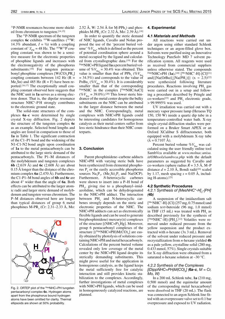
282 CHIMIA 2016, 70, No. 4 laureates: Junior prizes of the sCs fall meeting 2015
4. Experimental
4.1 Materials and MethodsAll reactions were carried out un-
der argon using either standard Schlenktechniques or an argon-filled glove box.Solvents were purified using an InnovativeTechnology PureSolv MD 7 solvent pu-rification system. All reagents were usedas received from commercial suppliersunless otherwise stated. The compoundsDippNHC=PH (1a),[10] [MesNHC*-H]-[Cl][21]and{[Na(OtBu)]
x[Na(PH
2)]} (x ≈ 2.5)[22]
were synthesized following literatureprocedures. Reactions involving PH
3gas
were carried out in a setup and follow-ing a procedure described by Pringle andco-workers[23] and PH
3electronic grade,
> 99.9995% was used.UV irradiation was carried out with a
mercury vapor pressure lamp (Heraeus TQ150, 150 W) inside a quartz dip tube in atemperature-controlled water bath. X-raysingle crystal diffraction studies were per-formed on a Bruker Smart APEX or anOxford XCalibur S diffractometer, bothequipped with a molybdenum X-ray tube(λ = 0.7107 Å).
Percent buried volume %VBur
was cal-culated using the user friendly online toolSambVca available at www.molnac.unisa.it/OMtools/sambvca.php with the defaultparameters as suggested by Cavallo andco-workers (sphere radius R = 3.5 Å, M–Pdistance d = 2.10 Å, Bondi radii[24] scaledby 1.17, mesh spacing s = 0.05 Å, includ-ing H-atoms).[25]
4.2 Synthetic Procedures4.2.1 Synthesis of [MesNHC*-H]
2-[PH]
(4b)A suspension of the imidazolium salt
[MesNHC*-H]-[Cl] (257mg,0.75mmol)andsodium tert-butoxide (96 mg, 1.0 mmol)in THF (15 mL) was treated with PH
3as
described previously for the synthesis of[DippNHC*-H]-[PH
2].[12] Volatiles were re-
moved under reduced pressure from theyellow suspension and the product ex-tracted with n-hexane (3x 3 mL). Removalof the solvent under reduced pressure andrecrystallization from n-hexane yielded 4bas a pale yellow, crystalline solid (280 mg,0.433mmol, 57%). Single crystals suitablefor X-ray diffraction were obtained from asaturated n-hexane solution at –30 °C.
4.2.2 Synthesis of the Complexes[(DippNHC=PH)M(CO)
5] (6a-c, M = Cr,
Mo, W)In a 200 mL Schlenk tube, 1a (210 mg,
0.500 mmol) and the equimolar amountof the corresponding metal hexacarbonylwere dissolved in THF (20 mL). The flaskwas connected to an argon Schlenk line fit-tedwith an overpressure valve set to 0.1 baroverpressure and exposed to UV radiation.
2.52 Å, W: 2.54 Å for M-PPh3) and phos-
phides M-PR2(Cr: 2.32 Å, Mo: 2.39 Å).[18]
In order to quantify the steric demandof NHC ligands, Cavallo and Nolan pro-posed the use of the ‘percent buried vol-ume’%V
Bur, which is defined as the percent
of potential coordination sphere around ametal occupied by the ligand and calculat-ed from crystallographic data.[19] For theDippNHC=PHligand1aapercent buriedvol-ume of %V
Bur= 30.4% was obtained. This
value is smaller than that of PPh3(%V
Bur= 34.5%) and corresponds to the value ofPnBu
3(%V
Bur= 30.4%). It is considerably
smaller than that of the correspondingDippNHC in the complex [DippNHCAuCl](%V
Bur= 39.0%).[20] This lower steric hin-
drance of themetal center despite the bulkysubstituents on the NHC can be attributedto the larger distance between the metaland the NHC. Correspondingly, metalcomplexes with NHC=PH ligands couldbe interesting candidates for homogenouscatalysis as their metal centers suffer fromless steric hindrance than their NHC coun-terparts.
3. Conclusion
Parent phosphinidene–carbene adductsNHC=PH with varying steric bulk havebeen synthesized from elemental phospho-rus P
4or the easily accessible phosphorus
sources Na3P7, (Me
3Si)
3P7and Na(OCP).
Furthermore, N-heterocyclic carbeneswere shown to insert into a P–H bond ofPH
3, giving rise to a phosphanyl–imid-
azolidine, which can be dehydrogenatedto the NHC=PH adduct. The interactionbetween PH
3and N-heterocyclic car-
benes strongly depends on the steric andelectronic properties of the NHC. TheNHC=PH adducts can act as electronicallyflexible ligands and can be used to generatebis(phosphinidene) mercury(ii) complexesof the structure [(NHC=P)
2Hg]. Moreover,
group 6 pentacarbonyl complexes of thestructure [(DippNHC=PH)M(CO)
5] are eas-
ily obtained by photolysis of solutions con-tainingNHC=PHandmetal hexacarbonyls.Calculations of the percent buried volumerevealed only low coverage of the metalcenter by the NHC=PH ligand despite itssterically demanding substituents. Thismight prove useful for the application inhomogenous catalysis, as the ligand keepsthe metal sufficiently free for catalyticinteraction and still provides kinetic sta-bilization to the complexes. Accordingly,further investigations of metal complexeswith NHC=PH ligands, which can be usedin homogeneously catalyzed reactions, areplanned.
31P-NMR resonances become more shield-ed from chromium to tungsten.[14,15]
The 31P-NMR spectrum of the tungstencomplex 6c exhibits 183W-satellites (183W:14.3% abundant, I = ½) with a couplingconstant of 1J
PW= 48 Hz. The 183W31P cou-
pling constant was shown to be a sensi-tive measure for the electronic propertiesof phosphine ligands and increases withthe electronegativity of the phosphorussubstituents.[16] For tungsten pentacar-bonyl phosphine complexes [W(CO)
5PR
3]
coupling constants between 142 Hz (R =Me
3Sn) and 485 Hz (R = F) have been re-
ported.[16,17] The exceptionally small cou-pling constant observed here suggests thatthe NHC unit in 1a serves as a strong elec-tron donor. That is, the dipolar resonancestructure NHC+-P-H strongly contributesto the electronic ground state.
The solid-state structures of the com-plexes 6a–c were determined by singlecrystal X-ray diffraction. Fig. 2 depictsthe structure of the tungsten complex 6cas an example. Selected bond lengths andangles are listed in comparison to those of1a in Table 1. The significant contractionof the C1–P1 bond and the widening of theN1-C1-N2 bond angle upon coordinationof 1a to the metal pentacarbonyls can beattributed to the large steric demand of thepentacarbonyls. The P1–M distances ofthe molybdenum and tungsten complexes6b (2.619 Å) and 6c (2.608 Å) are about0.15 Å longer than the distance of the chro-mium complex 6a (2.470 Å). Furthermore,the C1-P1–M bond angles of 6b and 6c areabout 4° wider than the angle of 6a. Botheffects can be attributed to the larger atom-ic radii and larger steric demand of molyb-denum and tungsten versus chromium. TheP–M distances observed here are longerthan typical distances of group 6 metalphosphines M-PR
3(Cr: 2.31–2.33 Å, Mo:
P1
C2C3
C1 N1N2
H
W
O1
Fig. 2. ORTEP plot of the DippNHC=PH tungstenpentacarbonyl complex 6c. Hydrogen atomsapart from the phosphorus bound hydrogenatoms have been omitted for clarity. Thermalellipsoids are shown at 50% probability.

laureates: Junior prizes of the sCs fall meeting 2015 CHIMIA 2016, 70, No. 4 283
The reaction was monitored by 31P-NMRspectroscopy. After radiation times of 5 to10 h, maximum conversions of 90–95%were observed. The solvent was removedunder reduced pressure and the residuewashedwith n-hexane (5x 5mL) to removeexcess starting materials. Recrystallizationfrom boiling toluene gave the complexesas yellow crystalline solids in 65–82 %yield. Single crystals suitable for X-raydiffraction were obtained from saturatedtoluene solutions at –30 °C (M = Cr) orfrom saturated toluene solutions layeredwith n-hexane (M = Mo, W).
Supplementary InformationThe structures discussed here have been
deposited with the Cambridge StructuralDatabase under the numbers CCDC 1446562,1446676, 1446858, 1446859.
AcknowledgementsWe thank the European Union (Marie Curie
ITN SusPhos, Grant Agreement No. 317404)for financial support. Furthermore, M.B. thanksthe Swiss Chemical Society and Metrohm forthe SCS-Metrohm Award for the best oralpresentation in Inorganic and CoordinationChemistry.
Received: January 18, 2016
[1] a) K. Lammertsma in ‘New Aspects inPhosphorus Chemistry III’, Ed.: J.-P. Majoral,Springer, Berlin Heidelberg, 2003; b) Z. Benko,R. Streubel, L. Nyulaszi, Dalton Trans. 2006,4321.
[2] H. Aktas, J. C. Slootweg, K. Lammertsma,Angew. Chem. Int. Ed. 2010, 49, 2102.
[3] a) L. J. Murphy, K. N. Robertson, J. D. Masuda,J. A. Clyburne in ‘N-Heterocyclic Carbenes’,Ed.: S. P. Nolan, Wiley-VCH Verlag GmbH &Co. KGaA, Weinheim, 2014; b) C. D. Martin,M. Soleilhavoup, G. Bertrand, Chem. Sci. 2013,4, 3020; c) D. J. D. Wilson, S. A. Couchman,J. L. Dutton, Inorg. Chem. 2012, 51, 7657;d) Y. Wang, G. H. Robinson, Inorg. Chem.2011, 50, 12326; e) O. Back, B. Donnadieu, P.Parameswaran, G. Frenking, G. Bertrand, Nat.Chem. 2010, 2, 369.
[4] a)A. J.Arduengo, J. C. Calabrese,A. H. Cowley,H. V. R. Dias, J. R. Goerlich, W. J. Marshall, B.Riegel, Inorg. Chem. 1997, 36, 2151; b) A. J.Arduengo, Dias,H. V. Rasika, J. C. Calabrese,Chem. Lett. 1997, 26, 143; c) A. J. ArduengoIII, J. Carmalt, Claire, A. C. Clyburne, Jason, H.Cowley, Alan, R. Pyati, Chem. Commun. 1997,981.
[5] K. Hansen, T. Szilvási, B. Blom, E. Irran, M.Driess, Chem. Eur. J. 2014, 20, 1947.
[6] A. M. Tondreau, Z. Benko, J. R. Harmer, H.Grützmacher, Chem. Sci. 2014, 5, 1545.
[7] A. Doddi, D. Bockfeld, T. Bannenberg, P. G.Jones, M. Tamm, Angew. Chem. Int. Ed. 2014,53, 13568.
[8] A. Beil, R. J. Gilliard, H. Grützmacher, DaltonTrans. 2016, DOI: 10.1039/C5DT03014E.
[9] L. Liu, D. A. Ruiz, F. Dahcheh, G. Bertrand,Chem. Commun. 2015, 51, 12732.
[10] M. Cicac-Hudi, J. Bender, S. H. Schlindwein,M. Bispinghoff, M. Nieger, H. Grützmacher, D.
Gudat,Eur. J. Inorg. Chem. 2015, DOI: 10.1002/ejic.201501017.
[11] B.A. Trofimov, S. N.Arbuzova, N. K. Gusarova,Russ. Chem. Rev. 1999, 68, 215.
[12] M. Bispinghoff, A. M. Tondreau, H.Grützmacher, C.A. Faradji, P.G. Pringle,DaltonTrans. 2015, DOI: 10.1039/C5DT01741F.
[13] K. J. Packer, J. Chem. Soc. 1963, 960.[14] S. O. Grim, D. A. Wheatland, W. McFarlane, J.
Am. Chem. Soc. 1967, 89, 5573.[15] M. F. Guns, E. G. Claeys, G. P. van der Kelen, J.
Mol. Struct. 1979, 54, 101.[16] E. O. Fischer, L. Knauss, R. L. Keiter, J. G.
Verkade, J. Organomet. Chem. 1972, 37, C7.[17] H. Schumann, H.-J. Kroth, Z. Naturforsch., B:
Chem. Sci. 1977, 32, 768.[18] F. H. Allen, O. Kennard, D. G. Watson, L.
Brammer, Orpen, A. Guy, R. Taylor, J. Chem.Soc., Perkin Trans. 2 1987, S1.
[19] A. C. Hillier, W. J. Sommer, B. S. Yong,J. L. Petersen, L. Cavallo, S. P. Nolan,Organometallics 2003, 22, 4322.
[20] H. Clavier, S. P. Nolan, Chem. Commun. 2010,46, 841.
[21] K. M. Kuhn, R. H. Grubbs, Org. Lett. 2008, 10,2075.
[22] M. Podewitz, J. D. van Beek, M. Wörle, T. Ott,D. Stein, H. Rüegger, B. H. Meier, M. Reiher,H. Grützmacher, Angew. Chem. Int. Ed. 2010,49, 7465.
[23] J. H. Downing, J. Floure, K. Heslop, M. F.Haddow, J. Hopewell, M. Lusi, H. Phetmung,Orpen, A. Guy, P. G. Pringle, R. I. Pugh, D.Zambrano-Williams, Organometallics 2008,27, 3216.
[24] A. Bondi, J. Phys. Chem. 1964, 68, 441.[25] A. Poater, B. Cosenza, A. Correa, S. Giudice, F.
Ragone, V. Scarano, L. Cavallo, Eur. J. Inorg.Chem. 2009, 1759.

284 CHIMIA 2016, 70, No. 4 laureates: Junior prizes of the sCs fall meeting 2015
doi:10.2533/chimia.2016.284 Chimia 70 (2016) 284–287 © Swiss Chemical Society
*Correspondence: Prof. Dr. S. LeutwylerE-mail: [email protected]ät BernDepartement für Chemie und BiochemieFreiestrasse 3CH-3012 Bern
Do Hydrogen Bonds Influence ExcitonicSplittings?
Franziska A. Balmer§, Philipp Ottiger, and Samuel Leutwyler*
§SCS-Metrohm Award for best oral presentation in Physical Chemistry
Abstract: The excitonic splitting and vibronic quenching of the inversion-symmetric homodimers of benzonitrile,(BN)2, andmeta-cyanophenol, (mCP)2, are investigatedby two-color resonant two-photon ionizationspectroscopy.These systems have very different hydrogen bond strengths: the OH…N≡Cbonds in (mCP)2 are ~10 times strongerthan the CH…N≡C hydrogen bonds in (BN)2. In (BN)2 the S
0( 1A
g) → S
1( 1A
g) transition is electric-dipole forbidden,
while the S0(1A
g) → S
2(1B
u) transition is allowed. The opposite holds for (mCP)2 due to the different transition
dipole moment vector alignment. The S0→ S
1/S
2spectra of the dimers are compared and their excitonic splittings
and vibronic quenchings are investigated by measuring the 13C-substituted heterodimer isotopomers, for whichthe centrosymmetry is broken and both transitions are allowed. The excitonic splittings are determined as∆exc = 2.1 cm–1 for (BN)2 and ∆exc = 7.3 cm–1 for (mCP)2. The latter exhibits a much stronger vibronic quenching,as the purely electronic splitting resulting from ab initio calculations is determined to be ∆calc= 179 cm–1, while in(BN)2 the calculated splitting is ∆calc= 10 cm–1. The monomer site-shifts upon dimerization and comparing certainvibrations that deform the hydrogen bonds confirm that the OH…N≡C hydrogen bond is much stronger than theCH…N≡C bond. We show that the H-bonds have large effects on the spectral shifts, but little or no influence onthe excitonic splitting.
Keywords: Benzonitrile · meta-Cyanophenol · Excitonic splitting · Hydrogen bonds · Vibronic coupling
1. Introduction
Hydrogen-bonded molecular dimersare model systems to investigate excitonicinteractions, which are of great importancein many photochemical and biologicalsystems such as conjugated polymers, mo-lecular crystals and photosynthetic light-harvesting complexes.[1] Such dimers ofbenzene derivatives are small enough to al-low detailed experimental, theoretical andcomputational investigations and are at thesame time representative of larger systems.Our group has been studying the excitonicS0→ S
1/S
2splittings in rigid, doubly hydro-
gen-bonded dimers such as (2-pyridone)2,
(2-aminopyridine)2, (benzoic acid)
2, (ben-
zonitrile)2and (ortho-cyanophenol)
2.[2–6]
They are centrosymmetric, meaning thatthe electronically excited S
1(and higher
Sn) states of monomers A and B are degen-
erate and excitonically coupled.[2–8] Upondimerization the transition dipole moment(TDM) vectors of the monomers combinein a parallel and antiparallel manner, giv-ing rise to an antisymmetric (A
u, B
u) and
a symmetric (Ag) excited state combi-
nation.[9] Thus, either the S0→ S
1or the
S0→ S
2transition is electric-dipole (g↔u)
allowed, while the other is electric-dipole(g↔g) forbidden. The inversion symmetrycan be broken by isotopic substitution ofeven a single atom, e.g. 12C/13C or H/D,rendering the forbidden transition slightlyallowed.[2–6] This enables us to experimen-tally determine the S
1/S
2splitting, denoted
∆obs.We have previously shown that the
displacements along the intra- and inter-molecular vibrational coordinates Q
ithat
occur upon electronic excitation and whichare quantified by the Huang-Rhys factorsSi, have a major impact on the magnitude
of the experimental splittings.[3,6] The ob-served splittings are up to a factor 40 small-er than the purely electronic (Davydov)exciton splittings ∆
calcthat are predicted
by high-level ab initio calculations. Thisso-called ‘vibronic quenching’ of the exci-tonic splitting ∆
calccan be obtained[3–10] by
taking the Huang-Rhys factors Siinto ac-
count within Förster’s perturbation theory
approach[7] or the Fulton-Gouterman mod-el.[11,12] Applying the resulting quenchingfactor Γ to the calculated exciton splitting∆calc
results in vibronic splittings ∆vibron
thatare very close to the experimentally ob-served S
1/S
2splittings.
∆vibron
= Γ· ∆calc
(1)
The strength of hydrogen bonds isdependent on the atoms involved and onstructural aspects. The shift in absorptionenergy of the dimer relative to the mono-mer allows us to draw conclusions on thestabilization gained upon dimerization.Further, vibrations deforming the hydro-gen bond also give us information on theH-bond strength. Here, we investigate ifhydrogen bond strength has a direct influ-ence on the excitonic splitting and vibroniccoupling on the examples of the benzoni-trile dimer, (BN)
2, with two CH…N≡C hy-
drogen bonds, and the meta-cyanophenoldimer, (mCP)
2, with two OH…N≡C hydro-
gen bonds.
2. Structural Aspects
The ground-state structures of (BN)2
and (mCP)2were optimized by the spin-
component-scaled variant of the approxi-

laureates: Junior prizes of the sCs fall meeting 2015 CHIMIA 2016, 70, No. 4 285
0 100 200 300 400 500 600Relative Wavenumber /cm-1
IonSignal/arb.units
a) (benzonitrile)2
b) (meta-Cyanophenol)2
0 (S2)0
0
0 (S1)0
0
36420cm
-133255cm
-1
χ1
0
χ1
0
σ1
0
δ1
0
β2
0
δCC-N1
0
δC-CN1
06a1
0
6a1
0θ2
0
δCC-N1
0
δC-OH1
06b1
0
Fig. 2. Two-color resonant two-photon ionization spectra of a) (benzonitrile)2 and b) (meta-cyano-phenol)2 (b) with assignments. Vibrations appearing in both spectra are color-coded.
(BN)2and 263 cm–1 in (mCP)
2. This shows
that the OH…N≡C H-bond is much stron-ger compared to the CH…N≡C bond, be-cause more energy is required to deform it.This is confirmed by the SCS-CC2/aVTZcalculated and counterpoise-corrected in-termolecular S
0binding energies, which
are 𝐷𝐷���� = –57.9 kJ/mol for (mCP)2and
𝐷𝐷���� = –23.1 kJ/mol for (BN)2, with cor-
responding basis-set superposition errorsof 9.5 kJ/mol and 4.3 kJ/mol, respectively.
Inter- and intramolecular vibrationsthat involve large motions of the H-bondedatoms and that appear in the spectra of both(BN)
2and (mCP)
2allow to draw conclu-
sions on H-bond strength. The intermo-lecular shear vibration χ and the in-planecyano-group bend δCCN appear in bothspectra. The respective fundamental exci-tations exhibit lower frequencies in (BN)
2compared to (mCP)
2: χ�� has an experimen-
tal frequency of 33 cm–1 in (BN)2and 117
cm–1 in (mCP)2; the δCCN�� is at 206 cm–1 in
mate second-order coupled-cluster (SCS-CC2) method using the aug-cc-pVTZ ba-sis set and are shown in Fig. 1. (BN)
2is
a planar, C2h-symmetric dimer,[13,14] while
(mCP)2is C
i-symmetric, with the mono-
mers slightly out of plane.[9,15]The calculated intermonomer distance
between the centers of mass in (mCP)2is
RAB= 5.34Å with an H…N distance of 2.04
Å; the center-of-mass distance in (BN)2
is RAB= 6.46 Å and the H…N distance is
2.36 Å. The presence of the OH group in(mCP)
2changes the transition dipole mo-
ment (TDM) orientation: In (BN)2the
angle of the monomer TDM relative to the𝑅𝑅�� vector is θ = 63°, which is larger thanthe ‘magic angle’ for which the magnitudeof the dipole–dipole interaction goes tozero, i.e. arccos(1/√3) = 54.7°.
Thus, the TDMs are oriented parallel/antiparallel, the S
0→ S
1transition is forbid-
den and the S0→ S
2transition is allowed.
For (mCP)2, the angle is θ = 11°, which is
smaller than the magic angle. In this case,the S
0→ S
1transition is allowed and the
S0→ S
2transition is forbidden.[9]
3. Two-Color Resonant Two-PhotonIonization Spectra
The dimers were produced and cooledin pulsed supersonic jet expansions. BNwas heated to 75 °C in an external vapor-izer and seeded into the neon carrier gas(backing pressure 1.2 bar), while mCP washeated inside the pulsed nozzle to 80 °C.Two-color resonant two-photon ionization(2C-R2PI) spectra were recorded by over-lapping the excitation and ionization lasersin the source of a time-of-flight mass spec-trometer. The spectra are shown in Fig. 2with both electronic origins set to zero forcomparison. The 0�� transition energies areindicated next to the corresponding band,namely 36420 cm–1 for (BN)
2and 33255
cm–1 for (mCP)2.[9,13] These energies cor-
respond to spectral shifts of δν =-92 cm–1
for (BN)2and δν =-1094 cm–1 for (mCP)
2relative to the respective monomer S
0→ S
1
transition energies. The much larger shiftfor (mCP)
2indicates a stronger hydrogen
bond of (mCP)2relative to that of (BN)
2.
This will be discussed in Section 5.The intramolecular normal modes are
denoted according to the Wilson nota-tion,[16] while for the intermolecular nor-mal modes we employ the nomenclatureintroduced in ref. [17]. The (BN)
2spec-
trum is dominated by intramolecular vi-bronic bands. Only one intermolecularvibration is observed, the shear vibrationχ��; otherwise the spectrum is very similarto that of BN.[13] By contrast, the spectrumof (mCP)
2showsmany excitations of inter-
molecular vibrations (δ, β, θ, σ, χ) as wellas several intramolecular vibrations.[9,15]
θA θBµA
µB
RAB
θA
µA
RAB µB
θB
a) (benzonitrile)2
b) (meta-cyanophenol)2
Fig. 1.a) (Benzonitrile)2 andb) (m-cyanophenol)2with indicated mono-mer transition dipolemoments (µ), inter-monomer distanceRAB and TDM align-ment angle θ.

286 CHIMIA 2016, 70, No. 4 laureates: Junior prizes of the sCs fall meeting 2015
5. Spectral Site-Shifts
The intermolecular interactions thatoccur upon dimerization act on the constit-uent monomers and result in changes of themonomer geometry as well as energy (theso-called deformation energy). The geom-etry deformation of the monomer leads toa change of its electronic structure. Thischange is different in the S
0and S
1states,
leading to a shift of the adiabatic S0→ S
1
absorption frequency. Thus the dimeriza-tion causes an electronic spectral site-shift.The site-shift can be evaluated from the av-erage of spectral shifts of the dimer S
1/S
2
0�� bands relative to the monomer S0 → S1
0�� band, see Fig. 4.Fig. 4 shows that the excitonic splitting
discussed above is relatively small anddoes not depend on the H-bond strength. Incontrast, the site-shift leads to much largerspectral effects and largely depends on theH-bond strength. The BN S
0→ S
10�� band
is observed at 36513 cm–1[18] and the aver-age of the dimer S
0→ S
1/S
2transitions is
36420 cm–1.[13] This result corresponds toa small site-shift of ∆
site= –92 cm-1, which
is 5% of the binding energy. The mCPmonomer origin lies at 34354 cm–1 whilethe average of the dimer S
0→ S
1/S
2origins
is at 33260 cm–1, resulting in a much largersite-shift of ∆
site= –1094 cm–1. Here, the
site-stabilization is 23% of the dimer bind-ing energy. This shows that the CH…N≡C
quenches the Davydov splittings by factorsof Γ–1 = 5 and 23, respectively, leading tovibronic S
1/S
2splittings of ∆
vibron= 2.1 cm–1
for (BN)2and ∆
vibron=7.7 cm-1 for (mCP)
2.
This is in excellent agreement with theexcitonic splittings determined above; allthe splittings are summarized in Table 1.We note that these splittings arise fromproperties of the monomers: The exciton-ic splitting results due to coupling of themonomer TDMs and the vibronic quench-ing is largely dependent on intramolecularmonomer vibrations Q
iand Huang-Rhys
factors Si. This implies that the H-bond
strength of the dimer does not affect theexcitonic splitting or vibronic quenching.
Table.1. Excitonic S1/S
2splitting (∆exc), Davydov
splitting (∆calc), quenching factor (Γ), resultingvibronic splitting (∆vibron=Γ·∆calc) and site-shift(δν).
(benzonitrile)2
(m-cyano-phenol)
2
∆exc
2.0 cm–1 7.3 cm–1
∆calc
10 cm–1 179 cm–1
Γ 0.213 0.043
∆vibron
2.1 cm–1 7.7 cm–1
δυ –92 cm–1 –1094 cm–1
4. Excitonic Splitting and VibronicCoupling
Isotopic 12C/13C substitution at a sin-gle atom breaks the inversion symmetryof the dimers and the symmetry descent,which is C
2h→ C
sfor (BN)
2and C
i→ C
1
for (mCP)2, renders the forbidden transi-
tion allowed. We take advantage of thenatural abundance of 13C. Fig. 3 shows themass-selective 2C-R2PI spectra of (BN)
2and (mCP)
2in the 0�� region, where the
top panels display the all-12C mass chan-nel and the lower panels the 13C-trace. Inthe case of (BN)
2-13C the S
0→ S
10�� transi-
tion appears at 36418.9 cm–1, red-shiftedby 1.2 cm–1 relative to (BN)
2, and with an
S1/S
2splitting of ∆
obs= 3.9 cm–1,[13] see Fig.
3(a). The S0→ S
2transition of (BN)
2-13C
is blue-shifted by 2.7 cm–1 relative to the(BN)
2S2origin, which is due to changes in
the zero-point vibrational energy (ZPVE)by isotopic substitution ∆
iso. The observed
S1/S
2splitting thus comprises an excitonic
contribution ∆excand an isotopic contribu-
tion ∆iso. According to degenerate pertur-
bation theory, these combine as:[4,13]
(2)��� = ���� + ���� . (
From monomer 2C-R2PI spectra of12C-BN and 13C-BN we have determinedthe purely isotopic contribution to be∆iso= 3.3 cm–1, leading to an excitonic split-
ting of ∆exc= 2.1 cm–1.[13]
In the case of (mCP)2the forbidden
S20�� band appears very weakly in the
(mCP)2-13C spectrum at 8.0 cm–1 above the
S1origin, see Fig. 3(b). The S
10�� band of
(mCP)2-13C is hardly shifted compared to
that of (mCP)2, indicating smaller changes
in ZPVE.[9] The S0→ S
1spectra of mCP
and mCP-13C were used to determine theisotopic contribution∆
iso= 3.3 cm–1 as well.
Employing Eqn. (2) with ∆obs= 8.0 cm–1
yields an excitonic splitting of ∆exc= 7.3
cm–1.The vertical excitation energies cal-
culated at the optimized SCS-CC2/aVTZground-state structures predict purelyelectronic splittings of ∆
calc= 10 cm–1 for
(BN)2and ∆
calc= 179 cm–1 for (mCP)
2.
However, these splittings do not take theredistribution of the electronic TDM intovibronic TDMs into account, and cantherefore not be directly compared to thedetermined excitonic splittings ∆
exc.[3–7,9,10]
To calculate the vibronic quenching factorΓ = Π
iexp(–S
i) = exp(–Σ
iSi), we employed
the Huang-Rhys factors 𝑆𝑆� = 𝑙𝑙� 2𝜇𝜇�ℏ𝜔𝜔��,where i numbers the totally-symmetric in-tramolecular vibrations of the BN or mCPmonomer.[3–6] The experimental S
iyielded
Γexp= 0.213 for (BN)
2and Γ
exp= 0.043 for
(mCP)2.[9,13] The vibronic coupling hence
36410 36420 36430
Wavenumber / cm-1
IonSignal/arb.units
(BN)2
(BN)2-13C
0 (S2)0
0
0 (S2)0
0
0 (S1)0
0
∆obs= 3.9 cm-1
a) (benzonitrile)2 b) (meta-cyanophenol)2
33250 33260 33270
Wavenumber / cm-1
IonSignal/arb.units
(mCP)2
(mCP)2-13C
0 (S1)0
0
0 (S1)0
0
0 (S2)0
0
∆obs= 8.0 cm-1
Fig. 3. 2C-R2PI spectra in the origin region of a) (benzonitrile)2 and b) (meta-cyanophenol)2 in the12C-mass channel (top) and the 13C-mass channel (bottom) with observed S1/S2 splitting in red.

laureates: Junior prizes of the sCs fall meeting 2015 CHIMIA 2016, 70, No. 4 287
larger than the excitonic splittings and areheavily influenced by the hydrogen bondstrengths. Thus the H-bonds have large ef-fects on the spectral shifts, but little or noinfluence on the exciton splitting.
Received: January 21, 2016
[1] F. C. Spano, Annu. Rev. Phys. Chem. 2006, 57,4563.
[2] P. Ottiger, S. Leutwyler, H. Köppel, J. Chem.Phys. 2009, 131, 204308.
[3] P. Ottiger, S. Leutwyler, H. Köppel, J. Chem.Phys. 2012, 136, 174308.
[4] P. Ottiger, S. Leutwyler, J. Chem. Phys. 2012,137, 204303.
[5] S. Kopec, P. Ottiger, S. Leutwyler, H. Köppel, J.Chem. Phys. 2015, 142, 084308.
[6] P. Ottiger, S. Leutwyler, H. Köppel, Chem. Sci.2015, 6, 6059.
[7] T. Förster, in ‘Modern Quantum Chemistry’,Ed. O. Sinanoglu, Academic, New York andLondon, 1965, Chap. IIIB, p. 93.
[8] A. Witkowski, W. Moffitt, J. Chem. Phys. 1960,33, 872.
[9] F.A. Balmer, P. Ottiger, S. Kopec, H. Köppel, S.Leutwyler, to be submitted.
[10] S. Kopec, P. Ottiger, S. Leutwyler, H. Köppel, J.Chem. Phys. 2012, 137, 184312.
[11] R. L. Fulton, M. Gouterman, J. Chem. Phys.1961, 35, 1059.
[12] R. L. Fulton, M. Gouterman, J. Chem. Phys.1964, 41, 2280.
[13] F. A. Balmer, P. Ottiger, S. Leutwyler, J. Phys.Chem. A 2014, 118, 11253.
[14] T. Kobayashi, K. Honma, O. Kajimoto, S.Tsuchiya, J. Chem. Phys. 1987, 86, 1111.
[15] N. Seurre, K. Le Barbu-Débus, F. Lahmani,A. Zehnacker-Rentien, J. Sepiol, Chem. Phys.2003, 295, 21.
[16] E. B. Wilson, Phys. Rev. 1934, 45, 706[17] A. Müller, F. Talbot, S. Leutwyler, J. Am. Chem.
Soc. 2002, 124, 14486.[18] P. Imhof, D. Krügler, R. Brause, K.
Kleinermanns, J. Chem. Phys. 2004, 121, 2598.
drogen bonds in (mCP)2are much stron-
ger compared to (BN)2, which is reflected
in (i) the binding energy Deof the com-
plexes, (ii) the site-stabilization δυ of thedimer relative to the corresponding mono-mer units and (iii) the shifts of inter- andintramolecular vibrational frequencies ofdimer modes such as the shear and the in-plane nitrogen bend, where the frequen-cy is much lower in (BN)
2compared to
(mCP)2. Yet, the strength of the hydrogen
bond only affects site-shift of the mono-mers upon dimerization, but not excitonicinteraction. The latter arises due couplingof the monomer TDMs and the vibronicquenching is well reproduced by only in-cluding intramolecular monomer modes.One sees that while the excitonic splittingis of great importance, it is energetically acomparatively weak interaction. The elec-tronic spectral site-shifts are 15–150 times
bonds have a much smaller influence thanthe OH…N≡C bonds, as the stabilization ismuch larger in (mCP)
2.
6. Conclusions
Jet-cooled two-color resonant two-photon ionization spectra of (benzoni-trile)
2and (meta-cyanophenol)
2and their
13C-isotopomers revealed excitonic split-tings of ∆
exc= 2.1 cm–1 and ∆
exc= 7.3 cm–1,
respectively. The vibronic quenching fac-tor derived from monomer fluorescencespectra was much smaller for (mCP)
2(Γ = 0.043) than for (BN)
2(Γ = 0.213),
since 2C-R2PI spectrum of mCP exhibitsmany more vibronic bands than BN. Thisleads to a much stronger quenching ofthe electronic exciton splitting by a factorΓ–1 = 23, while in (BN)
2Γ–1=5. The hy-
TransitionEnergy/cm
-1
∆exc
∆site
AA in dimer
environment
B in dimer
environment BAB
S0 S1
S0 S1
S0 S2
S0 S1
Fig. 4. Schematic representation of site- and excitonic splitting in a homodimer, where the S0→S
1
transition is forbidden. Experimentally accessible values are marked in full lines, while calculatedor indirectly determined transitions are dashed.
288 CHIMIA 2016, 70, No. 4 laureates: Junior prizes of the sCs fall meeting 2015
doi:10.2533/chimia.2016.288 Chimia 70 (2016) 288–291 © Swiss Chemical Society
*Correspondence: Prof. Dr. W. Meiera
E-mail: [email protected] of ChemistryUniversity of BaselKlingelbergstrasse 80CH-4056 BaselbSwiss Tropical and Public Health InstituteSocinstrasse 57 PostfachCH-4002 BaselcUniversity of BaselPetersgraben 2, CH-4002 Basel
Giant Host Red Blood Cell MembraneMimicking Polymersomes Bind ParasiteProteins and Malaria Parasites
Adrian Najer§abc, Sagana Thambooa, Cornelia G. Palivana, Hans-Peter Beckbc, and Wolfgang Meiera*
§SCS-Metrohm Award for best oral presentation in Polymers, Colloids and Interfaces
Abstract: Malaria is an infectious disease that needs to be addressed using innovative approaches to counteractspread of drug resistance and to establish or optimize vaccination strategies. With our approach, we aim for adual action with drug- and ‘vaccine-like’ activity against malaria. By inhibiting entry of malaria parasites into hostred blood cells (RBCs) – using polymer vesicle-based (polymersome) nanomimics of RBC membranes – the lifecycle of the parasite is interrupted and the exposed parasites are accessible to the host immune system. Here, wedescribe how host cell-sized RBC membrane mimics, formed with the same block copolymers as nanomimics,also bind the corresponding malaria parasite ligand and whole malaria parasites, similar to nanomimics. This wasdemonstrated using fluorescence imaging techniques and confirms the suitability of giant polymersomes (GUVs)as simple mimics for RBC membranes.
Keywords: Giant polymersomes · GUV · Malaria · Membrane mimics · Plasmodium
Introduction
Block copolymer-based vesicles, calledpolymersomes[1] due to their increased me-chanical stability compared to liposomesand broad chemical versatility to introducee.g. stimuli-responsiveness – are consid-ered ideal candidates for drug deliverypurposes, as nanoreactors, as artificial or-ganelles inside cells, or as simple cell mim-ics.[2,3] Our recently developed nanomimicstrategy against infectious diseases is basedon such polymer vesicles imitating a hostcell membrane at the nanoscale (nanomim-ics), which was achieved by mixing twodistinct block copolymers: vesicle-form-ing poly(2-methyl-2-oxazoline)-block-poly(dimethylsiloxane)-block-poly(2-methyl-2-oxazoline) (PMOXA-b-PDMS-b-PMOXA), and PDMS-b-heparin.[4,5]Giant polymersomes (giant unilamellarvesicles, GUVs), several micrometers indiameter, are increasingly acknowledgedas interesting model systems of very sim-plified cell mimics.[6]
Malaria, a disease caused byPlasmodium spp. parasites that are trans-mitted by Anopheles mosquitoes – killingmore than 600’000 people each year[7] –is our current target. When reaching theblood stage cycle, the malaria parasiteform called merozoite attaches to andenters red blood cells (RBCs), where theparasite asexually divides to form moreparasites, which egress from the infectedRBC (iRBC) after about 48 hours and in-fect more RBCs.[8] Heparan sulphate (verysimilar to heparin) on RBCs is consideredas being responsible for the initial attach-ment of merozoite to the RBC by interac-tion with Plasmodium falciparum mero-zoite surface protein 1-42 (PfMSP1
42).[9]
With our approach we block the ma-laria blood stage life cycle using RBC-membrane-mimicking polymersomes,which block the parasite after egress fromiRBCs, inhibit invasion, and expose the in-fective merozoites to the immune system.If successful in vivo, this artificial inhibi-tion of merozoite invasion could lead to animmune boost against extracellular mero-zoites.[4]
Here, we demonstrate that RBC mem-brane mimics formed at a larger size,but with similar hollow sphere architec-ture (heparin GUVs), can also bind theparasite protein PfMSP1
42and the whole
Plasmodium parasite (merozoite form)in a similar fashion as their nano-sizedcounterparts, the nanomimics.[4,5] Thisindicates that GUVs indeed represent asuitable model of small polymersomes,
with the advantage of a size close to thatof RBCs. Certain aspects, e.g. binding ofPfMSP1
42to polymersomes can be studied
with GUVs using methods that would notbe suitable for the nanostructure (e.g. con-focal laser scanning microscopy (CLSM)to study protein binding).
Experimental
MaterialsPDMS-b-heparin, PMOXA-b-PDMS-
b-PMOXA, and PfMSP142–OG488 syn-
thesis and characterization can be found inour previous publications.[4,5]
Giant Polymersome FormationGiant polymersome (GUV) formation
was accomplished according to the stan-dard electroformation technique[10] usinga Nanion Vesicle Prep Pro setup (NanionTechnologies,Munich, Germany).A fresh-ly cleaned ITO-coated glass slide was firstplasma-treated (Plasma Cleaner, PDC-002, Harrick Plasma, Ithaca, New York,USA), and then, a thin polymer film wasdeposited on it. For control GUVs (onlyPMOXA-b-PDMS-b-PMOXA) a solutionof PMOXA-b-PDMS-b-PMOXA in etha-nol (6 mg/mL, 40 µL) was dispersed onthe ITO-coated side of the glass slide andethanol was subsequently evaporated us-ing a vacuum chamber for 30 min (PlasmaCleaner, PDC-002, Harrick Plasma,Ithaca, New York, USA). For the heparinGUVs a mixture of PMOXA-b-PDMS-

laureates: Junior prizes of the sCs fall meeting 2015 CHIMIA 2016, 70, No. 4 289
ing polymersomes were formed at a largersize (micrometer scale, heparin GUVs) tostudy malaria protein and whole parasiteinteraction with RBC-sized polymersome-based mimics (Fig. 1). The same two blockcopolymers previously used for nano-mimic formation were also used to formthese giant mimics. PDMS
65-b-heparin
12was mixed with PMOXA
5-b-PDMS
58-
b-PMOXA5(or PMOXA
9-b-PDMS
67-b-
PMOXA9) using 15 wt% of the first block
copolymer and subsequent electroforma-tion, yielding heparin GUVs. GUVs madefrom PMOXA
5-b-PDMS
58-b-PMOXA
5without the heparin-functionalized copo-lymer were used as controls.
Giant Host Red Blood CellMembrane MimickingPolymersomes Bind PlasmodiumProteins
Formation of GUVs and heparin GUVswas successfully achieved in both cases us-ing electroformation. Usually, a large num-ber of mainly unilamellar, univesicularpolymersomes were formed, whereas onlyfew giants with multivesicular structureswere found. Interestingly, these artificialand flexiblemembranespossess a thicknessof about 11 nm,[4] whilst all the membranecomponents can laterally diffuse withinthe membrane,[12,13] but still form stablevesicles with several tens of micrometersin diameter. We performed CLSM to studythe interaction of fluorescently labelledPlasmodium ligand PfMSP1
42-OG488
and heparin, the receptor-like moleculeknown to bind PfMSP1
42,[9] on the surface
filters. They were immediately mixedwith 300 µL of a heparin GUV solutionin 300 mM sucrose, gently shaken, andincubated under static conditions at 37 °Cfor 15 min. Subsequently, 2 µL of 1 mg/mL Hoechst (DNA stain) was added andmerozoite/giant mixture was fixed in 2%paraformaldehyde/0.2% glutaraldehyde in150 mM phosphate buffer (pH 7.4) over-night. After gentle centrifugation (3500RCF) for 5 min, 10 µL of the pellet wasmounted on a microscopy slide.
Results and Discussion
Recently, we demonstrated that poly-mersome-based nanomimics of host RBCmembranes efficiently block malariaparasite invasion into host RBCs in vitro;nanomimics were more than two orders ofmagnitude more potent invasion inhibitorscompared to soluble receptor-like mol-ecules (heparin), when IC
50values were
compared based on the heparin concen-tration in the assays.[4,5] Furthermore, wemeasured that several heparin chains on asingle nanomimic bound parasite ligandsPfMSP1
42-OG488 with a high affinity (K
dof 12.1 ± 1.6 nM) using fluorescence cross-correlation spectroscopy (FCCS) of greenlabelled ligand and red labelled nanomim-ics.[5] This multiple binding with high af-finity is the basis for a strong multivalentinteraction of nanomimic and merozoite,explaining the potent invasion inhibitionby nanomimics.[5]
Herein, host RBC membrane-mimick-
b-PMOXA in ethanol (6 mg/mL, 40 µL)and PDMS-b-heparin in ethanol (4 mg/mL, 10 µL) corresponding to a ratio of wt/wt 85:15 was used. Afterwards, a greasedO-ring was put around the polymer film onthe glass slide to form a chamber and thenan aqueous sucrose solution (300 mM,600 µL) was added to hydrate the polymerfilm.After covering by another ITO-coatedglass slide, the sample was exposed to anac current (3.0 V, 3.0 Hz) at room tem-perature. For control GUVs we performedelectroformation overnight and for heparinGUVs electroformation was conducted forone hour. Finally, the GUV solutions weretransferred to Eppendorf tubes and storedat 4 °C.
Fluorescence MicroscopyFluorescence imaging of protein in-
teraction with giant polymersomes wasperformed on a confocal laser scanningmicroscope (Zeiss CLSM 510-META/Confocor2, Carl Zeiss, Jena, Germany).A freshly plasma-cleaned 8-well micros-copy chamber (Nunc Lab-Tek ChamberSlide System, Thermo Fisher Scientific)was filled with PBS (200 µL, pH 7.2),GUVs (20 µL in 300 mM sucrose),Bodipy630 (10 µL, 72 µM) in PBS, andPfMSP1
42-OG488 in PBS (5 µL, 2.5 µM).
For the CLSM measurements a water im-mersion objective (C-Apochromat, 40 xmagnification, NA of 1.2) was used. TheOG488 signal was obtained by excitationwith anAr laser (λ = 488 nm) with dichro-ic mirrors HFT (main beam splitter) 488and NFT (secondary beam splitter) 490with the band-pass BP 500-550 filter. HFT488/543/633, NFT 545 and the long-passLP 650 filters were used to collect the fluo-rescence signal of Bodipy630. The pinholesize was set to 70 µm for the green chan-nel, and 90 µm for the red channel, respec-tively. All settings (including laser power,detector gains) were kept constant for im-aging of positive and negative samples.
Fluorescence imagingof heparinGUVswith merozoites was performed on a LeicaDM 5000B fluorescence microscope usingthe appropriate filters.
Study Interaction of Giants andMerozoites
Plasmodium falciparum 3D7 parasiteculturing and merozoite isolation was per-formedasdescribed elsewhere.[4,11]Briefly,a 30 mL petri dish filled with human RBCsat 5% haematocrit and 5–6% parasitemia(iRBC late stages) was used for merozo-ite isolation. First, late stage iRBCs wereseparated from RBCs and young iRBCsby Percoll density gradient. After matura-tion to schizont stage parasites, they wereconcentrated to 1 mL in malaria culturemedium, and viable merozoites were me-chanically released using 1.2 µm syringe
b
~ 7 µm ~ 5 – 100 µm ~ 5 – 100 µm
a
5 58 5
12
65
Red Blood Cell (RBC) RBC-‐Membrane Mimic (Heparin GUV) Control GUVb
Fig. 1. a) Representation of the chemical structure of the two used block copolymers: PMOXA5-b-PDMS58-b-PMOXA5 and PDMS65-b-heparin12. R is either another heparin chain or hydrogen.b) Schematic representation of a red blood cell (RBC), a RBC-membrane-mimetic giant polymer-some (heparin GUV), and a control GUV without the heparin-containing copolymer.

290 CHIMIA 2016, 70, No. 4 laureates: Junior prizes of the sCs fall meeting 2015
mer vesicles canmimic – at a very primitivestage – the membrane of the host RBC tosuch an extent that binding of Plasmodiumproteins and whole malaria parasites isachieved. The large size of GUVs allowsthe analysis of certain aspects by conven-tional imaging methods. Naturally, for areal in vivo application the nano-scaled
merozoites slightly indented the mem-brane of heparin GUVs upon binding. Thisdeformation of the polymersome structureupon merozoite binding was also seen withnanomimics as previously demonstratedby electron microscopy of ultrathin slicesof merozoite-nanomimic complexes.[4]
This confirms that large-scaled poly-
of heparin GUVs. This is only possiblewith GUVs and not with the nanomim-ics, because of resolution limitations. Themembrane of control GUVs and heparinGUVs were labelled with the hydropho-bic dye Bodipy630 for visualizing all themembranes in the red channel (Fig. 2).The hydrophobic dye spontaneously in-corporates into the hydrophobic part of themembranes (PDMS).
The receptor-ligand interaction wasfollowed after mixing control GUVsand heparin GUVs with green-labelledPfMSP1
42-OG488, respectively. In the
control experiments, where GUVs with-out the receptor-like polysaccharide wereused, no significant interaction with theparasite protein was observed as expected(Fig. 2c,d). Only the heparin GUVs, whichmimic the host RBC membrane, were cov-ered with PfMSP1
42-OG488 proteins on
the surface, which appeared as green rings,whilst the exact same imaging settings asin the experiments with GUVs were used(Fig. 2a,b). Therefore, we could success-fully observe the interaction of heparinon GUVs with the Plasmodium ligandsPfMSP1
42using conventional CLSM im-
aging. This confirms measurements per-formed using nanomimics and fluores-cence-based diffusion measurements (flu-orescence correlation spectroscopy (FCS),and FCCS) with the same PfMSP1
42-
OG488 ligands of merozoites.[4,5] This in-dicates that the membrane-bound receptor-likemolecules (heparin) were also exposedon the large heparin GUVs after electro-formation, similarly to the nano-scaledversions thereof (nanomimics). Therefore,GUVs are well suited to study certain bio-physical aspects of the interaction betweenthe parasite proteins and membrane mim-ics with a size relevant biologically, whichsupport the conclusions already reportedfor the corresponding nanoversion system(nanomimics).[4,5]
Giant Host Red Blood CellMembrane MimickingPolymersomes Bind PlasmodiumMerozoites
Successful binding of parasite proteinsto heparin GUVs encouraged us to per-form experiments with whole Plasmodiummerozoites to check for binding of viableparasites to heparin GUVs. This was stud-ied using fluorescence microscopy afterincubation of heparin GUVs with purified,viablePlasmodium falciparummerozoites,which is the most aggressive, life-threaten-ing species of malaria parasites. The mem-brane of heparin GUVs was again visual-ized by red, hydrophobic dye (Bodipy630).Fluorescence images clearly demonstratethat merozoites also bind to RBC-sizedhost membrane-mimetic polymersomes(Fig. 3). In both examples shown, the
Fig. 2. CLSM images of heparin GUVs (a, b) and control GUVs (c, d), which were mixed with thePlasmodium parasite ligand PfMSP142-OG488 to investigate the ligand-receptor interaction; onlywhen the receptor was presented on the surface (heparin GUVs), the parasite ligand bound tothe GUVs, control GUVs showed no non-specific interaction with the protein. Left: DIC image.Middle left: Bodipy630 in the hydrophobic part of the GUV membranes. Middle right: OG488 onPfMSP142. Right: Merge. Scale bars: 10 µm.
Fig. 3. Fluorescence microscopy images of giant host RBC-mimicking polymersomes (red) thatbound Plasmodium merozoites (blue, DNA stain). Left: DIC image. Middle left: Hoechst, DNAstain. Middle right: Bodipy630, staining polymersome membranes. Right: Merge. Merozoites areabout 1.5 µm long.

laureates: Junior prizes of the sCs fall meeting 2015 CHIMIA 2016, 70, No. 4 291
[4] A. Najer, D. Wu, A. Bieri, F. Brand, C. G.Palivan, H.-P. Beck, W. Meier, ACS Nano 2014,8, 12560.
[5] A. Najer, S. Thamboo, J. T. Duskey, C. G.Palivan, H.-P. Beck, W. Meier, Macromol.Rapid Commun. 2015, 36, 1923.
[6] M. Marguet, C. Bonduelle, S. Lecommandoux,Chem. Soc. Rev. 2012, 42, 512.
[7] World Health Organization, ‘World HealthStatistics 2014’, Geneva, 2014.
[8] A. F. Cowman, D. Berry, J. Baum, J. Cell Biol.2012, 198, 961.
[9] M. J. Boyle, J. S. Richards, P. R. Gilson, W.Chai, J. G. Beeson, Blood 2010, 115, 4559.
[10] M. I. Angelova, D. S. Dimitrov, FaradayDiscuss. Chem. Soc. 1986, 81, 303.
[11] M. J. Boyle, D. W. Wilson, J. S. Richards, D. T.Riglar, K. K. Tetteh, D. J. Conway, S. A. Ralph,J. Baum, J. G. Beeson, Proc. Natl. Acad. Sci.USA 2010, 107, 14378.
[12] F. Itel, M. Chami, A. Najer, S. Lörcher, D. Wu,I. A. Dinu,W. Meier,Macromolecules 2014, 47,7588.
[13] F. Itel, A. Najer, C. G. Palivan, W. Meier, NanoLett. 2015, 15, 3871.
fining this concept and to advance closerto a real biomedical application.
AcknowledgementsThe Swiss National Science Foundation,
NCCR ‘Molecular Systems Engineering’, andthe University of Basel are acknowledged for fi-nancial support. A.N. gratefully acknowledgesMetrohm Schweiz AG and the Swiss ChemicalSociety for the award. Authors thank S. Kasperfor synthesizing PMOXA-b-PDMS-b-PMOXAand E. Angov (Division of Malaria VaccineDevelopment, Walter Reed Army Institute ofResearch, Silver Spring, MD, USA) is kindlyacknowledged for providing PfMSP1
42.
Received: December 27, 2015
[1] B. M. Discher, Science 1999, 284, 1143.[2] A. Feng, J. Yuan, Macromol. Rapid Commun.
2014, 35, 767.[3] C. G. Palivan, R. Goers, A. Najer, X. Zhang, A.
Car, W. Meier, Chem. Soc. Rev. 2016, 42, 377.
versions are preferable, but certain aspectsare difficult to analyze using only nano-sized structures.
Conclusion
Host RBC-membrane-mimickingpolymersomes (heparin GUVs) were suc-cessfully formed at a corresponding RBCsize (about 7 µm disk diameter) using twoblock copolymers and electroformationtechnique. Only when the second copoly-mer, containing the receptor-like mol-ecule (heparin), was present in the GUVmembrane, the parasite ligand PfMSP1
42bound to the GUVs. Furthermore, whole,viable, and RBC-infecting Plasmodiumfalciparum merozoites interacted and de-formed these large-sized RBC membranemimics (heparin GUVs). These are impor-tant steps towards understanding and de-


