Embed Size (px)
Citation preview

Journal of Experimental Botany, Vol. 66, No. 5 pp. 1325–1337, 2015doi:10.1093/jxb/eru485 Advance Access publication 24 December 2014This paper is available online free of all access charges (see http://jxb.oxfordjournals.org/open_access.html for further details)
ReseaRch PaPeR
Apoplastic and intracellular plant sugars regulate developmental transitions in witches’ broom disease of cacao
Joan Barau1, Adriana Grandis2, Vinicius Miessler de Andrade Carvalho1, Gleidson Silva Teixeira1, Gustavo Henrique Alcalá Zaparoli1, Maria Carolina Scatolin do Rio1, Johana Rincones1, Marcos Silveira Buckeridge2 and Gonçalo Amarante Guimarães Pereira1,*1 Laboratório de Genômica e Expressão, Departamento de Genética, Evolução e Bioagentes, Instituto de Biologia, Universidade Estadual de Campinas-UNICAMP, CP 6109, Campinas-SP, CEP 13083-970, Brazil2 Laboratório de Fisiologia Ecológica de Plantas, Departamento de Botânica, Instituto de Biociências, Universidade de São Paulo-USP, CP 11461, Rua do Matão 277, São Paulo-SP, CEP 05508-090, Brazil
* To whom correspondence should be addressed. E-mail: [email protected]
Received 3 July 2014; Revised 6 November 2014; Accepted 7 November 2014
Abstract
Witches’ broom disease (WBD) of cacao differs from other typical hemibiotrophic plant diseases by its unusually long biotrophic phase. Plant carbon sources have been proposed to regulate WBD developmental transitions; however, nothing is known about their availability at the plant–fungus interface, the apoplastic fluid of cacao. Data are provided supporting a role for the dynamics of soluble carbon in the apoplastic fluid in prompting the end of the biotrophic phase of infection. Carbon depletion and the consequent fungal sensing of starvation were identified as key signalling factors at the apoplast. MpNEP2, a fungal effector of host necrosis, was found to be up-regulated in an autophagic-like response to carbon starvation in vitro. In addition, the in vivo artificial manipulation of carbon availability in the apoplastic fluid considerably modulated both its expression and plant necrosis rate. Strikingly, infected cacao tis-sues accumulated intracellular hexoses, and showed stunted photosynthesis and the up-regulation of senescence markers immediately prior to the transition to the necrotrophic phase. These opposite findings of carbon depletion and accumulation in different host cell compartments are discussed within the frame of WBD development. A model is suggested to explain phase transition as a synergic outcome of fungal-related factors released upon sensing of extracellular carbon starvation, and an early senescence of infected tissues probably triggered by intracellular sugar accumulation.
Key words: Autophagy, hemibiotrophic, Moniliophthora perniciosa, senescence, starvation, Theobroma cacao.
Introduction
Nutrition is a key component of the trade-offs involved in the evolution of parasitism, and this importance is highlighted by the remarkable diversity of strategies that parasites exploit in order to feed at their host’s expense (Newton et al., 2010; Olson et al., 2012). Among plant pathogenic fungi, three dis-tinct classes have been described based on survival of host tissues during the course of nutrient acquisition: biotrophs, necrotrophs, and hemibiotrophs (Divon and Fluhr, 2007).
Biotrophs establish longer term infections and derive nutri-ents exclusively from living tissues, while necrotrophs feed on dead tissues by benefiting from prematurely induced host cell death (Oliver and Ipcho, 2004). Hemibiotrophs present fea-tures of both strategies, initially deriving nutrients from living plant tissues, but also requiring host cell death to grow and complete their life cycle (Munch et al., 2008). This short-term colonization of living tissues requires interaction-specific
This is an Open Access article distributed under the terms of the Creative Commons Attribution License (http://creativecommons.org/licenses/by/3.0/), which permits unrestricted reuse, distribution, and reproduction in any medium, provided the original work is properly cited.
© The Author 2014. Published by Oxford University Press on behalf of the Society for Experimental Biology.
at BIB
LIO
TE
CA
CE
NT
RA
L U
NIV
EST
AD
UA
L C
AM
PINA
S on Novem
ber 12, 2015http://jxb.oxfordjournals.org/
Dow
nloaded from

1326 | Barau et al.
strategies for obtaining nutrients while completely avoiding or delaying host defence mechanisms (Mendgen and Hahn, 2002; Spanu et al., 2010; Mukhtar et al., 2011). This is a cen-tral theme in the evolution of the very specialized biotrophic and hemibiotrophic life styles. It probably provides the expla-nation for their generally narrower host range specificities, as well as the convergent evolution of complex feeding/effector delivery structures such as specialized intracellular hyphae and haustoria (Voegele and Mendgen, 2011).
A particularly well-studied model of hemibiotrophic fungi is the rice blast ascomycete Magnaporthe oryzae (Wilson and Talbot, 2009). In M. oryzae, starvation and autophagy are essential to trigger pathogenesis-related development (Kershaw and Talbot, 2009), and a tight co-ordination between nutrient acquisition and fungal growth avoids the early triggering of plant defences and temporally co-ordi-nates the transition to the necrotrophic phase (Fernandez and Wilson, 2012; Fernandez et al., 2012). However, the com-plex intracellular nature of the interaction prevents a clearer understanding of the dynamics of nutrients during infection as well as its role, if any, in regulating disease development. In this regard, hemibiotrophs that have a strictly extracellular biotrophic phase may provide a somewhat simplified model for understanding how nutrient dynamics integrate into the hemibiotrophic life cycle.
Perhaps one of the most well-known hemibiotrophs with these characteristics is the tomato leaf mould Cladosporium fulvum. It enters leaves through stomata and cannot actively penetrate inside living host cells, growing restricted to the apoplast during the biotrophic phase (Perfect and Green, 2001). Carbon availability plays an important role in bio-trophic development, as infected tissues experience a decrease of sucrose associated with invertase activity (Joosten et al., 1990), and fungal proliferation occurs against the apoplastic sucrose gradient (Thomma et al., 2005). Starvation-regulated genes are also induced during infection (Coleman et al., 1997), and the importance of nutrient acquisition control is suggested by the decreased virulence of fungal mutants for the AREA transcription factor that act as a master regulator of preferable nitrogen source utilization in fungi (Thomma et al., 2006; Bolton and Thomma, 2008). Remarkably, the disruption of AREA homologues in many phytopathogens appears to have less impact on the virulence of hemibiotrophs with a relatively short biotrophic phase (e.g. M. oryzae with 3–4 d). However, it strongly impairs pathogenicity in those that grow in the apoplast for an extended time (e.g. C. fulvum with 7–10 d; Divon and Fluhr, 2007), suggesting that tight regulation of nutrient acquisition and utilization is even more crucial for pathogens with long-lasting biotrophic phases.
A peculiar example of a hemibiotrophic phytopathogen with an unusually long biotrophic phase is the basidiomy-cete fungus, Moniliophthora perniciosa, the causal agent of witches’ broom disease (WBD) in cacao (Meinhardt et al., 2008). Similarly to C. fulvum, it enters the host through sto-mata and thrives biotrophically at the apoplast (Frias et al., 1991). However, this phase is greatly extended in WBD, as it can endure for >60 d (Calle et al., 1982; Scarpari et al., 2005). One remarkable feature of the hemibiotrophic cycle
of WBD is the co-ordinated pleomorphic switch of M. per-niciosa hyphae (Evans, 1980) in which the developmental stages of the typical sexual life cycle of Basidiomycetes paral-lels the symptomatic transitions of the infected cacao tissues. Biotrophic development is characterized by strictly extracel-lular monokaryotic hyphae (asexual mycelia) growing at the cacao apoplast. During this phase, infected stems are called ‘green brooms’ after their conspicuous diseased phenotype of loss of apical dominance, resulting in uncontrolled pro-liferation of abnormally swollen axillary shoots. The second, necrotrophic, stage occurs upon death of infected cacao tis-sues (and hence called ‘dry brooms’), and it is characterized by proliferative growth of the invasive dikaryotic hyphae (sexual mycelia) that are observed inside cacao cells (Purdy and Schmidt, 1996).
A role for a plant-derived carbon signal in the co-ordination of this transition has long been proposed (Evans and Bastos, 1980); however, identification of the precise compounds involved in vivo has proven difficult. In vitro studies demon-strated that glycerol can be used to maintain a biotrophic-like development (Meinhardt et al., 2006), and the pharmacological blocking of the main respiratory chain sustained biotrophic-like growth in standard carbon-rich media (Thomazella et al., 2012). In addition, different carbon sources mediate altera-tions in colony morphology and in the secretion of necrosis-inducing proteins in vitro (Alvim et al., 2009), and evidence for a role of starvation and autophagy in WBD has been found both in vitro (da Hora Junior et al., 2012) and in vivo (Pungartnik et al., 2009). Previously a broad characterization of the biochemical alterations in whole infected tissues dur-ing WBD development was conducted (Scarpari et al., 2005); however, the availability of soluble carbon and its signalling role in the main plant–fungus interface of WBD, namely the apoplast of cacao plants, remains largely unknown.
The general belief that the apoplast of plants is a relatively nutrient-poor environment (Wilson et al., 2012) challenges the fact that M. perniciosa hyphae are capable of colonizing it for extended periods of time while inducing a remarkably strong set of phenotypic alterations in the host. In this study, the question of how the extracellular carbon availability is related to the long biotrophic phase of WBD in cacao is addressed, and its previously suggested role in the transition to the necro-trophic phase in vivo is explored. Efforts to build a detailed time course sampling of apoplastic soluble carbohydrates in healthy and diseased plants revealed remarkable dynamics which are strongly altered by WBD. This allowed the in vivo testing of the hypothesis that phase transition is prompted by carbon availability, followed by a stepwise collection of further physiological and molecular data depicting in unprecedented spatial–temporal detail the complex relationships of carbohy-drate metabolism and WBD developmental transitions.
Materials and methods
Plant and fungal materialExperiments were performed using the strain FA553 of M. pernici-osa (Mondego et al., 2008), routinely maintained in MYEA medium (2% malt extract, 0.5% yeast extract, 2% agar; w/v) in the dark at
at BIB
LIO
TE
CA
CE
NT
RA
L U
NIV
EST
AD
UA
L C
AM
PINA
S on Novem
ber 12, 2015http://jxb.oxfordjournals.org/
Dow
nloaded from
Sugar signalling and regulation of WBD development | 1327
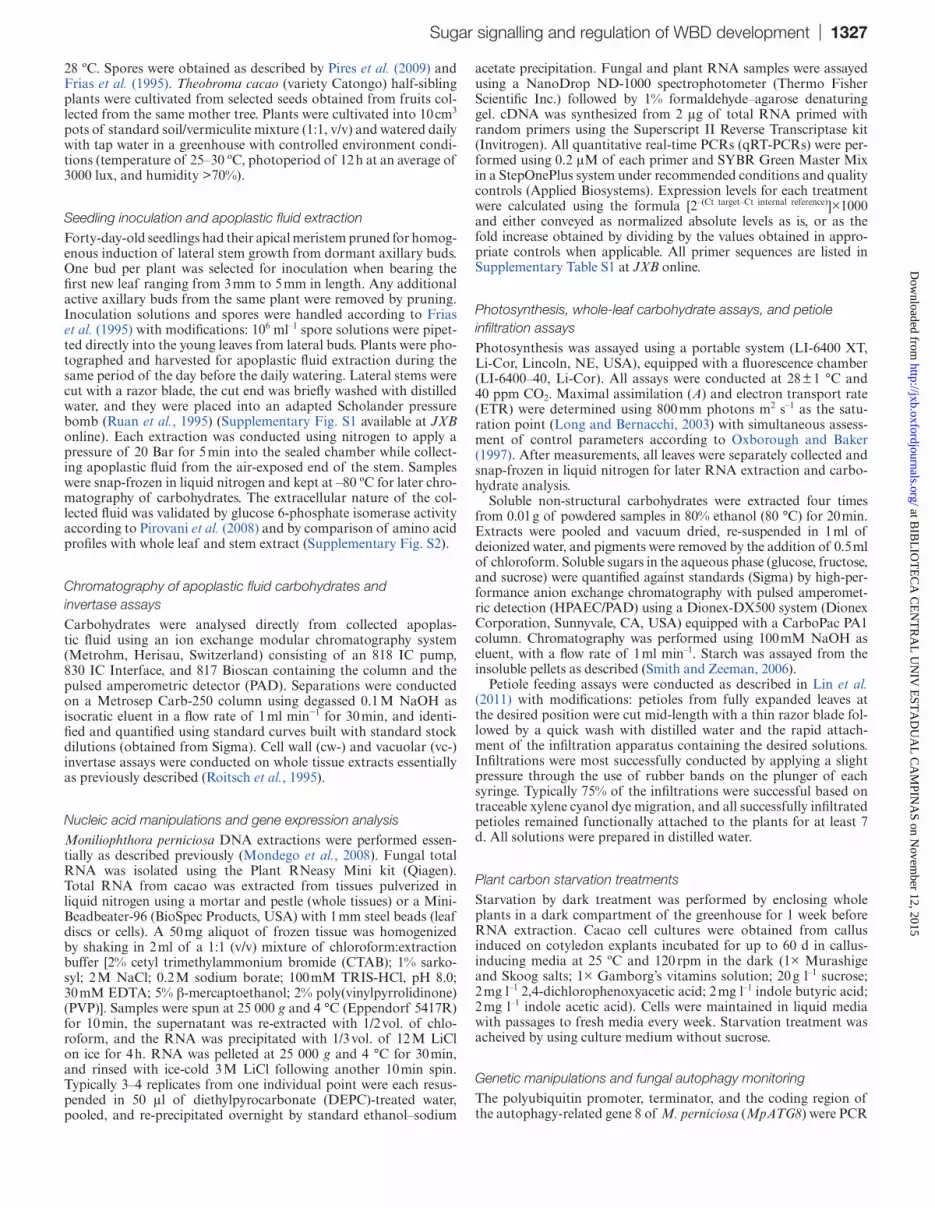
28 ºC. Spores were obtained as described by Pires et al. (2009) and Frias et al. (1995). Theobroma cacao (variety Catongo) half-sibling plants were cultivated from selected seeds obtained from fruits col-lected from the same mother tree. Plants were cultivated into 10 cm3 pots of standard soil/vermiculite mixture (1:1, v/v) and watered daily with tap water in a greenhouse with controlled environment condi-tions (temperature of 25–30 ºC, photoperiod of 12 h at an average of 3000 lux, and humidity >70%).
Seedling inoculation and apoplastic fluid extractionForty-day-old seedlings had their apical meristem pruned for homog-enous induction of lateral stem growth from dormant axillary buds. One bud per plant was selected for inoculation when bearing the first new leaf ranging from 3 mm to 5 mm in length. Any additional active axillary buds from the same plant were removed by pruning. Inoculation solutions and spores were handled according to Frias et al. (1995) with modifications: 106 ml–1 spore solutions were pipet-ted directly into the young leaves from lateral buds. Plants were pho-tographed and harvested for apoplastic fluid extraction during the same period of the day before the daily watering. Lateral stems were cut with a razor blade, the cut end was briefly washed with distilled water, and they were placed into an adapted Scholander pressure bomb (Ruan et al., 1995) (Supplementary Fig. S1 available at JXB online). Each extraction was conducted using nitrogen to apply a pressure of 20 Bar for 5 min into the sealed chamber while collect-ing apoplastic fluid from the air-exposed end of the stem. Samples were snap-frozen in liquid nitrogen and kept at –80 ºC for later chro-matography of carbohydrates. The extracellular nature of the col-lected fluid was validated by glucose 6-phosphate isomerase activity according to Pirovani et al. (2008) and by comparison of amino acid profiles with whole leaf and stem extract (Supplementary Fig. S2).
Chromatography of apoplastic fluid carbohydrates and invertase assaysCarbohydrates were analysed directly from collected apoplas-tic fluid using an ion exchange modular chromatography system (Metrohm, Herisau, Switzerland) consisting of an 818 IC pump, 830 IC Interface, and 817 Bioscan containing the column and the pulsed amperometric detector (PAD). Separations were conducted on a Metrosep Carb-250 column using degassed 0.1 M NaOH as isocratic eluent in a flow rate of 1 ml min−1 for 30 min, and identi-fied and quantified using standard curves built with standard stock dilutions (obtained from Sigma). Cell wall (cw-) and vacuolar (vc-) invertase assays were conducted on whole tissue extracts essentially as previously described (Roitsch et al., 1995).
Nucleic acid manipulations and gene expression analysisMoniliophthora perniciosa DNA extractions were performed essen-tially as described previously (Mondego et al., 2008). Fungal total RNA was isolated using the Plant RNeasy Mini kit (Qiagen). Total RNA from cacao was extracted from tissues pulverized in liquid nitrogen using a mortar and pestle (whole tissues) or a Mini-Beadbeater-96 (BioSpec Products, USA) with 1 mm steel beads (leaf discs or cells). A 50 mg aliquot of frozen tissue was homogenized by shaking in 2 ml of a 1:1 (v/v) mixture of chloroform:extraction buffer [2% cetyl trimethylammonium bromide (CTAB); 1% sarko-syl; 2 M NaCl; 0.2 M sodium borate; 100 mM TRIS-HCl, pH 8.0; 30 mM EDTA; 5% β-mercaptoethanol; 2% poly(vinylpyrrolidinone) (PVP)]. Samples were spun at 25 000 g and 4 °C (Eppendorf 5417R) for 10 min, the supernatant was re-extracted with 1/2 vol. of chlo-roform, and the RNA was precipitated with 1/3 vol. of 12 M LiCl on ice for 4 h. RNA was pelleted at 25 000 g and 4 °C for 30 min, and rinsed with ice-cold 3 M LiCl following another 10 min spin. Typically 3–4 replicates from one individual point were each resus-pended in 50 μl of diethylpyrocarbonate (DEPC)-treated water, pooled, and re-precipitated overnight by standard ethanol–sodium
acetate precipitation. Fungal and plant RNA samples were assayed using a NanoDrop ND-1000 spectrophotometer (Thermo Fisher Scientific Inc.) followed by 1% formaldehyde–agarose denaturing gel. cDNA was synthesized from 2 μg of total RNA primed with random primers using the Superscript II Reverse Transcriptase kit (Invitrogen). All quantitative real-time PCRs (qRT-PCRs) were per-formed using 0.2 μM of each primer and SYBR Green Master Mix in a StepOnePlus system under recommended conditions and quality controls (Applied Biosystems). Expression levels for each treatment were calculated using the formula [2–(Ct target–Ct internal reference)]×1000 and either conveyed as normalized absolute levels as is, or as the fold increase obtained by dividing by the values obtained in appro-priate controls when applicable. All primer sequences are listed in Supplementary Table S1 at JXB online.
Photosynthesis, whole-leaf carbohydrate assays, and petiole infiltration assaysPhotosynthesis was assayed using a portable system (LI-6400 XT, Li-Cor, Lincoln, NE, USA), equipped with a fluorescence chamber (LI-6400–40, Li-Cor). All assays were conducted at 28 ± 1 °C and 40 ppm CO2. Maximal assimilation (A) and electron transport rate (ETR) were determined using 800 mm photons m2 s–1 as the satu-ration point (Long and Bernacchi, 2003) with simultaneous assess-ment of control parameters according to Oxborough and Baker (1997). After measurements, all leaves were separately collected and snap-frozen in liquid nitrogen for later RNA extraction and carbo-hydrate analysis.
Soluble non-structural carbohydrates were extracted four times from 0.01 g of powdered samples in 80% ethanol (80 °C) for 20 min. Extracts were pooled and vacuum dried, re-suspended in 1 ml of deionized water, and pigments were removed by the addition of 0.5 ml of chloroform. Soluble sugars in the aqueous phase (glucose, fructose, and sucrose) were quantified against standards (Sigma) by high-per-formance anion exchange chromatography with pulsed amperomet-ric detection (HPAEC/PAD) using a Dionex-DX500 system (Dionex Corporation, Sunnyvale, CA, USA) equipped with a CarboPac PA1 column. Chromatography was performed using 100 mM NaOH as eluent, with a flow rate of 1 ml min–1. Starch was assayed from the insoluble pellets as described (Smith and Zeeman, 2006).
Petiole feeding assays were conducted as described in Lin et al. (2011) with modifications: petioles from fully expanded leaves at the desired position were cut mid-length with a thin razor blade fol-lowed by a quick wash with distilled water and the rapid attach-ment of the infiltration apparatus containing the desired solutions. Infiltrations were most successfully conducted by applying a slight pressure through the use of rubber bands on the plunger of each syringe. Typically 75% of the infiltrations were successful based on traceable xylene cyanol dye migration, and all successfully infiltrated petioles remained functionally attached to the plants for at least 7 d. All solutions were prepared in distilled water.
Plant carbon starvation treatmentsStarvation by dark treatment was performed by enclosing whole plants in a dark compartment of the greenhouse for 1 week before RNA extraction. Cacao cell cultures were obtained from callus induced on cotyledon explants incubated for up to 60 d in callus-inducing media at 25 ºC and 120 rpm in the dark (1× Murashige and Skoog salts; 1× Gamborg’s vitamins solution; 20 g l–1 sucrose; 2 mg l–1 2,4-dichlorophenoxyacetic acid; 2 mg l–1 indole butyric acid; 2 mg l–1 indole acetic acid). Cells were maintained in liquid media with passages to fresh media every week. Starvation treatment was acheived by using culture medium without sucrose.
Genetic manipulations and fungal autophagy monitoringThe polyubiquitin promoter, terminator, and the coding region of the autophagy-related gene 8 of M. perniciosa (MpATG8) were PCR
at BIB
LIO
TE
CA
CE
NT
RA
L U
NIV
EST
AD
UA
L C
AM
PINA
S on Novem
ber 12, 2015http://jxb.oxfordjournals.org/
Dow
nloaded from

1328 | Barau et al.
amplified from FA553 strain genomic DNA, and EGFP (enhanced green fluorescent protein) and the hygromycin (hph) selection cas-sette (Supplementary Fig. S5 at JXB online) were amplified from the pBGgHg vector (Chen et al. 2000) using the primers listed in Supplementary Table S1. Purified products were assembled by fusion PCR cloning as described (Szewczyk et al., 2006).
Fused cassettes were cloned in pGEM-T-easy (Promega) and confirmed by sequencing. The final vector for transformation of M. perniciosa was obtained by sequential digestion–ligation of the EGFP–MpATG8 expression cassette into the vector containing the hygromycin selection cassette using SacI and SalI restriction endo-nucleases. Transformation of M. perniciosa protoplasts followed the same procedures described by Lima et al. (2003), with the excep-tion that transformed protoplasts were plated into 20 ml of solid protoplast regeneration medium (0.5% peptone, 0.5% yeast extract, 0.3% glucose, 0.1% mannitol, 0.5% glycerol, 0.8% malt extract, 17% sucrose, and 1% agar) without selection for 3 d followed by the overlay of 5 ml of semi-solid (0.7% agar) protoplast regenera-tion medium containing 1 mg ml–1 hygromycin (for a final selective concentration of 200 μg ml–1 on the plate).
Confirmed transformants were cultivated into solid plates of 2% agar in tap water for 7 d and then transferred to defined solid medium according to Metzenberg (2004) modified by exchanging agar by agarose as the solidifying agent and either with or without carbon sources. Rapamycin treatments were conducted by over-laying 15-day-old colonies with 5 ml of semi-solid (0.7% agarose) defined media containing 500 ng ml–1 rapamycin. Hyphae for RNA extraction or microscopy were carefully scraped and collected from cracked open agarose layers after 12 h of treatment. Slides contain-ing live hyphae mounted in liquid medium were photographed by standard confocal laser microscopy (Carl Zeiss, LSM 510 Meta) and processed using LSM Image Browser (Carl Zeiss).
Results
A periodic carbon cycle in the apoplastic fluid of cacao
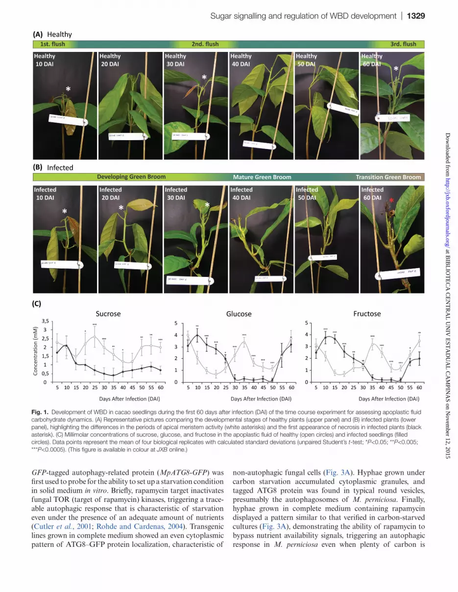
Apoplastic fluid was isolated from healthy and infected stems using an adapted Scholander pressure bomb (Supplementary Figs S1, S2 at JXB online), and the soluble carbohydrate pro-file was determined using liquid chromatography. In order to obtain a view of the soluble carbon sources available to M. perniciosa in the apoplast during infection, quantitative analysis was performed in a time course experiment span-ning the whole biotrophic and the initial transition to the necrotrophic phase of WBD in cacao seedlings. In healthy plants, concentrations of apoplastic carbohydrates followed an apparent cyclic pattern, successively alternating between higher and lower levels throughout the duration of the experi-ment. While the concentration of glucose and fructose peaked three times with very similar intervals, sucrose concentrations appeared to oscillate in an inverse manner (Fig. 1C; open cir-cles). The observed pattern coincided with the developmen-tal cycle of cacao seedlings as high levels of carbohydrates overlapped with periods of new leaf flushing and vegetative growth (Fig. 1A; Supplementary Fig. S3).
WBD disrupts the carbon cycle in the apoplast of cacao
Disease symptoms followed a similar developmental time frame to what was previously described when using seed-lings as models for WBD development (Scarpari et al., 2005).
Infected cacao shoots lost the periodic growth pattern char-acteristic of healthy controls (Fig. 1B), and this loss was also reflected in the disruption of the pattern of carbohydrates in the apoplastic fluid (Fig. 1C; filled circles). Glucose and fructose levels were found to be systematically higher in the apoplastic fluid of infected plants in the first 25 d of WBD development (Fig. 1C), during the tissue-proliferative phase of the disease (Fig. 1B; Supplementary Fig. S3 at JXB online). The increase of glucose and fructose correlated with the decrease of sucrose levels, suggesting enhanced activ-ity of invertases. Assays for cw- and vc-invertase activity revealed that cw-invertase is 3.5 times more active specifically in developing green brooms, showing little or no difference between healthy and infected tissues at later stages (Fig. 2A). Remarkably, vc-invertase is twice as active in infected tissues at the stage of mature and transition green brooms, with no difference at the beginning of infection or at later necrotic stages (Fig. 2A). In addition, gene orthologues of predicted cacao and M. perniciosa extracellular invertases appeared to be up-regulated in developing green brooms (Fig. 2B, C), indicating a possible cumulative role between fungal and plant enzymes in the maintenance of high apoplastic hexose levels from the breakdown of free sucrose.
The mature broom stage was phenotypically characterized by the cessation of growth of the infected tissues [40 days after infection (DAI); Fig. 1B; Supplementary Fig. S3 at JXB online]). In this stage, all apoplastic fluid carbohydrate concentrations reached the lowest levels (Fig. 1C; filled cir-cles), indicating that the cacao apoplast is relatively depleted of soluble carbon at advanced stages of infection. Hexoses appear again at measurable levels only after 50 DAI (Fig. 1C; filled circles), together with the appearance and spreading of leaf necrosis (Fig. 1B; 50–60 DAI), but with no clear increase in either activity or gene expression of apoplastic invertases (Fig. 2A–C), probably reflecting the leakage of the intracel-lular content of dying cacao cells. These results indicate that infection leads first to an extent of apoplastic fluid hexose accumulation and then to the depletion of soluble carbohy-drates before the transition to the necrotrophic phase. Based on the present findings of carbon depletion in the apoplas-tic fluid of mature brooms and on previous reports showing that fungal growth is strictly apoplastic up until much later in WBD development (Frias et al., 1991), it was further inves-tigated whether fungal sensing of carbon starvation could trigger the expression of fungal genes associated with the necrotrophic phase and the killing of infected tissues.
Carbon starvation is a signalling component controlling the duration of the biotrophic phase of WBD
It was previously shown that initiation of the necrotrophic phase of WBD in vivo is marked by the up-regulation of the fungal gene coding for an NEP-like effector of plant necrosis, MpNEP2, implicating its activity in the necrosis of infected cacao (Zaparoli et al., 2011). Therefore, a test was conducted to determine if fungal carbon starvation could be part of the signal that triggers the expression of MpNEP2. The drug rapamycin and a transgenic M. perniciosa strain expressing a
at BIB
LIO
TE
CA
CE
NT
RA
L U
NIV
EST
AD
UA
L C
AM
PINA
S on Novem
ber 12, 2015http://jxb.oxfordjournals.org/
Dow
nloaded from
Sugar signalling and regulation of WBD development | 1329
GFP-tagged autophagy-related protein (MpATG8-GFP) was first used to probe for the ability to set up a starvation condition in solid medium in vitro. Briefly, rapamycin target inactivates fungal TOR (target of rapamycin) kinases, triggering a trace-able autophagic response that is characteristic of starvation even under the presence of an adequate amount of nutrients (Cutler et al., 2001; Rohde and Cardenas, 2004). Transgenic lines grown in complete medium showed an even cytoplasmic pattern of ATG8–GFP protein localization, characteristic of
non-autophagic fungal cells (Fig. 3A). Hyphae grown under carbon starvation accumulated cytoplasmic granules, and tagged ATG8 protein was found in typical round vesicles, presumably the autophagosomes of M. perniciosa. Finally, hyphae grown in complete medium containing rapamycin displayed a pattern similar to that verified in carbon-starved cultures (Fig. 3A), demonstrating the ability of rapamycin to bypass nutrient availability signals, triggering an autophagic response in M. perniciosa even when plenty of carbon is
Fig. 1. Development of WBD in cacao seedlings during the first 60 days after infection (DAI) of the time course experiment for assessing apoplastic fluid carbohydrate dynamics. (A) Representative pictures comparing the developmental stages of healthy plants (upper panel) and (B) infected plants (lower panel), highlighting the differences in the periods of apical meristem activity (white asterisks) and the first appearance of necrosis in infected plants (black asterisk). (C) Millimolar concentrations of sucrose, glucose, and fructose in the apoplastic fluid of healthy (open circles) and infected seedlings (filled circles). Data points represent the mean of four biological replicates with calculated standard deviations (unpaired Student’s t-test; *P<0.05; **P<0.005; ***P<0.0005). (This figure is available in colour at JXB online.)
at BIB
LIO
TE
CA
CE
NT
RA
L U
NIV
EST
AD
UA
L C
AM
PINA
S on Novem
ber 12, 2015http://jxb.oxfordjournals.org/
Dow
nloaded from

1330 | Barau et al.
available. It was found that MpNEP2 expression levels were significantly higher in both carbon-starved and rapamycin-treated cultures (Fig. 3B), suggesting that starvation up-reg-ulates MpNEP2 under these conditions in vitro. In addition, cultures transferred into starvation medium presented a pecu-liar phenotype of slow growth followed by the development of faster growing sectors formed by very thin mycelia with marked hyphal branching (Fig. 3C), a typical characteristic of the invasive growth observed in infected plant tissues after phase transition (Frias et al., 1991).
Next, advantage was taken of the extracellular confine-ment of M. perniciosa in green brooms to perform a petiole feeding assay (Lin et al., 2010, 2011) to manipulate apoplas-tic fluid sugar availability artificially and test if the fungal sensing of carbon starvation could be linked to MpNEP2 expression and plant necrosis in vivo. Infiltration of a carbon solution on 40 DAI plants led to significant maintenance of higher carbon levels in the apoplastic fluid of infected tissues (Supplementary Fig. S4 at JXB online). This treatment led to a slowing down of the later necrotic process when compared with untreated controls (Fig. 4A), also followed by delayed MpNEP2 up-regulation (Fig. 4B). This result indicates that the lack of soluble carbon in the apoplast is probably per-ceived by the infecting fungus and used as a cue to the time-opportune expression of MpNEP2.
In order to account for the possibility that the observed delay of plant necrosis was due to an increase in the survival of plant cells fed with carbon and thus not related to a fun-gal response to starvation, rapamycin was also included in the infiltrations. Because plant TOR kinases are reported to be insensitive to the drug (Menand et al., 2002; Robaglia et al., 2012), presumably rapamycin only inactivates fungal TOR kinase. This ‘differential targeting’ due to drug sensitiv-ity should be able to rescue only the pathogen-related effects observed upon artificially increased sugar availability by infil-tration. Rapamycin infiltration on healthy plants did not affect development nor did it induce any detectable symptoms (data not shown). Infected plants infiltrated with a carbon solution containing rapamycin presented a necrosis progression more similar to that verified in controls (Fig. 4A), showing that rapamycin is able to rescue the delayed necrosis phenotype of carbon-infiltrated plants. However, the timing of MpNEP2 expression was only partially rescued (Fig. 4B), suggesting that other factors in addition to a response to carbon star-vation may also be part of the signalling leading to the up-regulation of this effector in vivo. Accordingly, the infiltration of rapamycin alone did not induce significant acceleration of necrotic symptoms (Fig. 4A), and it led to only a slightly ear-lier up-regulation of MpNEP2 (Fig. 4B). These results show that although MpNEP2 up-regulation is partially associated with the the sensing of carbon starvation at the apoplastic fluid, it is probably not the main cause of necrosis in infected tissues. This suggests that either other unknown WBD effec-tors might be taking part in the biotrophic to necrotrophic transition upon fungal carbon starvation or that additional key processes involving the physiology of the host might also be at play. Because substantial carbon depletion in the apo-plastic fluid of infected plants was found, it was decided to investigate further whether WBD-related plant carbon star-vation could lead to an early commitment of infected tissues to the necrotrophic phase.
WBD leads to a spatial pattern of intracellular sugar accumulation, photosynthesis decline, and up-regulation of carbon starvation markers within infected cacao tissues
The mature broom stage (30–40 DAI) precedes the appear-ance of the first signs of plant tissue death. This stage was characterized by the cessation of growth, with all apoplas-tic carbohydrate concentrations reaching the lowest levels measured during the experiment (Fig. 1B, C). This suggests that infected cacao tissues might have a compromised car-bon metabolism and thus are unable to provide further the sugars to be released in the apoplast at this stage of WBD development. Infected plant tissues have long been regarded as having down-regulated photosynthesis (Orchard and Hardwick, 1988; Scarpari et al., 2005), a condition known to set a scene of physiological carbon depletion leading to starvation and senescence in plants (Brouwer et al., 2013). To test if infected cacao tissues could be subject to carbon starvation, the expression of gene markers of acute carbon starvation in plants was assessed and photosynthesis and the
Fig. 2. Whole-tissue invertase enzymes activities and gene expression measured at key developmental stages of WBD, spanning from green broom stages (GB) to early necrosis. (A) Cell wall (cw-), and vacuolar (vac-) invertase activities portrayed as fold increase in infected tissues. (B) Gene expression of a conserved fungal secreted (extracellular) invertase present in the M. perniciosa genome. (C) Gene expression of a conserved cacao cw-invertase present in the T. cacao genome. Data points represent the mean value of three biological replicates with calculated standard deviations.
at BIB
LIO
TE
CA
CE
NT
RA
L U
NIV
EST
AD
UA
L C
AM
PINA
S on Novem
ber 12, 2015http://jxb.oxfordjournals.org/
Dow
nloaded from

Sugar signalling and regulation of WBD development | 1331
main intracellular carbon pools were measured in the first fully expanded leaf at both the base and the top of control and infected stems.
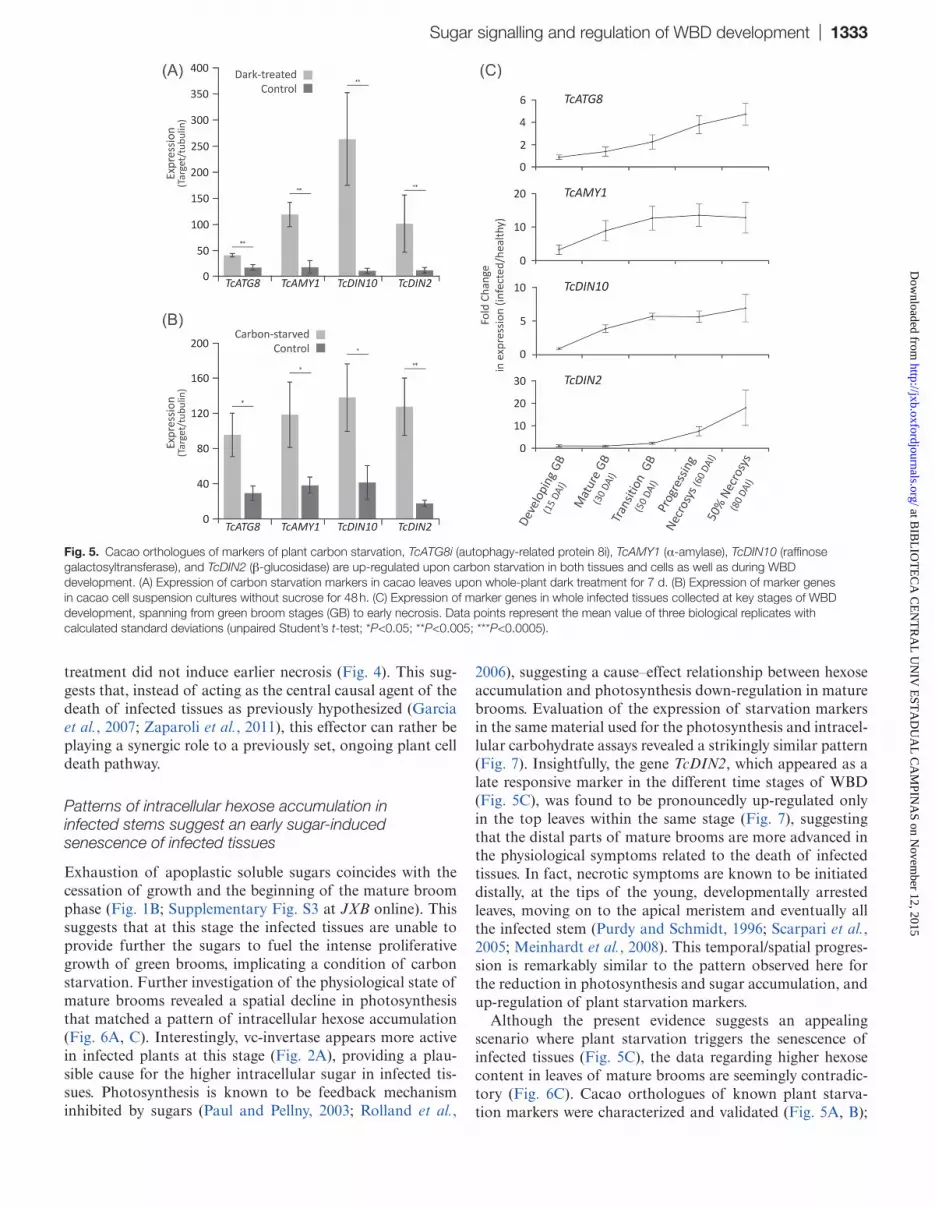
Cacao orthologues of previously characterized expres-sion markers of carbon starvation in plants (Fujiki et al. 2000; Contento et al., 2004; Rose et al., 2006) were identi-fied based on sequence similarity (Supplementary Table S1 at JXB online), and functionally validated in cacao tissues and cell suspensions cultures. qRT-PCR analysis showed an overall trend of up-regulation in the leaves of cacao plants incubated in the dark for 1 week (Fig. 5A) as well as in cacao cell culture without sucrose for 24 h (Fig. 5B), confirming that the selected genes are responsive to standard carbon starva-tion conditions in plants. Up-regulation was also evident in infected cacao tissues collected during selected time stages of WBD development; however, distinct patterns could be identified between markers. While transcripts of the gene TcATG8i showed a relatively gradual increase during the stages, TcAMY1 and TcDIN10 appeared as early respon-sive markers, showing up-regulation at 30 DAI (Fig. 5C). In contrast, TcDIN2 appeared as a late responsive marker, only being up-regulated at much later stages, at the spreading of infected tissue necrosis (Fig. 5C).
Photosynthesis (A) and electron transport rate (ETR) in the leaves at the top of 40 DAI stems were significantly reduced
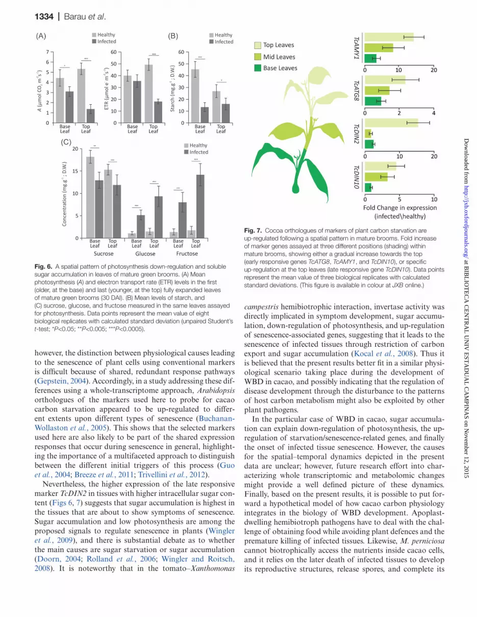
in infected plants when compared with leaves in the same stage and position of healthy controls (Fig. 6A). Although also affected, older leaves at the base had levels more simi-lar to those of corresponding healthy controls, suggesting a spatial symptomatic gradient. Starch levels were also found to be reduced in infected leaves, but no significant positional differences were found (Fig. 6B). Surprisingly, while intracel-lular leaf sucrose levels are only slightly decreased, a marked accumulation of intracellular glucose and fructose occurs in the leaves of infected tissues. Moreover, leaf hexose accumu-lation appeared to occur to a greater extent in the top infected leaves (Fig. 6C), following the same pattern observed for the photosynthetic decline.
In an attempt to correlate WBD-induced up-regulation of plant carbon starvation markers with the observed pattern of photosynthesis decline and intracellular hexose accumulation, their expression was evaluated in the same collected material used for photosynthesis and leaf carbon assays. Strikingly, it was found that the pattern of marker up-regulation followed the same basipetal gradient as observed for sugar accumula-tion and photosynthesis decay (Fig. 7), suggesting a direct correlation. Also, the patterns of early and late responsive-ness that were evident at different developmental time stages (Fig. 5C) appeared also to occur within the vertical axis of shoots prior to phase transition. While the genes TcAMY1,
Fig. 3. MpNEP2 is up-regulated under autophagic conditions triggered by carbon starvation in vitro. (A) Light, fluorescence, and merged microscopy images of a transgenic M. perniciosa strain expressing the autophagy reporter MpATG8-GFP. In complete medium (upper panel), hyphae show uniform cytoplasmic distribution of fluorescent-tagged ATG8 protein. In carbon starvation medium (middle panel) or in complete medium with rapamycin (lower panel), dense cytoplasmic granules (black arrows) and accumulation of protein into autophagic round vesicles are evident (white arrows). Scale bars=2 μm. (B) Relative MpNEP2 gene expression under carbon starvation in vitro and in the presence of carbon with rapamycin. Data points represent a mean value of three biological replicates with calculated standard deviations (unpaired Student’s t-test; ***P<0.0005). (C) M. perniciosa grows as dense colonies in complete defined medium in vitro, while under carbon starvation growth is stunted, with the occasional appearance of fast-growing sectors showing hyaline hyphae with pronounced branching (black arrow). (This figure is available in colour at JXB online.)
at BIB
LIO
TE
CA
CE
NT
RA
L U
NIV
EST
AD
UA
L C
AM
PINA
S on Novem
ber 12, 2015http://jxb.oxfordjournals.org/
Dow
nloaded from

1332 | Barau et al.
TcATG8i, and TcDIN10 showed a gradual increase in expres-sion along the vertical axis of the infected shoot, the late responsive marker TcDIN2 appears to be up-regulated only in the top leaves (Fig. 7). Taken together, the data suggest that physiological symptoms of WBD include dramatic changes in the primary carbon metabolism of cacao. Moreover, these changes appear to correlate well with the expression of genes involved in carbon starvation, occurring in a spatiotemporal gradient within the vertical axis of mature brooms.
Discussion
Apoplastic soluble sugars in the rhythmic growth of cacao and WBD development
Aiming for the characterization of the carbohydrates avail-able for M. perniciosa in the apoplastic fluid of cacao, a concentration pattern that matched the periodic growth of healthy cacao seedlings was unexpectedly found (Fig. 1A, C; Supplementary Fig. S3 at JXB online). Cacao is known to have a periodic, flush-type growth, and early investigations (Greathouse et al., 1971) showed exactly the same time frame of 30 d between early flushes that were verified in the present experiments (Fig. 1A). It was shown that cacao apoplastic fluid has considerably more carbohydra tes when the api-cal meristem is actively growing, precisely the only known
vegetative tissues that are susceptible to WBD. In infected plants, levels of hexoses in the apoplastic fluid increased in parallel with symptom development and the decrease of sucrose levels (Fig. 1C, filled circles), correlating with the up-regulation of fungal and plant extracellular invertases during most of the green broom phase (Fig. 2A–C). The induction of cw-invertase activity has been reported as a fea-ture common to many plant–pathogen interactions (Roitsch et al., 2003). It often induces infected tissues to behave as carbon sinks, providing the pathogen with increased car-bon resources (Berger et al., 2007); however, the array of mechanisms used to manipulate host metabolism are far less understood. Infected cacao tissues have typical symptoms of hormone imbalance, notably involving auxin and cytokinin (Melnick et al., 2012), and M. perniciosa has been shown both to possess the genetic pathways and to produce plant hormones in vitro (Kilaru et al., 2007; Mondego et al., 2008). Cw-invertases are known to be up-regulated in response to auxins and cytokinins (Roitsch et al., 2003), and it is possible that a similar mechanism might be operating during the bio-trophic phase of WBD. Recently, new evidence has emerged to support a major role of sugar availability as a determi-nant of apical dominance and bud dormancy break in plants (Mason et al., 2014). It is noteworthy that hexose concentra-tions in the apoplastic fluid during the episodes of periodic growth of healthy plants are comparable with the abnormal levels sustained during the proliferative phase of developing green brooms (Fig. 1C). In addition to artificially sustaining sink metabolism in infected tissues, increased cw-invertase activity and the consequent higher levels of hexoses may also act additively with plant hormones as proliferative signals leading to the characteristic meristem hyperactivity, and the loss of both apical dominance and periodicity of growth in developing green brooms.
Fungal carbon starvation at the cacao apoplast as a cue for the opportune expression of fungal effectors of plant necrosis
It was demonstrated that a decrease in the concentration of soluble carbohydrates occurs in the apoplastic fluid of mature green brooms (Fig. 1C), suggesting that M. perniciosa could be subject to a carbon starvation microenvironment immedi-ately prior to the death of infected tissues. Starvation condi-tions have long been reported to induce fungal pathogenicity factors (Talbot et al., 1997), and it has been shown here in vitro that the fungal NLP-like effector of plant necrosis, MpNEP2, is integrated downstream of a carbon starvation, autophagic response network (Fig. 3). Artificially increased carbohydrate concentrations in the apoplastic fluid of mature brooms led to a delay of plant necrosis and MpNEP2 expression (Fig. 4), suggesting that carbon starvation at the apoplast is a significant signalling component ruling the timing of these events in WBD. However, the extent of the contribution of MpNEP2 up-regulation in affecting the timing of necrosis was difficult to quantify. Although rapamycin was able to res-cue the delayed necrosis phenotype observed in carbon-only infiltrations, the earlier MpNEP2 expression observed in this
Fig. 4. Manipulation of apoplastic fluid carbon availability alters MpNEP2 expression and the necrosis rate of infected tissues. (A) Time (in days ater infection) that necrotic symptoms took to cover 50% of the length of infected stems after infiltrations with solutions containing carbon (sucrose, glucose, and fructose at 10 mM each), carbon with 400 ng ml–1 rapamycin, and rapamycin only. (B) MpNEP2 gene expression assayed at 10 d intervals after infiltration of infected stems. Data points represent the mean of four biological replicates with calculated standard deviations (unpaired Student’s t-test; *P<0.05; **P<0.005; ***P<0.0005). (This figure is available in colour at JXB online.)
at BIB
LIO
TE
CA
CE
NT
RA
L U
NIV
EST
AD
UA
L C
AM
PINA
S on Novem
ber 12, 2015http://jxb.oxfordjournals.org/
Dow
nloaded from
Sugar signalling and regulation of WBD development | 1333
treatment did not induce earlier necrosis (Fig. 4). This sug-gests that, instead of acting as the central causal agent of the death of infected tissues as previously hypothesized (Garcia et al., 2007; Zaparoli et al., 2011), this effector can rather be playing a synergic role to a previously set, ongoing plant cell death pathway.
Patterns of intracellular hexose accumulation in infected stems suggest an early sugar-induced senescence of infected tissues
Exhaustion of apoplastic soluble sugars coincides with the cessation of growth and the beginning of the mature broom phase (Fig. 1B; Supplementary Fig. S3 at JXB online). This suggests that at this stage the infected tissues are unable to provide further the sugars to fuel the intense proliferative growth of green brooms, implicating a condition of carbon starvation. Further investigation of the physiological state of mature brooms revealed a spatial decline in photosynthesis that matched a pattern of intracellular hexose accumulation (Fig. 6A, C). Interestingly, vc-invertase appears more active in infected plants at this stage (Fig. 2A), providing a plau-sible cause for the higher intracellular sugar in infected tis-sues. Photosynthesis is known to be feedback mechanism inhibited by sugars (Paul and Pellny, 2003; Rolland et al.,
2006), suggesting a cause–effect relationship between hexose accumulation and photosynthesis down-regulation in mature brooms. Evaluation of the expression of starvation markers in the same material used for the photosynthesis and intracel-lular carbohydrate assays revealed a strikingly similar pattern (Fig. 7). Insightfully, the gene TcDIN2, which appeared as a late responsive marker in the different time stages of WBD (Fig. 5C), was found to be pronouncedly up-regulated only in the top leaves within the same stage (Fig. 7), suggesting that the distal parts of mature brooms are more advanced in the physiological symptoms related to the death of infected tissues. In fact, necrotic symptoms are known to be initiated distally, at the tips of the young, developmentally arrested leaves, moving on to the apical meristem and eventually all the infected stem (Purdy and Schmidt, 1996; Scarpari et al., 2005; Meinhardt et al., 2008). This temporal/spatial progres-sion is remarkably similar to the pattern observed here for the reduction in photosynthesis and sugar accumulation, and up-regulation of plant starvation markers.
Although the present evidence suggests an appealing scenario where plant starvation triggers the senescence of infected tissues (Fig. 5C), the data regarding higher hexose content in leaves of mature brooms are seemingly contradic-tory (Fig. 6C). Cacao orthologues of known plant starva-tion markers were characterized and validated (Fig. 5A, B);
Fig. 5. Cacao orthologues of markers of plant carbon starvation, TcATG8i (autophagy-related protein 8i), TcAMY1 (α-amylase), TcDIN10 (raffinose galactosyltransferase), and TcDIN2 (β-glucosidase) are up-regulated upon carbon starvation in both tissues and cells as well as during WBD development. (A) Expression of carbon starvation markers in cacao leaves upon whole-plant dark treatment for 7 d. (B) Expression of marker genes in cacao cell suspension cultures without sucrose for 48 h. (C) Expression of marker genes in whole infected tissues collected at key stages of WBD development, spanning from green broom stages (GB) to early necrosis. Data points represent the mean value of three biological replicates with calculated standard deviations (unpaired Student’s t-test; *P<0.05; **P<0.005; ***P<0.0005).
at BIB
LIO
TE
CA
CE
NT
RA
L U
NIV
EST
AD
UA
L C
AM
PINA
S on Novem
ber 12, 2015http://jxb.oxfordjournals.org/
Dow
nloaded from
1334 | Barau et al.
however, the distinction between physiological causes leading to the senescence of plant cells using conventional markers is difficult because of shared, redundant response pathways (Gepstein, 2004). Accordingly, in a study addressing these dif-ferences using a whole-transcriptome approach, Arabidopsis orthologues of the markers used here to probe for cacao carbon starvation appeared to be up-regulated to differ-ent extents upon different types of senescence (Buchanan-Wollaston et al., 2005). This shows that the selected markers used here are also likely to be part of the shared expression responses that occur during senescence in general, highlight-ing the importance of a multifaceted approach to distinguish between the different initial triggers of this process (Guo et al., 2004; Breeze et al., 2011; Trivellini et al., 2012).
Nevertheless, the higher expression of the late responsive marker TcDIN2 in tissues with higher intracellular sugar con-tent (Figs 6, 7) suggests that sugar accumulation is highest in the tissues that are about to show symptoms of senescence. Sugar accumulation and low photosynthesis are among the proposed signals to regulate senescence in plants (Wingler et al., 2009), and there is substantial debate as to whether the main causes are sugar starvation or sugar accumulation (Doorn, 2004; Rolland et al., 2006; Wingler and Roitsch, 2008). It is noteworthy that in the tomato–Xanthomonas
campestris hemibiotrophic interaction, invertase activity was directly implicated in symptom development, sugar accumu-lation, down-regulation of photosynthesis, and up-regulation of senescence-associated genes, suggesting that it leads to the senescence of infected tissues through restriction of carbon export and sugar accumulation (Kocal et al., 2008). Thus it is believed that the present results better fit in a similar physi-ological scenario taking place during the development of WBD in cacao, and possibly indicating that the regulation of disease development through the disturbance to the patterns of host carbon metabolism might also be exploited by other plant pathogens.
In the particular case of WBD in cacao, sugar accumula-tion can explain down-regulation of photosynthesis, the up-regulation of starvation/senescence-related genes, and finally the onset of infected tissue senescence. However, the causes for the spatial–temporal dynamics depicted in the present data are unclear; however, future research effort into char-acterizing whole transcriptomic and metabolomic changes might provide a well defined picture of these dynamics. Finally, based on the present results, it is possible to put for-ward a hypothetical model of how cacao carbon physiology integrates in the biology of WBD development. Apoplast-dwelling hemibiotroph pathogens have to deal with the chal-lenge of obtaining food while avoiding plant defences and the premature killing of infected tissues. Likewise, M. perniciosa cannot biotrophically access the nutrients inside cacao cells, and it relies on the later death of infected tissues to develop its reproductive structures, release spores, and complete its
Fig. 7. Cocoa orthologues of markers of plant carbon starvation are up-regulated following a spatial pattern in mature brooms. Fold increase of marker genes assayed at three different positions (shading) within mature brooms, showing either a gradual increase towards the top (early responsive genes TcATG8, TcAMY1, and TcDIN10), or specific up-regulation at the top leaves (late responsive gene TcDIN10). Data points represent the mean value of three biological replicates with calculated standard deviations. (This figure is available in colour at JXB online.)
Fig. 6. A spatial pattern of photosynthesis down-regulation and soluble sugar accumulation in leaves of mature green brooms. (A) Mean photosynthesis (A) and electron transport rate (ETR) levels in the first (older, at the base) and last (younger, at the top) fully expanded leaves of mature green brooms (30 DAI). (B) Mean levels of starch, and (C) sucrose, glucose, and fructose measured in the same leaves assayed for photosynthesis. Data points represent the mean value of eight biological replicates with calculated standard deviation (unpaired Student’s t-test; *P<0.05; **P<0.005; ***P<0.0005).
at BIB
LIO
TE
CA
CE
NT
RA
L U
NIV
EST
AD
UA
L C
AM
PINA
S on Novem
ber 12, 2015http://jxb.oxfordjournals.org/
Dow
nloaded from
Sugar signalling and regulation of WBD development | 1335
life cycle. During senescence, nitrogen and carbohydrates that are immobilized for the normal functioning of plant cells are converted into high valuable resources that are optimal for efficient remobilization and rapid utilization in storage and further plant growth (Lim et al., 2007). By coupling the expres-sion of MpNEP2 to the onset of senescence using carbon starvation as a cue perceived at the apoplastic fluid, extracel-lular M. perniciosa hyphae can have early access to this valu-able resource otherwise enclosed inside senescing plant cells. Moreover, opportune speeding up of infected tissue necrosis by MpNEP2 activity might also prevent two important late features of plant senescence: remobilization and abscission. Accordingly, an intriguing characteristic of WBD in cacao is that dead infected tissues are preferentially kept attached to cacao trees for long periods of time (Purdy and Schmidt, 1996; Scarpari et al., 2005; Meinhardt et al., 2006). This can be advantageous to the pathogen by first allowing preferential access to the resources enclosed in dead tissues and thus not having to compete with other microorganisms that inhabit the ground litter of the forest. Secondly, it may also enhance the dispersal of spores and increase the chances of infecting nearby actively growing meristems of cacao trees, possibly completing the disease life cycle in a more efficient manner.
Supplementary data
Supplementary data are available at JXB online.Figure S1. Adapted Scholander pressure bomb to extract
apoplastic fluids from cocoa shoots.Figure S2. Validation of the pressure dehydration protocol
for the extraction of apoplastic fluid of cocoa tissues.Figure S3. Weight of control and infected tissues collected
during the time course experiment reflects the differences between periodic growth and proliferative growth in healthy and infected tissues.
Figure S4, Carbohydrate concentrations in the apoplastic fluid of mature brooms infiltrated for 7 d with a water solu-tion containing sucrose, glucose, and fructose, or water alone.
Figure S5. Outline of the cloned EGFP-MpATG8 autophagy monitoring and hygromycin selection cassettes for M. perniciosa transformation.
Table S1. Oligonucleotide primers used in this study.
AcknowledgementsThe authors are grateful for the assistance of Dr Lauro Tatsuo Kubota and Dr Jailson Cardoso Dias in the chromatographic detection and analysis of carbohydrates of cacao apoplastic fluid, and Marcelo Carazzolle, Osvaldo Reis Junior, and Gustavo Gilson Lacerda Costa for the bioinformatics sup-port and management of databases consulted in the conduct of this work. This work was financially supported by CNPq (472710/2008-7) and FAPESP (2006/56942-0, 2009/17507-5, and 2009/50119-9). The authors declare no conflicts of interest.
ReferencesAlvim FC, Mattos EM, Pirovani CP, Gramacho K, Pungartnik C, Brendel M, Cascardo JC, Vincentz M. 2009. Carbon source-induced changes in the physiology of the cacao pathogen Moniliophthora
perniciosa (Basidiomycetes) affect mycelial morphology and secretion of necrosis-inducing proteins. Genetics and Molecular Research 8, 1035–1050.
Berger S, Sinha AK, Roitsch T. 2007. Plant physiology meets phytopathology: plant primary metabolism and plant–pathogen interactions. Journal of Experimental Botany 58, 4019–4026.
Bolton MD, Thomma BPHJ. 2008. The complexity of nitrogen metabolism and nitrogen-regulated gene expression in plant pathogenic fungi. Physiological and Molecular Plant Pathology 72, 104–110.
Breeze E, Harrison E, McHattie S, et al. 2011. High-resolution temporal profiling of transcripts during Arabidopsis leaf senescence reveals a distinct chronology of processes and regulation. The Plant Cell 23, 873–894.
Brouwer B, Ziolkowska A, Bagard M, Keech O, Gardeström P. 2012. The impact of light intensity on shade-induced leaf senescence. Plant, Cell and Environment 35, 1084–1098.
Buchanan-Wollaston V, Page T, Harrison E, et al. 2005. Comparative transcriptome analysis reveals significant differences in gene expression and signalling pathways between developmental and dark/starvation-induced senescence in Arabidopsis. The Plant Journal 42, 567–585.
Calle H, Cook A, Fernando S. 1982. Histology of witches’-broom caused in cacao by Crinipellis perniciosa. Phytopathology 72, 1479–1481.
Chen X, Stone M, Schlagnhaufer C, Romaine CP. 2000. A fruiting body tissue method for efficient Agrobacterium-mediated transformation of Agaricus bisporus. Applied and Environmental Microbiology 66, 4510–4513.
Coleman M, Henricot B, Arnau J, Oliver RP. 1997. Starvation-induced genes of the tomato pathogen Cladosporium fulvum are also induced during growth in planta. Molecular Plant-Microbe Interactions 10, 1106–1109.
Contento AL, Kim S, Bassham DC. 2004. Transcriptome profiling of the response of Arabidopsis suspension culture cells to Suc starvation. Plant Physiology 135, 2330–2347.
Cutler NS, Pan X, Heitman J, Cardenas ME. 2001. The TOR signal transduction cascade controls cellular differentiation in response to nutrients. Molecular Biology of the Cell 12, 4103–4113.
da Hora Junior BT, Poloni Jde F, Lopes MA, et al. 2012. Transcriptomics and systems biology analysis in identification of specific pathways involved in cacao resistance and susceptibility to witches’ broom disease. Molecular Biosystems 8, 1507–1519.
Divon HH, Fluhr R. 2007. Nutrition acquisition strategies during fungal infection of plants. FEMS Microbiol Letters 266, 65–74.
Doorn WGV. 2004. Is petal senescence due to sugar starvation? Plant Physiology 134, 35–42.
Evans HC. 1980. Pleomorphism in Crinipellis perniciosa, causal agent of witches’ broom disease of cocoa. Transactions of the British Mycological Society 74, 515–523.
Evans HC, Bastos CN. 1980. Basidiospore germination as a means of assessing resistance to Crinipellis perniciosa (witches’ broom disease) in cocoa cultivars. Transactions of the British Mycological Society 74, 525–536.
Fernandez J, Wilson RA. 2012. Why no feeding frenzy? Mechanisms of nutrient acquisition and utilization during infection by the rice blast fungus Magnaporthe oryzae. Molecular Plant-Microbe Interactions 25, 1286–1293.
Fernandez J, Wright JD, Hartline D, Quispe CF, Madayiputhiya N, Wilson RA. 2012. Principles of carbon catabolite repression in the rice blast fungus: Tps1, Nmr1-3, and a MATE-family pump regulate glucose metabolism during infection. PLoS Genetics 8, e1002673.
Frias G, Purdy L, Schmidt R. 1991. Infection biology of Crinipellis perniciosa on vegetative flushes of cacao. Plant Disease 75, 552–556.
Frias G, Purdy L, Schmidt R. 1995. An inoculation method for evaluating resistance of cacao to Crinipellis perniciosa. Plant Disease 79, 787–791.
Fujiki Y, Ito M, Nishida I, Watanabe A. 2000. Multiple signaling pathways in gene expression during sugar starvation. Pharmacological analysis of din gene expression in suspension-cultured cells of Arabidopsis. Plant Physiology 124, 1139–1148.
Garcia O, Macedo JA, Tiburcio R, et al. 2007. Characterization of necrosis and ethylene-inducing proteins (NEP) in the basidiomycete Moniliophthora perniciosa, the causal agent of witches’ broom in Theobroma cacao. Mycological Research 111, 443–455.
at BIB
LIO
TE
CA
CE
NT
RA
L U
NIV
EST
AD
UA
L C
AM
PINA
S on Novem
ber 12, 2015http://jxb.oxfordjournals.org/
Dow
nloaded from

1336 | Barau et al.
Gepstein S. 2004. Leaf senescence—not just a ‘wear and tear’ phenomenon. Genome Biology 5, 212.
Greathouse D, Laetsch W, Phinney B. 1971. The shoot-growth rhythm of a tropical tree, Theobroma cacao. American Journal of Botany 58, 281–286.
Guo Y, Cai Z, Gan S. 2004. Transcriptome of Arabidopsis leaf senescence. Plant, Cell and Environment 27, 521–549.
Joosten MHAJ, Hendrickx LJM, Wit PJGM. 1990. Carbohydrate composition of apoplastic fluids isolated from tomato leaves inoculated with virulent or avirulent races of Cladosporium fulvum (syn. Fulvia fulva). Netherlands Journal of Plant Pathology 96, 103–112.
Kershaw MJ, Talbot NJ. 2009. Genome-wide functional analysis reveals that infection-associated fungal autophagy is necessary for rice blast disease. Proceedings of the National Academy of Sciences, USA 106, 15967–15972.
Kilaru A, Bailey BA, Hasenstein KH. 2007. Moniliophthora perniciosa produces hormones and alters endogenous auxin and salicylic acid in infected cocoa leaves. FEMS Microbiol Letters 274, 238–244.
Kocal N, Sonnewald U, Sonnewald S. 2008. Cell wall-bound invertase limits sucrose export and is involved in symptom development and inhibition of photosynthesis during compatible interaction between tomato and Xanthomonas campestris pv vesicatoria. Plant Physiology 148, 1523–1536.
Lim PO, Kim HJ, Nam HG. 2007. Leaf senescence. Annual Review of Plant Biology 58, 115–136.
Lima JO, dos Santos JK, Pereira JF, de Resende ML, de Araujo EF, de Queiroz MV. 2003. Development of a transformation system for Crinipellis perniciosa, the causal agent of witches’ broom in cocoa plants. Current Genetics 42, 236–240.
Lin YH, Ferguson BJ, Kereszt A, Gresshoff PM. 2010. Suppression of hypernodulation in soybean by a leaf-extracted, NARK- and Nod factor-dependent, low molecular mass fraction. New Phytologist 185, 1074–1086.
Lin YH, Lin MH, Gresshoff PM, Ferguson BJ. 2011. An efficient petiole-feeding bioassay for introducing aqueous solutions into dicotyledonous plants. Nature Protocols 6, 36–45.
Long SP, Bernacchi CJ. 2003. Gas exchange measurements, what can they tell us about the underlying limitations to photosynthesis? Procedures and sources of error. Journal of Experimental Botany 54, 2393–2401.
Mason MG, Ross JJ, Babst BA, Wienclaw BN, Beveridge CA. 2014. Sugar demand, not auxin, is the initial regulator of apical dominance. Proceedings of the National Academy of Sciences, USA 111, 6092–6097.
Meinhardt LW, Bellato Cde M, Rincones J, Azevedo RA, Cascardo JC, Pereira GA. 2006. In vitro production of biotrophic-like cultures of Crinipellis perniciosa, the causal agent of witches’ broom disease of Theobroma cacao. Current Microbiology 52, 191–196.
Meinhardt LW, Rincones J, Bailey BA, Aime MC, Griffith GW, Zhang D, Pereira GA. 2008. Moniliophthora perniciosa, the causal agent of witches’ broom disease of cacao: what’s new from this old foe? Molecular Plant Pathology 9, 577–588.
Melnick RL, Marelli J-P, Sicher RC, Strem MD, Bailey BA. 2012. The interaction of Theobroma cacao and Moniliophthora perniciosa, the causal agent of witches’ broom disease, during parthenocarpy. Tree Genetics and Genomes 8, 1261–1279.
Menand B, Desnos T, Nussaume L, Berger F, Bouchez D, Meyer C, Robaglia C. 2002. Expression and disruption of the Arabidopsis TOR (target of rapamycin) gene. Proceedings of the National Academy of Sciences, USA 99, 6422–6427.
Mendgen K, Hahn M. 2002. Plant infection and the establishment of fungal biotrophy. Trends in Plant Science 7, 352–356.
Metzenberg R. 2004. Bird medium: an alternative to Vogel medium. Fungal Genetics Newsletter 51, 19–20.
Mondego JM, Carazzolle MF, Costa GG, et al. 2008. A genome survey of Moniliophthora perniciosa gives new insights into Witches’ Broom Disease of cacao. BMC Genomics 9, 548.
Mukhtar MS, Carvunis AR, Dreze M, et al. 2011. Independently evolved virulence effectors converge onto hubs in a plant immune system network. Science 333, 596–601.
Munch S, Lingner U, Floss DS, Ludwig N, Sauer N, Deising HB. 2008. The hemibiotrophic lifestyle of Colletotrichum species. Journal of Plant Physiology 165, 41–51.
Newton AC, Fitt BD, Atkins SD, Walters DR, Daniell TJ. 2010. Pathogenesis, parasitism and mutualism in the trophic space of microbe–plant interactions. Trends in Microbiology 18, 365–373.
Oliver RP, Ipcho SV. 2004. Arabidopsis pathology breathes new life into the necrotrophs-vs.-biotrophs classification of fungal pathogens. Molecular Plant Pathology 5, 347–352.
Olson A, Aerts A, Asiegbu F, et al. 2012. Insight into trade-off between wood decay and parasitism from the genome of a fungal forest pathogen. New Phytologist 194, 1001–1013.
Orchard J, Hardwick K. 1988. Photosynthesis, carbohydrate translocation and metabolism of host and fungal tissues in cacao seedlings infected with Crinipellis perniciosa. In: Proceedings of the 10th International Cocoa Research Conference , 325.
Oxborough K, Baker N. 1997. Resolving chlorophyll a fluorescence images of photosynthetic efficiency into photochemical and non-photochemical components–calculation of qP and Fv-/Fm-. Photosynthesis Research 54, 1997.
Paul MJ, Pellny TK. 2003. Carbon metabolite feedback regulation of leaf photosynthesis and development. Journal of Experimental Botany 54, 539–547.
Perfect SE, Green JR. 2001. Infection structures of biotrophic and hemibiotrophic fungal plant pathogens. Molecular Plant Pathology 2, 101–108.
Pires AB, Gramacho KP, Silva DC, et al. 2009. Early development of Moniliophthora perniciosa basidiomata and developmentally regulated genes. BMC Microbiology 9, 158.
Pirovani CP, Carvalho HA, Machado RC, et al. 2008. Protein extraction for proteome analysis from cacao leaves and meristems, organs infected by Moniliophthora perniciosa, the causal agent of the witches’ broom disease. Electrophoresis 29, 2391–2401.
Pungartnik C, Melo SC, Basso TS, Macena WG, Cascardo JC, Brendel M. 2009. Reactive oxygen species and autophagy play a role in survival and differentiation of the phytopathogen Moniliophthora perniciosa. Fungal Genetics and Biology 46, 461–472.
Purdy LH, Schmidt RA. 1996. Status of cacao witches’ broom: biology, epidemiology, and management. Annual Review of Phytopathology 34, 573–594.
Robaglia C, Thomas M, Meyer C. 2012. Sensing nutrient and energy status by SnRK1 and TOR kinases. Current Opinion in Plant Biology 15, 301–307.
Rohde JR, Cardenas ME. 2004. Nutrient signaling through TOR kinases controls gene expression and cellular differentiation in fungi. Current Topics in Microbiology and Immunology 279, 53–72.
Roitsch T, Balibrea ME, Hofmann M, Proels R, Sinha AK. 2003. Extracellular invertase: key metabolic enzyme and PR protein. Journal of Experimental Botany 54, 513–524.
Roitsch T, Bittner M, Godt DE. 1995. Induction of apoplastic invertase of Chenopodium rubrum by d-glucose and a glucose analog and tissue-specific expression suggest a role in sink–source regulation. Plant Physiology 108, 285–294.
Rolland F, Baena-Gonzalez E, Sheen J. 2006. Sugar sensing and signaling in plants: conserved and novel mechanisms. Annual Review of Plant Biology 57, 675–709.
Rose TL, Bonneau L, Der C, Marty-Mazars D, Marty F. 2006. Starvation-induced expression of autophagy-related genes in Arabidopsis. Biology of the Cell 98, 53–67.
Ruan YL, Mate C, Patrick JW, Brady CJ. 1995. Non-destructive collection of apoplast fluid from developing tomato fruit using a pressure dehydration procedure. Australian Journal of Plant Physiology 22, 761.
Scarpari LM, Meinhardt LW, Mazzafera P, Pomella AW, Schiavinato MA, Cascardo JC, Pereira GA. 2005. Biochemical changes during the development of witches’ broom: the most important disease of cocoa in Brazil caused by Crinipellis perniciosa. Journal of Experimental Botany 56, 865–877.
Smith AM, Zeeman SC. 2006. Quantification of starch in plant tissues. Nature Protocols 1, 1342–1345.
at BIB
LIO
TE
CA
CE
NT
RA
L U
NIV
EST
AD
UA
L C
AM
PINA
S on Novem
ber 12, 2015http://jxb.oxfordjournals.org/
Dow
nloaded from

Sugar signalling and regulation of WBD development | 1337
Spanu PD, Abbott JC, Amselem J, et al. 2010. Genome expansion and gene loss in powdery mildew fungi reveal tradeoffs in extreme parasitism. Science 330, 1543–1546.
Szewczyk E, Nayak T, Oakley CE, Edgerton H, Xiong Y, Taheri-Talesh N, Osmani SA, Oakley BR. 2006. Fusion PCR and gene targeting in Aspergillus nidulans. Nature Protocols 6, 3111–3120.
Talbot N, McCfferty H, Ma M, Moore K, Hamer J. 1997. Nitrogen starvation of the rice blast fungus Magnaporthe grisea may act as an environmental cue for disease symptom expression. Physiological and Molecular Plant Pathology 50, 179–195.
Thomazella DP, Teixeira PJ, Oliveira HC, et al. 2012. The hemibiotrophic cacao pathogen Moniliophthora perniciosa depends on a mitochondrial alternative oxidase for biotrophic development. New Phytologist 194, 1025–1034.
Thomma B, Bolton M, Clergeot P, De Wit P. 2006. Nitrogen controls in planta expression of Cladosporium fulvum Avr9 but no other effector genes. Molecular Plant Pathology 7, 125–130.
Thomma BP, HP VANE, Crous PW, PJ DEW. 2005. Cladosporium fulvum (syn. Passalora fulva), a highly specialized plant pathogen as a model for functional studies on plant pathogenic Mycosphaerellaceae. Molecular Plant Pathology 6, 379–393.
Trivellini A, Jibran R, Watson LM, O’Donoghue EM, Ferrante A, Sullivan KL, Dijkwel PP, Hunter DA. 2012. Carbon deprivation-driven transcriptome reprogramming in detached developmentally arresting Arabidopsis inflorescences. Plant Physiology 160, 1357–1372.
Voegele R, Mendgen K. 2011. Nutrient uptake in rust fungi: how sweet is parasitic life? Euphytica 179, 41–55.
Wilson RA, Fernandez J, Quispe CF, Gradnigo J, Seng A, Moriyama E, Wright JD. 2012. Towards defining nutrient conditions encountered by the rice blast fungus during host infection. PLoS One 7, e47392.
Wilson RA, Talbot NJ. 2009. Under pressure: investigating the biology of plant infection by Magnaporthe oryzae. Nature Reviews Microbiology 7, 185–195.
Wingler A, Masclaux-Daubresse C, Fischer AM. 2009. Sugars, senescence, and ageing in plants and heterotrophic organisms. Journal of Experimental Botany 60, 1063–1066.
Wingler A, Roitsch T. 2008. Metabolic regulation of leaf senescence: interactions of sugar signalling with biotic and abiotic stress responses. Plant Biology (Stuttgart) 10 Suppl 1, 50–62.
Zaparoli G, Barsottini MR, de Oliveira JF, et al. 2011. The crystal structure of necrosis- and ethylene-inducing protein 2 from the causal agent of cacao’s Witches’ Broom disease reveals key elements for its activity. Biochemistry 50, 9901–9910.
at BIB
LIO
TE
CA
CE
NT
RA
L U
NIV
EST
AD
UA
L C
AM
PINA
S on Novem
ber 12, 2015http://jxb.oxfordjournals.org/
Dow
nloaded from


