Embed Size (px)
Citation preview

Ledford et al. Stem Cell Research & Therapy 2013, 4:134http://stemcellres.com/content/4/6/134
RESEARCH Open Access
Ixmyelocel-T, an expanded multicellular therapy,contains a unique population of M2-likemacrophagesKelly J Ledford, Frank Zeigler and Ronnda L Bartel*
Abstract
Introduction: M2 macrophages promote tissue repair and regeneration through various mechanisms includingimmunomodulation and scavenging of tissue debris. Delivering increased numbers of these cells to ischemictissues may limit tissue injury and promote repair. Ixmyelocel-T is an expanded, autologous multicellular therapycultured from bone-marrow mononuclear cells (BMMNCs). The purpose of this study was to characterize further aunique expanded population of M2-like macrophages, generated in ixmyelocel-T therapy.
Methods: Approximately 50 ml of whole bone marrow was obtained from healthy donors and shipped overnight.BMMNCs were produced by using density-gradient separation and cultured for approximately 12 days to generateixmyelocel-T. CD14+ cells were isolated from ixmyelocel-T with positive selection for analysis. Cell-surface phenotype wasexamined with flow cytometry and immunofluorescence, and expression of cytokines and chemokines was analyzedwith enzyme-linked immunosorbent assay (ELISA). Quantitative real-time PCR was used to analyze expression of genesin BMMNCs, ixmyelocel-T, the CD14+ population from ixmyelocel-T, and M1 and M2 macrophages. Ixmyelocel-T wascultured with apoptotic BMMNCs, and then visualized under fluorescence microscopy to assess efferocytosis.
Results: Macrophages in ixmyelocel-T therapy expressed surface markers of M2 macrophages, CD206, and CD163.These cells were also found to express several M2 markers, and few to no M1 markers. After stimulation withlipopolysaccharide (LPS), they showed minimal secretion of the proinflammatory cytokines interleukin-12 (IL-12)and tumor necrosis factor alpha (TNF-α) compared with M1 and M2 macrophages. Ixmyelocel-T macrophagesefficiently ingested apoptotic BMMNCs.
Conclusions: Ixmyelocel-T therapy contains a unique population of M2-like macrophages that are characterized byexpression of M2 markers, decreased secretion of proinflammatory cytokines after inflammatory stimuli, and efficientremoval of apoptotic cells. This subpopulation of cells may have a potential role in tissue repair and regeneration.
IntroductionMacrophages are a diverse population of cells that adaptand respond to a variety of signals, including cytokinesand microbial products [1]. Macrophages can be classifiedbased on their functional phenotypes; M1 macrophages areclassically activated by proinflammatory cytokines suchas gamma interferon (IFN-ɣ) and are T-helper 1 (Th1)associated, whereas M2 macrophages are alternativelyactivated by cytokines such as IL-4 and IL-13, and areT-helper 2 (Th2) associated [2]. Both in vitro and in vivostudies have demonstrated that M1 macrophages have an
* Correspondence: [email protected] Biosciences, Domino’s Farms, Lobby K, 24 Frank Lloyd Wright Drive,Ann Arbor, MI 48105, USA
© 2013 Ledford et al.; licensee BioMed CentraCommons Attribution License (http://creativecreproduction in any medium, provided the or
inflammatory phenotype that corresponds with the earlyphases of tissue injury [1], whereas M2 macrophages havean antiinflammatory and tissue-remodeling phenotypecorresponding with the late phases of tissue injury [1,3-8].M2 macrophages help promote clearance of inflammatorycells and the return of tissue homeostasis [7]. Recently, ithas become apparent that these macrophage classificationsare extremes of a wide spectrum of possible macrophagephenotypes [8-10].Several diseases are associated with a defect or alteration
in macrophage function [5,6,11]. M2 macrophages arecharacterized as immunosuppressive and reparative, andhave been implicated in stable areas of atheroscleroticlesions, myocardial infarction healing, and skeletal muscle
l Ltd. This is an open access article distributed under the terms of the Creativeommons.org/licenses/by/2.0), which permits unrestricted use, distribution, andiginal work is properly cited.
Ledford et al. Stem Cell Research & Therapy 2013, 4:134 Page 2 of 13http://stemcellres.com/content/4/6/134
repair [12,13]. Several studies have demonstrated thatmacrophages are differentially activated during cardiacremodeling after myocardial infarction, with M2 macro-phages being involved in the reparative phase [11,12,14].Studies have also shown that atherosclerotic lesions arecharacterized by the presence of proinflammatory M1macrophages that fail to switch to an antiinflammatory andreparative phenotype, thus promoting disease progression[5,15-17]. Therefore, increasing the proportion of M2macrophages in such disease states could be used to limittissue injury and promote repair.Ixmyelocel-T is an expanded, autologous multicellular
therapy containing a mixture of cell types cultured fromBMMNCs [18-20]. Recent clinical trials evaluating ixmye-locel-T therapy in the treatment of dilated cardiomyopathyand severe peripheral artery disease have shown clinicalpromise [19,21,22]. Ixmyelocel-T contains a mixture ofcells; the process used to generate this cell therapy expandsboth the CD90+ mesenchymal stromal cells (MSCs) andCD14+ macrophages, while retaining many of the CD45+
cells found in the bone marrow, because the process doesnot use any purification or enrichment steps, other thanphenotypic expansion. The MSCs have been previouslycharacterized both in vitro and in vivo [18,20]. Althoughearly development of ixmyelocel-T was focused on bone-regeneration studies due to the more obvious expansionof the MSCs which were extremely rare in bone marrow,an initial characterization of the CD14+ hematopoieticcells [20], demonstrated the simultaneous expansion ofmacrophages, which were primarily responsible for thesecretion of high levels of IL-10 and IL-1a receptor antag-onist, and were therefore consistent with these macro-phages being M2-like.The purpose of this study was to further characterize
ixmyelocel-T macrophages, and determine more preciselytheir phenotype. The population of macrophages generatedin ixmyelocel-T, may be beneficial in the treatment ofdiseases where tissue remodeling and immunomodulationare key components of successful clinical outcomes. Ixmye-locel-T macrophages are characterized as M2-like withminimal secretion of pro-inflammatory cytokines afterinflammatory challenge, as well as efficient removal ofapoptotic cells (efferocytosis). Additionally, this data pro-vides evidence that ixmyelocel-T contains a unique M2-like population of macrophages when compared to in vitrogenerated M1 and M2 macrophages.
MethodsCell cultureFor the generation of ixmyelocel-T commercially availablebone marrow aspirates (Lonza, MD, USA) were obtainedfrom healthy donors under informed consent. A smallvolume (~50 mL) of whole bone marrow was obtainedthrough needle aspiration of the posterior iliac crest, and
stored in heparinized tubes during shipment at ambienttemperature to a central processing facility. The mono-nuclear cell fraction was obtained via an automated,closed-system, Ficoll-based density gradient centrifugationseparation process. The isolated mononuclear cells werethen transferred to a sterile, single-use cell bioreactorcassette [20,23]. This proprietary system controlled tem-perature, culture medium exchange, and gas exchangeduring the culture period. After approximately 12 days,the cells were washed and harvested from the cassette by amultistep, automated process, and ready for experimentalstudy. For the generation of M1 and M2 macrophages,human peripheral blood monocytes from healthy donorswere purchased (AllCells LLC., Alameda, USA). The cellswere polarized into M1 and M2 macrophages using estab-lished protocols [24,25]. Briefly, monocytes were differ-entiated into macrophages by incubation with 100 ng/mL M-CSF (R&D Systems, Minneapolis, MN, USA) inRPMI-1640 (Invitrogen) supplemented with 20% FBS(Invitrogen) for 7 days. The media was then removedand the macrophages were then polarized with 10 pg/mLLPS plus 20 ng/mL interferon gamma (IFN-ɣ) for M1polarization, or 20 ng/mL IL-4 for M2 polarization (R&DSystems, Minneapolis, MN, USA) in RPMI-1640 with 5%FBS for 18 hours.
CD14+ cell purificationCD14+ cells were isolated from ixmyelocel-T by positiveselection using MACS beads (Miltenyi Biotec, BergischGladbach, Germany) as per the manufacturer’s instructions.After positive selection, the CD14+ ixmyelocel-T mac-rophages were transferred to 6-well culture plates forsubsequent experiments. Re-analyses of the selected cellsby flow cytometry indicated a relatively pure preparation(approximately >90%).
Flow cytometryFor cell surface staining, erythrocytes were lysed with lysingsolution for 10 minutes (Becton Dickinson, Sad Jose, CA).Fc receptors were blocked with Fc receptor blocking agent(Miltenyi Biotech, Auburn, CA, USA) for 15 minutes at4°C. Cells were then incubated with surface receptorantibodies for 15 minutes at 4°C, and then washed withphosphate buffered saline (PBS). Cell surface stainingwas analyzed using the Gallios flow cytometer (BeckmanCoulter, Brea, CA, USA). Kaluza software (BeckmanCoulter) was used to analyze the acquired data. The follow-ing antibodies were used for the analyses: anti-CD14 FITC,anti-CD14 APC, anti-CD206 PE, anti-CD16 ECD, anti-HLA-DR ECD and Annexin V FITC (Beckman Coulter),anti-CD163 APC and anti-CD163 PerCP5.5 (BioLegend,San Diego, CA, USA), and anti-MerTK (R&D Systems).
Ledford et al. Stem Cell Research & Therapy 2013, 4:134 Page 3 of 13http://stemcellres.com/content/4/6/134
Immunofluorescence stainingTo visualize alternatively activated macrophages in ixmye-locel-T, cells were seeded in chamber slides and immuno-stained with CD14, CD90, CD3 antibody (Santa CruzBiotechnology Inc) for 1 hour at room temperature prior toincubation with fluorochrome-tagged secondary antibodyfor 1 hour at room temperature. Counterstaining wasperformed with DAPI to visualize nuclei. Fluorescentmicroscopy was performed using a Nikon Eclipse TE2000-SMicroscope (Nikon, Tokyo, Japan) equipped with a SpotXplorer Leica digital camera (SPOT, Sterling Heights,MI, USA).
Real-time quantitative PCRFor real-time PCR analysis, CD14+ selected macrophagesfrom ixmyelocel-T were compared to CD14+ monocytesfrom the starting BMMNC population, M1 Macrophages,or M2 Macrophages. Total RNA was extracted with theRNeasy Mini Kit (Qiagen, Valencia, CA, USA) and 1 μg ofRNA was reverse transcribed using a high capacity cDNAreverse transcription kit per the manufacturer’s directions(Applied Biosciences, Carlsbad, CA, USA). Relative levelsof target gene expression were measured on the 7500Real-Time PCR system (Applied Biosystems). FAM-basedTaqman Gene Expression Assay Mix (Applied Biosystems)specific for each gene of interest and Taqman UniversalMaster Mix (Applied Biosystems) were used. Relativequantification PCR analysis was performed using the ABI7500 Software (Applied Biosystems). The relative amountof cDNA was calculated by normalization to the relativelevels of Gapdh.
Cytokine analysisEnzyme-linked immunosorbent assay (ELISA) kits wereused to determine the concentrations of the followingcytokines: interleukin (IL)-10, IL-1ra, tumor necrosis factoralpha (TNFα), and IL-12 p70 (R&D Systems, Minneapolis,MN, USA). For cytokine analysis of ixmyelocel-T vs.BMMNCs, cells were plated in the presence or absenceof 0.1 μg/mL lipopolysaccharide (LPS) overnight. AfterLPS challenge, supernatants were collected and assayedfor cytokines. For cytokine analysis of ixmyelocel-T mac-rophages vs. M1/M2 macrophages, cells were positivelyselected using CD14 MACS beads (Miltenyi Biotech) toisolate macrophages. After selection, cells were plated inthe presence or absence of 0.1 μg/mL lipopolysaccharide(LPS) overnight. After LPS challenge, supernatants werecollected and assayed for cytokines.
Efferocytosis assayBMMNCs were cryopreserved for use in the efferocytosisexperiments. Frozen BMMNCs were thawed, washed,and then labeled with the lipophilic dye PKH26 as recom-mended by the manufacturer (Sigma, St. Louis, MO, USA),
and apoptosis was induced with 2 μM staurosporine(Sigma). The apoptotic PKH26-labeled BMMNCs wereadded in a 1:1 concentration to ixmyelocel-T derivedfrom the same marrow donor. For fluorescent microscopy,ixmyelocel-T was labeled with PKH67 as recommendedby the manufacturer (Sigma). Ixmyelocel-T was incubatedwith the apoptotic PKH26-labeled BMMNCs for 3 hoursand then washed with PBS. The ixmyelocel-T samplesmixed with apoptotic cells were then analyzed by fluores-cent microscopy to determine if ixmyelocel-T macrophagesingested apoptotic cells.
Statistical analysisPaired t-test was performed to compare results. A p-valueless than 0.05 was considered statistically significant. Dataare reported as mean ± SEM.
ResultsIxmyelocel-T contains a population of M2-likemacrophagesIxmyelocel-T is composed of a mixture of cells, specificallythis mixture contains myeloid cells (macrophages, granu-locytes, monocytes, and mixed myeloid progenitors),lymphoid cells (T cells, B cells, and a mixture of lymphoidprecursors) and MSC/stromal cells [20]. As reported pre-viously, the two main cell types expanded from BMMNCsin Aastrom’s manufacturing process are CD90+ stromalcells and CD14+ macrophages [20]. On average there isabout a 90 fold increase in CD14+ macrophages inixmyelocel-T from BMMNCs [20]. Figure 1 displays amicroscopy image of ixmyelocel-T, and highlights the twopopulations which are expanded in this culture process(Figure 1).This mixture of cells has previously been presumptively
defined as anti-inflammatory, with a majority of the anti-inflammatory cytokines’ secretion being attributed to theexpanded population of CD14+ macrophages [20]. Thesecretory cytokine profile of ixmyelocel-T macrophageshas previously been reported to be consistent with aM2-like phenotype since it has been stated that M2macrophages regulate the inflammatory response by pro-ducing anti-inflammatory cytokines including IL-1ra andIL-10 [7,20,26]. This mixed cellular therapy is injected intodisease states which are often associated with inflamma-tion; therefore to determine if this anti-inflammatoryprofile would remain in the face of inflammatory challenge,the cells were stimulated with LPS overnight. These resultswere compared to BMMNC to determine if this effect isdue to the ex vivo expansion of the cells. Ixmyelocel-Tsecreted significantly elevated levels of the anti-inflammatorycytokine IL-10 before (583 ± 154 vs 5 ± 2 pg/ml, p < 0.01compared with BMMNCs, Figure 2A) and after (391 ± 11vs 1547 ± 173 pg/ml, p < 0.001 compared with BMMNCs,Figure 2A) LPS stimulation compared to BMMNCs.

Figure 1 Ixmyelocel-T contains a mixture of cells, mainly expanded CD14+ macrophages and CD90+ MSC. (A) Phase imaging depicts themixture of cells found in ixmyelocel-T. (B) Fluorescent imaging of CD90 (purple), CD14 (green), CD3 (red), and nuclei. Magnification: 20 ×.
Ledford et al. Stem Cell Research & Therapy 2013, 4:134 Page 4 of 13http://stemcellres.com/content/4/6/134
Additionally, ixmyelocel-T secreted significantly elevatedlevels of the anti-inflammatory cytokine IL-1ra before(4207 ± 934 vs 562 ± 68 pg/ml, p < 0.01 compared withBMMNCs, Figure 2B) and after (15685 ± 2656 vs 1898 ±81 pg/ml, p < 0.001 compared with BMMNCs, Figure 2B)LPS stimulation compared to BMMNCs. Both ixmyelocel-Tand BMMNCs secreted low levels of the pro-inflammatorycytokine TNFα before LPS stimulation (27 ± 11 vs 9 ±1 pg/ml, p = 0.17 compared with BMMNCs, Figure 2C).Ixmyelocel-T secretion of TNFα remained low after LPSstimulation in comparison to BMMNCs (158 ± 52 vs2872 ± 141 pg/ml, p < 0.001 compared with BMMNCs,Figure 2C) compared to BMMNCs. Both ixmyelocel-T
0
500
1000
1500
2000
BMMNC IXT
IL-1
0 (p
g/m
L)
-LPS
+LPS
A
0
500
1000
1500
2000
2500
3000
3500
BMMNC IXT
TN
Fα(
pg/m
L)
-LPS
+LPS
C
**
**
**
** #
# #
&
Figure 2 The expansion process used to produce ixmyelocel-T resultsTNFα, (D) IL-12 were quantified in BMMNCs and ixmyelocel-T supernatantspresented as mean ± SEM relative to control, *P <0.01, **P <0.001 vs BMMNLPS, lipopolysaccharide; SEM = standard error of the mean.
and BMMNCs secreted low levels of the pro-inflammatorycytokine IL-12 p70 before LPS stimulation (5 ± 2 vs 8 ±2 pg/ml, p = 0.21 compared with BMMNCs, Figure 2D).Ixmyelocel-T secretion of IL-12 remained low after LPSstimulation in comparison to BMMNCs (7 ± 5 vs 18 ±3 pg/ml, p = 0.07 compared with BMMNCs, Figure 2D)compared to BMMNCs. These results suggest that theexpansion process used to generate ixmyelocel-T fromBMMNCs generates this anti-inflammatory phenotype.Consistent with previously reported findings, quantitative
real-time PCR analysis demonstrated that the processused to generate ixmyelocel-T macrophages results in in-creased expression of M2 macrophage markers. Specifically,
0
5000
10000
15000
20000
BMMNC IXT
IL-1
ra (p
g/m
L)
-LPS
+LPS
0
5
10
15
20
25
BMMNC IXT
IL-1
2 p7
0 (p
g/m
L)
-LPS
+LPS
B
D
*
**
**
# #
&
in an anti-inflammatory cytokine profile. (A) IL-10, (B) IL-r1a, (C)treated with and without 0.1 μg/mL LPS (n = 5–11). Values areC + LPS. #P <0.01, ##P <0.001 vs IXT + LPS. &P <0.01 vs BMMNC -LPS.

Ledford et al. Stem Cell Research & Therapy 2013, 4:134 Page 5 of 13http://stemcellres.com/content/4/6/134
ixmyelocel-T macrophages express a statistically signifi-cantly higher level of 3 scavenger receptors that arereported to be expressed on M2 macrophages [5,27]:the mannose receptor (CD206) (1856 ± 730 vs 1 ± 0.65relative expression, p < 0.05 compared with BMMNCs;Figure 3A), the haptoglobin-hemoglobin scavenger receptor(CD163) (97 ± 9.26 vs 1 ± 0.54 relative expression, p < 0.001compared with BMMNCs; Figure 3B), and scavengerreceptor (SR)-B1 (16 ± 2.88 vs 1 ± 0.15 relative expression,p < 0.001 compared with BMMNCs; Figure 3C). Ixmyelo-cel-T macrophages were also found to express significantlyupregulated levels of MerTK (22 ± 4.98 vs 1 ± 0.43 relativeexpression, p < 0.01 compared with BMMNCs; Figure 3D),a receptor involved in the phagocytosis of apoptotic cells[28,29]. These cells also expressed PPAR γ (27 ± 3.68 vs.1 ± 0.58 relative expression, p < 0.001 compared withBMMNCs; Figure 3E), a ligand-activated nuclear receptorwhich is induced upon differentiation of monocytes intoM2 macrophages [1,30]. Expression of transforming growth
0
500
1000
1500
2000
2500
3000
BMMNC IXT
CD
206
(Rel
ativ
e E
xpre
ssio
n)
0
20
40
60
80
100
120
BMM
CD
163
(Rel
ativ
e E
xpre
ssio
n)
0
5
10
15
20
25
30
BMMNC IXT
Mer
TK
(Rel
ativ
e E
xpre
ssio
n)
05
101520253035
BMM
PP
AR
γ(R
elat
ive
Exp
ress
ion)
*
**
A B
D E
Figure 3 Ixmyelocel-T macrophages express genes of alternativelygene expression analysis of alternatively activated macrophage marker(n ≥ 4). Relative expression of (A) CD206, (B) CD163, (C) SR-B1, (D) MeValues are presented as mean ± SEM relative to control, *P <0.05 vs ixmGAPDH, glyceraldehyde 3-phosphate dehydrogenase; PPAR-γ, peroxisogrowth factor beta.
factor (TGF)-β, an indicator of the anti-inflammatoryphenotype, was also increased in ixmyelocel-T macrophages(21 ± 6.69 vs 1 ± 0.85 relative expression, p < 0.01 comparedwith BMMNCs; Figure 3F) [31,32]. Together these findingssuggest that ex vivo expansion produces a CD14+ popula-tion of cells with an M2-like phenotype.Flow cytometry analysis (Figure 4A) confirmed that
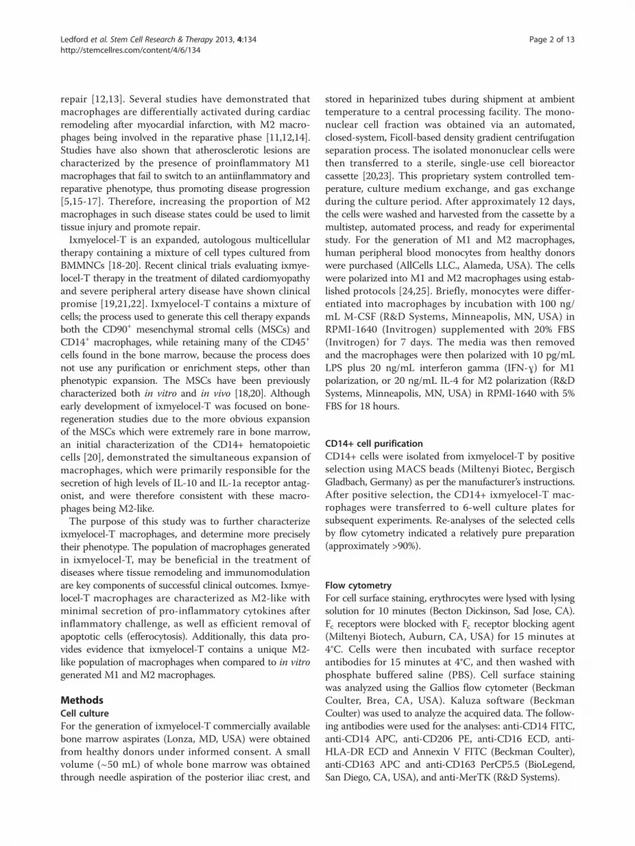
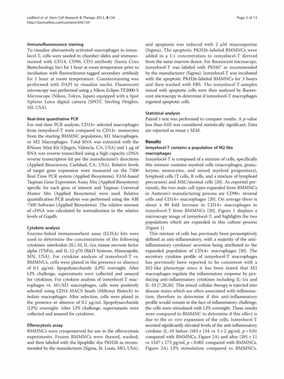
ixmyelocel-T macrophages express 2 well-known surfacereceptors of M2 macrophages: CD206 and CD163 [33].On average 17% of the cells in ixmyelocel-T expressCD206 (17 ± 0.6%) and 15% of the cells in ixmyelocel-Texpress CD163 (15 ± 1.3%). Since ixmyelocel-T consistsof a mixture of cells two markers of pro-inflammatorymacrophages were also examined. Flow cytometry analysisrevealed that the expression of the two pro-inflammatorymarkers associated withM1macrophages- CD16 (Figure 5A)and HLA-DR (Figure 5B) is minimal on ixmyelocel-Tmacrophages. These findings suggest that ixmyelocel-Tconsists mainly of macrophages with a M2-like phenotype.
NC IXT0
5
10
15
20
BMMNC IXT
SR-B
1 (R
elat
ive
Exp
ress
ion)
NC IXT0
5
10
15
20
25
30
BMMNC IXT
TG
Fβ
(Rel
ativ
e E
xpre
ssio
n)
**
*** ***
***
C
F
activated macrophage phenotype. Quantitative real-time PCRs within the ixmyelocel-T macrophages normalized to GAPDHrTK, (E) PPARγ, and (F) TGFβ in CD14+ ixmyelocel-T macrophages.yelocel-T, **P < 0.01 vs ixmyelocel-T, ***P < 0.001 vs ixmyelocel-T.
me proliferator-activated receptor gamma; TGFβ, transforming
CD14
Con
trol
CD14 CD14
CD
163
CD
206
A
Figure 4 Ixmyelocel-T macrophages express M2 macrophage surface receptors. (A) Ixmyelocel-T stained with the M2 macrophage markersCD163 and CD206.
Ledford et al. Stem Cell Research & Therapy 2013, 4:134 Page 6 of 13http://stemcellres.com/content/4/6/134
Ixmyelocel-T macrophages are a uniqueM2-like macrophageSince ixmyelocel-T macrophages were found to primarilyexpress surface expression of M2 markers, ixmyelocel-Tmacrophages were further characterized by direct com-parison to M1 and M2 macrophages generated in vitro.Quantitative real-time PCR analysis demonstrated that
Con
trol
Control CD
Con
trol
Con
trol
Control
Con
trol
C
A
B
Figure 5 Ixmyelocel-T macrophages do not express pro-inflammatorystained with the markers (A) CD16 and (B) HLA-DR.
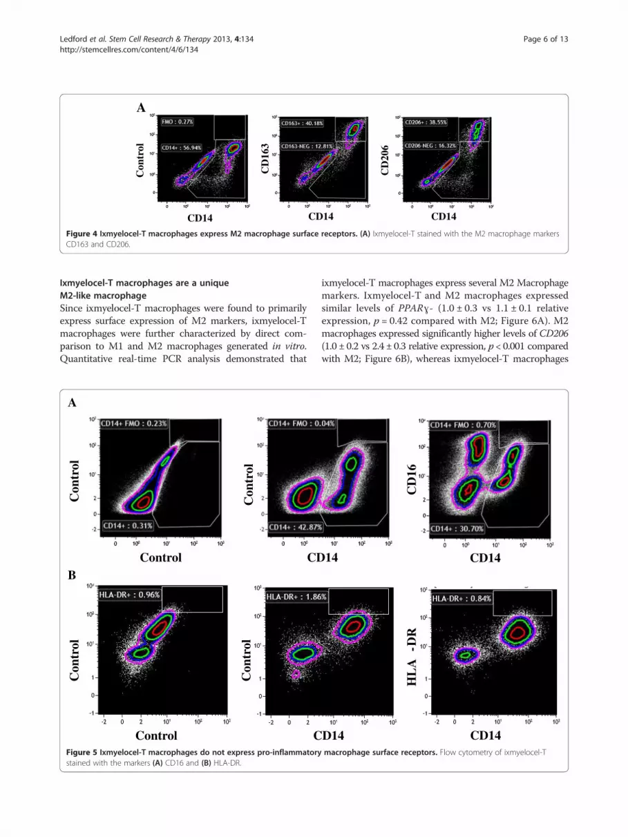
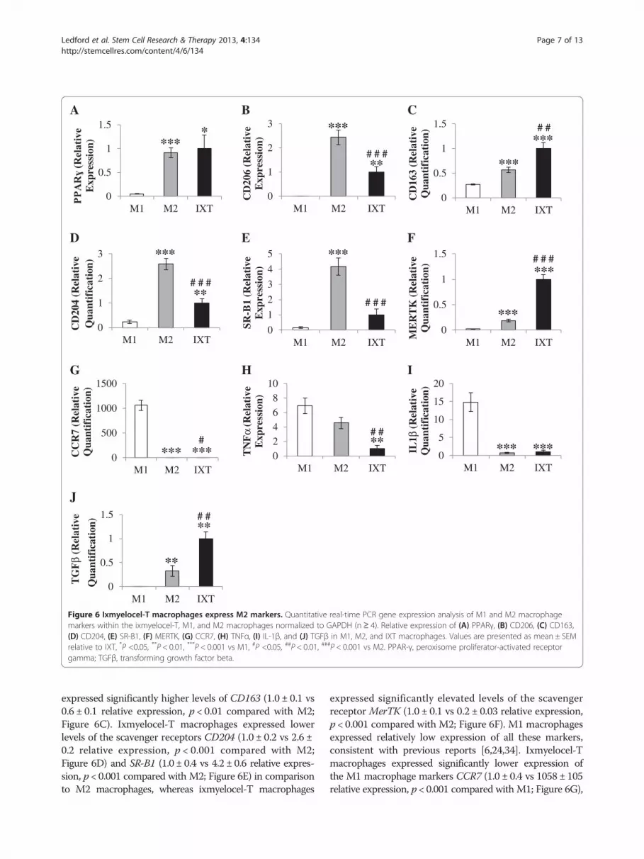
ixmyelocel-T macrophages express several M2 Macrophagemarkers. Ixmyelocel-T and M2 macrophages expressedsimilar levels of PPARɣ- (1.0 ± 0.3 vs 1.1 ± 0.1 relativeexpression, p = 0.42 compared with M2; Figure 6A). M2macrophages expressed significantly higher levels of CD206(1.0 ± 0.2 vs 2.4 ± 0.3 relative expression, p < 0.001 comparedwith M2; Figure 6B), whereas ixmyelocel-T macrophages
14 CD14
CD
16
D14
HL
A-D
R
CD14macrophage surface receptors. Flow cytometry of ixmyelocel-T
A
0
0.5
1
1.5
M1 M2 IXT
Exp
ress
ion)
0
0.5
1
1.5
M1 M2 IXT
ME
RT
K (R
elat
ive
Qua
ntif
icat
ion)
0
1
2
3
M1 M2 IXT
CD
204
(Rel
ativ
e Q
uant
ific
atio
n)
0
0.5
1
1.5
M1 M2 IXT
CD
163
(Rel
ativ
e Q
uant
ific
atio
n)
0
1
2
3
M1 M2 IXT
CD
206
(Rel
ativ
e E
xpre
ssio
n)
0
1
2
3
4
5
M1 M2 IXT
SR-B
1 (R
elat
ive
Exp
ress
ion)
0
0.5
1
1.5
M1 M2 IXT
TG
Fβ
(Rel
ativ
e
Qua
ntif
icat
ion)
0
5
10
15
20
M1 M2 IXT
IL1 β
(Rel
ativ
e Q
uant
ific
atio
n)
0
500
1000
1500
M1 M2 IXT
CC
R7
(Rel
ativ
e Q
uant
ific
atio
n)
02468
10
M1 M2 IXT
TN
Fα
(Rel
ativ
e E
xpre
ssio
n)
B C
D E F
G H I
J
**
# #
* ***
** # # #
*** *** # #
***
** # # #
***
# # #
*** *** # # #
***
*** #
*** ** # #
*** ***
**
Figure 6 Ixmyelocel-T macrophages express M2 markers. Quantitative real-time PCR gene expression analysis of M1 and M2 macrophagemarkers within the ixmyelocel-T, M1, and M2 macrophages normalized to GAPDH (n≥ 4). Relative expression of (A) PPARγ, (B) CD206, (C) CD163,(D) CD204, (E) SR-B1, (F) MERTK, (G) CCR7, (H) TNFα, (I) IL-1β, and (J) TGFβ in M1, M2, and IXT macrophages. Values are presented as mean ± SEMrelative to IXT, *P <0.05, **P < 0.01, ***P < 0.001 vs M1, #P <0.05, ##P < 0.01, ###P < 0.001 vs M2. PPAR-γ, peroxisome proliferator-activated receptorgamma; TGFβ, transforming growth factor beta.
Ledford et al. Stem Cell Research & Therapy 2013, 4:134 Page 7 of 13http://stemcellres.com/content/4/6/134
expressed significantly higher levels of CD163 (1.0 ± 0.1 vs0.6 ± 0.1 relative expression, p < 0.01 compared with M2;Figure 6C). Ixmyelocel-T macrophages expressed lowerlevels of the scavenger receptors CD204 (1.0 ± 0.2 vs 2.6 ±0.2 relative expression, p < 0.001 compared with M2;Figure 6D) and SR-B1 (1.0 ± 0.4 vs 4.2 ± 0.6 relative expres-sion, p < 0.001 compared with M2; Figure 6E) in comparisonto M2 macrophages, whereas ixmyelocel-T macrophages
expressed significantly elevated levels of the scavengerreceptor MerTK (1.0 ± 0.1 vs 0.2 ± 0.03 relative expression,p < 0.001 compared with M2; Figure 6F). M1 macrophagesexpressed relatively low expression of all these markers,consistent with previous reports [6,24,34]. Ixmyelocel-Tmacrophages expressed significantly lower expression ofthe M1 macrophage markers CCR7 (1.0 ± 0.4 vs 1058 ± 105relative expression, p < 0.001 compared with M1; Figure 6G),
Ledford et al. Stem Cell Research & Therapy 2013, 4:134 Page 8 of 13http://stemcellres.com/content/4/6/134
TNFα (1.0 ± 0.4 vs 6.9 ± 1.1 relative expression, p < 0.01compared with M1; Figure 6H), and IL-1β (1.0 ± 0.4 vs14.8 ± 2.6 relative expression, p < 0.001 compared with M1;Figure 6I). Additionally, ixmyelocel-T macrophages werefound to express elevated levels of the reparative cytokineTGF-β (Figure 6J). These findings suggest that ixmyelocel-Tmacrophages are mainly M2-like, as they share littlegene expression in common with M1 macrophages. Thesefinding also suggest that ixmyelocel-T macrophages havea unique M2-like profile since they express lower levels ofthe scavenger receptors CD204 and SR-B1, and elevatedexpression of CD163 and MerTK.M1 and M2 macrophage profiles are described as plastic,
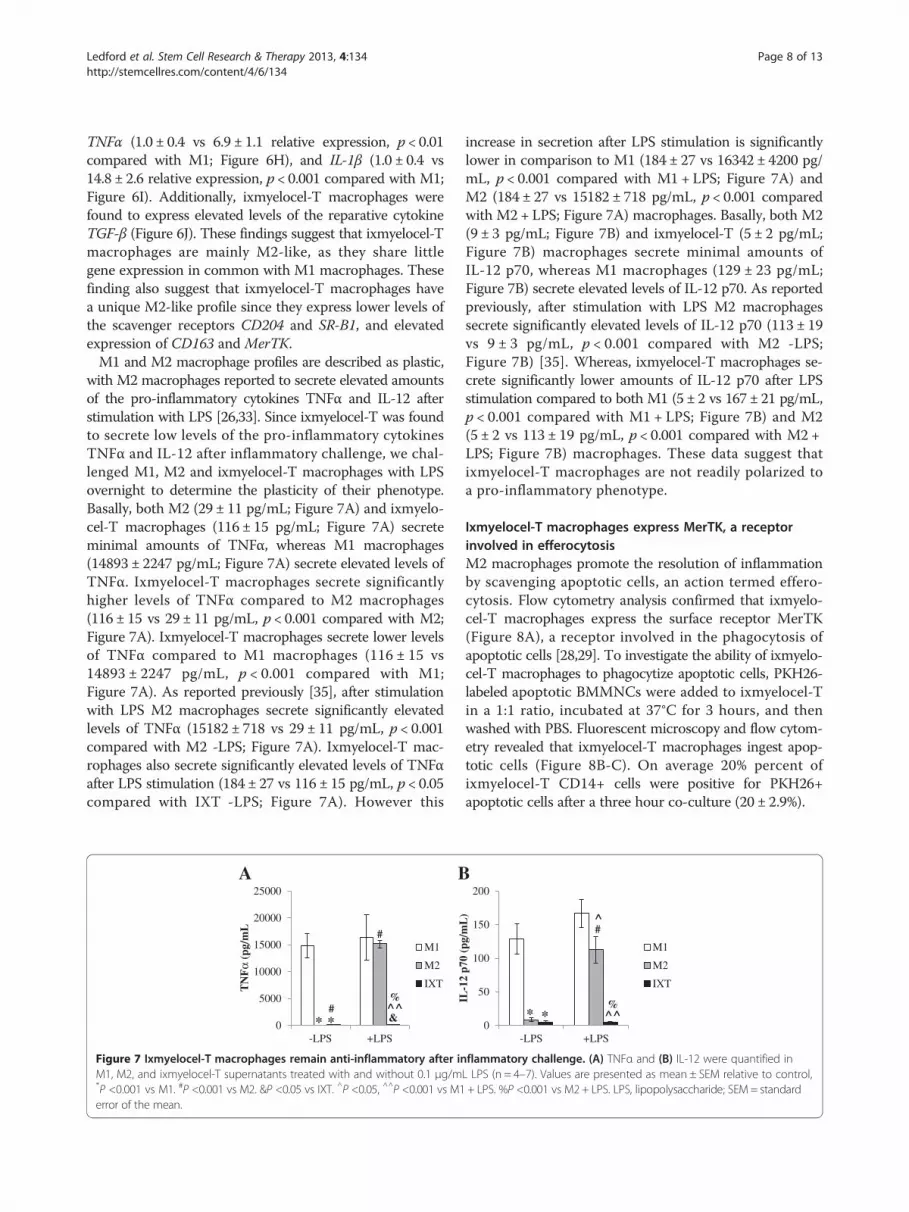
with M2 macrophages reported to secrete elevated amountsof the pro-inflammatory cytokines TNFα and IL-12 afterstimulation with LPS [26,33]. Since ixmyelocel-T was foundto secrete low levels of the pro-inflammatory cytokinesTNFα and IL-12 after inflammatory challenge, we chal-lenged M1, M2 and ixmyelocel-T macrophages with LPSovernight to determine the plasticity of their phenotype.Basally, both M2 (29 ± 11 pg/mL; Figure 7A) and ixmyelo-cel-T macrophages (116 ± 15 pg/mL; Figure 7A) secreteminimal amounts of TNFα, whereas M1 macrophages(14893 ± 2247 pg/mL; Figure 7A) secrete elevated levels ofTNFα. Ixmyelocel-T macrophages secrete significantlyhigher levels of TNFα compared to M2 macrophages(116 ± 15 vs 29 ± 11 pg/mL, p < 0.001 compared with M2;Figure 7A). Ixmyelocel-T macrophages secrete lower levelsof TNFα compared to M1 macrophages (116 ± 15 vs14893 ± 2247 pg/mL, p < 0.001 compared with M1;Figure 7A). As reported previously [35], after stimulationwith LPS M2 macrophages secrete significantly elevatedlevels of TNFα (15182 ± 718 vs 29 ± 11 pg/mL, p < 0.001compared with M2 -LPS; Figure 7A). Ixmyelocel-T mac-rophages also secrete significantly elevated levels of TNFαafter LPS stimulation (184 ± 27 vs 116 ± 15 pg/mL, p < 0.05compared with IXT -LPS; Figure 7A). However this
A
0
5000
10000
15000
20000
25000
-LPS +LPS
TN
Fα
(pg/
mL
M1
M2
IXT
B
* #
& *
#
%^^
Figure 7 Ixmyelocel-T macrophages remain anti-inflammatory after inM1, M2, and ixmyelocel-T supernatants treated with and without 0.1 μg/m*P <0.001 vs M1. #P <0.001 vs M2. &P <0.05 vs IXT. ^P <0.05, ^^P <0.001 vs M1error of the mean.
increase in secretion after LPS stimulation is significantlylower in comparison to M1 (184 ± 27 vs 16342 ± 4200 pg/mL, p < 0.001 compared with M1 + LPS; Figure 7A) andM2 (184 ± 27 vs 15182 ± 718 pg/mL, p < 0.001 comparedwith M2 + LPS; Figure 7A) macrophages. Basally, both M2(9 ± 3 pg/mL; Figure 7B) and ixmyelocel-T (5 ± 2 pg/mL;Figure 7B) macrophages secrete minimal amounts ofIL-12 p70, whereas M1 macrophages (129 ± 23 pg/mL;Figure 7B) secrete elevated levels of IL-12 p70. As reportedpreviously, after stimulation with LPS M2 macrophagessecrete significantly elevated levels of IL-12 p70 (113 ± 19vs 9 ± 3 pg/mL, p < 0.001 compared with M2 -LPS;Figure 7B) [35]. Whereas, ixmyelocel-T macrophages se-crete significantly lower amounts of IL-12 p70 after LPSstimulation compared to both M1 (5 ± 2 vs 167 ± 21 pg/mL,p < 0.001 compared with M1 + LPS; Figure 7B) and M2(5 ± 2 vs 113 ± 19 pg/mL, p < 0.001 compared with M2 +LPS; Figure 7B) macrophages. These data suggest thatixmyelocel-T macrophages are not readily polarized toa pro-inflammatory phenotype.
Ixmyelocel-T macrophages express MerTK, a receptorinvolved in efferocytosisM2 macrophages promote the resolution of inflammationby scavenging apoptotic cells, an action termed effero-cytosis. Flow cytometry analysis confirmed that ixmyelo-cel-T macrophages express the surface receptor MerTK(Figure 8A), a receptor involved in the phagocytosis ofapoptotic cells [28,29]. To investigate the ability of ixmyelo-cel-T macrophages to phagocytize apoptotic cells, PKH26-labeled apoptotic BMMNCs were added to ixmyelocel-Tin a 1:1 ratio, incubated at 37°C for 3 hours, and thenwashed with PBS. Fluorescent microscopy and flow cytom-etry revealed that ixmyelocel-T macrophages ingest apop-totic cells (Figure 8B-C). On average 20% percent ofixmyelocel-T CD14+ cells were positive for PKH26+apoptotic cells after a three hour co-culture (20 ± 2.9%).
0
50
100
150
200
-LPS +LPS
IL-1
2 p7
0 (p
g/m
L)
M1
M2
IXT
^
* *
#
%^^
flammatory challenge. (A) TNFα and (B) IL-12 were quantified inL LPS (n = 4–7). Values are presented as mean ± SEM relative to control,+ LPS. %P <0.001 vs M2 + LPS. LPS, lipopolysaccharide; SEM = standard

ixmyelocel-T Apoptotic Cellsixmyelocel-T + Apoptotic
Cells
A
B
CD14
Con
trol
CD14M
erT
K
C
CD14
PK
H26
CD14
PK
H26
CD14
PK
H26
Figure 8 Ixmyelocel-T alternatively activated macrophages readily phagocytize apoptotic cells. (A) Ixmyelocel-T stained with MerTK.(B-C) Adherent, healthy ixmyelocel-T stained with PKH67 was incubated with PKH26 labeled apoptotic cells. Apoptotic cells were washed away,and healthy ixmyelocel macrophages were analyzed using fluorescence microscopy and flow cytometry. On average 20% percent of ixmyelocel-TCD14+ cells were positive for PKH26+ apoptotic cells after a three hour co-culture apoptotic cells (20 ± 2.9%). (n≥ 5). Magnification: 60 ×.
Ledford et al. Stem Cell Research & Therapy 2013, 4:134 Page 9 of 13http://stemcellres.com/content/4/6/134
DiscussionIn this report, ixmyelocel-T was found to secrete signifi-cantly elevated levels of both IL-10 and IL-1ra beforeand after LPS stimulation, compared to BMMNCs. Add-itionally, both ixmyelocel-T and BMMNCs were found
to secrete minimal amounts of the pro-inflammatory cy-tokines TNFα and IL-12. After LPS challenge bothixmyelocel-T and BMMNCs secreted significantly moreTNFα, however this increase in secretion was significantlyhigher in BMMNCs. This expanded population of M2-like

Ledford et al. Stem Cell Research & Therapy 2013, 4:134 Page 10 of 13http://stemcellres.com/content/4/6/134
macrophages was also found to express significantlyelevated levels of the M2 markers CD206, CD163, SR-B1,MerTK, PPARɣ, and TGF-β. Together these findingssuggest that the 12 ± 1 day ex vivo expansion of autologousmarrow generates a population of M2-like macrophages inixmyelocel-T.Ixmyelocel-T macrophages were demonstrated to express
2 well-characterized surface markers of M2 macrophages,CD206 and CD163. Whereas surface expression of markersof pro-inflammatory macrophages, CD16 and HLA-DR[36,37], were found to be barely expressed. Studies havedemonstrated that macrophages with high expression ofCD16 efficiently produce pro-inflammatory cytokinesincluding TNFα and IL-12, while no or very little anti-inflammatory cytokines, whereas macrophages with lowexpression of CD16 produce low levels of pro-inflammatorycytokines and secrete elevated levels of anti-inflammatorycytokines such as IL-10 [37,38]. These findings suggest thatixmyelocel-T consists mainly of macrophages with anti-inflammatory M2-like phenotypes.In vivo, bone marrow-derived macrophages differentiate
from circulating peripheral blood monocytes after migra-tion into tissues, often in response to injury or insult.These macrophages can then be polarized into M1 or M2macrophages by their microenvironment [39]. As a cellulartherapy, ixmyelocel-T macrophages are generated ex vivofrom bone marrow and directly injected into areas whererepair is needed [20,22]. In order to further explore thedifferences between these cell phenotypes ixmyelocel-Tmacrophages were compared to M2 and M1 macrophages.Both Ixmyelocel-T and M2 macrophages were found toexpress similar levels of PPARɣ. Ixmyelocel-T macrophageswere found to express significantly lower levels of thescavenger receptors CD206, CD204, and SR-B1 comparedto M2 macrophages. However, ixmyelocel-T macrophageswere found to express significantly higher expression ofthe scavenger receptors CD163 and MerTK compared toM2 macrophages. Ixmyelocel-T macrophages were alsofound to have significantly higher expression of TGF-βcompared to M1 and M2 macrophages. When comparedto M1 macrophages both M2 and ixmyelocel-T macro-phages were found to express significantly lower levels ofthe M1 markers CCR7, TNFα, and IL-1β. Together thesedata suggest that ixmyelocel-T macrophages are moreM2-like. This data also suggests that ixmyelocel-T macro-phages may represent a unique M2-like macrophage, withslightly different expression of M2 markers. Future studieswill further examine the unique M2- like phenotype ofixmyelocel-T macrophages to determine just how similaror different they are to M2 macrophages.Macrophages are plastic cells that can switch from an
activated M1 state back to M2, and vice versa dependingon specific signals [26,33]. Previous reports have demon-strated that M2 macrophages can be polarized back to a
M1 state using pro-inflammatory stimuli such as LPS [35].To determine if ixmyelocel-T macrophages could also bepolarized back to a M1 state in a similar fashion M1, M2,and ixmyelocel-T macrophages were treated with LPS over-night. Before LPS stimulation both M2 and ixmyelocel-Tmacrophages secreted significantly less TNFα and IL-12p70 compared to M1 macrophages. However after over-night LPS stimulation, M2 macrophages secreted signifi-cantly elevated levels of TNFα and IL-12 p70 similar toM1 levels. This same amount of pro-inflammatory cytokinesecretion was not mimicked by ixmyelocel-T macrophages.While ixmyelocel-T macrophages did secrete significantlyelevated levels of TNFα after LPS stimulation, this amountof TNFα was significantly lower than that secreted by M2macrophages. Additionally, ixmyelocel-T macrophages se-creted significantly less IL-12 p70 after LPS stimulationcompared to M1 and M2 macrophages. These data suggestthat ixmyelocel-T macrophages are not readily polarized toa pro-inflammatory phenotype, suggesting that thesecells might be beneficial in the treatment of inflammatorydiseases. Future studies will further investigate this uniqueproperty of ixmyelocel-T macrophages to determine ifthey can affect the inflammatory state in vivo.M2 macrophages are potent phagocytes that favorably
bind and ingest early apoptotic cells [40]. Apoptotic cellsneed to be removed quickly to prevent the release oftissue-damaging intracellular components that can induceinflammatory responses [28,41]. Efferocytosis itself alsotriggers pro-resolving signals that promote dampeningof the immune response and restoring tissue homeostasis[14]. Defective clearance of apoptotic cells, or efferocytosis,is linked to the progression of several disease states includ-ing advanced atherosclerotic lesions, ischemic heart disease,and chronic wounds [28]. Ixmyelocel-T macrophages werefound to readily ingest apoptotic cells and express genes forthe scavenger receptors that promote the phagocytosis ofapoptotic cells. Of specific interest, ixmyelocel-T expressesthe scavenger receptor MerTK, which has been implicatedin mediating the anti-inflammatory clearance of apoptoticcells [29,42]. The expression of scavenger receptors andtheir M2-like phenotype predisposes ixmyelocel-T macro-phages to be potent phagocytes. This important functionof ixmyelocel-T macrophages could potentially promotehealing in disease states where efferocytosis is compro-mised. Specifically, advanced atherosclerotic lesions arecharacterized by defective efferocytosis, resulting in nec-rotic core formation [28,42]. In early atherosclerotic lesions,apoptotic cells are rapidly removed by macrophages, whichprevents the progression of lesions, but as the diseaseprogresses, apoptotic cells are not removed, resulting in apro-inflammatory cascade [28]. Efferocytosis is also requiredin healing the heart after ischemia where removing necroticdebris helps preserve the remaining cardiomyocytes [14].As such, the macrophages present in ixmyelocel-T may
Ledford et al. Stem Cell Research & Therapy 2013, 4:134 Page 11 of 13http://stemcellres.com/content/4/6/134
potentially promote tissue repair and regeneration throughthe removal of necrotic debris.Several different mechanisms have been reported in
the literature that may contribute to the development ofM2 macrophages, including ingestion of apoptotic cells,exposure to the Th2 cytokines IL-4 and IL-13, exposureto IL-10, and contact with mesenchymal stem cells[13,27,39,43]. The culture process used to generateixmyelocel-T results in a population of unique M2-likemacrophages. Th2 cytokines are not directly added toixmyelocel-T during the culture process to produceM2 macrophages [1,31,44]. However, due to the widemixture of cells found within ixmyelocel-T it is feasiblethat Th2 cytokines may be generated and secreted byother cells during this 12 ± 1 day process. Ixmyelocel-Tis composed of a mixture of cells including macrophages,granulocytes, monocytes, T cells, B cells, and MSC/stro-mal cells, and cytokine secretion from these different celltypes could influence the development of the M2-likephenotype [20]. For example, it has been previouslyreported that macrophages engage in bidirectional interac-tions with MSCs, resulting in MSC growth and M2phenotype polarization [7,12,45]. MSCs have been reportedto induce an IL-10 high and IL-12 low M2 phenotypein macrophages [39]. MSCs are expanded during theixmyelocel-T culture process; the interaction of thesecells with macrophages in culture could contribute to theM2-like population of cells found within ixmyelocel-T.Future studies will further examine the relationship be-tween these different cell types to determine if theyplay a role in the development of this M2-like populationof macrophages.Ixmyelocel-T is currently being evaluated in a Phase 2b
clinical trial of advanced heart failure. Tissue recovery afterinjury is a complex process involving an interplay betweenmacrophages, stem cells, and stromal cells to prevent tissuefibrosis, which can lead to ineffective tissue function [7].Recent work has highlighted the role of M2 macrophagesin these processes [7,17,46]. Macrophages are commonlyfound in association with areas of fibrosis in cardiac tissuein end-stage heart disease [9], and recent studies haveprovided evidence that M2-like macrophages have thecapacity to remove tissue debris and dampen inflammationin cardiac tissue promoting tissue repair [8]. Additionally,recent studies have found that macrophages found inhuman carotid artery atherosclerotic plaques are dominatedby a M1 phenotype [47], and that the number of pro-inflammatory M1 macrophages found in cardiac adiposetissue correlates with the severity of coronary artery ath-erosclerosis in human [12]. Therefore, ixmyelocel-T maypotentially promote tissue repair and regeneration in thesedisease states by providing a M2-like population of macro-phages described herein. The biological properties of thesemacrophages in ixmyelocel-T may potentially have clinical
utility for tissue repair and regeneration in the diseasestates where a population of M2 macrophages would becritical.This study further examined the expanded population
of macrophages found in ixmyelocel-T, since macrophageshave been reported to play specific and unique roles intissue regeneration and repair [7,17,46]. Several cellulartherapies are currently being explored for the treatment ofischemic cardiovascular diseases where tissue remodelingand immunomodulation are considered key componentsof successful clinical outcomes. BMMNCs, peripheral bloodmononuclear cells, endothelial progenitor cells (EPC), andMSCs are all currently being evaluated in the treatment ofischemic cardiovascular diseases [48]. Tissue regenerationis a complex process involving an interplay between mac-rophages, stem cells, and stromal cells to prevent tissuefibrosis, which can lead to ineffective tissue function, andit is hypothesized that a mixture of regenerative cells, ratherthan just a single cell type might be more advantageous[7,20,48,49]. Ixmyelocel-T consists of a mixture of cellsgenerated from BMMNCs, specifically an expandedpopulation of CD90+ stromal cells and CD14+ macro-phages which have been characterized with a M2-likephenotype [20]. It is thought that the mixture of cells foundin ixmyelocel-T might be more advantageous in long-termtissue regeneration and repair [20,48]. Future studies willfurther characterize the other cells types that make upixmyelocel-T in order to highlight the potential roles theseother subpopulations might play in tissue repair andregeneration.There are several limitations to this study. Mainly, the
findings and conclusions are based on in vitro experiments.The outcome of infiltrating macrophages to areas of inflam-matory injury is not fully understood as the inflammatoryenvironment may influence the outcome of the cells.Although we used human cells that would require the useof immune-compromised animals, in vivo experimentswould strengthen the findings. Additionally, the cells inthese experiments were obtained from healthy donors.It would be highly interesting to compare ixmyelocel-Tmacrophages to the peripheral blood macrophages (polar-ized M1 and M2) from a diseased patient. Studies have re-ported that several disease states, such as obesity and type2 diabetes, affect macrophage phenotype and polarizationin some cases limiting M2 polarization capacity [26,50].Therefore, it would useful to compare ixmyelocel-Tmacrophages to peripheral blood macrophages in clinicallyrelevant states; especially disease states where ixmyelocel-Tis injected as a treatment.
ConclusionOur data demonstrate that ixmyelocel-T therapy containsa unique population of M2-like macrophages that arecharacterized by secretion of anti-inflammatory cytokines
Ledford et al. Stem Cell Research & Therapy 2013, 4:134 Page 12 of 13http://stemcellres.com/content/4/6/134
and expression of M2 markers- CD206 and CD163.Furthermore, these cells are involved in efficient removalof apoptotic cells and have elevated expression of MerTK,which is imperative in limiting tissue injury and promotingrepair. The biological properties of the M2-like macro-phages in ixmyelocel-T may have clinical utility for tissuerepair and regeneration in the disease states where a popu-lation of M2 macrophages would be critical.
Abbreviations7-AAD: 7-amino-actinomycin D; BMMNC: Bone marrow blood mononuclearcell; cDNA: Complementary DNA; DAPI: 4′-6-diamidino-2-phenylindole;DMSO: Dimethyl sulfoxide; DNA: Deoxyribonucleic acid; ELISA: Enzyme-linkedimmunosorbent assay; EPC: Endothelial progenitor cell; FAM: 6-carboxyyfluorescein; GAPDH: Glyceraldehyde 3-phosphate dehydrogenase;IL: Interleukin; LPS: Lipopolysaccharide; MSCs: Mesenchymal stromal cells;PBS: Phosphate buffered saline; PCR: Polymerase chain reaction; PPAR-γ: Peroxisome proliferator-activated receptor gamma; RNA: Ribonucleic acid;SR-A: Scavenger receptor-A; TGF-β: Transforming growth factor; Th1: T helper1; Th2: T helper 2; TNF-α: Tumor necrosis factor alpha.
Competing interestsAll authors are employees of Aastrom Biosciences, Inc.
Authors’ contributionsKL conceived and designed research, acquired data (RT-PCR, efferocytosisanalysis, ELISA), analyzed and interpreted data, performed statistical analysis,and drafted the manuscript. FZ provided conceptual advice, analyzed data,and participated in the discussion of results. RB contributed to the scientificdirection, experimental approach, and interpretation of results. All authorsread and approved the final manuscript.
AcknowledgementsThe authors would like to thank Alden Wong, Judith Schmitt, Hillary Evens,John Osborne, and Nikki Murphy for excellent technical assistance and ChisParrish for his expertise with flow cytometry. Scientific editorial support wassponsored by Aastrom Biosciences, Inc, and provided by David E. Kaminsky,PhD, of AlphaBioCom, LLC.
Received: 19 March 2013 Revised: 26 August 2013Accepted: 23 October 2013 Published: 1 November 2013
References1. Bouhlel MA, Derudas B, Rigamonti E, Dievart R, Brozek J, Haulon S, Zawadzki
C, Jude B, Torpier G, Marx N, Staels B, Chinetti-Gbaguidi G: PPARgammaactivation primes human monocytes into alternative M2 macrophageswith anti-inflammatory properties. Cell Metab 2007, 6:137–143.
2. Mantovani A, Sica A, Locati M: New vistas on macrophage differentiationand activation. Eur J Immunol 2007, 37:14–16.
3. Jennewein C, Kuhn AM, Schmidt MV, Meilladec-Jullig V, von Knethen A,Gonzalez FJ, Brune B: Sumoylation of peroxisome proliferator-activatedreceptor gamma by apoptotic cells prevents lipopolysaccharide-inducedNCoR removal from kappaB binding sites mediating transrepression ofproinflammatory cytokines. J Immunol 2008, 181:5646–5652.
4. Mosser DM: The many faces of macrophage activation. J Leukoc Biol 2003,73:209–212.
5. Deonarine K, Panelli MC, Stashower ME, Jin P, Smith K, Slade HB, NorwoodC, Wang E, Marincola FM, Stroncek DF: Gene expression profiling ofcutaneous wound healing. J Transl Med 2007, 5:11.
6. Mantovani A, Garlanda C, Locati M: Macrophage diversity and polarizationin atherosclerosis: a question of balance. Arterioscler Thromb Vasc Biol2009, 29:1419–1423.
7. Ortega-Gomez A, Perretti M, Soehnlein O: Resolution of inflammation: anintegrated view. EMBO Mol Med 2013, 5:661–674.
8. Pinto AR, Paolicelli R, Salimova E, Gospocic J, Slonimsky E, Bilbao-Cortes D,Godwin JW, Rosenthal NA: An abundant tissue macrophage population inthe adult murine heart with a distinct alternatively-activated macrophageprofile. PLoS One 2012, 7:e36814.
9. Meznarich J, Malchodi L, Helterline D, Ramsey SA, Bertko K, Plummer T,Plawman A, Gold E, Stempien-Otero A: Urokinase plasminogen activatorinduces pro-fibrotic/m2 phenotype in murine cardiac macrophages.PLoS One 2013, 8:e57837.
10. Khallou-Laschet J, Varthaman A, Fornasa G, Compain C, Gaston AT,Clement M, Dussiot M, Levillain O, Graff-Dubois S, Nicoletti A, Caligiuri G:Macrophage plasticity in experimental atherosclerosis. PLoS One 2010,5:e8852.
11. Frangogiannis NG: Regulation of the inflammatory response in cardiacrepair. Circ Res 2012, 110:159–173.
12. Adutler-Lieber S, Ben-Mordechai T, Naftali-Shani N, Asher E, Loberman D,Raanani E, Leor J: Human macrophage regulation via interaction withcardiac adipose tissue-derived mesenchymal stromal cells. J CardiovascPharmacol Ther 2013, 18:78–86.
13. Kharraz Y, Guerra J, Mann CJ, Serrano AL, Munoz-Canoves P: Macrophageplasticity and the role of inflammation in skeletal muscle repair.Mediators Inflamm 2013, 2013:491497.
14. Thorp EB: Contrasting Inflammation Resolution during Atherosclerosisand Post Myocardial Infarction at the Level of Monocyte/MacrophagePhagocytic Clearance. Front Immunol 2012, 31:1–8.
15. Khanna S, Biswas S, Shang Y, Collard E, Azad A, Kauh C, Bhasker V,Gordillo GM, Sen CK, Roy S: Macrophage dysfunction impairsresolution of inflammation in the wounds of diabetic mice.PLoS One 2010, 5:e9539.
16. Waldo SW, Li Y, Buono C, Zhao B, Billings EM, Chang J, Kruth HS:Heterogeneity of human macrophages in culture and in atheroscleroticplaques. Am J Pathol 2008, 172:1112–1126.
17. Sindrilaru A, Peters T, Wieschalka S, Baican C, Baican A, Peter H, Hainzl A,Schatz S, Qi Y, Schlecht A, Weiss JM, Wlaschek M, Sunderkotter C,Scharffetter-Kochanek K: An unrestrained proinflammatory M1 macrophagepopulation induced by iron impairs wound healing in humans and mice.J Clin Invest 2011, 121:985–997.
18. Yin D, Wang Z, Gao Q, Sundaresan R, Parrish C, Yang Q, Krebsbach PH,Lichtler AC, Rowe DW, Hock J, Liu P: Determination of the fate andcontribution of ex vivo expanded human bone marrow stem andprogenitor cells for bone formation by 2.3ColGFP. Mol Ther 2009,17:1967–1978.
19. Powell RJ, Comerota AJ, Berceli SA, Guzman R, Henry TD, Tzeng E,Velazquez O, Marston WA, Bartel RL, Longcore A, Stern T, Watling S: Interimanalysis results from the RESTORE-CLI, a randomized, double-blindmulticenter phase II trial comparing expanded autologous bonemarrow-derived tissue repair cells and placebo in patients with criticallimb ischemia. J Vasc Surg 2011.
20. Bartel RL, Cramer C, Ledford K, Longcore A, Parrish C, Stern T, Watling S,Zeigler F: The Aastrom experience. Stem Cell Res Ther 2012, 3:26.
21. Comerota AJ, Link A, Douville J, Burchardt ER: Upper extremity ischemiatreated with tissue repair cells from adult bone marrow. J Vasc Surg 2010,52:723–729.
22. Powell RJ, Marston WA, Berceli SA, Guzman R, Henry TD, Longcore AT, SternTP, Watling S, Bartel RL: Cellular therapy with Ixmyelocel-T to treat criticallimb ischemia: the randomized, double-blind, placebo-controlledRESTORE-CLI trial. Mol Ther 2012, 20:1280–1286.
23. Gastens MH, Goltry K, Prohaska W, Tschope D, Stratmann B, Lammers D,Kirana S, Gotting C, Kleesiek K: Good manufacturing practice-compliantexpansion of marrow-derived stem and progenitor cells for cell therapy.Cell Transplant 2007, 16:685–696.
24. Martinez FO, Gordon S, Locati M, Mantovani A: Transcriptional profiling ofthe human monocyte-to-macrophage differentiation and polarization:new molecules and patterns of gene expression. J Immunol 2006,177:7303–7311.
25. Hirose K, Iwabuchi K, Shimada K, Kiyanagi T, Iwahara C, Nakayama H, DaidaH: Different responses to oxidized low-density lipoproteins in humanpolarized macrophages. Lipids Health Dis 2011, 10:1.
26. Bories G, Caiazzo R, Derudas B, Copin C, Raverdy V, Pigeyre M, Pattou F,Staels B, Chinetti-Gbaguidi G: Impaired alternative macrophage differentiationof peripheral blood mononuclear cells from obese subjects. Diab Vasc DisRes 2012, 9:189–195.
27. Mosser DM, Edwards JP: Exploring the full spectrum of macrophageactivation. Nat Rev Immunol 2008, 8:958–969.
28. Thorp EB: Mechanisms of failed apoptotic cell clearance by phagocytesubsets in cardiovascular disease. Apoptosis 2010, 15:1124–1136.

Ledford et al. Stem Cell Research & Therapy 2013, 4:134 Page 13 of 13http://stemcellres.com/content/4/6/134
29. Zizzo G, Hilliard BA, Monestier M, Cohen PL: Efficient Clearance of EarlyApoptotic Cells by Human Macrophages Requires M2c Polarization andMerTK Induction. J Immunol 2012, 189:3508–3520.
30. Gallardo-Soler A, Gomez-Nieto C, Campo ML, Marathe C, Tontonoz P,Castrillo A, Corraliza I: Arginase I induction by modified lipoproteins inmacrophages: a peroxisome proliferator-activated receptor-gamma/delta-mediated effect that links lipid metabolism and immunity.Mol Endocrinol 2008, 22:1394–1402.
31. Gordon S: Alternative activation of macrophages. Nat Rev Immunol 2003,3:23–35.
32. Laskin DL: Macrophages and inflammatory mediators in chemicaltoxicity: a battle of forces. Chem Res Toxicol 2009, 22:1376–1385.
33. Porcheray F, Viaud S, Rimaniol AC, Leone C, Samah B, Dereuddre-Bosquet N,Dormont D, Gras G: Macrophage activation switching: an asset for theresolution of inflammation. Clin Exp Immunol 2005, 142:481–489.
34. Badylak SF, Valentin JE, Ravindra AK, McCabe GP, Stewart-Akers AM: Macrophagephenotype as a determinant of biologic scaffold remodeling. Tissue Eng Part A2008, 14:1835–1842.
35. Zheng XF, Hong YX, Feng GJ, Zhang GF, Rogers H, Lewis MA, Williams DW,Xia ZF, Song B, Wei XQ: Lipopolysaccharide-induced M2 to M1macrophage transformation for IL-12p70 production is blocked byCandida albicans mediated up-regulation of EBI3 expression.PLoS One 2013, 8:e63967.
36. Ohri CM, Shikotra A, Green RH, Waller DA, Bradding P: The tissuemicrolocalisation and cellular expression of CD163, VEGF, HLA-DR, iNOS,and MRP 8/14 is correlated to clinical outcome in NSCLC. PLoS One 2011,6:e21874.
37. Hasan D, Chalouhi N, Jabbour P, Hashimoto T: Macrophage imbalance(M1 vs. M2) and upregulation of mast cells in wall of rupturedhuman cerebral aneurysms: preliminary results. J Neuroinflammation2012, 9:222.
38. Belge KU, Dayyani F, Horelt A, Siedlar M, Frankenberger M, Frankenberger B,Espevik T, Ziegler-Heitbrock L: The proinflammatory CD14 + CD16 + DR++monocytes are a major source of TNF. J Immunol 2002, 168:3536–3542.
39. Kim J, Hematti P: Mesenchymal stem cell-educated macrophages: a noveltype of alternatively activated macrophages. Exp Hematol 2009,37:1445–1453.
40. Xu W, Roos A, Schlagwein N, Woltman AM, Daha MR, van Kooten C:IL-10-producing macrophages preferentially clear early apoptoticcells. Blood 2006, 107:4930–4937.
41. Bottcher A, Gaipl US, Furnrohr BG, Herrmann M, Girkontaite I, Kalden JR, VollRE: Involvement of phosphatidylserine, alphavbeta3, CD14, CD36, andcomplement C1q in the phagocytosis of primary necrotic lymphocytesby macrophages. Arthritis Rheum 2006, 54:927–938.
42. Thorp E, Tabas I: Mechanisms and consequences of efferocytosis inadvanced atherosclerosis. J Leukoc Biol 2009, 86:1089–1095.
43. Weigert A, Jennewein C, Brune B: The liaison between apoptotic cells andmacrophages–the end programs the beginning. Biol Chem 2009,390:379–390.
44. Martinez FO, Helming L, Gordon S: Alternative activation of macrophages:an immunologic functional perspective. Annu Rev Immunol 2009,27:451–483.
45. Abumaree MH, Al Jumah MA, Kalionis B, Jawdat D, Al Khaldi A, AbomarayFM, Fatani AS, Chamley LW, Knawy BA: Human Placental MesenchymalStem Cells (pMSCs) Play a Role as Immune Suppressive Cells by ShiftingMacrophage Differentiation from Inflammatory M1 to Anti-inflammatoryM2 Macrophages. Stem Cell Rev 2013, 9:620–641.
46. Saclier M, Yacoub-Youssef H, Mackey AL, Arnold L, Ardjoune H, Magnan M,Sailhan F, Chelly J, Pavlath GK, Mounier R, Kjaer M, Chazaud B: Differentiallyactivated macrophages orchestrate myogenic precursor cell fate duringhuman skeletal muscle regeneration. Stem Cells 2013, 31:384–396.
47. Shaikh S, Brittenden J, Lahiri R, Brown PA, Thies F, Wilson HM: Macrophagesubtypes in symptomatic carotid artery and femoral artery plaques.Eur J Vasc Endovasc Surg 2012, 44:491–497.
48. Bartel RL, Booth E, Cramer C, Ledford K, Watling S, Zeigler F: From bench tobedside: review of gene and cell-based therapies and the slow advancementinto Phase 3 Clinical Trials, with a focus on Aastrom’s Ixmyelocel-T. Stem CellRev 2013, 9:373–383.
49. van Weel V, van Tongeren RB, van Hinsbergh VW, van Bockel JH, Quax PH:Vascular growth in ischemic limbs: a review of mechanisms and possibletherapeutic stimulation. Ann Vasc Surg 2008, 22:582–597.
50. Fadini GP, de Kreutzenberg SV, Boscaro E, Albiero M, Cappellari R, Krankel N,Landmesser U, Toniolo A, Bolego C, Cignarella A, Seeger F, Dimmeler S,Zeiher A, Agostini C, Avogaro A: An unbalanced monocyte polarisationin peripheral blood and bone marrow of patients with type 2diabetes has an impact on microangiopathy. Diabetologia 2013,56:1856–1866.
doi:10.1186/scrt345Cite this article as: Ledford et al.: Ixmyelocel-T, an expandedmulticellular therapy, contains a unique population of M2-like macro-phages. Stem Cell Research & Therapy 2013 4:134.
Submit your next manuscript to BioMed Centraland take full advantage of:
• Convenient online submission
• Thorough peer review
• No space constraints or color figure charges
• Immediate publication on acceptance
• Inclusion in PubMed, CAS, Scopus and Google Scholar
• Research which is freely available for redistribution
Submit your manuscript at www.biomedcentral.com/submit


