Embed Size (px)
Citation preview
Isoform-Specific Upregulation of Palladin in Human andMurine Pancreas TumorsSilvia M. Goicoechea1*, Brian Bednarski2,3, Christianna Stack1, David W. Cowan3, Keith Volmar4, Leigh
Thorne3,4, Edna Cukierman5, Anil K. Rustgi6, Teresa Brentnall7, Rosa F. Hwang8, Christopher A. G.
McCulloch9, Jen Jen Yeh2,3, David J. Bentrem10, Steven N. Hochwald11, Sunil R. Hingorani12, Hong Jin
Kim2,3., Carol A. Otey1,3.
1 Department of Cell and Molecular Physiology, University of North Carolina, Chapel Hill, North Carolina, United States of America, 2 Division of Surgical Oncology,
Department of Surgery, University of North Carolina, Chapel Hill, North Carolina, United States of America, 3 Lineberger Comprehensive Cancer Center, Chapel Hill, North
Carolina, United States of America, 4 Department of Pathology and Laboratory Medicine, University of North Carolina, Chapel Hill, North Carolina, United States of
America, 5 Cancer Biology Program, Fox Chase Cancer Center, Philadelphia, Pennsylvania, United States of America, 6 Departments of Medicine and Genetics, Abramson
Cancer Center, University of Pennsylvania, Philadelphia, Pennsylvania, United States of America, 7 Department of Medicine, University of Washington, Seattle, Washington,
United States of America, 8 Department of Surgical Oncology, M. D. Anderson Cancer Center, University of Texas, Houston, Texas, United States of America, 9 CIHR Group
in Matrix Dynamics, University of Toronto, Toronto, Canada, 10 Surgical Oncology, Northwestern Medical Faculty Foundation, Chicago, Illinois, United States of America,
11 Division of Surgical Oncology, University of Florida, Gainesville, Florida, United States of America, 12 Fred Hutchinson Cancer Research Center and University of
Washington, Seattle, Washington, United States of America
Abstract
Pancreatic ductal adenocarcinoma (PDA) is a lethal disease with a characteristic pattern of early metastasis, which is drivinga search for biomarkers that can be used to detect the cancer at an early stage. Recently, the actin-associated proteinpalladin was identified as a candidate biomarker when it was shown that palladin is mutated in a rare inherited form of PDA,and overexpressed in many sporadic pancreas tumors and premalignant precursors. In this study, we analyzed theexpression of palladin isoforms in murine and human PDA and explored palladin’s potential use in diagnosing PDA. Weperformed immunohistochemistry and immunoblot analyses on patient samples and tumor-derived cells using an isoform-selective monoclonal antibody and a pan-palladin polyclonal antibody. Immunoblot and real-time quantitative reversetranscription-PCR were used to quantify palladin mRNA levels in human samples. We show that there are two major palladinisoforms expressed in pancreas: 65 and 85–90 kDa. The 65 kDa isoform is expressed in both normal and neoplastic ductalepithelial cells. The 85–90 kDa palladin isoform is highly overexpressed in tumor-associated fibroblasts (TAFs) in bothprimary and metastatic tumors compared to normal pancreas, in samples obtained from either human patients orgenetically engineered mice. In tumor-derived cultured cells, expression of palladin isoforms follows cell-type specificpatterns, with the 85–90 kDa isoform in TAFs, and the 65 kDa isoform predominating in normal and neoplastic epithelialcells. These results suggest that upregulation of 85–90 kDa palladin isoform may play a role in the establishment of the TAFphenotype, and thus in the formation of a desmoplastic tumor microenvironment. Thus, palladin may have a potential usein the early diagnosis of PDA and may have much broader significance in understanding metastatic behavior.
Citation: Goicoechea SM, Bednarski B, Stack C, Cowan DW, Volmar K, et al. (2010) Isoform-Specific Upregulation of Palladin in Human and Murine PancreasTumors. PLoS ONE 5(4): e10347. doi:10.1371/journal.pone.0010347
Editor: Daniela Cimini, Virginia Tech, United States of America
Received December 17, 2009; Accepted March 30, 2010; Published April 26, 2010
Copyright: � 2010 Goicoechea et al. This is an open-access article distributed under the terms of the Creative Commons Attribution License, which permitsunrestricted use, distribution, and reproduction in any medium, provided the original author and source are credited.
Funding: Elsa U. Pardee Foundation, http://www.pardeefoundation.org/ (C.A. Otey and H.J. Kim), UNC Center for Gastrointestinal Biology and Disease, https://cgibd.med.unc.edu/index.php (C.A. Otey), National Institutes of Health (NIH) R01-GM081505 (C.A. Otey), National Cancer Institute (NCI)/NIH CA113451 and Fox Chase CancerCenter’s (FCCC) Kidney Keystone Program Grant (E. Cukierman). Mead Foundation, http://gileswmeadfoundation.org/; Rosenzweig Foundation, http://jrpancan.org/;and PanCAN, http://www.pancan.org/ (S. Hingorani), NIH, http://www.nih.gov/ to H.J. Kim (K08-CA098240), B. Bednarski (T32-CA09688), S. Hingorani (CA15704,CA114028, CA129357) and A. Rustigi (DK0606994). CIHR, http://www.cihr-irsc.gc.ca/ operating grant MOP-36332 (C.A.G. McCulloch). UNC SPORE in GastrointestinalCancers, http://cancer.med.unc.edu/patient/programs/gi.asp P50-CA10699 (H.J. Kim). NCI SPORE in Gastrointestinal Cancers, http://spores.nci.nih.gov/current/gi/index.htm P50-CA106991 (Jen Jjen Yeh). The funders had no role in study design, data collection and analysis, decision to publish, or preparation of the manuscript.
Competing Interests: The authors have declared that no competing interests exist.
* E-mail: [email protected]
. These authors contributed equally to this work.
Introduction
Pancreatic adenocarcinoma is the fourth leading cause of cancer
death in the United States [1]. This disease has an exceptionally
high lethality rate, due to its aggressive metastasis and the low
probability of diagnosis at an early stage. Approximately 80–90%
of patients with pancreatic cancer present with locally-advanced,
unresectable tumors or metastatic disease at the time of initial
diagnosis [2,3]. The dismal prognosis associated with pancreatic
adenocarcinoma has driven a search to identify the aberrant
signaling pathways that contribute to the development, growth,
and invasion of this disease, with the ultimate goal of developing
novel diagnostic biomarkers and effective targeted therapies [4].
Palladin is a cytoskeleton-associated scaffold protein that has
received attention recently in the pancreas cancer field [5,6].
Palladin’s function in normal cells has been defined previously by
PLoS ONE | www.plosone.org 1 April 2010 | Volume 5 | Issue 4 | e10347

knockdown and overexpression experiments in cultured cell
models, and it is clear that palladin is critically involved in actin-
dependent behaviors such as cell motility and contractility
[7,8,9,10]. In animal studies, palladin is upregulated during
wound-healing [11,12,13], and it is required for normal
mammalian embryogenesis [14]. In human breast cancer cells,
high levels of palladin expression are associated with increased
invasiveness [15,16], which suggests the possibility that abnormal-
ities in palladin expression or function might contribute to the
disregulated motility of metastatic cancer cells. Palladin’s precise
role in pancreas cancer has not yet been defined; however, a
mutation in the human palladin gene is associated with a rare
form of familial pancreatic cancer. Palladin was found to be
overexpressed in samples of sporadic pancreatic adenocarcinoma
and in tumor-derived cell lines [5]. These results were challenged
by a subsequent study that utilized immunohistochemical (IHC)
staining of a pancreas tumor array [6]. Although the follow-up
study confirmed that palladin is overexpressed in 96% of pancreas
tumors as compared to normal pancreas, it showed that palladin is
upregulated in stromal fibroblasts rather than in the neoplastic
cells of pancreas tumors [6]. Thus, the results provide evidence
that palladin is overexpressed specifically in pancreas tumors, yet
the identity of the cell type that is responsible for upregulating
palladin in these tumors remains unclear.
Palladin exists in all vertebrates as multiple size variants
generated from a single gene that possesses alternative promoters,
i.e. a ‘‘nested gene’’. There are three major palladin isoforms that
arise from alternative start sites (85–90, 140 and 200 kDa) and
multiple minor isoforms that result from alternative splicing
[10,17,18,19]. This rich diversity of isoforms raises the possibility
that human cells may express palladin variants that are not
detected by all antibodies, which could be the cause of previous
conflicting results. In the current study, w e set out to perform
Western blot analysis and immunohistochemical staining of
pancreatic tumors and tumor-derived cells using both isoform-
selective monoclonal and pan-palladin polyclonal antibodies, to
identify all of the palladin isoforms that are associated with the
different cell types found in pancreatic tumors. We show that there
are two major isoforms of palladin in pancreatic tumors: 65 kDa
and 85–90 kDa. PDA expresses predominantly the 85–90 kDa
isoform of palladin, while normal pancreas and non-PDA tumors
both express the 65 kDa isoform. We also show that palladin
overexpression occurs primarily in tumor-associated fibroblasts
(TAFs), and not the neoplastic epithelial cells, of human pancreatic
tumors. These results suggest the possibility that upregulation of
85–90 kDa palladin may be a critical step in the acquisition of the
activated fibroblast phenotype, which is key to the formation of a
pro-invasive tumor microenvironment.
Results
Human Palladin Isoforms are DiversePalladin is a highly conserved protein that is found in all
vertebrate species, and it arises from a single gene in mice and
humans (PALLD). The palladin gene is unusually large and
complex, spanning ,400 kb and containing at least 25 exons, and
it produces multiple palladin isoforms, including three well-
described size variants that resolve at 85–90, 140 and 200 kDa
[10,17,18,19]. In addition to the three classical palladin isoforms,
size variants resolving at ,115, 75, 65 and 50 kDa have been
reported in previous studies [6,10,14,19], suggesting that palladin
isoforms are more diverse than previously suspected. This
motivated us to carefully assess the existing databases of full-
length transcripts to look for evidence of additional palladin
isoforms. Examination of the Universal Protein database revealed
the existence of seven palladin variants in humans (http://beta.
uniprot.org/uniprot/Q8WX93), and alignment of their sequences
is shown in Figure 1A. It has been noted previously that some
palladin isoforms display a higher molecular weight by SDS-
PAGE (apparent MW) than would be predicted based upon their
sequence, and this is believed to be a consequence of their high
proline content, and this is reflected in Figure 1A. Palladin
contains two different conserved domains that contribute to its
role as an actin-binding scaffolding molecule: proline-rich
domains that are hot-spots for protein-protein interaction, and
Ig domains that mediate F-actin crosslinking [15]. Since the
various palladin isoforms each possess different combinations of
proline-rich and Ig domains, it is likely that they serve distinct
cellular functions. Therefore, we went on to determine which
palladin isoforms are expressed in pancreas, and if these palladin
isoforms follow cell-type specific patterns of expression in
pancreatic cells.
Two previous studies have explored palladin expression in
cultured cells derived from pancreas tumors, and both focused on
the widely-expressed 85–90 kDa palladin isoform [5,6], yet these
previous studies also presented evidence that pancreatic tumor-
derived cell types express smaller, uncharacterized palladin
variants (see, for example, the 75 kDa band in Fig. 2 of reference
6). Neither previous study investigated palladin expression in bulk
tumor samples, which contain a complex mixture of different cell
types. Therefore, we used Western blot analysis of donated patient
samples to determine which palladin isoforms are upregulated in
human tumors, and if palladin levels are associated with the
invasiveness of the tumors. For these experiments, we analyzed
samples of normal pancreas (Fig. 1B, lanes 1, 2) and primary
pancreatic ductal adenocarcinomas (PDA, lanes 3–5), and also two
different types of non-PDA tumors: a solid pseudopapillary tumor
of the pancreas (a tumor characterized as having lower metastatic
potential and favorable prognosis even with large primary tumors)
and a neuroendocrine carcinoma (which was diagnosed and
resected at an early, non-invasive stage). Lysates of these tumors
are shown in lanes 6 and 7, respectively, in Figure 1B. A polyclonal
palladin antibody designated 622 and an isoform-selective
monoclonal antibody called 1E6 (see Figure 1A) were used to
stain parallel immunoblots. Immunodetection with the 622
polyclonal revealed that normal pancreas and non-PDA tumors
both express an isoform resolving at ,65 kDa, while PDAs
express predominantly a palladin band resolving near 85–90 kDa
and a trace amount of the 65 kDa isoform. When parallel
immunoblots were stained with the 1E6 monoclonal, only the 85–
90 kDa band was detected (140 and 200 kDa palladin isoforms are
not expressed in pancreas). This is consistent with previous studies,
in which the 1E6 monoclonal was found to be selective for the 85–
90 kDa isoform of palladin [10,18]. It is important to note that the
upregulation of 85–90 kDa palladin shown in Figure 1B is
associated specifically with PDAs, which are both highly invasive
and strongly desmoplastic, and not with tumor types that are only
moderately invasive and associated with a less robust expansion of
the stroma. To confirm these results, we performed RT-qPCR in
an independent set of samples. The primers used for this study
should, in theory, recognize all palladin isoforms that contain the
Ig4 domain (i.e., all except isoform #6). Consistent with the
immunoblots results, Figure 1C shows that the levels of palladin
mRNA in PDA samples are highly elevated compared with
normal pancreas. This demonstrates that western blot and RT-
qPCR are feasible methods for quantifying the 85–90 kDa isoform
of palladin (protein and mRNA) and thus, may have a potential
use in the diagnosis of pancreatic ductal adenocarcinoma.
Palladin in Pancreatic TAFs
PLoS ONE | www.plosone.org 2 April 2010 | Volume 5 | Issue 4 | e10347

Figure 1. Analysis of human palladin isoforms in pancreatic tissues. A. Human palladin isoforms. Proline-rich domains are representedby red boxes, and Ig-like domains are shown as blue boxes. The epitope recognized by the 1E6 and 4D10 antibodies is highlighted in green. Theregion amplified by RT-qPCR is highlighted in light blue. Isoform #1, 3 and 4 are the primary products of the palladin gene and have been detectedby immunoblotting. The sequences of these isoforms are published. The sequences of isoforms #2, 5, 6, and 7 were obtained from genomicdatabases. ‘‘ND’’: not-determined. B. Western blot analysis of pancreas samples. Small pieces of fresh tissue were snap-frozen in liquid nitrogen,ground in a chilled mortar and pestle, extracted in a detergent-containing lysis buffer, and centrifuged at 15,0006g to remove any unsolubilizedparticulates. The supernatant was boiled in Laemmli sample buffer and resolved by SDS-PAGE, with 15 mg protein loaded per lane. The samples wereimmunoblotted and probed with two anti-palladin antibodies and an antibody to GADPH (a housekeeping gene) as a control for equal loading. Lanes1–2: normal pancreas. Lanes 3–5: primary adenocarcinoma tumors (PDA). Lane 6–7: Non-primary adenocarcinoma tumors (Non-PDA) (Lane 6: solidpseudopapillary tumor, Lane 7: neuroendocrine tumor). C. RT-qPCR. Total RNA was isolated from normal tissue (patients 1–4) and PDA tumors(patients 1–3), reverse transcribed, and subjected to RT-qPCR using gene-specific primers. Each bar represents the mean + SEM (0.06–0.35%) fromthree or more independent determinations.doi:10.1371/journal.pone.0010347.g001
Palladin in Pancreatic TAFs
PLoS ONE | www.plosone.org 3 April 2010 | Volume 5 | Issue 4 | e10347
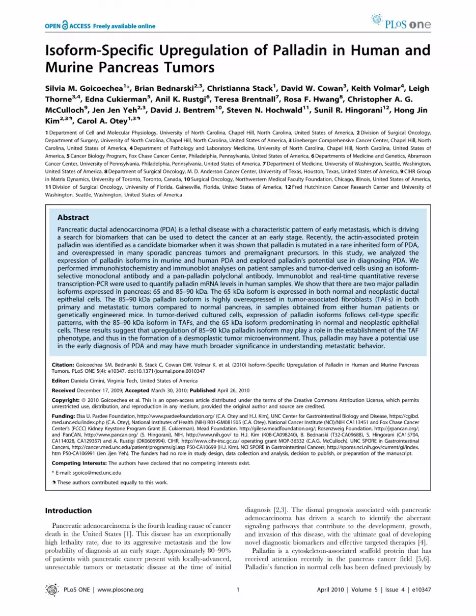
85–90 kDa Palladin Isoform is Strongly Upregulated inTumor-associated Fibroblasts
To resolve the existing confusion about the level of palladin
expression in different types of tumor-derived cells, we used both
the broadly-reactive polyclonal antibody 622 and the isoform-
selective monoclonal 1E6 to compare the pattern of isoforms
detected in tumor-associated fibroblasts and neoplastic tumor
cell lines. For comparison, we also analyzed the corresponding
non-tumor cells types. In Figure 2, human pancreatic ductal
epithelial (HPDE) cells were used as normal controls and probed
in parallel with three different lines of PDA-derived cells (Panc-1,
MiaPaCa, AsPC-1). The polyclonal antibody 622 detected a
single 65 kDa isoform in HPDE cells and all three tumor cell
lines, and the expression level of this palladin isoform did not
appear to vary significantly between normal and tumor cells.
When parallel blots were probed with monoclonal 1E6, no bands
were detected, even at long exposure times (Figure S1). In
contrast, when normal human fibroblasts (labeled NF) were
analyzed alongside lysates of primary pancreatic tumor-associ-
ated fibroblasts (TAF), the results showed that 85–90 kDa
palladin was strongly up-regulated in TAFs, and this band was
detected with both antibodies (1E6 and 622). In addition, the
polyclonal 622 antibody detected a minor band at 65 kDa in the
TAFs. To confirm these results, we also analyzed a newly
available line of immortalized pancreatic tumor-derived stellate
cells (PSC), which closely resemble TAFs in their phenotype and
patterns of gene expression [20], and the results obtained with
these cells were identical to those obtained with primary TAFs
(data not shown). These results suggest that cells of epithelial
origin may express different palladin isoforms than cells of
mesenchymal origin, and also confirm that a 85–90 kDa palladin
isoform is robustly upregulated in tumor-associated fibroblasts
when compared to normal adult fibroblasts, as reported
previously [6].
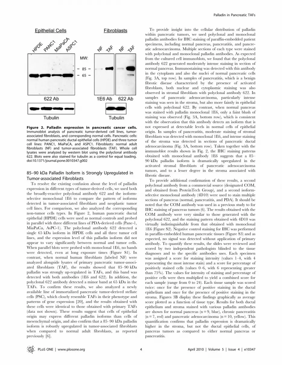
To provide insight into the cellular distribution of palladin
within pancreatic tumors, we used polyclonal and monoclonal
palladin antibodies for IHC staining of paraffin-embedded patient
specimens, including normal pancreas, pancreatitis, and pancre-
atic adenocarcinoma. Multiple sections of each type were stained
with polyclonal and monoclonal palladin antibodies. As expected
from the cultured cell immunoblots, we found that the polyclonal
antibody 622 generated moderately intense staining in section of
normal pancreas. Immunostaining was detected with this antibody
in the cytoplasm and also the nuclei of normal pancreatic cells
(Fig. 3A, top row). In samples of pancreatitis, which is a benign
fibrotic disease characterized by the presence of activated
fibroblasts, both nuclear and cytoplasmic staining was also
observed in stromal fibroblasts with polyclonal antibody 622. In
samples of pancreatic adenocarcinoma, particularly intense
staining was seen in the stroma, but also more faintly in epithelial
cells with polyclonal 622. By contrast, when normal pancreas
was stained with palladin monoclonal 1E6, only a faint blush of
staining was observed (Fig. 3A, bottom row), which is consistent
with the observation that this antibody detects an isoform that is
not expressed at detectable levels in normal cells of epithelial
origin. In samples of pancreatitis, moderate staining of stromal
fibroblasts was detected with monoclonal 1E6, and intense staining
of the stroma was detected in sections of pancreatic ductal
adenocarcinoma (Fig. 3A, bottom row). Taken together with the
immunoblot results shown in Fig. 2, the IHC staining patterns
obtained with monoclonal antibody 1E6 suggests that a 85–
90 kDa palladin isoform is dramatically upregulated in the
activated stromal fibroblasts of pancreatic adenocarcinoma
tumors, and to a lesser degree in the stroma associated with
fibrotic disease.
To provide additional confirmation of these results, a second
polyclonal antibody from a commercial source (designated COM,
and obtained from ProteinTech Group), and a second isoform-
selective monoclonal antibody (4D10) were used to stain multiple
sections of pancreas (normal, pancreatitis, and PDA). It should be
noted that the COM antibody was used in a previous study to for
IHC staining of pancreas tumors (6). The results obtained with the
COM antibody were very similar to those generated with the
polyclonal 622, and the staining pattern obtained with 4D10 was
virtually indistinguishable from that obtained with monoclonal
1E6 (Figure S2). Negative control staining for IHC was performed
in paraffin-embedded human pancreatic tissues (Figure S3) and as
expected, no signal was detected without application of primary
antibody. To quantify these results, the slides were reviewed and
scored by two independent pathologists blinded to the tissue
diagnoses and to the specific antibodies uses. Each specimen
was assigned a score for staining intensity (values 1–4, with 4
representing the most intense stain) and a score for percentage of
positively stained cells (values 0–6, with 6 representing greater
than 75%). The values for intensity of staining and percentage of
positive cells were then multiplied to yield a combined score for
each sample (range from 0 to 24). Each tissue sample was scored
twice: once for the presence of positive staining in the ductal
epithelium and once for the presence of positive staining in the
stroma. Figures 3B display these findings graphically as average
score plotted as a function of tissue type. Results for both ductal
epithelium and stroma stained with various palladin antibodies
are shown for normal pancreas (n = 9, blue), chronic pancreatitis
(n = 7, red) and pancreatic adenocarcinoma (n = 10, yellow). This
quantification confirms that palladin expression is dramatically
higher in the stroma, but not the ductal epithelial cells, of
pancreas tumors as compared to either normal pancreas or
pancreatitis.
Figure 2. Palladin expression in pancreatic cancer cells.Immunoblot analysis of pancreatic tumor-derived cell lines, tumor-associated fibroblasts, and corresponding normal cells. Pancreatic cells:normal human pancreatic ductal epithelial cells (HPDE) and three tumorcell lines: PANC1, MiaPaCA, and ASPC1. Fibroblasts: normal adultfibroblasts (NF) and tumor-associated fibroblasts (TAF). Whole celllysates were analyzed by western blot using the polyclonal antibody622. Blots were also stained for tubulin as a control for equal loading.doi:10.1371/journal.pone.0010347.g002
Palladin in Pancreatic TAFs
PLoS ONE | www.plosone.org 4 April 2010 | Volume 5 | Issue 4 | e10347
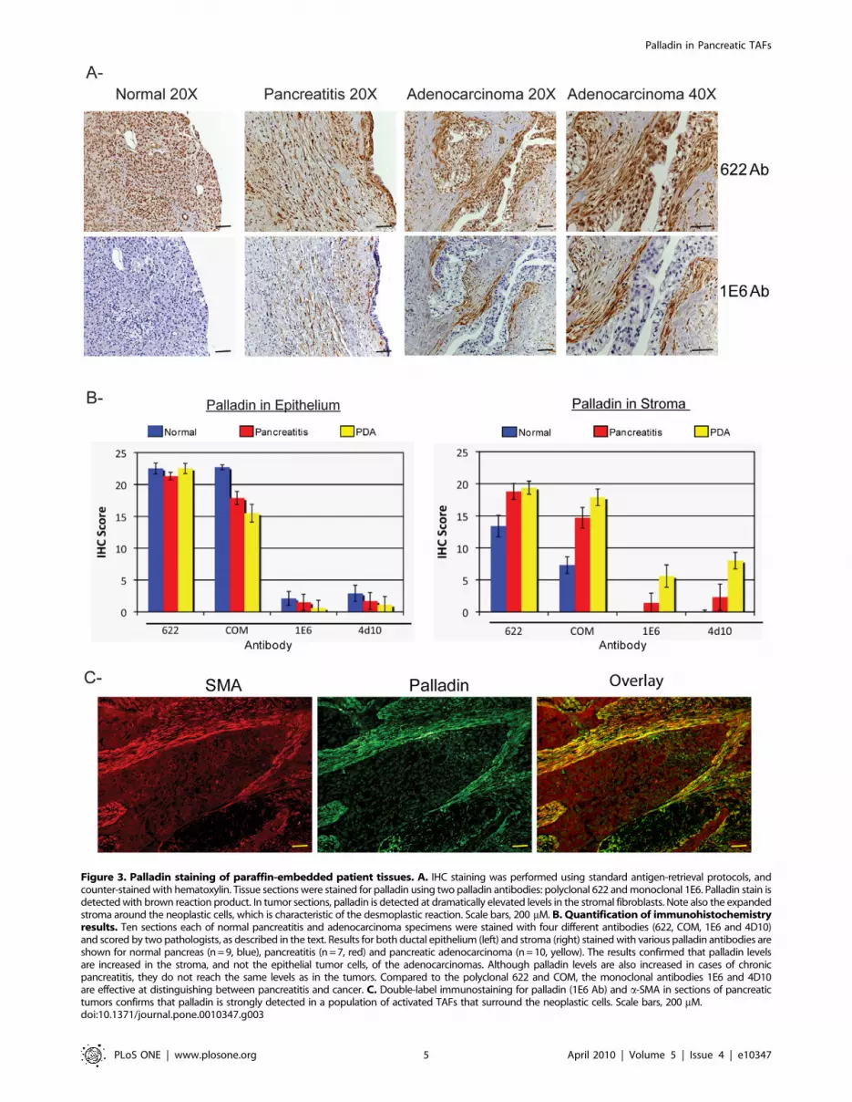
Figure 3. Palladin staining of paraffin-embedded patient tissues. A. IHC staining was performed using standard antigen-retrieval protocols, andcounter-stained with hematoxylin. Tissue sections were stained for palladin using two palladin antibodies: polyclonal 622 and monoclonal 1E6. Palladin stain isdetected with brown reaction product. In tumor sections, palladin is detected at dramatically elevated levels in the stromal fibroblasts. Note also the expandedstroma around the neoplastic cells, which is characteristic of the desmoplastic reaction. Scale bars, 200 mM. B. Quantification of immunohistochemistryresults. Ten sections each of normal pancreatitis and adenocarcinoma specimens were stained with four different antibodies (622, COM, 1E6 and 4D10)and scored by two pathologists, as described in the text. Results for both ductal epithelium (left) and stroma (right) stained with various palladin antibodies areshown for normal pancreas (n = 9, blue), pancreatitis (n = 7, red) and pancreatic adenocarcinoma (n = 10, yellow). The results confirmed that palladin levelsare increased in the stroma, and not the epithelial tumor cells, of the adenocarcinomas. Although palladin levels are also increased in cases of chronicpancreatitis, they do not reach the same levels as in the tumors. Compared to the polyclonal 622 and COM, the monoclonal antibodies 1E6 and 4D10are effective at distinguishing between pancreatitis and cancer. C. Double-label immunostaining for palladin (1E6 Ab) and a-SMA in sections of pancreatictumors confirms that palladin is strongly detected in a population of activated TAFs that surround the neoplastic cells. Scale bars, 200 mM.doi:10.1371/journal.pone.0010347.g003
Palladin in Pancreatic TAFs
PLoS ONE | www.plosone.org 5 April 2010 | Volume 5 | Issue 4 | e10347
Our IHC results are in agreement with an earlier study, which
reported that palladin is strongly overexpressed in the stroma of
171 of 177 sections (96.6%) of PDA tumors [6]. In that study,
palladin-positive stromal cells were identified as tumor-associated
fibroblasts based upon their morphology. To definitively confirm
the identity of palladin-positive cells within the pancreatic tumors,
we performed double-label immunofluorescence staining on tumor
sections, using the palladin monoclonal 1E6 and a monoclonal
antibody to a-smooth muscle actin (a-SMA), a marker for
fibroblast activation. As shown in Figure 3C, in sections of
pancreatic adenocarcinoma, the staining patterns for palladin and
a-SMA co-localized, which supports the conclusion that palladin is
expressed in stromal cells expressing a-SMA, which include
activated TAFs and stellate cells.
Palladin is Upregulated in Lymph Node and LiverMetastases and in Other Human Cancers
Two previous reports have suggested a potential role for palladin
in the metastasis of breast cancer by showing that palladin levels are
elevated in a highly invasive subpopulation of human breast cancer
cells [15,16]. The propensity for early invasion and metastasis is a
characteristic trait of pancreas tumors, such that we were prompted
to explore palladin expression in samples of pancreatic cancer
metastases to lymph nodes and liver to determine if palladin is
upregulated in the tumor cells or the TAFs in these samples. As
demonstrated in Figure 4A, palladin staining was strongly detected
in TAFs, and only weakly in the neoplastic epithelial cells, in both
types of metastases. These results suggest that neoplastic cells
are able to induce palladin upregulation in neighboring stromal
cells as a mechanism to recreate a favorable microenvironment
following metastatic invasion to a new location.
The results shown so far indicate that the 85–90 kDa palladin
isoform is upregulated in PDA tumor-associated fibroblasts in both
primary and metastatic tumors. To extend our observations to
other human cancers we performed immunoblot analysis of
tumor-associated fibroblasts isolated from five different tumors
obtained from breast, lung, ovary, kidney and skin. Pancreas was
included as a control. We used both the polyclonal antibody 622
and the monoclonal 1E6 to compare the pattern of isoforms
detected in tumor-associated fibroblasts. As expected, Figure 4B
shows that the 85–90 kDa palladin isoform is upregulated tumor-
associated fibroblasts from all tumors analyzed indicating that 85–
90 kDa palladin upregulation in the stromal microenvironment
may be a common feature of human cancers.
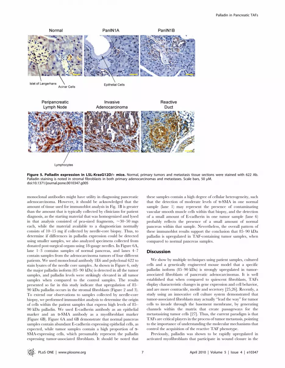
Palladin Expression in Murine PDA Recapitulates thePattern Seen in Human PDA
Important insights into the mechanisms of pancreatic adeno-
carcinoma pathogenesis have been gained from the use of
genetically engineered mice that express an activating mutation
in the Kras gene (KrasG12D) targeted to the pancreas [21,24]. We
performed immunohistochemical analyses on primary tumors and
metastases harvested from these mice to characterize palladin
expression during disease progression. Beginning with the earliest
precursor ductal lesions (i.e. PanIN-1A and -1B), associated
fibroblasts show staining for palladin using either antibody 622
(Figure 5) or the commercially available polyclonal antibody (data
not shown). Control staining for IHC of paraffin-embedded mouse
pancreatic tissue is shown in Figure S3. Interestingly, palladin
expression in the reactive fibrosis associated with acinar-ductal
metaplasia regions (around the so-called ‘‘reactive ducts’’) was
even more robust (Figure 5). We note that the ductal epithelial
cells in the adjacent PanINs do not show palladin expression, nor
do the surrounding normal acinar parenchymal cells. Palladin-
expressing fibroblasts increase in number and staining intensity
with disease progression, as strongly positive TAFs were seen
diffusely infiltrating the invasive ductal adenocarcinomas, along
with occasional faint cytoplasmic staining of the primary tumor
cells (Figure 5). Interestingly, in other areas of invasive disease with
scant to non-existent stromal cells, the tumor cells are completely
negative. The specificity of clustering of palladin-positive TAFs
around tumor cells is clearly demonstrated in peripancreatic
lymph nodes with infiltrating disease (Figure 5). The TAFs form
distinct rings around nests of tumor cells while the intervening
regions containing lymphocytes are completely negative. Taken
together with the results from human PDAs, these findings suggest
that upregulation of palladin by TAFs is likely to be a common
feature of desmoplastic tumors in multiple species.
Palladin Isoforms in Needle-Core Biopsy SamplesThe results of both IHC staining and immunoblot analysis
suggest that immunodetection of palladin by isoform-selective
Figure 4. Palladin in human pancreatic cancer metastasis andother human cancers. A. Immunohistochemistry of lymph nodeand liver metastases. Formalin-fixed, paraffin-embedded tissuesections were stained with COM antibody. Left: Low and highmagnification images of lymph node metastasis. Right: Low and highmagnification of liver metastasis. Arrow heads, lightly-stained tumorepithelial cells. Arrows, intensely-stained tumor-associated fibroblasts.Scale bars, 100 mM. B. Immunoblot analysis of tumor-associatedfibroblasts from different human cancers. Tumor-associatedfibroblasts were isolated from different human cancers: normal, kidney,lung, pancreas, breast and skin. Whole cell lysates were analyzed bywestern blot using the monoclonal antibody 1E6 and the polyclonalantibody 622. Blots were also stained for GAPDH as a control for equalloading.doi:10.1371/journal.pone.0010347.g004
Palladin in Pancreatic TAFs
PLoS ONE | www.plosone.org 6 April 2010 | Volume 5 | Issue 4 | e10347
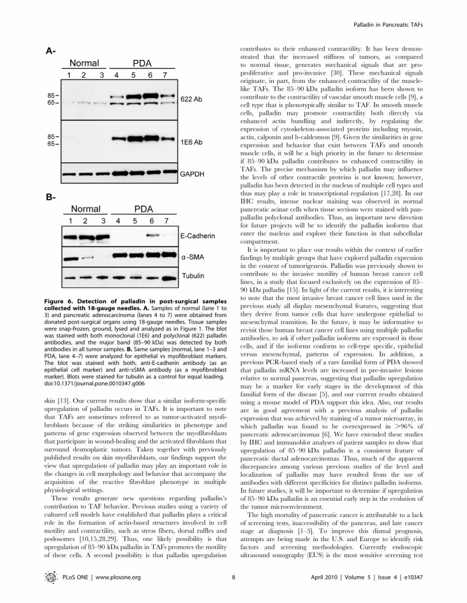
monoclonal antibodies might have utility in diagnosing pancreatic
adenocarcinoma. However, it should be acknowledged that the
amount of tissue used for immunoblot analysis in Fig. 1B is greater
than the amount that is typically collected by clinicians for patient
diagnosis, as the starting material that was homogenized and lysed
in that analysis consisted of pea-sized fragments, ,30–50 mgs
each, while the material available to a diagnostician normally
consists of 10–15 mg if collected by needle-core biopsy. Thus, to
determine if differences in palladin expression could be detected
using smaller samples, we also analyzed specimens collected from
donated post-surgical organs using 18-gauge needles. In Figure 6A,
lane 1–3 contains samples of normal pancreas, and lanes 4–7
contain samples from the adenocarcinoma tumors of four different
patients. We used monoclonal antibody 1E6 and polyclonal 622 to
stain lysates of the needle core samples. As shown in Figure 6, only
the major palladin isoform (85–90 kDa) is detected in all the tumor
samples, and palladin levels were strikingly elevated in all tumor
samples when compared to the control samples. The results
presented so far in this study indicate that upregulation of 85–
90 kDa palladin occurs in the stromal fibroblasts (Figure 2 and 3).
To extend our observations to samples collected by needle-core
biopsy, we performed immunoblot analysis to determine the origin
of cells within the patient samples that express high levels of 85–
90 kDa palladin. We used E-cadherin antibody as an epithelial
marker and an a-SMA antibody as a myofibroblast marker
(Figure 6B). Figure 6A and 6B demonstrate that normal pancreas
samples contain abundant E-cadherin expressing epithelial cells, as
expected, while tumor samples contain a high proportion of a-
SMA-expressing cells, which presumably represent the palladin
expressing tumor-associated fibroblasts. It should be noted that
these samples contain a high degree of cellular heterogeneity, such
that the detection of moderate levels of a-SMA in one normal
sample (lane 1) may represent the presence of contaminating
vascular smooth muscle cells within that biopsy, and the detection
of a small amount of E-cadherin in one tumor sample (lane 6)
probably reflects the presence of a small amount of normal
pancreas within that sample. Nevertheless, the overall pattern of
these immunoblot results support the conclusion that 85–90 kDa
palladin is upregulated in TAF-containing tumor samples, when
compared to normal pancreas samples.
Discussion
We show by multiple techniques using patient samples, cultured
cells and a genetically engineered mouse model that a specific
palladin isoform (85–90 kDa) is strongly upregulated in tumor-
associated fibroblasts of pancreatic adenocarcinomas. It is well
established that when compared to quiescent fibroblasts, TAFs
display characteristic changes in gene expression and cell behavior,
and are more contractile, motile and secretory [25,26]. Recently, a
study using an innovative cell culture system demonstrated that
tumor-associated fibroblasts may actually ‘‘lead the way’’ for tumor
cells to invade through the basement membrane, by generating
channels within the matrix that create passageways for the
metastasizing tumor cells [27]. Thus, the current paradigm is that
TAFs are critical players in the process of tumor metastasis, pointing
to the importance of understanding the molecular mechanisms that
control the acquisition of the reactive TAF phenotype.
Previously, palladin was shown to be rapidly upregulated in
activated myofibroblasts that participate in wound closure in the
Figure 5. Palladin expression in LSL-KrasG12D/+ mice. Normal, primary tumors and metastasis tissue sections were stained with 622 Ab.Palladin staining is noted in stromal fibroblasts in both primary adenocarcinomas and metastases. Scale bars, 50 mM.doi:10.1371/journal.pone.0010347.g005
Palladin in Pancreatic TAFs
PLoS ONE | www.plosone.org 7 April 2010 | Volume 5 | Issue 4 | e10347
skin [13]. Our current results show that a similar isoform-specific
upregulation of palladin occurs in TAFs. It is important to note
that TAFs are sometimes referred to as tumor-activated myofi-
broblasts because of the striking similarities in phenotype and
patterns of gene expression observed between the myofibroblasts
that participate in wound-healing and the activated fibroblasts that
surround desmoplastic tumors. Taken together with previously
published results on skin myofibroblasts, our findings support the
view that upregulation of palladin may play an important role in
the changes in cell morphology and behavior that accompany the
acquisition of the reactive fibroblast phenotype in multiple
physiological settings.
These results generate new questions regarding palladin’s
contribution to TAF behavior. Previous studies using a variety of
cultured cell models have established that palladin plays a critical
role in the formation of actin-based structures involved in cell
motility and contractility, such as stress fibers, dorsal ruffles and
podosomes [10,15,28,29]. Thus, one likely possibility is that
upregulation of 85–90 kDa palladin in TAFs promotes the motility
of these cells. A second possibility is that palladin upregulation
contributes to their enhanced contractility. It has been demon-
strated that the increased stiffness of tumors, as compared
to normal tissue, generates mechanical signals that are pro-
proliferative and pro-invasive [30]. These mechanical signals
originate, in part, from the enhanced contractility of the muscle-
like TAFs. The 85–90 kDa palladin isoform has been shown to
contribute to the contractility of vascular smooth muscle cells [9], a
cell type that is phenotypically similar to TAF. In smooth muscle
cells, palladin may promote contractility both directly via
enhanced actin bundling and indirectly, by regulating the
expression of cytoskeleton-associated proteins including myosin,
actin, calponin and h-caldesmon [9]. Given the similarities in gene
expression and behavior that exist between TAFs and smooth
muscle cells, it will be a high priority in the future to determine
if 85–90 kDa palladin contributes to enhanced contractility in
TAFs. The precise mechanism by which palladin may influence
the levels of other contractile proteins is not known; however,
palladin has been detected in the nucleus of multiple cell types and
thus may play a role in transcriptional regulation [17,28]. In our
IHC results, intense nuclear staining was observed in normal
pancreatic acinar cells when tissue sections were stained with pan-
palladin polyclonal antibodies. Thus, an important new direction
for future projects will be to identify the palladin isoforms that
enter the nucleus and explore their function in that subcellular
compartment.
It is important to place our results within the context of earlier
findings by multiple groups that have explored palladin expression
in the context of tumorigenesis. Palladin was previously shown to
contribute to the invasive motility of human breast cancer cell
lines, in a study that focused exclusively on the expression of 85–
90 kDa palladin [15]. In light of the current results, it is interesting
to note that the most invasive breast cancer cell lines used in the
previous study all display mesenchymal features, suggesting that
they derive from tumor cells that have undergone epithelial to
mesenchymal transition. In the future, it may be informative to
revisit those human breast cancer cell lines using multiple palladin
antibodies, to ask if other palladin isoforms are expressed in those
cells, and if the isoforms conform to cell-type specific, epithelial
versus mesenchymal, patterns of expression. In addition, a
previous PCR-based study of a rare familial form of PDA showed
that palladin mRNA levels are increased in pre-invasive lesions
relative to normal pancreas, suggesting that palladin upregulation
may be a marker for early stages in the development of this
familial form of the disease [5], and our current results obtained
using a mouse model of PDA support this idea. Also, our results
are in good agreement with a previous analysis of palladin
expression that was achieved by staining of a tumor microarray, in
which palladin was found to be overexpressed in .96% of
pancreatic adenocarcinomas [6]. We have extended these studies
by IHC and immunoblot analyses of patient samples to show that
upregulation of 85–90 kDa palladin is a consistent feature of
pancreatic ductal adenocarcinomas. Thus, much of the apparent
discrepancies among various previous studies of the level and
localization of palladin may have resulted from the use of
antibodies with different specificities for distinct palladin isoforms.
In future studies, it will be important to determine if upregulation
of 85–90 kDa palladin is an essential early step in the evolution of
the tumor microenvironment.
The high mortality of pancreatic cancer is attributable to a lack
of screening tests, inaccessibility of the pancreas, and late cancer
stage at diagnosis [1–3]. To improve this dismal prognosis,
attempts are being made in the U.S. and Europe to identify risk
factors and screening methodologies. Currently endoscopic
ultrasound sonography (EUS) is the most sensitive screening test
Figure 6. Detection of palladin in post-surgical samplescollected with 18-gauge needles. A. Samples of normal (lane 1 to3) and pancreatic adenocarcinoma (lanes 4 to 7) were obtained fromdonated post-surgical organs using 18-gauge needles. Tissue sampleswere snap-frozen, ground, lysed and analyzed as in Figure 1. The blotwas stained with both monoclonal (1E6) and polyclonal (622) palladinantibodies, and the major band (85–90 kDa) was detected by bothantibodies in all tumor samples. B. Same samples (normal, lane 1–3 andPDA, lane 4–7) were analyzed for epithelial vs myofibroblast markers.The blot was stained with both, anti-E-cadherin antibody (as anepithelial cell marker) and anti-aSMA antibody (as a myofibroblastmarker). Blots were stained for tubulin as a control for equal loading.doi:10.1371/journal.pone.0010347.g006
Palladin in Pancreatic TAFs
PLoS ONE | www.plosone.org 8 April 2010 | Volume 5 | Issue 4 | e10347
available. It can identify early pancreatic changes caused by an
IPMN (pre-malignant intraductal papillary mucinous neoplasm),
mucinous cysts, or malignant lesions (small masses and PanIN
lesions, i.e., preneoplastic pancreatic intraepithelial neoplasia), or
early features of chronic pancreatitis [31]. Our current results
indicate that upregulation of a 85–90 kDa palladin isoform is
specific to pancreatic ductal adenocarcinoma, and is not detected
in less invasive tumor types. Our results also show that palladin
expression can be detected in small amounts of tissue (10–15 mg).
Taken together, these results point to the possibility that
immunodetection of the 85–90 kDa palladin isoform in samples
collected by EUS-guided fine-needle aspiration may have
diagnostic utility as an early, specific marker for the development
of pancreatic adenocarcinoma, which may provide new avenues
for diagnosing pancreatic cancer at a treatable stage of the disease.
Materials and Methods
Ethics StatementThis study was conducted according to the principles expressed
in the Declaration of Helsinki. The study was approved by the
Institutional Review Board of UNC Hospital (#07-2046). All
patients provided written informed consent for the collection of
samples and subsequent analysis. All animals were handled in
strict accordance with good animal practice as defined by the
relevant national and/or local animal welfare bodies, and all
animal work was approved by the ‘‘Fred Hutchinson Cancer
Research Center Institutional Animal Care and Use Committee’’
(Institutional Animal Welfare Assurance Number A3226-01).
MaterialsFour different antibodies against palladin were used: two rabbit
polyclonals, 622 and commercial (ProteinTech Group) and two
monoclonals, 1E6 and 4D10. The commercial antibody was
utilized previously for IHC and immunoblot studies [6], polyclonal
622 is the sister antibody of the previously characterized 621 [5],
and monoclonals 1E6 and 4D10 were characterized previously
[10,15]. To control for equal sample loading, antibodies to alpha
tubulin (Lab Vision Corporation), GAPDH (Santa Cruz) and a-
smooth muscle actin (Sigma) were used. The protease inhibitor
cocktail for mammalian tissues was from Sigma. Alexafluor-488
and Alexafluor-568 anti-mouse IgG and anti-rabbit IgG conju-
gated secondary antibodies were from Molecular Probes.
Cell linesHuman cell types used in this study include: pancreatic ductal
epithelial cells (HPDE) [5], three pancreatic cancer cell lines
(PANC1, MiaPaCa and ASPC1), primary normal gingival
fibroblasts (NF), primary tumor-associated fibroblasts obtained
from human pancreatic adenocarcinomas (TAF), and immortal-
ized pancreatic stellate cells. Pancreatic cancer cell lines were
obtained from ATCC: PANC1 (ATCC # CRL-1469), ASPC1
(ATCC # CRL-1682) and MiaPaCa (ATCC # CRL 1420).
Primary cultures of human gingival fibroblasts were obtained from
biopsies of normal gingiva in patients aged 10 to 16 years, as
described previously [32,33], and were used at passages 3–12 for
all experiments. Human pancreatic tumor-associated fibroblasts
were isolated as described previously [34] and used at passage 2–4.
Immortalized human pancreatic stellate cells were characterized
previously [20]. NF were cultured in MEM Alpha plus 15% FBS
and antibiotics, while TAF and stellate cells were cultured in
DMEM plus 10% FBS and antibiotics. HPDE cells were cultured
in Keratinocyte SFM with 50 mg Bovine Pituitary Extract and
2.5 mg Epidermal Growth Factor. Panc1 and MiaPaCa cells were
cultured in DMEM plus 10% FBS and antibiotics. ASPC1 were
cultured in RPMI 1640 plus 10% FBS and antibiotics. All cultured
cells were grown at 37uC and 5% carbon dioxide.
Cell Lysis and Immunoblot AnalysisCells were cultured on 100 mm tissue culture dishes, rinsed
briefly with phosphate-buffered saline, and then scraped into a
lysis buffer containing 50 mM Tris (pH 7.0), 150 mM NaCl, 1%
Triton X-100, and a protease inhibitor cocktail for mammalian
tissues. The supernatant was collected after centrifugation at
14,000 rpm for 15 min. The cell lysates were analyzed by
immunoblot or frozen with liquid nitrogen and stored at 280uCfor future use. For the immunoblot, lysates were boiled in 2X
Laemmli buffer, and 20 mg of protein were resolved by SDS-
PAGE in each lane of a 4–12% gel. The proteins were transferred
to nitrocellulose and immunoblotted. Immunocomplexes were
visualized using the Western Lights Chemiluminescence Detection
kit from Perkin-Elmer.
ImmunohistochemistryDe-identified patient samples of primary and metastatic tumors
were obtained through an IRB-approved study supported through
the tissue procurement facility of the Lineberger Comprehensive
Cancer Center. The protocol supports the procurement of
malignant and non-malignant tissue for cancer-related research,
and informed consent is obtained from patients who agree to
participate. Five micron-thick, formalin-fixed paraffin-embedded
(FFPE) tissue sections were collected on a coated glass slide, dried
vertically overnight at room temperature and heated in a 60uCdrying oven for one hour. The tissue sections were de-paraffinized
in two changes of xylene, hydrated using descending grades of
ethanol, and rinsed in a Tris buffer containing Tween 20
(DakoCytomation TRIS-Buffered Saline S1968/DakoCytomation
Tween 20 S1966). Endogenous peroxidase activity was blocked by
incubating in a 3% hydrogen peroxide/methanol solution for 10
minutes. (Fisher Scientific Hydrogen Peroxide ACS H325/
Mallinckrodt Chemicals Methanol 3016-16). Antigen retrieval
was performed by heating with steam in a citra buffer (BioGenex
Antigen Retrieval Citra HK086-pK) for 30 minutes, cooling for 15
minutes at room temperature and placing in Tris buffered solution
with Tween for 5 minutes. Immunostaining was performed on the
Dako Autostainer platform (DakoCytomation) at room tempera-
ture. Briefly, each tissue section was incubated in normal horse
serum (Vector Laboratories Elite kit 6102) for 15 minutes; the
primary antibody was applied for 60 minutes, and detection was
completed by incubating with a biotinylated link. An avidin-biotin
complex (Vectastain Elite Kit 6102) was applied for 30 minutes
followed by diaminobenzidine (Innovex NB314SB) chromogen for
2 minutes. Signal contrast was maximized by counterstaining in
hematoxylin (DakoCytomation Mayer’s Hematoxylin S3309) for 1
minute, rinsing in deionized water and finally in a bluing solution
(Richard-Allan Scientific 7301) for 30 seconds. Samples were then
rinsed in Tris buffer for 5 minutes, dehydrated in ethanol and
placed in xylene, and mounted using Permount (Fisher Scientific
Permount SP15).
Immunoblot of patient samplesPea-sized pieces of fresh tissue were snap-frozen in liquid
nitrogen, ground in a chilled mortar and pestle, and extracted in
lysis buffer containing 50 mM Tris pH 7.5, 8 M urea, 5% SDS,
10 mM EDTA, and protease inhibitor cocktail for mammalian
tissues. Samples were then centrifuged at 15,0006g to remove any
unsolubilized particulates, and the supernatant was resolved by
SDS-PAGE, using 15 mg protein loaded per lane.
Palladin in Pancreatic TAFs
PLoS ONE | www.plosone.org 9 April 2010 | Volume 5 | Issue 4 | e10347
Mouse TumorsPrimary pancreatic ductal adenocarcinomas and metastases
were isolated from genetically engineered mice in which an
activating mutation in Kras is targeted to progenitor cells of the
developing pancreas [21]. These animals develop pre-invasive
ductal lesions stochastically and manifest the full spectrum of
PanIN lesions culminating in invasive and metastatic disease with
clinical, histopathological and molecular features that faithfully
mimic the human disease [21,22,23]. Tissue specimens were
dissected, formalin-fixed and paraffin-embedded and sectioned for
immunohistochemistry, as described above for the human
samples.
RNA isolation and quantitative reverse transcription-PCRDe-identified patient samples were collected after approval by
each individual IRB and use of all samples for this study was
approved by the UNC IRB. Samples of matched normal
pancreas and pancreatic cancer were obtained at the time of
operation and flash frozen in liquid nitrogen. Total RNA was
extracted from the snap-frozen tumor samples using Allprep Kits
(Qiagen) and quantified using nanodrop spectrophotometry
(ThermoScientific). Reverse transcriptase reactions were per-
formed using the commercial kit High-Capacity cDNA Reverse
Transcription (Applied Biosystem) and contained up to 2 mg of
total RNA. Real-time quantitative PCR was performed using an
Applied Biosystems 7500 Fast Real Time PCR System in a a total
volume of 25 ml mixture containing 2.5 ml of 10-fold diluted
cDNA, 0.2 pM of sense and antisense primers and the Power
SYBR@ Green Master Mix (Applied Biosystem). Because the
coding sequence of the 85–90 kDa isoform of palladin is
contained and/or overlaps with the coding sequence of other
palladin isoforms, RT-PCR primers were designed to amplify
mRNA of the Ig3 domain containing isoforms (isoforms #1, #2,
#3, #4, #5 and #7). The primers used in the experiment were
as follows: sense 59 – AAC CGA GCA GGA CAG AAC- 39 and
antisense 59- TGG TGG CAC TCC CAA TAC-39. The thermal
cycling conditions were 50uC for 2 min, then 95uC for 10 min
followed by 40 cycles of initial denaturation step of 95uC/15 s
and 60uC/60 s. No-template reactions were included as negative
controls in every plate. Sequence Detection Software (Applied
Biosystems, Foster City, USA) results were imported into
Microsoft Excel for further analysis. Raw expression levels were
calculated from the average of the triplicate ddCT (RQ) values.
Standard curve obtained for the primer and PCR product from a
pool of samples was used as template. Each PCR reaction was
carried out in triplicates.
Supporting Information
Figure S1 Immunoblot analysis of pancreatic tumor-derived cell
lines using palladin 1E6 Ab. Normal fibroblasts were used as a
positive control. Pancreatic cells: normal human pancreatic ductal
epithelial cells (HPDE) and three tumor cell lines: PANC1,
MiaPaCA, and ASPC1. Whole cell lysates were analyzed by
western blot using the monoclonal antibody 1E6. Blots were
subjected to long exposure times and also stained for tubulin as a
control for equal loading.
Found at: doi:10.1371/journal.pone.0010347.s001 (0.24 MB TIF)
Figure S2 Immunohistochemistry of paraffin-embedded patient
specimens. IHC staining was performed using standard antigen-
retrieval protocols, and counter-stained with hematoxylin. Tissue
sections were stained for palladin using two palladin antibodies:
polyclonal COM from ProteinTech group, and monoclonal
4d10.
Found at: doi:10.1371/journal.pone.0010347.s002 (8.42 MB TIF)
Figure S3 Control staining for IHC of paraffin-embedded
human and mouse pancreatic tissues. IHC staining was performed
as in Figure 2, except that normal rabbit serum was substituted for
the primary antibody.
Found at: doi:10.1371/journal.pone.0010347.s003 (10.34 MB
TIF)
Acknowledgments
The authors thank Dr. Vivekanand Gupta (Fox Chase Cancer Center) for
assistance in generating the multi-fibroblast immunoblot, Dr. Brian Law
(University of Florida) for assistance with isolation of the pancreatic tumor
associated fibroblasts, and Dr. Tanja Crnogorac-Jurcevic (Barts and the
London School of Medicine and Dentistry) for critical reading of the
manuscript. The authors also thank Mary Beth Ward, Ross Gagnon and
Dustin Cashman (University of North Carolina at Chapel Hill) for
technical assistance. RT-qPCR was performed by the Microbiome Core
Facility at the University of North Carolina at Chapel Hill, North
Carolina.
Author Contributions
Conceived and designed the experiments: SMG BKB HJK CAAO.
Performed the experiments: SMG CS EC SH. Analyzed the data: SMG
BKB KV LT SH HJK CAAO. Contributed reagents/materials/analysis
tools: DWC KV LT AKR TAB RFH CAM JJY DJB SH SH HJK CAAO.
Wrote the paper: SMG CAAO.
References
1. Jemal A, Siegel R, Ward E, Hao Y, Xu J, et al. (2009) Cancer statistics, 2009.CA Cancer J Clin 59: 225–49.
2. Cleary SP, Gryfe R, Guindi M, Greig P, Smith L, et al. (2004) Prognostic factorsin resected pancreatic adenocarcinoma: analysis of actual 5-year survivors. J Am
Coll Surg 198: 722–731.
3. Conlon KC, Klimstra DS, Brennan MF (1996) Long-term survival after curative
resection for pancreatic ductal adenocarcinoma. Clinicopathologic analysis of 5-year survivors. Ann Surg 223: 273–279.
4. Sebolt-Leopold JS, English JM (2006) Mechanisms of drug inhibition ofsignalling molecules. Nature 441: 457–462.
5. Pogue-Geile KL, Chen R, Bronner MP, Crnogorac-Jurcevic T, Moyes KW,et al. (2006) Palladin mutation causes familial pancreatic cancer and suggests a
new cancer mechanism. PLoS Med 3: e516.
6. Salaria SN, Illei P, Sharma R, Walter KM, Klein AP, et al. (2007) Palladin is
overexpressed in the non-neoplastic stroma of infiltrating ductal adenocarcino-mas of the pancreas, but is only rarely overexpressed in neoplastic cells. Cancer
Biol Ther 6: 324–328.
7. Goicoechea S, Arneman D, Disanza A, Garcia-Mata R, Scita G, et al. (2006)
Palladin binds to Eps8 and enhances the formation of dorsal ruffles and
podosomes in vascular smooth muscle cells. J Cell Sci 119: 3316–3324.
8. Goicoechea SM, Arneman D, Otey CA (2008) The role of palladin in actinorganization and cell motility. Eur J Cell Biol 87: 517–525.
9. Jin L, Yoshida T, Ho R, Owens GK, Somlyo AV (2008) The actin associatedprotein palladin is required for development of normal contractile properties of
smooth muscle cells derived from embryoid bodies. J Biol Chem.
10. Parast MM, Otey CA (2000) Characterization of palladin, a novel protein
localized to stress fibers and cell adhesions. J Cell Biol 150: 643–656.
11. Boukhelifa M, Hwang SJ, Valtschanoff JG, Meeker RB, Rustioni A, et al. (2003)
A critical role for palladin in astrocyte morphology and response to injury. MolCell Neurosci 23: 661–668.
12. Jin L, Kern MJ, Otey CA, Wamhoff BR, Somlyo AV (2007) Angiotensin II,focal adhesion kinase, and PRX1 enhance smooth muscle expression of lipoma
preferred partner and its newly identified binding partner palladin to promote
cell migration. Circ Res 100: 817–825.
13. Ronty MJ, Leivonen SK, Hinz B, Rachlin A, Otey CA, et al. (2006) Isoform-specific regulation of the actin-organizing protein palladin during TGF-beta1-
induced myofibroblast differentiation. J Invest Dermatol 126: 2387–2396.
14. Luo H, Liu X, Wang F, Huang Q, Shen S, et al. (2005) Disruption of palladin
results in neural tube closure defects in mice. Mol Cell Neurosci 29: 507–
515.
Palladin in Pancreatic TAFs
PLoS ONE | www.plosone.org 10 April 2010 | Volume 5 | Issue 4 | e10347

15. Goicoechea SM, Bednarski B, Garcia-Mata R, Prentice-Dunn H, Kim HJ, et al.
(2008) Palladin contributes to invasive motility in human breast cancer cells.
Oncogene 28: 587–98.
16. Wang W, Goswami S, Lapidus K, Wells AL, Wyckoff JB, et al. (2004)
Identification and testing of a gene expression signature of invasive carcinoma
cells within primary mammary tumors. Cancer Res 64: 8585–8594.
17. Mykkanen OM, Gronholm M, Ronty M, Lalowski M, Salmikangas P, et al.
(2001) Characterization of human palladin, a microfilament-associated protein.
Mol Biol Cell 12: 3060–3073.
18. Rachlin AS, Otey CA (2006) Identification of palladin isoforms and
characterization of an isoform-specific interaction between Lasp-1 and palladin.
J Cell Sci 119: 995–1004.
19. Wang HV, Moser M (2008) Comparative expression analysis of the murine
palladin isoforms. Dev Dyn 237: 3342–3351.
20. Hwang RF, Moore T, Arumugam T, Ramachandran V, Amos KD, et al. (2008)
Cancer-associated stromal fibroblasts promote pancreatic tumor progression.
Cancer Res 68: 918–926.
21. Hingorani SR, Wang L, Multani AS, Combs C, Deramaudt TB, et al. (2005)
Trp53R172H and KrasG12D cooperate to promote chromosomal instability
and widely metastatic pancreatic ductal adenocarcinoma in mice. Cancer Cell 7:
469–483.
22. Hingorani SR, Petricoin EF, Maitra A, Rajapakse V, King C, et al. (2003)
Preinvasive and invasive ductal pancreatic cancer and its early detection in the
mouse. Cancer Cell 4: 437–450.
23. Izeradjene K, Combs C, Best M, Gopinathan A, Wagner A, et al. (2007)
Kras(G12D) and Smad4/Dpc4 haploinsufficiency cooperate to induce mucinous
cystic neoplasms and invasive adenocarcinoma of the pancreas. Cancer Cell 11:
229–243.
24. Tuveson DA, Hingorani SR (2005) Ductal pancreatic cancer in humans and
mice. Cold Spring Harb Symp Quant Biol 70: 65–72.25. Kalluri R, Zeisberg M (2006) Fibroblasts in cancer. Nat Rev Cancer 6: 392–401.
26. Mahadevan D, Von Hoff DD (2007) Tumor-stroma interactions in pancreatic
ductal adenocarcinoma. Mol Cancer Ther 6: 1186–1197.27. Gaggioli C, Hooper S, Hidalgo-Carcedo C, Grosse R, Marshall JF, et al. (2007)
Fibroblast-led collective invasion of carcinoma cells with differing roles forRhoGTPases in leading and following cells. Nat Cell Biol 9: 1392–1400.
28. Endlich N, Schordan E, Cohen CD, Kretzler M, Lewko B, et al. (2009) Palladin
is a dynamic actin-associated protein in podocytes. Kidney Int 75: 214–226.29. Liu XS, Luo HJ, Yang H, Wang L, Kong H, et al. (2007) Palladin regulates cell
and extracellular matrix interaction through maintaining normal actincytoskeleton architecture and stabilizing beta1-integrin. J Cell Biochem 100:
1288–1300.30. Paszek MJ, Weaver VM (2004) The tension mounts: mechanics meets
morphogenesis and malignancy. J Mammary Gland Biol Neoplasia 9: 325–342.
31. Rozen P, Liphshitz I, Rosner G, Barchana M, Lachter J , et al. PancreaticCancer Consortium (2009) Pancreatic cancer in Israel: the epidemiology,
possibilities of prevention, early detection and screening. Isr Med Assoc J.11(12):710–713.
32. Arora PD, Janmey PA, McCulloch CA (1999) A role for gelsolin in stress fiber-
dependent cell contraction. Exp Cell Res 250: 155–167.33. McCulloch CA, Knowles GC (1993) Deficiencies in collagen phagocytosis by
human fibroblasts in vitro: a mechanism for fibrosis? J Cell Physiol 155:461–471.
34. Corsino P, Davis B, Law M, Chytil A, Forrester E, et al. (2007) Tumors initiatedby constitutive Cdk2 activation exhibit transforming growth factor beta
resistance and acquire paracrine mitogenic stimulation during progression.
Cancer Res 67: 3135–3144.
Palladin in Pancreatic TAFs
PLoS ONE | www.plosone.org 11 April 2010 | Volume 5 | Issue 4 | e10347


