Embed Size (px)
Citation preview
E
Ica
DEC
a
ARA
KPCSNL
1
dbtn
i
F
4
0h
Resuscitation 84 (2013) 1143– 1149
Contents lists available at SciVerse ScienceDirect
Resuscitation
jo ur nal homep age: www.elsev ier .com/ locate / resusc i ta t ion
xperimental paper
schemic post-conditioning and vasodilator therapy during standardardiopulmonary resuscitation to reduce cardiac and brain injuryfter prolonged untreated ventricular fibrillation�,��
emetris Yannopoulos ∗, Nicolas Segal, Timothy Matsuura, Mohammad Sarraf, Marit Thorsgard,mily Caldwell, Jennifer Rees, Scott McKnite, Karen SantaCruz, Keith G. Lurie
ardiovascular Division at The University of Minnesota, United States
r t i c l e i n f o
rticle history:eceived 3 January 2013ccepted 21 January 2013
eywords:ost-conditioningardiopulmonary resuscitationurvivaleurological functioneft ventricular function
a b s t r a c t
Aim of the study: We investigated the effects of ischemic postconditioning (IPC) with and without car-dioprotective vasodilatory therapy (CVT) at the initiation of cardiopulmonary resuscitation (CPR) oncardio-cerebral function and 48-h survival.Methods: Prospective randomized animal study. Following 15 min of ventricular fibrillation, 42 Yorkshirefarm pigs weighing an average of 34 ± 2 kg were randomized to receive standard CPR (SCPR, n = 12),SCPR + IPC (n = 10), SCPR + IPC + CVT (n = 10), or SCPR + CVT (n = 10). IPC was delivered during the first 3 minof CPR with 4 cycles of 20 s of chest compressions followed by 20-s pauses. CVT consisted of intravenoussodium nitroprusside (2 mg) and adenosine (24 mg) during the first minute of CPR. Epinephrine was givenin all groups per standard protocol. A transthoracic echocardiogram was obtained on all survivors 1 and4 h post-ROSC. The brains were extracted after euthanasia at least 24 h later to assess ischemic injury in 7regions. Ischemic injury was graded on a 0–4 scale with (0 = no injury to 4 ≥50% neural injury). The sumof the regional scores was reported as cerebral histological score (CHS). 48 h survival was reported.Results: Post-resuscitation left ventricular ejection (LVEF) fraction improved in SCPR + CVT,SCPR + IPC + CVT and SCPR + IPC groups compared to SCPR (59% ± 9%, 52% ± 14%, 52% ± 14% vs. 35% ± 11%,respectively, p < 0.05). Only SCPR + IPC and SCPR + IPC + CVT, but not SCPR + CVT, had lower mean CHS
compared to SCPR (5.8 ± 2.6, 2.8 ± 1.8 vs. 10 ± 2.1, respectively, p < 0.01). The 48-h survival amongSCPR + IPC, SCPR + CVT, SCPR + IPC + CVT and SCPR was 6/10, 3/10, 5/10 and 1/12, respectively (Coxregression p < 0.01).Conclusions: IPC and CVT during standard CPR improved post-resuscitation LVEF but only IPC was inde-pendently neuroprotective and improved 48-h survival after 15 min of untreated cardiac arrest in pigs.. Introduction
An estimated 350,000 patients suffer from out-of-hospital car-iac arrest (OHCA) each year in the United States.1 Even with theest clinically documented methods of cardiopulmonary resuscita-
ion (CPR), more than 85–90% of OHCA patients die or have severeeurological deficits.2 Cerebral and cardiac dysfunction following� A Spanish translated version of the summary of this article appears as Appendixn the final online version at http://dx.doi.org/10.1016/j.resuscitation.2013.01.024.�� The Institutional Animal Care Committee of the Minneapolis Medical Researchoundation approved the protocol (number 11-05, approved on 5/10/2011).∗ Corresponding author at: Department of Cardiology, University of Minnesota,
20 Delaware Street, SE, MMC 508, Minneapolis, MN 55455-0341, United States.E-mail address: [email protected] (D. Yannopoulos).
300-9572/$ – see front matter © 2013 Elsevier Ireland Ltd. All rights reserved.ttp://dx.doi.org/10.1016/j.resuscitation.2013.01.024
© 2013 Elsevier Ireland Ltd. All rights reserved.
successful resuscitation from cardiac arrest is the major cause ofdeath and long-term morbidity.3,4
We recently have shown that after 15 min of untreated car-diac arrest due to ventricular fibrillation, ischemic postconditioning(IPC) at the initiation of standard CPR, can improve neurologicalintact survival.5 IPC with four, 20 s pauses during the first 3 minof CPR have been shown to be synergistic with sodium nitroprus-side “enhanced” CPR (SNPeCPR) which utilizes active compressionand decompression CPR with an inspiratory impedance thresholddevice and abdominal binding.6
In this investigation we try to build upon our previous publishedstudies and evaluate the effects of cardioprotective vasodilatortherapy (CVT) alone and in combination with IPC in a model of
standard CPR (SCPR). We sought to provide evidence of reducedglobal reperfusion injury after prolonged ischemia with histolog-ical and biomarker based endpoints in addition to the clinicalendpoints.
1 uscita
tSsfip
2
ipIca
2
rvSafioa(tmIsstcd
2
tairmIiodpotTRtR
2
it3dnas
144 D. Yannopoulos et al. / Res
We hypothesized that by using a simple CPR strategy designedo control the initial reintroduction of blood flow during Basic Lifeupport (BLS), we could protect vital organs from injury and sub-tantially improve outcomes after 15 min of untreated ventricularbrillation. Further, we hypothesized that the addition of cardio-rotective vasodilatory agents would act synergistically with IPC.
. Materials and methods
All studies were performed on Yorkshire farm pigs weigh-ng an average of 34 ± 2 kg. A certified and licensed veterinarianrovided a blinded neurologic assessment at 24 and 48 h. The
nstitutional Animal Care Committee of the Minneapolis Medi-al Research Foundation approved the protocol (number 11-05,pproved on 5/10/2011).7
.1. Preparatory phase
The anesthesia, surgical preparation, data monitoring, andecording procedures used in this study have been described pre-iously in detail and the study protocol was used unaltered fromegal et al.5 After endotracheal intubation, inhaled isoflurane at
dose of 0.8–1.2% was used for anesthesia up until ventricularbrillation (VF) induction. Anesthesia was restarted after returnf spontaneous circulation (ROSC). The animal’s bladder temper-ture was maintained at 37.5 ± 0.5 ◦C with a warming blanketBair Hugger, Augustine Medical, Eden Prairie, MN). Central aor-ic and right atrial pressures were recorded continuously with
icromanometer-tipped catheters (Mikro-Tip Transducer, Millarnstruments, Houston, TX). The left internal carotid artery wasurgically exposed and an ultrasound flow probe (Transonic 420eries multichannel, Transonic Systems, Ithaca, NY) placed to quan-ify blood flow (mL/min). Compression force, rate and depth, wereontinuously recorded throughout all experiments and controlleduring CPR to assure all groups received identical CPR quality.
.2. Experimental protocol
After the surgical preparation was complete, oxygen satura-ion on room air was >95%, and ETCO2 was stable between 35nd 42 mmHg for 5 min, VF was induced by delivering directntracardiac current. Standard chest compression cardiopulmonaryesuscitation was performed with a pneumatically driven auto-atic piston device (Pneumatic Compression Controller, Ambu
nternational, Glostrup, Denmark) as previously described.8 Dur-ng SCPR, we delivered uninterrupted chest compressions at a ratef 100 compressions/min, with a 50% duty cycle and a compressionepth of 25% of the anteroposterior chest diameter. Asynchronousositive-pressure ventilations were delivered with room air (FIO2f 0.21) with a manual resuscitator bag. The tidal volume was main-ained at ∼10 mL/kg and the respiratory rate was 10 breaths/min.he investigators were blinded to hemodynamics during CPR. IfOSC was not achieved, defibrillation was delivered every 2 minhereafter during CPR. Resuscitation efforts were continued untilOSC was achieved or for a total of 15 min.
.3. Protocol
We tested two interventions in this study, independently, andn combination during SCPR. These interventions included, (a) IPC,hat was delivered with four cycles of 20-s pauses for the first
min of the resuscitation effort5 and (b) administration of car-
ioprotective vasodilator therapy (CVT). CVT consisted of sodiumitroprusside (SNP) and adenosine. SNP was given as a 2 mg bolust minute 1 and a second 1 mg bolus at minute 3 of CPR.6 Adeno-ine was given as a single 24 mg bolus after the first SNP bolustion 84 (2013) 1143– 1149
(after preliminary studies demonstrated superiority of this dosein improving post-resuscitation left ventricular (LV) dysfunction.9
Epinephrine was administered in all groups in a 0.5 mg (∼15 �g/kg)bolus at minute 4 of CPR, 60 s before first defibrillation.
Following 15 min of untreated VF, 42 pigs were random-ized prospectively using a computer-generated program into fourgroups:
I. SCPR group as controls: received only SCPR and epinephrine (12animals).
II. IPC group: received four 20-s pauses during the first 3 min ofSCPR.
III. CVT group: received SNP and adenosine as described abovewhile performing SCPR.
IV. IPC + CVT group: received both four 20-s pauses of CPR (IPC) andCVT, as described above. Groups II–IV had 10 animals each.
2.4. Post-ROSC care
The protocol for post resuscitation care has been described indetail by Segal et al.5 Supplemental oxygen was added only ifarterial saturation was lower than 90%. Animals were observedunder general anesthesia with isoflurane until hemodynamicallystable. Hemodynamic stability was defined as a mean aortic pres-sure >55 mmHg without pharmacologic support for 10 min aswell as normalization of ETCO2 and acidosis. Animals that werehypotensive post-ROSC received increments of 0.1 mg intravenousepinephrine every 5 min until mean arterial pressure rose above50 mmHg. If pH was lower than 7.20, 50–100 mEq of NaHCO3 weregiven intravenously.
All groups received post-resuscitation therapeutic hypothermiaas recommended by the American Heart Association for comatosepatients resuscitated from VF to simulate best practice and opti-mize the chances of the control group for neurological recovery.10
Target temperature was set at 34 ◦C and was maintained at thatlevel with the use of a cutaneous cooling device (Arctic Sun, Medi-vance Inc., Louisville, CO). Central temperature was measured atthe bladder of the animals. Total hypothermic time was 12 h.10
Survivors were given intramuscular injections of nonsteroidalanalgesics.11 Animals were returned to their runs and wereobserved every 2 h for the first 6 h for signs of distress or acceler-ated deterioration of their function If animals met predeterminedcriteria of an adverse outcome such as status epilepticus, severecardio-respiratory distress or deep coma after 24 h based on thejudgment of a veterinarian, blinded to the intervention, they wereeuthanized per protocol.
2.5. Neurologic assessment
24 and 48 h after ROSC, a certified veterinarian, blinded to theintervention, assessed the pigs’ neurologic function based on amodified cerebral performance category (CPC) scoring system forpigs. That has been described in detail by our group.5 The fol-lowing scoring system was used: 1 = normal; 2 = slightly disabled;3 = severely disabled but conscious; 4 = vegetative state; and 5 wasgiven to animals that died in the lab due to unachievable ROSC ordied in the run following ROSC.
2.6. Echocardiographic evaluation of the left ventricle
A transthoracic echocardiogram was obtained on all survivors
1 and 4 h post-ROSC. Parasternal long and short axis views wereobtained at each time point. Ejection fraction (EF) was assessedby visual estimation from two independent clinical echocardiog-raphers blinded to the treatments and if there was more than 10%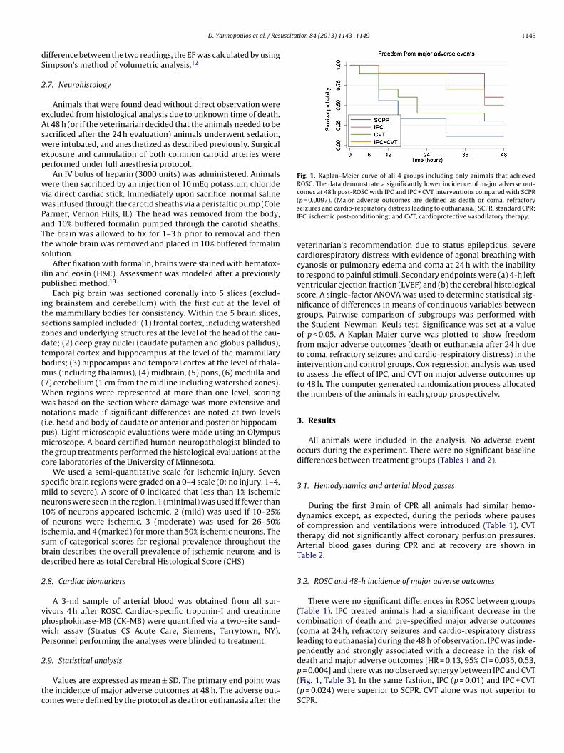
uscitation 84 (2013) 1143– 1149 1145
dS
2
eAswep
wvwPaTts
ip
itszdtbm(Wwn(pmtc
smn1oisbd
2
vpwP
2
tc
Fig. 1. Kaplan–Meier curve of all 4 groups including only animals that achievedROSC. The data demonstrate a significantly lower incidence of major adverse out-comes at 48 h post-ROSC with IPC and IPC + CVT interventions compared with SCPR(p = 0.0097). (Major adverse outcomes are defined as death or coma, refractory
D. Yannopoulos et al. / Res
ifference between the two readings, the EF was calculated by usingimpson’s method of volumetric analysis.12
.7. Neurohistology
Animals that were found dead without direct observation werexcluded from histological analysis due to unknown time of death.t 48 h (or if the veterinarian decided that the animals needed to beacrificed after the 24 h evaluation) animals underwent sedation,ere intubated, and anesthetized as described previously. Surgical
xposure and cannulation of both common carotid arteries wereerformed under full anesthesia protocol.
An IV bolus of heparin (3000 units) was administered. Animalsere then sacrificed by an injection of 10 mEq potassium chloride
ia direct cardiac stick. Immediately upon sacrifice, normal salineas infused through the carotid sheaths via a peristaltic pump (Cole
armer, Vernon Hills, IL). The head was removed from the body,nd 10% buffered formalin pumped through the carotid sheaths.he brain was allowed to fix for 1–3 h prior to removal and thenhe whole brain was removed and placed in 10% buffered formalinolution.
After fixation with formalin, brains were stained with hematox-lin and eosin (H&E). Assessment was modeled after a previouslyublished method.13
Each pig brain was sectioned coronally into 5 slices (exclud-ng brainstem and cerebellum) with the first cut at the level ofhe mammillary bodies for consistency. Within the 5 brain slices,ections sampled included: (1) frontal cortex, including watershedones and underlying structures at the level of the head of the cau-ate; (2) deep gray nuclei (caudate putamen and globus pallidus),emporal cortex and hippocampus at the level of the mammillaryodies; (3) hippocampus and temporal cortex at the level of thala-us (including thalamus), (4) midbrain, (5) pons, (6) medulla and
7) cerebellum (1 cm from the midline including watershed zones).hen regions were represented at more than one level, scoringas based on the section where damage was more extensive andotations made if significant differences are noted at two levelsi.e. head and body of caudate or anterior and posterior hippocam-us). Light microscopic evaluations were made using an Olympusicroscope. A board certified human neuropathologist blinded to
he group treatments performed the histological evaluations at theore laboratories of the University of Minnesota.
We used a semi-quantitative scale for ischemic injury. Sevenpecific brain regions were graded on a 0–4 scale (0: no injury, 1–4,ild to severe). A score of 0 indicated that less than 1% ischemic
eurons were seen in the region, 1 (minimal) was used if fewer than0% of neurons appeared ischemic, 2 (mild) was used if 10–25%f neurons were ischemic, 3 (moderate) was used for 26–50%schemia, and 4 (marked) for more than 50% ischemic neurons. Theum of categorical scores for regional prevalence throughout therain describes the overall prevalence of ischemic neurons and isescribed here as total Cerebral Histological Score (CHS)
.8. Cardiac biomarkers
A 3-ml sample of arterial blood was obtained from all sur-ivors 4 h after ROSC. Cardiac-specific troponin-I and creatininehosphokinase-MB (CK-MB) were quantified via a two-site sand-ich assay (Stratus CS Acute Care, Siemens, Tarrytown, NY).
ersonnel performing the analyses were blinded to treatment.
.9. Statistical analysis
Values are expressed as mean ± SD. The primary end point washe incidence of major adverse outcomes at 48 h. The adverse out-omes were defined by the protocol as death or euthanasia after the
seizures and cardio-respiratory distress leading to euthanasia.) SCPR, standard CPR;IPC, ischemic post-conditioning; and CVT, cardioprotective vasodilatory therapy.
veterinarian’s recommendation due to status epilepticus, severecardiorespiratory distress with evidence of agonal breathing withcyanosis or pulmonary edema and coma at 24 h with the inabilityto respond to painful stimuli. Secondary endpoints were (a) 4-h leftventricular ejection fraction (LVEF) and (b) the cerebral histologicalscore. A single-factor ANOVA was used to determine statistical sig-nificance of differences in means of continuous variables betweengroups. Pairwise comparison of subgroups was performed withthe Student–Newman–Keuls test. Significance was set at a valueof p < 0.05. A Kaplan Maier curve was plotted to show freedomfrom major adverse outcomes (death or euthanasia after 24 h dueto coma, refractory seizures and cardio-respiratory distress) in theintervention and control groups. Cox regression analysis was usedto assess the effect of IPC, and CVT on major adverse outcomes upto 48 h. The computer generated randomization process allocatedthe numbers of the animals in each group prospectively.
3. Results
All animals were included in the analysis. No adverse eventoccurs during the experiment. There were no significant baselinedifferences between treatment groups (Tables 1 and 2).
3.1. Hemodynamics and arterial blood gasses
During the first 3 min of CPR all animals had similar hemo-dynamics except, as expected, during the periods where pausesof compression and ventilations were introduced (Table 1). CVTtherapy did not significantly affect coronary perfusion pressures.Arterial blood gases during CPR and at recovery are shown inTable 2.
3.2. ROSC and 48-h incidence of major adverse outcomes
There were no significant differences in ROSC between groups(Table 1). IPC treated animals had a significant decrease in thecombination of death and pre-specified major adverse outcomes(coma at 24 h, refractory seizures and cardio-respiratory distressleading to euthanasia) during the 48 h of observation. IPC was inde-pendently and strongly associated with a decrease in the risk ofdeath and major adverse outcomes [HR = 0.13, 95% CI = 0.035, 0.53,p = 0.004] and there was no observed synergy between IPC and CVT
(Fig. 1, Table 3). In the same fashion, IPC (p = 0.01) and IPC + CVT(p = 0.024) were superior to SCPR. CVT alone was not superior toSCPR.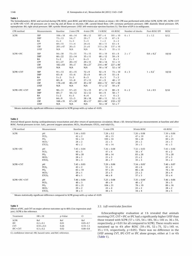
1146 D. Yannopoulos et al. / Resuscitation 84 (2013) 1143– 1149
Table 1The hemodynamics, ROSC and survival during CPR, ROSC, post-ROSC and 48 h.Values are shown as mean ± SD. CPR was performed with either SCPR, SCPR + IPC, SCPR + CVTor SCPR + IPC + CVT. All pressures are in mm Hg and all flows in mL/min. CBF, carotid blood flow; CPP, coronary perfusion pressure; DBP, diastolic blood pressure; EPI,epinephrine; RA, right atrial pressure; SBP, systolic blood pressure; LVEF, left ventricular ejection fraction (%). The dose of EPI is in milligrams.
CPR method Measurements Baseline 2 min CPR 4 min CPR 1 h ROSC 4 h ROSC Number of shocks Post-ROSC EPI ROSC
SCPR SBP 104 ± 18 44 ± 14 68 ± 12 107 ± 4 101 ± 10 6 ± 1 2 ± 1.2 9/12DBP 73 ± 13 14 ± 7 29 ± 7 67 ± 15 67 ± 9RA 4 ± 2 3 ± 3 5 ± 3 1 ± 2 4 ± 2CPP 69 ± 15 11 ± 4 24 ± 6 66 ± 11 63 ± 4CBF 201 ± 47 26 ± 5 21 ± 6 113 ± 26 137 ± 16LVEF N/A N/A N/A 36 ± 5 35 ± 11
SCPR + IPC SBP 94 ± 30 73 ± 31 75 ± 15 95 ± 19 83 ± 8 2 ± 1* 0.8 ± 0.2* 10/10DBP 66 ± 22 22 ± 14 35 ± 11 68 ± 12 59 ± 9RA 5 ± 2 2 ± 1 6 ± 3 9 ± 3 8 ± 1CPP 61 ± 27 20 ± 15* 29 ± 15 58 ± 14 51 ± 11CBF 182 ± 83 39 ± 3* 56 ± 27* 214 ± 49* 227 ± 86*
LVEF N/A N/A N/A 56 ± 14* 52 ± 14*
SCPR + CVT SBP 91 ± 13 41 ± 15 72 ± 9 92 ± 9 73 ± 10 4 ± 3 1 ± 0.2* 10/10DBP 65 ± 8 15 ± 6 35 ± 9 69 ± 9 55 ± 8RA 5 ± 2 3 ± 3 8 ± 3 8 ± 3 7 ± 2CPP 60 ± 8 12 ± 8 27 ± 13 61 ± 9 48 ± 17CBF 178 ± 43 48 ± 19* 45 ± 19* 264 ± 72* 167 ± 66LVEF N/A N/A N/A 57 ± 12* 59 ± 9*
SCPR + IPC + CVT SBP 98 ± 11 57 ± 21 72 ± 15 87 ± 13 88 ± 9 6 ± 2 1.4 ± 0.5 9/10DBP 69 ± 7 18 ± 12 32 ± 12 64 ± 9 58 ± 7RA 5 ± 2 6 ± 3 4 ± 4 4 ± 1 6 ± 2CPP 64 ± 9 12 ± 5 28 ± 18 60 ± 11 52 ± 12CBF 168 ± 35 67 ± 30* 49 ± 17* 263 ± 66* 218 ± 53*
LVEF N/A N/A N/A 54 ± 18* 52 ± 7*
* Means statistically significant difference compared to SCPR group with a p value of <0.05.
Table 2Arterial blood gasses during cardiopulmonary resuscitation and after return of spontaneous circulation. Mean ± SD. Arterial blood gas measurements at baseline and afterROSC. Partial pressures in torr. SaO2, percent oxygen saturation; HCO3, bicarbonate; ETCO2, end-tidal CO2.
CPR method Measurement Baseline 5-min CPR 30 min ROSC 4 h ROSC
SCPR pH 7.44 ± 0.03 7.28 ± 0.2 7.26 ± 0.08 7.39 ± 0.06PCO2 42 ± 2 49 ± 13 47 ± 10 41 ± 3PO2 96 ± 14 100 ± 34 98 ± 28 80 ± 25HCO3
− 28 ± 2 22 ± 3 20 ± 2 24 ± 3ETCO2 40 ± 2 43 ± 14 39 ± 5 41 ± 5
SCPR + IPC pH 7.45 ± 0.04 7.26 ± 0.08 7.32 ± 0.03 7.43 ± 0.06PCO2 40 ± 5 47 ± 4 38 ± 4* 41 ± 2PO2 90 ± 20 89 ± 14 83 ± 26 75 ± 20HCO3
− 28 ± 1 21 ± 3 21 ± 2 27 ± 3ETCO2 39 ± 3 45 ± 7 35 ± 3 35 ± 2
SCPR + CVT pH 7.45 ± 0.03 7.29 ± 0.06 7.34 ± 0.03* 7.40 ± 0.07PCO2 43 ± 2 51 ± 5 43 ± 6 42 ± 3PO2 83 ± 19 97 ± 25 86 ± 26 76 ± 18HCO3
− 29 ± 1 25 ± 5 23 ± 2 26 ± 4ETCO2 40 ± 1 44 ± 8 37 ± 3 35 ± 4
SCPR + IPC + CVT pH 7.46 ± 0.04 7.25 ± 0.08 7.35 ± 0.04* 7.46 ± 0.06PCO2 40 ± 3 49 ± 9 40 ± 2* 39 ± 5PO2 83 ± 21 104 ± 21 78 ± 19 89 ± 16HCO3
− 29 ± 2 22 ± 4 22 ± 2 28 ± 2ETCO2 38 ± 3 44 ± 5 34 ± 4 34 ± 5
* Means statistically significant difference compared to SCPR group with a p value of <0
Table 3Effects of IPC, and CVT on major adverse outcomes up to 48 h (Cox regression anal-ysis). SCPR is the reference.
Treatment HR ± SE p-Value CI
SCPR Ref Ref RefIPC 0.2 ± 0.1 0.01 0.05–0.7CVT 0.55 ± 0.3 0.26 0.19–1.5IPC + CVT 0.3 ± 0.2 0.02 0.08–0.8
CI, confidence interval; HR, hazard ratio; and Ref, reference.
.05
3.3. Left ventricular function
Echocardiographic evaluation at 1 h revealed that animalsreceiving CVT, CVT + IPC or IPC had a significantly higher LVEF thanthose treated with SCPR (57 ± 12%, 54 ± 18%, 56 ± 14% vs. 36 ± 5%,respectively, p < 0.01 for all compared to SCPR). These results were
sustained up to 4 h after ROSC (59 ± 9%, 52 ± 7%, 52 ± 14% vs.35 ± 11%, respectively, p < 0.05). There was no difference in theLVEF among CVT, IPC-CVT or IPC alone groups, either at 1 or 4 h(Table 1).
D. Yannopoulos et al. / Resuscitation 84 (2013) 1143– 1149 1147
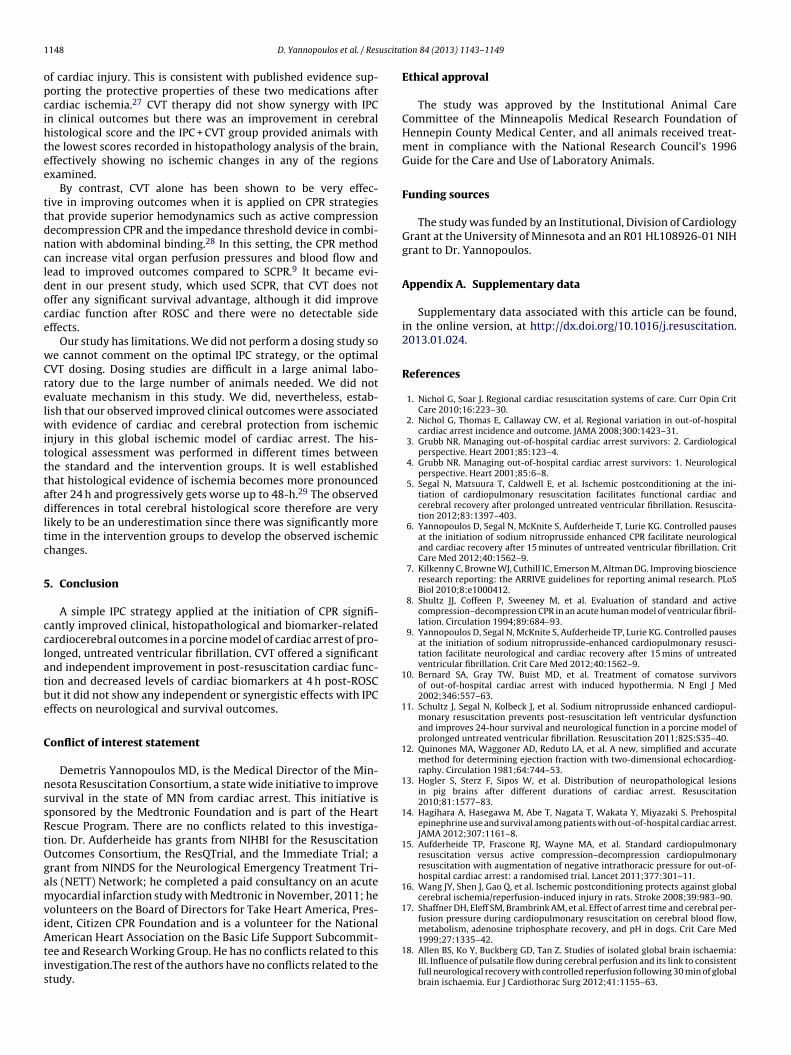
Fig. 2. Left panel: severe ischemic brain injury (SCPR), right panel: minimal ischemic brain injury (IPC) representative samples of severe versus minimal histologic ischemicb ealthyi
3
a(wIaId
3
2I2wCC1i
3
gcwfhcS(gd
4
cibdt
rain injury. Purple condensed nuclei are a marker of ischemic insult (red arrow). Hn this figure legend, the reader is referred to the web version of the article.)
.4. Cardiac biomarkers
Plasma was obtained at 4 h in all animals. IPC, CVT + IPC and CVTlone resulted in significantly lower CK-MB and Troponin-I levelsall in �g/L) at 4 h compared to SCPR controls. The Troponin-I levelas 31 ± 34 at 4 h in SCPR as compared to 8.5 ± 7, 7 ± 7, 5 ± 5 for
PC, CVT, IPC + CVT, respectively (p < 0.05). CK-MB was measuredt 37 ± 24 in SCPR group compared to 13 ± 10, 18 ± 13, 11 ± 9 forPC, CVT and IPC + CVT groups, respectively (p < 0.05). There was noifference the cardiac biomarkers among the intervention groups.
.5. Neurologic function
Blinded assessment of cerebral performance category (CPC) at4 h on the live animals showed improvement in the CVT, CVT-
PC and IPC groups compared to SCPR group (2.6 ± 0.9, 2.25 ± 1,.75 ± 0.4 vs. 3.5 ± 0.5, respectively, p < 0.05 for all). At 48 h, thereas only one animal that survived in the SCPR group and hadPC score of 3 (severe deficit). Surviving animals treated with CVT,VT + IPC, and IPC alone at 48 h had a mean CPC score of 2.3 ± 1.6,.8 ± 0.8, 2.2 ± 0.9 with no difference between the groups, but with
mprovement in all intervention groups at 48 h compared with 24 h.
.6. Neurohistopathology
The mean time for brain harvest was shorter in the SCPRroup since more animals died earlier or had major adverse out-omes requiring euthanasia. The mean harvest time for SCPRas 20 ± 12 h compared to 39.0 ± 12.4, 38.4 ± 13 and 42.0 ± 12.0 h
or IPC, CVT + IPC, CVT, respectively. Despite later evaluation ofistopathological samples, IPC and IPC + CVT resulted in signifi-antly lower total cerebral histological score (CHS) compared toCPR group (5.8 ± 2.6, 2.8 ± 1.8 vs. 10 ± 2.1, respectively, p < 0.01)Fig. 2). One animal in the IPC group and two animals in the IPC + CVTroup showed no ischemic damage at 48 h (≤2 total CHS). CVT aloneid not improve CHS (6.6 ± 3.7) compared to SCPR group (p = 0.1).
. Discussion
Results from this investigation demonstrate that cardiac anderebral function can be preserved after prolonged global ischemic
nsult of 15 min of untreated ventricular fibrillation cardiac arresty early application of ischemic post-conditioning and use of car-ioprotective vasodilators during CPR. These findings provide, forhe first time, strong support for a simple BLS strategy that includesneurons are shown with blue arrow. (For interpretation of the references to color
four controlled pauses of compressions during the first 3 min ofCPR improves post-resuscitation LV function. This strategy alsodecreases the levels of cardiac biomarkers of injury at 4 h and leadsto a significant decrease of the ischemic histological injury of thebrain leading to better neurological outcomes at 24 and 48 h com-pared to standard CPR.
In this study, ROSC was not a predictor of neurological and car-diac function post-resuscitation. This finding that is consistent withlarge human studies showing dissociation between ROSC rates andimproved survival with good neurological function.14,15
Our data suggest that IPC is the most important factor lead-ing to a significant improvement in cardiac function and survivalwith good neurological function. IPC decreased that risk of deathand major adverse events by almost 80% compared to SCPR. Itwas also associated with significant improvement in cerebral his-tological injury; a combination of improved survival and cerebralpreservation is currently considered the ultimate outcome for anyintervention evaluated during cardiac arrest.
Our study is also in concordance with the published study byWang et al. that showed that IPC was effective in protecting thebrains of rats from a global 10-min ischemic insult that were not incardiac arrest.16 This new observation should cause reassessmentof the notion that cerebral recovery is not feasible after 10–12 minof cardiac arrest.17 In a recent series of papers, Allen et al. haveshown that controlled reperfusion of the brain with the use ofbypass and a special reperfusion solution that mitigates reperfusioninjury can result in the absence of ischemic changes in the braineven after 30 min of isolated global cerebral ischemia.18,19 Basedon similar principles, we have provided a simple method of IPC(with four 20-s pauses in compressions and ventilation during thefirst 3 min of CPR) that could be easily applied in the clinical settingand translated to patients receiving CPR for treatment of cardiacarrest.
We used IPC during CPR because of the described mechanismsof protection from reperfusion injury which currently is thought tobe modulation of the opening of mitochondrial permeability transi-tion pores (mPTP) and KATP channels.20–26 Our results support thecontention that the previously described protective mechanismscan also be observed during the global ischemia and reperfusion ofcardiac arrest, decreasing vital organ injury as documented, in thisstudy, with the histological evaluation of the brain and functional
and biomarker assessment of the surviving animals.CVT with sodium nitroprusside and adenosine did not improveneurological outcomes but led to an independent improvement inpost-resuscitation cardiac function and lower levels of biomarkers
1 uscita
opcihtee
ttdncldoce
wCrelwitttadltc
5
cclatbe
C
nssRtOgamviAtis
1
1
1
1
1
1
1
1
148 D. Yannopoulos et al. / Res
f cardiac injury. This is consistent with published evidence sup-orting the protective properties of these two medications afterardiac ischemia.27 CVT therapy did not show synergy with IPCn clinical outcomes but there was an improvement in cerebralistological score and the IPC + CVT group provided animals withhe lowest scores recorded in histopathology analysis of the brain,ffectively showing no ischemic changes in any of the regionsxamined.
By contrast, CVT alone has been shown to be very effec-ive in improving outcomes when it is applied on CPR strategieshat provide superior hemodynamics such as active compressionecompression CPR and the impedance threshold device in combi-ation with abdominal binding.28 In this setting, the CPR methodan increase vital organ perfusion pressures and blood flow andead to improved outcomes compared to SCPR.9 It became evi-ent in our present study, which used SCPR, that CVT does notffer any significant survival advantage, although it did improveardiac function after ROSC and there were no detectable sideffects.
Our study has limitations. We did not perform a dosing study soe cannot comment on the optimal IPC strategy, or the optimalVT dosing. Dosing studies are difficult in a large animal labo-atory due to the large number of animals needed. We did notvaluate mechanism in this study. We did, nevertheless, estab-ish that our observed improved clinical outcomes were associated
ith evidence of cardiac and cerebral protection from ischemicnjury in this global ischemic model of cardiac arrest. The his-ological assessment was performed in different times betweenhe standard and the intervention groups. It is well establishedhat histological evidence of ischemia becomes more pronouncedfter 24 h and progressively gets worse up to 48-h.29 The observedifferences in total cerebral histological score therefore are very
ikely to be an underestimation since there was significantly moreime in the intervention groups to develop the observed ischemichanges.
. Conclusion
A simple IPC strategy applied at the initiation of CPR signifi-antly improved clinical, histopathological and biomarker-relatedardiocerebral outcomes in a porcine model of cardiac arrest of pro-onged, untreated ventricular fibrillation. CVT offered a significantnd independent improvement in post-resuscitation cardiac func-ion and decreased levels of cardiac biomarkers at 4 h post-ROSCut it did not show any independent or synergistic effects with IPCffects on neurological and survival outcomes.
onflict of interest statement
Demetris Yannopoulos MD, is the Medical Director of the Min-esota Resuscitation Consortium, a state wide initiative to improveurvival in the state of MN from cardiac arrest. This initiative isponsored by the Medtronic Foundation and is part of the Heartescue Program. There are no conflicts related to this investiga-ion. Dr. Aufderheide has grants from NIHBI for the Resuscitationutcomes Consortium, the ResQTrial, and the Immediate Trial; arant from NINDS for the Neurological Emergency Treatment Tri-ls (NETT) Network; he completed a paid consultancy on an acuteyocardial infarction study with Medtronic in November, 2011; he
olunteers on the Board of Directors for Take Heart America, Pres-dent, Citizen CPR Foundation and is a volunteer for the National
merican Heart Association on the Basic Life Support Subcommit-ee and Research Working Group. He has no conflicts related to thisnvestigation.The rest of the authors have no conflicts related to thetudy.
1
tion 84 (2013) 1143– 1149
Ethical approval
The study was approved by the Institutional Animal CareCommittee of the Minneapolis Medical Research Foundation ofHennepin County Medical Center, and all animals received treat-ment in compliance with the National Research Council’s 1996Guide for the Care and Use of Laboratory Animals.
Funding sources
The study was funded by an Institutional, Division of CardiologyGrant at the University of Minnesota and an R01 HL108926-01 NIHgrant to Dr. Yannopoulos.
Appendix A. Supplementary data
Supplementary data associated with this article can be found,in the online version, at http://dx.doi.org/10.1016/j.resuscitation.2013.01.024.
References
1. Nichol G, Soar J. Regional cardiac resuscitation systems of care. Curr Opin CritCare 2010;16:223–30.
2. Nichol G, Thomas E, Callaway CW, et al. Regional variation in out-of-hospitalcardiac arrest incidence and outcome. JAMA 2008;300:1423–31.
3. Grubb NR. Managing out-of-hospital cardiac arrest survivors: 2. Cardiologicalperspective. Heart 2001;85:123–4.
4. Grubb NR. Managing out-of-hospital cardiac arrest survivors: 1. Neurologicalperspective. Heart 2001;85:6–8.
5. Segal N, Matsuura T, Caldwell E, et al. Ischemic postconditioning at the ini-tiation of cardiopulmonary resuscitation facilitates functional cardiac andcerebral recovery after prolonged untreated ventricular fibrillation. Resuscita-tion 2012;83:1397–403.
6. Yannopoulos D, Segal N, McKnite S, Aufderheide T, Lurie KG. Controlled pausesat the initiation of sodium nitroprusside enhanced CPR facilitate neurologicaland cardiac recovery after 15 minutes of untreated ventricular fibrillation. CritCare Med 2012;40:1562–9.
7. Kilkenny C, Browne WJ, Cuthill IC, Emerson M, Altman DG. Improving bioscienceresearch reporting: the ARRIVE guidelines for reporting animal research. PLoSBiol 2010;8:e1000412.
8. Shultz JJ, Coffeen P, Sweeney M, et al. Evaluation of standard and activecompression–decompression CPR in an acute human model of ventricular fibril-lation. Circulation 1994;89:684–93.
9. Yannopoulos D, Segal N, McKnite S, Aufderheide TP, Lurie KG. Controlled pausesat the initiation of sodium nitroprusside-enhanced cardiopulmonary resusci-tation facilitate neurological and cardiac recovery after 15 mins of untreatedventricular fibrillation. Crit Care Med 2012;40:1562–9.
0. Bernard SA, Gray TW, Buist MD, et al. Treatment of comatose survivorsof out-of-hospital cardiac arrest with induced hypothermia. N Engl J Med2002;346:557–63.
1. Schultz J, Segal N, Kolbeck J, et al. Sodium nitroprusside enhanced cardiopul-monary resuscitation prevents post-resuscitation left ventricular dysfunctionand improves 24-hour survival and neurological function in a porcine model ofprolonged untreated ventricular fibrillation. Resuscitation 2011;82S:S35–40.
2. Quinones MA, Waggoner AD, Reduto LA, et al. A new, simplified and accuratemethod for determining ejection fraction with two-dimensional echocardiog-raphy. Circulation 1981;64:744–53.
3. Hogler S, Sterz F, Sipos W, et al. Distribution of neuropathological lesionsin pig brains after different durations of cardiac arrest. Resuscitation2010;81:1577–83.
4. Hagihara A, Hasegawa M, Abe T, Nagata T, Wakata Y, Miyazaki S. Prehospitalepinephrine use and survival among patients with out-of-hospital cardiac arrest.JAMA 2012;307:1161–8.
5. Aufderheide TP, Frascone RJ, Wayne MA, et al. Standard cardiopulmonaryresuscitation versus active compression–decompression cardiopulmonaryresuscitation with augmentation of negative intrathoracic pressure for out-of-hospital cardiac arrest: a randomised trial. Lancet 2011;377:301–11.
6. Wang JY, Shen J, Gao Q, et al. Ischemic postconditioning protects against globalcerebral ischemia/reperfusion-induced injury in rats. Stroke 2008;39:983–90.
7. Shaffner DH, Eleff SM, Brambrink AM, et al. Effect of arrest time and cerebral per-fusion pressure during cardiopulmonary resuscitation on cerebral blood flow,metabolism, adenosine triphosphate recovery, and pH in dogs. Crit Care Med
1999;27:1335–42.8. Allen BS, Ko Y, Buckberg GD, Tan Z. Studies of isolated global brain ischaemia:III. Influence of pulsatile flow during cerebral perfusion and its link to consistentfull neurological recovery with controlled reperfusion following 30 min of globalbrain ischaemia. Eur J Cardiothorac Surg 2012;41:1155–63.

uscita
1
2
2
2
2
2
2
2
2
2
D. Yannopoulos et al. / Res
9. Allen BS, Ko Y, Buckberg GD, Tan Z. Studies of isolated global brain ischaemia: II.Controlled reperfusion provides complete neurologic recovery following 30 minof warm ischaemia – the importance of perfusion pressure. Eur J CardiothoracSurg 2012;41:1147–54.
0. Zhao ZQ. Postconditioning in reperfusion injury: a status report. CardiovascDrugs Ther 2010;24:265–79.
1. Argaud L, Gateau-Roesch O, Raisky O, Loufouat J, Robert D, Ovize M.Postconditioning inhibits mitochondrial permeability transition. Circulation2005;111:194–7.
2. Cour M, Gomez L, Mewton N, Ovize M, Argaud L. Postconditioning: from thebench to bedside. J Cardiovasc Pharmacol Ther 2011;16:117–30.
3. Eldaif SM, Deneve JA, Wang NP, et al. Attenuation of renal ischemia–reperfusion
injury by postconditioning involves adenosine receptor and protein kinase Cactivation. Transpl Int 2010;23:217–26.4. Kin H, Zatta AJ, Lofye MT, et al. Postconditioning reduces infarct size viaadenosine receptor activation by endogenous adenosine. Cardiovasc Res2005;67:124–33.
2
tion 84 (2013) 1143– 1149 1149
5. Mykytenko J, Reeves JG, Kin H, et al. Persistent beneficial effect of postcon-ditioning against infarct size: role of mitochondrial K(ATP) channels duringreperfusion. Basic Res Cardiol 2008;103:472–84.
6. Ovize M, Baxter GF, Di Lisa F, et al. Postconditioning and protection from reper-fusion injury: where do we stand? Position paper from the Working Group ofCellular Biology of the Heart of the European Society of Cardiology. CardiovascRes 2010;87:406–23.
7. Hillegass WB, Dean NA, Liao L, Rhinehart RG, Myers PR. Treatment of no-reflowand impaired flow with the nitric oxide donor nitroprusside following percu-taneous coronary interventions: initial human clinical experience. J Am CollCardiol 2001;37:1335–43.
8. Yannopoulos D, Matsuura T, Schultz J, Rudser K, Halperin HR, Lurie KG. Sodium
nitroprusside enhanced cardiopulmonary resuscitation improves survival withgood neurological function in a porcine model of prolonged cardiac arrest. CritCare Med 2011;39:1269–74.9. Back T, Schuler OG. The natural course of lesion development in brain ischemia.Acta Neurochir Suppl 2004;89:55–61.


