Embed Size (px)
Citation preview
1057
BIOLOGY OF REPRODUCTION 65, 1057–1066 (2001)
Involvement of Sex Steroid Hormones in the Early Stages of Spermatogenesisin Japanese Huchen (Hucho perryi )1
Mohamed A. Amer,3 Takeshi Miura,2,4 Chiemi Miura,3 and Kohei Yamauchi3
Division of Marine Biosciences, Graduate School of Fisheries Science,3 and Field Science Center for NorthernBiosphere,4 Hokkaido University, Hakodate 041-8611, Hokkaido, Japan
ABSTRACT
In higher vertebrates, considerable progress has been madein understanding the endocrine regulation of puberty; however,in teleosts, the regulatory mechanisms of spermatogenesis dur-ing the first annual cycle remain unclear. The present study wasconducted to understand the regulatory mechanisms of sper-matogenesis throughout the different stages of the first sper-matogenic cycle and to check the ability of various steroids andhormones to induce in vitro spermatogonial proliferation in Jap-anese huchen (Hucho perryi ). The results indicate that the se-rum level of 11-ketotestosterone (11-KT) was positively associ-ated with germ cell type; the level first began to rise with theappearance of late-type B spermatogonia and continued to in-crease gradually throughout the active spermatogenic stages andspermiogenesis, reaching a peak value 2 wk before spawning,and then declined. During the spermatogenic stages, the serumconcentration of 17a,20b-dihydroxy-4-pregnen-3-one (17a,20b-DP) was undetectable. Only a small peak was detected with theappearance of spermatocytes and spermatids, and at the time ofspawning, the level increased dramatically, reaching its maxi-mum value with the onset of milt production. Despite the highvariation in serum levels of 17b-estradiol (E2) both betweenmonths and among the individuals, E2 was found during thewhole reproductive cycle. From these results, we concluded that1) 11-KT is necessary for the initiation of spermatogenesis andsperm production, and it probably plays a role in spermiation,2) 17a,20b-DP is essential for the final maturation stage, couldplay a significant role in the mitosis phase and meiosis process,and probably participates in the regulation of spawning behav-ior, and 3) estrogen is an indispensable male hormone that playsa physiological role in some aspects of testicular functions, es-pecially during the mitotic phase. The three steroids were alsoable to induce DNA synthesis, spermatogonial renewal, and/orspermatogonial proliferation in vitro.
gametogenesis, puberty, spermatogenesis, steroid hormones, tes-tis
INTRODUCTION
In vertebrates, puberty is the period during which a ju-venile acquires the capacity to reproduce. It is characterizedby an activation of the brain-pituitary gonadal axes. Inhigher vertebrates, considerable progress has been made inunderstanding the endocrine regulation of puberty, but in
1Supported by a doctoral scholarship from the Mission Department of theMinistry of Education, Egypt, and in part by grants-in-aid from the Ministryof Agriculture, Forestry and Fisheries of Japan (BDP-01-1V-2-1), and fromThe Japan Society for the Promotion of Science.2Correspondence. FAX: 81 138 40 5545;e-mail: [email protected]
Received: 26 March 2001.First decision: 16 April 2001.Accepted: 21 May 2001.Q 2001 by the Society for the Study of Reproduction, Inc.ISSN: 0006-3363. http://www.biolreprod.org
teleosts, the regulatory mechanisms underlying this acti-vation are still poorly understood [1, 2]. Spermatogenesisis a continuum of cellular differentiation in which threeprincipal phases can be discerned: spermatogonial renewaland proliferation, meiosis, and spermiogenesis [3, 4]. Invertebrates, including nonmammals, germ cell developmentand maturation appear to proceed in a similar fashion [5].In most teleost species, including salmonids, the testis is ofunrestricted spermatogonial-testis type, characterized byspermatogonia along the entire length of the testicular lob-ules, and the unit of spermatogenesis is the spermatocyst.Within each cyst, germ cell development occurs synchro-nously [6–10]. During the prepubertal period, spermato-gonial stem cells and Sertoli cells occur as solid cords with-in the testicular lobule [5]. With the onset of spermatogen-esis, spermatogonial stem cells divide to produce genera-tions of spermatogonia, which enter the spermatogeniccycle. Spermatogonia type A divide several times to pro-duce spermatogonia type B, which, in turn, develop intospermatocytes and then undergo meiosis to produce sper-matids. The spermatids then proceed through a morpholog-ical metamorphosis during spermiogenesis to form sper-matozoa. Spermiation results from the breakdown of Sertolicells that form the spermatogenic cyst wall. In salmonids,the two major events of the testicular cycle (spermatogen-esis and spermiation) are temporally separated by a stageof spermatozoal maturation during which the spermatozoaundergo physiological changes [6].
In teleosts, as in other vertebrates, spermatogenesis isessentially regulated by pituitary gonadotropin and testic-ular factors, including androgens. Gonadotropin stimulatesandrogen production by Leydig cells and, eventually, con-trols spermatogenesis and spermiation [6, 11–13]. Amongthe androgenic steroids, 11-ketotestosterone (11-KT) is themajor androgen in most teleost species [14], especially dur-ing the later stages of spermatogenesis [15–20]. The pro-gestin 17a,20b-dihydroxy-4-pregnen-3-one (17a,20b-DP)could play a direct or indirect role in the mechanism ofspermiation of male salmonids via regulation of the ioniccomposition of the seminal fluid and the acquisition ofsperm motility [21, 22]. A number of studies have men-tioned that a distinct shift from androgen to 17a,20b-DPappears to occur in the testes of several teleost fish aroundthe onset of spermiation. These results suggest that, in sev-eral species of salmonids and in the Japanese eel (Anguillajaponica), 17a,20b-DP is essential for the final testicularor sperm maturation [18, 19, 22–29]. Estrogens, derivedeither from local aromatization of androgens or producedby the testis, can exert feedback effects on the neuroen-docrine components of the male reproductive axis, but re-cent evidence suggests that estrogen may also exert para-crine actions within the testis itself [30]. Recently, it hasbeen reported that implantation of 17b-estradiol (E2) pro-motes spermatogonial renewal in Japanese eel [31] and in-duces a full, qualitatively normal spermatogenesis in hy-
Dow
nloaded from https://academ
ic.oup.com/biolreprod/article/65/4/1057/2723386 by guest on 25 M
arch 2022

1058 AMER ET AL.
b
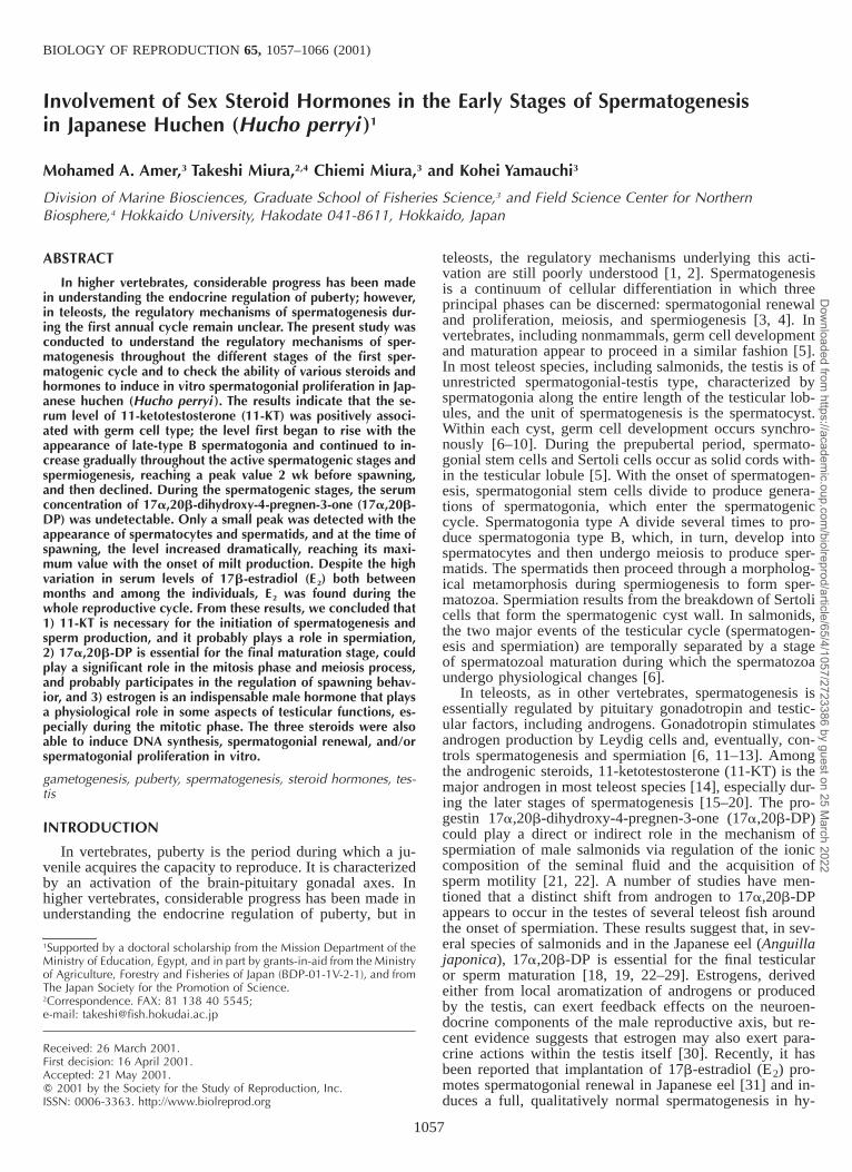
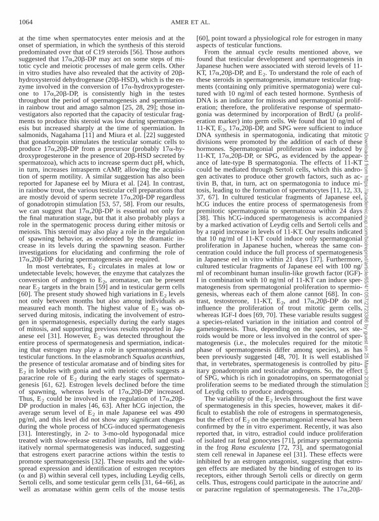
FIG. 1. Monthly changes in water temperature (A), total body length (B),and body weight (C) during the sampling period of male Japanese huchenfrom the prepubertal stage (42 mo of age) to the spawning time (57 moof age). Values are displayed as mean 6 SEM. During March 1999, sam-pling were carried out weekly, so the value represents the mean of allsamplings.
pogonadal mice [32], suggesting that estrogen is an indis-pensable male hormone. Thus, in fish, differentiation ofspermatogonia into spermatozoa requires several steroidhormones. The role of androgen and progestin has beenelucidated, in large part, through the correlation of seasonalchanges in serum hormone levels with the different stagesof the annual testicular cycle in a variety of fish species.However, it is not clear whether the progression of germinalcells into different stages of spermatogenesis is regulatedby changes in these hormonal signals; in other words, thespecific role played by a specific hormone of these steroidsthroughout the different stages of spermatogenesis remainsunclear. Previously, our in vivo and in vitro studies on therole of these steroids in Japanese eel (an excellent systemfor analyzing the mechanisms controlling spermatogenesisdue to the presence of only type A and early type B sper-matogonia) showed that 1) 11-KT is necessary for the ini-tiation of spermatogenesis [33], 2) E2 promotes stem cellrenewal [31], and 3) 17a,20b-DP is essential for the ac-quisition of sperm motility and milt production [24].
Although salmonids have been the subject of more pub-lished papers than any other group of teleosts, one of theirsubgroups, the huchen (Hucho perryi), has not been wellstudied. In Japan, huchen is the largest salmonid. It is alsoa late-maturing species, so the quantity of available spermfrom a mature male is very limited [34]. Recently, huchenhas been considered as a new species for aquaculture; there-fore, a source of gametes is necessary for propagating andpreserving the species. To our knowledge, no informationexists concerning its reproductive process, particularly sper-matogenesis and serum sex-steroid profiles of male Japa-nese huchen. The present study was conducted using huch-en as a new system for the analysis of mechanisms con-trolling spermatogenesis to understand the physiologicalchanges that occur both before and during puberty. Duringthis period, we studied the testicular development, histo-logical changes, and serum steroid profiles of 11-KT,17a,20b-DP, and E2. For this purpose, fish were sampledfrom the prepubertal stage (testes contained only primitivespermatogonia) to the first sexually mature stage (testesfilled with spermatozoa, milt could be stripped by abdom-inal massage). The in vitro study was carried out on pre-pubertal fish to check the ability of various hormones andsteroids to induce spermatogonial proliferation in culturedtesticular fragments.
MATERIALS AND METHODS
FishThree to six male huchen were sampled monthly starting from the
prepuberty stage (December 1997) until the spawning season (March1999). In March 1999, sampling was carried out weekly. The fish werereared at the Nanae Fish Culture Experimental Station, Faculty of Fish-eries, Hokkaido University, in outdoor ponds supplied with river waterunder natural water temperature and photoperiod and were fed ad libitumwith commercial salmon food (Trout Fodder; Oriental Yeast Co., Ltd.,Tokyo, Japan). During the period of study, the lowest water temperature(1.88C) and the highest (18.78C) were recorded on January 7, 1998 andAugust 21, 1998, respectively (Fig. 1A). The fish were killed immediatelyby a blow to the head, and blood samples were collected from the caudal
Dow
nloaded from https://academ
ic.oup.com/biolreprod/article/65/4/1057/2723386 by guest on 25 M
arch 2022
1059STEROID HORMONES AND EARLY STAGES OF SPERMATOGENESIS
vein with a syringe, transferred to centrifuge tubes, and placed on crushedice for 1–2 h. Blood samples were centrifuged at 3000 rpm for 15 min.The serum was then collected and stored at 2208C until hormone assayswere performed. Body weight and total body length were recorded. Thetestes were excised and weighed for calculation of the gonadosomaticindex (GSI; gonad weight/body weight 3 100).
HistologyHistological analysis of spermatogenic activity was performed to cor-
relate testicular development with steroid production. A small piece takenfrom the midportion of the testis of each male huchen was fixed in Bouinsolution, dehydrated through an ascending series of ethanol, and embeddedand blocked in paraffin. Then, 5-mm sections were cut and stained withhematoxylin and eosin. Sections were analyzed under light microscopyand classified into several distinct spermatogenic stages according to themethod of Grier [8]. From each male huchen, five standard histologicalgrids were chosen randomly, and the number of the cysts of each germcell type was counted and expressed as a percentage of the total numberof germ cell cysts. From October 1998 until the end of study period,counts could not be done, because testicular sections contained sperma-tozoa either in cysts or free in lobular lumens or testicular ducts. Therefore,the values of each germ cell type were calculated based on the relativearea of each type in photographs. Light micrographs of testicular sectionsstained with hematoxylin and eosin at different stages of development (seeFig. 3) were prepared from plastic sections (HistoResin Plus; HeraeusKulzer GmbH, Heidelberg, Germany) according to the manufacturer’s in-structions.
RadioimmunoassayAfter two extractions of steroids with diethyl ether, serum levels of
steroids were measured by RIA according to the method of Ueda et al.[27] and Sakai et al. [29] for 11-KT, the method of Young et al. [35] for17a,20b-DP, and the method of Kagawa et al. [36] for E2.
Testicular Organ CultureThe organ culture technique employed was the floating method de-
scribed by Miura et al. [37, 38], with some modifications. Briefly, eightimmature male huchen were killed by a blow to the head, and their tailswere cut. All the following operations were carried out under sterile con-ditions. The fish were then sterilized in 70% (w/v) ethanol, and the testeswere excised and cut into 1 3 1 3 0.5-mm fragments. For each testedtreatment, two testicular fragments of each fish were placed on elder pith(treated with 100% ethanol, washed with distilled water, autoclaved, dried,and covered with a nitrocellulose membrane) in a 24-well plastic culturedish. Each elder pith was floated separately in a culture well containing 1ml of culture medium. The basal culture medium consisted of Leibovitz-15 medium supplemented with 0.1 mM aspartic acid, 0.1 mM glutamicacid, 1.7 mM L-proline, 0.5% (w/v) bovine serum albumin fraction V(Sigma, St. Louis, MO), 1 mg/L bovine insulin, and 10 mM Hepes, ad-justed to pH 7.4 with 1 M NaOH. Steroids were first dissolved in ethanol,except for cortisol, which was dissolved in distilled water, and then allhormones were diluted with the culture medium. The same concentrationof ethanol was added to the cortisol- and nonhormonal (control)-treatedfragments. Testicular fragments were cultured in a medium containing oneof the following hormones: 11-KT, testosterone, 5b-dihydrotestosterone(5b-DHT), androsterone, androstenedione, dehydroepiandrosterone(DHEA), cortisol, E2, 17a,20b-DP, and salmon pituitary glycoprotein frac-tion (SPG, rich in gonadotropin (GTH)-II and contaminated with GTH-Iand probably with thyroid stimulating hormone (TSH) at a dose of 10 ng/ml, then incubated for 15 days in humidified air at 128C. The medium waschanged after 1 wk of culture.
5-Bromo-2-Deoxyuridine LabelingLabeling of 5-bromo-2-deoxyuridine (BrdU) was carried out according
to the manufacturer’s instructions, with minor modification. Because BrdUis incorporated into replicating DNA, detection of the proliferating cell ispossible. Testicular fragments were incubated with BrdU (1 ml/well) forthe last 24 h in culture. After culture, testicular fragments were fixed inBouin solution, and 5-mm paraffin sections were cut and stained immu-nohistochemically. Sections were then counterstained with hematoxylin.The number of immunolabeled germ cells was counted and expressed asa percentage of the total number of germ cells (BrdU index; immunola-beled germ cells/total germ cells 3 100). For each tested hormone, three
randomly chosen, standard histological grids from different sections ofeach animal were viewed with a light microscope.
Statistics
All data except serum steroid concentrations were analyzed using aone-way ANOVA followed by Duncan New Multiple Range Test(SuperAnova; Abacus Concepts, Inc., Berkeley, CA). Steroid data of 11-KT, 17a,20b-DP, and E2 did not follow a normal distribution; therefore,these data were analyzed by a nonparametric statistical test (Kruskal-Wal-lis ANOVA), followed by the Bonferroni multiple-comparison test. In allcases, significance was accepted at P , 0.05. Data are presented as themean 6 SEM.
RESULTS
Body Size
Throughout the sampling period, total body length grad-ually increased from 37.2 6 1.2 cm at 42 mo of age (be-ginning of the sampling) to 49.9 6 1.1 cm at 54 mo of age(December 1998). From January to March 1999, bodylength remained at a relatively constant range of 47–49.5cm. During the same period, average body weight increasedfrom 477 6 51.7 g to 1267.5 6 44.4 g at 42 and 54 moof age, respectively. For both body length and body weight,the differences between values obtained during the prepu-berty and early spermatogenic stages were significantlylower than those obtained during the active spermatogenicstage (Fig. 1, B and C). This indicates that fish may haveto reach a certain size before puberty can begin.
Testicular Development and Serum Steroid Profiles
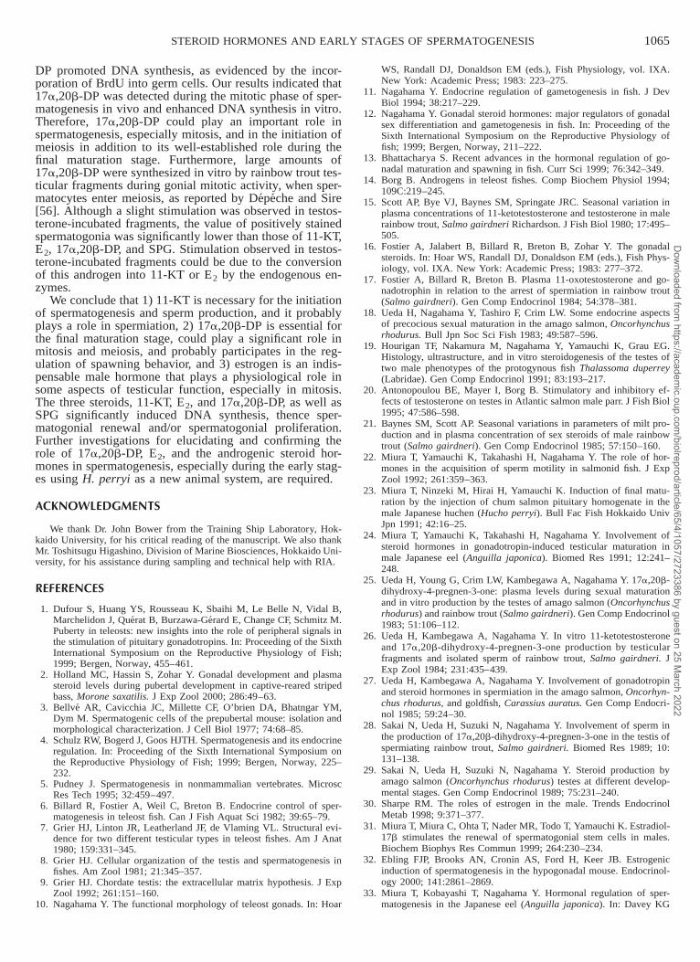
The mean GSIs remained very low (0.01%–0.05%) fromDecember 1997 to May 1998. With the onset of the activespermatogenesis stage, as evidenced by histological data,the GSI increased rapidly, reaching the highest value(1.4%) in September 1998. From November 1998 untilMarch 1999, the GSI remained relatively constant, but thevalues were significantly lower than in September and Oc-tober 1998 (Fig. 2A).
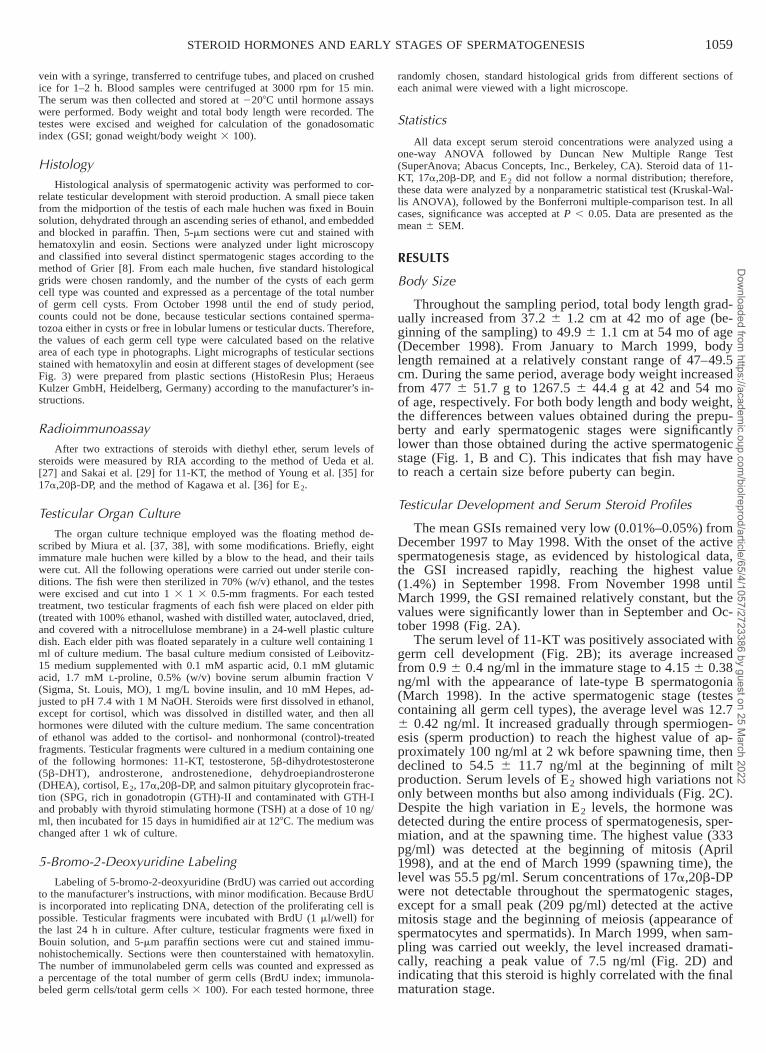
The serum level of 11-KT was positively associated withgerm cell development (Fig. 2B); its average increasedfrom 0.9 6 0.4 ng/ml in the immature stage to 4.15 6 0.38ng/ml with the appearance of late-type B spermatogonia(March 1998). In the active spermatogenic stage (testescontaining all germ cell types), the average level was 12.76 0.42 ng/ml. It increased gradually through spermiogen-esis (sperm production) to reach the highest value of ap-proximately 100 ng/ml at 2 wk before spawning time, thendeclined to 54.5 6 11.7 ng/ml at the beginning of miltproduction. Serum levels of E2 showed high variations notonly between months but also among individuals (Fig. 2C).Despite the high variation in E2 levels, the hormone wasdetected during the entire process of spermatogenesis, sper-miation, and at the spawning time. The highest value (333pg/ml) was detected at the beginning of mitosis (April1998), and at the end of March 1999 (spawning time), thelevel was 55.5 pg/ml. Serum concentrations of 17a,20b-DPwere not detectable throughout the spermatogenic stages,except for a small peak (209 pg/ml) detected at the activemitosis stage and the beginning of meiosis (appearance ofspermatocytes and spermatids). In March 1999, when sam-pling was carried out weekly, the level increased dramati-cally, reaching a peak value of 7.5 ng/ml (Fig. 2D) andindicating that this steroid is highly correlated with the finalmaturation stage.
Dow
nloaded from https://academ
ic.oup.com/biolreprod/article/65/4/1057/2723386 by guest on 25 M
arch 2022

1060 AMER ET AL.
FIG. 2. Monthly changes in GSI (A), se-rum steroid profiles of 11-KT (B), E2 (C),and 17a,20b-DP (D) in male Japanesehuchen from the prepubertal stage to thetime of spawning. During March 1999,blood sampling were carried out weeklyto detect any hormonal changes before thebeginning of milt production. Values aredisplayed as mean 6 SEM. The GSI forMarch 1999 represents the mean of allfish sampled during that month. A star (orstars) above a bar indicate a significantvalue. *P , 0.05, **P , 0.01.
Spermatogenesis and Histology
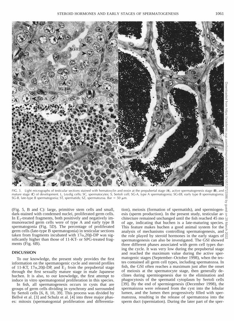
Histologically, the testis of Japanese huchen is of theunrestricted spermatogonial-type according to the termi-nology of Billard et al. [6] and Grier et al. [7]. During theprepubertal stage, the testes consisted mainly of connectivetissue, interstitial cells of Leydig, and the primitive (typeA and early type B) spermatogonia, which occurred withSertoli cells as solid cords within the testicular lobules (Fig.3A). In March 1998, a few late-type B spermatogonial cystsbecame apparent in some individual testicular lobules. InJune 1998, spermatocytes started to appear for the firsttime, and very few cysts of spermatids were observed, in-dicating the initiation of puberty. By August 1998, the tes-ticular lobules had increased in size and were filled withcysts of late-type B spermatogonia, spermatocytes, andspermatids, and occasionally, a few isolated clusters ofspermatozoa were observed in some individuals (beginningof spermiogenesis). The testes underwent very rapid growthand development from September to October 1998, andeach testicular lobule contained all germ cell types (Fig.3B): type A spermatogonia (13%), late-type B spermato-gonia (19%), spermatocytes (17%), spermatids (21%), andspermatozoa (30%). In November and December 1998,more than 60% of germ cells were spermatozoa (Fig. 4).From January 1999 onward, the testicular sections werefilled with spermatozoa either in the testicular lumen or inthe sperm duct (Figs. 3C and 4), and no other germ celltypes were observed, except type A spermatogonia, whichare the starting point for the next spermatogenic cycle. Atthe end of March 1999, milt could be stripped by abdom-
inal massage for the first time, indicating that puberty hadbeen completed.
In Vitro Spermatogonial Proliferation
Immature testicular fragments were cultured in L-15 me-dium with or without one of the following hormones: 11-KT, testosterone, 5b-DHT, androsterone, androstenedione,DHEA, cortisol, E2, 17a,20b-DP, and SPG at a dose of 10ng/ml. After 15 days of culture in testicular sections takenfrom 11-KT-, E2-, 17a,20b-DP-, and SPG-treated frag-ments, BrdU was incorporated into numerous spermatogo-nial cells, compared with the control and all other testedhormones, indicating that DNA was synthesized (Fig. 5).The effect of these hormones on the incorporation of BrdU(as a proliferating marker) by the testicular germ cells wasexamined by calculation of the BrdU index. The BrdU in-dex of the testicular fragments incubated with 11-KT,17a,20b-DP, E2, and SPG was significantly (P , 0.01)higher than those of the initial, control, and all other hor-monal-treated fragments (Fig. 6A). The highest value(34.5% 6 1.7%) was detected in 11-KT-treated fragments,but this value was not significantly different from those of17a,20b-DP-, E2-, and SPG-treated fragments. A slight, butstatistically significant, stimulation was observed in testos-terone-treated fragments compared with that in the control,but the values in other hormonal treatments were not sig-nificantly different from those in the control and with tes-tosterone. On the other hand, two types of immunoreactedgerm cells were observed in testicular sections taken fromeither 11-KT-, 17a,20b-DP-, or SPG-treated fragments
Dow
nloaded from https://academ
ic.oup.com/biolreprod/article/65/4/1057/2723386 by guest on 25 M
arch 2022
1061STEROID HORMONES AND EARLY STAGES OF SPERMATOGENESIS
FIG. 3. Light micrographs of testicular sections stained with hematoxylin and eosin at the prepubertal stage (A), active spermatogenesis stage (B), andmature stage (C) of development. L, Leydig cells; SC, spermatocytes; S, Sertoli cell; SG-A, type A spermatogonia; SG-EB, early type B spermatogonia;SG-B, late-type B spermatogonia; ST, spermatids; SZ, spermatozoa. Bar 5 50 mm.
(Fig. 5, B and C): large, primitive stem cells and small,dark-stained with condensed nuclei, proliferated germ cells.In E2-treated fragments, both positively and negatively im-munoreacted germ cells were of type A and early type Bspermatogonia (Fig. 5D). The percentage of proliferatedgerm cells (late-type B spermatogonia) in testicular sectionstaken from fragments incubated with 17a,20b-DP was sig-nificantly higher than those of 11-KT- or SPG-treated frag-ments (Fig. 6B).
DISCUSSION
To our knowledge, the present study provides the firstinformation on the spermatogenic cycle and steroid profilesof 11-KT, 17a,20b-DP, and E2 from the prepubertal stagethrough the first sexually mature stage in male Japanesehuchen. It is also, to our knowledge, the first attempt toinduce in vitro spermatogonial proliferation in this species.
In fish, all spermatogenesis occurs in cysts that aregroups of germ cells dividing in synchrony and surroundedby Sertoli cells [6, 8, 10, 39]. This process was divided byBellve et al. [3] and Schulz et al. [4] into three major phas-es: mitosis (spermatogonial proliferation and differentia-
tion), meiosis (formation of spermatids), and spermiogen-esis (sperm production). In the present study, testicular ar-chitecture remained unchanged until the fish reached 45 moof age, indicating that huchen is a late-maturing species.This feature makes huchen a good animal system for theanalysis of mechanisms controlling spermatogenesis, andthe role played by steroid hormones in the early stages ofspermatogenesis can also be investigated. The GSI showedthree different phases associated with germ cell types dur-ing the cycle. It was very low during the prepubertal stageand reached the maximum value during the active sper-matogenic stages (September–October 1998), when the tes-tes contained all germ cell types, including spermatozoa. Infish, the GSI often reaches a maximum just after the onsetof meiosis at the spermatocyte stage, then generally de-clines during spermiogenesis due to the elimination andphagocytosis of the spermatid cytoplasm by Sertoli cells[39]. By the end of spermiogenesis (December 1998), thespermatozoa were released from the cyst into the lobularlumen, and the lumen then progressively filled with sper-matozoa, resulting in the release of spermatozoa into thesperm duct (spermiation). During the later part of the sper-
Dow
nloaded from https://academ
ic.oup.com/biolreprod/article/65/4/1057/2723386 by guest on 25 M
arch 2022
1062 AMER ET AL.
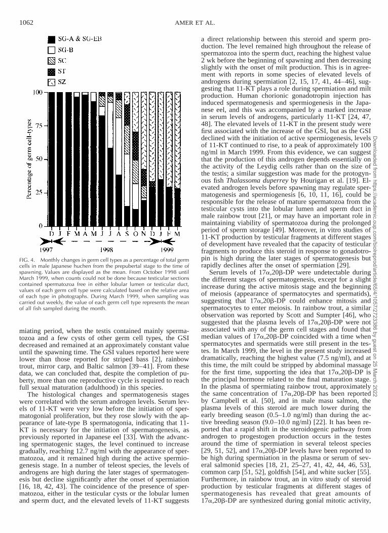
FIG. 4. Monthly changes in germ cell types as a percentage of total germcells in male Japanese huchen from the prepubertal stage to the time ofspawning. Values are displayed as the mean. From October 1998 untilMarch 1999, when counts could not be done because testicular sectionscontained spermatozoa free in either lobular lumen or testicular duct,values of each germ cell type were calculated based on the relative areaof each type in photographs. During March 1999, when sampling wascarried out weekly, the value of each germ cell type represents the meanof all fish sampled during the month.
miating period, when the testis contained mainly sperma-tozoa and a few cysts of other germ cell types, the GSIdecreased and remained at an approximately constant valueuntil the spawning time. The GSI values reported here werelower than those reported for striped bass [2], rainbowtrout, mirror carp, and Baltic salmon [39–41]. From thesedata, we can concluded that, despite the completion of pu-berty, more than one reproductive cycle is required to reachfull sexual maturation (adulthood) in this species.
The histological changes and spermatogenesis stageswere correlated with the serum androgen levels. Serum lev-els of 11-KT were very low before the initiation of sper-matogonial proliferation, but they rose slowly with the ap-pearance of late-type B spermatogonia, indicating that 11-KT is necessary for the initiation of spermatogenesis, aspreviously reported in Japanese eel [33]. With the advanc-ing spermatogenic stages, the level continued to increasegradually, reaching 12.7 ng/ml with the appearance of sper-matozoa, and it remained high during the active spermio-genesis stage. In a number of teleost species, the levels ofandrogens are high during the later stages of spermatogen-esis but decline significantly after the onset of spermiation[16, 18, 42, 43]. The coincidence of the presence of sper-matozoa, either in the testicular cysts or the lobular lumenand sperm duct, and the elevated levels of 11-KT suggests
a direct relationship between this steroid and sperm pro-duction. The level remained high throughout the release ofspermatozoa into the sperm duct, reaching the highest value2 wk before the beginning of spawning and then decreasingslightly with the onset of milt production. This is in agree-ment with reports in some species of elevated levels ofandrogens during spermiation [2, 15, 17, 41, 44–46], sug-gesting that 11-KT plays a role during spermiation and miltproduction. Human chorionic gonadotropin injection hasinduced spermatogenesis and spermiogenesis in the Japa-nese eel, and this was accompanied by a marked increasein serum levels of androgens, particularly 11-KT [24, 47,48]. The elevated levels of 11-KT in the present study werefirst associated with the increase of the GSI, but as the GSIdeclined with the initiation of active spermiogenesis, levelsof 11-KT continued to rise, to a peak of approximately 100ng/ml in March 1999. From this evidence, we can suggestthat the production of this androgen depends essentially onthe activity of the Leydig cells rather than on the size ofthe testis; a similar suggestion was made for the protogyn-ous fish Thalassoma duperrey by Hourigan et al. [19]. El-evated androgen levels before spawning may regulate sper-matogenesis and spermiogenesis [6, 10, 11, 16], could beresponsible for the release of mature spermatozoa from thetesticular cysts into the lobular lumen and sperm duct inmale rainbow trout [21], or may have an important role inmaintaining viability of spermatozoa during the prolongedperiod of sperm storage [49]. Moreover, in vitro studies of11-KT production by testicular fragments at different stagesof development have revealed that the capacity of testicularfragments to produce this steroid in response to gonadotro-pin is high during the later stages of spermatogenesis butrapidly declines after the onset of spermiation [29].
Serum levels of 17a,20b-DP were undetectable duringthe different stages of spermatogenesis, except for a slightincrease during the active mitosis stage and the beginningof meiosis (appearance of spermatocytes and spermatids),suggesting that 17a,20b-DP could enhance mitosis andspermatocytes to enter meiosis. In rainbow trout, a similarobservation was reported by Scott and Sumpter [46], whosuggested that the plasma levels of 17a,20b-DP were notassociated with any of the germ cell stages and found thatmedian values of 17a,20b-DP coincided with a time whenspermatocytes and spermatids were still present in the tes-tes. In March 1999, the level in the present study increaseddramatically, reaching the highest value (7.5 ng/ml), and atthis time, the milt could be stripped by abdominal massagefor the first time, supporting the idea that 17a,20b-DP isthe principal hormone related to the final maturation stage.In the plasma of spermiating rainbow trout, approximatelythe same concentration of 17a,20b-DP has been reportedby Campbell et al. [50], and in male masu salmon, theplasma levels of this steroid are much lower during theearly breeding season (0.5–1.0 ng/ml) than during the ac-tive breeding season (9.0–10.0 ng/ml) [22]. It has been re-ported that a rapid shift in the steroidogenic pathway fromandrogen to progestogen production occurs in the testesaround the time of spermiation in several teleost species[29, 51, 52], and 17a,20b-DP levels have been reported tobe high during spermiation in the plasma or serum of sev-eral salmonid species [18, 21, 25–27, 41, 42, 44, 46, 53],common carp [51, 52], goldfish [54], and white sucker [55].Furthermore, in rainbow trout, an in vitro study of steroidproduction by testicular fragments at different stages ofspermatogenesis has revealed that great amounts of17a,20b-DP are synthesized during gonial mitotic activity,
Dow
nloaded from https://academ
ic.oup.com/biolreprod/article/65/4/1057/2723386 by guest on 25 M
arch 2022

1063STEROID HORMONES AND EARLY STAGES OF SPERMATOGENESIS
FIG. 5. Immunoreaction of BrdU in testicular sections from fragments cultured in L-15 medium alone (A) or with 11-KT (B), 17a,20b-DP (C), or E2
(D). DC, Dividing cell; PC, proliferating cell; SG-A, type A spermatogonia; SG-EB, early type B spermatogonia. Bar 5 50 mm.
FIG. 6. Effects of various steroids andhormones on in vitro spermatogonial pro-liferation. Testicular fragments from imma-ture males were cultured in L-15 mediumin the presence or absence of one of thefollowing steroids and hormones: 11-KT,testosterone (T), 5b-DHT, androsterone (A),androstenedione (AD), DHEA, cortisol (C),E2, 17a,20b-DP, and SPG at a dose of 10ng/ml. Fragments were then incubated for15 days in humidified air at 128C. Theproliferation was assessed using incorpora-tion of BrdU (a proliferation marker) as de-scribed in Materials and Methods. A) BrdUindex. The number of positively immuno-reacted germ cells is expressed as a per-centage of the total number of germ cells.B) Percentage of the proliferated germcells (late-type B spermatogonia). Thenumber of proliferated germ cells is ex-pressed as a percentage of the total num-ber of germ cells. Results are given as themean 6 SEM. Values with the same lower-case letter(s) are not significantly different(P , 0.05).
Dow
nloaded from https://academ
ic.oup.com/biolreprod/article/65/4/1057/2723386 by guest on 25 M
arch 2022
1064 AMER ET AL.
at the time when spermatocytes enter meiosis and at theonset of spermiation, in which the synthesis of this steroidpredominated over that of C19 steroids [56]. Those authorssuggested that 17a,20b-DP may act on some steps of mi-totic cycle and meiotic processes of male germ cells. Otherin vitro studies have also revealed that the activity of 20b-hydroxysteroid dehydrogenase (20b-HSD), which is the en-zyme involved in the conversion of 17a-hydroxyprogester-one to 17a,20b-DP, is consistently high in the testesthroughout the period of spermatogenesis and spermiationin rainbow trout and amago salmon [25, 28, 29]; those in-vestigators also reported that the capacity of testicular frag-ments to produce this steroid was low during spermatogen-esis but increased sharply at the time of spermiation. Insalmonids, Nagahama [11] and Miura et al. [22] suggestedthat gonadotropin stimulates the testicular somatic cells toproduce 17a,20b-DP from a precursor (probably 17a-hy-droxyprogesterone in the presence of 20b-HSD secreted byspermatozoa), which acts to increase sperm duct pH, which,in turn, increases intrasperm cAMP, allowing the acquisi-tion of sperm motility. A similar suggestion has also beenreported for Japanese eel by Miura et al. [24]. In contrast,in rainbow trout, the various testicular cell preparations thatare mostly devoid of sperm secrete 17a,20b-DP regardlessof gonadotropin stimulation [53, 57, 58]. From our results,we can suggest that 17a,20b-DP is essential not only forthe final maturation stage, but that it also probably plays arole in the spermatogenic process during either mitosis ormeiosis. This steroid may also play a role in the regulationof spawning behavior, as evidenced by the dramatic in-crease in its levels during the spawning season. Furtherinvestigations for elucidating and confirming the role of17a,20b-DP during spermatogenesis are required.
In most vertebrates, E2 circulates in males at low orundetectable levels; however, the enzyme that catalyzes theconversion of androgen to E2, aromatase, can be presentnear E2 targets in the brain [59] and in testicular germ cells[60]. The present study showed high variations in E2 levelsnot only between months but also among individuals asmeasured each month. The highest value of E2 was ob-served during mitosis, indicating the involvement of estro-gen in spermatogenesis, especially during the early stagesof mitosis, and supporting previous results reported in Jap-anese eel [31]. However, E2 was detected throughout theentire process of spermatogenesis and spermiation, indicat-ing that estrogen may play a role in spermatogenesis andtesticular functions. In the elasmobranch Squalus acanthias,the presence of testicular aromatase and of binding sites forE2 in lobules with gonia and with meiotic cells suggests aparacrine role of E2 during the early stages of spermato-genesis [61, 62]. Estrogen levels declined before the timeof spawning, whereas levels of 17a,20b-DP increased.Thus, E2 could be involved in the regulation of 17a,20b-DP production in males [46, 63]. After hCG injection, theaverage serum level of E2 in male Japanese eel was 490pg/ml, and this level did not show any significant changesduring the whole process of hCG-induced spermatogenesis[31]. Interestingly, in 2- to 3-mo-old hypogonadal micetreated with slow-release estradiol implants, full and qual-itatively normal spermatogenesis was induced, suggestingthat estrogens exert paracrine actions within the testis topromote spermatogenesis [32]. These results and the wide-spread expression and identification of estrogen receptors(a and b) within several cell types, including Leydig cells,Sertoli cells, and some testicular germ cells [31, 64–66], aswell as aromatase within germ cells of the mouse testis
[60], point toward a physiological role for estrogen in manyaspects of testicular functions.
From the annual cycle results mentioned above, wefound that testicular development and spermatogenesis inJapanese huchen were associated with steroid levels of 11-KT, 17a,20b-DP, and E2. To understand the role of each ofthese steroids in spermatogenesis, immature testicular frag-ments (containing only primitive spermatogonia) were cul-tured with 10 ng/ml of each tested hormone. Synthesis ofDNA is an indicator for mitosis and spermatogonial prolif-eration; therefore, the proliferative response of spermato-gonia was determined by incorporation of BrdU (a prolif-eration marker) into germ cells. We found that 10 ng/ml of11-KT, E2, 17a,20b-DP, and SPG were sufficient to induceDNA synthesis in spermatogonia, indicating that mitoticdivisions were promoted by the addition of each of thesehormones. Spermatogonial proliferation was induced by11-KT, 17a,20b-DP, or SPG, as evidenced by the appear-ance of late-type B spermatogonia. The effects of 11-KTcould be mediated through Sertoli cells, which this andro-gen activates to produce other growth factors, such as ac-tivin B, that, in turn, act on spermatogonia to induce mi-tosis, leading to the formation of spermatocytes [11, 12, 33,37, 67]. In cultured testicular fragments of Japanese eel,hCG induces the entire process of spermatogenesis frompremitotic spermatogonia to spermatozoa within 24 days[38]. This hCG-induced spermatogenesis is accompaniedby a marked activation of Leydig cells and Sertoli cells andby a rapid increase in levels of 11-KT. Our results indicatedthat 10 ng/ml of 11-KT could induce only spermatogonialproliferation in Japanese huchen, whereas the same con-centration could induce the full process of spermatogenesisin Japanese eel in vitro within 21 days [37]. Furthermore,cultured testicular fragments of Japanese eel with 100 ng/ml of recombinant human insulin-like growth factor (IGF)-I in combination with 10 ng/ml of 11-KT can induce sper-matogenesis from spermatogonial proliferation to spermio-genesis, whereas each of them alone cannot [68]. In con-trast, testosterone, 11-KT, E2, and 17a,20b-DP do notinfluence the proliferation of trout mitotic germ cells,whereas IGF-I does [69, 70]. These variable results suggesta species-related variation in the initiation and control ofgametogenesis. Thus, depending on the species, sex ste-roids would be more or less involved in the control of sper-matogenesis (i.e., the molecules required for the mitoticphase of spermatogenesis differ among species), as hasbeen previously suggested [48, 70]. It is well establishedthat, in vertebrates, spermatogenesis is controlled by pitu-itary gonadotropin and testicular androgens. So, the effectof SPG, which is rich in gonadotropins, on spermatogonialproliferation seems to be mediated through the stimulationof Leydig cells to produce androgens.
The variability of the E2 levels throughout the first waveof spermatogenesis in this species, however, makes it dif-ficult to establish the role of estrogens in spermatogenesis,but the effect of E2 on the spermatogonial renewal has beenconfirmed by the in vitro experiment. Recently, it was alsoreported that, in vitro, estradiol could induce proliferationof isolated rat fetal gonocytes [71], primary spermatogoniain the frog Rana esculenta [72, 73], and spermatogonialstem cell renewal in Japanese eel [31]. These effects wereinhibited by an estrogen antagonist, suggesting that estro-gen effects are mediated by the binding of estrogen to itsreceptors, either through Sertoli cells or directly on germcells. Thus, estrogens could participate in the autocrine and/or paracrine regulation of spermatogenesis. The 17a,20b-
Dow
nloaded from https://academ
ic.oup.com/biolreprod/article/65/4/1057/2723386 by guest on 25 M
arch 2022
1065STEROID HORMONES AND EARLY STAGES OF SPERMATOGENESIS
DP promoted DNA synthesis, as evidenced by the incor-poration of BrdU into germ cells. Our results indicated that17a,20b-DP was detected during the mitotic phase of sper-matogenesis in vivo and enhanced DNA synthesis in vitro.Therefore, 17a,20b-DP could play an important role inspermatogenesis, especially mitosis, and in the initiation ofmeiosis in addition to its well-established role during thefinal maturation stage. Furthermore, large amounts of17a,20b-DP were synthesized in vitro by rainbow trout tes-ticular fragments during gonial mitotic activity, when sper-matocytes enter meiosis, as reported by Depeche and Sire[56]. Although a slight stimulation was observed in testos-terone-incubated fragments, the value of positively stainedspermatogonia was significantly lower than those of 11-KT,E2, 17a,20b-DP, and SPG. Stimulation observed in testos-terone-incubated fragments could be due to the conversionof this androgen into 11-KT or E2 by the endogenous en-zymes.
We conclude that 1) 11-KT is necessary for the initiationof spermatogenesis and sperm production, and it probablyplays a role in spermiation, 2) 17a,20b-DP is essential forthe final maturation stage, could play a significant role inmitosis and meiosis, and probably participates in the reg-ulation of spawning behavior, and 3) estrogen is an indis-pensable male hormone that plays a physiological role insome aspects of testicular function, especially in mitosis.The three steroids, 11-KT, E2, and 17a,20b-DP, as well asSPG significantly induced DNA synthesis, thence sper-matogonial renewal and/or spermatogonial proliferation.Further investigations for elucidating and confirming therole of 17a,20b-DP, E2, and the androgenic steroid hor-mones in spermatogenesis, especially during the early stag-es using H. perryi as a new animal system, are required.
ACKNOWLEDGMENTS
We thank Dr. John Bower from the Training Ship Laboratory, Hok-kaido University, for his critical reading of the manuscript. We also thankMr. Toshitsugu Higashino, Division of Marine Biosciences, Hokkaido Uni-versity, for his assistance during sampling and technical help with RIA.
REFERENCES
1. Dufour S, Huang YS, Rousseau K, Sbaihi M, Le Belle N, Vidal B,Marchelidon J, Querat B, Burzawa-Gerard E, Change CF, Schmitz M.Puberty in teleosts: new insights into the role of peripheral signals inthe stimulation of pituitary gonadotropins. In: Proceeding of the SixthInternational Symposium on the Reproductive Physiology of Fish;1999; Bergen, Norway, 455–461.
2. Holland MC, Hassin S, Zohar Y. Gonadal development and plasmasteroid levels during pubertal development in captive-reared stripedbass, Morone saxatilis. J Exp Zool 2000; 286:49–63.
3. Bellve AR, Cavicchia JC, Millette CF, O’brien DA, Bhatngar YM,Dym M. Spermatogenic cells of the prepubertal mouse: isolation andmorphological characterization. J Cell Biol 1977; 74:68–85.
4. Schulz RW, Bogerd J, Goos HJTH. Spermatogenesis and its endocrineregulation. In: Proceeding of the Sixth International Symposium onthe Reproductive Physiology of Fish; 1999; Bergen, Norway, 225–232.
5. Pudney J. Spermatogenesis in nonmammalian vertebrates. MicroscRes Tech 1995; 32:459–497.
6. Billard R, Fostier A, Weil C, Breton B. Endocrine control of sper-matogenesis in teleost fish. Can J Fish Aquat Sci 1982; 39:65–79.
7. Grier HJ, Linton JR, Leatherland JF, de Vlaming VL. Structural evi-dence for two different testicular types in teleost fishes. Am J Anat1980; 159:331–345.
8. Grier HJ. Cellular organization of the testis and spermatogenesis infishes. Am Zool 1981; 21:345–357.
9. Grier HJ. Chordate testis: the extracellular matrix hypothesis. J ExpZool 1992; 261:151–160.
10. Nagahama Y. The functional morphology of teleost gonads. In: Hoar
WS, Randall DJ, Donaldson EM (eds.), Fish Physiology, vol. IXA.New York: Academic Press; 1983: 223–275.
11. Nagahama Y. Endocrine regulation of gametogenesis in fish. J DevBiol 1994; 38:217–229.
12. Nagahama Y. Gonadal steroid hormones: major regulators of gonadalsex differentiation and gametogenesis in fish. In: Proceeding of theSixth International Symposium on the Reproductive Physiology offish; 1999; Bergen, Norway, 211–222.
13. Bhattacharya S. Recent advances in the hormonal regulation of go-nadal maturation and spawning in fish. Curr Sci 1999; 76:342–349.
14. Borg B. Androgens in teleost fishes. Comp Biochem Physiol 1994;109C:219–245.
15. Scott AP, Bye VJ, Baynes SM, Springate JRC. Seasonal variation inplasma concentrations of 11-ketotestosterone and testosterone in malerainbow trout, Salmo gairdneri Richardson. J Fish Biol 1980; 17:495–505.
16. Fostier A, Jalabert B, Billard R, Breton B, Zohar Y. The gonadalsteroids. In: Hoar WS, Randall DJ, Donaldson EM (eds.), Fish Phys-iology, vol. IXA. New York: Academic Press; 1983: 277–372.
17. Fostier A, Billard R, Breton B. Plasma 11-oxotestosterone and go-nadotrophin in relation to the arrest of spermiation in rainbow trout(Salmo gairdneri). Gen Comp Endocrinol 1984; 54:378–381.
18. Ueda H, Nagahama Y, Tashiro F, Crim LW. Some endocrine aspectsof precocious sexual maturation in the amago salmon, Oncorhynchusrhodurus. Bull Jpn Soc Sci Fish 1983; 49:587–596.
19. Hourigan TF, Nakamura M, Nagahama Y, Yamauchi K, Grau EG.Histology, ultrastructure, and in vitro steroidogenesis of the testes oftwo male phenotypes of the protogynous fish Thalassoma duperrey(Labridae). Gen Comp Endocrinol 1991; 83:193–217.
20. Antonopoulou BE, Mayer I, Borg B. Stimulatory and inhibitory ef-fects of testosterone on testes in Atlantic salmon male parr. J Fish Biol1995; 47:586–598.
21. Baynes SM, Scott AP. Seasonal variations in parameters of milt pro-duction and in plasma concentration of sex steroids of male rainbowtrout (Salmo gairdneri). Gen Comp Endocrinol 1985; 57:150–160.
22. Miura T, Yamauchi K, Takahashi H, Nagahama Y. The role of hor-mones in the acquisition of sperm motility in salmonid fish. J ExpZool 1992; 261:359–363.
23. Miura T, Ninzeki M, Hirai H, Yamauchi K. Induction of final matu-ration by the injection of chum salmon pituitary homogenate in themale Japanese huchen (Hucho perryi). Bull Fac Fish Hokkaido UnivJpn 1991; 42:16–25.
24. Miura T, Yamauchi K, Takahashi H, Nagahama Y. Involvement ofsteroid hormones in gonadotropin-induced testicular maturation inmale Japanese eel (Anguilla japonica). Biomed Res 1991; 12:241–248.
25. Ueda H, Young G, Crim LW, Kambegawa A, Nagahama Y. 17a,20b-dihydroxy-4-pregnen-3-one: plasma levels during sexual maturationand in vitro production by the testes of amago salmon (Oncorhynchusrhodurus) and rainbow trout (Salmo gairdneri). Gen Comp Endocrinol1983; 51:106–112.
26. Ueda H, Kambegawa A, Nagahama Y. In vitro 11-ketotestosteroneand 17a,20b-dihydroxy-4-pregnen-3-one production by testicularfragments and isolated sperm of rainbow trout, Salmo gairdneri. JExp Zool 1984; 231:435–439.
27. Ueda H, Kambegawa A, Nagahama Y. Involvement of gonadotropinand steroid hormones in spermiation in the amago salmon, Oncorhyn-chus rhodurus, and goldfish, Carassius auratus. Gen Comp Endocri-nol 1985; 59:24–30.
28. Sakai N, Ueda H, Suzuki N, Nagahama Y. Involvement of sperm inthe production of 17a,20b-dihydroxy-4-pregnen-3-one in the testis ofspermiating rainbow trout, Salmo gairdneri. Biomed Res 1989; 10:131–138.
29. Sakai N, Ueda H, Suzuki N, Nagahama Y. Steroid production byamago salmon (Oncorhynchus rhodurus) testes at different develop-mental stages. Gen Comp Endocrinol 1989; 75:231–240.
30. Sharpe RM. The roles of estrogen in the male. Trends EndocrinolMetab 1998; 9:371–377.
31. Miura T, Miura C, Ohta T, Nader MR, Todo T, Yamauchi K. Estradiol-17b stimulates the renewal of spermatogonial stem cells in males.Biochem Biophys Res Commun 1999; 264:230–234.
32. Ebling FJP, Brooks AN, Cronin AS, Ford H, Keer JB. Estrogenicinduction of spermatogenesis in the hypogonadal mouse. Endocrinol-ogy 2000; 141:2861–2869.
33. Miura T, Kobayashi T, Nagahama Y. Hormonal regulation of sper-matogenesis in the Japanese eel (Anguilla japonica). In: Davey KG
Dow
nloaded from https://academ
ic.oup.com/biolreprod/article/65/4/1057/2723386 by guest on 25 M
arch 2022

1066 AMER ET AL.
(ed.), Perspectives in Comparative Endocrinology. Ottawa: NationalResearch Council of Canada; 1994: 631–635.
34. Kimura S, Hara A. Culture and artificial fertilization of Japanese huch-en Hucho perryi. Jpn Suisanzoshoku 1989; 37:121–128.
35. Young G, Crim LW, Kagawa H, Kambegawa A, Nagahama Y. Plasma17a,20b-dihydroxy-4-pregnen-3-one levels during sexual maturationof amago salmon (Oncorhynchus rhodurus): correlation with plasmagonadotropin and in vitro production by ovarian follicles. Gen CompEndocrinol 1983; 51:96–106.
36. Kagawa H, Young G, Adachi S, Nagahama Y. Estradiol-17b produc-tion in amago salmon (Oncorhynchus rhodurus) ovarian follicles: roleof the thecal and granulosa cells. Gen Comp Endocrinol 1982; 47:440–448.
37. Miura T, Yamauchi K, Takahashi H, Nagahama Y. Hormonal induc-tion of all stages of spermatogenesis in vitro in the male Japanese eel(Anguilla japonica). Proc Natl Acad Sci U S A 1991; 88:5774–5778.
38. Miura T, Yamauchi K, Takahashi H, Nagahama Y. Human chorionicgonadotropin induces all stages of spermatogenesis in vitro in the maleJapanese eel (Anguilla japonica). Dev Biol 1991; 146:258–262.
39. Billard R. Reproduction in rainbow trout: sex differentiation, dynam-ics of gametogenesis, biology and preservation of gametes. Aquacul-ture 1992; 100:263–298.
40. Billard R, Weil C, Bieniarz K, Mikolajczyk T, Breton B, Epler P,Bougoussa M. Testicular and some hormonal changes during the firstfour years of life in the mirror carp, Cyprinus carpio L. J Fish Biol1992; 41:473–487.
41. Mayer I, Lundqvist H, Berglund I, Schmitz M, Schulz R, Borg B.Seasonal endocrine changes in Baltic salmon, Salmo salar, immatureparr and mature male parr. I. Plasma levels of five androgens, 17a-hydroxy-20b-dihydroprogesterone, and 17b-estradiol. Can J Zool1990; 68:1360–1365.
42. Mayer I, Schmitz M, Borg B, Schulz R. Seasonal endocrine changesin male and female Arctic charr (Salvelinus alpinus). I. Plasma levelsof three androgens, 17a-hydroxy-20b-dihydroprogesterone, and 17b-estradiol. Can J Zool 1992; 70:37–42.
43. Amiri BM, Maebayashi M, Adachi S, Yamauchi K. Testicular devel-opment and serum sex steroid profiles during the annual sexual cycleof the male sturgeon hybrid, the bester. J Fish Biol 1996; 48:1039–1050.
44. Mayer I, Berglund I, Rydevik M, Borg B, Schulz R. Plasma levels offive androgens, 17a-hydroxy-20b-dihydroprogesterone in immatureand mature male Baltic salmon (Salmo salar) parr, and the effects ofcastration and androgen replacement in mature parr. Can J Zool 1990;68:263–267.
45. Liley NR, Breton B, Fostier A, Tan ESP. Endocrine changes associatedwith spawning behavior and social stimuli in a wild population ofrainbow trout (Salmo gairdneri). I. Males. Gen Comp Endocrinol1986; 62:145–156.
46. Scott AP, Sumpter JP. Seasonal variations in testicular germ cell stagesand in plasma concentrations of sex steroids in male rainbow trout(Salmo gairdneri) maturing at 2 years old. Gen Comp Endocrinol1989; 73:46–58.
47. Miura T, Yamauchi K, Nagahama Y, Takahashi H. Induction of sper-matogenesis in male Japanese eel, Anguilla japonica, by a single in-jection of human chorionic gonadotropin. Zool Sci 1991; 8:63–73.
48. Miura T, Kawamura S, Miura C, Yamauchi K. Impaired spermato-genesis in the Japanese eel, Anguilla japonica: possibility of the ex-istence of factors that regulate entry of germ cells into meiosis. DevGrowth Differ 1997; 39:685–691.
49. Malison JA, Procarione LS, Barry TP, Kapuscinski AR, Kayes TB.Endocrine and gonadal changes during the annual reproductive cycleof the freshwater teleost, Stizostedion vitreum. Fish Physiol Biochem1994; 13:473–484.
50. Campbell CM, Fostier A, Jalabert B, Truscott B. Identification andquantification of steroids in the serum of rainbow trout during sper-miation and oocyte maturation. J Endocrinol 1980; 85:371–378.
51. Barry TP, Aida K, Okumura T, Hanyu I. The shift from C-19 to C-21 steroid synthesis in spawning male common carp, Cyprinus carpio,is regulated by the inhibition of androgen production by progestogensproduced by spermatozoa. Biol Reprod 1990; 43:105–112.
52. Barry TP, Santos AJG, Furukawa K, Aida K, Hanyu I. Steroid profiles
during spawning in male common carp. Gen Comp Endocrinol 1990;80:223–231.
53. Vizziano D, LeGac F. Effect of gonadotropin type II and 17-hydroxy-4-pregnene-3,20-dione on 17,20b-dihydroxy-4-pregnen-3-one produc-tion by rainbow trout testes at different developmental stages. FishPhysiol Biochem 1998; 19:269–277.
54. Kobayashi M, Aida K, Hanyu I. Gonadotropin surge during spawningin male goldfish. Gen Comp Endocrinol 1986; 62:70–79.
55. Scott AP, MacKenzie DS, Stacey NE. Endocrine changes during nat-ural spawning in the white sucker, Catostomus commersoni. II. Steroidhormones. Gen Comp Endocrinol 1984; 56:349–359.
56. Depeche J, Sire O. In vitro metabolism of progesterone and 17a-hydroxyprogesterone in the testis of the rainbow trout, Salmo gaird-neri Rich., at different stages of spermatogenesis. Reprod Nutr Dev1982; 22:427–438.
57. Loir M. Trout steroidogenic testicular cells in primary culture. II. Ste-roidogenic activity of interstitial cells, Sertoli cells, and spermatozoa.Gen Comp Endocrinol 1990; 78:388–398.
58. Vizziano D, Fostier A, LeGac F, Loir M. 20b-Hydroxysteroid dehy-drogenase activity in nonflagellated germ cells of rainbow trout testis.Biol Reprod 1996; 54:1–7.
59. Schlinger BA, Arnold AP. Circulating estrogen in a male songbirdoriginate in the brain. Proc Natl Acad Sci U S A 1992; 89:7650–7653.
60. Nitta H, Bunick D, Hess RA, Janulis L, Newton SC, Millette CF,Osawa Y, Shizuta Y, Toda K, Bahr JM. Germ cells of the mouse testisexpress p450 aromatase. Endocrinology 1993; 132:1396–1401.
61. Callard GV, Pudney JA, Mak P, Canick JA. Stage-dependent changesin steroidogenic enzymes and estrogen receptors during spermatogen-esis in the testis of the dogfish, Squalus acanthias. Endocrinology1985; 117:1328–1335.
62. Callard GV. Autocrine and paracrine role of steroids during spermato-genesis: studies in Squalus acanthias and Necturus maculosus. J ExpZool 1992; 261:132–142.
63. Vizziano D, LeGac F, Fostier A. Effect of 17b-estradiol, testosteroneand 11-ketotestosterone on 17,20b-dihydroxy-4-pregnen-3-one pro-duction in the rainbow trout testis. Gen Comp Endocrinol 1996; 104:179–188.
64. Cooke PS, Young P, Hess RA, Cunha GR. Estrogen receptor expres-sion in developing epididymis, efferent ductules, and other male re-productive organs. Endocrinology 1991; 128:2874–2879.
65. Fisher JS, Millar MR, Majdic G, Saunders PTK, Fraser HM, SharpeRM. Immunolocalization of estrogen receptor alpha (ERa) within thetestis and excurrent ducts of the rat and marmoset monkey from peri-natal life to adulthood. J Endocrinol 1997; 153:485–495.
66. Saunders PTK, Fisher JS, Sharpe RM, Millar MR. Expression of es-trogen receptor beta (ERb) occurs in multiple cell types, includingsome germ cells, in the rat testis. J Endocrinol 1998; 156:R13–R17.
67. Miura T, Miura C, Yamauchi K, Nagahama Y. Human recombinantactivin induces proliferation of spermatogonia in vitro in the Japaneseeel Anguilla japonica. Fish Sci 1995; 61:434–437.
68. Nader MR, Miura T, Ando N, Miura C, Yamauchi K. Recombinanthuman insulin-like growth factor I stimulates all stages of 11-ketotes-tosterone-induced spermatogenesis in the Japanese eel, Anguilla ja-ponica, in vitro. Biol Reprod 1999; 61:944–947.
69. Loir M. In vitro approach to the control of spermatogonia proliferationin the trout. Mol Cell Endocrinol 1994; 102:141–150.
70. Loir M. Spermatogonia of rainbow trout: II. In vitro study of theinfluence of pituitary hormones, growth factors and steroids on mitoticactivity. Mol Reprod Dev 1999; 53:434–442.
71. Li H, Papadopoulos V, Vidic B, Dym M, Culty M. Regulation of rattestis gonocyte proliferation by platelet-derived growth factor and es-tradiol: identification of signaling mechanisms involved. Endocrinol-ogy 1997; 138:1289–1298.
72. Cobellis G, Pierantoni R, Minucci S, Pernas-Alonso R, MeccarielloR, Fasano S. c-fos Activity in Rana esculenta testis: seasonal andestradiol-induced changes. Endocrinology 1999; 140:3238–3244.
73. Minucci S, Di Matteo L, Chieffi P, Pierantoni R, Fasano S. 17b-Es-tradiol effects on mast cell number and spermatogonial mitotic indexin the testis of the frog, Rana esculenta. J Exp Zool 1997; 278:93–100.
Dow
nloaded from https://academ
ic.oup.com/biolreprod/article/65/4/1057/2723386 by guest on 25 M
arch 2022


