Embed Size (px)
Citation preview
Copyright 0 199 1 by the Genetics Society of America
Introduction of the Transposable Element mariner into the Germline of Drosophila melanogaster
Dan Garza, Meetha Medhora, Akihiko Koga and Daniel L. Hart1 Department of Genetics, Washington University School of Medicine, St. Louis, Missouri 631 10-1095
Manuscript received September 2 1, 199 1 Accepted for publication February 16, 199 1
ABSTRACT A chimeric white gene ( z P h ) and other constructs containing the transposable element mariner from
Drosophila mauritiana were introduced into the germline of Drosophila melanogaster using transfor- mation mediated by the P element. In the absence of other mariner elements, the WPLh allele is genetically stable in both germ cells and somatic cells, indicating that the peach element ( i e . , the particular copy of mariner inserted in the usCh allele) is inactive. However, in the presence of the active element Mosl , the u9" allele reverts, owing to excision of the peach element, yielding eye-color mosaics and a high rate of germline reversion. In strains containing Mosl virtually every fly is an eye-color mosaic, and the rate of wp" germline reversion ranges from 10 to 25%, depending on temperature. The overall rates of mariner excision and transposition are approximately sixfold greater than the rates in comparable strains of Drosophila simulans. The activity of the Mosl element is markedly affected by position effects at the site of Mosl insertion. In low level mosaic lines, dosage effects of Mosl are apparent in the heavier level of eye-color mosaicism in Mosl homozygotes than in hetero- zygotes. However, saturation occurs in high level mosaic lines, and then dosage effects are not observed. A pBluescribe M13+ plasmid containing Mosl was injected into the pole plasm of D. melanogaster embryos, and the Mosl element spontaneously integrated into the germline at high effkiency. These transformed strains of D. melanogaster presently contain numerous copies of mariner and may be useful in transposon tagging and other applications.
T RANSPOSABLE elements provide powerful tools for molecular genetics. They are able to
be maintained in a variety of related genomes, serve as agents of chromosomal insertion, deletion, or re- arrangement, and provide the basis for transforma- tion of somatic or germ cells. At the same time, the biological mechanisms that control the dissemination of transposable elements among species and their persistence within species are poorly understood. The mariner family of transposable elements in the genus Drosophila is a case in point (reviewed in HARTL 1989). Elements in the mariner family occur in most species of the melanogaster species subgroup of Drosophila, but they have not yet been found in natural isolates of D. melanogaster itself (JACOBSON, MEDHORA and HARTL 1986). Comparisons of mariner sequences present in species most closely related to D. melano- gaster (D. simulans, D. mauritiana and D. sechellia) and other, more distantly related members of the species subgroup (D. yakuba and D. teissieri) suggest that mar- iner invaded the genome prior to diversification of the species subgroup (MARUYAMA and HARTL 1991). The early invasion implies that the element was lost in the lineages leading to D. melanogaster, D. erecta and D. orena. On the other hand, the occurrence of highly similar mariner sequences in very distantly related species of the genus Zaprionus implies a rela- Genetics 12% 303-310 (June, 1991)
tively recent horizontal transfer (K. MARUYAMA and D. L. HARTL, unpublished).
The mariner transposable element is 1286 base pairs in length. It terminates in 28-base-pair inverted repeat sequences and contains a long open reading frame capable of coding for a polypeptide of 345 amino acids (JACOBSON, MEDHORA and HARTL 1986). The element was discovered in D. mauritiana through a mutant allele, white-peach (Wp""), which is genetically unstable in both somatic and germ cells (JACOBSON and HARTL 1985). Various strains of D. mauritiana contain 20-40 copies of mariner, and the Wpc" allele has a copy of mariner inserted in the 5' end of the white gene. A low level of somatic excision of the mariner element from UP" occurs in most strains of D. mauritiana, yielding pigmented patches in an other- wise peach-colored eye. The infrequent occurrence of such mosaics implies that most copies of mariner in the genome are not highly active. Other mariner ele- ments, called mosaic (Mos) factors, are very active. For example, in most strains, the factor Mosl results in eye color mosaicism in virtually every fly that carries it, and rates of germline reversion of Wpc" in MosZ strains are up to 1% (BRYAN, JACOBSON and HARTL 1987).
The species distribution and genetic characteristics of mariner provide several strong motivations for in-
304 D. Garza et al.
troducing the element into the genome of D. melano- gaster. First, the presence of the element in closely related species suggests that the element will be active in the D. melanogaster genome, adding to the already impressive repertoire of genetic tools available in this species and providing another system for transposon tagging and perhaps germline transformation. Sec- ond, the capability for genetic manipulation in D. melanogaster enables detailed genetic analysis of the mariner element itself and host factors that may be necessary for its activity. Third, the absence of mariner from the normal genome of D. melanogaster means that genetic studies can be carried out without com- plications due to other mariner elements in the genetic background.
In this paper we report the transfer of the mariner system into the genome of D. melanogaster. A chimeric white gene composed mainly of D. melanogaster se- quence, but including a mariner-containing restriction fragment from the Wpc" allele of D. mauritiana, was introduced into the germline of D. melanogaster using P element-mediated transformation (RUBIN and SPRA- DLING 1982), yielding @* transformants. Transfor- mation using the P element was also used to introduce the mariner mosaic element Mosl (BRYAN, JACOBSON and HARTL 1987) into the D. melanogaster germline. Crosses between the D. melanogaster U S c h and Mosl strains reconstituted the system of high level somatic mosaicism observed in D. mauritiana. Studies of the transformed D. melanogaster strains have demon- strated that the Mosl element is approximately eight- fold more active in the genome of D. melanogaster than in D. mauritiana. However, the particular copy of mariner inserted in the UgCh allele is apparently nonfunctional. Dosage and saturation effects of Mosl are evident in the levels of somatic mosaicism and in the rates of germline reversion of U S c h , and there is a pronounced temperature effect on Mosl activity. In addition, the autonomous Mosl element is capable of spontaneous integration into the germline of D. mel- anogaster, providing the potential for the development of a general transformation system using mariner.
MATERIALS AND METHODS
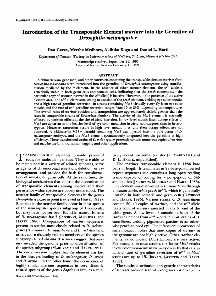
The white-peach transformation vector pUChsneo:uPh was constructed as illustrated in Figure 1A. First, a P element transformation vector containing the wild-type white gene from Canton S was constructed from the pUChsneo vector (STELLER and PIRROTTA 1985) by isolating a 1 1.7-kb EcoRI fragment containing white from plasmid pP[(w, ry)]A (HA- ZELRIGG, LEVIS and RUBIN 1984) and inserting this fragment into the EcoRI cloning site of pUChsneo. The pUChsneo vector was provided by V. PIRROTTA, the pP[(w, ry)]A plasmid by G . RUBIN. The resulting plasmid was recovered after transformation into Escherichia coli HB101 and was partially digested with BamHI to remove the 3.0-kb BamHI fragment from white spanning coordinates 1 .O-4.0 (BINCHAM, LEVIS and RUBIN 1981). The excised BamHI
fragment was then replaced with the analogous mariner- containing 4.3-kb BamHI fragment from plasmid pJ1 (JA- COBSON, MEDHORA and HARTL 1986). Clones were isolated after transformation into HBlOl, and the constructs having the proper orientation of the inserted BamHI fragment were confirmed by restriction mapping using various combina- tions of BamHI, Sal1 and SacI. It should be noted that pUChsneo:uPh contains a chimeric white gene: the material within the 4.3 kb BamHI fragment is from D. mauritiana, the rest of the gene is from D. melanogaster.
The transformation vector Car20:Mosl was constructed as outlined in Figure 1B. A 5.0-kb BamHI-Hind111 fragment containing Mosl (MEDHORA, MACPEEK and HARTL 1988) was isolated, the ends made flush with the Klenow fragment of DNA polymerase I, and inserted into the HpaI site of the P element transformation vector Carnegie 20 (RUBIN and SPRADLINC 1983). The orientation of the inserted fragment was determined by enzyme digestion with SalI, which cleaves once at nucleotide 349 in the 1286-base-pair mariner element (orientation as in JACOBSON, MEDHORA and HARTL 1986). The 5.0-kb Mosl-containing fragment includes Mosl along with approximately 3.5 kb of upstream sequence and 0.2 kb of downstream sequence from D. simulans (MED- HORA, MARUYAMA and HARTL 199 1).
The plasmid used for autonomous mariner transforma- tion of the D. melanogaster germline was constructed by inserting the same 5.0-kb BamHI-Hind111 fragment contain- ing Mosl into pBluescribe M13+ after digestion of the plasmid with BamHI and HindIII.
Plasmid DNA was purified by CsC1-ethidium bromide equilibrium density gradient centrifugation and used for injection of embryos. Transformations were carried out essentially as described in RUBIN and SPRADLINC (1982). DNA concentrations in the injection needle were approxi- mately 150 rg/ml, and embryos were injected and incubated under oil at 18". Emerging larvae were transferred to standard Drosophila medium and maintained at 25 O . In the transformations with P element we used the wings-clipped helper plasmid pr25.7WC (KARESS and RUBIN 1984) at a concentration of approximately 75 pg/ml.
General procedures for DNA isolation, enzyme digestion, electrophoresis and blotting are described in BRYAN, GARZA and HARTL (1990). General procedures for recombinant DNA manipulation are described in MANIATIS, FRITSCH and SAMBROOK (1982).
RESULTS
Transformation of wpFh and Mod: D. melanogaster embryos of genotype y w67c23 (chosen because it is a partial deletion of white) were injected with the chi- meric &" gene contained in the P element vector pUChsneo:&*, diagrammed in Figure lA, and co- injected with the wings-clipped helper plasmid (KA- RESS and RUBIN 1984). Among 138 surviving GO off- spring, 108 were fertile. One of these gave offspring with peach-like eye color, and these were individually crossed with the y w67c23 stock for further analysis. Genetic studies indicated two independent insertion events had occurred in the transformed Go. Two of the white-peach offspring carried insertions into an identical position in the X chromosome (designated sublines P735 and P739), and the other white-peach offspring had an insertion into chromosome 3 (subline P734). The insertion into chromosome 3 is associated
mariner in D. melanogaster 305
I I Em R I Born HI 8omHI EcoRI
-" Y
P 8omHI hs +nee f "-
A. pUChsneo:wPch
mariner
p J J l
PP [(we rv IA]
DUChsneo
Hpo I I "_ "- Carnegie 20
P rY+ f
0. C20: Mos I FIGURE 1 .-Transformation vectors for the introduction of mar-
iner into D. melanogaster. (A) The U P h vector was created by replac- ing the indicated BamHI fragment in the D. melanogaster white gene with the corresponding fragment from the U P h allele from D. mauritiana. This fragment contains the mariner element designated peach. (B) The Mosf vector was created by inserting a Mosfcontain- ing BamHI-Hind111 fragment into the HpaI cloning site in Carnegie 20.
with a recessive lethal, which has thus far been insep- arable from the site of the insertion.
None of the transformed lines give any evidence of somatic or germinal instability of W p c h . Somatic insta- bility is indicated by excision events that give occa- sional pigmented facets in the eye, and germinal insta- bility is indicated by reverse mutation of wpCh to w+ or forward mutation to w-. The results with D. melano- gaster are in marked contrast to those with D. mauri- tiana or D. simulans, in which W p c h always gives some detectable level of somatic excision (JACOBSON and HARTL 1985; HAYMER and MARSH 1986). The differ- ence between the species suggests that the mariner element inserted in W p c h is inactive and that the insta- bility of W p c h in the other species results from trans- activation by other copies of mariner in the genetic background. We therefore designate the particular mariner element present in W p c h as the peach element. The sequence of this element has been determined (JACOBSON, MEDHORA and HARTL 1986). It is possible that the peach element in D. melanogaster might have undergone mutations in the course of subcloning and in vitro manipulation. However, the conclusion that the peach element is inactive is supported by genetic experiments in a strain of D. simulans that contains
1 2 3 4 5
9.46 kb .
4.30 kb .
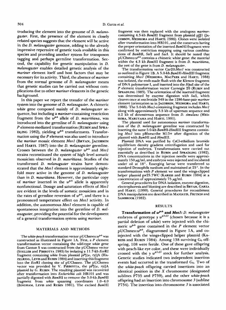
FIGURE 2.-Transformants of- D. melanogaster containing the mariner element. Filter hybridizations were carried out with DNA from various strains digested with BamHI and probed with mariner DNA. The lanes are as follows: (1) strain P735, which contains an insertion of UP*; (2) P735 insertion in combination with a single insertion of M o s f ; (3)-(5) single insertions of Mosf at different positions in the genome, in the absence of UP*.
no other copies of mariner in the genetic background. When the wpCh allele was introduced into this strain by repeated backcrossing, and all other copies of mariner in the genetic background were eliminated by further backcrossing, the resulting Wpch strains were somati- cally and germinally stable (G. J. BRYAN and D. L. HARTL, unpublished), as observed in the D. melano- gaster UgCh strains.
Transformants of D. melanogaster carrying Mosl were obtained using the vector C20:Mosl dia- grammed in Figure l B , which was injected along with the wings-clipped helper into the strain ry506. Survival of injected embryos to eclosion was approximately 5- 10%. About 80% of the eclosing Go individuals were fertile, and germline transformants were obtained from 13% of the fertile Go. Most of the transformed lines contained a single copy of the P[ry+ Mosl ] transposon (Figure 2). Three transformed lines bear- ing single insertions-M3, M16 and R8-were chosen for further study. Lines M3 and M16 have independ- ent insertions of P[ry+ M o s l ] into chromosome 2, and line R8 has an insertion into chromosome 3. All three lines are homozygous viable.
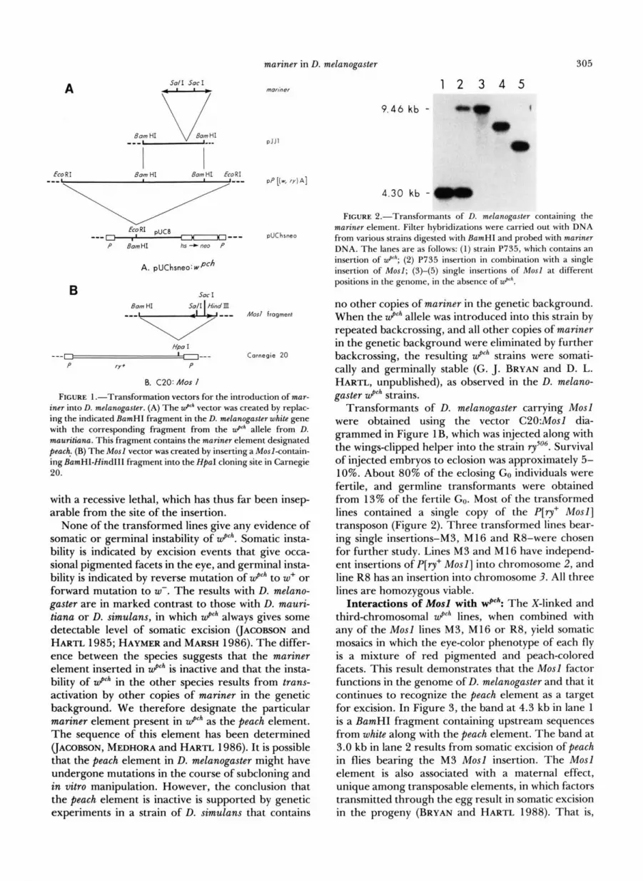
Interactions of Mosl with wPch: The X-linked and third-chromosomal Wpch lines, when combined with any of the Mosl lines M3, M16 or R8, yield somatic mosaics in which the eye-color phenotype of each fly is a mixture of red pigmented and peach-colored facets. This result demonstrates that the Mosl factor functions in the genome of D. melanogaster and that it continues to recognize the peach element as a target for excision. In Figure 3, the band at 4.3 kb in lane 1 is a BamHI fragment containing upstream sequences from white along with the peach element. The band at 3.0 kb in lane 2 results from somatic excision of peach in flies bearing the M3 Mosl insertion. The Mosl element is also associated with a maternal effect, unique among transposable elements, in which factors transmitted through the egg result in somatic excision in the progeny (BRYAN and HARTL 1988). That is,
306 D. Garza et al.
1 2 3 4 7.0 kb --" 4.3 kb -
3.0 kb - FIGURE 3.-Filter hybridizations of D. melanoguster DNA di-
gested with EamHl and probed with a 3.0-kb BamHl fragment of white DNA. The 7.0-kb fragment is characteristic of the w67r23 deletion, which is in the common genetic background of the strains. The 3.0-kb fragment is derived from the wildtype gene (in both D. melanogaster and D. mauritiana), and the 4.3-kb fragment is derived from the D. mauritiana u P h fragment containing the peach element. In each case the u P h insertion is that in strain P735. Lanes are as follows: (1) w ~ " ~ ' , u P h ; (2) w67r23, u P h ; Mosl (M3 insertion); (3) w67r23, u P h maternal-effect mosaic; (4) w67"3, w+ revertant.
females that are heterozygous for Mosl have predom- inantly mosaic offspring, even though half of the progeny lack M o s l . In most stocks the maternal-effect mosaics can be identified by phenotype because they show very few reversion events that occur early in development, resulting in one (occasionally more) large sectors of pigmented facets, whereas their M o s l / + siblings have frequent reversion events occurring at different stages of eye development resulting in a more speckled phenotype (e.g., Figure 1 in BRYAN and HARTL 1988). DNA from maternal-effect mosaics of D. melanogaster is shown in lane 3 in Figure 3. The occurrence of somatic excision of peach in these indi- viduals is clear, although the excision occurs to a lesser extent than in sibs carrying the P[ry+ M o s l ] transpo- son.
The character of the eye-color mosaicism differs somewhat among the M3, M16 and R8 lines. In combination with WPC', the Mosl line M3 gives heavy mosaicism (defined as large, overlapping patches of pigmented tissue), line M16 gives light mosaicism (defined as small, scattered patches of pigmented tis- sue), and line R8 gives an intermediate level of mo- saicism.
One straightforward explanation of the different levels of mosaicism in the Mosl lines is that Mosl expression is affected by position effects resulting from genomic sequences at or near the site of inser- tion. This interpretation is supported by additional genetic experiments, described in the next section, in which the Mosl factor had undergone transposition to still other positions in the genome.
Position effects on Mosl expression: Mobilization of the Mosl transposon can occur either autonomously or as a result of mobilization by the modified P ele- ment P[ry+ A2-3](99B), which produces high levels of P transposase activity but low levels of transposition
and excision (ROBERTSON et al. 1988). Various types of crosses were carried out to obtain Mosl factors in different positions in the genome.
For the Mosl insertions in chromosome 2, males of genotype P[ry+ Mosl] /CyO, Cy; rySo6 Sb P[ry+ A2-31 (99B) lry were crossed with ry females, and the non- rosy, nonstubble, CyO/+ offspring were crossed with Wpch in order to detect eye-color mosaicism. Among 15 transpositions obtained from the M3 and M16 Mosl lines in this manner, eight had the P[ry+Mos l ] transposon located in the C y 0 balancer and seven were in the third chromosome. In an analogous scheme using the R8 insertion into chromosome 3, CyO, Cy/ Sp; rySo6 Sb P[ry+ A2-3](99B)/P[ry+ Mosl] flies were crossed with ry, and the nonrosy, stubble offspring were crossed with f l h in order to detect eye-color mosaicism. Among 12 transpositions detected in this manner, three were in the X chromosome, two in chromosome 2, and seven in chromosome 3. In the absence of P[ry+ A2-3](99B) , among 12 autonomous transpositions of the Mosl factor in the M3 strain, three transpositions were into the X chromosome and nine into chromosome 3 . As expected, these trans- posed Mosl elements were no longer associated with
Altogether we examined 39 lines with transposi- tions of either the P[ry' M o s l ] transposon or of Mosl itself. The degree of mosaicism among these lines varied markedly, from one extreme of a very light speckle of pigmented facets to the other extreme of extremely heavy mosaicism with almost fully pig- mented eyes. That a wide range of mosaic types can result from transpositions of the identical Mosl factor implies that much of the variation in phenotype among the lines is attributable to position effects on expression of MosZ. Marked variation in level of mo- saicism was also observed in the transpositions bearing the P[ry+ M o s l ] transposon. Insertions of this element contain approximately 11 kb of ry+ and D. simulans DNA separating the Mosl element from genomic se- quences upstream of the insertion site (Figure lB), indicating that the position effects may extend over substantial distances. On the other hand, there are approximately 200 base pairs of flanking DNA on the downstream side of the P[ry+ Mosl ] transposon, and perhaps some of the position effects are mediated from this end.
Dosage and saturation effects: In addition to po- sition effects on Mosl expression are clear effects of gene dosage. For comparative purposes, rates of germline reversion are preferable to subjective assess- ments of degree of somatic mosaicism because they are more quantitative. Reversion data are summarized for the M16 (light mosaic) and M3 (heavy mosaic) lines in Table 1. The numbers tabulated are the frequencies of phenotypically w+ offspring resulting
ry+.
mariner in D. melanogaster 307
TABLE 1
Reversion rates of UP* allele
M16 M3 Genotype (light mosaic) (heavy mosaic)
zPh/Y; Mosl /C@dd 11.6 f 0.6 (2742) 15.6 f 0.9 (1640) zPh/Y; M o s l / M o s l & 24.0 f 2.2 (379) 17.4 f 1.4 (703) &h/&h; Mosl/CyO $9 9.9 f 0.5 (3475) 15.3 f 0.6 (3453) f i h / f l h ; Mosl/Mosl $9 ND" 15.7 f 1.3 (795)
Tabulated values are percentages. The numbers in parentheses
a Not determined. are the total numbers of progeny.
from reversion of wp" to w+ in various genotypes. Although the revertants are phenotypically wild type, DNA sequencing in D. mauritiana indicates that the reversion events are usually somewhat imprecise (BRYAN, GARZA and HARTL 1990). The frequencies of reversion were estimated by the proportion of wild- type progeny, since there was no significant tendency for wild-type progeny to occur in clusters.
Overall, the rate of wp" reversion in D. melanogaster is much greater than in D. mauritiana . The average rate of reversion among homozygous Mosl males from the M16 and M3 lines is approximately 20%, which is about eightfold greater than the rate of reversion in homozygous Mosl males in the E25H line in D. maur- itiana (BRYAN, JACOBSON and HARTL 1987). More- over, there is virtually no difference in the rate of up" reversion in males and females in D. melanogaster ( P = 0.15), whereas in D. mauritiana there is a 2.5-fold greater rate of reversion in males (JACOFSON and HARTL 1985).
As noted, the degree of somatic mosaicism is mark- edly less in the M16 line than in the M3 line. A significant difference is also reflected in the rate of germline reversion, which in heterozygous Mosl gen- otypes is 34% greater in males and 54% greater in females (P values in x* tests are both less than 0.001).
Dosage effects of Mosl are apparent in the M16 line, in which the homozygotes have an approximately twofold greater rate of reversion than the heterozy- gotes. The differences are also clear in the degree of somatic mosaicism: Mosl homozygotes in the M 16 line have larger pigmented patches than the heterozy- gotes, and the genotypes can usually be distinguished on this basis. However, a difference between hetero- zygotes and homozygotes is not seen in the heavy mosaic line M3. The M3 Mosl heterozygotes and homozygotes cannot be distinguished by degree of somatic mosaicism, and the rate of Wpch reversion in the homozygotes is only slightly (and not significantly) different from what it is in the heterozygotes ( P = 0.6 in a x* test). The absence of a dosage effect with M3 suggests a saturation effect in which the amount of Mosl gene product is no longer limiting to the rate of peach excision.
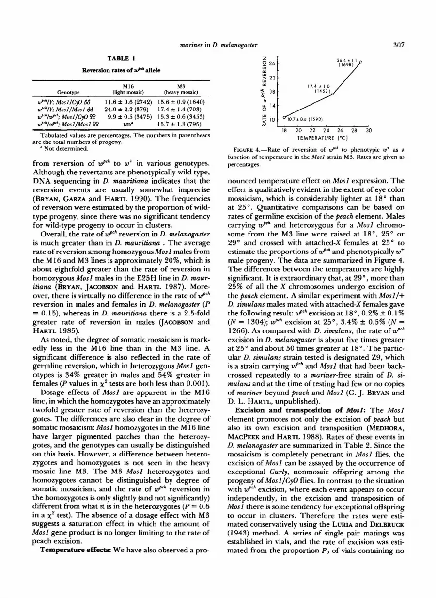
Temperature effects: We have also observed a pro-
0 2 4 Z
Ln
f I f 14
w
CL 2 10
17.4 t 1.0
I f 14
w
CL
18 20 22 24 26 28 30 I
18 20 22 24 26 28 30 TEMPERATURE ('C)
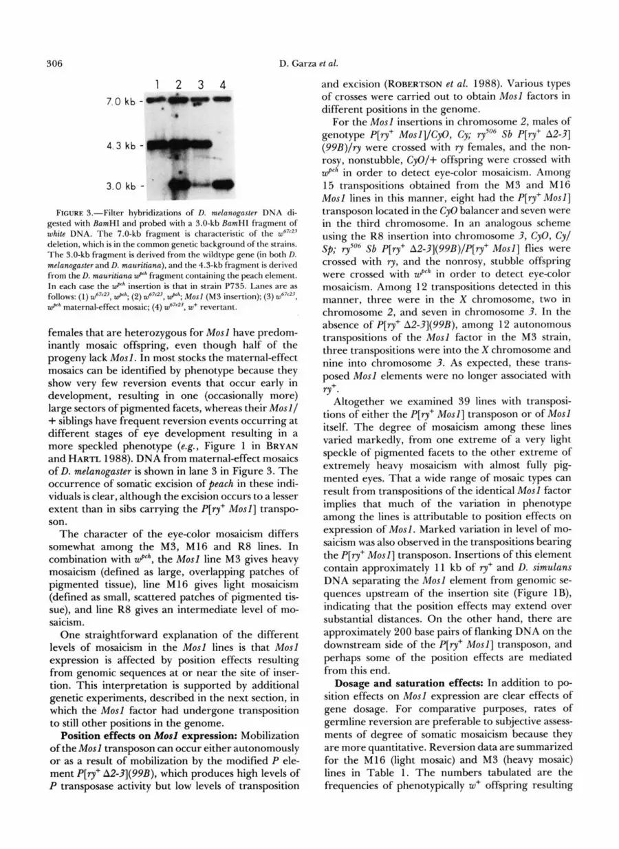
FIGURE 4.-Rate of reversion of z P h to phenotypic w+ as a function of temperature in the Mosl strain M3. Rates are given as percentages.
nounced temperature effect on Mosl expression. The effect is qualitatively evident in the extent of eye color mosaicism, which is considerably lighter at 18" than at 25 ". Quantitative comparisons can be based on rates of germline excision of the peach element. Males carrying Wpch and heterozygous for a Mosl chromo- some from the M3 line were raised at 18", 25" or 29" and crossed with attached-X females at 25" to estimate the proportions of Wpch and phenotypically w+ male progeny. The data are summarized in Figure 4. The differences between the temperatures are highly significant. It is extraordinary that, at 29", more than 25% of all the X chromosomes undergo excision of the peach element. A similar experiment with Mosl /+ D. simulans males mated with attached-X females gave the following result: W p c h excision at 18 ", 0.2% 0.1 % (N = 1304); wp" excision at 25", 3.4% & 0.5% ( N = 1266). As compared with D. simulans, the rate of W p c h
excision in D. melanogaster is about five times greater at 25" and about 50 times greater at 18". The partic- ular D. simulans strain tested is designated Z9, which is a strain carrying Wpch and Mosl that had been back- crossed repeatedly to a mariner-free strain of D. si- mulans and at the time of testing had few or no copies of mariner beyond peach and Mosl (G. J. BRYAN and D. L. HARTL, unpublished).
Excision and transposition of Mosl: The Mos2 element promotes not only the excision of peach but also its own excision and transposition (MEDHORA, MACPEEK and HARTL 1988). Rates of these events in D. melanogaster are summarized in Table 2. Since the mosaicism is completely penetrant in Mosl flies, the excision of Mosl can be assayed by the occurrence of exceptional Curly, nonmosaic offspring among the progeny of Mosl/CyO flies. In contrast to the situation with U g c h excision, where each event appears to occur independently, in the excision and transposition of Mosl there is some tendency for exceptional offspring to occur in clusters. Therefore the rates were esti- mated conservatively using the LURIA and DELBRUCK (1943) method. A series of single pair matings was established in vials, and the rate of excision was esti- mated from the proportion Po of vials containing no

308 D. Garza
TABLE 2
Excision and transposition rates of Mosl
et al.
M16 M3 Genotype (light mosaic) (heavy mosaic)
Excision (non-Cy nonmosaics) zPh/Y; MosllCyO W (41) 1.9 f 0.4 (70.3) 1.0 f 0.2 (55.5) @ h / z P h ; MosllCyO PP 1.3 rf: 0.3 (33.9) 2.0 f 0.4 (26.3)
(52) Transposition (Cy mosaics)
z P * / Y ; MosllCyO 68 (32) 2.4 f 0.5 (56.8) 1.0 f 0.3 (37.7) z P h / z P h ; MosllCyO 99 3.8 * 0.7 (29.6) 2.2 f 0.4 (24.8)
(53)
Tabulated values are percentages. The numbers in parentheses after the genotypes are the numbers of matings; those in parenthe- ses after the standard errors are the (harmonic) mean numbers of progeny. In each case the rate of excision was estimated from the proportion Po of vials containing no exceptional offspring as rate = -(l/N)ln(Po), where N is the average number of offspring of the relevant genotype per vial. The approximate standard errors were estimated by the asymptotic delta method as (r/N)'(I/P,)'Va7(P,), where Var(P,) is binomial.
exceptional offspring as rate = -( l/N)ln(Po), where N is the average number of offspring of the relevant genotype (e.g., CyO) per vial. The number of vials tested is given in parenthesis after each genotype, and the average number of offspring of the relevant gen- otype is given in parenthesis after the standard errors.
The estimated rates of Mosl excision are given in the upper part of Table 2. The rates are much lower than those of peach excision, and there is virtually no difference between the M3 and M16 lines in spite of their obviously different levels of eye-color mosaicism.
A similar result was found with respect to Mosl transposition, summarized in the lower part of Table 2. Transposition events were assayed by the occur- rence of exceptional Curly mosaic progeny among the progeny of Mosl/CyO flies, and the rates were esti- mated as described above. By the nature of the mating scheme, any transpositions of Mosl to new locations along the same chromosome are undetectable. In the MI6 line the rate of detectable transposition of Mosl appears to be somewhat greater than the rate of excision, but in the M 16 line the rates are comparable.
Germline transformation with mariner: We have also investigated the capability of the Mosl element to insert autonomously into the genome when injected into the pole plasm of Drosophila embryos, using a vector with the 5.0-kb BamHI-Hind111 fragment con- taining Mosl inserted into the cloning site of pBluescribe M 13+. Embryos of a cn; y strain obtained from P. M. BINCHAM were injected. Surviving GO flies were crossed individually with V p c h D. melanogaster and the progeny examined visually for eye-color mosai- cism. Groups of GI progeny were sib mated and their offspring examined for mosaicism in the G:! and Gs generations. Among 150 injected cn; y embryos, 28 hatched, 19 survived to eclosion and 16 were fertile. Five of the 16 fertile Go adults yielded at least one
9.46 kb -
4.30 kb -
1 2 3 4 5
FIGURE 5.-Accumulation of mariner elements in spontaneously transformed lines of D. melanogaster containing Mosl. In each case the genetic background contains the z P h insertion P735. Genomic DNA was digested with BamHI and Hind111 and probed with an Sspl-NheI fragment of mariner. Lanes are as foIIows: (1) w6'"', z P h ;
(2)-(5) independent lines transformed with Mosl, designated A, D, H and X, respectively.
mosaic offspring from which a Mosl-bearing strain was established by backcrossing to wp" and sib mating.
A few generations subsequent to the establishment of the transformed Mosl lines, chromosomal DNA was prepared and digested with BamHI and Hind11 and probed with a 1-kb internal SspI-NheI fragment of mariner deriving from coordinates 56-1 183 UA- COBSON, MEDHORA and HARTL 1986). Multiple mari- ner-containing bands were observed in the trans- formed lines, indicating that the integrated Mosl ele- ment was promoting active transposition and accumulation of copies (Figure 5).
DISCUSSION
The system of inherited eye-color mosaicism result- ing from excision of the transposable element mariner from the V p c h allele has been transferred into the genome of D. melanogaster, a species in which mariner does not normally occur (JACOBSON, MEDHORA and HARTL 1986). The particular V p c h allele transformed into D. melanogaster is a chimeric gene consisting of the D. melanogaster w+ sequence into which has been substituted a 4.3-kb BamHI fragment derived from coordinates 1 .O-4.0 of the D. mauritiana w+ gene that contains the particular mariner element designated peach (coordinates defined as in JACOBSON, MEDHORA and HARTL 1986). The peach element is inactive, and so, in the absence of other mariner elements, the chimeric V p c h allele present in strains of D. melanogaster is genetically stable in both somatic and germ cells. However, in the presence of the active mariner ele- ment Mosl , excision of the peach element occurs,
mariner in D. melanogaster 309
resulting eye-color mosaicism and a rate of germline reversion of 10-25% or more, depending on temper- ature.
One noteworthy feature of Table 2 is that the rate of transposition of Mosl, relative to the rate of exci- sion, is substantially greater than observed among other Drosophila transposons (see reviews in BERG and HOWE 1989). In the M16 line, the rate of trans- position of Mosl may actually exceed the rate of excision; and in the M3 line, the rates of transposition and excision are approximately equal. Such a high rate of Mosl transposition, relative to the rate of excision, should favor the persistence of Mosl ele- ments in the genome. Consistent with this suggestion, CAPY et al. (1990) have found that active mariner transposable elements are widespread in natural pop- ulations of D. simulans.
The presence of mariner in D. melanogaster provides a new means of genetic manipulation in this species that may be advantageous for certain purposes. Since the mariner system has been used for transposon tag- ging of genes in D. mauritiana (BRYAN, GARZA and HARTL 1990), it may be useful for this purpose in D. melanogaster as well. Since mariner has a different target-site specificity than the P element (O'HARE and RUBIN 1983; BRYAN, GARZA and HARTL 1990), genes that are poor targets for one element may be better targets for the other. One potentially useful feature of the mariner system is that new mutations resulting from mariner insertions can be identified by their high rate of somatic and germline excision in the presence of Mosl. Novel applications might include the analysis of developmental mutations induced by mariner inser- tions, since somatic excision will yield large numbers of somatic mosaics, and various temperatures can be used to control the rate and also perhaps the timing of the excision events. The high rate of somatic exci- sion could also be used to identify nonautonomous mariner insertions that have mutant phenotypes only in the absence of Mosl-induced excision.
The finding that Mosl itself can efficiently insert into the D. melanogaster genome also suggests the possibility of developing an alternative system for germline transformation, toward which the construc- tion of suitable cloning vectors and preliminary tests are presently underway. The mariner element con- tains a single, long, uninterrupted open reading frame (JACOBSON, MEDHORA and HARTL 1986), and the ele- ment may be able to function in a wide variety of genomes. In this connection it is interesting to note the presence of a mariner-like element in the genome of the Cecropia moth Hyalophora cecropia. The partic- ular copy of the Cecropia element that has been sequenced is 125 1 base pairs in length and contains a protein-coding region that has 55% similarity in amino acid composition with the putative coding re-
gion of mariner from D. mauritiana (LIDHOLM, GUD- MUNDSSON and BOMAN, personal communication). However, the presence of several frameshift differ- ences in the Cecropia element suggests that it is non- functional. There are approximately 2000 copies of these mariner-like elements in the Cecropia genome. The presence of mariner-like elements in this Lepi- dopteran suggests that a successful transformation system based on mariner might have a broad host range.
Presence of the mariner element in D. melanogaster opens up new experimental approaches to the study of transposable element function. For example, dif- ferent Mos factors and chimeric constructs can be studied in D. melanogaster using the Wpc" allele (MED- HORA, MARUYAMA and HARTL 1991), and dosage effects of Mos factors and mariner target sequences can be determined. The complete penetrance of eye- color mosaicism in u P h ; Mosl flies also provides easy phenotypic detection of mutations that eliminate the excision of WPC". Such mutations in the peach element will affect the ability of the element to serve as a target or to excise, and those in Mosl will affect expression or activity of the element. However, the system should also identify mutations in chromosomal genes that are necessary for mariner target recognition or excision. It should also be noted that the u9"*-Mosl system can serve as an enhancer trap for insertions of Mosl near enhancers that stimulate differential expression across the eye. During routine stockkeeping a mosaic strain of D. simulans has been isolated in which the mosaic patches occur primarily in the anterior portion of the eye (BRYAN 1989).
The rate of germline excision of the peach element in Mosl-bearing strains of D. melanogaster is approxi- mately 17% at 25 O , as compared with approximately 3% in D. simulans-about a sixfold difference. Rates of Mosl excision and transposition are not as high as that of peach excision, but they are still substantially greater than in D. simulans (MEDHORA, MACPEEK and HARTL 1988). We do not know why the rates of excision and transposition are so much greater in D. melanogaster than in D. simulans. Since strains of D. simulans containing only W p c h and Mosl do not have rates of peach excision comparable to those in D. melanogaster (G. J. BRYAN and D. L. HARTL, unpub- lished), the difference is not likely to result merely from other copies of mariner in the genetic back- ground. A more likely possibility is that the species differ because of host factors with which mariner or its products interact-and many host factors must be involved in the transcription, translation, transport, and function of the mariner product. It is even possible that the different mariner behavior in the two species reflects a more general process, not yet understood, by which, on average, the abundance of transposable
310 D. Garza et al.
elements in D. melanogaster is some sevenfold greater than in D. simulans or D. mauritiana (DOWSETT and
It is also unclear why the rate of peach excision is so much greater than that of Mosl, but the likely candi- dates are differences in recognition sequences in the elements themselves (MEDHORA, MARUYAMA and HARTL 1991) or differences in their accessibility in the genome. Whatever the mechanism, the result is that the level of eye-color mosaicism is not necessarily correlated with the rate of Mosl excision or transpo- sition.
Transposable elements are capable of undergoing horizontal transfer between species. The population dynamics of P element in a new host genome has been studied by experimental transfer of this element into D. simulans (SCAVARDA and HARTL 1984,1987; DAN- IELS, STRAUSBAUGH and ARMSTRONG 1985; DANIELS, CHOVNICK and KIDWELL 1989). The introduction of mariner into D. melanogaster provides experimental opportunities that are in some ways superior because of the repertoire of mutants and chromosome re- arrangements that are available in D. melanogaster. The Mosl-transformed strains of D. melanogaster pres- ently contain numerous copies of mariner at many scattered positions throughout the genome. It will be of some interest to determine whether the D. mela- nogaster populations evolve toward a state comparable to that in many widely distributed natural populations of D. simulans that contain active Mos-like elements (CAPY et al. 1990).
YOUNG 1982).
We are grateful to A. MACPEEK and K. MARUYAMA for their help in completing the manuscript, V. PIRROTTA and G. RUBIN for the gift of clones, and W. ENGEIS for the P[q' A2-3](99B) strain. This work was supported by National Institutes of Health grant GM33741.
LITERATURE CITED
BERG, D. E., and M. M. HOWE (Editors), 1989 Mobile DNA. American Society for Microbiology, Washington, D.C.
BINGHAM, P. M., R. LEVIS and G . M. RUBIN, 1981 Cloning of DNA sequences from the white locus of D. melanogaster by a novel and general method. Cell 25: 693-704.
BRYAN, G. J. , 1989 Molecular analysis of transposon-induced mutations and their derivatives in Drosophila. Ph.D. thesis, Washington University, St. Louis.
BRYAN, G. J., D. GARZA and D. L. HARTL, 1990 Insertion and excision of the transposable element mariner in Drosophila. Genetics 1 2 5 103-1 14.
BRYAN, G. J., and D. L. HARTL, 1988 Maternally inherited tran- sposon excision in Drosophila simulans. Science 2 4 0 2 15-21 7 .
BRYAN, G. J., J. W. JACOBSON and D. L. HARTL, 1987 Heritable somatic excision of a Drosophila transposon. Science 2 3 5 1636- 1638.
CAPY, P., F. CHAKRANI, F. LEMEUNIER, D. L. HARTL and J. R. DAVID, 1990 Active mariner transposable elements are wide- spread in natural populations of Drosophila simulans. Proc. Roy. SOC. Lond. B 242: 57-60.
DANIELS, S. B., A. CHOVNICK and M. G. KIDWELL, 1989 Hybrid dysgenesis in Drosophila simulans lines transformed with auton- omous P elements. Genetics 121: 281-291.
DANIELS, S. B., L. D. STRAUSBAUGH and R. A. ARMSTRONG, 1985 Molecular analysis of P element behavior in Drosophila simulans. Mol. Gen. Genet. 2 0 0 258-265.
DOWSETT, A. P., and M. W. YOUNG, 1982 Differing levels of dispersed repetitive DNA among closely related species of Drosophila. Proc. Natl. Acad. Sci. USA 7 9 4570-4574.
HARTL, D. L. , 1989 Transposable element mariner in Drosophila species, pp. 531-536 in Mobile DNA, edited by D. E. BERG and M. HOWE. American Society for Microbiology, Washington, D.C.
HAYMER, D. S., and J. L. MARSH, 1986 Germ line and somatic instability of a white mutation in Drosophila mauritiana due to a transposable element. Dev. Genet. 6: 281-291.
HAZELRIGG, T., R. LEVIS^^^ G. M. RUBIN, 1984 Transformation of white locus DNA in Drosophila: dosage compensation, zeste interaction, and position effects. Cell 3 6 469-481.
JACOBSON, J. W., and D. L. HARTL, 1985 Coupled instability of two X-linked genes in Drosophila mauritiana: Germinal and somatic mutability. Genetics 111: 57-65.
JACOBSON, J. W., M. M. MEDHORA and D. L. HARTL, 1986 Molecular structure of a somatically unstable transpos- able element in Drosophila. Proc. Natl. Acad. Sci. USA 83:
KARESS, R. E., and G. M. RUBIN, 1984 Analysis of transposable element functions in Drosophila. Cell 38: 135-146.
LURIA, S., and M. DELBRUCK, 1943 Mutations of bacteria from virus sensitivity to virus resistance. Genetics 2 8 491-51 1 .
MANIATIS, T., E. F. FRITSCH and J. SAMBROOK, 1982 Molecular Cloning: A Laboratory Manual, Cold Spring Harbor Laboratory, Cold Spring Harbor, N.Y.
MARUYAMA, K., and D. L. HARTL, 1991 Evolution of the trans- posable element mariner in Drosophila species. Genetics 128: 319-329.
MEDHORA, M. M., A. H. MACPEEK and D. L. HARTL, 1988 Excision of the Drosophila transposable element mari- ner: indentification and characterization of the Mos factor.
MEDHORA, M., K. MARUYAMA and D. L. HARTL, 1991 Molecular and functional analysis of the mariner mutator element Mosl in Drosophila. Genetics 1 2 8 3 1 1-3 18.
O'HARE, K., and G. M. RUBIN, 1983 Structure of P transposable elements and their sites of insertion and excision in the Dro- sophila melanogaster genome. Cell 34: 25-35.
ROBERTSON, H. M., C. R. PRESTON, R. W. PHILLIS, D. JOHNSON- SCHLITZ, W. K. BENZ and W. R. ENGELS, 1988 A stable genomic source of P element transposase in Drosophila mela- nogaster. Genetics 119: 75-83.
RUBIN, G. M., and A. C. SPRADLING, 1982 Genetic transformation of Drosophila with transposable element vectors. Science 218: 348-353.
RUBIN, G. M., and A. C . SPRADLING, 1983 Vectors for P eiement- mediated gene transfer in Drosophila. Nucleic Acids Res. 11: 6341-6351.
SCAVARDA, N. J., and D. L. HARTL, 1984 Interspecific DNA transformation in Drosophila. Proc. Natl. Acad. Sci. USA 81:
SCAVARDA, N. J., and D. L. HARTL, 1987 Germline abnormalities in Drosophila simulans transfected with the transposable P ele- ment. J. Genet. 66: 1-15.
STELLER, H., and V. PIRROTTA, 1985 A transposable P vector that confers selectable G418 resistance to Drosophila larvae.
8684-8688.
EMBO J. 7: 2185-2189.
7515-7519.
EMBO J. 4: 167-171. Communicating editor: M. J. SIMMONS


