Embed Size (px)
Citation preview
Foundation block - Anatomy - Lecture 5
INTRODUCTION OF CARDIOVASCULAR SYSTEM (CVS)
Objectives
2
By the end of this session, student should be able to:
❖ Identify the components of the cardiovascular system.
❖ Describe the Heart as regards (position, chambers and valves).
❖ Describe the Blood vessels (Arteries, Veins and Capillaries).
❖ Describe the Portal System.
❖ Describe the Sinusoids.
❖ Describe the Functional and Anatomical end arteries.
❖ Describe the Arteriovenous Anastomosis.
Color guide :Only in boys slides in GreenOnly in girls slides in Purpleimportant and doctors note in RedExtra information in Blue
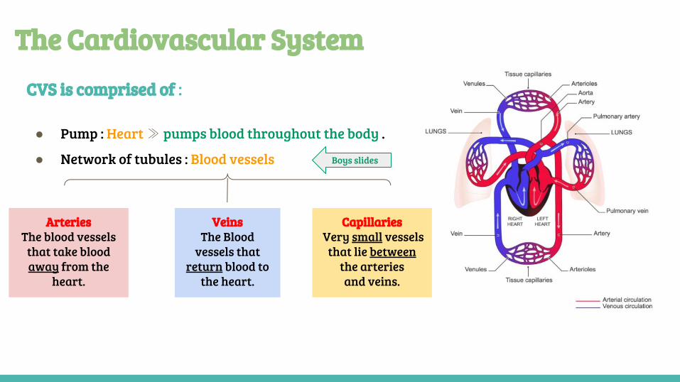
CVS is comprised of :
● Pump : Heart ≫ pumps blood throughout the body .
● Network of tubules : Blood vessels
The Cardiovascular System
ArteriesThe blood vessels
that take blood away from the
heart.
VeinsThe Blood
vessels that return blood to
the heart.
Capillaries Very small vessels
that lie between the arteriesand veins.
Boys slides
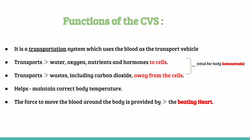
● It is a transportation system which uses the blood as the transport vehicle
● Transports ≫ water, oxygen, nutrients and hormones to cells.
● Transports ≫ wastes, including carbon dioxide, away from the cells.
● Helps - maintain correct body temperature.
● The force to move the blood around the body is provided by ≫ the beating Heart.
Functions of the CVS :
(vital for body homeostasis)
The heart
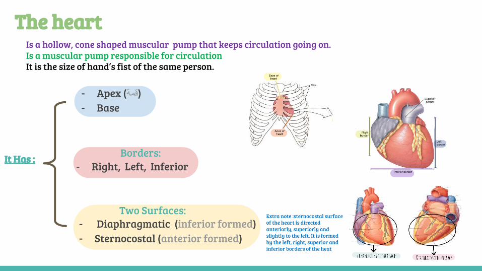
- Apex (قمة)- Base
- Diaphragmatic (inferior formed) - Sternocostal (anterior formed)
Two Surfaces:
Borders: - Right, Left, Inferior
It Has :
Is a hollow, cone shaped muscular pump that keeps circulation going on.Is a muscular pump responsible for circulationIt is the size of hand’s fist of the same person.
Extra note :sternocostal surface of the heart is directed anteriorly, superiorly and slightly to the left. It is formed by the left, right, superior and inferior borders of the heat
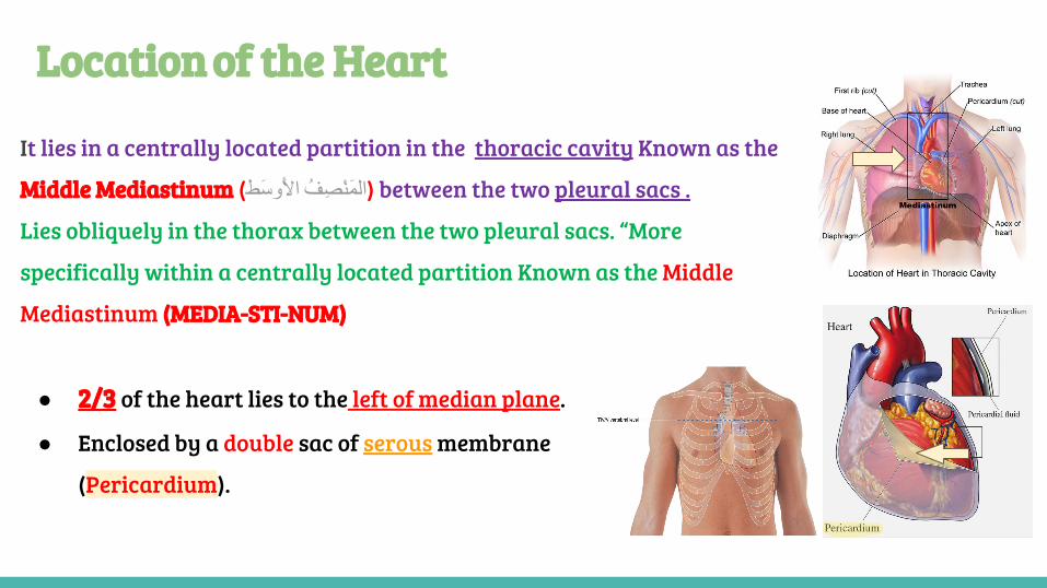
Location of the Heart
It lies in a centrally located partition in the thoracic cavity Known as the
Middle Mediastinum (الَمْنِصُف األَوَسط) between the two pleural sacs .
Lies obliquely in the thorax between the two pleural sacs. “More
specifically within a centrally located partition Known as the Middle
Mediastinum (MEDIA-STI-NUM)
● 2/3 of the heart lies to the left of median plane.
● Enclosed by a double sac of serous membrane
(Pericardium).
Chambers of the Heart
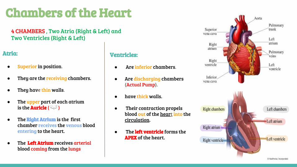
Atria:
● Superior in position.
● They are the receiving chambers.
● They have thin walls.
● The upper part of each atrium is the Auricle ( أَُذینَة )
● The Right Atrium is the first chamber receives the venous blood entering to the heart.
● The Left Atrium receives arterial blood coming from the lungs
Ventricles:
● Are inferior chambers.
● Are discharging chambers (Actual Pump).
● have thick walls.
● Their contraction propels blood out of the heart into the circulation.
● The left ventricle forms the APEX of the heart.
4 CHAMBERS , Two Atria (Right & Left) andTwo Ventricles (Right & Left)
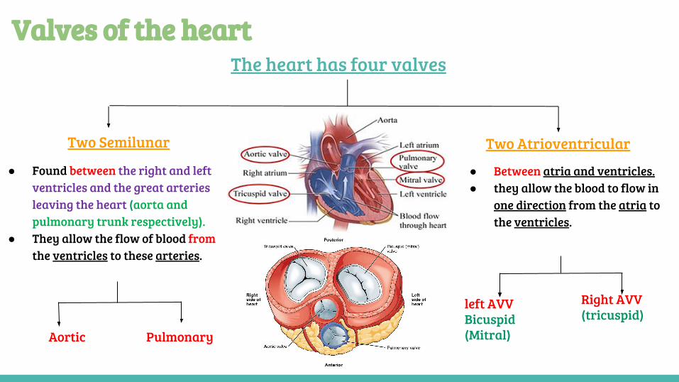
Valves of the heartThe heart has four valves
Two AtrioventricularTwo Semilunar
● Between atria and ventricles.● they allow the blood to flow in
one direction from the atria to the ventricles.
Right AVV(tricuspid)
left AVVBicuspid(Mitral)
● Found between the right and left ventricles and the great arteries leaving the heart (aorta and pulmonary trunk respectively).
● They allow the flow of blood from the ventricles to these arteries.
PulmonaryAortic
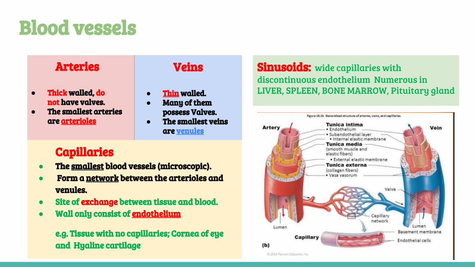
Blood vessels
Arteries
● Thick walled, do not have valves.
● The smallest arteries are arterioles
Veins
● Thin walled.● Many of them
possess Valves.● The smallest veins
are venules
Capillaries● The smallest blood vessels (microscopic).● Form a network between the arterioles and
venules.● Site of exchange between tissue and blood. ● Wall only consist of endothelium
e.g. Tissue with no capillaries; Cornea of eye and Hyaline cartilage
Sinusoids: wide capillaries with discontinuous endothelium Numerous in LIVER, SPLEEN, BONE MARROW, Pituitary gland
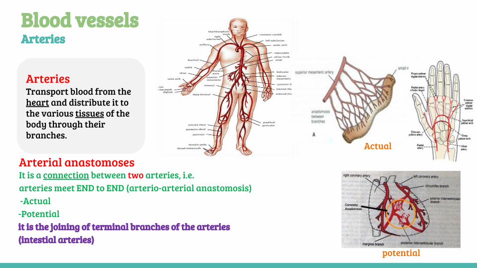
Blood vessels Arteries
Arteries Transport blood from the heart and distribute it to the various tissues of the body through their branches.
Actual
Arterial anastomosesIt is a connection between two arteries, i.e.arteries meet END to END (arterio-arterial anastomosis) -Actual -Potentialit is the joining of terminal branches of the arteries (intestial arteries)
potential
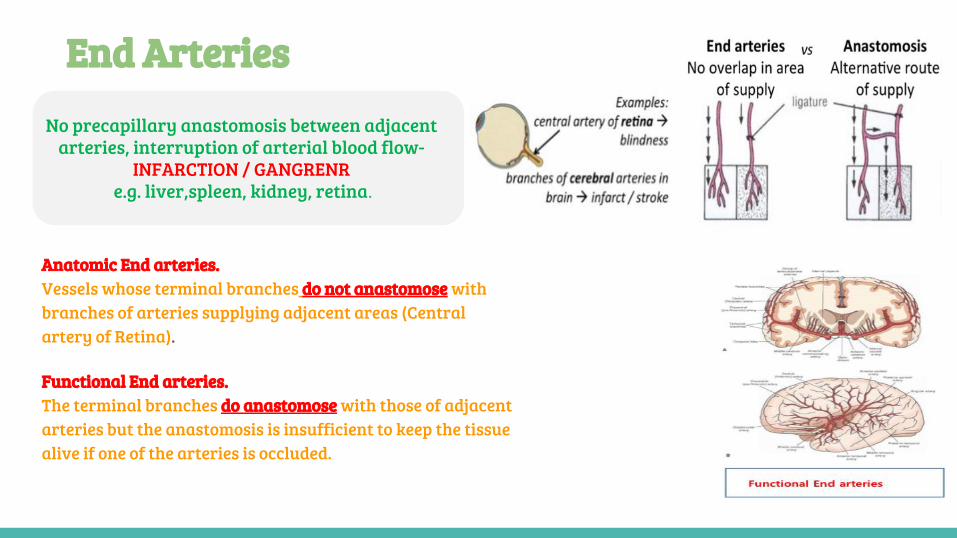
Anatomic End arteries. Vessels whose terminal branches do not anastomose with branches of arteries supplying adjacent areas (Central artery of Retina).
Functional End arteries. The terminal branches do anastomose with those of adjacent arteries but the anastomosis is insufficient to keep the tissue alive if one of the arteries is occluded.
No precapillary anastomosis between adjacent arteries, interruption of arterial blood flow-
INFARCTION / GANGRENR e.g. liver,spleen, kidney, retina.
End Arteries
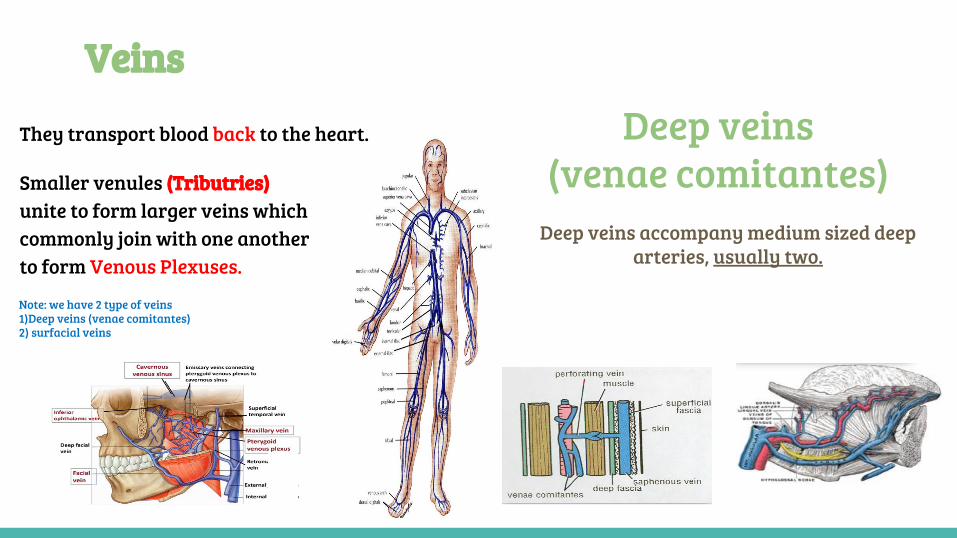
Veins
Deep veins accompany medium sized deep arteries, usually two.
Deep veins(venae comitantes)
They transport blood back to the heart.
Smaller venules (Tributries) unite to form larger veins which commonly join with one another to form Venous Plexuses.
Note: we have 2 type of veins 1)Deep veins (venae comitantes)2) surfacial veins
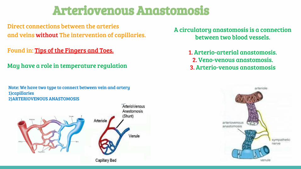
Arteriovenous AnastomosisDirect connections between the arteriesand veins without The intervention of capillaries.
Found in: Tips of the Fingers and Toes.
May have a role in temperature regulation
A circulatory anastomosis is a connection between two blood vessels.
1. Arterio-arterial anastomosis.
2. Veno-venous anastomosis.3. Arterio-venous anastomosis
Note: We have two type to connect between vein and artery 1)capillaries 2)ARTERIOVENOUS ANASTOMOSIS
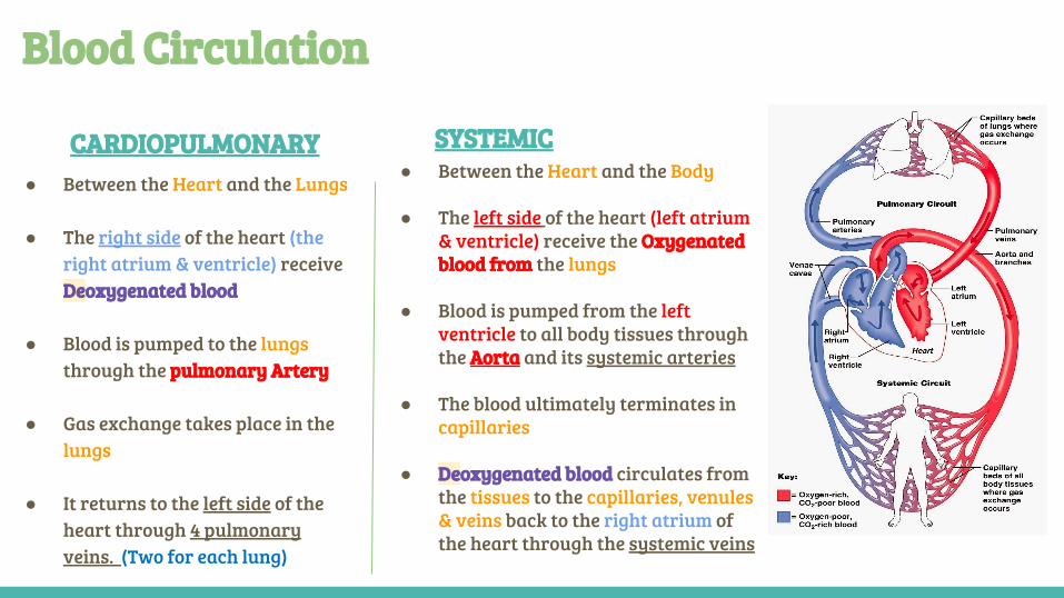
CARDIOPULMONARY
Blood Circulation
SYSTEMIC● Between the Heart and the Lungs
● The right side of the heart (the right atrium & ventricle) receive Deoxygenated blood
● Blood is pumped to the lungs through the pulmonary Artery
● Gas exchange takes place in the lungs
● It returns to the left side of the heart through 4 pulmonary veins. (Two for each lung)
● Between the Heart and the Body
● The left side of the heart (left atrium & ventricle) receive the Oxygenated blood from the lungs
● Blood is pumped from the left ventricle to all body tissues through the Aorta and its systemic arteries
● The blood ultimately terminates in capillaries
● Deoxygenated blood circulates from the tissues to the capillaries, venules & veins back to the right atrium of the heart through the systemic veins
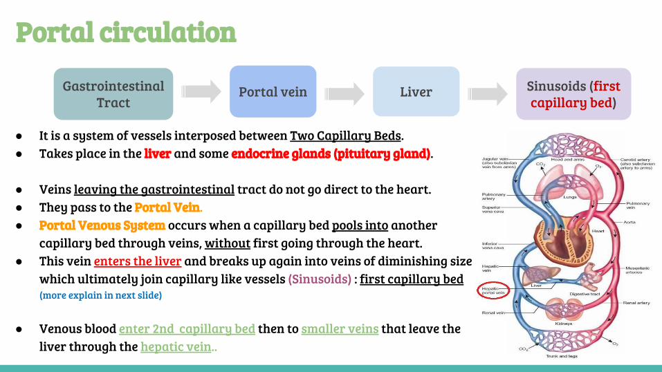
Portal circulation
● It is a system of vessels interposed between Two Capillary Beds. ● Takes place in the liver and some endocrine glands (pituitary gland).
● Veins leaving the gastrointestinal tract do not go direct to the heart.● They pass to the Portal Vein.● Portal Venous System occurs when a capillary bed pools into another
capillary bed through veins, without first going through the heart.● This vein enters the liver and breaks up again into veins of diminishing size
which ultimately join capillary like vessels (Sinusoids) : first capillary bed (more explain in next slide)
● Venous blood enter 2nd capillary bed then to smaller veins that leave the liver through the hepatic vein..
Gastrointestinal Tract
Portal vein Liver Sinusoids (first capillary bed)
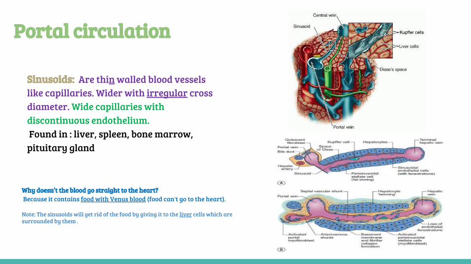
Sinusoids: Are thin walled blood vessels like capillaries. Wider with irregular cross diameter. Wide capillaries with discontinuous endothelium. Found in : liver, spleen, bone marrow, pituitary gland
Portal circulation
Why doesn’t the blood go straight to the heart? Because it contains food with Venus blood (food can't go to the heart).
Note: The sinusoids will get rid of the food by giving it to the liver cells which are surrounded by them .
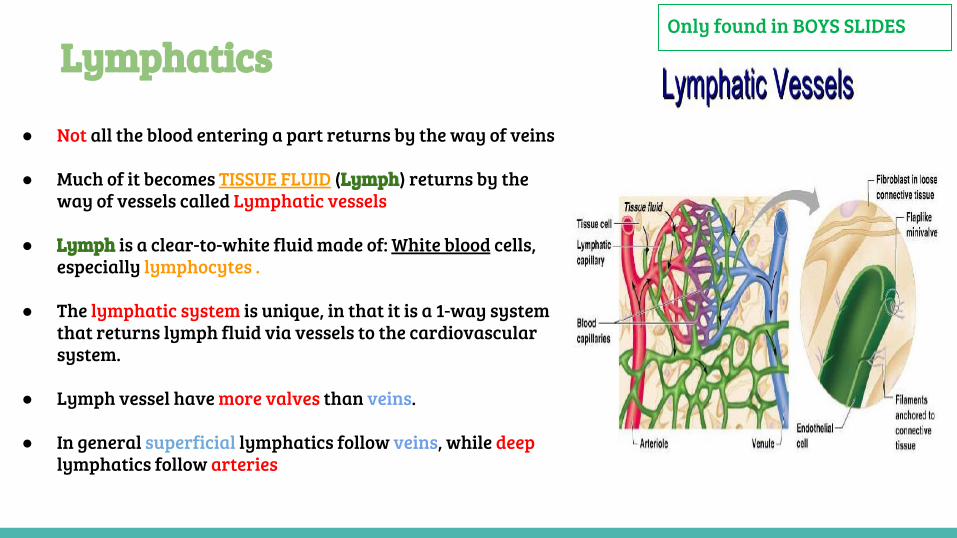
LymphaticsOnly found in BOYS SLIDES
● Not all the blood entering a part returns by the way of veins
● Much of it becomes TISSUE FLUID (Lymph) returns by the way of vessels called Lymphatic vessels
● Lymph is a clear-to-white fluid made of: White blood cells, especially lymphocytes .
● The lymphatic system is unique, in that it is a 1-way system that returns lymph fluid via vessels to the cardiovascular system.
● Lymph vessel have more valves than veins.
● In general superficial lymphatics follow veins, while deep lymphatics follow arteries
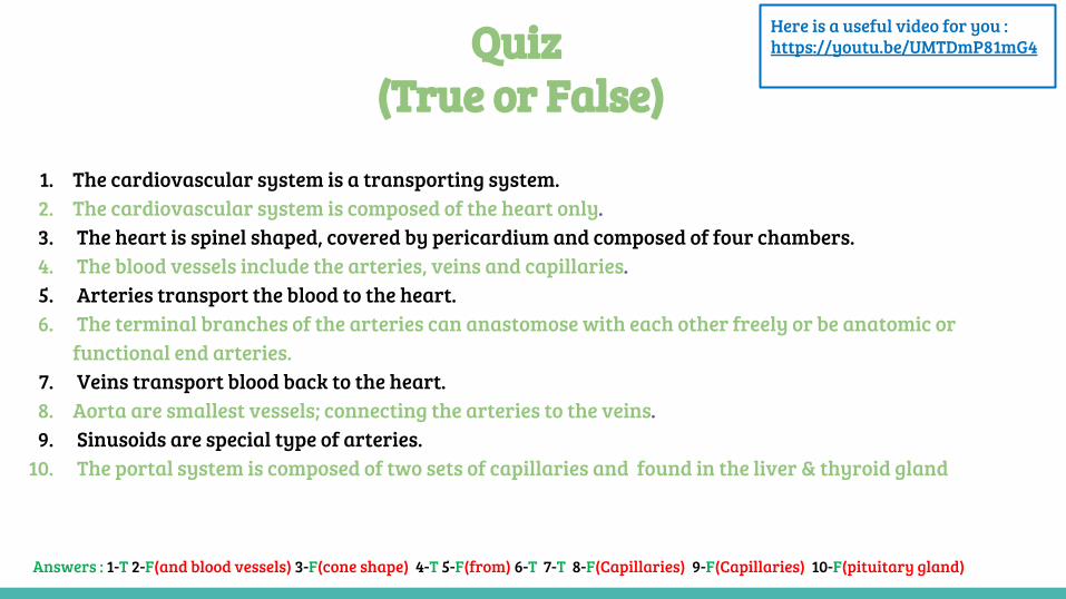
Quiz (True or False)
1. The cardiovascular system is a transporting system. 2. The cardiovascular system is composed of the heart only.3. The heart is spinel shaped, covered by pericardium and composed of four chambers.4. The blood vessels include the arteries, veins and capillaries.5. Arteries transport the blood to the heart. 6. The terminal branches of the arteries can anastomose with each other freely or be anatomic or
functional end arteries.7. Veins transport blood back to the heart.8. Aorta are smallest vessels; connecting the arteries to the veins. 9. Sinusoids are special type of arteries.
10. The portal system is composed of two sets of capillaries and found in the liver & thyroid gland
Answers : 1-T 2-F(and blood vessels) 3-F(cone shape) 4-T 5-F(from) 6-T 7-T 8-F(Capillaries) 9-F(Capillaries) 10-F(pituitary gland)
Here is a useful video for you : https://youtu.be/UMTDmP81mG4
● Abdulrahman Shadid● Ateen Almutairi
Contact us: Twitter : @Anatomy438
Boys team:
● Khalid AL-Dossari● Naif Al-Dossari● Faisal Alqifari ● Salman Alagla● Ziyad Al-jofan● Suhail Basuhail● Ali Aldawood● Khalid Nagshabandi
Girls team :
● Ajeed AlRashoud● Taif Alotaibi● Noura Alturki● Amirah Al-zahrani● Alhanouf Al-haluli● Sara Al-Abdulkarim● Rawan Alzayed● Reema Almasoud● Renad Alhaqbani● Nouf Alhumaidhi● Fay AlBuqami● Jude Alkhalifah● Nouf Alhussaini
Team members
Team leaders
Good luck to you all
A special thanks to the 436 anatomy team, who inspired
our work.


